drosophila adult muscle precursors form a network of ... · 1966 materials and methods drosophila...
TRANSCRIPT
1965DEVELOPMENT AND STEM CELLS RESEARCH ARTICLE
INTRODUCTIONSeveral populations of stem cells have been identified in the fruitfly over the last few years (for a review, see Pearson et al.,2009). Studies on Drosophila germline stem cells (GSCs)(Spradling et al., 2001) and, more recently, intestinal stem cells(ISCs) (Micchelli and Perrimon, 2006; Ohlstein and Spradling,2006; Takashima et al., 2008) that persist in adult flies havehelped establish niche models of stem cell self-renewal.Conversely, during development, populations of transient stemcells play crucial roles in the formation of specific tissues(Pearson et al., 2009). For example, transient populations ofneural stem cells called neuroblasts undergo a series ofasymmetric cell divisions that ensure self-renewal and at thesame time give rise to a large range of neural lineages thatundergo differentiation (Yu et al., 2006). In the embryonicmesoderm, muscle progenitor cells were shown to divideasymmetrically like the neuroblasts (Ruiz Gomez et al., 1997),but unlike neuroblasts, they divide only once, and give rise eitherto two distinct muscle founder cells that enter the differentiationprocess or to a muscle founder and a cell called an adult muscleprecursor (AMP) that keeps an undifferentiated state (RuizGomez et al., 1997; Figeac et al., 2007). As the AMPs expressmarkers specific to muscle progenitors, such as the b-HLHtranscription factor Twist (Bate et al., 1991; Figeac et al., 2007),the asymmetric cell division leading to the production of an
AMP resembles the asymmetric cell division of the neuroblaststhat ensure self-renewal. The key role of AMP cells in adultmuscle growth and in the regeneration of a subset of thoracicmuscles also indicates that the AMPs share properties withvertebrate satellite cells (Maqbool and Jagla, 2007). Thus, theAMPs emerge as a novel, muscle-committed population oftransient Drosophila stem cells. Based on this assumption, weaimed to gain insights into AMP cell behaviour and the geneticcontrol of their specification to improve our knowledge onmuscle stem cells in general. To address these issues, we firstattempted to identify new cell markers capable of tracking AMPsduring development. We found that two targets of Notchsignalling, E(spl)M6 and Him, as well as two transcriptionfactors, Zfh1 and Cut, are specifically expressed in DrosophilaAMPs. Both Him (Liotta et al., 2007) and Zfh1 (Postigo et al.,1999) are able to counteract Mef2-driven myogenicdifferentiation, probably acting as Mef2 repressors, whereas Cutis known to play a role in the diversification of flight muscles(Sudarsan et al., 2001) and to act as a neural selector gene(Bodmer et al., 1987). The AMP-specific roles of these geneshave not yet been investigated. Using the Notch-responsiveelement of E(spl)M6 to drive membrane-targeted GFP in theAMPs, we observed that AMPs send long cellular processes andare interconnected. We also designed a genetic screen to identifygenes affecting AMP cell pattern and found that rhomboid (rho)and other EGF pathway components control AMP specificationand subsequently protect them against apoptosis. A key role forEGFR signalling was further supported by the identification ofEGF-secreting cells that ensure AMP cell maintenance and bythe finding that regulatory modules driving expression in lateralAMPs carry functional EGF (ETS)-responsive motifs.
Development 137, 1965-1973 (2010) doi:10.1242/dev.049080© 2010. Published by The Company of Biologists Ltd
GReD, INSERM U931, CNRS UMR6247, Clermont University, Faculté de Médecine,28 Place Henri Dunant, Clermont-Ferrand, 63000, France.
*Author for correspondence ([email protected])
Accepted 18 April 2010
SUMMARYIn Drosophila, a population of muscle-committed stem-like cells called adult muscle precursors (AMPs) keeps an undifferentiatedand quiescent state during embryonic life. The embryonic AMPs are at the origin of all adult fly muscles and, as we demonstratehere, they express repressors of myogenic differentiation and targets of the Notch pathway known to be involved in muscle cellstemness. By targeting GFP to the AMP cell membranes, we show that AMPs are tightly associated with the peripheral nervoussystem and with a subset of differentiated muscles. They send long cellular processes running along the peripheral nerves and, bythe end of embryogenesis, form a network of interconnected cells. Based on evidence from laser ablation experiments, the mainrole of these cellular extensions is to maintain correct spatial positioning of AMPs. To gain insights into mechanisms that lead toAMP cell specification, we performed a gain-of-function screen with a special focus on lateral AMPs expressing the homeoboxgene ladybird. Our data show that the rhomboid-triggered EGF signalling pathway controls both the specification and thesubsequent maintenance of AMP cells. This finding is supported by the identification of EGF-secreting cells in the lateral domainand the EGF-dependent regulatory modules that drive expression of the ladybird gene in lateral AMPs. Taken together, ourresults reveal an unsuspected capacity of embryonic AMPs to form a cell network, and shed light on the mechanisms governingtheir specification and maintenance.
KEY WORDS: AMP, Stem cell, Muscle, Drosophila, Ladybird
Drosophila adult muscle precursors form a network ofinterconnected cells and are specified by the rhomboid-triggered EGF pathwayNicolas Figeac, Teresa Jagla, Rajaguru Aradhya, Jean Philippe Da Ponte and Krzysztof Jagla*
DEVELO
PMENT

1966
MATERIALS AND METHODSDrosophila stocksThe M6-GFP (Rebeiz et al., 2002), Hid A329 (Bergmann et al., 2002) andUAS-Lb (Jagla et al., 1998) lines have been previously described. Thecollection of EP gain-of-function lines, including the EP3704 (rho) line,was provided by Szeged Stock Center. UAS-EGFRDN (BL5364), UAS-Htl (BL5367), UAS-pointed1 (BL869), UAS-GAP-GFP (BL4522),69BGAL4 (BL1774) and mutant alleles for Star (BL2772), spitz (BL1859),yan (BL3101) and H99 apoptosis inducers (BL1576) were kindly providedby Bloomington Stock Center. Duf-GAL4 and Duf-lacZ lines were fromK. Vijay Raghavan (NCBS, Bangalore, India). An M6-GAL4 driver linewas generated in-lab as follows. A 5� region of E(spl)m6 gene from –2098to +37, corresponding to the region previously used to drive GFPexpression in the M6-GFP construct (Rebeiz et al., 2002), was amplifiedusing the following primers: forward, ATATCTAG AC GA CGCTTA -TTATCAGCCCAA and reverse, ATAGGAT CC GAGT TCTTAGC -GCGTTGATTC. The resulting 2135 bp PCR product directionally cloned(XbaI/BamHI) into pPTGAL vector (Sharma et al., 2002) was injected intow1118 embryos to produce transgenic lines.
Antibodies and RNA probesWholemount embryos were stained using the following primary antibodies:anti-Twi (rabbit, 1/300, made in-lab; see below), anti-Twi (guinea pig,1/300) from E. Furlong (European Molecular Biology Laboratory,Heidelberg, Germany), anti-Twi (rabbit, 1/300) from S. Roth (CologneUniversity, Germany), anti-b3 Tubulin (rabbit, 1/2000) from R. Renkawitz-Pohl (Friedrich-Alexander University of Erlangen-Nuremberg, Erlangen,Germany), anti-Mef2 (rabbit, 1/1000) from H. Nguyen (Friedrich-Alexander University of Erlangen-Nuremberg, Erlangen, Germany), anti-Kr (guinea pig, 1/800) from M. Frasch (Friedrich-Alexander University ofErlangen-Nuremberg, Erlangen, Germany), anti-Zfh1 (rabbit, 1/500) fromR. Bodmer (The Burnham Institute for Medical Science, La Jolla, CA,USA) and anti-Lbe (mouse, 1/2500) (Jagla et al., 1998). We also usedmouse monoclonal anti-Elav (1/500) and anti-Cut (1/200) from DSHB,anti-DpERK (mouse, 1/100) and anti-LacZ (goat, 1/1000) from Sigma, andanti-GFP (goat 1/300) from Biogenesis. Secondary antibodies coupledto CY3, CY5, FITC or Alexa488 were obtained from JacksonImmunoResearch, and TSA-fluorescein was obtained from PerkinElmer.
Fluorescent in situ hybridization was performed according to Nagaso etal. (Nagaso et al., 2001) using previously described Dig-labelled RNAprobes for Him (19) and rho (22). A Zeiss LSM510 microscope was usedfor confocal imaging with Volocity software for image analysis and 3Dmovie generation. Polyclonal antibodies were generated against the N-terminal part of the Twist protein encoded by the first exon. We used thefollowing pair of primers to generate the expression vector: forward,ATAGAGCTCGAGCGCTCGCTCGGTGTCG and reverse, ATAGGTA -CCTGTGGGAGTTTGAGGGTCTG. The 1215 bp PCR product digestedwith SacI and KpnI was cloned in-frame into pRSET B His-tagged vector(Invitrogen). His-Twi fusion protein was purified using NI-NTA agarose(Qiagen). Antibody was then generated in rabbit by Proteogenix SA andpurified on an affinity column carrying recombinant Twi protein.
Time-lapse and laser ablation experimentsAge-matched M6-GAL4; UAS-GAP-GFP embryos were dechorionated,aligned laterally on long coverslips and used for time-lapse experiments ona Leica MP-SP5 RS inverted confocal microscope. Images were takenevery 5 minutes over a 3-6 hour period and converted into 4D files usingImaris Suite (Bitplane). The multiphoton point ablation device available onthe Leica MP-SP5 confocal microscope was used to destroy connectionsbetween the AMPs. Three independent ablation experiments wereperformed and analyzed on stage-14 and stage-15 embryos. We applied thefollowing IR laser settings: wavelength, 920 nm; time, 20 ms; gain, 80%;offset, 50%. Post-ablation time-lapse movies were generated as the wild-type movies.
LME and LAMPE regulatory regions and site-specific mutagenesisLME (ladybird muscle enhancer)-lacZ transgenic lines were generatedby PCR amplification of a 563 bp fragment with primers ATAGGTAC-CTTCATAAGCCAAATGTATCGGC (forward) and ATATCTAGA-
CACAGATTCTCCTTCTTCTTTC (reverse) carrying KpnI and XbaIrestriction sites, respectively, then directionally cloned into aPWHSPLAC vector. In a similar manner, LAMPE (lateral adult muscleprecursor enhancer)-lacZ lines were generated by cloning a 156 bpgenomic fragment amplified with ATATCTAGATCTTTGACCAAAG-CAAGTCC (forward) and ATAGGTACCCGCGGAAG CAAT -AAAATCTC (reverse) primers. Site-specific mutagenesis wasperformed to produce transgenic lines carrying LME and LAMPEregions with mutated Mef2, ETS and Lb binding sites. The following TFbinding sites were found within the LME and LAMPE sequences and amutated version of each site, generated by PCR site-specificmutagenesis, is specified (mutated nucleotides are in bold): wild-typeMef2 (Junion et al., 2005), CTCATAAATAG; mutated Mef2,CTCCCGGATAG; wild-type Lb (Junion et al., 2007), VYTAAYHA;mutated Lb, VYTSSYHA; wild-type ETS1, aCMGGAWGt; mutatedETS1, aCAGAGCAg; wild-type ETS2, gCWTCCKCg; mutated ETS2,tAGATCGCg. VG/C/A; YT/C; HA/C/T; SC/G; MA/C; WA/T;KT/G. At least three independent transgenic lines were generated andanalyzed for each genetic context.
RESULTSNotch targets and repressors of myogenicdifferentiation are expressed in AMPsIn late Drosophila embryos, each abdominal hemisegmentfeatures six AMPs at stereotypical positions associated withdifferentiating muscle fibres (Fig. 1A-C,K). To bettercharacterize these cells, we first tested whether the Notchpathway, which is known to be required for generation ofsatellite cells from muscle progenitors (Vasyutina et al., 2007)and for keeping them ready to engage in muscle regeneration(Carlson et al., 2008), is also active in AMPs. Analysis of a GFPreporter line, M6-GFP, described as a read-out of the Notchpathway in Drosophila (Lai et al., 2000), revealed that it is co-expressed with Twist in AMPs (Fig. 1D). Also, transcripts ofanother Notch target, Him (Rebeiz et al., 2002; Liotta et al.,2007), specifically accumulated in AMPs (Fig. 1G). By testingseveral mesodermal cell markers, we found that, in addition toTwist, two other transcription factors, Zfh1 and Cut, areexpressed in all AMPs (Fig. 1F,H). Zfh1 expression in embryonicAMPs has also been reported by Sellin et al. (Sellin et al., 2009),whereas cut has previously been used to reveal a subset of AMPsassociated with larval wing (Sudarsan et al., 2001) and legimaginal discs (Soler et al., 2006). Despite expressing commonmarkers, the AMPs are heterogenous and differ by theexpression of muscle identity genes (Fig. 1K). For example,slouch (S59) and Pox meso are specifically expressed in ventral(V) AMPs (Knirr et al., 1999; Duan et al., 2007) whereasladybird (lb) and Kruppel (Kr) display lateral (L) AMP-specificexpression (Fig. 1I-K).
Embryonic AMPs are interconnected and form acell networkTo gain insights into AMP cell shapes and their behaviour, wegenerated an M6-GAL4 line that recapitulates M6-GFPexpression (compare Fig. 1D with 1E) and used it to drive amembrane-targeted GFP. It has been previously reported thatAMPs are associated with the larval peripheral nervous system(PNS) and that in daughterless mutant embryos lacking all thelarval sensory system, the final pattern of AMPs is deranged(Bate et al., 1991). Here, we show (Fig. 1L,N,O; see Movie 1 inthe supplementary material) that all embryonic AMPs are closelyassociated with both the PNS and the differentiated muscles,sitting either at the top of muscle fibres [LAMPs and dorsal (D)
RESEARCH ARTICLE Development 137 (12)
DEVELO
PMENT

AMPs] or on their internal face [dorsolateral (DL) AMPs andVAMPs)]. We demonstrate that, in late embryos, the AMPs forma network of cells displaying irregular shapes and that areinterconnected by long cellular processes aligning PNS nerves(Fig. 1L-O; see Movies 1-3 in the supplementary material).Connections between the AMPs initially form within theparasegments, but the AMPs very quickly send filopodiaposteriorly and make contact with DLAMPs of the adjacentsegment, thus interlinking all AMPs. In addition to theinterconnected M6+/twi+ AMPs, we also identified a populationof morphologically distinct M6+/twi– cells of unknown fate,located more internally in central and posterior regions of theabdomen (Fig. 1L; see Movie 1 in the supplementary material).
To understand how the network of AMPs is formed duringembryonic development, we performed a series of time-lapseexperiments. Our data show (Fig. 2A-E; see Movie 4 in thesupplementary material) that, starting from early stage 14, VAMPssend two main filopodia dorsally, one growing along theintersegmental nerve and targeting DAMPs and another one thatfollows the segmental nerve in the direction of LAMPs. As a result,by the end of stage 15, within each abdominal segment the VAMPsbecome connected to LAMPs as well as DLAMPs and DAMPs(see Movie 4 in the supplementary material). Interestingly, theLAMPs of a given segment also make contact with the major
ventral-dorsal extension of the adjacent posterior segment. They doso via an intermediary M6-positive cell that is twi-negative (Fig.1M; Fig. 2D,E) so that, at the end of embryogenesis, all AMPs areinterconnected. As these connections are no longer seen in thesecond instar larvae when the AMPs start to proliferate and migrateto different locations (Farell and Keshishian, 1999) (data notshown), we hypothesize that they might play a role in spatiallypositioning AMPs and/or keeping them quiescent.
To investigate the role of the interconnections, we usedmultiphoton laser ablation to analyze AMP cell behaviour inembryos in which the main ventral-dorsal extensions weredisrupted. Our time-lapse experiments (Fig. 2F-J; see Movie 5 inthe supplementary material) clearly show that the AMPsdisconnected by ablation from the AMP cell network becomehighly mobile and are no longer detected at their stereotypicallocations. They move randomly and fail to re-establish contactswith other AMPs (Fig. 2I,J; see Movie 5 in the supplementarymaterial). In embryos ablated at a late stage of development (afterstage 15), when the cellular connections are already completelyformed, the AMPs send cellular processes laterally and contactAMPs from adjacent segments (data not shown). This allows themto keep an approximately correct dorsoventral position.Interestingly, ablation at stage 14, when the AMP cells are not yetinterconnected, leads to the disruption of the AMP cell network and
1967RESEARCH ARTICLEEGF controls specification of AMP cells
Fig. 1. Markers of embryonic AMP cells. (A-C)Lateralviews of stage-15 embryos stained with anti-Twi antibody toreveal AMP patterns. (A)Wild type. (B,C)Embryos were co-stained with anti-3-Tubulin to reveal muscle fibres (B) oranti-Dmef2 (C) to reveal muscle nuclei. Arrowheads point toAMP cells located ventrally (VAMP), laterally (LAMP),dorsolaterally (DLAMP) and dorsally (DAMP). (D)Notch-responsive element from the E(spl)m6 gene drives GFPexpression in Twi-positive AMPs (arrowheads). Yellowarrows point to cytoplasmic extensions of AMP cells.(E)Membrane-targeted GFP expression driven by anE(spl)m6 element reveals that AMPs send long processes ina dorsoventral direction and are interconnected(arrowheads). (F,G)Repressors of myogenic differentiationZfh1 (F) and Him (G) are expressed in AMPs (arrowheads).(H)Homeobox selector gene cut marks AMPs (arrowheads).(I,J)Differential expression of Kruppel (Kr) in one of twoLAMPs (arrow), which are both Lb-positive and Twi-positive.Insets at right corner of (I) show a high Kr expression in themore anterior LAMP (arrow) compared with a weak Krexpression in the posterior LAMP (arrowhead). Both LAMPsare Lb and Twi positives (see insets in J). (K)A schemeillustrating the location of AMPs within a hemisegment anda code of differential gene expression. (L-O)Snapshots from3D reconstructions of GAP-GFP reveal AMPs (arrowheads).(L)Nuclei of PNS neurons lie close to the AMP extensions.The arrow points to M6-GFP-positive and Twist-negativeAMP-like cells connecting LAMPs with DLAMPs. M6-GAP-GFP staining also reveals a group of Twist-negative and Elav-negative cells displaying a more regular morphology (doublearrowhead; also see Fig. S3 in the supplementary material).(M)Interconnections of AMPs at the end of embryogenesisand their alignment (N) with the intersegmental nerves ofthe PNS. Arrows show AMP-like cells. (O)General view ofAMPs, PNS and body wall muscles.
DEVELO
PMENT

1968
to an increased number of free M6-GFP cells of roundedmorphology, some of which undergo mitosis (yellow arrows, Fig.2I,J; see Movie 5 in the supplementary material).
Thus, we conclude that the one important reason for whichAMPs are interconnected is to ensure their precise spatialpositioning. As in certain ablation conditions we observesupernumerary free M6-positive cells undergoing cellular division,we believe that formation of a network promotes the quiescent stateof AMPs.
Dual role of the EGFR pathway in specificationand maintenance of AMPsBased on the premise that AMPs represent a novel population oftransient Drosophila stem cells, we performed a gain-of-functionscreen to identify genes affecting their specification. This was doneusing a previously described collection of EP lines (Bidet et al.,2003) and two of the AMP markers, i.e. Twi to reveal all AMPsand Lb to specifically visualize LAMPs. Among the identifiedcandidates affecting the number of AMPs, we found that the pan-mesodermal overexpression of rho, required for maturation ofEGFR ligand Spitz (Fig. 3I), leads to the specification of a muchhigher number of AMPs (Fig. 3B; Table 1). Interestingly, AMPsare only overproduced in the dorsal, dorsolateral and lateralregions, reflecting the specificity of their response to EGFR
signalling. The promoter effect of the EGFR pathway is supportedby the specification of supernumerary AMPs in embryosexpressing a constitutively active form of EGFR (EGFCA) in themesoderm (Fig. 3C; Table 1) and a loss of the majority of AMPswhen a dominant-negative form of EGFR (EGFRDN) wasoverexpressed (Fig. 3F; Table 1). Loss-of-function mutations ofspitz, which encodes an EGFR ligand, and Star, which is requiredfor targeting Spitz to the Golgi, result in an EGFRDN-likephenotype (Fig. 3D,E; Table 1). Here again, only the DAMPs,DLAMPs and LAMPs are affected, while the number of VAMPsremains unchanged (Fig. 3C-F; Table 1). Moreover, we observedthat the pan-mesodermal expression of the constitutively activeform of RAS leads to a phenotype resembling that of EGFRCA(Table 1; see Fig. S1 in the supplementary material), whichstrongly suggests that the signal transduced by EGFR has a majorimpact on RAS-dependent AMP specification. This assumption isfurther supported by the minor changes in AMP cell number inembryos expressing the constitutively active fibroblast growthfactor (FGF) receptor Heartless (see Fig. S1 in the supplementarymaterial).
As discussed previously, AMPs arise from a subset of muscleprogenitors, which segregate from a group of cells calledpromuscular clusters. It is thought that non-segregating cells frompromuscular clusters give rise to fusion-competent myoblasts, but
RESEARCH ARTICLE Development 137 (12)
Fig. 2. Formation of the AMP cell network duringembryogenesis and behaviour of AMP cells separatedfrom the network by laser ablation. (A-E)Selected time-point views from Movie 4 in the supplementary material and(F-J) selected time-point views from the Movie 5 in thesupplementary material. Panels show dorsolateral views ofdeveloping wild-type M6-GAL4; UAS-GAP-GFP embryos (A-E)or embryos in which connections between LAMPs andDLAMPs in three abdominal segments were disrupted by laserablation (F-J). The first time-points (A,F) correspond to stage14 of embryogenesis. AMP cells are indicated by arrows (D,E).Asterisks in D and E point to intermediary M6-positive cellsthat make connections between LAMPs and the ventral-dorsalextension of the posterior segment. Moving, non-connectedM6-positive cells are indicated by the arrowheads in D and E.Ablation points are indicated by target symbols (G). Theembryo shown in F-J was ablated at stage 14 at the time theventral-dorsal extensions are still growing dorsally, when AMPcells are not yet interconnected. This led to disruption of theAMP network. Compared with non-ablated embryos (D,E), anincreased number of free M6-GFP cells of roundedmorphology is seen 4-5 hours after ablation (arrowheads inI,J). Notice that the adjacent non-ablated segments are alsoaffected. The yellow arrow in I points to a dividing M6-positivecell, which gives rise to two rounded cells indicated by yellowarrows in J (also see Movie 5 in the supplementary material).
DEVELO
PMENT

the developmental destination of non-segregating cells from thepromuscular clusters that give rise to AMPs is unknown. Toinvestigate this issue, we tested whether they are eliminated byapoptosis. A few supplementary AMPs per hemisegment wereobserved in embryos with impaired apoptosis (Fig. 3G), thussupporting a view that non-segregating cells can adopt AMP-likefate and become eliminated by apoptotic events. Interestingly, theanalysis of the mutants deficient in three Drosophila activators ofapoptosis, i.e. reaper, grim and Hid (W, Wrinkled – FlyBase),showed that LAMPs, DLAMPs and DAMPs were overproduced(Fig. 3G), whereas VAMP numbers remained unchanged,highlighting a phenotype reminiscent of that observed in the EGFRgain-of-function situation (Fig. 3B,C). As the EGFR pathway isknown to protect cells from apoptosis by repressing Hid(Bergmann et al., 2002), we investigated whether AMP number isregulated by Hid-induced apoptosis. We found an excess of AMPsin Hid mutants (Fig. 3H; Table 1), which demonstrates that Hid isthe major component of the apoptotic pathway controlling AMPcell numbers. To further investigate the potential role of EGFRsignalling in AMP cell survival, we focused on the lateral regionand attempted to identify EGF-sending cells by monitoring rho
expression. We did not find rho expression in PNS neurons (seeFig. S2 in the supplementary material). However, our data clearlyshow that in each hemisegment, one mesodermal cellcorresponding to lateral oblique 1 (LO1) muscle founder (Fig.3L,M; see Fig. S3, Movie 6 in the supplementary material) andseveral epidermal cells (Fig. 3N,O; see Movie 7 in thesupplementary material) all express high levels of rho and are thusexpected to secrete the EGF ligand Spitz. Consistent with thesefindings and with the concomitant accumulation of phospho-ERKin AMPs (see Fig. S4 in the supplementary material), theoverexpression of rho in specified muscle founders (Fig. 3J) or inectodermal cells (Fig. 3K) leads to an increased number of AMPs,similar to the pattern found in embryos deficient for apoptosis(Table 1). Taken together, our data show that EGFR signallingplays an active role in AMP cell specification and, in later stages,is reactivated in AMPs to protect them against apoptosis (Fig. 3I).
Regulatory modules operating in LAMPsWe have previously reported (Jagla et al., 1998) that lb homeoboxgenes are expressed in LAMPs and are required for theirspecification, suggesting that they might act as targets of the EGFR
1969RESEARCH ARTICLEEGF controls specification of AMP cells
Fig. 3. EGFR pathway components regulate specificationand maintenance of AMPs. (A-I)AMP pattern revealed usinganti-Twi antibody. (A)Wild type. (B,C)Pan-mesodermalexpression of rho (B) and a constitutively active form of EGFR(C) leads to a strong increase in the number of AMPs at lateral,dorsolateral and dorsal positions. Background staining in thetracheal system seen in A-C is due to a different anti-Twiantibody originating from F. Perrin-Schmidt (IGBMC,Strasbourg). Notice that VAMPs are unaffected. The number ofLAMPs, DLAMPs and DAMPs is dramatically reduced inembryos with affected processing of EGF ligand (D), lacking theligand (E) or expressing a dominant-negative form of EGFR (F).Arrowheads point to AMPs, whereas asterisks indicate theirloss. (G,H)Blocking apoptosis by deleting all inducers ofapoptosis (G) or by specific mutation of Hid (H) results in amoderately increased number of AMPs (arrowheads).(I)Scheme illustrating the anti-apoptotic role of EGFR. (J,K)Thesupernumerary AMPs are generated in embryos in which latemesodermal (J) or epidermal (K) gain-of-function of rho isinduced, suggesting that EGF signalling plays a dual role and,in later stages, is involved in AMP survival. (L-O)The anti-apoptotic function of EGF is supported by specific expression ofrho in Duf-positive LO1 founder (arrowheads in L,M) and 69B-positive epidermal cells (arrowheads in N,O) overlying theLAMPs (arrows).
DEVELO
PMENT

1970
pathway. Interestingly, lb genes are also expressed in theneighbouring segment border muscle (SBM) muscle and arerequired for its fibre-type-specific differentiation (Jagla et al., 1998;Junion et al., 2007). This raises questions as to the regulatorymodules that drive lb expression in differentiating SBM versus theregulatory modules allowing LAMP-specific expression. In orderto identify the lb enhancers, we systematically tested non-codingsequences lying upstream and downstream of lb genes by lacZreporter transgenesis (see Fig. S5 in the supplementary material).We started by testing, in vivo, 16 overlapping DNA fragmentsranging from 1.2-4.5 kb. This lead us to the identification ofepidermal, neural and mesodermal regulatory modules (see Fig. S5in the supplementary material). They included a 2.1 kb fragmentlocated 3.8 kb downstream to the lbe (ladybird early) gene that wasfound to drive expression in both mesodermal cell types, i.e. SBMand LAMPs, whereas a 1.5 kb fragment lying 11.1 kb upstream tothe lbe transcription start site was able to drive expression inLAMPs only. Several constructs were then tested within theseregions to identify minimal enhancers (Fig. 4A). A 560 bpsequence called LME (ladybird muscle enhancer), capable ofdriving lacZ expression in both SBM and LAMPs, was dissectedfrom the initial 2.1 kb fragment. Importantly, the LME-lacZ linerecapitulates all aspects of lbe expression in both SBM and LAMPs(Fig. 4B,C). We attempted to separate SBM and LAMP modulesbut failed to get lacZ transgenic lines displaying specific SBM-onlyor LAMP-only expression. Dissection of the upstream 1.5 kbfragment resulted in the identification of a 156 bp DNA modulenamed LAMPE (lateral adult muscle precursor enhancer), whichdrives expression in LAMPs (Fig. 4D,E) in a similar manner to the1.5 kb fragment. To understand how LME and LAMPE enhancersfunction and to determine whether they can act as transcriptional
targets for effectors of EGFR signalling, we analyzed theirsequences searching for evolutionarily conserved motifs andtranscription factor binding sites, with special focus on the ETSsites to which EGFR effectors bind. We found that both enhancerscarry potential ETS binding sites. Within the LME enhancer, inaddition to an ETS site, there were two potential Lb binding sitesand one Mef2 binding site that were found to be a part of theconserved sequence boxes (Fig. 5A), whereas the LAMPE elementhoused two perfectly conserved boxes carrying two potential ETS-binding sequences and three homeodomain-binding (Lb) motifs(Fig. 5A). To test whether the identified ETS binding sites arefunctional, we generated transgenic lines carrying disrupted ETSmotifs (ETSmut). We observed that lacZ expression wasspecifically downregulated in the ETSmut LME line or lost in theETSmut LAMPE line (Fig. 5J-M), demonstrating that ETS sites areessential for driving expression in LAMPs. As documented byectopic LAMPE-lacZ expression in EGFR gain-of-functionembryos (see Fig. S6 in the supplementary material), the identifiedmotifs act as transcriptional targets of the EGFR pathway in vivo.The presence of homeodomain binding motifs within both LMEand LAMPE elements also suggested that Lb itself is important forthe activity of both enhancers. To test this, we generated a series oftransgenic lines with disrupted homeodomain binding sites. Wefound that lacZ expression driven either by Lbmut LAMPE (Fig.5I) or by Lbmut LME elements (data not shown) was no longerdetected in lb-positive lineages. Further evidence of a key role ofLb autoregulation is the capacity of Lbe to induce LAMPE-drivenlacZ expression in an increased number of mesodermal cells (seeFig. S6 in the supplementary material). Thus, the homeodomain-binding motifs appear crucial for both enhancers. As mentionedearlier, the LME element driving expression in differentiated SBM
RESEARCH ARTICLE Development 137 (12)
Table 1. Number of Twist-labelled wild-type AMPs and different gain- and-loss-of-function mutant embryosGenotype VAMP LAMP DLAMP DAMP
Wt 1.0 (±0.0) 2.3 (±0.5) 2.7 (±0.7) 1.0 (±0.2)24B Gal4>EP3704 (rho) 1.4 (±0.5) 9.0 (±4.6) 8.0 (±4.3) 2.4 (±1.1)Duf Gal4>EP3704 (rho) 1.0 (±0.1) 3.0 (±0.9) 3.6 (±0.6) 1.5 (±0.7)69B Gal4>EP3704 (rho) 1.1 (±0.4) 3.4 (±1.1) 3.8 (±1.1) 2.4 (±1.1)star (2772) 1.2 (±0.4) 0.5 (±0.7) 1.4 (±0.9) 1.1 (±0.5)spitz1 (1859) 1.0 (±0.0) 1.3 (±1.0) 2.1 (±0.9) 1.5 (±1.0)24B Gal4>EGFRCA (4846) 1.2 (±0.4) 10.5 (±4.2) 12.1 (±5.0) 6.4 (±3.1)Twist Gal4>EGFRDN (5364) 1.0 (±0.0) 0.7 (±0.7) 1.2 (±0.9) 1.0 (±0.5)24B Gal4>UAS Ras 85D (4847) 1.8 (±1.1) 11.6 (±3.6) 19.8 (±3.4) 5.5 (±2.0)H99 (1576) 0.9 (±0.2) 4.2 (±1.5) 2.9 (±0.8) 1.2 (±0.6)Hid A329 1.0 (±0.3) 3.1 (±1.6) 2.9 (±1.0) 2.2 (±1.2)
Values represent averages of 50 hemisegments with errors. Bloomington Stock Center reference numbers are indicated in brackets.
Fig. 4. Minimal lb enhancers driving expression inlateral AMPs and in differentiated SBM muscle.(A)(Upper) Scheme illustrating the location ofmesodermal lb enhancers. (Lower) Schematicrepresentation of the identification of minimal enhancers(LME within the P5 and LAMPE within ERE90 regions),showing the location of different sub-fragments tested inan in vivo lacZ reporter assay in transgenic Drosophilalines. Fragments found to drive lacZ expression in LAMPsand/or SBM lineage are flagged by a red ‘+’ sign.Positions of LME and LAMPE minimal enhancers areindicated. (B,C) LME drives lacZ expression in lb-positivemesodermal lineages: LAMPs (arrowheads) and the SBM(arrows). (D,E)LAMPE-directed lacZ expression coincideswith lb expression in LAMP cells (arrowheads). D
EVELO
PMENT

and undifferentiated LAMP cells carries a perfect Mef2 bindingmotif that is not present in the LAMPE enhancer drivingexpression in the LAMP lineage only. This prompted us to testwhether the Mef2 site plays a role in the SBM-specific activity ofLME. As shown in Fig. 5F and 5G, the majority of the SBM-specific expression of lacZ is lost in transgenic embryos carryingLME with a mutated Mef2 site, whereas lacZ expression inLAMPs remains unaffected.
Taken together, the in vivo analyses of LME and LAMPEregulatory modules reveal a pivotal role of ETS binding sites andEGFR signalling in driving AMP-specific expression, and a keyrole for the Mef2 binding site in driving expression indifferentiating muscle cells.
DISCUSSIONUnderstanding how different populations of stem cells arespecified, how they maintain their undifferentiated state duringdevelopment and, from there on, how they are activated to enterdifferentiation is of prime importance for further progress inregenerative biology. One of the future challenges is to developnew animal models and tools making it possible to follow stemcells in vivo. Recent studies performed in the fruit fly have led tothe identification of several stem cell populations (for a review, see
Pearson et al., 2009), making Drosophila an attractive modelsystem for stem cell biology that is well adapted to in vivoapproaches. Here, we exploit the amenability of the Drosophilasystem to gain insights into the specification and behaviour ofAMPs, which emerge as a novel population of muscle-committedtransient stem cells in Drosophila.
Repressors of myogenic differentiation andtargets of the Notch pathway are specificallyexpressed in AMPsIt has been previously reported (Ruiz Gomez and Bate, 1997)that a subset of muscle progenitors divides asymmetrically andgives rise to numb-positive founder cells that undergodifferentiation and to Notch-expressing AMPs. Through thispathway, six AMPs are born in each abdominal hemisegment. Incontrast to founders, AMPs express the Notch target Him(Rebeiz et al., 2002; Liotta et al., 2007) and Zfh1, the Drosophilahomolog of ZEB (27), both of which are able to counteractMef2-driven myogenic differentiation. Interestingly, anothergeneral AMP marker, E(spl)M6, also corresponds to a Notchtarget (Rebeiz et al., 2002), suggesting that Notch signallingcould play an evolutionarily conserved role in muscle cellstemness. It operates not only in vertebrate satellite cells(Conboy and Rando, 2002; Vasyutina et al., 2007; Carlson et al.,2008) but also, as we show here, in Drosophila AMPs. Finally,we report that, similar to muscle progenitors, the AMPs areheterogenous and express different muscle identity genes, suchas lb or slou (Jagla et al., 1998; Knirr et al., 1999). This stronglysuggests that AMPs acquire a positional identity that makes themcompetent to form a given type of muscles during adultmyogenesis. For example, the lateral AMPs expressing lb are atthe origin of all lateral body wall muscles of the adult fly. Insupport of the specific positional identities of AMPs comes alsothe analysis of lame duck (lmd) mutant embryos known to bedevoid of fusion-competent myoblasts (FCMs) (Sellin et al.,2009). In this mutant context, the number of Twi-positive andZfh1-positive AMP-like cells is highly increased, while thenumber of Lbe- and Twi-positive LAMPs committed to thelateral lineage remains unchanged (Sellin et al., 2009). Thus inthe absence of lmd, some presumptive FCMs can adopt theAMP-like fate but they do not carry positional informationtransmitted by the identity genes such as lb.
Embryonic AMPs form a network ofinterconnected cellsBased on the premise that the AMPs correspond to a novelpopulation of transient stem cells, we attempted to analyze theirshapes and behaviour in living embryos carrying M6-GAL4 andUAS-GAP-GFP transgenes. To our surprise, we found that shortlyafter their specification, the AMPs start to send cellular processesthat align along the nerves of the PNS, with the result that, by theend of embryogenesis, all AMPs become linked together.Interestingly, the intersegmental connections are made via anintermediary M6+ twi– cell of unknown fate. In addition to thisparticular cell, which ensures the intersegmental link betweenAMPs, the embryos also contained other M6+ twi– non-neuralcells of rounded morphology located more internally that wereunconnected to the AMP cell network. The origin and identity ofthese cells remain unknown.
Exploiting the possibility of following AMPs in vivo, we testedhow AMPs would behave if we broke their connections. As theAMPs separated from the network by laser ablation changed shape
1971RESEARCH ARTICLEEGF controls specification of AMP cells
Fig. 5. Transcription factor binding sites required for theenhancer activities of LME and LAMPE. (A)Scheme showingdistribution of Lbe/Hom (green boxes), ETS/Pointed (blue boxes) andMef2 (black box) binding sites within LME and LAMPE. Sequencemotifs conserved between D. melanogaster, D. virilis and D.pseudobscura are depicted as shaded boxes. Corresponding sequencealignments are shown below. (B,C)LME-lacZ expression in SBM (arrows)and in LAMPs (arrowheads). (D,E)LAMPE-driven lacZ expression inLAMPs (arrowheads). (F,G)Mutation of conserved Mef2 binding sitewithin the LME enhancer leads to a partial loss of lacZ expression inSBM cells (arrows) but not in LAMPs (arrowheads). (H,I)Disrupting allthree potential Lb binding sites resulted in a complete loss of LAMPE-driven lacZ expression. (J,K)Disruption of the ETS binding site results inpartial loss of LME lacZ expression in LAMPs without affectingexpression in SBM lineage (arrows). (L,M)Mutation of both of theconserved ETS binding sites within LAMPE leads to ectopic lacZexpression and loss of lacZ in LAMPs. Arrowheads in I and M point toLb expression in LAMPs.
DEVELO
PMENT
1972
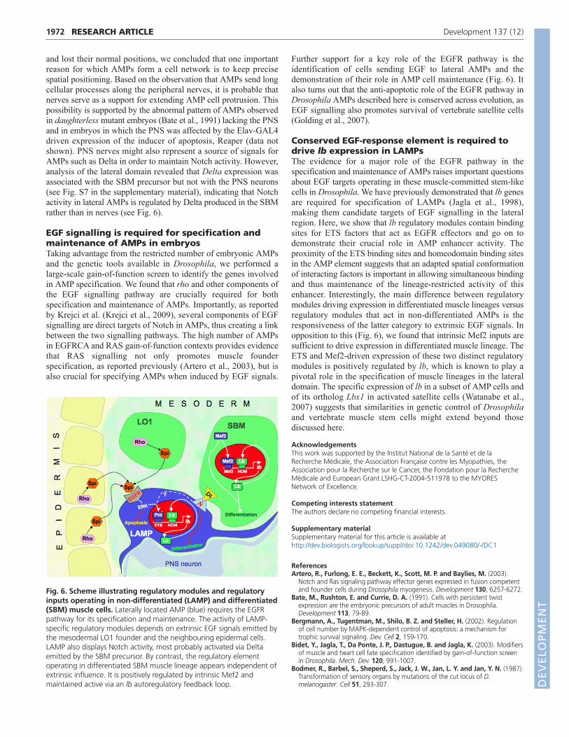
and lost their normal positions, we concluded that one importantreason for which AMPs form a cell network is to keep precisespatial positioning. Based on the observation that AMPs send longcellular processes along the peripheral nerves, it is probable thatnerves serve as a support for extending AMP cell protrusion. Thispossibility is supported by the abnormal pattern of AMPs observedin daughterless mutant embryos (Bate et al., 1991) lacking the PNSand in embryos in which the PNS was affected by the Elav-GAL4driven expression of the inducer of apoptosis, Reaper (data notshown). PNS nerves might also represent a source of signals forAMPs such as Delta in order to maintain Notch activity. However,analysis of the lateral domain revealed that Delta expression wasassociated with the SBM precursor but not with the PNS neurons(see Fig. S7 in the supplementary material), indicating that Notchactivity in lateral AMPs is regulated by Delta produced in the SBMrather than in nerves (see Fig. 6).
EGF signalling is required for specification andmaintenance of AMPs in embryosTaking advantage from the restricted number of embryonic AMPsand the genetic tools available in Drosophila, we performed alarge-scale gain-of-function screen to identify the genes involvedin AMP specification. We found that rho and other components ofthe EGF signalling pathway are crucially required for bothspecification and maintenance of AMPs. Importantly, as reportedby Krejci et al. (Krejci et al., 2009), several components of EGFsignalling are direct targets of Notch in AMPs, thus creating a linkbetween the two signalling pathways. The high number of AMPsin EGFRCA and RAS gain-of-function contexts provides evidencethat RAS signalling not only promotes muscle founderspecification, as reported previously (Artero et al., 2003), but isalso crucial for specifying AMPs when induced by EGF signals.
Further support for a key role of the EGFR pathway is theidentification of cells sending EGF to lateral AMPs and thedemonstration of their role in AMP cell maintenance (Fig. 6). Italso turns out that the anti-apoptotic role of the EGFR pathway inDrosophila AMPs described here is conserved across evolution, asEGF signalling also promotes survival of vertebrate satellite cells(Golding et al., 2007).
Conserved EGF-response element is required todrive lb expression in LAMPsThe evidence for a major role of the EGFR pathway in thespecification and maintenance of AMPs raises important questionsabout EGF targets operating in these muscle-committed stem-likecells in Drosophila. We have previously demonstrated that lb genesare required for specification of LAMPs (Jagla et al., 1998),making them candidate targets of EGF signalling in the lateralregion. Here, we show that lb regulatory modules contain bindingsites for ETS factors that act as EGFR effectors and go on todemonstrate their crucial role in AMP enhancer activity. Theproximity of the ETS binding sites and homeodomain binding sitesin the AMP element suggests that an adapted spatial conformationof interacting factors is important in allowing simultaneous bindingand thus maintenance of the lineage-restricted activity of thisenhancer. Interestingly, the main difference between regulatorymodules driving expression in differentiated muscle lineages versusregulatory modules that act in non-differentiated AMPs is theresponsiveness of the latter category to extrinsic EGF signals. Inopposition to this (Fig. 6), we found that intrinsic Mef2 inputs aresufficient to drive expression in differentiated muscle lineage. TheETS and Mef2-driven expression of these two distinct regulatorymodules is positively regulated by lb, which is known to play apivotal role in the specification of muscle lineages in the lateraldomain. The specific expression of lb in a subset of AMP cells andof its ortholog Lbx1 in activated satellite cells (Watanabe et al.,2007) suggests that similarities in genetic control of Drosophilaand vertebrate muscle stem cells might extend beyond thosediscussed here.
AcknowledgementsThis work was supported by the Institut National de la Santé et de laRecherche Médicale, the Association Française contre les Myopathies, theAssociation pour la Recherche sur le Cancer, the Fondation pour la RechercheMédicale and European Grant LSHG-CT-2004-511978 to the MYORESNetwork of Excellence.
Competing interests statementThe authors declare no competing financial interests.
Supplementary materialSupplementary material for this article is available athttp://dev.biologists.org/lookup/suppl/doi:10.1242/dev.049080/-/DC1
ReferencesArtero, R., Furlong, E. E., Beckett, K., Scott, M. P. and Baylies, M. (2003).
Notch and Ras signaling pathway effector genes expressed in fusion competentand founder cells during Drosophila myogenesis. Development 130, 6257-6272.
Bate, M., Rushton, E. and Currie, D. A. (1991). Cells with persistent twistexpression are the embryonic precursors of adult muscles in Drosophila.Development 113, 79-89.
Bergmann, A., Tugentman, M., Shilo, B. Z. and Steller, H. (2002). Regulationof cell number by MAPK-dependent control of apoptosis: a mechanism fortrophic survival signaling. Dev. Cell 2, 159-170.
Bidet, Y., Jagla, T., Da Ponte, J. P., Dastugue, B. and Jagla, K. (2003). Modifiersof muscle and heart cell fate specification identified by gain-of-function screenin Drosophila. Mech. Dev. 120, 991-1007.
Bodmer, R., Barbel, S., Sheperd, S., Jack, J. W., Jan, L. Y. and Jan, Y. N. (1987).Transformation of sensory organs by mutations of the cut locus of D.melanogaster. Cell 51, 293-307.
RESEARCH ARTICLE Development 137 (12)
Fig. 6. Scheme illustrating regulatory modules and regulatoryinputs operating in non-differentiated (LAMP) and differentiated(SBM) muscle cells. Laterally located AMP (blue) requires the EGFRpathway for its specification and maintenance. The activity of LAMP-specific regulatory modules depends on extrinsic EGF signals emitted bythe mesodermal LO1 founder and the neighbouring epidermal cells.LAMP also displays Notch activity, most probably activated via Deltaemitted by the SBM precursor. By contrast, the regulatory elementoperating in differentiated SBM muscle lineage appears independent ofextrinsic influence. It is positively regulated by intrinsic Mef2 andmaintained active via an lb autoregulatory feedback loop. D
EVELO
PMENT
Carlson, M. E., Hsu, M. and Conboy, I. M. (2008). Imbalance between pSmad3and Notch induces CDK inhibitors in old muscle stem cells. Nature 454, 528-532.
Conboy, I. M. and Rando, T. A. (2002). The regulation of Notch signalingcontrols satellite cell activation and cell fate determination in postnatalmyogenesis. Dev. Cell 3, 397-409.
Duan, H., Zhang, C., Chen, J., Sink, H., Frei, E. and Noll, M. (2007). A key roleof Pox meso in somatic myogenesis of Drosophila. Development 134, 3985-3997.
Farrell, E. R. and Keshishian, H. (1999). Laser ablation of persistent twist cells inDrosophila: muscle precursor fate is not segmentally restricted. Development126, 273-280.
Figeac, N., Daczewska, M., Marcelle, C. and Jagla, K. (2007). Muscle stem cellsand model systems for their investigation. Dev. Dyn. 236, 3332-3342.
Golding, J. P., Calderbank, E., Partridge, T. A. and Beauchamp, J. R. (2007).Skeletal muscle stem cells express anti-apoptotic ErbB receptors during activationfrom quiescence. Exp. Cell Res. 313, 341-356.
Jagla, T., Bellard, F., Lutz, Y., Dretzen, G., Bellard, M. and Jagla, K. (1998).Ladybird determines cell fate decisions during diversification of Drosophilasomatic muscles. Development 125, 3699-3708.
Junion, G., Jagla, T., Duplant, S., Tapin, R., Da Ponte, J. P. and Jagla, K.(2005). Mapping Dmef2-binding regulatory modules by using a ChIP-enriched insilico targets approach. Proc. Natl. Acad. Sci. USA 102, 18479-18484.
Junion, G., Bataillé, L., Jagla, T., Da Ponte, J. P., Tapin, R. and Jagla, K. (2007).Genome-wide view of cell fate specification: ladybird acts at multiple levelsduring diversification of muscle and heart precursors. Genes Dev. 21, 3163-3180.
Knirr, S., Azpiazu, N. and Frasch, M. (1999). The role of the NK-homeobox geneslouch (S59) in somatic muscle patterning. Development 126, 4525-4535.
Krejci, A., Bernard, F., Housden, B. E., Collins, S. and Bray, S. J. (2009). Directresponse to Notch activation: signaling crosstalk and incoherent logic. Sci.Signal. 2, ra1.
Lai, E. C., Bodner, R. and Posakony, J. W. (2000). The enhancer of split complexof Drosophila includes four Notch-regulated members of the bearded genefamily. Development 127, 3441-3455.
Liotta, D., Han, J., Elgar, S., Garvey, C., Han, Z. and Taylor, M. V. (2007). TheHim gene reveals a balance of inputs controlling muscle differentiation inDrosophila. Curr. Biol. 17, 1409-1413.
Maqbool, T. and Jagla, K. (2007). Genetic control of muscle development:learning from Drosophila. J. Muscle Res. Cell Motil. 28, 397-407.
Micchelli, C. A. and Perrimon, N. (2006). Evidence that stem cells reside in theadult Drosophila midgut epithelium. Nature 439, 475-479.
Nagaso, H., Murata, T., Day, N. and Yokoyama, K. K. (2001). Simultaneousdetection of RNA and protein by in situ hybridization and immunologicalstaining. J. Histochem. Cytochem. 49, 1177-1182.
Ohlstein, B. and Spradling, A. (2006). The adult Drosophila posterior midgut ismaintained by pluripotent stem cells. Nature 439, 470-474.
Pearson, J., López-Onieva, L., Rojas-Ríos, P. and González-Reyes, A. (2009).Recent advances in Drosophila stem cell biology. Int. J. Dev. Biol. 53, 1329-1339.
Postigo, A. A., Ward, E., Skeath, J. B. and Dean, D. C. (1999). zfh-1, theDrosophila homologue of ZEB, is a transcriptional repressor that regulatessomatic myogenesis. Mol. Cell. Biol. 19, 7255-7263.
Rebeiz, M., Reeves, N. L. and Posakony, J. W. (2002). SCORE: a computationalapproach to the identification of cis-regulatory modules and target genes inwhole-genome sequence data. Site clustering over random expectation. Proc.Natl. Acad. Sci. USA 99, 9888-9893.
Ruiz Gómez, M. and Bate, M. (1997). Segregation of myogenic lineages in Drosophila requires numb. Development 124, 4857-4866.Sellin, J., Drechsler, M., Nguyen, H. T. and Paululat, A. (2009). Antagonistic
function of Lmd and Zfh1 fine tunes cell fate decisions in the Twi and Tinpositive mesoderm of Drosophila melanogaster. Dev. Biol. 326, 444-455.
Sharma, Y., Cheung, U., Larsen, E. W. and Eberl, D. F. (2002). PPTGAL, aconvenient Gal4 P-element vector for testing expression of enhancer fragmentsin Drosophila. Genesis 34, 115-118.
Soler, C., Daczewska, M., Da Ponte, J. P., Dastugue, B. and Jagla, K. (2006).Coordinated development of muscles and tendons of the Drosophila leg.Development 131, 6041-6051.
Spradling, A., Drummond-Barbosa, D. and Kai, T. (2001). Stem cells find theirniche. Nature 414, 98-104.
Sudarsan, V., Anant, S., Guptan, P., VijayRaghavan, K. and Skaer, H. (2001).Myoblast diversification and ectodermal signaling in Drosophila. Dev. Cell 1,829-839.
Takashima, S., Mkrtchyan, M., Younossi-Hartenstein, A., Merriam, J. R. andHartenstein, V. (2008). The behaviour of Drosophila adult hindgut stem cells iscontrolled by Wnt and Hh signalling. Nature 454, 651-655.
Vasyutina, E., Lenhard, D. C., Wende, H., Erdmann, B., Epstein, J. A. andBirchmeier, C. (2007). RBP-J (Rbpsuh) is essential to maintain muscle progenitorcells and to generate satellite cells. Proc. Natl. Acad. Sci. USA 104, 4443-4448.
Watanabe, S., Kondo, S., Hayasaka, M. and Hanaoka, K. (2007). Functionalanalysis of homeodomain-containing transcription factor Lbx1 in satellite cells ofmouse skeletal muscle. J. Cell Sci. 120, 4178-4187.
Yu, F., Kuo, C. T. and Jan, Y. N. (2006). Drosophila neuroblast asymmetric celldivision: recent advances and implications for stem cell biology. Neuron 51, 13-20.
1973RESEARCH ARTICLEEGF controls specification of AMP cells
DEVELO
PMENT