drowning & burns
TRANSCRIPT

DROWNING & BURNS J O D I E G E R D I N D V M D A C V P A U S T R A L I A 2 0 1 8

DROWNING

OVERVIEW: DROWNING
• Definitions • Nx findings • Histo findings

ASPHYXIA
• An umbrella term for death due to body-wide lack of oxygen (hypoxia/ anoxia) • Asphyxia is a mechanism of death, not a COD
• Previously terminology inconsistent, recent review/ standardization (Sauvageau 2012)
• Asphyxia is classified into 4 broad categories: 1. Suffocation 2. Strangulation 3. Mechanical asphyxia 4. Drowning


DROWNING DEFINITION
• Fatal respiratory impairment from submersion / immersion, with the airway covered by liquid* • A liquid/air interface is present at the entrance of the
airway, preventing breathing air • Not “filling the respiratory tract with liquid”
• Small amounts of liquid can RARELY cause drowning • Drowning after any
H2O-related activities • Playing in pools, sprinklers,
lakes, streams, baths, etc.
*World Congress on Drowning 2002

DEFUNCT DEFINITIONS
• Non-fatal drowning • Water aspiratedà rescue à survives
• Fatal drowning • Water aspirationà rescue à dies
• No longer used: • Active & Passive drowning, Dry & Wet drowning,
Near-drowning, Secondary drowning • Confusing terminology arose due to drowning victims
with few/mild initial respiratory signs • Most signs are immediate, nearly all in 8 hrs, up to 24 hours later

H2O in pharynx
Aspiration
Cough reflex
More aspiration
Hypoxemia
Loss of consciousness
Apnea
Cardiac arrest
The Drowning Process
Struggle & airway submersion
Salt & Fresh H2O:
Surfactant destruction &
washoutà alveolar wall
damage

DROWNING: NX GOALS
• If Hx of exposure to liquid, consider drowning • For bodies is found in H20, determine:
• Animal alive or dead at the time of submersion • Not all bodies recovered from H20 drowned;
• Often, bodies are disposed of in water • Death due to something else while in water
• Rule in/out drowning, other CODs • Contributing illness making
submersion fatal • Seizures, ataxia, blindness, etc.

DROWNING: NX FINDINGS
• Body wet (“spiked” haircoat) • Foam / froth in upper airways
• Mix of aspirated H2O, mucus, & surfactant
• Emphysematous & edematous lungs • Soggy (edema) and/ or crepitant (emphysema) • +/- Rib impressions • Copious fluid exudes from the cut surface
• Multifocal, patchy, red areas in lungs • Due to congestion, atelectasis, & hemorrhage
• If no significant pulmonary edema, ascribing the COD to drowning is unwise
• Water, mud, sand, plant matter in alveoli or stomach

Drowned wallaby. Wet hair coat looks “spiked”. (Also head trauma)

Drowned cat with liver fracture. Lungs have not collapsed, & small scattered areas of hemorrhage.

Drowned raccoon. Lungs are look “full” & failed to collapse.

Drowned squirrel. Lungs hyper-inflated, failed to collapse.

Drowned dog. Multifocal areas of congestion / hemorrhage & failure to collapse.

DROWNING: HISTOLOGY
• H2O does not passively seep into the deep lung tissue in deceased or unconscious people– Aspiration requires active ventilation
• HISTO: • Alveolar edema & hemorrhage • Expansion / coalescing alveolar spaces with torn (blunted/ clubbed) alveolar walls • AKA Emphysematous change
• Foreign material (plant, sand, etc.) in airways • Especially in terminal /deeper airways


Lung, drowned cat. Unidentified foreign material in bronchioles.

EXCEPTION: DIVING ANIMALS
• Lunged species that spend a significant % of time in water rarely if ever aspirate, even though drowning is certain based on circumstances (caught in nets, etc.) – JG opinion/ communication • Ex: Seals, sea turtles, otter…
• These animals may never involuntarily aspirate (gasp); larynx stays closedà hypoxiaà death
• COD is suffocation

BURNS

OVERVIEW: BURNS
• Classification of burns • Depth • Cause / 6 types
• Nx goals • Assess depth & estimate the extent
• How to evaluate burned / charred remains

BURNS
• Burn = Wound due to excessive heat • Severity depends on:
• Temperature • Duration of exposure • Ability of the tissue to dissipate heat

SKIN BURN DEPTH
• #1 organ burned • Classified by thickness (degree) • Superficial (1st degree)
• Some/all epidermis à erythema
• Partial thickness (2nd degree): • Entire epidermis & some/all dermis à blisters, skin necrosis
• Full thickness (3rd & 4th degree) • Epidermis & dermis plus some/all SQ à charred tissue, exposure of fat & muscle
• Painless (nerves dead)

Normal
Partial
Superficial
Full thickness

• Epidermis only • Red +/- swollen (erythema & edema) • Mildly painful • Do not scar. No injury to
basement membrane / stem cells
SUPERFICIAL BURNS

• Entire epidermis + some Dermis injured • Ooze blood/ serumà Scab; Humans blister • +/- Scar, +/- Alopecia
• Depends on whether stem cells were injured
• Painful
PARTIAL THICKNESS BURNS
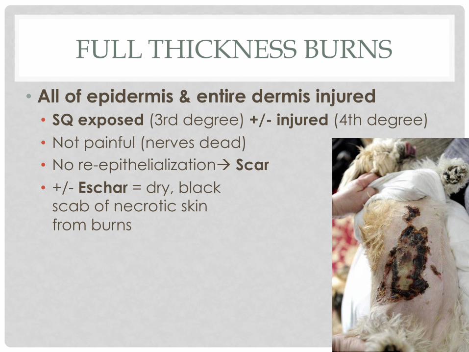
• All of epidermis & entire dermis injured • SQ exposed (3rd degree) +/- injured (4th degree) • Not painful (nerves dead) • No re-epithelializationà Scar • +/- Eschar = dry, black
scab of necrotic skin from burns
FULL THICKNESS BURNS

• Rate depth based on worst-affected area • Often challenging; most are a mix of depths • Full extent often peaks several days after
exposure • Histo may be
helpful to determine depth
EVALUATING BURNS

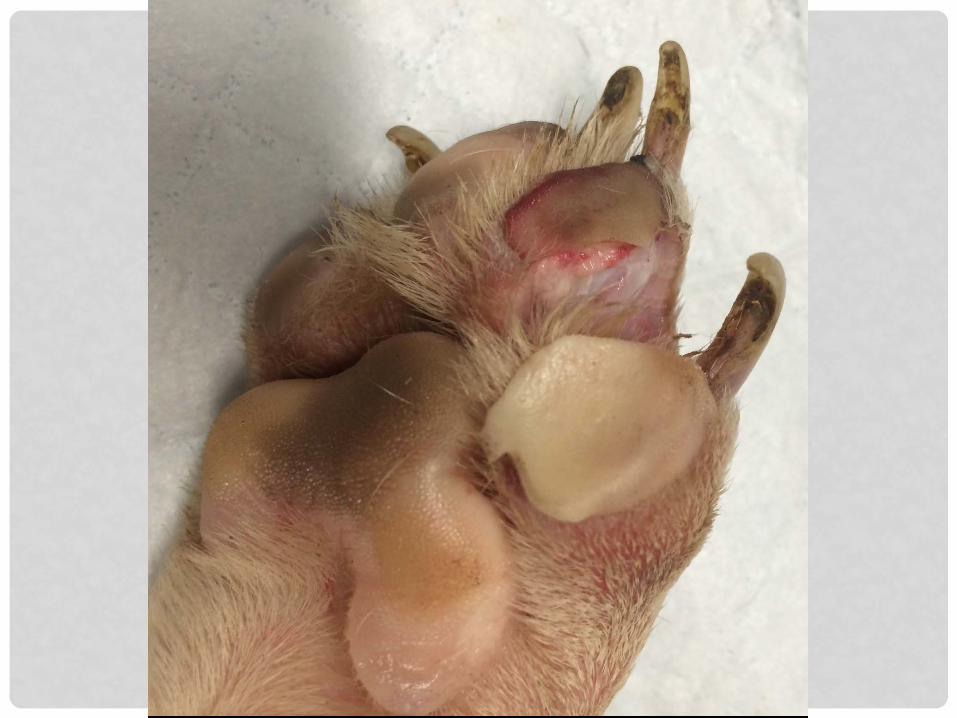
Dog. Burn depth (thickness)?

Partial thickness / 2nd degree

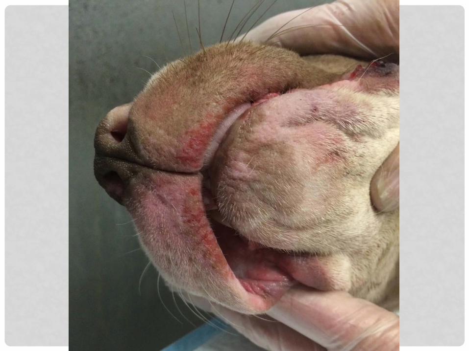
Dog. Depth (thickness)?

Dog. Full thickness burn, cause unknown. An eschar is present.

BURN TYPES: EXAMPLES
1. Scalds • Ex: Garden hoses left in the sun
2. Fire/ flame • Ex: House fires
3. Electrical • Ex: Electrical cords
4. Contact • Ex: Heat rocks, Car mufflers, Brands
5. Radiation • Ex: Sunburns (UV), microwaves, Radiation Tx
6. Chemical burns • Ex: Petro-chemical burns, Severe contact dermatitis

CIGARETTE BURNS
• Purposeful burns made by holding the cigarette perpendicular to skinà ~1.0 cm diameter round crater, well-defined edge
• Accidental brushing up against a cigarette à “Comet” lesion: Round spot & tapering tail

SCALDS
• Contact with wet heat • Ex: Boiling H2O, steam, etc.
• Pattern • 1 or more usually coalescing burns, often on dorsum • Margins irregular, elongated dorsal to ventral (gravity)
• Severity lessens ventrally (liquid cools & drips off)
• Tiny satellite burns d/t Drips & Splashes
• Even superficial scalds can produce significant scarring
• No singeing of hair

Healing scald with peripheral re-epithelialization.
Photos courtesy Dr. Robert Reisman, ASPCA

Healing scald with peripheral re-epithelialization. Note dorsal distribution &“splash / drip” pattern (arrows).
Photos courtesy Dr. Robert Reisman, ASPCA

CONTACT BURNS
• A hot surface directly contacts the body • Ex: Heat rocks, Car mufflers, Brands, irons, etc.
• Dog. Contact burn (hot pavement) • Thickness?

CONTACT BURNS
• A hot surface directly contacts the body • Ex: Heat lamps / heat rocks, Car mufflers, Brands
• Dog. Contact burn (hot pavement) • Partial
thickness

ELECTRICAL BURNS
• May cause focal or branching (arborizing) skin lesions • Ex: Cautery, bit electrical cords, & lightning
• High voltage: Central crater w/brown-yellow margin • May be see in combo w/ flame burn if the hair coat
catches on fire
• Low voltage: No lesions OR Central chalky white crater with erythema
• Electricity causes distinct histological changes • “Windblown” (elongated) nuclei

Electric collar (invisible fence) collar wound- NOT A BURN. Pressure necrosis. No gross signs of a burn.

MICROWAVE BURNS
• Microwaves heat water, inc. water in tissues • Tissue with a high H2O content reaches a higher
temp than tissues with less water • Primarily affects skin, muscle & internal organs;
spares SQ fat (contains little water) • Well-demarcated & unevenly distributed
• Focal “hot spots” where 1 tissue abuts another • The severity of the injuries corresponds to the
duration of exposure

MICROWAVE BURNS
• 2008 Munro: Fatal feline cases • Flexure of the forelimbs at the carpus with or
without ex-sheathing of the claws (~pugilistic posture)
• Fragility of the skin +/- splitting with sharp, well delineated, edges
• Crumpling &reddening of the tips of the ears • Congestion of all lung lobes • Internal organs readily disintegrate & have the odor
of cooked chicken • Absence of singed hair

Woman killed cat for eating her goldfish by putting it in a microwave. (Sentence: Jail-14 weeks)
https://www.express.co.uk/news/uk/464624/Woman-jailed-for-14-weeks-after-putting-cat-in-microwave

FIRES & FLAME BURNS
• Skin is in direct contact with a flame • Severity depends on duration of exposure
• Singes hair, then chars skin, nails, & deeper tissues
• Flash burns-- sudden ignition / explosion of a volatile substance (accelerants) • Produces a uniform burn (1st or 2nd degree) on all
exposed areas & singes the hair

Singed whiskers. Only seen with fire / flame burns.

Flash Burn. Cat doused with lighter fluid & set on fire. Even singeing, charring & contraction of the skin (heat). Found alive
but quickly euthanized.

Cat; body burned on a fire after death in an attempt to dispose of the body/ destroy evidence.
Well delineated areas of singed hair.

Burned cat, section of lung. Small pieces burned hair in the bronchi & alveolar spaces (circled).

Young Pit Bull put in oven. Not a fire / flame burn, but similar?


Dog that was in a house fire with thermal burns.

Toxic epidermal necrolysis (TEN) in a dog & cat. Similar “clown-face” appearance to flame burns.
Function of thinness of skin? Lack of improvement with supportive care, lack of accelerant
odor, & histo of the affected areas differentiate TEN from burns.

BURNED REMAINS: NX GOALS
• Was death due to fire, or was the body burned? • Soot in upper airway = Evidence of smoke inhalation
(“vital change”) = proves animal was alive to inhale smoke • Have area set aside for examination of pluck • Avoid cross contamination of soot on body into organs
• Use new/ clean gloves & clean knife to get histo samples
• +/-Accelerant testing • +/- Blood carbon monoxide [CO] (standard in people)
• Look for cherry red livor mortis • CO-Hb is very stable with no exposure to light
• Test likely valid for days • EDTA heart blood sample • Human lab?

BURNED REMAINS: ARTIFACTS
• Artifacts of extreme heat: • Bone Fx including skull • Epidural hematomas • Skin splitting • “Pugilistic posture”
flexion of the elbows & carpi
• Internal organs typically preserved
Brain of burned cat with small epidural hematoma

FIRES: ACCELERANT TESTING
• Animals not spontaneously combustible; Accelerants must be used
• Collect ASAP! • Accelerants (volatiles) evaporate quickly
• Collect anything that smells • Ex: collars, haired skin
• Collect least-burned areas • Accelerant least–consumed
• Clean metal or glass container

Dog in house fire (hind end). Pugilistic posture: Flexion of the hips, stifles & digits & extension of hocks, due to heat contraction of collagen in muscle & tendons.

Dog (same as previous). Flexed shoulders, elbows & carpi, contracture of skin & curled back lips. Well delineated area of spared skin & hair (white patch). Tracheal ulceration (thermal injury)à COD= smoke inhalation.

CHEMICAL BURNS
• Strong acids & alkalis cause direct cell damage • Severity depends on the agent, strength /
concentration, & duration of contact • Alkaline agents (pH greater than 11.5) tend to produce more
severe (full thickness) injury compared to acids
• Gross lesions resemble other burns, especially scalds • Predominantly skin
• Tissue necrosis • +/- Blistering (people) • More superficial compared to thermal burns

CHEMICAL BURNS
• Ddx chemical from thermal difficult • Histo *might* help
• Heat “wicked” by hairs, disproportionate damage to follicles
• Chemical residue - Odor or liquid itself
• Ddx accidental from purposeful may be difficult • Hx / investigation dependent • Severe irritant contact dermatitis
• Idiosyncratic reactions to topical Rx, especially flea/tick preparations

Suspected chemical burn with ventral distribution: Paws, rump, elbows, from sitting/walking in the chemical, & mouth from licking it off.
Photos courtesy Dr. Robert Reisman, ASPCA

The dorsal midline burn is a common pattern, seen with a wide variety of accidental & purposeful causes.
Determining the cause without a history may be impossible.
LEFT: Severe irritant contact dermatitis from a reaction to topical flea/ tick medication (right).
RIGHT: Scald caused by garden
hose.

ESTIMATING % AFFECTED
• “Rule of 9s” not accurate for other species
• How many credit cards does it take to cover the burn? • Determine Body Surface Area based
on weight -- standard conversion charts (as for chemo)

% BSA = [# cards x 0.45] / total BSA
EXAMPLE: 6kg dog; 22 card burn
22 x 0.45 %BSA burned = --------------
0.33m2 = 30%

BURNS: NX GOALS
1. Document location(s) affected • Remember to check oral cavity
2. Estimate % body surface affected 3. Assess the depth 4. Diagnostic features • Eschar, blisters, “splashes”
5. Cause of death = Burn
• ID type (if possible): Scalding, Contact, Flame, Electrical, Microwave, & Chemical
• Does the burn fit with the explanation?
Healing obscures

SUMMARY
• 6 types of Burns: • Scalding, Contact, Flame, Electrical, Microwave, &
Chemical • Burns should be described in terms of: 1. Depth: superficial, partial, complete thickness 2. Extent: % total body surface area affected 3. Distribution (Pattern): Anatomic location(s), drips/
splashes? 4. Features of the burn: singed or charred tissue, or eschar
• Animals exposed to fires – evaluate for exposure to: • Smoke/ fumes & Carbon monoxide • Exposure to heat.
• Consider accelerant testing

REFERENCES • Szpilman D, et al. “Dry drowning” and other myths. Cleve Clin J Med.
2018;85(7):529–535. • Munro R, Munro HMC. Animal abuse and unlawful killing: Forensic
veterinary pathology. Elsevier Ltd; 2008. • Reedy LM, Clubb FJ Jr. Microwave burn in a toy poodle: A case
report. The Journal of the American Animal Hospital …. 1991;27:497–500.
• Budd R. Burns associated with the use of microwave ovens. J Microw Power Electromagn Energy. 1992;27(3):160–163.
• Sobhakumari et al. Pathology of carbon monoxide poisoning in two cats. BMC Veterinary Research (2018) 14:67
• Malic CC, et al. Resuscitation burn card--a useful tool for burn injury assessment. Burns. 2007 Mar;33(2):195-9.
• Sauvageau A, Boghossian, E. Classification of asphyxia: The need for standardization. J Forensic Sci 2012;55:1259-67