drug-induced glaucoma - fmshk.org · drug-induced glaucoma open-angle steroid is a group of drugs...
TRANSCRIPT

Medical Bulletin VOL.15 NO.10 OCTOBER 2010
29
Drug-induced Glaucoma
Dr. Nafees BAIGMBBS(HK), MRCS(Edin), FCOphthHK, FHKAM(Ophthalmology)Resident (Specialist), Hong Kong Eye Hospital
Dr. Nafees BAIG
IntroductionGlaucoma is a form of optic neuropathy with specific visual field loss. It is usually associated with raised intraocular pressure (IOP). Several drugs have the potential to cause raised IOP; this can occur via an open-angle or a angle-closure mechanism.1 One of the most important drugs is steroid.2 Steroid-induced glaucoma is a form of open-angle glaucoma. It is usually associated with topical steroids. However other forms of administration such as inhaled, oral, intravenous, periocular and intravitreal, can also lead to raised IOP. Medications for treating a variety of systemic conditions including depression, allergies, Parkinson disease etc., can produce pupillary dilation and precipitate an attack of acute angle-closure glaucoma in anatomically predisposed eyes that have narrow angles.
Mechanisms of IOP Elevation in Drug-induced GlaucomaOpen-angleSteroid is a group of drugs that may produce IOP elevation by open-angle mechanism. Not all patients taking steroid will develop glaucoma. Risk factors include preexisting primary open-angle glaucoma, a family history of glaucoma, high myopia, diabetes mellitus and young age.3 It has been shown that 18-36% of the general population and 46–92% of patients with primary open-angle glaucoma respond to topical ocular administration of corticosteroids with an elevation of IOP, usually within 2–4 weeks after therapy has been instituted.1
Topically applied eyedrops, topically applied creams to the periorbital area and intravitreal injections are more likely to cause IOP elevation. The incidence of elevated IOP is less with intravenous, parenteral and inhaled routes of administration. Since IOP elevation can be gradual and asymptomatic, patients on chronic corticosteroid therapy can remain undiagnosed, which can result in glaucomatous optic nerve damage.
Steroid-induced IOP elevation typically occurs within a few weeks after commencing steroid therapy. In most cases, IOP returns spontaneously to the baseline within a few weeks to months upon stopping the steroid. In rare situations, the IOP remains high that requires prolonged glaucoma medication or even surgery.
Closed-angleSome drugs have contraindications or adverse effects concerning with acute angle-closure glaucoma. These drugs will incite an attack in those individuals with very narrow anterior chamber angles that are prone to occlusion especially when the pupils are dilated. The classes of medications that have the potential to induce angle-closure are topical anticholinergic or sympathomimetic pupil dilating drops, tricyclic antidepressants, monoamine oxidase inhibitors, antihistamines, antiparkinsonism drugs, antipsychotic medications and antispasmolytic agents.
Sulfonamide-containing medications may induce angle-closure glaucoma by a different mechanism, involving the anterior rotation of the ciliary body. Typically, the angle-closure is bilateral and occurs within the first few doses. Patients with narrow or wide open angles are potentially susceptible to this rare and idiosyncratic reaction.
Pathophysiology of Drug-induced GlaucomaOpen-angleThe exact pathophysiology of steroid-induced glaucoma is unknown. It is known that steroid-induced IOP elevation is secondary to increased resistance to aqueous outflow. Some evidence shows that there could be increased accumulation of glycosaminoglycans or increased production of trabecular meshwork-inducible glucocorticoid response (TIGR) protein, which could mechanically obstruct the aqueous outflow. Other evidence suggests that the corticosteroid-induced cytoskeletal changes could inhibit pinocytosis of aqueous humour or inhibi t the c lear ing of glycosaminoglycans, resulting in the accumulation of this substance and blockage of the aqueous outflow.
Closed-angleAqueous humour is secreted by the ciliary body and circulates through the pupil to the anterior chamber angle. (Fig. 1) The pathophysiology of angle-closure glaucoma is usually due to pupillary blockage, i.e. iris-lens contact at the pupillary border resulting from pupillary dilation. Medications have a direct or secondary effect, either in stimulating sympathetic or inhibiting parasympathetic activation causing pupillary dilation, which can precipitate acute angle-closures in patients with occludable anterior chamber angles. These include adrenergic agonists (e.g.

Medical BulletinVOL.15 NO.10 OCTOBER 2010
30
phenylephrine), β2-specific adrenergic agonists (e.g. salbutamol), noncatecholamine adrenergic agonists (e.g. amphetamine, dextroamphetamine, methamphetamine and phendimetrazine) and anticholinergics (e.g. tropicamide). Histamine H1receptor antagonists ( a n t i h i s t a m i n e s ) a n d h i s t a m i n e H 2 r e c e p t o r antagonists (e.g. cimetidine and ranitidine) have weak anticholinergic adverse effects. Antidepressants such as fluoxetine, paroxetine, fluvoxamine and venlafaxine have been associated with acute angle-closures. It is believed to be induced by either the anticholinergic adverse effects or the increased levels of serotonin that cause mydriasis.
Sulfa-containing medications result in acute angle-closures in a different mechanism. This involves the anterior rotation of the ciliary body with or without choroidal effusions, resulting in a shallow anterior chamber and blockage of the trabecular meshwork by the iris. Pupillary dilation and a preexisting shallow anterior chamber angle are not necessary. The exact reason causing ciliary body swelling is unknown in susceptible individuals.
Topiramate is a sulfa-containing anticonvulsant. There were reports about patients on topiramate developing acute angle-closures. However, a pilot study was conducted in the Hong Kong Eye Hospital and the Prince of Wales Hospital recently which showed that short-term use of topiramate did not induce asymptomatic angle narrowing.4 Therefore it was suggested that topiramate-induced secondary angle-closure glaucoma may be an all-or-none phenomenon.
Fig. 1. Flow of aqueous humor indicated in red.
Clinical Assessment for Drug-induced GlaucomaHistoryThe patient's current medications should be carefully elicited.
Symptoms With steroid-induced glaucoma, the pressure elevation is gradual. Therefore, there are very few symptoms during the early stage of disease. At a later stage, patients may complain of loss of the peripheral visual field. At the more advanced stage, when the central vision is also affected, patients may complain of blurring of vision.
In drug-induced acute angle-closure glaucoma, the symptoms are the same as in primary acute angle-closure glaucoma. These include sudden eye pain, headache associated with nausea and/ or vomiting, blurring of vision and halos around bright objects.
Past Ocular History/Past Medical History History of systemic medical disease, which could require chronic corticosteroid use (e.g., uveitis, collagen vascular disease, asthma, dermatitis) should be elicited. Patients with preexisting primary open-angle glaucoma, a family history of primary open-angle glaucoma, diabetes mellitus, high myopia, or connective tissue diseases are at greater risk to be steroid responders.
Physical ExaminationA complete ophthalmic examination should be performed including the followings:
Visual acuity and refractionPatients with acute angle-closures have significant drops in visual acuity. Patients with hyperopia are at higher risks for narrow anterior chamber angles.
Pupil reflexAcute angle-closure presents with a fixed, mid-dilated pupil (Fig. 2) while an afferent pupillary defect indicates unilateral optic nerve damage.
Fig. 2. Slit lamp photo of an eye with acute angle closure showing shallow anterior chamber, mid dilated pupil and ciliary injection
Intraocular pressureAcute angle-closure usually presents with a much higher IOP than steroid-induced glaucoma which presents with a gradual IOP elevation.
Slit lamp examinationExamination of the anterior chamber is essential to look for signs of other secondary glaucomas such as uveitic, pigment dispersion and pseudoexfoliation glaucoma. It can also assess the depth of the anterior chamber and to exclude pupillary block. Fig. 2 shows a shallow anterior chamber in an acute angle-closure. Cataract is also associated with chronic steroid use.
Gonioscopic examinationGonioscopy can evaluate the angle anatomy (i.e. open
Medical Bulletin VOL.15 NO.10 OCTOBER 2010
31
or narrow) and to determine whether the angle is occludable during pupil dilation.
Optic disc evaluationStereoscopic examination of the optic disc is necessary to exclude glaucomatous damage. The signs of glaucoma optic nerve damage include increased cup-to-disc ratio in horizontal and vertical meridians; progressive enlargement of the cup; evidence of nerve fibre layer damage with red-free filter; notching or thinning of disc rim; pallor; presence of haemorrhage; asymmetry between discs; and peripapillary atrophy. Fig. 3 shows a pink optic disc with normal cup-disc ratio while Fig. 4 shows a pale glaucomatous disc with increased cup-disc ratio.
Fig. 3. Normal optic disc with normal cup to disc ratio
Fig. 4. Pale glaucomatous optic disc with increased cup to disc ratio
Investigations
PerimetryVisual Field testing such as Humphrey or Goldman is used to evaluate the severity of optic neuropathy.
Optical Coherence Tomography (OCT)OCT is an optical signal acquisition and processing method. It captures micrometer-resolution, three-dimensional images from within the optical scattering media (e.g., biological tissue). OCT is an interferometric technique, typically employing near-infrared light. It is used to evaluate the retinal nerve fibre thickness around the optic disc in glaucoma patients. Serial scans can be used to demonstrate any progression of disease.
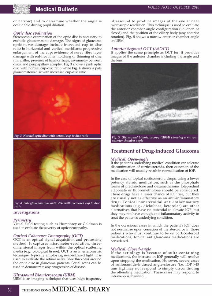
Ultrasound Biomicroscopy (UBM)UBM is an imaging technique that uses high frequency
ultrasound to produce images of the eye at near microscopic resolution. This technique is used to evaluate the anterior chamber angle configuration (i.e. open or closed) and the position of the ciliary body (any anterior rotation). Fig. 5 shows a narrow anterior chamber angle on UBM.
Anterior Segment OCT (ASOCT)It applies the same principle as OCT but it provides images of the anterior chamber including the angle and the lens.
Fig. 5. Ultrasound biomicroscopy (UBM) showing a narrow anterior chamber angle
Treatment of Drug-induced GlaucomaMedical: Open-angle If the patient's underlying medical condition can tolerate discontinuation of corticosteroids, then cessation of the medication will usually result in normalisation of IOP.
In the case of topical corticosteroid drops, using a lower potency steroid medication, such as the phosphate forms of prednisolone and dexamethasone, loteprednol etabonate or fluorometholone should be considered. These drugs have a lesser chance of IOP rise, but they are usually not as effective as an anti-inflammatory drug. Topical nonsteroidal ant i - inf lammatory medications (e.g., diclofenac, ketorolac) are other alternatives that have no potential to elevate IOP, but they may not have enough anti-inflammatory activity to treat the patient's underlying condition.
In the occasional cases in which the patient's IOP does not normalise upon cessation of the steroid or in those patients who must continue to be on corticosteroid medications, topical antiglaucoma medications are considered.
Medical: Closed-angle If the aetiology is because of sulfa-containing medications, the increase in IOP generally will resolve upon stopping the medication. However, severe cases of sulfonamide-induced angle-closure (i.e. IOP >45 mm Hg) may not respond to simply discontinuing the offending medication. These cases may respond to intravenous mannitol.

Medical BulletinVOL.15 NO.10 OCTOBER 2010
32
For other aetiologies of drug-induced angle-closure, they are treated similar to primary acute angle-closure glaucoma by using antiglaucoma medications including topical beta blockers, prostaglandin analogues, cholinergic agonists and often oral acetazolamide.5
Laser treatmentFor open-angle steroid-induced glaucoma, Argon laser trabeculoplasty or selective laser trabeculoplasty can be applied in the absence of ocular inflammation if the IOP is suboptimal with medication.
In closed-angle glaucoma, argon laser peripheral iridoplasty (ALPI) may be applied to deepen the anterior chamber and widen the angle. Laser iridotomy (LI) can be performed to reverse pupillary block or to prevent further pupillary block. Fig. 6 shows evidence of argon laser peripheral iridoplasty (in green) and laser iridotomy (in red).
Fig. 6. Slit-lamp photo showing argon laser irioplasty marks (green circles) and an inferior laser iridotomy (red circle)
Surgical: Open-angle When medical therapy is ineffective in lowering the IOP to target pressure or the patient is intolerant of medical therapy, then surgical therapy is indicated.
In patients whom both medical and laser therapy have failed to lower the IOP adequately, surgical treatment is warranted. Usually, trabeculectomy, a guarded filtration procedure, with or without intraoperative antimetabolites, is the primary procedure. In cases of eyes with active neovascularisation or inflammation, a glaucoma drainage implant may be used as the primary procedure.
Surgical: Closed-angleTrabeculectomy can also be performed with similar indications as open-angle glaucoma. However the
surgery is more difficult since the anterior chamber is shallower and the cornea is usually hazier due to the acute IOP rise.
P r e v e n t i o n o f D r u g - i n d u c e d GlaucomaOpen-angleUnnecessary prolonged use of steroid should be avoided. Ophthalmic evaluation is recommended for patients treated with long-term steroids especially with risk factors such as family history of primary open-angle glaucoma.
Closed-angleProphylactic laser iridotomy may be performed in patients requiring frequent mydriasis such as frequent fundus examinations for diabetic retinopathy. Medications causing secondary angle-closure are avoided in susceptible individuals as far as possible.
ConclusionThe prognosis of steroid-induced glaucoma depends on the duration of the IOP elevation and the control of IOP after diagnosis. Uncontrolled increase in IOP can lead to permanent optic nerve damage and hence permanent blindness. In patients with controlled IOP, the prognosis can be favourable depending on the severity of disease on presentation. Drug-induced IOP rise can be asymptomatic initially especially in open-angle type. General practitioners should be aware of the risk factors for glaucoma before prescribing a drug that has the potential to cause, precipitate or exacerbate glaucoma. Topical steroids can cause IOP rise in susceptible individuals in a fairly short period of time and therefore it is advised that topical steroids should be prescribed by doctors capable of measuring IOP. Whenever in doubt, an ophthalmologist should be consulted.
References1.
2.
3.
4.
5.
Tripathi RC, Tripathi BJ, Haggerty C, et al. Drug-induced glaucomas: mechanism and management. Drug Safety 2003;26:749-767.Tripathi RC, Parapuram SK, Tripathi BJ, et al. Corticosteroids and glaucoma risk. Drugs Aging 1999;15:439-450.Shukla D, Vidhya N, Prasad NM, et al. Evaluation of patient age as a risk factor for intraocular pressure elevation after intravitreal triamcinolone. Am J Ophthalmol 2007;144:453-454.Leung DY, Leung H, Baig N, et al. Topiramate and asymptomatic ocular angle narrowing: a prospective pilot study. Eye 2009;23:2079-2081.Lam DS, Tham CC, Lai JS, Leung DY. Current approaches to the management of acute primary angle closure. Curr Opin Ophthalmol 2007;18:146-151.