druginduced gastrointestinal inury digi pdates reflections ... · pdf filedruginduced...
TRANSCRIPT

PATHOLOGICA 2017;109:97-109
Review
Drug-induced gastrointestinal injury (DIGI). Updates, reflections and key points
G. DE PETRIS1, L. DE MARCO2, J.I. LÓPEZ3
1 Department of Pathology, Penrose-St. Francis Hospital, Colorado Springs, CO USA; 2 Anatomia Patologica, Arcispedale Santa Maria Nuova, Reggio Emilia (RE), Italy; 3 Department of Pathology, Cruces University Hospital, BioCruces Research Institute, University of
the Basque Country (UPV/EHU), Barakaldo, Spain
Key words
Drug-induced gastrointestinal injury • Identifiable drugs • Angiotensin receptor inhibitors • Immuno-modulators
Summary
The goals of this short narrative review are 1) to provide an update in recent developments in the field of drug-induced gastrointestinal injury (DIGI), and 2) to distill few key points to approach with con-fidence a difficult and vast area of gastrointestinal pathology. DIGI is a challenging diagnosis as it can produce almost any pattern of the injury of the gastrointestinal tract. The recognition of a pattern and the knowledge of which drugs can produce that pattern, are the first step of the diagnostic process; communication with the clinical
team and a high level of suspicion are then paramount. The patholo-gist can be the leading clinicians of the care team in few situations in which she/he can recognize the drug at the microscope. Knowl-edge of the most relevant differential diagnoses of DIGI is essential to avoid significant pitfalls. Finally, several DIGIs due to recently developed immunomodulators used in oncology have shown rel-evance given their sometimes fatal outcome and the accumulating evidence of a common morphological appearance among them.
In alphabetical order:AFB: acid fast bacilli stain; ARB: angiotensin receptor blocker; BAS: bile acid sequestrants; CMV: cytomegalovirus; CRF: chronic renal failure; DDC: dilated damaged crypt; DIGI: drug-induced gastrointestinal injury; GI: gastrointestinal tract; IBD: inflammatory Bowel Disease; I-C: inhibitor of CTLA-4 receptor; IELs: intraepithelial lymphocytes; GVHD: graft versus host disease; LC: lanthanum carbonate; NSAID’s: non-steroidal anti-inflammatory drugs; PI: PD1 receptor inhibitor; PI3K: phosphatydilinositol-3-kinase; SPS: sodium polystyrene Sulphonate (Kayexalate®)
CorrespondenceGiovanni De Petris, Penrose St. Francis Hospital, Department of Patholog, 2222 N Nevada Ave, Colorado Springs, CO 80907 USA - E-mail: [email protected]
Abbreviations
AcknowledgementsThe authors wish to thank Dr. M. Dupre, Dr. R. Gonzalez, Dr. M. Makino, and Dr. K. Matsukuma for their kindness, and for sharing original photographs of their cases.
Introduction
Drugs are known noxae with significant aggressive impact in the gastrointestinal tract (GI). Bates et al. 1 showed that the overall rate of adverse GI drug effects is 6.5 per 100 hospital admissions. Iatrogenic effects on the GI outside of conditions requiring hospital’s care remain unknown and are probably unknowable. To add, it is significant that doctors may not be aware of the existence, quality and/or severity of drug-induced GI injury (DIGI).Excellent reviews of DIGI are published almost year-ly 2-4. This short narrative review intends not only to pro-vide a comprehensive update on those previous works
(to which we suggest strongly to refer to and upon which we build this update) but also, and above all, to crystal-lize in key points what we have learnt about DIGI along the years. The goal is to help the practicing pathologist to diagnose DIGI or, at least, to introduce DIGI in a meaningful differential diagnosis.
The basic knowledge
The interaction of drugs with intestine depends mainly on the drug’s physical-chemical characteristics, dosage, administration route, size of the pill, formulation (e.g.

G. DE PETRIS ET AL.98
sustained release) and on the recipient variables (e.g. other drugs that may interact, how the drug is ingested, the motility of the intestine, anatomic features, associ-ated diseases). DIGI can be highly specific but more commonly it is not, thus requiring communications with clinical teams and temporal correlation with drug intake or drug stoppage to allow for diagnosis. Diverse con-troversial clinical situations can appear. On one hand, the intestine can respond to noxae with a very limited repertoire of histological responses/patterns (morpho-logical “funneling”), i.e., various diseases or conditions may overlap in their histological manifestations. On the other, the opposite is also true, that is, one DIGI or one disease can “blossom” into multiple different histo-logical patterns. When should the pathologist consider DIGI? Is DIGI frequent absence of diagnostic specific-ity a nihilistic sinkhole for the morphologist? In brief the answer is no. In general DIGI should be considered when an increase in eosinophils, in apoptotic activity, in intraepithelial lymphocytes (IELs) does appear, or when vacuolation of cytoplasm is noted in the mucosa cells 5. We learned also that DIGI should be considered when:multiple portions of the GI are affected by various inju-ry types (e.g. collagenous colitis and increased IELs in stomach), microscopic findings do not fit neatly into a known disease (e.g. apoptotic bodies that are markedly increased in what appears to be Inflammatory Bowel Dis-ease, (IBD)], the clinical context is unusual [e.g. ischemic colitis in a young patient or ischemic gastritis), the patient is unresponsive to what appears to be an appropriate treat-ment (e.g. gluten free-diet in celiac disease). Nowadays it is fashionable to try to work up the differential diagnosis of gastrointestinal medical diseases using morphological patterns of injury; DIGI, however, escapes this approach as it is not a single disease and it can generate almost any type of injury pattern. The clinical background of the pa-tient is essential not only because the clinician, hopefully, knows the drugs taken by the patient but also because knowing what conditions affect the patient may heighten the pathologist awareness of a DIGI. Certain conditions are treated with drugs that are well known to cause DIGI. A list of such diseases is in Table I.
KEY POINT #1: DIGI is most often non-specific histologically and can manifest with almost any pattern of injury. Clinical data, awareness and knowledge of DIGI pathology can help in the diagnosis.
It cannot be overemphasized that the nonspecific drug-induced changes are also clinically significant: an ex-ample is the impact of apoptotic bodies due to drugs in the diagnosis of Graft Versus Host Disease (GVHD); less than 6 apoptotic bodies/10 crypts post bone marrow transplant (BMT) in the colon are not sufficient for the diagnosis of GVHD since several cases resolve without therapy 6. Drugs are the most likely cause of this low-grade apoptosis 6. The diagnosis of “indeterminate for GVHD” post BMT is therefore due to overlap with a nonspecific histological DIGI.
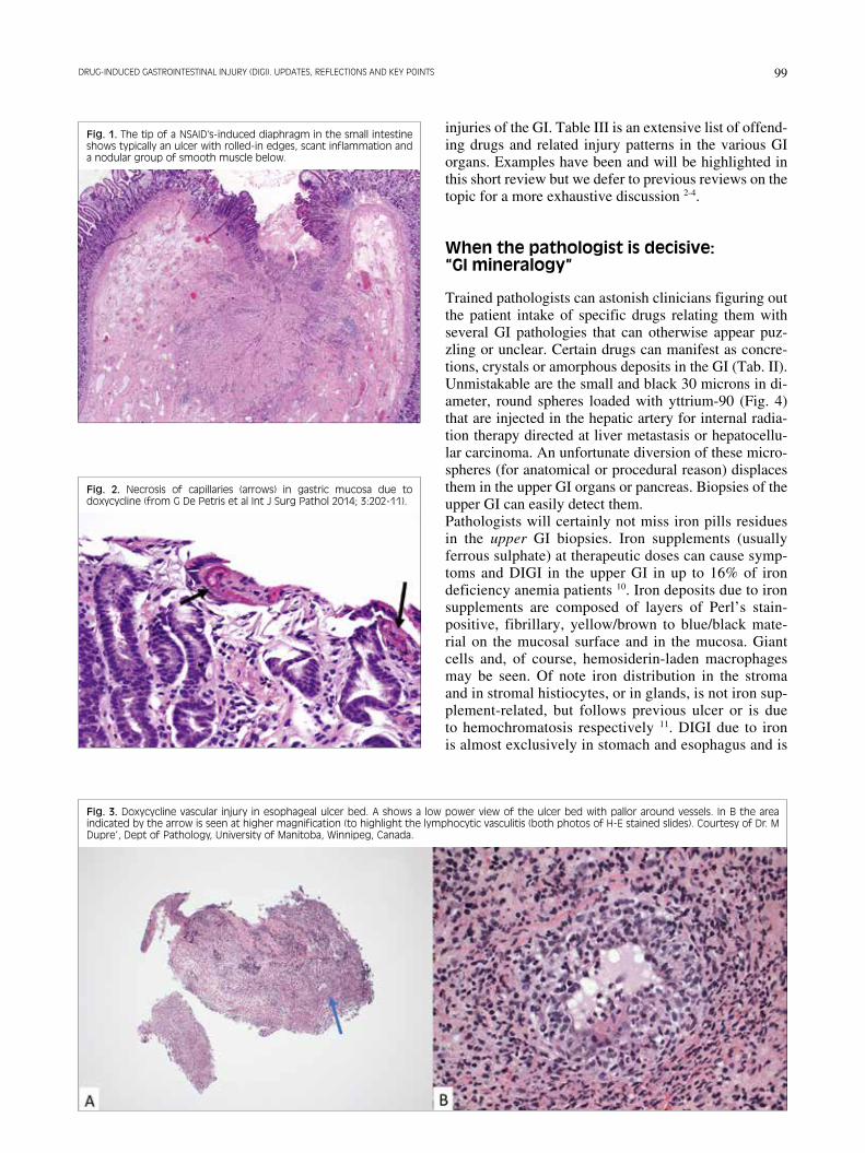
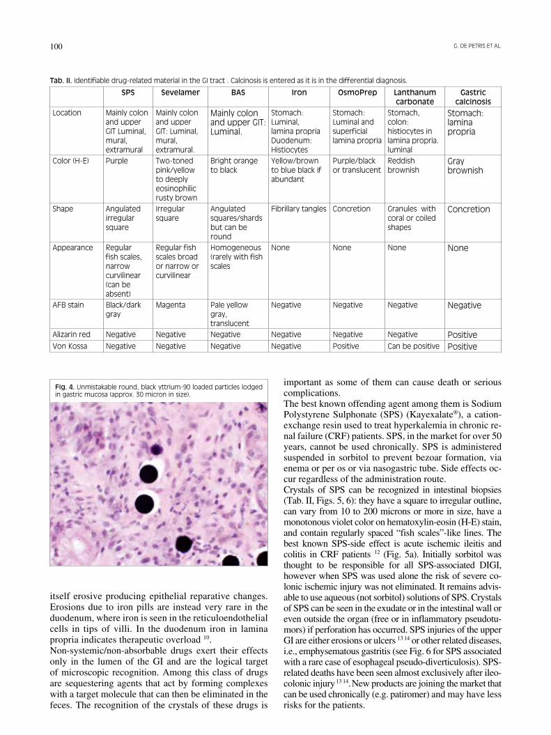
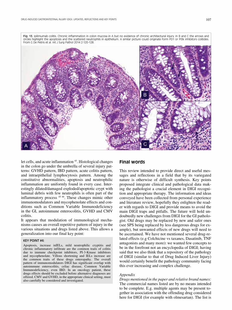
A more common situation is that of a nonspecific GI ulcer. Drugs are considered the main causes of ulcer-ations of unclear origin routinely encountered in the GI. Ulcers due to drugs are in the overwhelming number of cases histologically nonspecific. Pill fragments are the most direct evidence of ulcer being a DIGI but this event is rare. Too numerous are the drugs capable of causing erosion/ulcer to be listed here, we provide instead clues for the differential diagnosis. Location of the erosion/ulcer can help. Esophageal “hang-up” areas (such as the aortic arch, the imprinting due to an enlarged left atrium, the gastroesophageal junction) lead to prolonged contact of the drug with subsequent chemical burn. Proximal esophagitis/ulcer is almost always pathognomonic of drug-induced esophagitis (lichen planus is the main dif-ferential diagnosis in these cases). The isolated proximal location effectively eliminates gastro-esophageal reflux disease as a cause. In addition to antrum and duodenal bulb, the right colon, and terminal ileum are locations for Non-Steroidal Anti-inflammatory Drug (NSAIDs) ulcers. In case of NSAIDs-induced diaphragms forma-tion the ulcer would typically involve the tips of the dia-phragms 7 (Fig. 1). Histology can provide some help: NSAIDs-induced ul-cer is generally inflammation-poor, superficial, rarely involving the muscularis propria. Tetracyclines notori-ous esophago-gastro-toxic antibiotics include doxycy-cline: this antibiotic has gained possible morphological relevance. Doxycycline can cause erosions/ulcers in the entire upper GI but, uniquely so far, doxycycline can cause capillary vascular degeneration with a peculiar “necrotic” appearance and microthrombi 8 (Fig. 2). In addition, as first described in the esophagus by Medli-cott and Dupré 9, doxycycline causes a lymphocytic vas-culitis with endotheliitis and peculiar perivascular pallor due to edema and fibroblast proliferation (Fig. 3). Such pattern should promote an inquiry into doxycycline in-take. As previously stated drugs can induce all the pattern of
Tab. I. Clinical conditions that should alert the pathologist of drug-induced injury.
Condition Drugs used affecting GIChronic renal failure Sodium polystyrene
Sulphonate, Sevelamer, Lanthanum carbonate
Autoimmune disorders (e.g lupus, myasthenia gravis, psoriasis)Solid organ transplant
Mycophenolate
Cancer patients (CLL, FL, Melanoma, breast esophagus lung ovary cancer etc)
Taxanes, PDL-1 inhibitors, CCTLA inhibitors, Adelalisib,
Polypharmacy, especially if in elderly or neurologically impaired patients
NSAIDs, Doxycycline, Clindamycin, Bisphosphonates
Hypertension Angiotensin Receptor Blockers (Olmesartan, Valsartan, Telmersartan)
99DRUG-INDUCED GASTROINTESTINAL INJURY (DIGI). UPDATES, REFLECTIONS AND KEY POINTS
injuries of the GI. Table III is an extensive list of offend-ing drugs and related injury patterns in the various GI organs. Examples have been and will be highlighted in this short review but we defer to previous reviews on the topic for a more exhaustive discussion 2-4.
When the pathologist is decisive: “GI mineralogy”
Trained pathologists can astonish clinicians figuring out the patient intake of specific drugs relating them with several GI pathologies that can otherwise appear puz-zling or unclear. Certain drugs can manifest as concre-tions, crystals or amorphous deposits in the GI (Tab. II). Unmistakable are the small and black 30 microns in di-ameter, round spheres loaded with yttrium-90 (Fig. 4) that are injected in the hepatic artery for internal radia-tion therapy directed at liver metastasis or hepatocellu-lar carcinoma. An unfortunate diversion of these micro-spheres (for anatomical or procedural reason) displaces them in the upper GI organs or pancreas. Biopsies of the upper GI can easily detect them. Pathologists will certainly not miss iron pills residues in the upper GI biopsies. Iron supplements (usually ferrous sulphate) at therapeutic doses can cause symp-toms and DIGI in the upper GI in up to 16% of iron deficiency anemia patients 10. Iron deposits due to iron supplements are composed of layers of Perl’s stain-positive, fibrillary, yellow/brown to blue/black mate-rial on the mucosal surface and in the mucosa. Giant cells and, of course, hemosiderin-laden macrophages may be seen. Of note iron distribution in the stroma and in stromal histiocytes, or in glands, is not iron sup-plement-related, but follows previous ulcer or is due to hemochromatosis respectively 11. DIGI due to iron is almost exclusively in stomach and esophagus and is
Fig. 1. The tip of a NSAID’s-induced diaphragm in the small intestine shows typically an ulcer with rolled-in edges, scant inflammation and a nodular group of smooth muscle below.
Fig. 2. Necrosis of capillaries (arrows) in gastric mucosa due to doxycycline (from G De Petris et al Int J Surg Pathol 2014; 3:202-11).
Fig. 3. Doxycycline vascular injury in esophageal ulcer bed. A shows a low power view of the ulcer bed with pallor around vessels. In B the area indicated by the arrow is seen at higher magnification (to highlight the lymphocytic vasculitis (both photos of H-E stained slides). Courtesy of Dr. M Dupre’, Dept of Pathology, University of Manitoba, Winnipeg, Canada.
G. DE PETRIS ET AL.100
itself erosive producing epithelial reparative changes. Erosions due to iron pills are instead very rare in the duodenum, where iron is seen in the reticuloendothelial cells in tips of villi. In the duodenum iron in lamina propria indicates therapeutic overload 10.Non-systemic/non-absorbable drugs exert their effects only in the lumen of the GI and are the logical target of microscopic recognition. Among this class of drugs are sequestering agents that act by forming complexes with a target molecule that can then be eliminated in the feces. The recognition of the crystals of these drugs is
important as some of them can cause death or serious complications.The best known offending agent among them is Sodium Polystyrene Sulphonate (SPS) (Kayexalate®), a cation-exchange resin used to treat hyperkalemia in chronic re-nal failure (CRF) patients. SPS, in the market for over 50 years, cannot be used chronically. SPS is administered suspended in sorbitol to prevent bezoar formation, via enema or per os or via nasogastric tube. Side effects oc-cur regardless of the administration route.Crystals of SPS can be recognized in intestinal biopsies (Tab. II, Figs. 5, 6): they have a square to irregular outline, can vary from 10 to 200 microns or more in size, have a monotonous violet color on hematoxylin-eosin (H-E) stain, and contain regularly spaced “fish scales”-like lines. The best known SPS-side effect is acute ischemic ileitis and colitis in CRF patients 12 (Fig. 5a). Initially sorbitol was thought to be responsible for all SPS-associated DIGI, however when SPS was used alone the risk of severe co-lonic ischemic injury was not eliminated. It remains advis-able to use aqueous (not sorbitol) solutions of SPS. Crystals of SPS can be seen in the exudate or in the intestinal wall or even outside the organ (free or in inflammatory pseudotu-mors) if perforation has occurred. SPS injuries of the upper GI are either erosions or ulcers 13 14 or other related diseases, i.e., emphysematous gastritis (see Fig. 6 for SPS associated with a rare case of esophageal pseudo-diverticulosis). SPS-related deaths have been seen almost exclusively after ileo-colonic injury 13 14. New products are joining the market that can be used chronically (e.g. patiromer) and may have less risks for the patients.
Tab. II. Identifiable drug-related material in the GI tract . Calcinosis is entered as it is in the differential diagnosis.
SPS Sevelamer BAS Iron OsmoPrep Lanthanum carbonate
Gastric calcinosis
Location Mainly colon and upper GIT Luminal, mural, extramural
Mainly colon and upper GIT: Luminal, mural, extramural.
Mainly colon and upper GIT: Luminal.
Stomach: Luminal, lamina propria Duodenum: Histiocytes
Stomach: Luminal and superficial lamina propria
Stomach, colon: histiocytes in lamina propria. luminal
Stomach: lamina propria
Color (H-E) Purple Two-toned pink/yellow to deeply eosinophilic rusty brown
Bright orange to black
Yellow/brown to blue black if abundant
Purple/black or translucent
Reddish brownish
Gray brownish
Shape Angulated irregular square
Irregular square
Angulated squares/shards but can be round
Fibrillary tangles Concretion Granules with coral or coiled shapes
Concretion
Appearance Regular fish scales, narrow curvilinear (can be absent)
Regular fish scales broad or narrow or curvilinear
Homogeneous (rarely with fish scales
None None None None
AFB stain Black/dark gray
Magenta Pale yellow gray, translucent
Negative Negative Negative Negative
Alizarin red Negative Negative Negative Negative Negative Negative PositiveVon Kossa Negative Negative Negative Negative Positive Can be positive Positive
Fig. 4. Unmistakable round, black yttrium-90 loaded particles lodged in gastric mucosa (approx. 30 micron in size).

101DRUG-INDUCED GASTROINTESTINAL INJURY (DIGI). UPDATES, REFLECTIONS AND KEY POINTS
Sevelamer carbonate (Rengela®, Renagel®) is a resin targeting phosphate ions used in the treatment of hyper-phosphatemia of CRF or tumor lysis syndrome patients. Sevelamer causes chronic diarrhea and constipation. DIGI associated with sevelamer are reported in the up-
per GI (gastric pneumatosis, esophageal ulceration) and, especially, in the lower GI (colitis with crypt distortion and Paneth cell metaplasia, inflammatory polyps, recto-colonic ulcers, colonic stricture, colonic inflammatory mass formation and ischemia, colonic perforation of di-
Tab. III. Offending drugs and related lesion patterns in various organs of the GIT.
EsophagusLichenoid pattern Known event but specific drugs studies absent
(antimalarial, gold, NSAIDs Thiazides, dental amalgam associated with it)Eosinophilic pattern Known event but specific drugs studies absentDysplasia-like changes Taxanes, colchicineDrug deposits/crystals Crospovidone, cellulose, iron, SPS, sevelamer, BAS, OsmoPrepAcute esophagitis Tetracyclines, bisphosphonates, vitamin C, clindamycin etc (>100 drugs have been
involved)StomachIschemic Oxygen peroxide, resins (SPS, sevelamer)reactive gastropathy NSAIDs, OsmoPrep, resins, iron, mycophenolate and many moreCollagenous and lymphocytic OlmesartanAcute gastritis Resins (SPS, sevelamer)Drugs deposits/crystals Resins, OsmoPrep, Iron, crospovidone microcrystalline celluloseInfiltrative cellular process Lanthanum carbonate, clofazimine5Dysplasia-like changes TaxanesGranulomatous Lanthanum carbonateSmall intestineCollagenous and atrophic Olmesartan, methotrexate, NSAIDs, mycophenolate, azathioprineLymphocytic Olmesartan, PPIs, NDSAIDs, PDL-1 inhibitors, CTCLA inhibitors, PI-3-K inhibitorsInfiltrative ClofazimineErosion /strictures/ulcers NSAIDsColonIBD-like Mycophenolate, ipilimumab, rituximab, TNFα inhbitors, NSAIds, Idelalisib,Ischemic Digitalis, estrogens, cocaine, ergotamine, Kayexalate, sevelamer, Glutaraldehyde NSAIDs,
PD1 inhibitorsFocal active and self-limited colitis NSAIDs, sodium phosphate, mycophenolate, ipilimumabLymphocytic and collagenous colitis PPI inhibitors, ticlopidine, NSAIDs, Statins, Idelalisib and many othersDysplasia-like Taxanes, colchicine, cyclosporine
Fig. 5. Ischemic colitis due to Kayexalate (left). On the right the crystals from the superficial exudate (purple with internal lines) are highlighted.

G. DE PETRIS ET AL.102
verticulum) 15-20, although direct causation is not demon-strated. The features of the luminal crystals are shown in Table II and Figure 7. They are scaly in appearance with the crystal displaying a 2 toned-color appearance: pink and yellow (but also occasionally brown/red/purple). They stain magenta after Acid Fast Bacilli stain (AFB). SPS and sevelamer crystals identification will require a callback to the clinician. However, one must first ex-clude mimickers: the best known are other resins such as the bile acid sequestrants (BAS) (cholestyramine, co-lesevelam, cholestipol) crystals. BAS have been innocu-ous so far and their importance lies in their distinction from the ion-binder resins described above. Table II and Figures 5 to 7 provide the clues necessary for identifica-tion.Lanthanum carbonate (LC) is a phosphate binder, de-void of aluminum and calcium, effective in the control of hyperphosphatemia in patients with CRF. No serious side effects have so far been linked to the use of LC. Makino et al. 21 described LC deposition in gastric mu-cosa in CRF in 2015. LC deposition is frequent (seen in 14 of 19 CRF patients according to Goto 22) mainly in the stomach but also in duodenum 23 and colon 22. LC has interesting characteristics: 1) is one of the few drugs causing an infiltrative cellular pattern of injury in the GI (another is clofazimine, a third tier anti-tuber-culosis antibiotic); 2) it is radio-opaque and detectable using radiological imaging 24 while displaying white/chalky granules or polyps at endoscopy with erosions
and ulcers. LC is found in the lamina propria but also on the mucosal surface. The histology is that of a foreign body reaction characterized by an infiltrate of eosino-philic, large, often multinucleated, histiocytes contain-ing colorless or brownish needle-like or branched, coiled or crescent-shaped inclusion bodies (Fig. 8). Formation of well-defined epithelioid granulomas (Fig. 8) is occa-sionally seen, highlighting again how a DIGI can mimic other conditions, in this case granulomatous gastritis. Regional lymph nodes have similar LC infiltrates 24. The differential diagnosis of crystals/concretions/ crys-talline deposits in the GI has been enriched recently by additional compounds: crospovidone, microcrystal-line cellulose and sodium phosphate oral tablets (Os-moPrep®) creating a veritable “GI mineralogy” for the pathologist. Crospovidone (Fig. 9) and microcrystalline cellulose (Fig. 10) are not active principles: they are widely utilized drugs stabilizers and fillers that, when in oral medications, can be recognized in the GI, espe-cially in the small intestine (in 9% of patients according to Shaddy et al. 25).Crosposvidone ranges from 0.4 to 1.5 mm in size, has a coral shape, generally with pink cores and purple coats in each segment on H-E (Fig. 9). It is not birefringent under polarized light and is dark orange after von Kossa stain. Microcrystalline cellulose is instead brightly bire-fringent under polarized light, and is transparent in H-E-stained slides (Fig. 10). These two compounds are well known to pulmonary pathologists as their presence in
Fig. 6. An unusual association in the upper GI: esophageal pseudo-diverticulosis, with the superficial kayexalate crystal (in the circle) magnified in picture on the right.

103DRUG-INDUCED GASTROINTESTINAL INJURY (DIGI). UPDATES, REFLECTIONS AND KEY POINTS
the lung indicates aspiration or intravenous drug abuse. The appearance in the lungs and GI is however different: while in the lung crospovidone is solid blue or black on H-E-stained sections, in the GI has a two-toned color.Sodium phosphate tablet OsmoPrep (USA: OsmoPrep®, France: Colokit®) is used for colonoscopy preparation, occasionally employed in patients that do not wish to ingest the large volumes of liquids otherwise required for the procedure. The drug in its solution preparation (Phosphosoda) is well known to cause colonic aphthous ulcers and focal active colitis. The tablets instead cause deposits in the stomach where they mimic calcinosis and iron-pill injury 26. The injury is of the gastropathy/ero-sive type. The deposition occurs in the gastric superficial lamina propria where it appears similar to crushed pill fragments, purple to black, less often translucent, less than 100 microns in size (Fig. 11). OsmoPrep is von Kossa stain positive (von Kossa stains the phosphate moiety) but alizarine red negative. This staining pat-tern distinguishes OsmoPrep from gastric calcinosis in which calcium is reactive for both stains 26.
Fig. 7. A collection of crystals of non-absorbable resins as seen in biopsies of the GI. From left to right: (A) Kayexalate (Sodium Polystyrene Sulpho-nate), (B) Cholestyramine, (C) Sevelamer, and (D) another Bile Acid Sequestrant (BAS), Colesevelam on the far right. Notice how Cholestyramine and Colesevelam, both BAS, are indistinguishable. The colors of Kayexalate and Sevelamer are distinctive and diagnostic in the majority (but not all) cases. From G. De Petris et al. Int J Surg Pathol. 2014; 2:120-8.
Fig. 8. Lanthanum carbonate (LC) deposits in histiocytes in gastric lamina propria (A), higher magnification of histiocyte with crystals of LC in B. The lamina propria may contain well-formed granulomas (C) (notice histiocytes with LC inclusion below the granuloma). Figure 8A and 8B courtesy of Dr. M Makino Shinonoi-Ai, Nagano, Japan.
Fig. 9. Crospovidone (H-E stained slide, 40X magnification). Note the pink cores and purple coats in the tangles of the coral-like structure of crospovidone. Courtesy of Dr. R. Gonzales Univ. of Rochester, Ro-chester NY, USA).

G. DE PETRIS ET AL.104
Finally, the pathologist should be aware of the possibil-ity of multiple compounds deposits and crystals being present in the same patient. The clinical background in which the drugs are used may also heighten the need to look for and/or exclude associated conditions (e.g. amy-loid in case of CRF with GI resins crystals).
KEY POINT #2: The pathologist can be decisive and recognize drugs in H-E stained slides. Identification is clinically useful.
Some drug effects are traps we must know about
Olmesartan medoxomil, a widely-prescribed angioten-sin II receptor blocker antihypertensive drug, is used by millions of patients, a minuscule minority of them develops a significant DIGI after months to years of exposure 27. Olmesartan can cause sprue-like enteropa-
thy especially in elderly patients. Partial or total villous blunting, increase in IELs and collagenous enteritis are reported frequently in this DIGI. The stomach and co-lon have similar findings consisting of lymphocytic and collagenous gastritis and colitis in isolation or associ-ated with enteropathy (Fig. 12A). A detailed exam of the cases in the literature shows however that the occasional patient may sometime present without duodenal IELs in-crease (Fig. 12A) in olmesartan-induced villous atrophy, a significant difference with celiac disease 28. The recognition of this DIGI is perhaps the most im-portant of all. The patients are often hospitalized with uncontrollable diarrhea and electrolytes disorders, typi-cally with a diagnosis of sero-negative celiac disease; “suspicious for lymphoma” is another preliminary diagnosis that will also be given due to the refractori-ness to all therapies and the severity of symptoms. The suspension of olmesartan will prevent malnutrition and expensive testing, and is spectacularly effective in re-lieving the symptoms in few days and in restituting the morphology to normal, or almost normal, in few months. Symptoms do recur after reintroduction of olmesartan in the few cases in which this was attempted28. Of note other members of the Angiotensin Receptor Blocker (ARB) family are capable of causing severe enteropathy and enterocolitis (e.g. Valsartan and Telmersartan) and Losartan (Dr De Marco personal communication) 28 29. The dramatic enteropathy certainly dominates our clini-cal interaction with ARBs DIGI, however it is likely that ARBs have milder histological presentations (e.g. increase in chronic inflammation, isolated duodenal or colonic IELs increase) 28-30.Taxanes (Paclitaxel, Docetaxel, Cabazitaxel) used in cancer patients (breast, ovary, digestive and prostate cancers) disrupt mitotic tubules preventing tubule de-polymerization (mitotic arrest-drugs) and have a direct antitumoral apoptotic effect (Docetaxel in particular). Taxanes can mimic dysplasia anywhere in the GI and to perfection in mucosa of Barrett’s esophagus. Atypical
Fig. 10. Microcrystalline cellulose appears as transparent in A (H-E stain) B) and brightly birefringent under polarized light (B). Courtesy of Dr. R. Gonzales Univ. of Rochester, Rochester NY, USA).
Fig. 11. OsmoPrep deposition in superficial gastric lamina propria. The bottom right figure shows positivity for Von Kossa stain. Courtesy of Dr. K. Matsukuma, Univ of California, Davis, CA, USA).

105DRUG-INDUCED GASTROINTESTINAL INJURY (DIGI). UPDATES, REFLECTIONS AND KEY POINTS
nuclei with numerous mitoses (some appearing as ring mitoses, some others with a central bar) and increased apoptosis are the results of taxanes intake (Fig. 12B). To help in the differential diagnosis between true dys-plasia and taxanes it has been reported how taxanes af-fects only the proliferative areas of the glands and that hyperchromasia and nuclear pleomorphism are absent in taxanes effect 31. The differential diagnosis remains highly difficult in Barrett’s esophagus or other condi-tions prone to dysplasia and exposed to taxanes: only awareness of this DIGI and clinical correlation will help prevent this pitfall.Mycophenolate is used in solid organ graft maintenance therapy and in autoimmune disorders (e.g lupus, psoria-sis, myasthenia gravis). Mycophenolate colitis is well described 2-4: the main features are (Fig. 12 C) architec-tural disarray of the mucosa with crypts drop out and
“exhausted” apoptotic crypts (that is: thinned stretched eosinophilic epithelium around a dilated/damaged-ap-pearing crypt (DDC) with eosinophilic luminal debris and increased apoptotic bodies) 4 32 33. Occasionally my-cophenolate colitis is ischemic in appearance, in indi-vidual cases the colitis is worse in the proximal colon. Of note the colitis can have skip areas further mimicking Crohn’s disease to the unaware. In the small intestine villous atrophy and inflammation due to mycopheno-late can mimic celiac disease in the individual case. The DDC pattern of mycophenolate is reminiscent of, and needs to be distinguished from, cytomegalovirus infec-tion and especially GVHD. These conditions must be excluded due to vastly different therapies. Star et al. 34
summarized features to help distinguish the two as fol-lows: mycophenolate colitis features much more promi-nent lamina propria inflammation with eosinophils than
Fig. 12. A illustrates a collagenous enteritis due to olmesartan. Notice how IELs are not prominent in this case, an event occurring in a minority of cases. In B is Taxol effect in Barrett’s esophagus, a mitosis with a central bar in it is present in the center. C1 and C2 show mycophenolate colitis. In C1 eosinophilic exhausted epithelium stretches along a injured crypt. In C2 the appearance of the mucosa with crypts drop out, distortion and damaged crypts is present.

G. DE PETRIS ET AL.106
GVHD, while GVHD displays endocrine nests and apoptotic microabscesses more commonly than myco-phenolate.
KEY POINT #3: The most relevant pitfalls due to DIGI are those related to olmesartan and other ARBs (vs celiac disease and/or collagenous enteropathy), mycophenolate (vs. GVHD and CMV) and taxanes (vs. dysplasia).
Oncology is a fertile ground for DIGI
New oncologic therapies are not exempt of side-effects in GI. DIGI provoked by the recently developed immune checkpoint inhibitors and Phosphatydilinositol-3-Kinase (PI3K) inhibitors will be analyzed here.Immune checkpoint inhibitors. Under this category are CTLA-4 (cytotoxic T-lymphocyte associated protein-4) inhibitors (ipilimumab and tremelimumab) and Pro-grammed Death-1 receptor inhibitors (PDI) (such as pembrolizumab, nivolumab and atezolimumab that tar-gets the PD-1 ligand). These two classes of inhibitors are sometimes used together in modern oncology therapy. The severity of the GI side effects is higher after CT-LA-4 inhibitors 35 36 41.
CTLA-4 is a receptor whose activation inhibits cyto-toxic T-cells. Inhibitors of CTLA-4 (I-C) remove this inhibition and prolong cytotoxic T-cells activation and action on tumor cells. I-C are approved for the treatment of advanced melanoma, they are under investigation in the treatment of non-small cell carcinoma of the lung, as well as prostate, renal and ovarian carcinoma, and in mesothelioma. The mechanism of action of I-C leads to loss of systemic tolerance and a host of immune-relat-ed disorders including DIGI. Between 20 and 50% of patients suffer GI side effects, the most common being watery diarrhea appearing within 6 weeks into treatment sometimes as early as few days from the beginning of the therapy 35. Life threatening colitis is seen in 16% of patients36. Less than 1 percent of patients suffer serious intestinal side effect or death 36. The endoscopic appearance of diffuse colitis was seen in 21% of patients in a study by Beck et al. 37. Ipilimumab offends also stomach and small intes-tine, sometimes in isolation. Endoscopy shows granu-larity, exudate, and erosion/ulcers but can be entirely normal even though colitis is present: biopsies must always be obtained. The histopathological findings 38 39
are similar throughout the intestine. The histology dem-onstrates villous blunting, diffuse chronic inflammation of mucosa, and increase in apoptosis, rare neutrophilic cryptitis, and intraepithelial lymphocytosis. They re-semble autoimmune enterocolitis, an otherwise utterly rare disease in adults. The diagnosis of AIE in an adult should be made after DIGI has been excluded. The pro-cess may occasionally suggest IBD on a superficial ob-servation, however in the colon there are no classic IBD findings such as basal plasmacytosis and crypts distor-tion. As a reminder, classic IBD would not have severe
apoptosis or marked increase in IELs (Fig. 13). Rare is lymphocytic colitis and even rarer is ipilimumab associ-ated lymphocytic plexitis of the colon. An exceptional case from Mayo Clinic showed the activation of a latent celiac disease into overt disease after ipilimumab 40.PD-1 receptor activation prevents cytotoxic T-cell ac-tivation by self-antigens. Tumors use this mechanism to defuse the antitumoral immunological response. PD-1 inhibitors (PI) unbridle the T-cell response to ma-lignancies. PI are employed in melanoma, urothelial carcinoma, non-small cell carcinoma of the lung, re-nal carcinoma and mismatch repair deficient colorectal carcinoma. It is not surprising that the enhanced T-cell response is accompanied by a series of side effects that appear to be immune-related. PIs cause mild watery di-arrhea in 20-30% of patients, approximately 6 weeks into treatment 41 (severe in less than 10% of cases), and other GI symptoms (nausea vomiting, abdominal pain occult blood in stool. The endoscopy shows erythema friability and aphthous ulcers. The histology of PI in-jury has been recently described in 19 patients 42. Any portion of the GI was found affected in at least one patient. The upper GI showed increased mononuclear cells in the lamina propria of stomach and duodenum, villous blunting, increased eosinophils, neutrophilic villitis, neutrophils in foveolar epithelium. On the other hand, increased apoptosis was less common. Similar findings were seen in the terminal ileum. Distortion of mucosal architecture was not a feature of PI DIGI. The colon had similar findings with, however, more com-mon apoptotic bodies and, interestingly, ischemic coli-tis. Increased intraepithelial lymphocytes were seen in both upper and lower GI. Collagenous colitis was seen in one case. An unusual finding was the development of well-formed granulomas associated with crypt rup-tures.Phosphatydilinositol-3-kinase (PI3K) inhibitors. PI3K are a family of kinases that regulates several cancer cells functions. Idelalisib (Zydelig®) is a selective in-hibitor of the PI3K delta isoenzyme that promotes apoptosis in cells of hematopoietic malignancies. Idelalisib is used in therapy of relapsed or refractory chronic lymphocytic leukemia, small lymphocytic lymphoma and follicular lymphoma. Idealisib therapy can causes immune-mediated toxicities affecting liver and colon 43, side effects that may require cessation of the therapy. Diarrhea is usually watery and appears in 20-45% of patients 44. Early diarrhea (in the first two months) is responsive to symptomatic therapy without need of stopping the drug. Late diarrhea is considered a symptom of Idelalisib-induced autoimmune entero-colitis and generally requires discontinuation of the drug44. Endoscopy of the colon often reveals nothing of relevance or, in a minority of cases, pseudo-mem-branes, aphthous ulcers and erythema.Histological changes in the small bowel include in-crease in mononuclear cells infiltrate in lamina propria, increased apoptosis, villous atrophy, and increased in-traepithelial lymphocytes, less commonly reduced gob-
107DRUG-INDUCED GASTROINTESTINAL INJURY (DIGI). UPDATES, REFLECTIONS AND KEY POINTS
let cells, and acute inflammation 45. Histological changes in the colon go under the umbrella of several injury pat-terns: GVHD pattern, IBD pattern, acute colitis pattern, and intraepithelial lymphocytosis pattern. Among the constitutive abnormalities, apoptosis and neutrophilic inflammation are uniformly found in every case. Inter-estingly dilated/damaged exploded/apoptotic crypt with luminal debris with few neutrophils is often part of the inflammatory process 45 46. These changes mimic other immunomodulators and mycophenolate effects and con-ditions such as Common Variable Immunodeficiency in the GI, autoimmune enterocolitis, GVHD and CMV colitis.It appears that modulation of immunological mecha-nisms causes an overall repetitive pattern of injury in the various situations and drugs listed above. This allows a generalization into our final key point:
KEY POINT #4: Apoptosis, increase inIELs, mild neutrophilic cryptitis and chronic inflammatory infiltrate are the common traits of colitis due to immune checkpoint inhibitors, PI-3-Kinase inhibitors and mycophenolate. Villous shortening and IELs increase are the common traits of these drugs enteropathy. The overall pattern of immunomodulators DIGI has significant overlap with autoimmune enterocolitis, celiac disease, Common Variable Immunodeficiency, even IBD. In an oncology patient, these drugs effects should be excluded before alternative diagnoses are offered. CMV and GVHD, in the appropriate clinical setting, must also carefully be considered and investigated.
Final words
This review intended to provide direct and useful mes-sages and reflections in a field that by its variegated nature is otherwise of difficult synthesis. Key points proposed integrate clinical and pathological data mak-ing the pathologist a crucial element in DIGI recogni-tion and appropriate therapy. The information and ideas conveyed have been collected from personal experience and literature review, hopefully they enlighten the read-er with regards to DIGI and provide means to avoid the main DIGI traps and pitfalls. The future will hold un-doubtedly new challenges from DIGI for the GI patholo-gist. Old drugs may be replaced by new and safer ones (see SPS being replaced by less dangerous drugs for ex-ample), but unwanted effects of new drugs will need to be ascertained. We have not mentioned several drug-re-lated effects (e.g Colchicine vs taxanes, Dasatinib, TNF antagonists and many more): we wanted few concepts to be in the forefront not an encyclopedia of DIGI; having said that we also think that a repository of the pathology of DIGI (similar to that of Drug Induced Liver Injury) would certainly benefit the pathology community facing this ever increasing and complex challenge.
AppendixDrugs mentioned in the paper and relative brand names:The commercial names listed are by no means intended to be complete. E.g. multiple agents may be present to-gether in association with the offending drug considered here for DIGI (for example with olmesartan). The list is
Fig. 13. Ipilimumab colitis. Chronic inflammation in colon mucosa in A but no evidence of chronic architectural injury. In B and C the arrows and circles highlight the apoptosis and the scattered neutrophils in epithelium. A similar picture could originate form PD1 or PI3k inhibitors colitides. From G De Petris et al. Int J Surg Pathol 2014 2:120-128.

G. DE PETRIS ET AL.108
only meant to be helpful to recognize some of the most commonly used name brands associated with the drugs mentioned in the paper.NSAIDs (a very long list of aspirine and COX inhibitors could be entered here, please refer to textbooks or web-sites), doxycycline (Vibramicin® and many other brand names); Ytrrium-90 loaded particles (Syrtex SYR-spheres®); Iron supplements (several formulations and brand names), Sodium polystyrene sulphate (Kayexa-late®); Patiromer (Veltassa®); Sevelamer (Rengela®, Re-nagel®); Cholestyramine (Questran®, Prevalite®); Cole-sevelam (Welchol®); Cholestipol (Colestid®); Lantha-num carbonate (Fosrenol®); Sodium phosphate tablets (OsmoPrep®), Olmesartan (Benicar®, Azor® etc); Val-sartan (Diovan®, Entresto® etc.); Ilbesartan (Avapro®, Karvea®, Aprovel®); Losartan (Cozaar®, Hyzaar®); Tax-anes: Paclitaxel (Taxol®, Onxol®, Abraxane®); Docetax-el (Taxotere®, Docefrez®), Cabazitaxel (Jevtana®); My-cophenolate (Mycophenolate mofetil: Cellcept® and Mycophenolate sodium: Myofortic®); Ipilimumab (Yer-voy®); Tremelimumab; Pembrolizumab (Keytruda®); Nivolumab (Opdivo®); Atezolimumab (Tecentriq®); Idelalisib (Zydelig®).
References
1 Bates DW, Forster AJ, Gandhi TK, et al. The incidence and sever-ity of adverse events affecting patients after discharge from the hospital. Ann Intern Med 2003;138:161-7.
2 McCarthy AJ, Lauwers GY, Sheahan K. Iatrogenic pathology of the intestines. Histopathology 2015;1:15-28.
3 De Petris G, Gatius Caldero S, Chen L, et al. Histopathological changes in the gastrointestinal tract due to drugs: an update for the surgical pathologist (part I of II). Int J Surg Pathol 2014;2:120-8.
4 De Petris G, Caldero SG, Chen L, et al. Histopathological changes in the gastrointestinal tract due to medications: an update for the surgi-cal pathologist (part II of II). Int J Surg Pathol 2014;3:202-11.
5 Price AB. Pathology of drug-associated gastrointestinal disease. Br J Clin Pharmacol 2003;56:477-82.
6 Lin J, Fan R, Zhao Z, et al. Is the presence of 6 or fewer crypt apoptotic bodies sufficient for diagnosis of graft versus host dis-ease? A decade of experience at a single institution. Am J Surg Pathol 2013;4:539-47.
7 De Petris G, Lopez JI. Histopathology of diaphragm disease of the small intestine: a study of 10 cases from a single institution. Am J Clin Pathol 2008;4:518-25.
8 Xiao SY, Zhao L, Hart J, et al. Gastric mucosal necrosis with vascular degeneration induced by doxycycline. Am J Surg Pathol 2013;2:259-63.
9 Medlicott SA, Ma M, Misra T, et al. Vascular wall degeneration in doxycycline-related esophagitis. Am J Surg Pathol 2013;7:1114-5.
10 Kaye P, Abdulla K, Wood J, et al. Iron-induced mucosal pathology of the upper gastrointestinal tract: a common finding in patients on oral iron therapy. Histopathology 2008;3:311-7.
11 Marginean EC, Bennick M, Cyczk J, et al. Gastric siderosis: pat-terns and significance. Am J Surg Pathol 2006;4:514-20.
12 Rashid A, Hamilton SR. Necrosis of the gastrointestinal tract in uremic patients as a result of sodium polystyrene sulfonate (Kayex-alate) in sorbitol: an underrecognized condition. Am J Surg Pathol 1997;1:60-9.
13 Abraham SC, Bhagavan BS, Lee LA, et al. Upper gastrointestinal
tract injury in patients receiving kayexalate (sodium polystyrene sulfonate) in sorbitol: clinical, endoscopic, and histopathologic findings. Am J Surg Pathol 2001;5:637-44.
14 Harel Z, Harel S, Shah PS, et al. Gastrointestinal adverse events with sodium polystyrene sulfonate (Kayexalate) use: a systematic review. Am J Med 2013;3:264.e9-24.
15 Swanson BJ, Limketkai BN, Liu TC, et al. Sevelamer crystals in the gastrointestinal tract (GIT): a new entity associated with mu-cosal injury. Am J Surg Pathol 2013 11:1686-93.
16 Yamaguchi T, Ohyama S, Furukawa H, et al. Sigmoid colon di-verticula perforation associated with sevelamer hydrochloride ad-ministration: a case report. Ann Med Surg (Lond) 2016;10:57-60.
17 Desai M, Reiprich A, Khov N, et al. Crystal-associated colitis with ulceration leading to hematochezia and abdominal pain. Case Rep Gastroenterol 2016;10:332-7.
18 Kim J, Olson K, Butani L. Sevelamer crystals in the mucosa of the gastrointestinal tract in a teenager with end-stage renal disease. Pediatr Nephrol 2016;2:339-41.
19 Okwara C, Choi C, Park JY. Sevelamer-induced colitis presenting as a pseudotumor. Clin Gastroenterol Hepatol 2015;7:A39-A40.
20 Amer S, Nguyen C, De Petris G. Images of the month: gastric pneumatosis due to sevelamer-mediated necrosis. Am J Gastroen-terol 2015;6:799.
21 Makino M, Kawaguchi K, Shimojo H, et al. Extensive lanthanum deposition in the gastric mucosa: the first histopathological re-port. Pathol Int 2015;1:33-7.
22 Goto K, Ogawa K. Lanthanum deposition is frequently observed in the gastric mucosa of dialysis patients with lanthanum carbonate therapy: a clinicopathologic study of 13 cases, including 1 case of lanthanum granuloma in the colon and 2 nongranulomatous gas-tric cases. Int J Surg Pathol 2016;1:89-92.
23 Iwamuro M, Tanaka T, Urata H, et al. Lanthanum phosphate de-position in the duodenum. Gastrointest Endosc 2016 Jun 20 doi: 10.1016/j.gie.2016.06.012. [Epub ahead of print]
24 Yabuki K, Shiba E, Harada H, et al. Lanthanum deposition in the gastrointestinal mucosa and regional lymph nodes in dialysis pa-tients: analysis of surgically excised specimens and review of the literature. Pathol Res Pract 2016;10:919-26.
25 Shaddy SM, Arnold MA, Shilo K, et al. Crospovidone and micro-crystalline cellulose: a novel description of pharmaceutical fillers in the gastrointestinal tract. Am J Surg Pathol 2017;41:564-9.
26 Matsukuma K, Gui D, Olson KA, et al. OsmoPrep-associated gas-tritis: a histopathologic mimic of iron pill gastritis and mucosal calcinosis. Am J Surg Pathol 2016;11:1550-6.
27 Rubio-Tapia A, Herman ML, Ludvigsson JF, et al. Severe spru-elike enteropathy associated with olmesartan. Mayo Clin Proc 2012;8:732-8.
28 Burbure N, Lebwohl B, Arguelles-Grande C, et al. Olmesartan-associated sprue-like enteropathy: a systematic review with em-phasis on histopathology. Hum Pathol 2016;50:127-34.
29 Mårild K, Lebwohl B, Green PH, et al. Blockers of angiotensin other than olmesartan in patients with villous atrophy: a nation-wide case-control study. Mayo Clin Proc 2015;6:730-7.
30 Marietta EV, Cartee A, Rish A, et al. Drug-induced enteropathy. Dig Dis 2015;2:215-20.
31 Daniels JA, Gibson MK, Xu L, et al. Gastrointestinal tract epi-thelial changes associated with taxanes: marker of drug toxicity versus effect. Am J Surg Pathol 2008;3:473-7.
32 Liapis G, Boletis J, Skalioti C, et al. Histological spectrum of my-cophenolate mofetil-related colitis: association with apoptosis. Histopathology 2013;5:649-58.
33 Lee S, de Boer WB, Subramaniam K, et al. Pointers and pitfalls of mycophenolate-associated colitis. J Clin Pathol 2013;1:8-11.

109DRUG-INDUCED GASTROINTESTINAL INJURY (DIGI). UPDATES, REFLECTIONS AND KEY POINTS
34 Star KV, Ho VT, Wang HH, et al. Histologic features in colon biopsies can discriminate mycophenolate from GVHD-induced colitis. Am J Surg Pathol 2013;9:1319-28.
35 Weber JS, Dummer R, de Pril V, et al. Patterns of onset and reso-lution of immune-related adverse events of special interest with ipilimumab: detailed safety analysis from a phase 3 trial in pa-tients with advanced melanoma. Cancer 2013;9:1675-82.
36 Slovin SF, Higano CS, Hamid O, et al. Ipilimumab alone or in combination with radiotherapy in metastatic castration-resistant prostate cancer: results from an open-label, multicenter phase I/II study. Ann Oncol 2013;7:1813-21.
37 Beck KE, Blansfield JA, Tran KQ, et al. Enterocolitis in patients with cancer after antibody blockade of cytotoxic T-lymphocyte-associated antigen 4. J Clin Oncol 2006;15:2283-9.
38 Gupta A, De Felice KM, Loftus EV Jr, et al. Systematic review: colitis associated with anti-CTLA-4 therapy. Aliment Pharmacol Ther 2015;4:406-17.
39 Oble DA, Mino-Kenudson M, Goldsmith J, et al. Alpha-CTLA-4 mAb-associated panenteritis: a histologic and immunohistochemi-cal analysis. Am J Surg Pathol 2008;8:1130-7.
40 Gentile NM, D’Souza A, Fujii LL, et al. Association between ipili-mumab and celiac disease. Mayo Clin Proc 2013;4:414-7.
41 Weber JS, Dummer R, de Pril V, et al; MDX010-20 Investiga-tors. Patterns of onset and resolution of immune-related adverse events of special interest with ipilimumab: detailed safety analysis from a phase 3 trial in patients with advanced melanoma. Cancer 2013;9:1675-82.
42 Gonzalez RS, Salaria SN, Bohannon CD, et al. PD-1 inhibitor gas-troenterocolitis: case series and appraisal of ‘immunomodulatory gastroenterocolitis. Histopathology 2017 Mar;70:558-67.
43 ZYDELIG (Idelalisib tablets). Full prescribing information, Gil-ead Sciences, Inc. Foster City CA, 2014.
44 Coutré SE, Barrientos JC, Brown JR, et al. Management of adverse events associated with idelalisib treatment: expert panel opinion. Leuk Lymphoma 2015;10:2779-86.
45 Louie CY, Di Maio MA, Matsukuma KE, et al. Idelalisib-associ-ated enterocolitis: clinicopathologic features and distinction from other enterocolitides. Am J Surg Pathol 2015;12:1653-60.
46 Weidner AS, Panarelli NC, Geyer JT, et al. Idelalisib-associat-ed colitis: histologic findings in 14 patients. Am J Surg Pathol 2015;12:1661-7.