dtic file copr · irreversible phase consisting of those changes which occur after cell death. ......
TRANSCRIPT

DTIC FILE COPR 'I
AD
00NN ION MOVEMENTS IN SHOCK IN RELATION TO SURVIVAL
00 AND ITS MODIFICATIONS
Final Report
Toshihide Sato, M.D.
January 1985
Supported by
U.S. ARMY MEDICAL RESEARCH AND DEVELOPMENT COMMANDFort Detrick, Frederick, Maryland 21702-5012
Contract No. DAMD17-83-C-3164
University of Maryland at Baltimore DTICBaltimore, Maryland 21201 A j% ELECTE
DOD DISTRIBUTION STATEMENT
Approved for public release; distribution unlimited
The findings in this report are not to be construed asan official Department of the Army position unless sodesignated by other authorized documents.

=R'J0I"Y CLASSIFICATION OF THIS PAGE
REPORT DOCUMENTATION PAGE Form Approved
la. REPORT SECURITY CLASSIFICATION lb. RESTRICTIVE MARKINGS II
2a. SECURITY CLASSIFICATION AUTHORITY 3. DISTRIBUTION /AVAILABILITY OF REPORT
2b. DECLASSIFICATION/DOWNGRADING SCHEDULE Approved for public release;distribution unlimited
4. PERFORMING ORGANIZATION REPORT NUMBER(S) 5. MONITORING ORGANIZATION REPORT NUMBER(S)
6a. NAME OF PERFORMING ORGANIZATION 6b. OFFICE SYMBOL 7a. NAME OF MONITORING ORGANIZATIONUniversity of Maryland (If applicable)
- ADDRESS (City, State, and ZIP Code) 7b. ADDRESS (City, State, and ZIP Code)
10 South Pine StreetBaltimore, MD 21201
8a. NAME OF FUNDINGISPONSORING J8b. OFFICE SYMBOL 9. PROCUREMENT INSTRUMENT IDENTIFICATION NUMBERORGANIZATION U.S. Army Medical (If applicable)
Research & Development Command Contract No. DAMD17-83-C-3164
&c. ADDRESS (City, State, and ZIP Code) .10. SOURCE OF FUNDING NUMBERS
PROGRAM PROJECT |TASK WORK UNITFort Detrick ELEMENT NO. NO. 3M1- NO. CCSSION NO.Frederick, Maryland 21702-5012 61102A ll02BSl0 BA !45l11. TITLE (include Security Classification)
Ion Movements in Shock in Relation to Survival and its Modifications
12. PERSONAL AUTHOR(S)
Toshihide Sato, M.D.
13a. TYPE OF REPORT 13b. TIME COVERED [14. DATE OF REPORT (Year, Month, Day) 15. PAGECOUNTFinal
FROM 83/6/11TO 84/12/3 1985 January
16. SUPPLEMENTARY NOTATION
17. COSATI CODES 18. SUBJECT TERMS (Continue on reverse if necessary and identify by block number)
FIELD GROUP SUB.GROUP ! Hemorrhagic and Bacteremic Shock, Pathophysiology,
06 01 Biochemistry, Morphology, X-ray Microanalysis, RA V06 I 16,
19 ABSTRACT (Continue on reverse if necessary and identify by block number)- 'In order to elucidate the mechanisms of cell injury and to correlate such with ion
'hifts in tissues, comprehensive physiologic, biochemical, and morphological studies as well*I s x-ray microanalysis measurements of ions in liver and heart were performed using both-n vivo hemorrhagic and bacteremic shock models in rats. In the hemorrhagic shock model,eterioration of physiologic parameters and increases in serum potassium concentrations andecreases in serum calcium concentrations were seen. In the bacteremic shock model, a hypo-
dynamic state and increases in the activity of various enzymes released from vital organs,such as the liver or heart, were observed. Morphological results suggested that local produc-tion of free radicals from migrating and aggregated leukocytes into the sinusoids of the liverfter bacteremia may play a possible role in the hepatocellular injury. In both models,-ray microanalysis of ions in freeze-dried sections of liver and heart showed increases insodium, chlorine and calcium and decreases in potassium and phosphorus. -Data obtained as aesult of these comprehensive studies not only showed a good correlation between ion shifts
20. DISTRIBUTION /AVAILABILITY OF ABSTRACT 21. AJSTRACT SECURITY CLASSIFICATONOUNCLASSIFIED/UNLIMITED M SAME AS RPT 0 DTIC USERS Unclassified
22a. NAME OF RESPONSIBLE INDIVIDUAL 22b. TELEPHONE (Include Area Code) 22c. OFFICE SYMBOL
Mrs. Virginia Miller (301) 663-7325 / SGRD-RMI-S
DD Form 1473, JUN 86 Previous editicns are obsolete. SECURIrY CLASSIFICATION OF THIS PAGE
FOREWORD
In conducting research using animals, theinvestigator(s) adhered to the "Guide for the Care andUse of Laboratory Animals," prepared by the Committeeon Care and Use of Laboratory Animals of the Instituteof Laboratory Animal Resources, National ResearchCouncil (NIH Publication No. 86-23, Revised 1985).
Citations of commercial organizations and tradenames in this report do not constitute an officialDepartment of the Army endorsement or approval of theproducts or services of these organizations.
Accession ForXTIS GRA&I
DTIC TABUnannounced 5Justification 0
ByDistribution/Availability Codes
Avail and/or, t [Special
2

3
TABLE OF CONTENTSTitle Page ...................................... . *0*.... 1
Foreword .............................................. 2Table of Contents ......... .. .. .... ................ 3List of Illustrattons and Tables .................... 4Report .............................................. 5A. Problem ........................................... 5B. Background ........................................ 5C. Approach to the Problem .... ...................... 8
1. Hemorrhagic Shock Model in the Rat .......... 82. Bacteremic Shock Model in the Rat ............ 93. Methods ............................ .. .. .. 10
a. Physiological Studies .................. 10b. Biochemistry ...................... 10c. Serum Electro.ytes .................. 10d. Histochemist-y ...................... .. 10e. Morphologica,, Studies .................. 11
i) Light Microscopy ............ 0 ... 11ii) High Resolution Light Microscopy.11iii) Transmission Electron Microscopy.11
f. X"ray Microanalysis .................... 11g. Statistical Analysis ................... 12
D. Results and Discussion of the Results ............ 121. Hemorrhagic Shock Model in the Rat .......... 12
a. Physiological Studies .................. 12b. Biochemistry ........................... 13c. Serum Electrolytes ................... 1.14d. X-ray Microanalysis ................... 16
2. Bacteremic Shock Model in the Rat...........17
i. Sublethal Bacteremia ................... 17a. Physiologyical Studies ........... 17b. Biochemistry ..................... 17c. Serum Electrolytes ............... 20
ii. Lethal Bacteremia ...................... 20a. Physiological Studies ............. 20b. Biochemistry .......... .. ........ 20c. Serum Electrolytes ................ 20d. Histochemistry ............... 23e. Morphological Studies ........ 23f. X-ray Microanalysis ............... 24
E. Conclusions .................................. 25F. Recommendations .................................. 27
Literature Cited .................... ........ ....... 29Glossary ........................................... ... 33Appendix .............................................. 34A. Flowchart of Our Hypothesis .......... ........... 34B. List of Publications Supported By This Contract...35C. Personnel Who Received Contract Support ........... 39
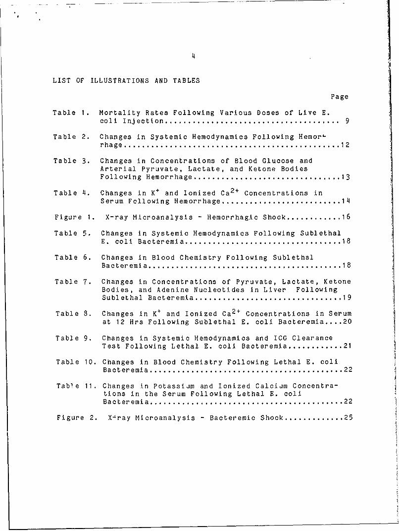
4
LIST OF ILLUSTRATIONS AND TABLES
Page
Table 1. Mortality Rates Following Various Doses of Live E.coli Injection ...................................... 9
Table 2. Changes in Systemic Hemodynamics Following Hemorlrhage ............................................... 12
Table 3. Changes in Concentrations of Blood Glucose andArterial Pyruvate, Lactate, and Ketone BodiesFollowing Hemorrhage ................................ 13
Table 4. Changes in K+ and Ionized Ca 2 + Concentrations in
Serum Following Hemorrhage .......................... 14
Figure 1. X-ray Microanalysis - Hemorrhagic Shock ............ 16
Table 5. Changes in Systemic Hemodynamics Following SublethalE. coli Bacteremia .................................. 18
Table 6. Changes in Blood Chemistry Following SublethalBacteremia .......................................... 18
Table 7. Changes in Concentrations of Pyruvate, Lactate, KetoneBodies, and Adenine Nucleotides in Liver FollowingSublethal Bacteremia ................................ 19
Table 8. Changes in K+ and Ionized Ca 2 + Concentrations in Serumat 12 Hrs Following Sublethal E. coli Bacteremia .... 20
Table 9. Changes in Systemic Hemodynamics and ICG ClearanceTest Following Lethal E. coli Bacteremia ............ 21
Table 10. Changes in Blood Chemistry Following Lethal E. coliBacteremia .................................. ........ 22
Table 11. Changes In Potassiam and Ionized Calcidm Concentra-tions in the Serum Following Lethal E. coliBacteremia ......................................... 22
Figure 2. X-ray Microanalysis - Bacteremic Shock ............. 25

REPORT
A. PROBLEM
The understanding of the events following various types ofcell injury is crucial to the development of our knowledgeconcerning the pathogenesis, treatment, and prevention of humandiseases. Despite the advanced knowledge and treatment ofhemorrhagic and bacteremic shock, mortality and morbiditycontinue to remain high. For better treatment of thesediseases in man, much firther knowledge and understanding of thecellular pathophysiology of shock is needed. It is ourhypothesis that many of the cellular changes which lead the cellfrom normal to irreversibly injured are initiated and modifiedby primary and/or secondary effects of ion redistributionstaking place between the cell and the extracellular compartmentand between various compartments in the cell. With the recentavailability of x-ray microanalysis as a tool to measureintracellular ions and thus being able to correlate structurewith the chemical composition of cells, a new dimension to theanalysis of cellular reactions to injury by the shock state hasbecome possible. However, spcclflc aspects concerning effectsat the cellular and subcellular levels need to be furtherclarified. Therefore, the aim of this study was to characterizethe cellular and subcellular effects of hemorrhagic andbacteremic shock in the liver and heart using in vivo ratmodels.
B. BACKGROUND
The focus of our laboratory for the past two decades hasbeen directed toward the understanding of the events followingvarious types of cell injury with the aim of developingknowledge which will be useful in the understanding of thepathogenesis, treatment and prevention of human diseases. Sincethe work of Virchow El], the dominant concept of pathologyregards disease as a result of the reactions of cells to injury.An injury can be defined as any physical or chemical stimuluswhich pertarbs cellular homeostasis. Such a perturbation can betransient and readily adapted to by the cell with no subsequenteffect or it may be a more prolonged effect to which the cellcan adapt only by a series of structural and fundamentalmodifications or to which it succumbs, resulting in cell death.Following an injury, cell reactions can be classified into twophases: a reversible phase which precedes cell death and anirreversible phase consisting of those changes which occur aftercell death. Therefore, as has now become evident, all diseasestates are most meaningfully expressed at the cellular andsubcellular levels with such changes forming the basis of thephysiologic and morphologic alterations which are observed.

6
Although a number of critical steps occur followinginjury, many of the cellular changes leading the cell fromnormal to irreversibly injured are initiated as well as modified
by primary and secondary effects of ion redistributions.Diffusible ions such as Na, Mg, P, Cl, K, and Ca are all veryimportant to the fine tuning of metabolic cell processes and arecontrolled within very narrow limits in various intracellularcompartments and between the cell and the extracellular space.As a result, numerous studies over the past decade have stronglyimplicated an extremely important if not key role for theirmovements and redistributions in the pathophysiology of diversetypes of cell injury such as shock, myocardial infarction, acuterenal failure, metaplasia, regeneration and malignant transfor-mation. Although all of these physiologically active ions areimportant, it has not been until recently that the unique roleof calcium as activator and regulator of many diverse cellularactivities such as contractile processes, cell division,secretory processes, enzyme activation, etc., has been reali:,.?d.Since the extracellular calcium concentration is higher byseveral orders of mafnitude than the estimated cytosolicconcentration of 10- to IO 5 M and enters the cell by diffusiondown a steep electrochemical gradient, this continuous influx ofcalciam implies the existence of regulatory systems formaintaining low cytosolic concentrations. Current evidenceindicates that the mitochondria, plasma membrane and endoplasmicreticulum may each play a role in this control.
Although the study of ion distribution and redistributionsin tissue using such methods as atomic absorption spectro"photometry, precipitation techniques, autoradiography, isotopelabeling, etc. have been utilized for many years, frequently,inaccurate and misleading data have been obtained due to thedifficulties associated with maintaining ion composition duringthe technical procedures. However, with the recent introductionof x-ray microanalysis, detailed assays of elements with atomicweights equal to Na or greater offers great promise in the studyof electrolyte shifts in both normal and disease states. Thetechnique is based on measurements of characteristic radiationresulting from interactions of electrons with matter [2] andpermits, in many cases, complete non-destructive analysis bydetection of characteristic x-rays which can produce informationabout the distribution, quantity and chemical form of an elemen,at the cellular and subcellular levels in biological sampleswith a sensitivity uf approximately 10 g. Areas as small as1000 A can be measured which permit qualitative and quantitativemeasurements to be made on organelles and even parts oforganelles. this is obviously of great value not only instudying many types of cellular inclusions but also in studyingthe organelle distribution of elements of physiologic importanceincluding Na, Mg, P, Cl, K, and Ca. All of these ions have beenpreviously impossible to localize at this level using other

7
techniques. Therefore, in the study of ion redistributionsfollowing cell injury, the importance of x44ray microanalysiscannot be overestimated.
Many theories have been proposed to explain the sequence ofevents which may lead to irreversible shock and to account forthe severe changes in multiple organ systems which occur duringeach of these phases. Such theories have included lysosomaldisruption, deficits in energy metabolism, and damage to cellmembranes among others [3]. It has not been clear, however,which, if any, of these events is primary and which issecondary. In addition to the above, several studies havesuggested that one important alteration involved in cellfunction is manifested by altered plasma membrane potential andion shifts between extra- and intracellular compartments [3,4].
Although there have been a number of publications detailingclinical measurements by atomic absorption spectrophotometry ofion shifts following sepsis cnd hemorrhagic shock, to ourknowledge, only the work performed in our laboratory [5,6] andthat of Nichols et al. [7] has utilized the methodology of x-raymicroanalysis to determine such shifts in tis-sue. Data fromboth of these studies were in agreement in that increases werenoted in Na and Cl and decreases in K following shock.
Other investigations, using flame atomic absorption or ionspecific microelectrodes, have shown leakage of K ions fromcells at a relatively early stage following both septic andhemo. rhagic shock [8,9]. Such leakage has been found to eitherprecede, accompany or be followed by mitochondrial damage andcell swelling [10,11,12,13]. In order to determine if thisleakage of K+ is due to a failure in energy metabolism or to adirect effect of endotoxins on ion pumps, Kilpatrick-Smith etal. [14] investigated the effects of endotoxin in suspensions ofcultured mouse neuroblastoma C"1300 cells and concluded thatthere were two phases of endotoxin action on these cells: theinitial very rapid reaction in which mitochondrial metabolism isaltered but fully compensated and the second phase in whichcellular energy production by the damaged mitochondria cannotprovide ATP at a sufficient rate to maintain normal cellularactivities, thus leading to gradual cell death. Although themechanism for the rapid response in energy metabolism is notclear, it could be either through a direct interaction of theendotoxin with mitochondrial membranes or enzymes or indirectthrough a second messenger such as intracellular Ca2 + . Thelatter is particularly intriguing to us since, if this is thecase, it could link possible, subtle changes in plasma membranepermeability, which may accompany binding and transport of thetoxin, and subsequent response of mitochondrial energypr uction reactions. In accordance with this possible role ofCa l', Spitzer et al. [15] and Liu et al. [16] observed that

8
suppressed oxidation of fatty acids by hearts of dogs which hadbeen given endotoxin could be reversed by the addition of EDTA,therefore leading to the hypothesis that the increased level offree Ca 2 + may have been responsible for the observed changes inmetabolism. However, further experimentation is necessary toprove this point.
In direct connection with the above, and the role of ionmovements in shook, it has become evident that volume regulationby cells, control of cell Na+ and Ca 2 + and modulation of.membranes and the cytoskeleton comprise very important if notdeterminate roles in a number of sublethal and lethal cellreactions to injury. Moreover, interactions of these with cellmembrane modification and cytoskeletal control produce a modelwhich, we believe, relates many important disease processes tobiochemical and ultrastructural changes. Therefore, based onrecent morphological, immunocytochemical and x-4ray analyticaldata from our laboratories and those of others, ve have recentlyadvanced a hypothesis (see Appendix) which proposes that alteredNa+ and Ca 2 + regulation play extremely important roles inseemingly diverse pathological phenomena ranging from acute celldeath to chronic processes such as shock, trauma, myocardialinfarction and neoplasia and, moreover, that all of thesephenomena are interrelated in a common series of cellularreactions [17,18].
It was the aim of the present study, therefore, to inves-tigate the role of ion movements in in vivo animal modelsfollowing exposure to sublethal and lethal episodes ofhemorrhagic and bacteremic shock by direct analyses ofintracellular ion distributions using x-ray microanalysis and tocorrelate these data with physiological, biochemical, andmorphological observations.
C. APPROACH TO THE PROBLEM
1. Hemorrhagic Shock Model in the Rat
Male Sprague Dawley rats, weighing 250-300 g, werefasted overnight and anesthetized with phenobarbital. They wererestrained in supine positions on surgical boards and allowed tobreathe spontaneously without a respirator. The left carotidartery was cannulated with polyethlene tubing (PE-50) and theproximal end inserted into the aorta. Hemorrhagic shock wasproduced by removal of blood using suction for a period of oneminute and the animals divided randomly into the following threegroups:
LD 50 group = (3.16-0.18BW) ml blood/100 g body weight.LD 84 group = (3.25-0.17BW) ml blood/100 g body weight.Control group = cannulation only

9
Parameters, as described below, were measured at 0 time and15 min after hemorrhage.
2. Bacteremic Shock Model in the Rat
Recently, we developed a bacteremic shock model
in rats to study the cellular and subcellular alterations inresponse to lethal and sublethal Gram-negative bacteremia [19].To establish the model, various doses of a live E. colisuspension (Serotype: 0-18) were Injected in conscious ratsthrough the tail veins. Doses and mortality rates are summar-ized in Table 1.
Table 1. MORTALITY RATES FOLLOWING VARIOUS DOSES OF LIVEE. COLI INJECTION
Dose (x 109 Mortality RateGroup Organisms/100 g BW) 6 Hrs 12 Hrs 24 Hrs
A 2.8-3.3 100% (14/14)B 1.8-2.0 14% ( 4/29) 62% (18/29) 100% (29/29)C 1.3"1.5 0% ( 0/25) 8% ( 2/25) 96% (24/25)D 0.4-0.5 0% ( 0/15) 7% ( 1/15) 7% ( 1/15)
Light microscopic studies performed on the dead animalsafter E. coli treatment revealed the following: 1) a dose-related depletion of white pulp in the spleen; 2) foci of smallE. coli colonies in the heart only in group C in which theanimals survived beyond 12 hours; 3) focal necrosis of the liver
parenchyma with increased number and size of necrosis along withprolonged survival time; 4) no characteristic changes in thelung; 5) mucosal hemorrhage, vascular congestion and desqua-mation of the epithelium in the small intestine; and 6)medullary congestion in the kidney [19]. Groups C and D wereselected as the lethal and sublethal models respectively.
On the basis of the above data, theref-re, in thepresent contract, bacteremic shock was induced in rats by IVinjection of live E. coli organisms (Sero type: 0-18). E. coliorganisms were inoculated in a tryptic soy broth medium from astock culture maintained on tryptic soy agar. After stationaryculture in the tryptic soy broth medium for 15 hrs at 370 C, theorganisms .,ere washed three times with sterile saline solutionand resuspended in a saline solution. The number of E. coliorganisms was predetermined spectrophotometrically by readingthe dilute E. coli suspension at 580 nm. Twenty-four hrs later,the number of viable organisms was confirmed by colon counting.A sublethal (0.4-0.5 x 10 9 ) or a lethal (1.3-2.0 x 10 ) dose ofE. coli organisms in 0.5 ml saline per 100 g body weight wasinjected into the tail vein with a one minute period of

10
injection. Saline,-injected rats were used as c( Lrols. At 6,9, 12, and 24 hrs following injection, physiological, morpho.Plogic, histochemical, biochemical and x-ray microanalysisstudies were carried out as described as below.
3. Methods
The following methods were used in both the hemorrhagicand the bacteremic models.
a. Physiological Studies
An arterial catheter was introduced into the lowerabdominal aorta through the left femoral artery and its externalend connected to a transducer. A transpulmonary thermodilutionmethod, which we recently developed [20), was used for themeasurement of cardiac output. A thermistor probe was insertedinto the right carotid artery until its tip lay in front of theaortic valve. As an indicator, 40-60 ul saline solution wasinjected into a central vein catheter (PE 50), which was placedclose to the right atrium, through the right jugular vein.After injection of the saline, changes in blood temperature andarterial blood pressure, as well as EKGs, were simultaneouslyrecorded on a polygraph.
b. Biochemistry
For blood chemistry determinations, theconcentrations of glucose, pyruvate, lactate, creatinine,bilirubin, and the activities of S-GPT, S-OCT, CPK, LDH, andamylase were determined according to standard methods. Afteracid extraction from freeze-clamped livers, the concentrationsof pyruvate, lactate, and adenine nucleotides were enzymaticallymeasured.
c. Serum Electrolytes
Serum K and ionized Ca2 + concentrations were measuredby ion selective electrodes.
d. Histochemistry
Routine frozen sections were cut from liver andheart and air'dried for the determination of the followingenzyme activities: AlPase, AcPase, ATPase, SDH, G6Pase, andG6PDH.
11
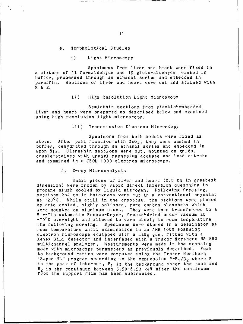
e. Morphological Studies
i) Light Microscopy
Specimens from liver and heart were fixed ina mixture of 4% formaldehyde and 1% glutaraldehyde, washed inbuffer, processed through an ethanol series and embedded inparaffin. Sections of liver and heart were cut and stained withH & E.
ii) High Resolution Light Microscopy
Semi-thin sections from plastic-embeddedliver and heart were prepared as described below and examinedusing high resolution light microscopy.
iii) Transmission Electron Microscopy
Specimens from both models were fixed asabove. After post fixation with OsO4, they were washed inbuffer, dehydrated through an ethanol series and embedded inEpon 812. Ultrathin sections were cut, mounted on grids,double-stained with uranyl magnesium acetate and lead citrateand examined in a JEOL 1OOB electron microscope.
f. X-ray Microanalysis
Small pieces of liver and heart (0.5 mm in greatestdimension) were frozen by rapid direct immersion quenching inpropane slush cooled by liquid nitrogen. Following freezing,sections 2:4 um in thickness were cut in a conventional cryostatat -200 C. While still in the cryostat, the sections were pickedup onto cooled, highly polished, pure carbon planchets which4ere mounted on aluminum stubs. They were then transferred to aViri-Tis Automatic Freezet-Dryer, freeze-dried under vacuum at-700 C overnight and allowed to warm slowly to room temperaturethe following morning. Specimens were stored in a dessicator atroom temperature until examination in an AMR 1000 scanningelectron microscope equipped with a LaB 6 gun, fitted with aKevex SiLi detector and interfaced with a Tracor Northern NS 880multiohannel analyzer. Measurements were made in the scanningmode with microscope parameters as previously described. Peakto background ratios were computed using the Tracor Northern"Super ML" program according to the expression P-BI/B 2 where Pis the peak of interest, B1 is the background under the peak andB2 is the continuum between 5.50-6.50 keV after the continuumfrom the support film has been subtracted.

12
g. Statistical Analysis
In the hemorrhagic shock model, the paired two tailedStudent's t test was performed to compare the pre-shock andpost-shock val,'es and Analysis of Variance was ased to comparegroups. In th! bacteremic model, Analysis of Variance was usedto determine the significant difference among groups. A valueof p < 0.05 was considered significant.
D. RESULTS AND DISCUSSION ON THE RESULTS
1. Hemorrhagic Shock Model in the Rat
a. Physiological Studies
Detailed results of changes in CI, MABP, HR, TPR,and SVI are summarized in Table 2.
In the LD5O hemorrhagic shock group, CI decreased to 22.7%of pre-,shock values at 15 min, while in the LD84 group, CIdropped to 15.1% of pre-shock values. MABP dropped to 27.8% and23.8% of pre-shock values in LD5O and LD84 groups, respectively.TPR increased to 120% and 203% of pre-shock values in LD5O andLD84 groups, respectively. in sham-operated controls, nosignificant changes were noted. EKGs showed changes in the STsegment in both the LD50 and LD84 groups, indicating ischemicchanges.
These data suggest that the presence of very powerfulsympathetic mechanisms, induced by released cathecholamines,may, to some extent, prevent hypoperfuslon in vital organs byinducing relatively well maintained HR and increased TPR.
Table 2. CHANGES IN SYSTEMIC HEMODYNAMICS FOLLOWING HEMORRHAGE
LD50 (n=3) LD84 (n=6)0 min 15 min 0 min 15 min
Cl (ml/min/kg) 312+60 71+13 306+65 46+34^MABP (mmHg) 141-8 39711 * 139+8 33+13*
HR (beats/mn) 417+21 3097-40* 416+23 303+66TPR (mmHg/ml/min/kg) 0.46;0.07 0.55+0.01 0.47+0.11 0.950.46SVI (ml/kg) 0.75+0.11 0.2370.311* 0.7370.12 0.1570.10*
Values given are means + S.D.; *=Significant difference (p < 0.05)between 0 and 15 min groups using paired t test

13
b. Biochemistry
Blood glucose, pyruvate, lactate, and ketone bodieswere measured. Due to limited available blood after hemorrhage,fluorospectrometry for measurement of pyruvate, lactate, andketone bodies was used. Detailed results are summarized inTable 3.
Increases in the concentrations of arterial pyruvate andlactate with decreases in the ratio of pyruvate/lactate wereseen in both the LD50 and LD84 groups. However, hypoglycemiawas observed only in the LD84 group.
In arterial ketone bodies, the concentration of AcAc in boththe LD50 and LD84 groups decreased but that of BHOH did notsignificantly change, thus decreasing the ratio of AcAc/BHOH inboth groups. Since the main source of production of AcAc andBHOB is the liver and since these substrates are diffusable andwell regulated by the oxidorreduction state of free NAD+/NADH inliver mitochondria, the ratio of arterial AcAc/BHOB indirectlyreflects the redox state of free NAD+/NADH in liver mito-chondria. Despite the powerful sympathetic reflex, as indicatedby the physiological monitor mentioned above, these data suggestthat the oxygen supply to liver mitochondria is insufficient toproduce NADH through the mitochondrial respiratorychain in both the LD5O and LD84 groups.
Table 3. CHANGES IN CONCENTRATIONS OF BLOOD GLUCOSE AND ARTERIALPYRUVATE, LACTATE, AND KETONE BODIES FOLLOWING HEMORRHAGE
LD5O LD840 min 15 min 0 min 15 min
Glucose (6)109.1+14.1 109.4+20.9 (6)112.5+12.0 80.3 +10.4*Pyruvate (5)0.174;70.031 0. 23370.067* (5)0.148+0.01 0.191+0.046*Lactate (5) 2.57+0.63 11.29+6.39* (5) 2.0470.23 7.69+1.79*P/Lxl0 2 (5) 6.95+1.46 2.47 0.98* (5) 7.31+0.97 2.490.73*AcAc (6)0.046;70.013 0.026+0.014* (5)0.06770.029 0.027+0.010*BHOB (6)0.1700.116 0.171+0.113* (5)0.165;0.107 0.15270.067A+B (6)0.21670.11 7 0.197+0.119 (5)0.25270.095 0.17970.068A/B (6)0.36870.183 0.202+0.120* (5)0.432+0.208 0.203+0.109*
Values = means+S.D.Glucose mg/dl-Lactate, Pyruvate, AcAc, BHOB = amoles/ml(n) = number of animals* = significant difference (p < 0.05) between 0 and 15 min groups
using paired t test

114
c. Serum Electrolytes
K + and ionized Ca 2 + concentrations inserum were measured by ion selective electrodes. Isotonic 2, 4and 6 mM KC1 solutions for K+ and 0.5, 1.0, and 1.5 mMCaC1 2 solutions for ionized Ca 2+ were used as standards.Measurements were performed in a 2500 water bath. Results aresummarized in Table 4.
Significant increases in K+ concentrations afterhemorrhage were noted, with the more severe hemorrhage inducinghigher values. On the other hand, decreases in ionized Ca 2 +
concentrations in the serum after hemorrhage were observed.
Table 4. CHANGES IN K+ AND IONIZED Ca2 + CONCENTRATIONS IN SERUMFOLLOWING HEMORRHAGE
LD50 LD840 min 15 min 0 min 15 min
K+ (mEq/L) (8) 3.2+0.6 6.5+0.9* (7) 3.2+0.7 6.9+1.0*Ca2 + (mEq/L) (8) 1.0870.25 0.085+0.24* (7) 1.06+0.36 0.82+0.31*
Values given are means+S.D.* significant difference (p < 0.05) between 0 and 15 min groups
using paired t test
The availability of mini ion selective electrodes hasenabled the measurement of specific ion concentrations in smallamounts of serum. Since these ion selective electrodes arehighly specific to certain ions and the technique is quitesimple, reproducibility, therefore, is quite high [21,22,23,24].
Although the exact mechanism of hyperkalemia is unclear,x-ray microanalysis of ions in tissues, as described below,showed decreases in the concentration of K+ in the liver andheart after hemorrhage. Thus, released K+ from cells in theirenergy-depleted state may well provide the mechanism for theserum increase.
In our rat hemorrhagic shock model, the mechanismresponsible for the hypocalcemia is also unclear. However,decreased oxygen supply to tissies, as mentioned above, leads todepression or depletion of cellular energy, especially ATP. Thisdecreased cellular energy causes cessation of the Na-Ca and/orH+-Ca pumps, causing accumulation of Ca 2 + in intracellularcompartments, potentially in mitochondria and/or the endoplasmicreticulum (ER). This accumulation of Ca2 + in cells may causedecreased Ca 2 + concentration in the serum. Another possibleexplanation is that severe hemorrhage induces cathecholaminerelease, as mentioned above, leading to peripheral hemostasis

15
which may result in intra-vascular coagulation. This requiresCa 2 + in the serum. Thus, in addition to the accumulation ofCa 2 + in cells, Ca 2 + in the serum may decrease due to binding.
Harrigan et al. [25] studied changes in Ca 2 + levels insevere shock patients during and after resuscitation. Totalserum proteins, serum albumin, total Ca 2 + , and iouized Ca 2 + weresignificantly reduced. The severity of hypocalcemia paralleledthe hypoproteinemia, the number of transfusions given duringresuscitation, and the duration of shock. From their data, theysuggested that increased extravascular Ca 2 + flux occurs withsevere shock. As mentioned above, this may, indeed, relate tothe ion shifts observed in cells here in our study.
Carpenter et al. [22], in a study of hemorrhagic shock inbaboons who were then resuscitated, found significantdiminutions (51%) in Ca 2 + in serum following resuscitation. Sucha finding is consistent with our hypothesis in the sense of thereflow phenomenon. Reflow into previously ischemic energydepleted areas would be expected to result in an increase inintracellular Ca2+ , as is typically seen following reflow inischemic myocardium. The above authors infer that such anincrease in intracellular Ca 2 + is a manifestation of the "sickcell" syndrome. This is, of course, also consistent with ourown work and, indeed, emphasizes the desirability of measuringintracellular electrolytes as we have done in this study, sincewithout measuring intracellular electrolytes, one is onlydealing with an inference.
The differences in changes noted in serum Ca 2 + during shockbetween our rat hemorrhagic shock model and the above-mentionedbaboon model may depend upon the severity of shock, since theblood pressure in the baboons was maintained at 60 mmHg whereasthe blood pressure was less than 30 mmHg in our rat model whenblood samples were taken. The severe shock may cause a greaterinflux of Ca 2 + into the cells, resulting in decreases in Ca 2 + tnthe serum.
Carpenter et al. [22] also showed that there were changesin all forms of Mg; namely, an increase in serum Mg 2 + duringshock which then returned to normal levels after resucitation.We have not thus far observed significant changes in intra-cellular Mg2 + levels in our hemorrhagic shock model but therehas been some evidence of such decreases in our bacteremic shockmodel as described below. These same investigators [22] alsonoteo increases in serum phosphate levels which might wellcorrelate with the decreased cellular phosphate levels observedin our studies as detected by x-ray microanalysis (see below).

16
d. X-ray Microanalysis
Typical ion measurements, using x-ray micro-analysis, of 4 um freeze4,dried sections of liver and heart afterhemorrhage are shown in Fig. 1. 'SHAM"LIVER' and 'SHAMmHEART'represent tissues obtained from an animal in which only surgicalmanipulation was performed while 'EXPT-LIVER' and 'EXPT-HEART'were obtained from an animal following a 15 min interval of LD84hemorrhagie as described in the 'Methods' section above. P-Bj/B 2represents the peak (P) to background (B) ratios for eachelement as computed according the previously describedmethodology. Note the increases in Na, Cl, and Ca and thedecreases in K and P in both the liver and heart from thehemorrhagic animal as compared to the sham control values. Thesedata are in good agreement with the above serum electrolyte datasince, as discussed above, a decrease in serum electrolytescorresponds to an increase in intracellular electrolytes.
HEMORRHAGIC SHOCK - 15 MIN
I
OL.
0.
i. 1is
= SH4A-IVR
VAN-WART UPT-IWART
Fig. 1
17
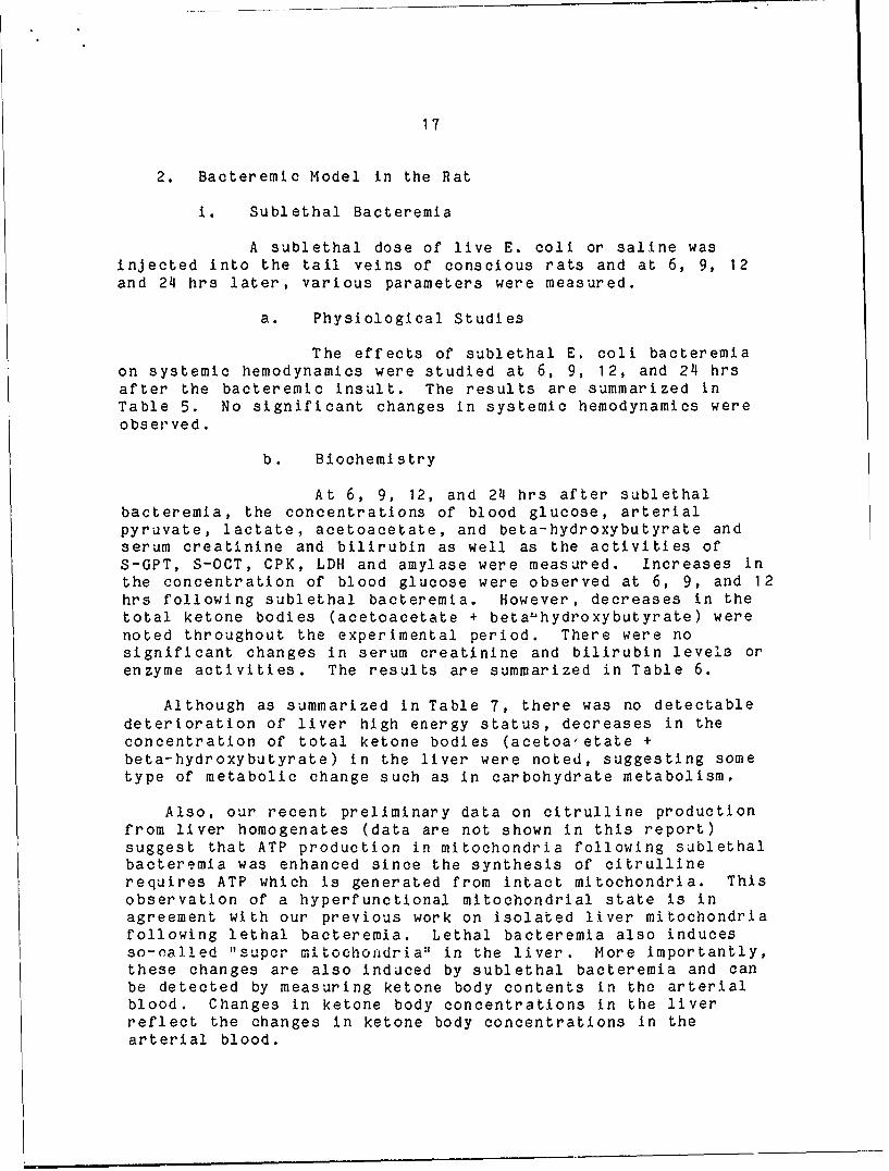
2. Bacteremic Model in the Rat
i. Sublethal Bacteremia
A sublethal dose of live E. coli or saline wasinjected into the tail veins of conscious rats and at 6, 9, 12and 24 hrs later, various parameters were measured.
a. Physiological Studies
The effects of sublethal E. coli bacteremiaon systemic hemodynamics were studied at 6, 9, 12, and 24 hrsafter the bacteremic insult. The results are summarized inTable 5. No significant changes in systemic hemodynamics wereobserved.
b. Biochemistry
At 6, 9, 12, and 24 hrs after sublethalbacteremia, the concentrations of blood glucose, arterialpyruvate, lactate, acetoacetate, and beta-hydroxybutyrate andserum creatinine and bilirubin as well as the activities ofS-GPT, S-OCT, CPK, LDH and amylase were measured. Increases inthe concentration of blood glucose were observed at 6, 9, and 12hrs following sublethal bacteremia. However, decreases in thetotal ketone bodies (acetoacetate + beta' hydroxybutyrate) werenoted throughout the experimental period. There were nosignificant changes in serum creatinine and bilirubin levels orenzyme activities. The results are summarized in Table 6.
Although as summarized in Table 7, there was no detectabledeterioration of liver high energy status, decreases in theconcentration of total ketone bodies (acetoaetate +beta-hydroxybutyrate) in the liver were noted, suggesting sometype of metabolic change such as in carbohydrate metabolism.
Also, our recent preliminary data on citrulline productionfrom liver homogenates (data are not shown in this report)suggest that ATP production in mitochondria following sublethalbacteremia was enhanced since the synthesis of citrullinerequires ATP which is generated from intact mitochondria. Thisobservation of a hyperfunctional mitochondrial state is inagreement with our previous work on isolated liver mitochondriafollowing lethal bacteremia. Lethal bacteremia also inducesso-nalled "supcr mltoochondria" in the liver. More importantly,these changes are also induced by sublethal bacteremia and canbe detected by measuring ketone body contents in the arterialblood. Changes in ketone body concentrations in the liverreflect the changes in ketone body concentrations in thearterial blood.

.18
- ~ ~ C t- a' N30 0 -"
-4 It'. CA. * i 'j -00 N' 000 *NVM 100rI. ** 0 *-O- 0 ) ~t'.- a~ 000 ~"-"I
CY N O I T0T -.I4 .444 **4 144400 Mo~i 0 . m 9 T -.Ln 0 %DO t- z0 C
C U2NeA -- -0 M-'. *N- 8-~ul N.'C"! NSA * 0 .V
I.) m'0 -TUN 0 00(m 00 -71 0 0
0C9f1LO' SL 4 . ' . . 'A A.. ~ '
(- 0o 3- MLt *snm (7o 1% LoN~~~u LAI-O L. ow'. ' toL
0Oc %A0~m t- 0 0. M-ot 0M1
0~ ~ Ln ( 7 r oSO onSF- 0o U"-0 03-0 % - m
O'N c N00 t- T.0 M -0 *N
00 C"S0~ -4* 0-u's -cm 0 3-*0C)...1111. - -N . *N'.0S
0co MC 0L .- 0 0
IV 1 t 1A D t- -,. 00'. 00 NN
L' 00 N0 0%- 0)0 00."
CC4 C'LLA3 NOO *-0 1 CC' l li r; 0In CN N 9 -. 0= -m t.sL . 10 00
0-~f 055' C,..+9- '. 0 '.' 0 ,"-'it D - = 0% 0 N 7 00 m
cl +' U 0 ) .. ++ 1.1.1 L41o4141- ~ 0Mm D % 41,=+1MM
c0 m1 r l ot-r l 0 NL(1 0 M 0 00F- ..
U 5) 00 1
0 C -4 0 M O' .0 00-*
.. 2 vs -r u' U CO e0 *1 +0 + n.110%0 ID 0 0 - Ol m.1- . 0 000 V% 0- -0
t. 1O - -3l~ U n 000- - N MID0' 10 t 0 451n4141r 141+1 -- Goas oo %0N'h
.. 2 CC aO 0 - 0 r..I.- - O '.-0 0
00 0 h o CDc1- 0 - 0'> ' Z .
Z '.:0'. 0 0'.-:00L C Snn- ~ % t- L. co Do.. ..- N 0 m m
.0'.- 4 co ' -0c 0 .3
0 . - (V .0 0 0 Ln CO N'010 000 M aOMS0''.00M LC
3 1.1 171.1 -3u- . I 10 U 2MU
- 0 00 t Cto oo '0 co 0. 0= l )t4% 0. ' . S O
32 3 *a'. *'00 -0 N Na'.; 03- 5)5.'0 *C-l0 . . . U' '03
0 .o - J 0LA'.. SO NO 000 .- 0Snn00 0'' Sn~~~~~ * COO - CD *114. '1 17;1 ... + .,
-, :. + +I+5 +
Ch00100 t I-3 Cl (1 0C '
En 50032 0=0 1 cL. 'D'. V".!N ma C 1
LA.'.4CC. 14 m.1C- -4-' 0E t. ;%
Z 0 '-o t. M. 00C: CY- Y- .4- ".5 .- l'. . Dt-
o~~( 03. '--' 05 C. IlIIT1,c '~ ' TT1V4 - 0 %0 )nt 5 - 10 C%- D M O -
1'4, U+*l 1 o to9'. u D o o C M n
SC 00 M c xN "4.0% 0...4 AU ~ m _r ~ -Q- Ct!-
cm 0to... oa.CCC C.0 Le.0-0 -"~..C
SL0 z~ 8)I '- - .LA~~~~~~ xFL L C. >) .I) .'5
10~ C. ' m0 '~' --. ~. -~g 5 -4.C*.. C)'.. 0. .00.5to0 0.4 .4 0 F-O0.0 ~ 0-0 O U..2 to
40cc. E0> .An UFLA EUF-L 1

19
0 t- en - t- oo cn- -o
co co -o m 01MO. 000 0
10 Lq -0 t- a0a
cu NO0mC/) =
cm N '0 "I eni"I N
D 0 L. cma N~~C. 1. -0 01 .0N.
+ I 1 +1.11+1 + + 4 l.I '?o m a.,-O0 CO r- 0 NI
0~ ~~ ~ UU 1 . *rnu n'ee0a cmOO '0 ~0m
e0 NO enC.Y)
0 N 0cmAc
0 +1 "+1 '114 +1 1,41 40 0 t-00 0 m mco DO m
0 - en L ANL N.Z0 0W -8 - ,-~ '0 N G
F- 0. LAOOCNC%
0% t-- %0 N en (,j Ain 0 C IAN C 0.
+. C 1 1 +11+1.4 1 .. 'Zl 0 D 05 n ~Oene n n.C00 N
-, UP zen It C05C'0' N0 0
C.) r. 0 0
0 0~ 0 N-0cmi h (7
0 0 co en -- Go-r o c
0 LA CeO 0 00 0 9C) +1 +I +111.. '.1. 1 +1
0' ~ ~ ; C0 0-A 5L LAO m-~ ~ ~ t co C)' *n-A F nO.
0%o O)-
0 C.0D00
to00 0~'o n c 0' t- L.- LA.Ot.oC . , 0 n AC-.o 0 0
c 71 41 .4++. 1111 .o N 0' f- C)>0)t-.-- OsC'-O nc
C.) S Ln N %, (-'O 0 -
(- 0 % m00
> > ') AJ 0A ) > V
C.)~~ 0 0)~ . . .
C.) 04) 0s .-0 O10' 0A. 6aC) E E 0).
> - 0
0 0 0 0 )- NOO-o L ej 0- co to 0-n0
06 C.L mn 0 E L).. . .O I- a en zA N 000 0
-4 ~ ~ ~ ~ ~ ~ - > 1 . IIII. 1111 .

20
c. Serum Electrolytes
Serum electrolyte measurements following 12 hrs of sublethalbacteremia showed a decrease in ionized Ca 2 + and an increase inK+ concentrations (Table 8).
Table 8. CHANGES IN K+ AND IONIZED Ca2 + CONCENTRATIONS IN SERUMAT 12 HRS FOLLOWING SUBLETHAL E. COLI BACTEREMIA
Saline 12 Hrs
K+ (mEq/L) 3.3 3.9Ca 2 + (mEq/L) 2.2 1.5
ii. Lethal Bacteremia
a. Physiological Studies
The effects of lethal E. coli bacteremia onsystemic hemodynamics and ICG clearance test were studied at 6,9, and 12 hrs after a bacteremic insult. A hypodynamic statewas noted at 9 and 12 hrs. The results are summarized in Table9.
Insignificant decreases in TPR at 6 hrs was followed bysignificant increase. at 9 and 12 hrs. This is compatible withthe early hyperdynamlc state seen in various experimentalanimals, including baboons, dogs, and rats [26,20], The latehypodynamic state seen at 9 and 12 hrs is probably due to acombination of decreased venous return following sustainedperipheral pooling and myocardial dysfunction as indicated byhigh CPK release in the serum (see below).
The ICG clearance test, which is a good indicator of hepatic
blood flow, decreased following the bacteremic insult.
b. Biochemistry
At 6, 9, and 12 hrs after bacteremia, the concen-trations of blood glucose, pyruvate, and lactate and serumcreatinine and bilirubin and the activities of S-GPT, S-OCT,CPK, LDH, and amylase were measured. Hypoglycemia and lacticacidemia and increases in the concentrations of serum creatinineaid bilirubin were observed. Increases in the activities ofS-GPT, S-OCT and LDH and no increases in amylase were noted. Theresults are summarized in Table 10. As mentioned above, highCPK values were seen at 12 hrs when the hypodynamic state waswell established.

21
Table 9. CHANGES IN SYSTEMIC HEMODYNAMICS AND ICG CLEARANCETEST FOLLOWING LETHAL E. COLI BACTEREMIA
Control 6 Hrs 9 Hrs 12 Hrs(n-8) (n=6) (n-6) (n=6)
CI 254+23 244+46 177+63* 182+22*TPR 0.533+0.041 0.501+0.109 0.703+0.226* 0.679+0.086*SVI 0.57770.076 0.49670.097 0.433'0.130* 0.37370.045*BP 13576 114-7* 11410* 122721HR 444728 475711 402+37* 488T25*ICG 2.7+-0.4 7.6+3.3* 8.674.1* 8.11.3*
Value = Mean+S.D.CI = ml/min/kgTPR mmHg/ml/min/kgSVI = ml/kgMABP = mmHgHR = beats/minICG = t 1/2 (min)* = significant difference (p < 0.05) vs control
c. Serum Electrolytes
Concentrations of serum K+ and Ca 2 + were measuredusing ion selective electrodes. Increases in K+ with time andinitial decreases in ionized Ca 2 + were noted. The results areshown in Table 11.
Prominent among the laboratory characteristics ofstaphylococcal toxic.-shock syndrome in patients is hypocalcemia.Recently, Chesney et al. [24] demonstrated that hypocalcemiarepresented a reduction in both total Ca 2 + and ionized Ca 2 + withelevated immunoreactive calcitonin. In experimental animals,Trunkey et al. [23] reported that the serum ionized Ca 2 +
concentration decreased during septic shock in the baboon as didthe skeletal muscle extracellular pool of Ca 2 + [27]. Using redblood cells, Shires et al. [28] demonstrated the cellular uptakeof Cl-, Na + and water, and a loss of K+ during septic shock inbaboons. They suggested decreased energy utilization ratherthan decreased energy production as a factor leading todiminished active ion transport during septic shock.
Carli et al. [29] demonstrated that septic shock sera frompatients, when compared to normal sera, increased the actionpotential duration and depressed contractility of beatingcardiac cells. Addition of Ca 2 + reversed these two actions.

22
S) * .. N . '- - z 0
'-4~ +j1+1-+I+t+I+1+14+I+ +1+1 :TT+ I t- r M - N M0 0%0D
U3) (\J mNm - N t-0aW'fN .3 C~j . Y
C-))
('\J . 0 COC\.-T Ln -*0) -C>0 00\-r 000C nmC
Ci) T- +I+I+I+I+I+I+i+i+i+I+ U) +1+1i)~0 O U.C N N-0 o N = 00%
-:4~fL C'' 0
C.)
.IO'MM-0 *CO LA + c.. . . .. CM OM M (\J
w n 00000 Cw V2 ) 00 CS.. - +I+I+1+I+I+17+++1 L +1+1
>- +I-=n -l L-- m t- -x 0 Ln '-i = 1~-1aCJ 0 V- rC\ -= M 0 N. .\ 0
E- 10 CO . . * LA.- I- zI- . . ~ Lod) 0 ~00 N~ .z- NE - .
Ci)m .- - 0.aa''o O 0 Oi ' 0w. CO - c\. M . %D CO a% 0 -W
C) 0N 0D 0)
o -1 o-C'.JO -0 C) H r-I CO C.- ) 0 -x . . . * M) L'0 + A- 0 .on S. - 00 0-A 0) 0 )0nCl= DC 40 t- 00 0)
.) +1+1+1+1+++7 +1+1+1+1 4-I o .4.) +1+1-I 4--z r_ .=0 0 C7 l=k oa = )3V 00 w0'I 0 M M ~f- 0(N - co cl f4-I * o *- - f
o o '- .4-t -- ( C) 0 =. ~ .4 .
Cl) C. 0 CM n - Co)w V~U0U~L-.O-' w1 -I t'-t- C
0)C zrt-r om w t- -% 1-4 r--t-O
m .02 E- C 00~~r m r%3 024 0-o' 2
-o C4d) fl,.0 r=. E4-.
-. c 0 43 C--- '.- 5 -C -i r-i M o M (W )
a) 0 > M 4.) 'r1- .- t- 00M )1 0o 9a0)2 )U)Q0 0 4 54 rH a- - ~j
cl - ) 0 1 1 . I C-13 r- cis + it r02 a4. 02 -. I 02 C- 02 a0 02 C)

23
Again, as mentioned above, all oP these results are compatiblenot only with our serum electrolyte data but also with our x-raymicroanalysis data since a decrease in serum electrolytescorresponds to an increase in intracellular tissue electrolytes.
d. Histochemistry
After lethal bacteremia, AlPase, AcPase, ATPase,SDH, G6Pase and G6PDH were performed on liver frozen sections.Increases in AlPase activity, due to the positive activity overthe leukocytes which had migrated into the sinusoid, andincreases in AcPase activity in the parenchymal cell were noted.The increases in AcPase activity in the parenchymal cellsindicated the quantitative increases in the number of secondarylysosomes. ATPase, SDH, G6Pase, and G6PDH showed no significantchanges.
e. Morphological Studies
Routine light microscopic observations showedinfiltration of leukocytes surrounding foci of bacterial colonyin the heart during hypodynamic state after E. coli bacteremia.Recently, Manson and Hess [30] hypothesized that oxygen freeradicals from leukocytes contribute to the characteristicmyocardial dysfunction of endotoxin shock. Free radicals candirectly affect the function and activity of the excitation-contraction coupling system of cardiac muscle.
Plastic-embedded semi-thin sections of liver were examinedusing high resolution light microscopy. Foci of hepatocellularinjury were observed as increasing in size and number with time.Focal accumulation of fibrin and degeneration and debris ofleukocytes in the sinusoids were observed. Focal accumulationof lipid droplets with time was also noted.
Electron microscopy showed swollen mitochondria andincreases in the number of autophagic vacuoles with time. Theincreased number of autophagic vacuoles observed in thehepatocytes of these animals is of great interest. In previousstudies, we have characterized and analyzed this phenomenon invarious models including that in which autophagy is induced byinfusion by glucagon or administration of vinblastine [31,32].We also have been observing massive increases in the number ofautophagic vanvoles in tissucs from biopsies and/or immediateautopsies of patients with severe sepsis in our trauma unit[33). . This phenomenon appears to be related to modificationof the cytoskeleton, perhaps occasioned by intracellularmessengers including Ca2 + . This might explain the induction ofthis phenomenon by glucagon as well as vinblastine. In in vitrostudies in our laboratory have been successful in reproducingautophagy using the Ca ionophore A23187, which increases Ca

24
influx [34]. The increased number of autophagic vacuoles, notedin the present study, also correlated with the light microscopicdemonstration of increased activity of acid phosphatase, awellknown lysosomal marker. Such increased autophagy is one ofthe principle known means of organelle turnover and maycorrelate with the increased protein catabolism observed inanimals and patients with sepsis.
The mitochondrial swelling observed in the hepatocytes isalso of interest and represents an early, although reversible,change following a variety of types of cell injury. It is ourhypothesis that this swelling directly correlates withmodification of intracellular electrolytes. An increase ofionized Ca 2 + in the cytosol could activate phospholipases whichmodify mitochondrial inner membrane permeability.
The mechanism of damage to the hepatocytes in bacteremiaand/or sepsis, of course, is an important question. In ourbacteremic model, these lesions were predominantly seen in theareas where leukocytes had migrated and aggregated, suggestingthat there is some correlation between the infiltration ofleukocytes and the damage to hepatic parenchymal cells. Onepossible mechanism, as shown in our working hypothesis [17,18],is membrane damage from complement, endotoxins, or toxicproducts of leukocytes subh as superoxide and related meta-bolites.
f. X-ray Microanalysis
Typical ion measurements, using x-ray microanalysis,of 4 um freeze-dried sections of liver and heart afterbacteremia are shown in Fig. 2. 'SALINE C - LIVER' and 'SALINEC 4 HEART' represent tissies obtained from an animal in whichsaline only was injected into the tail vein while '9 HR-BACT-LIVER' and '9 HR4BACT-HEART' were obtained from an animal 9 hrsfollowing an injection of a lethal dose of E. coli organisms.P-BI/B 2 represents peak (P) to background (B) ratios for eachelement as computed according to previously described methodol-ogy. Note the increases in Na, Cl, and Ca and the decrease in Pin both the liver and heart following the bacteremic episode ascompared to the saline-injected controls. Note also that Kdecreases in the liver but increases in the heart. Such anincrease in K has been noted by Clemens et al. [35] in hepato-cytes following sepsis in micropuncture areas of hyperpolari-zation. Again, as mentioned above, x-ray microanalysis ofintracellular ions adds considerably to our knowledge of changesoccurring in bacteremia by indicating clearly that such shiftsor redistributions do, indeed, occur.

25
BACTEREM I A
1.20
1.08
0.96
0.84
0 .2
NA MG P S C K CA
UNOPER C-LIVER 9 HR-BA(,T-.I1VER
UNOPER C-fIEART 9 HR-SBACT-+IEAT
Fig. 2
E. CONCLUSIONS
In our in vivo rat hemorrhagic shock model, measurements ofintracellular ion shifts, as determined by x ray microanalysisof freeze-dried 4 um sections of liver and heart, correlatedwith the significant changes seen in serum K+ and ionized Ca2 +
concentrations as well as with the physiologic, biochemical, andmorphological paramcters described above. The depressed energyproduction, due to the decreased oxygen delivery, resulted ininhibition of the active membrane transport, causing theintracellular ion shifts. These intracellular ion shifts maywell provide the mechanism for the increase in K4 and thedecrease in the ionized Ca 2 + concentration in the serum.
In addition, x~ray microanalysis measurements revealeddecreases in phosphorus. Although serum phosphate levels werenot measured in our study, Carpenter et al. [22] have shown thatthey did increase significantly during hemorrhagic shock in the
26
baboon. The above would, therefore, agree with the hypothesisthat a decrease in serum electrolyte levels corresponds toincreases in intracellular ion levels.
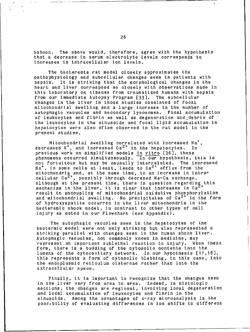
The bacteremia rat model closely approximates thepathophysiology and subcellular changes seen in patients withsepsis. It is striking that the morphological changes in theheart and liver correspond so closely with observations made inthis laboratory on tissues from traumatized humans with sepsisfrom our Immediate Autopsy Program [33]. The subcellularchanges in the liver in those studies consisted of focalmitochondrial swelling and a large increase in the number ofautophagic vacuoles and secondary lysosomes. Focal accumulationof leukocytes and fibrin as well as degeneration and debris ofthe leukocytes in the sinusoids and focal lipid accumulation inhepatocytes were also often observed in the rat model in thepresent studies.
Mitochondrial swelling correlated with increased Na + ,
decreased K+ , and increased Ca 2 + in the hepatocytes. Inprevious work on simplified models in vitro [36], thesephenomena occurred simultaneously. Ln our hypothesis, this isnot fortuitous but may be causally interrelated. The increasedNa + , in some cells at least, leads to Ca 2 + efflux from themitochondria and, at the same time, to an increase in intra-cellular Ca 2 +, possibly through decresed Na-Ca exchange.Although at the present time, there is question regarding thismechanism in the liver, it is clear that increases in Ca +
result in uncoupling of mitochondrial oxidative phosphorylationand mitochondrial swelling. No precipitates of Ca 2 + in the formof hydroxyapatite occurred in the liver mitochondria in thebacteremic shock model, in contrast to other types of cellinjury as noted in our Flowchart (see Appendix).
The autophagic vacuoles seen in the hepatocytes of thebactermic model were not only striking but also represented astriking parallel with changes seen in the human shock liver.Autophagic vacuoles, not commonly known in medicine, mayrepresent an important sublethal reaction to injury. When theseform, there is a budding of the cytosolic contents into thelumens of the cytocavitary network. In our hypothesis [17,18],this represents a form of cytosolic blebbing, in this case, intothe endoplasmic reticulum cisternae rather than into theextracellular space.
Finally, it is important to recognize that the changes seenin the liver vary from area to area. Indeed, in histologicsections, the changes are regional, involving local degenerationand local accumulation of leukocytes and fibrin in thesinusoids. Among the advantages of x-ray microanalysis is thepossibility of evaluating differences in ion shifts in different

27
cells and to correlate these with changes in subcellularsystems, other intracellular events, and other changes such asaccumulation of leukocytes. It is our working hypothesis thatone mechanism of cell injury is from membrane damage. The toxicproducts of infiltrated leukocytes such as superoxide andrelated metabolites may play a possible role in such regionalcell injury.
F. RECOMMENDATION
The studies conducted during this contract period haveclearly validated the utility of these two in vivo models forthe study of hemorrhagic and bacteremic shock, respectively. Theresults with the bacteremic model have been particularlyexciting and, therefore based on the results and observationsdescribed in this report, it is logical that we extend ourstudies with the following goals:
1. to characterize ion shifts in more detail at theorganelle level, using x-ray microanalysis, in livers and heartsfrom our in vivo bacteremic shock animal model and to correlatesuch with physiological, biochemical, and morphological data;
2. to explore the role of oxygen free radicals releasedfrom infiltrated leukocytes on hepatocellular injury and cardiacdysfunction seen in the hypodynamic state;
3. to explore the nature of protein catabolism as indicatedby our observation of increased autophagic vacuoles andsecondary lysosomes;
4. to explore the effects of various interventions such as
a. non-steroidal anti'inflammatory agents and cortico-steroids which alter produccion of oxygen freeradicals as well as membrane stability, and;
b. calcium entry blockers which modify intracellularion shifts;
5. to study ion shifts in tissues from the in vivo model,using x-ray microanalysis, following various interventions andto correlate such with physiological, biochemical, andmorphological data.
In future studies, in vitro rat hepatocyte and myocytemodels could be used and the same biochemical and morphologicaltechniques as described above performed, including x-raymicroanalysis of intracellular ions. Comparison of responses ofthe rat hepatocyte model with a human hepatocyte model could beincluded in future studies.

28
LITERATURE CITED
1. Virchow, R.: Vorlesungen uber Pathologie, Berlin,Hirschwald, 1863.
2. Chandler, J.A.: X-ray microanalysis in the electronmicroscope. In: Practical Methods in Electron Microscopy.Vol. 6 (Glauert, A.M., ed.), North-Holland Publ. Co.,Amsterdam, pp. 317P-547, 1977.
3. Baue, A.E.: Multiple, progressiver or sequential systemfailure. A syndp me of the 1970's. Arch. Surg.110:779-781, 1975.
4. Trump, B.F. : The role of cellular membrane systems in
shock. In: The Cell in Shock, The Upjohn Co., Kalamazoo,MI., pp. 16-29, 1974.
5. Trump, B.F., Berezesky, I.K., Chang, S.H., Pendergrass,R.E., and Mergner, W.J.: The role of ion shifts in cellinjury. Scanning Electr. Microsc. 3:1-13, 1979.
6. Trump, B.F., Laiho, K.U., and Berezesky, I.K.: The role ofion movements in cell injury and shock. Circ. Shock 6:182,1979.
7. Nichols, B.L., Bilbrey, G.L., Hazelwood, C.F., Kimzey,S.L., Liu, C.T., Viteri, F., Alvarado, J., and Beisel,W.R. : Sequential changes in body composition duringinfection; Electron probe study. IV. Amer. J. Clin. Nutr.30:1439-1446, 1977.
S. Cunningham, J.N., Shires, G.T., and Wagner, Y.: Cellulartransport defects in hemorrhagic shock. Surgery70:215-222, 1971.
9. Silver, I.A.: Ion movements induced by endotoxin incultured cells. Circ. Shock 5:2214222, 1978.
10. DePalma, R.G., Harano, Y., Robinson, A.V., and Holden,W.D.: Structure and function of hepatic mitochondria inhemorrhage and endotoxemia. Surg. Forum 21:3-6, 1970.
11. DePalma, R.G., Levy, S., and Holden, W.D.: Ultorastructureand oxidative phosphorylation of liver mitochondria inexperimental hemorrhagic shock. J. Trauma 10:122:4132,1970.
12. Schumer, W., Erve, P.R., and Obernolte, R.P.: Endotoxemiceffect on cardiac and skeletal muscle mitochondria. Surg.Gynecol. Obstet. 133:433-436, 1970.

29
13. Schumer, W., Gupta, T.K.D., Moss, G.S., and Nhyus, L.M.:Effect of endotoxemia on liver cell mitochondria in man.Ann. Surg. 171:875-882, 1970.
14. Kilpatrick'Smith, L., Erecinska, M., and Silver, I.A.Early cellular responses in vitro to endotoxin administra-tion. Ciro. Shock 8:5851-600, 1981.
15. Spitzer, J.J., Bechtel, A.A., Archer, L.T., Black, M.R.,and Hinshaw, L.B.: Myocardial substrate utilization in dogsfollowing endotoxin administration. Amer. J. Physiol.227:132-136, 197 4.
16. Liu, M.S., and Spitzer, J.J.: In vitro effects of E. coliendotoxin on fatty acid and lactate oxidation in caninemyocardijm. Circ. Shock 4:181-190, 1977.
17. Trump, B.F., Berezesky, I.K., and Phelps, P.C.: Sodium andcalcium regulation and the role of the cytoskeleton in thepathogenesis of disease: A review and hypothesis. ScanningElectr. Microsc. 2:434-454, 1981.
18. Trump, B.F. and Berezesky, I.K.: The role of sodium andcalcium regulation in toxic cell injury. In: DrugMetabolism and Drug Toxicity (Mitchell, J.R., and Horning,M.G., eds.), Raven Press, New York, pp. 261-300, 1984.
19. Tanaka, J., Sato, T., Jones, R.T., Trump, B.F., and Cowley,RA: The pathophysiology of septic shock: Responses todifferent doses of live Escherichia coli injection in rats.Adv. Shock Res. 9:101-114, 1983.
20. Sato, T., Isoyama, T., Tanaka, J., Jones, R.T., Cowley, RA., and Trump, B.F. : The pathophysiology of septic shock:Changes in hemodynamics in rats following live E. coliinjection. An application of the thermodilution method formeasurement of cardiac output. Adv. Shock Res. 7:25-42,1982.
21. Moore, E.W.: Ionized calcium in normal serum, ultra-filtrates, and whole blood determined by ion-exchangeelectrodes. J. Clin. Invest. 49:318-334, 1970,
22. Carpenter, M.A., Trunkey, D.D., and Holcroft, J.: Ionizedcalcium and magnesium in the baboon: Hemorrhagic shock andresuscitation. Circ. Shock 5:163-'172, 1978.
23. Trunkey, D., Carpenter, M.A., and Holoroft, J.: Ionizedcalcium and magnesium: The effect of septic shock in thebaboon. J. Trauma 18:166-172, 1978.

30
24. Chesney, R.W., McCarron, D.M., Haddad, J.G., Hawker, C.D.,DiBella, F.P., Chesney, P.J., and Davis, J.P.: Pathogenicmechanisms of the hypocalcemia of the staphylococcaltoxiclishock syndrome. J. Lab. Clin. Med. 101:576-585,1983.
25. Harrigan, C., Lucas, C.E., and Ledgerwood, A.M.: Signifi-cance of hypocalcemia following hypovolemic shock. J.Trauma 23:488-493, 1983.
26. Hinshaw, L.B., Brackett, D.J., Archer, L.T., Belier, B.K.,and Wilson, M.F.: Detection of the "hyperdynamic state" ofsepsis in the baboon during lethal E. coli infusion. J.Trauma 23:361-365, 1983.
27. Holcroft, J.W., Trunkey, D.D., and Carpenter, M.A.: Extra-cellular calcium pool decreases during deep septic shock inthe baboon. Ann. Surg. 192:683-686, 1980.
28. Shires, G.T., III, Peitzman, A.B., Illner, H., and Shires,G.T. : Changes in red blood cell transmembrane potential,electrolytes, and energy content in septic shock. J.Trauma 23:769-774, 1983.
29. Carli, A., Auclair, M.-C., Vernimmen, C., and Jourdon, P.:Reversal by calcium of rat heart cell dysfunction inducedby human sera in septic shock. Circ. Shock 6:147-157,1 979.
30. Manson, N.H., and Hess, H.L.: Interaction of oxygen freeradicals and cardiac sarcoplasmic reticulum: Proposed rolein the pathogenesis of endotoxin shock. Circ. Shock10:205-213, 1983.
31. Arstila, A.U., Shelburne, J.D., and Trump, B.F.: Studieson cellular autophagocytosis: A histochemical study onsequential alterations of mitochondria in the glucagon-induced autophagic vacuoles of rat liver. Lab. Invest.27:317, 1972.
32. Hirsimaki, P., Trump, B.F., and Arstila, A.U.: Studies onvinblastine4induced autophagocytosis in the mouse liver. I.The relation of lysosomal changes to general injuriouseffects. Virchows Arch. Cell Pathol. 22:89, 1976.
33. Champion, H.R., Jones, R.T., Trump, B.F., Decker, R.,Wilson, S., Miginski, M., and Gill, W.: A clinico-pathologic study of hepatic dysfunction following shock.Surg. Gynecol. Obset. 142:657, 1976.

31
34. Jokinen, I., Hirsimaki, Y., and Arstila, A.U.: Auto-phagocytosis induced by ionophore A23187 and low calciummedium in EATC. J. Ultrastructr. Res. 69:1149, 1979.
35. Clemens, M.G., Chaudry, I.H., and Baue, A.E.: Hepaticmembrane potentials in early and late sepsis. Ciro. Shock10:245, 1983.
36. Laiho, K.U., and Trump, B.F. Mitochondrial changes, ionand water shifts in the cellular injury of Ehrlich ascitestumor cells. Beitr. Pathol. Bd. 155:237-247, 1975.

32
GLOSSARY
AcAc -acetoacetateA+B -acetoacetate + beta-hydroxybutyrateA/B -acetoacetate/beta~hydroxybutyrateAcPase - acid phosphataseAlPase -alkaline phosphataseATPase -adenosine triphosphataseBHOB =beta'*hydroxybutyrateCa -calciumCa++ -ionized calciumCI cardiac index (mi/mmn/kg)CPK creatine phosphokinaseCreatinine = mg/dlD. Bilirubin =direct bilirubin (mg/dl)Energy Charge (ATP + 1/2 ADP)/(ATP + ADP + AMP)ER =endoplasmic reticulumGlucose -mg/dlGPT -alanine aminotransf eraseG6Pase =glucoseE16L"phosphatase
G6PDH glucose-6-phosphatase dehydrogenaseH-R -heart rateLactate =nmoles/m.LDH -lactate dehydrogenaseMABP = mean arterial blood pressureMg++ =ionized magnesiumOCT = ornithine car bamoyl transf erasePyruvate =nmoles/mlP/L =pyruvate/lactateSDH = succinate dehydrogenaseSVI = stroke volume indexTPR -total peripheral resistanceT. Bilirubin = total bilirubin (mg/dl)

APPENDIX
A.* FLOWCHART ILLUSTRATING OUR HYPOTHESIS OF THE RELATIONSHIPS AMONG Na+ AND
Ca2 + REGULATION, THE CYTOSKELETON AND THE CELLULAR REACTIONS WHICH ENSUE.Iminlow or~ ENperY Snm~uts PLASM~ WMIAME IXTESaIrY
AmaIA ExVIOTOxinI SCffIA PEROXIDATIONINIITORS MODIFY SH M tS t1A ENTRY
tCOWLEfINT APWTEoRICIN
AT? DEFIIENCY ATP~sE INHI&ITORS (OwAII) GERIA PERNASILITY NA C4INELS
;A ATPASE IAINFLUXACTIVITY
ICA ATPASE fWA1
INA-CA EXCHANZE
(90.-CA ENTRY BLOCXERS. [N']
tYn=OXYAPATITE 14fITOCwOHDQtIA CA-CALAODULAIN CEWUtXES (A...----..wE IUlAEPRECIPITATES UPAK
INJURYCALMODULIN
LAP ,~ M#cZtusa. INR ATIVAES STIRILTE IIMj SYNTHESIS
CONT A"TI Ob. 1ISSOLUTION CELL DIVISION
MODIFY PEN34ANEPHOSPHOL I .
VACIJOLESI
FREE FATTY.ACIDS ANDLTSOPHOSPI4ATIDES
TfIUAICs #EMOL (KEY'ERSISLE) A A
#PERPIEASILITY (IREERSISLE)
MilmonorDRIAL0± FLOCCUL.ENT DENSITIS IN 1IITOCHONDRIA Loss OF PM INTEGRITYCALCIFICATION
QD&LY IF "IO~A M nCTION IS NOT INHIsIT
(From Trump, B.F. and Berezesky, I.K.: Role of sodium and calcium regulationin toxic cell injury. IN: Drug Metabolism and Drug Toxicity, Chapter 13,Mitchell, J.R. and Horning, M.G., eds, Raven Press, New York, pp. 261-300,1984.)

34
B. LIST OF PUBLICATIONS SUPPORTED BY THIS CONTRACT.
Berezesky, I.K., Sato, T., Hirai, F., Nakatani, T., andTrump, B.F. : The role of ion shifts in a rat hemorrhagicshock model. Fed. Proc. 43:326, 1984.
Berezesky, I.K., Sato, T., Nakatani, T., Hirai, F., andTrump, b.F.: Intracellular ion movements in an in vivobacteremic shock animal model. Ciro. Shock 13:100, 1984.
Sato, T., Berezesky, I.K., Hirai, F., Nakatani, T., andTrump, B.F.: Ultrastructural changes in migratingleukocytes in the liver following lethal E. coli bacteremiain rats. Proc. Electr. Microsc. Soc. Amer. pp. 182-183,1984.

C - ;I ': I, to DTIC do u t nl
pwrmit fully Iagib'1 leproduction .35 Fed. Proc. 1984 43:326
3 SHOCK 1: MYOCARDIUM/MODELS (240-2431 MONDAY
M4 241
TKE EFDC Or SPI.2NDCtCMY ON AN EXPIEOAL HkT DMa.lY A O O ISD Aik ~ CS
AML ABSCES MOEL TiKTEA= O O SHIMT IN A PAT HDawic SHOCK mooaABDOMNAL MOIXo T 1?toL I. "Iklatnl, F. IK. Urezeskye r T. S~to
eF lse .Iktr
s n
Hirai*, and l.F. Trump. Univriy it Iryland 5chool at *~r'Tup nvr i o"Ptlrdsho {;eii~
Medicine, Department of Pathology, Baltimore# MD 21201. ,D 21201.Recently, we developed a highly reproducible lintra- xtra- and intr&cellular ion shifts may play a key role
abdominal abscess model In the rat by Inoculation of a cat in cell injury. lo correlate ion shifts and cell Injuryfecal pellet with or without 107 t. cli into the abdominal using physiologic and biochemical parameters, W04 6c LD50cavity which produced 100 abscess formation at the abscess hemorrhagic shock rats were produced by wlthdrawin ast"e (Fed Proc 42:1251, 1983). In the present experi- precalculated volume of blood within a one sminute periodments, the effects of splenectomy on this model were stud- and parameters monitored before and at 15 minutes afterled. Splenectomay was perforred sismltaneously with inocu- hemorrhage. Cardiac output was proportional to the sever-lation of the fecal pellet in young Spraque-Dawley rats. ity of hemorrhage and E1s showed ischemic changes. AfterSplenectoy induced a higher mortality rate at the peri- hemorhage, hypoglycemia and lactic acidosis were noted buttonitis stage (by 48 hra after inoculation of the pellet) no significant Increases in LCI and CCT release in serumas also did the lower doses of E. colt (103). Continuous were observed. Serum electrolyte concontrations. as mas-body weight loss was observed in half of the rats with ured by Ion selective electrodes, showed increases in pa-splenectomy and Z. coil during the abscess stage. An E. tassium and decreases in calcium after hemorrhage. X-raycall abscess with or without splenectery resulted in a microanalysis of Ions in freeze-dried sections of heart andhyperdynamic state, high cardiac output and lo peripheral liver shcved increases in sodium and chlorine aWn decreasesresistance. The hepatic energy status deteriorated with in potassium concentrations. These data not only show goodthe abscess. Splenectomy alone or splenectoamy with a ster- correlation between ion shifts and hemorrhagic asck slie fecal pellet incwuced no death and no body weight loss monitored by physiologic, biochemical and x-ray micro-during the abscess stages. These data sujgest that the analytical parameters but also that ion shifts do play anspleen plays a key role in systemic resistance and that irtarant If not determinant role In cell injury. iup-splenectomy ray change the stable abscess stage to an un- ported by Army Contract #il.DW7-83-C-3164,)stable abscess stage. (upported in part by NIH Grant*lRO1 0432084.)
242 243
EFFECT OF ENDOTOXIN ON BLOOD GASES .pH IN CONSCIOUS RATS. .RELATIONSHIP or STRUCTURE AND FUNCTION F5 THE U.S.W. Law*, P. 0onahue'. N. Brottman* P. Gams Filho f. Davis. REFERENCE STANDARD ENDOTOXIN EXOSED TO CO-RADIAIION.&J. Ferguson, U of IL at Chicago H.$.C., Chicago, L 60680. Gyorgy Casko, Eva A. Sub&, Alice Ahigren, Cho-Ming Tal and
Uts are used extensively in models of endotoxin shock. Ronald J. Elio. National Institutes of Nealth, Federal DrugHowever, little is known about the effects of endotoxin on Administration and Naval Medical Research Instituteblood gases in rats. In this study male, Sprague-Dawley rats, Bethesda, MD.weighing 300-400 g, were anesthetized with 2 mi/kg Equl- The relationship berween the structure and function ofThesin (len-Sal Co.) i.p. for cannulation of blood vessels bacterisl endotoxts is poorly understood. V. used ionizing(right carotid artery and Jugular vein) with PE-50 poly- radiation from a Co-source to physically detoxify theethylene tubing (Fisher Sci. Co.). 24 hours after surgery, highly purified bacterial endotoxin, the U.S. Referenceeach conscious rat received endotoxin (6 or 10 m/9g I.v.; E. Standard Endotoxin (ISE). Alterations in the structure ofcoil lipopolysaccharide; Difco). Blood gases were measured the ISE were asessed with the electron microscope and byon a Corning 168 blood gas/pH analyzer. Total hemoglobin (Hb) electrophoresIs. The biological function of the irradiatedwas measured according to the method of Henry. Results from LSE was assessed with the lioulus amebocyte lysare test,arteri al samoles are presented in the table below. souse lethality, anticomplementary activity for guinea pigDose Time oH P H jO Nb complement, local Shwartzuan reaction in rabbits, sirt-6 mg 0 ,7.44 37.0 90.6 25.6 26.7 14.8 genicity for mouse spleen cells and platelet aggregation/kg 10 7.41 33.9 96.7* 21.8 22.8' 16.3 using dog platelets. The results show a direct relationship
30 7.38 28.7* 103.6' 17.1l 17.9' 15.8 between the degradation of the molecular and supra-smolecular60 7.39 23.4* 108.4 14.2' 14.9' 13.3 structure and lose of biological function. lowver, there
10 mg 0 7.49 36.4 87.6 27.7 28.8 .. vs wa variability among the functional assays in their rate of/kg 10 7.46 24.1* 96.7 17.91 18.6" .... chsnge with proiressve irradiation of the ISE. These
30 7.41 12.9' 117.9'- 8.4* 8.86 ---- studies sugest that it may be possible to selectively alter
- 0 7.03 18.7' 120.9' S.2* S.8' ---- the endotoxin molecule to preserve positive factors for the_*of0.1'b, LS after anova "sin. post-endotoxln, host while eliminating toxicity.
Theseresul ts indicate that endotox n 5, cause draticdose related changes in arterial blood gases in rats that areunrelated to pH. Supported by Lola Wilson Grant #48309.
BIOMEDICAL ENGINEERING (244-245)
244 24S
PULSE DUPLICATOR APPARATUS FOR SIMULATIHG VASCULAR FE0DY- HODEL BASED STUDY OF THE CLOSED-LOOP BAROREFLEX CON"TROL OF TO-HAwICS IN VITRO. Harvey S. Borovetz, Arthur M. Brant*, Eva M. TAL PERIPHERAL RESISTANCE IN THE CAT. Roberto Burattini, PietSevick , E. Christie Farrell*, Edwin C. Klein*. and Conrad f*(Dept. of Electronics & Automatica, Univ. of Ancona,
all.* Dept. of Surgery, University of Pittsburgh, Pgh., Pa.A pulse duplicator apparatus (PDA) has been developed for Italy. Lab. for Physiol., Free Univ., Amserdan, The Nethrlinde)
simulation in vitro of vascular hemodynanics. The FDA is We quantified the steady-state baroreflex regulation of to-characterized by the realistic pulsatile flow of radiolabelled tal peripheral resistance in closed-loop condition. In eight(14C-4) serum cholesterol through excised canine carotids. A lightly anesthetized cats ve varied cardiac output by gradedunique feature of its design is that such variables as mean caval vein occlusion. The static relationship between man ar-pressure, transmural presure, pulse pressure, heart rate, and teriovenous pressure gradient and man flow, which was convexarterial flow rate may be independently varied, and the heno-dynamic response of the carotid artery to these flow processes to the pressure axis, was described by a model. The model caostudied in detail. Sixteen perfusion studies, ech of two sists of a nonlinear negative-feedback control system wherehour duration, have been conducted to date using freshly ex- control pressure was assumed as reference pressure. The ratiocised canine carotids in the PrA. The hemodynamic simulation of the steady-state change in peripheral resistance to the chan-corresponded to the normal vascular case with pulse frequency ge in arterial pressure (resiatance gain, CR) is a constant pa-- I/sec, T'-37o, ,-an flow rate % 150 cc/mm, and the pressure rameter in the model. We used an automatic identification pro-waveform ,i20 = Hg/80 a Hg (mean 100 m Hg). Among theinteresting findings from these experiments are: 1) the over- cedure to estimate C1 from the experimental pressure-flow data.all uptake of 14C-4 cholesterol by the artery is not control- The estimates varied, in the different animals, from 0.002 toled by boundary layer phenomena; 2) the distribution of 0.010 min/nl. When the baroreflex sensitivity was diminishedradiolabelled serum within the various layers of the wall of by deepening anesthesia, GCt decreased by 35 to 50 and couldthe artery shows a steep gradient within the intim/media become zero with very deep anesthesia. We then linearized ourwhich reduces to zero in the adventitial region; 3) the wave- model about the control operating point and computed the over-form for the instantaneous radial dilation lags that forproximal pressure by 16100. The peak Instantaneous flow rate all open-loop gain Go. The values of Go varied from 0.64 toI~, . .,4.1 Al-4- , U- - .. 0 20 urin Iiht anesthesia anA decreaad by the same ercen-

36
CLrc. Shock 1984 13:100
100 Abstrcts
These studies demonstrate that irreversible injury to ATP generating mechanismsoccurs during hypoxia. Loss of AMP may play a role. Cellular inorganic phosphatelevels may be a ore sensitive Indicator of oxygen-deprivation Injury than ATP.The mechanism of this injury is independent of Intracellular acidosis.
140
REVERSE TRIIODOTHYRONINE EXACERBATES MORTALITY AFTER SMAO SHOCK. S.HALEVY. M. LIU-BARNETT, 9.M. ALTURA. Dept. of Anesthesiology, SUNYot Sny Brook,NY 11554 and Dept. of Physiology, SUNY Downslate Medical Center, rooklyn, NY 11203.
Reven triiodothyronine (rT) Is an ubiquitous thyroid hormone found.in large concentra-tions In the blood of patients and experimental animals with a variety of acute or chronic non-thyroidal illnesses. Although ihe cardiovascular actions of the thyroid hormones, T3 and T4are well-cneem, the precise effects, If any, that rT3 exerts on the cardiovascular system havenot been described. During some of our experiments, we noticed that an increase in serum rT3seemed to parallel the severity of vascular changes and mortality after shock. In view of thelatter and the paucity of data on rT3, we initiated experiments n rats subjected to superiormesentefic arterial occlusion (SMAO) shock. The Immediate and long-term effects of a singleodministration of 3, 3, 5-trlodothyronine (rT3) on survival and plasma T3 and T4 was studied Inmole Wilsar rats. Thyroid hormone levels were assessed by radioirmrunomssay. Mortality wasfou d to increase by 160 percent in rT3-treoted animals. Plasma T3 and T4 levels decreasedin control onimak treated with rT3. However, rT3-treated animals e&ib;ted significontlylower T3 and T4 tan controls or untreated animals when subjected to SMAO for 20 min. Theseresults suggest that rT3 may play an important role in the pathophysiology of circulatoy shock.
S141INTRACELUJLAR ION MOVEMNTS IN AN IN VI B CTEREMIC SHXK ANIMAL ML. I. K.BEREZESKY*, Tz SATO*, T. NAYATANI', F. HIRAI*, B.F. TRUMP*. (Introduced by: .Jones). Department of Pathology, University of Maryland School of Medicine,Baltimore, MD 21201.
In order to elucidate the mechanism of cell injury following bacteremic shock inan in vivo rat model, intracellular ion shifts were measured in serm using ion se-lective electrodes and in unfixed, freeze-dried cryosections of liver and heart us-ing x-ray microanalysis (XA). Bacteremic shock was induced by IV injection intothe tail vein of a lethal dose (1.3-2.0 x 10⁹) of live E. coli organism. Saline-injected rats were used as controls. At 6, 9, and 12 hrs following the bacteremicinsult, ser-. K+ and Ca2+ levels were measured. For 1aX , stall pieces of liverand heart were rapidly quench-frozen, cryosectioned, freeze-dried and analyzed.Serum electrolyte measureents showed increases in K+ and initial decreases in Ca2+with time as coopared to controls. X revealed increases in Na, Cl and Ca anddecreases in K, P, and Mg in both organs following bacteremia. These data are con-sonant with each other in that decreases in serum electrolytes correspond to in-creases in intracellular ions. However, -even rore importantly, the excellent cor-relation of these data with our physiological, biochemical, and morphological re-sults illustrates the extrapolation of ion shifts as measured by x-ray microanaly-sis to the structural and functional manifestations of bacteremia. As to themechanisms Involved, the results are conpatible with our hypothesis that deregula-tion of ions, particularly Ca2+, triggers a variety of events involving the cellmerbrane and the cytoskeleton which lead to irreversible injury and thus celldeath. (Supported by Army Contract No. DWD7-83-C-3164 and NIH G32084.)
142PROTECTION AGAINST SALMONELLA TYPHIMURIUM BACTEREMIA IN RODENTS BYIMMUNOGLOBULIN THERAPY. M.S. COLLINS*, J.M. LURTON*, M.M. EVERETTO,T.E. EMERSON, JR. Cutter Group of Miles Labs, Berkeley, CA 94710
Mortality during bacteremia shock remains high despite treatmentwith specific antibodies and other agents. This study was undertakento determine the efficacy of a recently developed "native" 5% IgGpreparation for i.v. use (IGIV) against severe bacjeremia.

37
G W lNlI). Ed. ho. rrlJt he a .42 nJ.d.4..rI .y v/,. uththe Vegi r ,i M. rsar r ugi iti lf 4-retCop gh C M|4 by San Ffancioa P,"u. Inc. Box eO. San frwinuco. CA 941016,90. USA
ULTRASMLMURAL CQ1A S IN, MIGRATING LEUOTE IN 7M LIVER FOUf.INGLZ11AL E. COLI BACTEXIA IN RATS
T. Sato, I.K. Berezesky, F. Hirai, T. Nakatani and B.F. Trump
University of Maryland School of Medicine, Department of Pathology,Baltimore, MD 21201
Hepatocellular injury and dysfunction after Gram-negative sepsis have beenwell recognized in both humans and experimental animal models (1,2) withcirculating septic components being attributed to producing the hepato-cellular injury. We have previously shown, in a lethal E. coli bacteremicrat model, the progressive migration of leukocytes and foci of hepaticlesions along with release of liver enzymes (2). In those studies, hepaticlesions were randomly scattered throughout the liver but were prominent inmidzonal regions where ndgrated and aggregated leukocytes were frequentlyseen, suggesting that there may be some correlation between the presence ofleukocytes and damage to hepatic parenchymal cells (2). In the same model,infiltration of leukocytes into the myocardial interstitium was also noted(3), possibly contributing to the cbserved myocardial dysfunction.
In the present study, ultrastructural changes in migrating leukocytes inliver following lethal E. coli bacteremia in rats were studied. Adult malerats were fasted for 15 hrs. A lethal dose of live E. coli (Serotype:0-18; 1.3 to 2.0 x 10⁹ organisms per 100 gn of body weight) was injected inconscious rats through the tail vein within a 1 min period. Saline-injected rats were used as controls. Animals were sacrificed at 3,6, and12 hrs after injection. Livers were fixed, processed and ultrathinsections examined with a JEOL 100 electron microscope.
After bacteremic treat-nent, progressive migration of leukocytes andfibrinoid deposits in sinusoids were apparent with time. Migratingleukocytes with dense cytoplasm frequently contained engulfed E. coliorganisms (Fig. 1). With digestion of organisms, the leukocyte cytoplasmbecame enlarged and less dense and contained heteroscres and vacuoles (Fig.2). Some of the leukocytes contained numerous heteroscmes or seconddrylysoscmes with degeneration of cytoplasnic organelles (Fig. 3). Frequently#degenerated leukocytes, with a "watery" cytoplast and containing vacuolesand secondary lysoscmes, were phagocytosed by Kuppfer cells (Fig. 4).These degenerated leukocytes in the sinusoids Increased in number withtime. Hepatccellular damage, such as dilatation of the ER and swelling cfmitochondria along Vith increased numbers of phagocytoscmes or secondarylysoscmes, also increased with time.
Knowledge of the possible mechanisms involved in bacteremia or sepsis whichcause hepatocellular injury and dysfunction needs much further investigati-on. In addition to altered microcirculation and circulating septicccmponents such as endotoxins produced by bacterenia or sepsis, onepossible mechanism may be membrane damage fram the toxic products of themigrating leukocytes such as oxygen free radicals and related metabolites.These oxygen free radicals have been shown to cause extensive cellulardcuwge in several tissues following endotoxemda including endothelial celldamage, phospholipid membrane lysis, damaged mitochondria, lysoscmaldisruption and increased vascular permeability (4). Acid hydrolasesreleased during the digestion of these microorganisms and from thedegenerated leukocytes in the sinusoids may also be involved. Thus, theregional production of oxygen free radicals and related metabolites as wellas acid hydrolases may play a key role in the epatccellular injury anddysfunction seen in bacteremia or sepsis. (Supported by NIH G432084 andArmy Contract DAMD 17-83C-3164).
182

38
1. II.R. Champion et al. Surg. Gynecol. Cbstet. 142(1976)657.2. T. Sato et al. Lab. lIv. 47(1982)304.3. . 1ana3ka et al. Adiv. Shock Res. 9(1983)101.4. N.H. Manson and H.L. Hess. Circ. Shock 10(1983)205.
14~:
4, 'A v .
kz.
.1.
-a :7
"R
R. f
Fi. .Lakcya mgato cntiin ngledE cl ()Fig.2. eukcyte cotaiir~gnimrou secrday 1sosc~s arr7Ts
Fig. ~ ~ ~ ~ ~ Z 3.rgnea4 euoyeFig. 4.Elenrate 1e4-R uirongacytssb K frcel()
L Or,183

39
C. PERSONNEL WHO RECEIVED CONTRACT SUPPORT.
S. Kawashima, M.D.
1.K. Berezesky, B.A.