duplicated axillary arch muscles arising from the ...the inferolateral border of the right side...
TRANSCRIPT

Case Report
Corresponding author: Sitthichai IamsaardDepartment of Anatomy, Faculty of Medicine, Khon Kaen University, 123 Mitraparp Road, Amphoe Muang, Khon Kaen 40002, ThailandTel: +66-4336-3212, Fax: +66-4336-3212, E-mail: [email protected]
This is an Open Access article distributed under the terms of the Creative Commons Attribution Non-Commercial License (http://creativecommons.org/licenses/by-nc/3.0/) which permits unrestricted non-commercial use, distribution, and reproduction in any medium, provided the original work is properly cited.
Copyright © 2012. Anatomy & Cell Biology
http://dx.doi.org/10.5115/acb.2012.45.4.288pISSN 2093-3665 eISSN 2093-3673
Duplicated axillary arch muscles arising from the latissimus dorsi Sitthichai Iamsaard1,2, Nongnut Uabundit1,2, Kimaporn Khamanarong1, Kittisak Sripanidkulchai1, Kowit Chaiciwamongkol1, Malivalaya Namking1, Somsiri Ratanasuwan1, Porntip Boonruangsri1, Wiphawi Hipkaeo1,2
1Department of Anatomy, Faculty of Medicine, Khon Kaen University, 2Integrative Complementary Alternative Medicine (ICAM) Research and Development Group, Faculty of Medicine, Khon Kaen University, Khon Kaen, Thailand
Abstract: Many origins and insertions of an axillary muscular slip (also known as Langer’s or axillary arch muscles) have been documented previously. In this report, we found duplicated axillary arch muscles (two variant muscular slips) originating from the inferolateral border of the right side latissimus dorsi muscle. Obviously, these axillary arch muscles can be distinguished as short and long muscular strips. While the origin was the same, the short muscular slip inserts into the fascia covering on the pectoralis minor, whereas the longer one inserts on/into the aponeurosis of pectoralis major. For the surgery in the axillary region, this rare variation should be considered a cause of surgical interventions.
Key words: Duplicated axillary arch, Muscular slip, Latissimus dorsi, Aponeurosis
Received June 18, 2012; Revised September 12, 2012; Accepted October 5, 2012
as axillary arch) is described as the presence of variant thin muscular/tendinous slip (also called Langer’s muscle) arising from the medial border of the latissimus dorsi with no insertion onto typical floor of intertubercular groove of the humerus [5].
The variant origins and insertions of the axillary arch muscles have been described [6-8]. In most cases the origin of a classical variant muscular slip arises from the latissimus dorsi muscle whereas the insertions varied from single to other structures at multiple sites [7, 8]. In the present obser-vation, we report duplicated axillary arch muscular variations that arise from the latissimus dorsi muscle but have two different insertions. This is a type of axillary arch that has not been previously described.
Case Report
During the routine dissection of embalmed cadavers for teaching medical students at the Medical Gross Anatomy Laboratory in the Department of Anatomy, Faculty of Medicine, Khon Kaen University, the upper limbs were
Introduction
The axillary region is a clinically important area because it possesses neurovascular and lymph node structures associated between the neck and the upper limb. Anatomical variations of axillary muscular slips may cause obstructions of axillary vessels and nerves. Muscular variations in the axillary region may be involved in thoracic outlet syndrome, shoulder instability, development of lymph edema of the upper limb, and surgical interventions such as breast surgery [1-3]. By shoulder magnetic resonance imaging examination, Guy et al. [4] suggested that the axillary arch can cause lymph node concealment and brachial plexus impingement.
In general, the muscular variation of the axilla (known

Duplicated axillary arch muscles with different insertions
http://dx.doi.org/10.5115/acb.2012.45.4.288
Anat Cell Biol 2012;45:288-290 289
www.acbjournal.org
dissected and observed carefully to study the compartments of the pectoral and axillary regions. Unexpectedly, in a 33-year-old male cadaver, we found two variant muscular slips arising from a common inferolateral border of the right side latissimus dorsi muscle (Fig. 1). We considered these muscular variations duplicated axillary arch muscles (also called Langer’s muscle). These muscular slips were distinguishable as long and short muscular bands (Fig. 1A, B). The long slip is about 13.5 cm in length, 1.7 cm in width measured at the broadest point, and 0.4 cm in thickness. The short one is approximately about 9.1 cm in length, 1.6 cm in width, and 0.4 cm in thickness. These two slips arise from the same origin (inferolateral border of the latissimus dorsi), but their insertions are totally different (Fig. 1). For the origin location, the short and long muscular slips arise near the location of the second and third ribs, respectively. The short muscular slip inserts into the deep fascia covering on the pectoralis minor, whereas the longer one runs on/into the aponeurosis of pectoralis major (Fig. 1C). We could not find separate innervations and arterial supplies of each muscular slip. However, small branches of the first and second intercostobrachial nerves were present under this muscle arch around the first intercostal space. It seemed that thoracodorsal vessels and nerve, lateral thoracic vessels and long thoracic nerve, and lymph nodes were closely located underneath the
muscular slips. In contrast, the anomalous muscles of the left side axillary region were not observed.
Discussion
The axillary region is a complicated area that contains the neurovascular bundles and lymphatic glands, surrounded by a large quantity of fibrous capsules and adipose tissue. Therefore, it is possible that axillary arch muscles might cause lymph node concealment or brachial plexus impingement. In addition, the muscular variations in this region must be considered before performing any surgical intervention such as axillary lymphadenectomy for breast carcinoma [3, 9]
Most muscular variation of the axilla is the presence of unusual muscle slips, typically called “axillary arch muscle.” Classically, the origin of variant axillary muscular slips comes from the border of either latissimus dorsi or pectoralis major, whereas the insertions vary as discussed in Lama and Tamang [8]. The latissimus dorsi-arising axillary arch muscle inserts into the fascia covering the common tendon of short head of biceps brachii and coracobrachialis [6], tendon of pectoralis major [10, 11], or aponeurosis of the coracobrachialis muscle [11]. In addition, Lama and Tamang [8] reported a rare axil-lary arch muscle slip that originated from the latissimus dorsi and inserted onto the medial epicondyle of the humerus and
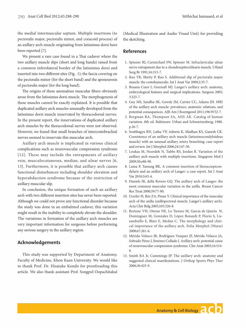
Fig. 1. Dissected right axilla showing (A) duplicated axillary arch muscles (two variant muscular slips) originating from latissimus dorsi muscle, (B) schematic sketching, and (C) their aponeurotic insertions, considered as the axillary arch of langer. A1, short axillary arch muscular slip 1; A2, long axillary arch muscular slip 2; A1*, aponeurosis of A1; A2*, aponeurosis of A2; Co, coracobrachialis; De, deltoid; LD, latissimus dorsi; MdN, median nerve; PMa, pectoralis major (reflected); PMi, pectoralis minor; SeA, seratus anterior; ULN, ulnar nerve.
Anat Cell Biol 2012;45:288-290 Sitthichai Iamsaard, et al290
www.acbjournal.orghttp://dx.doi.org/10.5115/acb.2012.45.4.288
the medial intermuscular septum. Multiple insertions (to pectoralis major, pectoralis minor, and coracoid process) of an axillary arch muscle originating from latissimus dorsi have been reported [7].
We present a rare case found in a Thai cadaver where the two axillary muscle slips (short and long bands) raised from a common inferolateral border of the latissimus dorsi and inserted into two different sites (Fig . 1); the fascia covering on the pectoralis minor (for the short band) and the aponeurosis of pectoralis major (for the long band).
The origins of these anomalous muscular fibers obviously arose from the latissimus dorsi muscle. The morphogenesis of these muscles cannot be exactly explained. It is possible that duplicated axillary arch muscles unusually developed from the latissimus dorsi muscle innervated by thoracodorsal nerves. In the present report, the innervations of duplicated axillary arch muscles by the thoracodorsal nerves were not observed. However, we found that small branches of intercostobrachial nerves seemed to innervate this muscular arch.
Axillary arch muscle is implicated in various clinical complications such as neurovascular compression syndrome [12]. These may include the entrapments of axillary vein, musculocutaneous, median, and ulnar nerves [6, 13]. Furthermore, it is possible that axillary arch causes functional disturbances including shoulder elevation and hyperabduction syndrome because of the restriction of axillary muscular slip.
In conclusion, the unique formation of such an axillary arch with two different insertion sites has never been reported. Although we could not prove any functional disorder because the study was done in an embalmed cadaver, this variation might result in the inability to completely elevate the shoulder. The variations in formation of the axillary arch muscles are very important information for surgeons before performing any serious surgery in the axillary region.
Acknowledgements
This study was supported by Department of Anatomy, Faculty of Medicine, Khon Kaen University. We would like to thank Prof. Dr. Hisatake Kondo for proofreading this article. We also thank assistant Prof. Songpol Oopachitakul
(Medical Illustration and Audio Visual Unit) for providing the sketching.
References
1. Spinner RJ, Carmichael SW, Spinner M. Infraclavicular ulnar nerve entrapment due to a chondroepitrochlearis muscle. J Hand Surg Br 1991;16:315-7.
2. Rao TR, Shetty P, Rao S. Additional slip of pectoralis major muscle: the costohumeralis. Int J Anat Var 2009;2:35-7.
3. Besana-Ciani I, Greenall MJ. Langer's axillary arch: anatomy, embryological features and surgical implications. Surgeon 2005; 3:325-7.
4. Guy MS, Sandhu SK, Gowdy JM, Cartier CC, Adams JH. MRI of the axillary arch muscle: prevalence, anatomic relations, and potential consequences. AJR Am J Roentgenol 2011;196:W52-7.
5. Bergman RA, Thompson SA, Afifi AK. Catalog of human variation. 8th ed. Baltimore: Urban and Schwartzenberg; 1988. p.26-7.
6. Soubhagya RN, Latha VP, Ashwin K, Madhan KS, Ganesh CK. Coexistence of an axillary arch muscle (latissimocondyloideus muscle) with an unusual axillary artery branching: case report and review. Int J Morphol 2006;24:147-50.
7. Loukas M, Noordeh N, Tubbs RS, Jordan R. Variation of the axillary arch muscle with multiple insertions. Singapore Med J 2009;50:e88-90.
8. Lama P, Tamang BK. A common insertion of thoracoepicon-dylaris and an axillary arch of Langer: a case report. Int J Anat Var 2010;3:63-4.
9. Daniels IR, della Rovere GQ. The axillary arch of Langer: the most common muscular variation in the axilla. Breast Cancer Res Treat 2000;59:77-80.
10. Ucerler H, Ikiz ZA, Pinan Y. Clinical importance of the muscular arch of the axilla (axillopectoral muscle, Langer's axillary arch). Acta Chir Belg 2005;105:326-8.
11. Bertone VH, Ottone NE, Lo Tartaro M, García de Quirós N, Dominguez M, Gonzalez D, López Bonardi P, Florio S, Lis-sandrello E, Blasi E, Medan C. The morphology and clini-cal importance of the axillary arch. Folia Morphol (Warsz) 2008;67:261-6.
12. Mérida-Velasco JR, Rodríguez Vazquez JF, Mérida Velasco JA, Sobrado Pérez J, Jiménez Collado J. Axillary arch: potential cause of neurovascular compression syndrome. Clin Anat 2003;16:514-9.
13. Smith RA Jr, Cummings JP. The axillary arch: anatomy and suggested clinical manifestations. J Orthop Sports Phys Ther 2006;36:425-9.