durian seed as a potential substrate …eprints.usm.my/31796/1/fairuzdzah_binti_ahmad_lothfy.pdffor...
TRANSCRIPT
i
DURIAN SEED AS A POTENTIAL SUBSTRATE
FOR BOLUS IN RADIOTHERAPY
FAIRUZDZAH BINTI AHMAD LOTHFY
UNIVERSITI SAINS MALAYSIA
2015

i
DURIAN SEED AS A POTENTIAL SUBSTRATE FOR
BOLUS IN RADIOTHERAPY
By
FAIRUZDZAH BINTI AHMAD LOTHFY
Thesis submitted in fulfilment of the requirements
for the Master of Science
JUN 2015
ii
BIJI DURIAN SEBAGAI POTENSI BAHAN UNTUK
BOLUS DALAM RADIOTERAPI
Oleh
FAIRUZDZAH BINTI AHMAD LOTHFY
Tesis yang diserahkan bagi memenuhi keperluan Sarjana Sains
JUN 2015
iii
ACKNOWLEDGMENT
I would like to express my thankfulness to Almighty God for giving me an opportunity to
finish this study.
This research project would not have been possible without the support of many people. I
would like to express my sincere gratitude to my supervisor, Dr Iskandar Shahrim bin Mustafa
for his scientific advice, for always finding time for my problems, for sharing his scientific
experience, support and supervision. I also wish to thank my co-supervisor Dr Sivamany
Kandaiya for her support in my work.
I also would also like to convey thanks to the Higher Education Ministry and Universiti
Teknologi Mara for providing the financial support during my study.
I would like to thanks Alurtron Manager, Encik Zahidee and his staff Encik Shaari from
Malaysia Nuclear Agency for their significant technical assistance. I also thank Dr. Zary
Shariman for helping me in my research study to give permission to use Biology School
equipment.
Most of all I thank to my husband Zakri for his love, encouragement and most valuable
support and my dear children, Danysh Asyraf Luqman, Damya Qalesya and Damya Qaisara
Soffiyea for an abundance of joyful moments.
ii

iv
iii
TABLE OF CONTENTS
Page
ACKNOWLEDGMENT ii
TABLE OF CONTENTS iii
LIST OF TABLES vi
LIST OF FIGURES vii
LIST OF SYMBOLS x
ABSTRAK xiii
ABSTRACT xv
CHAPTER 1: INTRODUCTION
1.1 Background 1
1.2 Scope of study 3
1.3 Problem statement 4
1.4 Research Objectives 5
1.5 Significant of Study 5
CHAPTER 2: LITERATURE REVIEW
2.1 Important of bolus in radiotherapy 7
2.2 Characteristics of bolus 11
2.3 Current bolus technology in radiotherapy 12
2.4 Durian seed gum composition and properties 14

iv
2.5 Polyvinyl alcohol improve mechanical properties of natural polymer 19
2.6 Effect of electron beam on polymer 22
2.7 Human body composition 29
2.8 Tissue equivalent properties 31
2.9 Interaction radiation with material
31
CHAPTER 3: MATERIALS AND EXPERIMENTAL WORK
3.1 Materials 34
3.2 Instrumentations 34
3.3 Methodology 34
3.3.1 Durian seed gum preparation 35
3.3.2 Bolus production 36
3.4 Validation the characteristics of fabricated bolus 37
3.4.1 Measurement mechanical properties 37
3.4.2 Mass density and electron density measurement 37
3.4.3 Transmission factor
39
CHAPTER 4: RESULTS AND DISCCUSIONS
4.1 Mechanical properties of material 42
4.1.1 Tensile strength 42
4.1.2 Strain 47
4.1.3 Young‟s Modulus 50

v
4.2 Dosimetric properties 51
4.2.1 Mass density 52
4.2.2 Electron density 58
4.2.3 Transmission factor 63
CHAPTER 5: CONCLUSIONS AND FUTURE WORK
5.1 Conclusions 70
5.2 Recommendations for Future Work
71
REFERENCES
72
APPENDICES
Appendix A 78
Appendix B 79
Appendix C 81
Appendix D 82
Appendix E 83
Appendix F 85
vi
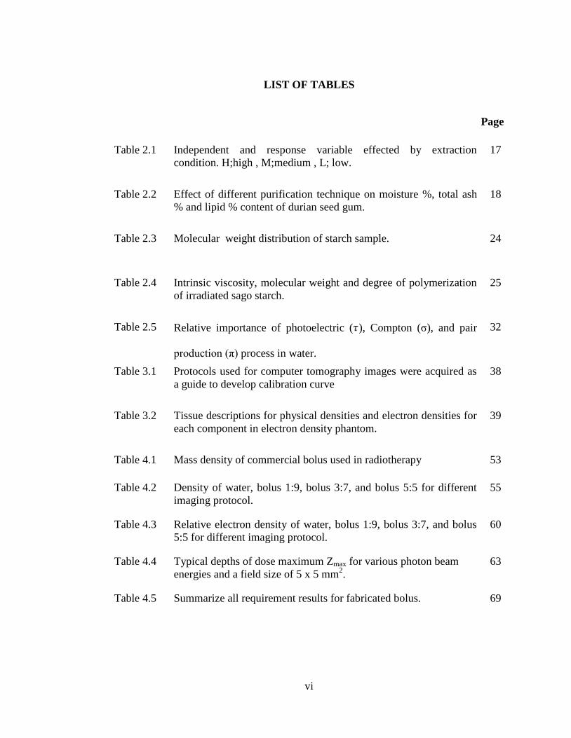
LIST OF TABLES
Page
Table 2.1 Independent and response variable effected by extraction
condition. H;high , M;medium , L; low.
17
Table 2.2 Effect of different purification technique on moisture %, total ash
% and lipid % content of durian seed gum.
18
Table 2.3 Molecular weight distribution of starch sample. 24
Table 2.4 Intrinsic viscosity, molecular weight and degree of polymerization
of irradiated sago starch.
25
Table 2.5 Relative importance of photoelectric ( ), Compton (σ), and pair
production (π) process in water.
32
Table 3.1 Protocols used for computer tomography images were acquired as
a guide to develop calibration curve
38
Table 3.2 Tissue descriptions for physical densities and electron densities for
each component in electron density phantom.
39
Table 4.1 Mass density of commercial bolus used in radiotherapy 53
Table 4.2 Density of water, bolus 1:9, bolus 3:7, and bolus 5:5 for different
imaging protocol.
55
Table 4.3 Relative electron density of water, bolus 1:9, bolus 3:7, and bolus
5:5 for different imaging protocol.
60
Table 4.4 Typical depths of dose maximum Zmax for various photon beam
energies and a field size of 5 x 5 mm2.
63
Table 4.5 Summarize all requirement results for fabricated bolus. 69
vii
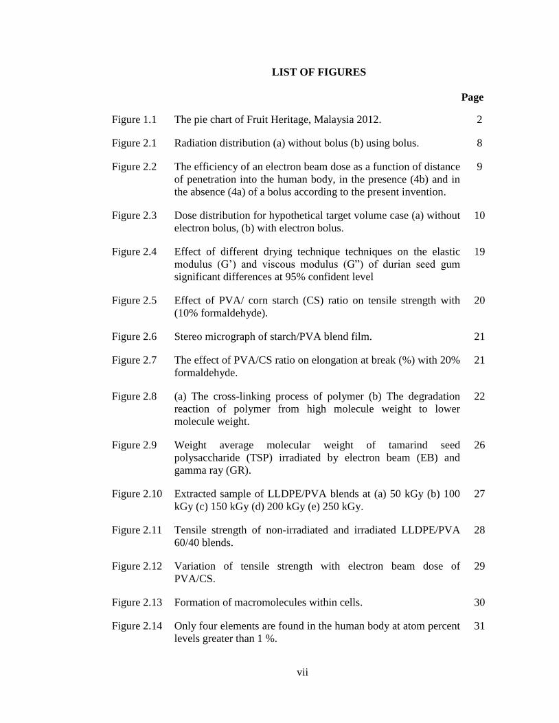
4 LIST OF FIGURES
Page
Figure 1.1 The pie chart of Fruit Heritage, Malaysia 2012. 2
Figure 2.1 Radiation distribution (a) without bolus (b) using bolus. 8
Figure 2.2 The efficiency of an electron beam dose as a function of distance
of penetration into the human body, in the presence (4b) and in
the absence (4a) of a bolus according to the present invention.
9
Figure 2.3 Dose distribution for hypothetical target volume case (a) without
electron bolus, (b) with electron bolus.
10
Figure 2.4 Effect of different drying technique techniques on the elastic
modulus (G‟) and viscous modulus (G”) of durian seed gum
significant differences at 95% confident level
19
Figure 2.5 Effect of PVA/ corn starch (CS) ratio on tensile strength with
(10% formaldehyde).
20
Figure 2.6 Stereo micrograph of starch/PVA blend film. 21
Figure 2.7 The effect of PVA/CS ratio on elongation at break (%) with 20%
formaldehyde.
21
Figure 2.8 (a) The cross-linking process of polymer (b) The degradation
reaction of polymer from high molecule weight to lower
molecule weight.
22
Figure 2.9 Weight average molecular weight of tamarind seed
polysaccharide (TSP) irradiated by electron beam (EB) and
gamma ray (GR).
26
Figure 2.10 Extracted sample of LLDPE/PVA blends at (a) 50 kGy (b) 100
kGy (c) 150 kGy (d) 200 kGy (e) 250 kGy.
27
Figure 2.11 Tensile strength of non-irradiated and irradiated LLDPE/PVA
60/40 blends.
28
Figure 2.12 Variation of tensile strength with electron beam dose of
PVA/CS.
29
Figure 2.13 Formation of macromolecules within cells. 30
Figure 2.14 Only four elements are found in the human body at atom percent
levels greater than 1 %.
31
viii
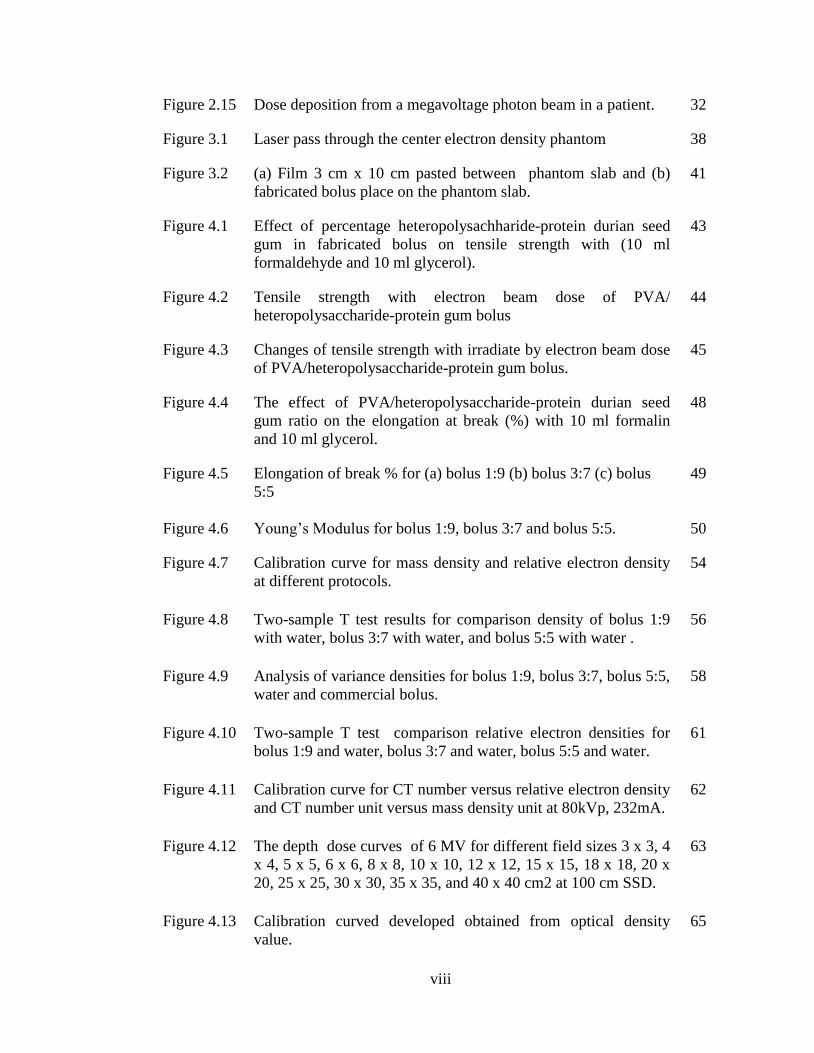
Figure 2.15 Dose deposition from a megavoltage photon beam in a patient. 32
Figure 3.1 Laser pass through the center electron density phantom 38
Figure 3.2 (a) Film 3 cm x 10 cm pasted between phantom slab and (b)
fabricated bolus place on the phantom slab.
41
Figure 4.1 Effect of percentage heteropolysachharide-protein durian seed
gum in fabricated bolus on tensile strength with (10 ml
formaldehyde and 10 ml glycerol).
43
Figure 4.2 Tensile strength with electron beam dose of PVA/
heteropolysaccharide-protein gum bolus
44
Figure 4.3 Changes of tensile strength with irradiate by electron beam dose
of PVA/heteropolysaccharide-protein gum bolus.
45
Figure 4.4 The effect of PVA/heteropolysaccharide-protein durian seed
gum ratio on the elongation at break (%) with 10 ml formalin
and 10 ml glycerol.
48
Figure 4.5 Elongation of break % for (a) bolus 1:9 (b) bolus 3:7 (c) bolus
5:5
49
Figure 4.6 Young‟s Modulus for bolus 1:9, bolus 3:7 and bolus 5:5. 50
Figure 4.7 Calibration curve for mass density and relative electron density
at different protocols.
54
Figure 4.8 Two-sample T test results for comparison density of bolus 1:9
with water, bolus 3:7 with water, and bolus 5:5 with water .
56
Figure 4.9 Analysis of variance densities for bolus 1:9, bolus 3:7, bolus 5:5,
water and commercial bolus.
58
Figure 4.10 Two-sample T test comparison relative electron densities for
bolus 1:9 and water, bolus 3:7 and water, bolus 5:5 and water.
61
Figure 4.11 Calibration curve for CT number versus relative electron density
and CT number unit versus mass density unit at 80kVp, 232mA.
62
Figure 4.12 The depth dose curves of 6 MV for different field sizes 3 x 3, 4
x 4, 5 x 5, 6 x 6, 8 x 8, 10 x 10, 12 x 12, 15 x 15, 18 x 18, 20 x
20, 25 x 25, 30 x 30, 35 x 35, and 40 x 40 cm2 at 100 cm SSD.
63
Figure 4.13 Calibration curved developed obtained from optical density
value.
65

ix
Figure 4.14 Four efficiencies curves as a function of depth of penetration of
photon for the source of 6 MeV after using all fabricated bolus;
bolus 1:9, bolus 3:7, bolus 5:5 on the surface on slab phantom,
and open field.
66
Figure 4.15 Two-sample T test results by comparing transmission factor of
water phantom 2967 with bolus 1:9, bolus 3:7, and bolus 5:5.
67

x
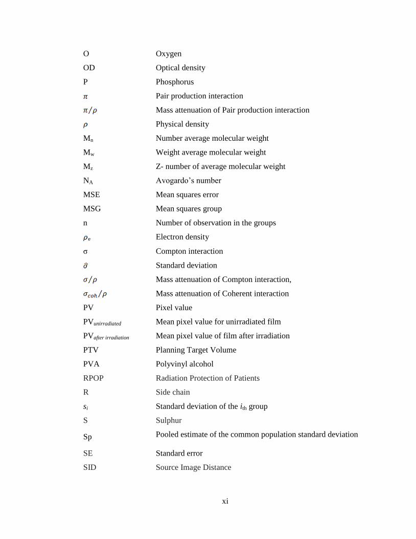
5 LIST OF SYMBOLS
A Cross sectional area of the sample
B N-Containing base
C Carbon
CS Corn Starch
CT Computed Tomography
DDS
Dehulled Durian Seed
DP
Degree polymerization
dpi Dots per inch
Dmax Dose at reference depth of maximum dose
Ds Surface dose at beam entrance side
Dd Dose at any depth
Dex Surface dose at the beam exit side
DNA Deoxyribonucleic acid
E Young's modulus
EB
Electron beam
F
Force
oF Fahrenheit
FDA Food and Drug Administration
FSCF Field size correction factor
G‟ Elastic modulus
G‟‟ Viscous modulus
GR Gamma ray
H Hydrogen
HU Hounsfield Unit
H2O Water
k Number of groups
LLDPE Linear low-density polyethylene
MU Monitor unit
N Nitrogen
xi
O Oxygen
OD Optical density
P Phosphorus
Pair production interaction
Mass attenuation of Pair production interaction
Physical density
Mn Number average molecular weight
Mw Weight average molecular weight
Mz Z- number of average molecular weight
NA Avogardo‟s number
MSE Mean squares error
MSG Mean squares group
n Number of observation in the groups
e Electron density
σ Compton interaction
Standard deviation
Mass attenuation of Compton interaction,
Mass attenuation of Coherent interaction
PV Pixel value
PVunirradiated Mean pixel value for unirradiated film
PVafter irradiation Mean pixel value of film after irradiation
PTV Planning Target Volume
PVA Polyvinyl alcohol
RPOP Radiation Protection of Patients
R Side chain
si Standard deviation of the ith group
S Sulphur
Sp Pooled estimate of the common population standard deviation
SE Standard error
SID Source Image Distance
xii
SSE Sum of square error
SSG Sum of square group
TSP Tamarind seed polysaccharide
τ Photoelectric
τ⁄ρ Mass attenuation of Photoelectric interaction
µ Linear attenuation coefficient
µm Mass absorption coefficient
Total mass attenuation coefficient
WDS Whole durian seed
Zeff Effective atomic number

xiii
BIJI DURIAN SEBAGAI POTENSI BAHAN UNTUK
BOLUS DALAM RADIOTERAPI
ABSTRAK
Tiga jenis bolus telah dihasilkan dengan menggunakan tiga jenis komposisi
dengan beza nisbah polivinil alkohol (PVA) terhadap gam biji durian. Nisbah PVA
kepada gam biji durian untuk bolus jenis pertama ialah 1:9; 3:7 dan 5:5. Bolus yang
dihasilkan diciri berdasarkan kepada sifat-sifat mekanikal seperti kekuatan
ketegangan, terikan, dan kelenturan. Akhirnya, kesemua bolus yang dihasilkan
mempunyai kekuatan ketegangan yang kecil. Kekuatan tegangan maksimum yang
boleh dicapai untuk bolus 1:9 ialah 0.083 MPa, bolus 3:7 ialah 0.323 MPa dan bolus
5:5 ialah 0.786 MPa. Peratus terikan bagi setiap jenis bolus yang dihasilkan memenuhi
keperluan minimum iaitu melebihi 50 %. Kesemua bolus yang dihasilkan mempunyai
nilai Modulus Young yang rendah di mana memenuhi satu keperluan bagi bolus yang
digunakan dalam radioterapi iaitu kurang daripada 100 MPa. Nilai ini penting untuk
menghasilkan bolus dengan kelenturan yang baik. Bolus yang dihasilkan perlu
mempunyai sifat kesetaraan tisu. Justeru, adalah satu keperluan untuk mengukur
ketumpatan fizikal, ketumpatan elektron relatif dan faktor transmisi. Nilai ketumpatan
fizikal untuk bolus 1:9 ialah 1.07 ± 0.01 g cm-3
, bolus 3:7 ialah 1.00 ± 0.01 g cm-3
dan bolus 5:5 ialah 1.00 ± 0.01 g cm-3
. Ketumpatan bolus 1:9 berbeza dengan
ketumpatan air sebanyak 7 %. Bolus 3:7 dan bolus 5:5 tidak mempunyai beza ketara
dengan ketumpatan air. Walau bagaimanapun, dengan membandingkan semua

xiv
ketumpatan bolus yang dihasilkan dengan senarai ketumpatan bolus komersial, nilai-
nilai tidak menunjukkan beza ketara terhadap ketumpatan. Ketumpatan elektron relatif
untuk bolus 1:9 ialah 1.028 ± 0.019, bolus 3:7 ialah 1.002 ± 0.003 dan bolus 5:5 ialah
1.002 ± 0.002. Dengan membandingkan ketumpatan elektron relatif bolus 1:9 dengan
ketumpatan elektron relatif air ia menunjukkan perbezaan ketara manakala bolus 3:7
dan bolus 5:5 menunjukkan tiada beza ketara. Faktor transmisi diukur dengan
mengira peratus kedalaman dos (PDD) pada kedalaman dos maksimum selepas
menggunakan bolus. PDD pada kedalaman dos maksimum untuk bolus 1:9 ialah
85.63%, bolus 3:7 ialah 98.13%, dan bolus 5:5 ialah 98.88%. Perbandingan faktor
transmisi bolus 3:7 dan bolus 5:5 dengan phantom air 2967 menunjukkan tiada
perbezaan ketara dan faktor transmisi bolus 1:9 dan phantom air 2967 menunjukkan
perbezaan ketara.

xv
DURIAN SEED AS A POTENTIAL SUBSTRATE FOR
BOLUS IN RADIOTHERAPY
ABSTRACT
Three types of bolus were fabricated using three types of composition with
different ratios of polyvinyl alcohol (PVA) to durian seed gums. The ratio of PVA to
durian seed gum were 1:9; 3:7 and 5:5. The fabricated bolus was characterized based
on the mechanical properties such as tensile strength, strain, and flexibility.
Eventually, all bolus composition that were prepared had low tensile strength. The
maximum tensile strength for bolus 1:9 was 0.083 MPa, bolus 3:7 was 0.323 MPa and
bolus 5:5 was 0.786 MPa. The strain of every type of fabricated bolus fulfilled the
minimum requirement which was more than 50%. All types of bolus had less than 100
MPa value of Young‟s Modulus which fulfilled one of the requirements for bolus to
be used in radiotherapy. This value was important to produce bolus with good
flexibility. The fabricated bolus had tissue equivalent properties. Hence, physical
density, relative electron density and transmission factor were measured. Physical
densities for bolus 1:9 was 1.07 ± 0.01 g cm-3
, bolus 3:7 was 1.00 ± 0.01 g cm-3
and
bolus 5:5 was 1.00 ± 0.01 g cm-3
. The density bolus 1:9 differed from water density by
7 %. However, density bolus 3:7 and bolus 5:5 had no significant difference with
water density. All types of fabricated bolus density were compared with a list of
commercial bolus density. The values did not show significant difference towards the
density. Relative electron density for bolus 1:9 was 1.028 ± 0.019, bolus 3:7 was

xvi
1.002 ± 0.003 and bolus 5:5 was 1.002 ± 0.002. There was a significant difference in
electron density between bolus 1:9 and water. However, bolus 3:7 and bolus 5:5
showed no significant differences. Transmission factor was measured by calculating
the percentage depth dose (PDD) of the maximum depth dose.. PDD of the maximum
depth dose for bolus 1:9 was 85.63%, bolus 3:7 was 98.13%, and bolus 5:5 was
98.88%. The comparison of transmission factor for bolus 3:7 and bolus 5:5 with
water phantom type 2967 showed no significant difference, and transmission factor of
with bolus 1:9 and water type 2967 showed significant difference.
1
6 CHAPTER 1
7 INTRODUCTION
1.1 Background
In radiotherapy, the correct amount of radiation is desired to extinguish the
abnormal tissue, like cancer in head and neck, brain, breast, cervical, prostate and
skin cancer. It is capable to devastate some benign diseases (Radiation Protection of
Patients, 2013).
The radiotherapy treatment inadvertently to gives exposure to normal tissues. A
case in point is patients may get the effect to damage of the normal tissue during the
course of therapy at a few weeks, months or years after therapy. As an example, the
salivary glands are often affected when radiotherapy is given for head and neck
cancer. Rectums are often affected by pelvic irradiation for treatment of prostate and
cervical, skin, mucosa, subcutaneous tissues, and bone cancer (Stone et al., 2003).
Megavoltage photon radiotherapy penetrates the skin to irradiate deep-seated
tumors, with skin-sparing property. Consequently, one must be a layer on the skin to
act as the skin surface as purpose to treat superficial lesions. Hence, bolus is used in
order to deliver the full prescribed dose to the skin surface and underlying tissue (Vyas
et al., 2013). Bolus is also applied to the surface of a patient to compensate irregular
patient contour, negating skin sparing, and extra attenuating. Bolus is put in close
contact with the human body to correct for the distribution of absorbed dose in
radiotherapy.
2
The required properties for the bolus are Young‟s Modulus of less than 100
MPA, ability to withstand 50 % strain and the substance must have radiation
properties equivalent to human body tissue. The standard bolus should not sag during
treatment. Basically, bolus should have good plasticity, flexibility and adhesion to a
living body besides free from air bubble (Cooney et al., 2006; U.S Patent no.
6,231,858 B1, 2001).
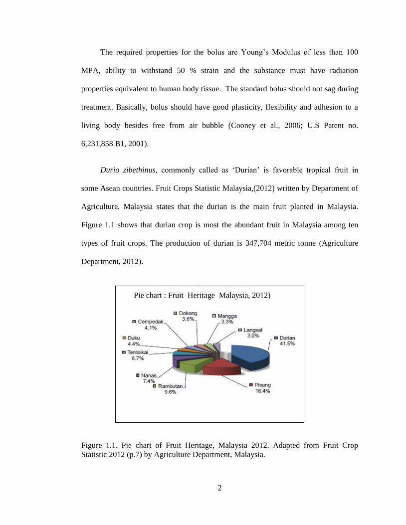
Durio zibethinus, commonly called as „Durian‟ is favorable tropical fruit in
some Asean countries. Fruit Crops Statistic Malaysia,(2012) written by Department of
Agriculture, Malaysia states that the durian is the main fruit planted in Malaysia.
Figure 1.1 shows that durian crop is most the abundant fruit in Malaysia among ten
types of fruit crops. The production of durian is 347,704 metric tonne (Agriculture
Department, 2012).
Figure 1.1. Pie chart of Fruit Heritage, Malaysia 2012. Adapted from Fruit Crop
Statistic 2012 (p.7) by Agriculture Department, Malaysia.
Pie chart : Fruit Heritage Malaysia, 2012)
3
Durian seed consists aproximately 20 % to 25% of the total fruit mass (Amid et al.,
2012) and has 54.90 % moisture, 1.58 % ash, 3.40 % protein, 1.32 % fat and 18.92 %
starch in durian seed (Srianta et al., 2012). The biopolymer from durian seed consists
of a heteropolysaccharide-protein structure (Amid & Mirhosseini, 2012a). Natural
biopolymer from plant sources provides variety of functional properties and has more
advantages than synthetic polymer. It is due to its biocompatibility, non-toxicity, and
inexpensive properties (Navaratne & Nawarathne, 2014). Chemical composition and
molecular structure of the polymer induce the physiochemical and functional
properties of natural plant-based biopolymer (Amid et al., 2012). The processes such
as extraction, purification, drying and/or further modification significantly affects the
chemical composition, molecular structure, rheological properties and viscoelastic
behavior. As a consequence, it will involve the functional properties of biopolymers
(Amid & Mirhosseini, 2012a; Amid & Mirhosseini, 2012b). Therefore, this
modification allows the polysaccharide-protein biopolymer from Durio zibethinus
seed to be a new substrate for bolus production. This study will focus on the
fabrication of the bolus for radiotherapy by using a new substrate which is durian seed.
1.2 Scope of study
The scope of this study was to make the durian seed as a new substrate for bolus
in radiotherapy. Bolus was fabricated by applying the the blended durian seed gum
and polyvinyl alcohol (PVA). The best methods such as extraction, purification and
drying of the seeds have to be selected to get it as an appropriate gum for producing
the bolus. All of these methods can influence the chemical composition, molecular
4
structure, rheological properties and viscoelastic behavior from the durian seed gum
(Amid et al., 2012; Amid & Mirhosseini, 2012b, 2012c).
In general, the fabricated bolus must be equivalent to human body tissue,
homogeneous, excellent plasticity, good form-compatibility and adhesive to the
human body. Other criteria such as toxicity level are important due to direct contact of
bolus on human skin surface during treatment. The fabricated bolus must fulfill all the
aforementioned properties and conditions.
1.3 Problem Statement
Requirement of consumable materials that are disposed such as bolus was
practiced as a way to prevent bacterial and viral infections in the hospital through
medical apparatus and instruments. Although the price of conventional boluses using
plastics, paraffin, silicone, or synthetic rubbers as their base material is very high, but
they must be disposed as industrial waste. This is due to that, used boluses cannot be
disposed effortlessly without treatment. It is impracticable to dispose the bolus after
only one use due to high cost. If water is used as a bolus material, the container
enclosing water is difficult to discard (U.S. Patent no. 6,231,858 B1, 2001).
Nowadays, many types of synthetic polymers are used to produce bolus such as
elastometric polymer, synthetic rubber and paraffin (U.S. Patent no. US 6,231,858 B1,
2001). Most of these substances originate from petroleum and most of the
conventional ones are regarded as non-renewable petroleum sources. Petroleum
5
resources are limited and the use of non-degradable polymers causes environmental
problems (Shafik et al., 2014).
Durio zibethinus is a commercial farming industry. Approximately 20 % of
durian mostly thrown away after consumption (Amid et al., 2012). Natural polymers
are suitable sources for their inherent biodegradability, accessibility, and relatively
low cost. Thus, crop waste such as durian seed can be used to develop a new product.
1.4 Research Objectives
The objectives of this study are:
1. To fabricate a bolus for radiotherapy by using durian seed as a new
substrate.
2. To ensure that the fabricated bolus satisfies the dosimetric properties,
physical and mechanical characteristics needed.
1.5 Significant of Study
Natural organic polymers such as Durio zibethinus seeds, ordinarily be as crop
waste after consumption. Hence, this crop waste can be a new resource of raw material
which encourages development of value added product. The features of Durio
zibethinus seeds make it appropriate as the bolus for radiotherapy.

6
In addition, by using the Durio zibethinus seed to replace the current material
used as bolus, the cost for producing the bolus will be inexpensive. In addition, the
natural organic polymers will preserve the petrochemical resources and reduce
environmental impact.

7
8 CHAPTER 2
9 LITERATURE REVIEW
2.1 Importance of bolus in radiotherapy
Megavoltage photon, such as gamma-rays or x-rays causes deoxyribonucleic
acid (DNA) devastation of cell in target area in radiotheraphy. The most familiar
radiation treatment used is external radiation from the machine outside out of the
patient‟s body such as a linear accelerator (U.S. Patent no. 2008/0123810 A1,
2008).
By destroying the genetic material that controls how the cells develop and
divide, the radiation is able to trigger cell injury and devastation. By using the
higher energy radiation, both healthy and malignant cells are destroyed (American
Cancer Society, 2014).
Radiotherapy treats a target volume, which can be allocated at the breast or
thoracic wall, the axillary region, the supraclavicular region, and the internal
mammary chain. The presence of healthy organs in close proximity of the target to
be treated can lead to side effect of radiation. It must be avoided especially the
lungs, the heart, the brachial plexus, the cervical spinal cord, the larynx and the
thyroid (U.S. Patent no. 2010/0070236 A1, 2010).
The main purpose of radiotherapy bolus is to equate the irregular patient
contour and provide a flat surface for normal beam incidence. Hence the bolus
consists of tissue equivalent material properties which is positioned directly on the

8
skin surface. Theoretically, the use of bolus is useful in radiotherapy treatment
(Podgorsak, 2005).
(a)
(b)
Figure 2.1: Radiation Distribution (a) without bolus (b) using bolus (U.S. Patent
no. 6231,858 B1, 2001).
Figures 2.1 (a) and 2.1 (b) illustrated the difference in dose distribution of
rays with or without a bolus in radiotherapy for head. The bolus controls dose
dissemination of X-rays to irradiate the maximum absorbed dose to the focus target
volume.
The second purpose of the bolus is to treat the lesions on or close to the skin
surface. Bolus material is placed on top of the skin region undergoing radiation
therapy to raise the radiation dosage at the skin surface. Hence, the aim to treat the
9
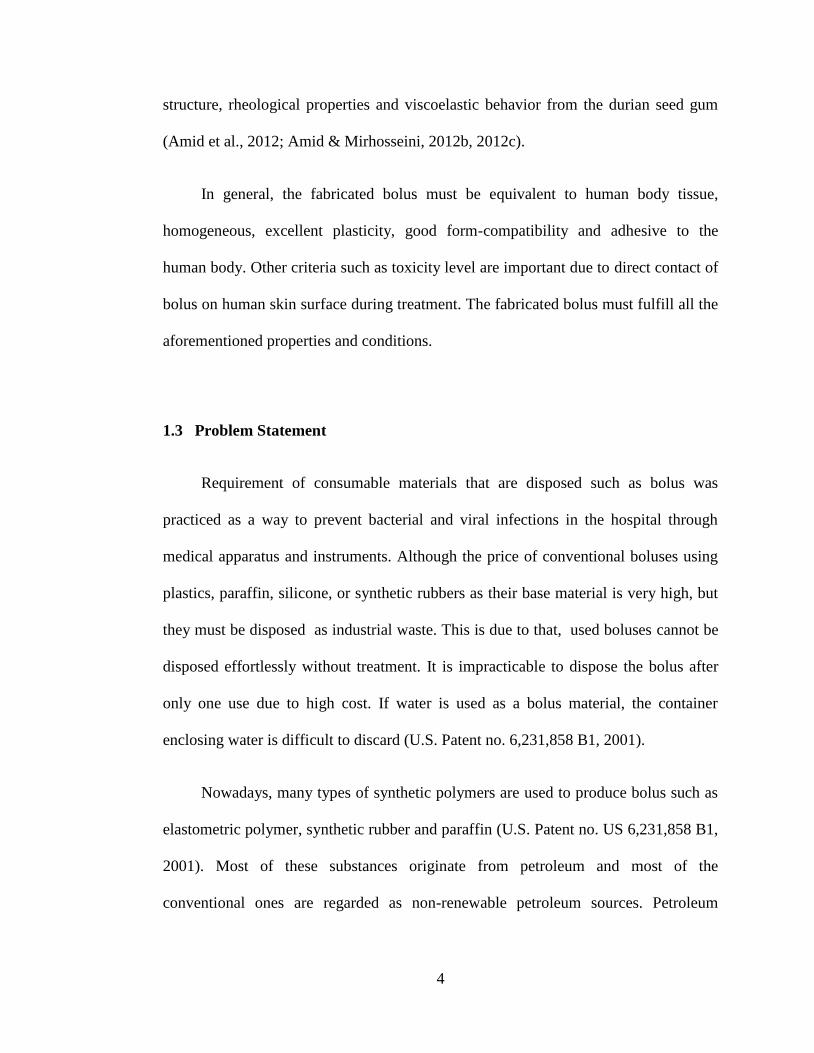
lesions on or close to the skin surface can be achieved by increasing dose on or
close the skin surface.
Figure 2.2: The efficiency of an electron beam dose as a function of distance of
penetration into the human body, in the presence (4b) and in the absence (4a) of a
bolus according to the present invention (U.S. Patent no. 2010/0070236 A1, 2010).
Figure 2.2 illustrated two efficiency curves 4a and 4b as a function of the
depth of penetration of the electron beam for the source of 9 MeV electrons and for
1 cm bolus. A bolus with tissue equivalent properties of 1 cm bring about the
maximum dose shifted about 1 cm. Thus, the maximum dose is achieved at the
depth of 1 cm, then the curve decreases rapidly. Consequently, it allows the
healthy organs close to the target is prevented from being damaged (U.S. Patent
no. 2010/0070236 A1, 2010).
10
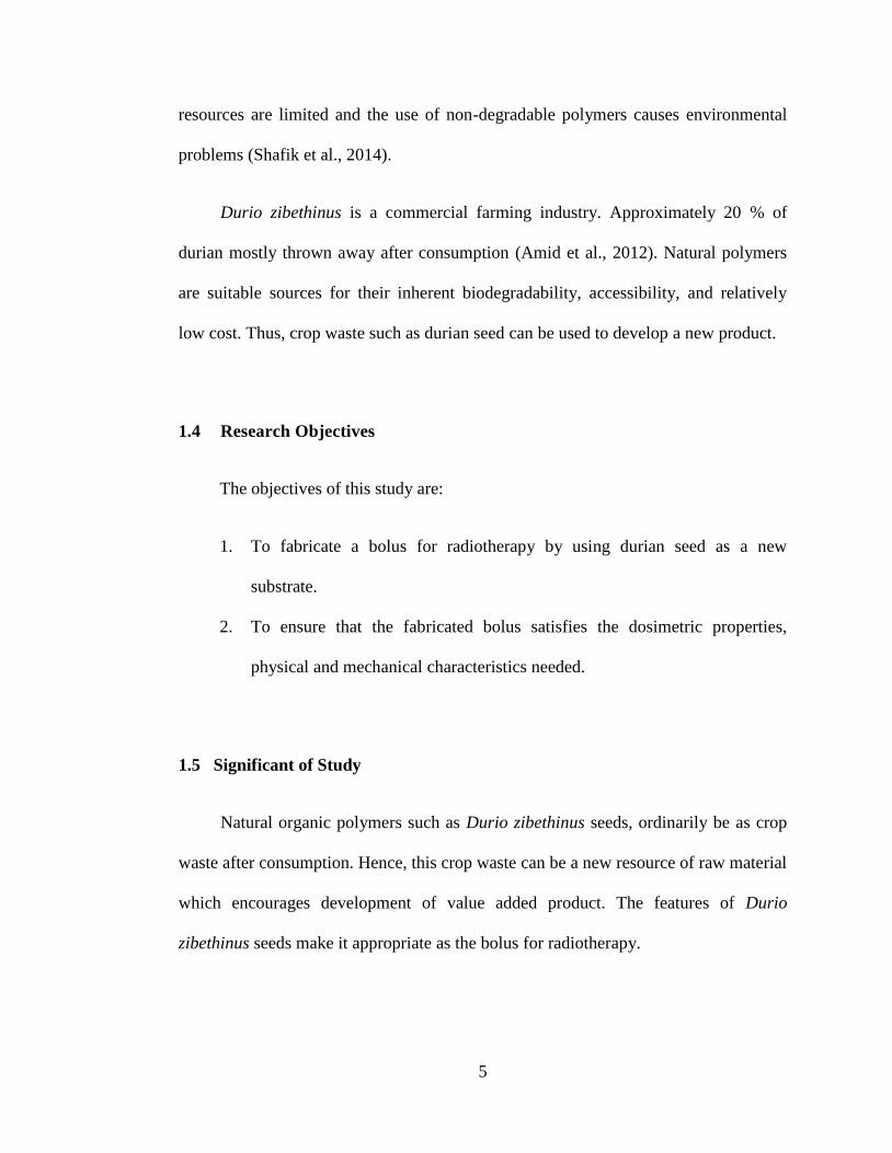
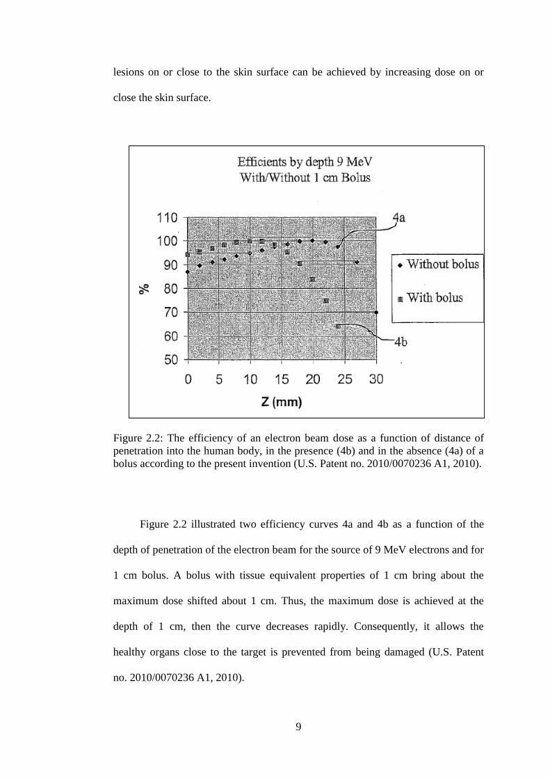
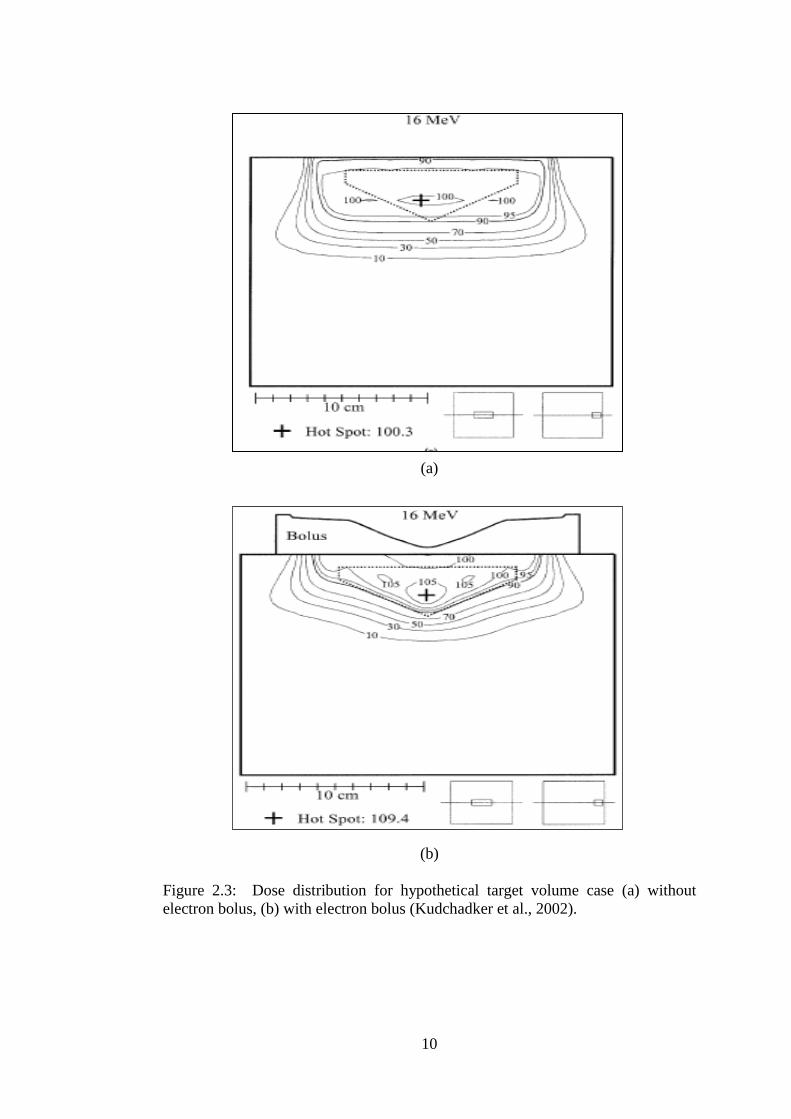
(a)
(b)
Figure 2.3: Dose distribution for hypothetical target volume case (a) without
electron bolus, (b) with electron bolus (Kudchadker et al., 2002).

11
Figure 2.3 (a) illustrated that planning target volume (PTV) is covered by
the 90 % isodose, and the dose variation is about 10 %. As a consequence of that, a
significant volume of healthy tissue outside the PTV is being exposed to higher
dose (> 90 %). Figure 2.3 (b), the 90 % isodose line now follows the distal
boundary of PTV. As a result, it minimizes the potential of damage the healthy
tissue outside the PTV (Kudchadker et al., 2002). However, tumors will receive
needed doses in an actual situation for the patient without bolus, but normal tissue
received overdoses. It is therefore essential to use electron bolus to compensate for
above irregular. Hence, to make sure that the required dose is received by the
patient, bolus with adequate thickness must be placed correctly. Bolus
requirements must be clearly documented to ensure the delivered doses to patient
are homogeneneous (Kudchadker et al., 2002).
2.2 Characteristics of bolus
Commonly the usable bolus must fulfill the following properties and
conditions. For physical properties, the bolus should be homogenous, resilient, has
excellent plasticity, form-compatibility and adhesiveness to a living body. In
addition, the bolus materials are non- toxic, have even thickness and do not contain
air (U.S. Patent no. 6,231,858 B1, 2001).
The mechanical properties of bolus such as Young‟s Modulus must be less
than 100 MPa and ability to withstand over 50 % strain. Bolus materials must have
stability between 40 oF and 125
oF. The bolus should be odorless, non- tacky, non-
adherent, and its types of materials have been approved by Food and Drug
Administration (FDA) for human skin contact (Cooney et al., 2006).

12
Boluses should have tissue-equivalent (within 2 %) dosimetric properties. The
tissue equivalent properties need to comprise a density that is equal to water. Thus,
the density of bolus material can be about 1 g cm-3
. Alternative ways need to be
taken if the density value is not equal to 1 g cm-3
in order to achieve tissue
equivalent properties. The thickness of the bolus material needs to be adjusted
based on the ratio of thickness to obtain the desired tissue equivalent properties.
For instance, a bolus material of an oil gel comprising mineral oil and suitable
styrenic block copolymer have density between about 0.86 g cm-3
and 0.91 g cm-3
.
This bolus material may achieve the desired tissue-equivalent dosimetric properties
by adjusting the bolus thickness (U.S. Patent no 2008/0123810 A1, 2008).
Preferably, the bolus material should be checked by comparing the depth
dose distribution in the bolus and the water. If scaling factor is needed, it should be
documented and used in treatment planning while using the bolus (Khan &
P.Gibbons, 2014).
2.3 Current bolus technology in radiotherapy
Commercial materials that are often used as bolus are such as paraffin wax,
polystyrene, lucite, superstuff, and superflab. Superflab bolus is a good bolus. This
material has fulfilled the requirement of the bolus for radiotherapy which are
transparency, flexibility, and almost water equivalent (Khan & P. Gibbons, 2014).
All the bolus material must obtain Food Drug and Administration (FDA)
approval for human contact. Bolus consists of pre-formed gel sheets that are
draped over the target area and usually comes in form of ready made such as
13
superflab and elasto-gel. Bolus also comes in form of powders and mix with water,
then molded onto the target area where they solidify such as Super Stuff and
Aquaplast RT (Cooney et al., 2006).
Superflab bolus (Figure A.1) is the most popular, best confirming, not
expensive and non-sticking (Feaster, 2014). The composition of these bolus
materials of gels are vinyl plastic w/di-isodecyl phthalate and water-based gels
with acrylic polymer. It fulfills the requirements of producing bolus which are
flexible, have uniform thickness, and conform to the skin. The density of Superflab
bolus is 1.02 g cm-3
. Maximum strain for superflab bolus 1.6, tensile stress is 120
MPa, and Young‟s modulus 37 MPa at low strain to 97 MPa high strain (Cooney et
al., 2006). Superflab bolus material can be washed with soap and water, followed
by a talcum or corn starch that can be cut with scissors and approved for human
contact. Superflab bolus material is provided in varying thicknesses as to provide
maximum dose build-up for relevant photon energies. A superflab bolus needs to
be handled with a full care due to synthetic oils that can damage plastic surface
(Feaster, 2014).
Elasto-gel bolus (Figure A.2) has self-adhesive nature of gel material that
provides excellent skin contact without air gaps and maybe layered together for
different thicknesses (Radiation Products Design Inc., 2013). Elasto-gel bolus
consists of non-toxic material and transparent and does not have any changes on
the physical properties such as appearance and property of the elasto-gel pad after
irradiating with high doses (Chang et al., 1992). Elasto-gel bolus is easy to use
which can be easily cut into the desired size and shape, mildly adhesive, reusable
on the same patient, and conforms exceptionally well to body contours (JRT

14
Associates, 2012). Elasto-gel bolus is prepared by mixing the water, glycerine, and
acrylic polymer, which gels the whole mixture. Density elasto-gel bolus is 1.20 g
cm-3
(Chang et al., 1992).
Aquaplast RT bolus (Figure A.3) is used for single patient use. This bolus
is placed in hot water, then the material softens and can be molded to almost any
area of the body without air pockets. Aquaplast RT bolus material has a density
about 1.7 g cm-3
(JRT Associates, 2012).
2.4 Durian seed gum composition and properties
“Gum” is defined as a group of naturally occurring polysaccharides and / or
proteins initiate from different sources such as animal, plant and microbial (Amid
et al., 2012). Gums are substances contain of hydrophilic, long-chain, high
molecular weight molecules, commonly with colloidal properties, that in water-
based systems produce gels. It has highly viscous suspensions or solutions in low
dry-substance content. Ordinarily, primary purpose of gum for thickening and/or
gelatin (Hoefler, 2004). Natural plant gums are usually safe for oral consumption
due to their safety (non-toxic), low cost and availability (Amid et al., 2012). pH
value of the durian seed gum solution is 6.93 at room temperature, which are quite
similar with guar gum. It is stable in condition of pH between 2.0 to 10.0 (Amin et
al., 2007). The durian seed can be made up of 54.90 % moisture, 1.58 % ash, 3.40
% protein, 1.32 % fat and 18.92 % starch (Srianta et al., 2012). Amin et al (2007)
studied the composition of durian seed gum for the whole durian seed (WDS) flour
and dehulled durian seed (DDS) flour. This study found that WDS contains 6.5 %
water, 6.0 % protein, 3.1 % ash, 0.4 % fat, 10.1 % crude fiber and 73.9 %

15
carbohydrate. For DDS, it contains 6.6 % moisture, 7.6 % protein, 3.8 % ash, 0.4
% fat, 4.8 % crude fiber and 76.8 % carbohydrate (Amin & Arshad, 2009).
The highest monosaccharide in the carbohydrate composition of durian seed
gum are galactose (48.6-59.9 %), glucose (37.1-45.1 %), arabinose (0.58-3.41 %),
and xylose (0.3-3.21 %) (Amid et al., 2012). The large fatty acid of the lipid
fraction from the durian seed gum comprises palmitic acid, palmitoleic acid, stearic
acid, oleic acid, linoleic acid, and linolenic acid. The largest amino acids of durian
seed gum consists of leucine (30.9-37.3 %), lysine (6.04-8.36 %), aspartic acid
(6.10-7.19 %), glycine (6.07-7.42 %), alanine (5.24-6.14 %), glutamic acid (5.57-
7.09 %), valine (4.5-5.50 %), proline (3.87-4.81 %), serine (4.39-5.18 %),
threonine (3.44-6.50 %), isoleucine (3.30-4.07 %), and phenylalanine (3.11-9.04
%) (Amid et al., 2012).
Amin et al (2007) found that the main sugar of durian seed gum are
rhamnose, D-galactose, and glucose in the ratio of 3:1:9. This study found that
durian seed gum do not have galactomannan (Amin et al., 2007). The major
element in forming of gum in durian seed is L-rhamnose, which is a C-5
polysaccharide (Amin et al., 2007). Polysaccharide can form the structure of arrest
water molecules when rhamnose sugar contacts with water. As a result, a thick gel
is formed after arresting water molecules by C-5 polysaccharide (Navaratne and
Nawarathne, 2014).
The natural polymer from durian seed has more elasticity (gel- like behavior)
than viscous (liquid-like) characteristic at a low frequency (Amid & Mirhosseini,
2012c). The natural heteropolysaccharide/protein polymer from durian seed gum
has a relatively low solubility ranging from 9.1 % to 36.0 % (Amid & Mirhosseini,

16
2012c). It is caused by the presence of impurities, insoluble matter and large
particles present in the chemical structure of the natural polymer from the durian
seed gum. The viscosity of crude durian seed gum consists of diverse values from
3.3 mPa.s to 24.3 mPa.s reliant on the extraction condition. The protein content of
crude durian seed gum ranges from 3.8 % to 9.3 %, and it depends on chemical
extraction condition. Degree of elastic modulus (G‟) ranges between 1.48 Pa to
9.46 Pa and degree of viscous modulus (G‟‟) varies from 0.28 Pa to 4.51 Pa (Amid
& Mirhosseini, 2012c).
The functional characteristics of polysaccharide plant gums are governed by
the chemical composition, molecular weight, sequence of monosaccharide,
configuration of glycoside linkages, and the position of glicoside linkages in the
backbone and side chain (Amid et al., 2012). The composition, molecular
structure, rheological behavior and functional properties of durian seed depend on
the method of extraction condition, purification and drying process(Amid et al.,
2012; Amid & Mirhosseini, 2012a, 2012b, 2012c; Mirhosseini et al., 2013).
Chemically-extracted durian seed gum has a typical gel network and
revealed gel like behavior at low concentration (0.5 % w/w) and low frequency
(0.1 Hertz). Degree of gel-like or viscosity behavior depends on gum
concentration. When elastic modulus is higher than the viscous modulus, the
sample behaves more gel-like behavior and weak viscous behavior. Table 2.1
showed extraction condition effect extraction yield, viscosity, elastic modulus,
viscous modulus, and solubility (Amid & Mirhosseini, 2012a).

17
Table 2.1: Independent and response variable effected by extraction condition.
H;high , M;medium , L; low (Amid & Mirhosseini, 2012a).
Variable Decoloring times
(minutes)
Soaking times
(hours)
Soaking
temperatures (Co)
60 120 180 4 8 12 25 40 55
Extraction yield L M H L M H L M H
Viscosity H M L H M L M L H
Protein content P ≥ 0.05 H M L M H L
Elastic modulus L M H H M L -
Viscous modulus - H M L H M L
Solubility H M L L M H L M H
Based on a previous study, purified seed gum contains 17.9 % of moisture
content and 29.8 % total ash content (Amin et al., 2007). Ash content of durian
seed gum was higher compared to the others commercial gum and only 0.2 % of
the ash as water soluble (Amin et al., 2007). The ash will lead the natural gum to
less solubility. The effect of purification process to minimize the ash contents
(Table 2.2) (Amid et al., 2012).

18
Table 2.2: Effect of different purification technique on moisture %, total ash %
and lipid % content of durian seed gum. A; isopropanol and ethanol, B;
isopropanol and acetone, C; saturated barium hydroxide, D; Fehling solution
(Amid et al., 2012).
Test
Crude
gum
(Aqueous)
Crude gum
(Chemical)
Purified
gum A
Purified
gum B
Purified
gum C
Purified
gum D
Moisture % 26.8 ±
1.09a
24.6
±1.22ab
23.2 ±
1.03b
22.8 ±
0.79bc
20.5 ±
0.35c
21.7 ±
1.13bc
Total
ash %
32.8 ±
1.57a
34.3 ± 1.78a
20.6 ±
0.89b
23.4 ±
1.34b
15.8 ±
1.13c
12.1 ±
0.94d
Soluble
ash % 1.7 ± 0.03
a 1.5 ± 0.01
a
0.9 ±
0.00b
1.0 ±
0.14b
0.7 ±
0.03c
0.5 ±
0.06d
Lipid % 1.92 ±
0.07a
0.78 ± 0.11b
0.14 ±
0.04c
0.16 ±
0.02c
0.21 ±
0.06d
0.19 ±
0.03d
All drying methods lead to significantly lower the elasticity (G‟) and viscous
modulus (G”) of durian seed gum. The freeze-dried gum and oven-dried (105 0C)
gum reveal the highest and lowest viscous modulus (G‟). All drying techniques
significantly (p<0.05) change the dynamic viscoelastic properties durian seed gum.
All drying techniques show significant (p<0.05) reduction of both elastic (G‟) and
viscous modulus (G”) (Figure 2.4) (Amid & Mirhosseini, 2012c).

19
Figure 2.4: Effect of different drying technique techniques on the elastic modulus
(G‟) and viscous modulus (G”) of durian seed gum significant differences at 95 %
confident level (Amid and Mirhosseini, 2012b).
2.5 Polyvinyl alcohol improve mechanical properties of natural polymer
Polyvinyl alcohol (PVA) is a synthetic biodegradable polymer and possesses
excellent mechanical properties. PVA has physical properties such as adhesive
strength, tensile strength and flexibility. For instance , pure PVA films have tensile
modulus at 1.9 ± 0.2 GPa (Zhang et al., 2003). PVA has improved mechanical
properties of natural biopolymer since natural biopolymer have poor mechanical
properties.
20
Previous study on the different ratios of PVA to starch shows that it
improves the dimensional stability and strength (Sadhu et al., 2014). By increasing
the ratio of PVA to starch, the mechanical strength improves (Sadhu et al., 2014).
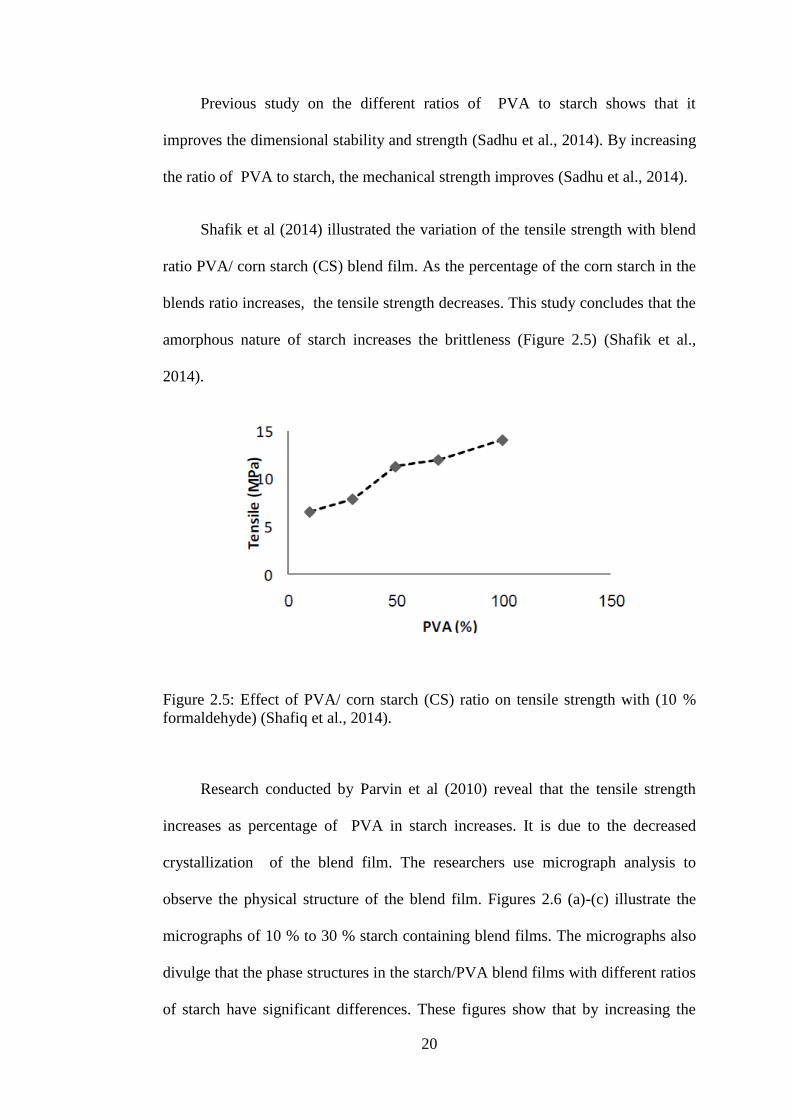
Shafik et al (2014) illustrated the variation of the tensile strength with blend
ratio PVA/ corn starch (CS) blend film. As the percentage of the corn starch in the
blends ratio increases, the tensile strength decreases. This study concludes that the
amorphous nature of starch increases the brittleness (Figure 2.5) (Shafik et al.,
2014).
Figure 2.5: Effect of PVA/ corn starch (CS) ratio on tensile strength with (10 %
formaldehyde) (Shafiq et al., 2014).
Research conducted by Parvin et al (2010) reveal that the tensile strength
increases as percentage of PVA in starch increases. It is due to the decreased
crystallization of the blend film. The researchers use micrograph analysis to
observe the physical structure of the blend film. Figures 2.6 (a)-(c) illustrate the
micrographs of 10 % to 30 % starch containing blend films. The micrographs also
divulge that the phase structures in the starch/PVA blend films with different ratios
of starch have significant differences. These figures show that by increasing the

21
concentration of starch from 10 % to 30 %, starch micro domains reverse to be
continued phase from disseminate phase, which implies that the amorphous starch
is partially miscible with PVA (Parvin et al., 2010).
Figure 2.6: Stereo micrograph of starch/PVA blend film (Parvin et al., 2010).
Shafik et al (2014) found that the smallest PVA weight percent shows the lowest
percentage of elongation at break for PVA/CS films. This is due to the amorphous
nature of starch led to a lower elongation at break of the films (Figure 2.7) (Shafik
et al., 2014).
Figure 2.7: The effect of PVA/CS ratio on elongation at break (%) with 20 %
formaldehyde (Shafiq et al., 2014).
22
2.6 Effect of electron beam on polymer
Electron beam is ionizing energy that is generally characterized by low
penetration and high dose rate. The use of higher dose rate causes less exposure
time and brought about reduction in potential degradation of polymers (Silindir &
Ozer, 2010).
Electron beam treatment can modify the mechanical properties of the
surfaces and interface of synthetic polymers and biopolymers (Schwars, 2010).
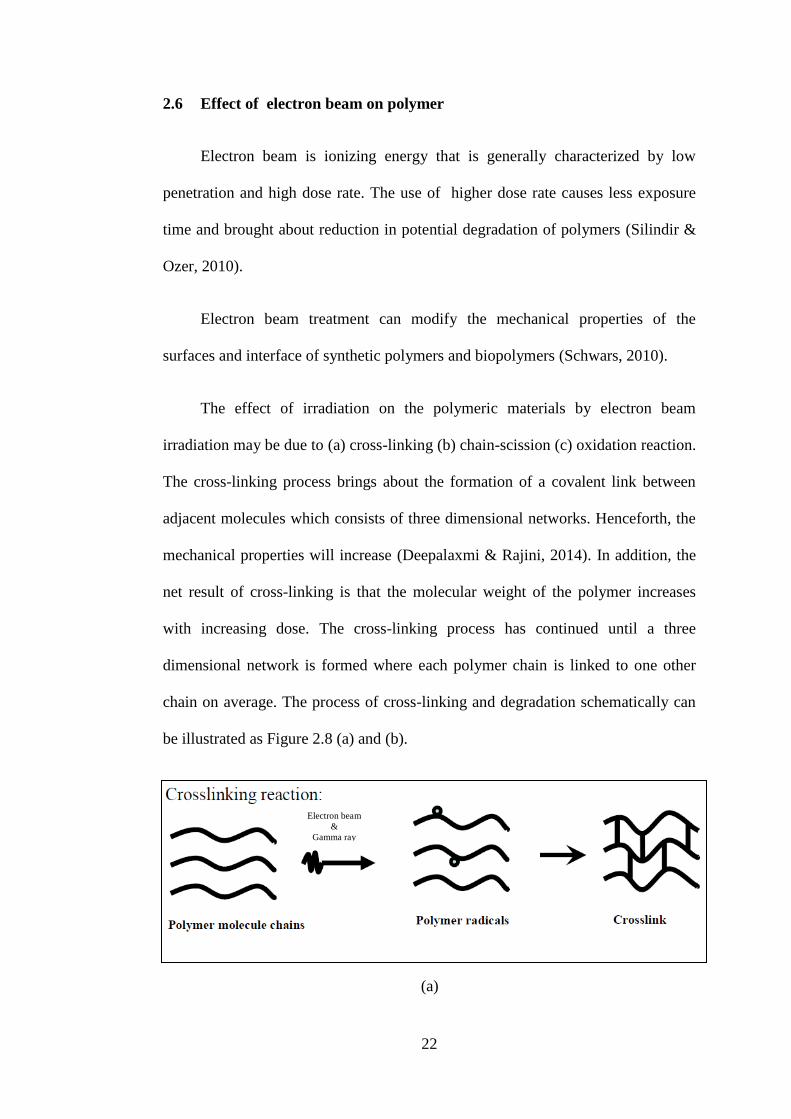
The effect of irradiation on the polymeric materials by electron beam
irradiation may be due to (a) cross-linking (b) chain-scission (c) oxidation reaction.
The cross-linking process brings about the formation of a covalent link between
adjacent molecules which consists of three dimensional networks. Henceforth, the
mechanical properties will increase (Deepalaxmi & Rajini, 2014). In addition, the
net result of cross-linking is that the molecular weight of the polymer increases
with increasing dose. The cross-linking process has continued until a three
dimensional network is formed where each polymer chain is linked to one other
chain on average. The process of cross-linking and degradation schematically can
be illustrated as Figure 2.8 (a) and (b).
(a)
Electron beam
&
Gamma ray

23
(b)
Figure 2.8: (a) The cross-linking process of polymer (b) The degradation reaction
of polymer from high molecule weight to lower molecule weight (Darwis, 2010).
When scission of polymer molecule chains are in the majority in an
irradiated polymer, the molecular weight decreases as dose increases. The final
product might be a low molecular weight liquid in some cases (Darwis, 2010).
A method to modify the structure without introducing new chemical group
could prove the benefit of the electron beam. For instance, novel cross-linking of
arabic gum occurred without the need to any additive and only cross-link by
irradiation by using high solute concentrations Gum arabic will degrade once
irradiated with electron beam in aqueous solution at low concentration.
Conversely, cross-linking occurs with minimal degradation in highly concentrated
aqueous solution (Guven, 2012). Commonly, electron beam irradiation is a method
to degrade polymers both in solid and liquid state. Radiation processing of
polysaccharides is based on the generation of free radicals which are capable to
induce molecular changes by decomposition of macromolecules and the creation of
molecules with smaller chains. This method changes some physicochemical
degradation rate of molecular weight fractions depending on the botanical source
of starches (Nemtanu et al., 2010). Polysaccharides can be degraded due to a
Electron beam
&
Gamma ray
24
scission of glycoside bonds with irradiate by ionizing radiation (Yoshii, 2004).
These intermediates can continue several reaction paths process which cause
rearrangements and/or formation of new bonds. These reactions can be the
formation of oxidized products, grafts, scission of main chain (degradation) or side
chains or cross-linking (Guven, 2012).
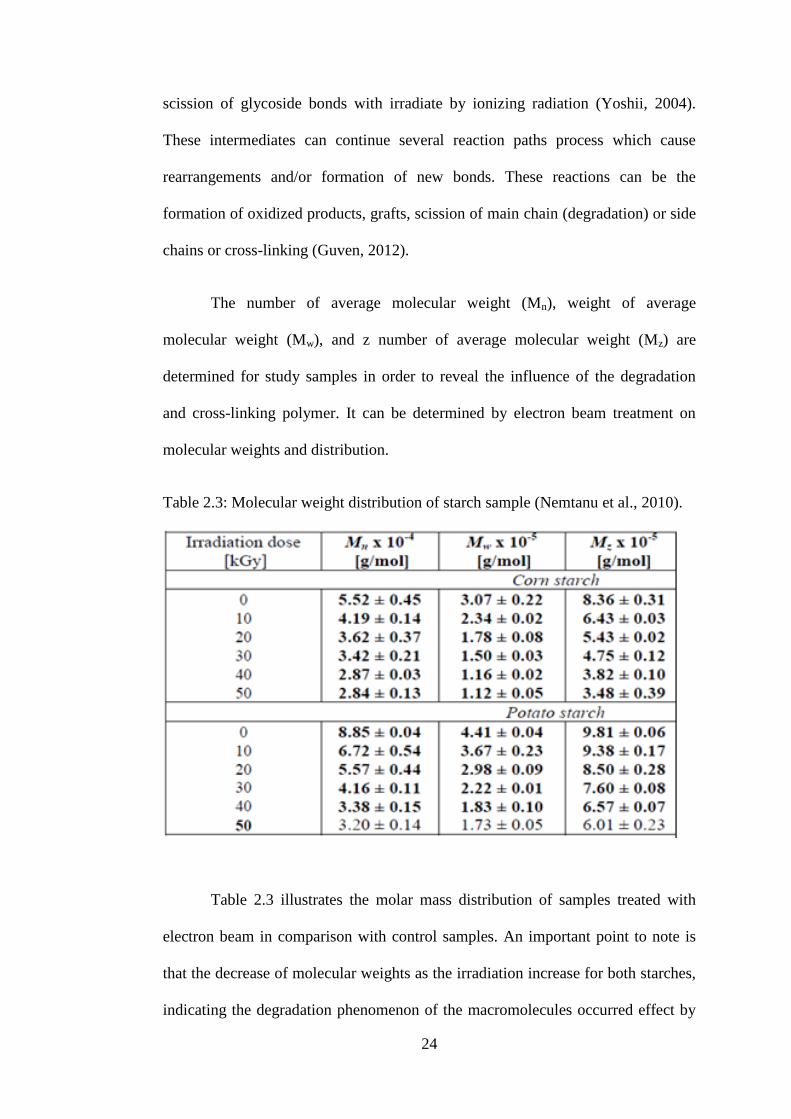
The number of average molecular weight (Mn), weight of average
molecular weight (Mw), and z number of average molecular weight (Mz) are
determined for study samples in order to reveal the influence of the degradation
and cross-linking polymer. It can be determined by electron beam treatment on
molecular weights and distribution.
Table 2.3: Molecular weight distribution of starch sample (Nemtanu et al., 2010).
Table 2.3 illustrates the molar mass distribution of samples treated with
electron beam in comparison with control samples. An important point to note is
that the decrease of molecular weights as the irradiation increase for both starches,
indicating the degradation phenomenon of the macromolecules occurred effect by