dynamic contrast-enhanced and diffusion mri show rapid and...
TRANSCRIPT

Dynamic Contrast-Enhanced and Diffusion MRI Show Rapidand Dramatic Changes in Tumor Microenvironment inResponse to Inhibition of HIF-1A Using PX-4781
Benedicte F Jordany Matthew Runquist z Natarajan Raghunand Amanda Bakersect Ryan WilliamssectLynn Kirkpatrick b Garth Powissect and Robert J Gillies
Department of Biochemistry University of Arizona Health Sciences Center Tucson AZ 85724 USAyLaboratory of Biomedical Magnetic Resonance Universite Catholique de Louvain Brussels B-1200Belgium zDepartment of Biotechnology University of Arizona Health Sciences Center TucsonAZ 85724 USA sectArizona Cancer Center University of Arizona Tucson AZ 85724 USAbProlx Pharmaceuticals Tucson AZ USA
Abstract
PX-478 is a new agent known to inhibit the hypoxia-
responsive transcription factor HIF-1A in experimental
tumors The current study was undertaken in prepa-
ration for clinical trials to determine which noninvasive
imaging endpoint(s) is sensitive to this drugrsquos actions
Dynamic contrast-enhanced (DCE) and diffusion-
weighted (DW) magnetic resonance imaging (MRI)
were used to monitor acute effects on tumor hemo-
dynamics and cellularity respectively Mice bearing
human xenografts were treated either with PX-478 or
vehicle and imaged over time DW imaging was per-
formed at three b values to generate apparent diffu-
sion coefficient of water (ADCw) maps For DCE-MRI
a macromolecular contrast reagent BSA-Gd-DTPA
was used to determine vascular permeability and vas-
cular volume fractions PX-478 induced a dramatic
reduction in tumor blood vessel permeability within
2 hours after treatment which returned to baseline by
48 hours The anti-VEGF antibody Avastin reduced
both the permeability and vascular volume PX-478 had
no effect on the perfusion behavior of a drug-resistant
tumor system A-549 Tumor cellularity estimated from
ADCw was significantly decreased 24 and 36 hours
after treatment This is the earliest significant response
of ADC to therapy yet reported Based on these pre-
clinical findings both of these imaging endpoints will
be included in the clinical trial of PX-478
Neoplasia (2005) 7 475ndash485
Keywords PX-478 HT-29 tumors Dynamic Contrast-Enhanced MagneticResonance Imaging Diffusion Magnetic Resonance Imaging molecularimaging
Introduction
Solid tumors with areas of hypoxia are the most aggres-
sive and difficult tumors to treat [1] Even micrometastases
have areas of hypoxia at the growing edge where tumor
growth outstrips new blood vessel formation [23] Hypoxic
cancer cells survive the hostile hypoxic environment by chang-
ing to a glycolytic metabolism [4] becoming resistant to
programmed cell death (apoptosis) [5] and producing fac-
tors such as vascular endothelial growth factor (VEGF) that
stimulate new blood vessel formation from existing vascula-
ture (angiogenesis) leading to increased tumor oxygenation
and growth [6] The cancer cell response to hypoxia is medi-
ated through the hypoxia-inducible factor-1 (HIF-1) transcrip-
tion factor [78] HIF-1 is a heterodimer consisting of HIF1-a
and HIF-1b subunits both members of the basicndashhelixndash loopndash
helix Per-ARNT-SIM (PAS) family of transcription factors [9]
HIF-1a and HIF-1b associate in the cytosol prior to transport to
the nucleus [10] where they bind to hypoxic regulated element
(HRE) DNA sequences in the 3V and 5V regions of hypoxia-
regulated genes [11] HIF-1b is constitutively expressed and
its levels are not changed by hypoxia [7] HIF-1a is constitu-
tively expressed but under aerobic conditions it is rapidly
degraded by the ubiquitinndash26S proteasome pathway so that
HIF-1a levels are almost nondetectable [12] Under condi-
tions of hypoxia HIF-1a degradation is inhibited and HIF-1a
protein levels increase resulting in an increase in HIF-1 trans-
activating activity
HIF-1a expression has been detected in the majority of
solid tumors examined including brain bladder breast colon
ovarian pancreatic renal and prostate tumors [13] whereas
Abbreviations DCE-MRI dynamic contrast-enhanced MRI DW-MRI diffusion-weighted
MRI ADCw apparent diffusion coefficient of water VEGF vascular endothelial growth
factor HIF-1 hypoxia-inducible factor-1 MMCM macromolecular contrast media PSP
permeability ndash surface area product
Address all correspondence to R J Gillies PhD Arizona Cancer Center 1515 North
Campbell Avenue Tucson AZ 85724-5024 E-mail gilliesuarizonaedu1This work was supported by PHS grants U54 CA90821 and CA077575 and infrastructure
grants R24 CA083148 P30 CAQ3074 and CA98920 Benedicte Jordan was supported by
the Belgian National Fund for Scientific Research (FNRS) as lsquolsquoCharge de Recherchesrsquorsquo
Received 27 September 2004 Revised 19 November 2004 Accepted 23 November 2004
Copyright D 2005 Neoplasia Press Inc All rights reserved 1522-800205$2500
DOI 101593neo04628
Neoplasia Vol 7 No 5 May 2005 pp 475 ndash485 475
wwwneoplasiacom
RESEARCH ARTICLE
no expression was detected in surrounding normal tissues
nor was it detected in benign tumors [14] Clinically HIF-1a
overexpression has been shown to be a marker of highly
aggressive diseases and has been associated with poor
prognosis and treatment failure in a number of cancers in-
cluding breast ovarian cervical oligodendroglioma esopha-
geal and oropharyngeal cancers [15ndash19] HIF-1a presence
correlates with tumor grade as well as vascularity [2021]
High-grade glioblastoma multiforme has significantly higher
levels of VEGF expression and neovascularisation com-
pared with low-grade gliomas [2223] Studies such as these
suggest that HIF-1 mediates hypoxia-induced VEGF expres-
sion in tumors leading to highly aggressive tumor growth
PX-478 (S-2-amino-3-[4V-NN-bis(2-chloroethyl)amino]-
phenyl propionic acid N-oxide dihydrochloride) is a novel
agent that suppresses both constitutive and hypoxia-induced
levels of HIF-1a in cancer cells [24] The inhibition of tumor
growth by PX-478 is positively associated with HIF-1a levels
in a variety of different human tumor xenografts in scidmice
Magnetic resonance imaging (MRI) is a noninvasive
technique that can be used to obtain information regarding
tumor vascularization metabolism and pathophysiology and
allows early assessment of therapeutic effects of cancer
drugs [2526] One approach is dynamic contrast-enhanced
(DCE) MRI which measures tumor vascular characteris-
tics after administration of a contrast medium [2728] MRI
enhanced with small-molecular-weight contrast agents is
extensively used in the clinic to differentiate benign from
malignant lesions as well as to monitor tumor microvascular
characteristics during treatment However the advantage
of using large molecular agents (macromolecular contrast
media or MMCM) designed for prolonged intravascular
retention has been demonstrated in several preclinical stud-
ies [29ndash32] Correlations betweenMMCM-enhancedparame-
ters and angiogenic markers such as microvessel density and
VEGF levels have been studied [3334] Diffusion-weighted
(DW) MRI allows noninvasive characterization of biologic
tissues based on the random microscopic motion of water
proton measurement referred to as the apparent diffusion
coefficient of water (ADCw) [35] Preclinical studies have
shown that DWI allows early detection of tumor response to
chemotherapy [36ndash41] Most likely changes in the diffusion
characteristics are caused by a shift of water to the extra-
cellular space [42] It is therefore anticipated that DW-MRI
will detect early changes in cellular volume fractions resulting
from apoptosis-associated cell shrinkage necrosis or vaso-
genic edema [4344] Because water is not as diffusionally
restricted in the extracellular space compared to the intra-
cellular space a decrease in cell volume fraction will result
in an overall increase in the ADCw We have previously
characterized the capability of DWI to detect early changes
in tumor ADCw following antitumor therapy in preclinical
models [4546] and in the clinical setting [47]
This study monitored the antitumor activity of PX-478
an HIF-1a inhibitor soon to enter clinical testing on HT-29
human colon xenografts using both DCE and DW-MRI and
assessed the use of these techniques as early and surrogate
endpoints for the antitumor response to the drug These
noninvasive magnetic resonance techniques provide in-
sights on tumor microvessel characteristics such as PSP
and vascular volume fraction and on cellular volume ratios
(cellularity and necrotic fraction) which may be early markers
and even predictors of tumor response
Materials and Methods
Cell Line and Tumor Implantation
HT-29 a tumorigenic nonmetastatic human colon carci-
noma cell line and A-549 a non small cell human lung
cancer cell line were obtained from the American Tissue
Type Collection (Rockville MD) Cells were passaged twice
weekly with a 12 split and cultured in Dulbeccorsquos modified
Eaglersquos medium (DMEMF12) supplemented with 10 fetal
bovine serum (HyClone Fort Collins CO) For inoculation
approximately 106 cells in 01 ml of media were injected
subcutaneously into the right flank of female severe com-
bined immunodeficient (SCID) mice of ages 5 to 6 weeks
(Arizona Cancer Center Experimental Mouse Shared Ser-
vices Tucson AZ) Mice developed palpable tumors within a
week of inoculation Tumors were allowed to grow to 100 to
500 mm3 prior to imaging All animal protocols were ap-
proved by the University of Arizona Institutional Animal Care
and Use Committee (IACUC Tuczon AZ)
Treatments
PX-478 was provided by Prolx Pharmaceuticals (Tucson
AZ) and prepared fresh each day in 09NaCl as a 10mgml
solution and administered intraperitoneally to the mice
within 30 minutes of preparation Mice were treated with
either vehicle or 125 mgkg PX-478 and were studied 2
12 24 or 48 hours later A minimum of eight animals were
examined with MRI at each time point (four to six controls
and four to six treated) An additional 36-hour time point
was included in the DW-MRI protocol For imaging mice
were anesthesized using 10 to 20 isoflurane carried
in oxygen Body temperature was maintained at 37jCwith a circulating water blanket and was monitored using
a rectal Luxtron fluoroptic thermometer (Luxtron Santa
Clara CA) Contrast agent Gd-DTPA coupled to albumin
(Gd-BSA 06 mgg in 015 ml of saline) was injected by
a tail vein catheter comprising a 30-gauge needle connected
to PE-20 polyethylene tubing The Gd-BSA was synthesized
by the Arizona Cancer Center Synthetic Chemistry Core
(Tucson AZ) Chemical analysis indicated that there were
an average of 38 Gd bound per protein molecule The
human anti-VEGF antibody Avastin (bevacizumab Genen-
tech San Francisco CA) was administered intravenously at
a dose of 20 ml30 g
MRI
All imagings were performed on a 47-T horizontal
bore MR imager (Bruker Billerica MA) Mice were posi-
tioned into a 24-mm ID Litzcage coil (Doty Scientific Colum-
bia SC) Sagittal scout images were obtained to determine
the position of tumors
476 Monitoring Therapy Response with MRI Jordan et al
Neoplasia Vol 7 No 5 2005
DW-MRI methodology Contiguous axial 20-mm slices
covering the entire tumor were imaged as per the follow-
ing protocol DW images were obtained using the DIFRAD
sequence [48] with the acquisition parameters TR = 2 sec-
onds TE = 36 milliseconds D = 13 milliseconds d = 5 milli-
seconds matrix size = 128 128 and FOV = 4 4 cm
where d and D represent the duration and separation of dif-
fusion gradients respectively At each slice location images
were obtained at three b values (25 500 and 950 secmm2)
with a time resolution of 13 minutes for a complete data set
The b value is equal to c2Gd2d2(D(d3)) where Gd is the
strength of the diffusion weighting gradient and c is the
gyromagnetic ratio for protons Images were reconstructed
using a filtered backprojection algorithm of magnitude data
to minimize motion artifacts ADCw maps were generated
by fitting the three b values to the Stejskal-Tanner equa-
tion S = S0ebADCw where S0 is the signal intensity in the
absence of diffusion weighting and S is the signal intensity
with diffusion weighting ADCw maps were analyzed using
programs written in Interactive Data Language (Research
Systems Boulder CO) Hand-drawn regions of interest
(ROIs) corresponding to tumor localized on the scout scans
were cloned onto the ADCw maps and ADCw distribution
histograms were obtained for each tumor For each time
point (2 12 24 36 and 48 hours after vehicle or PX-478
injection) two groups (one control and one treated) of four
to six mice were imaged In addition four mice were moni-
tored over the full time course independently of the DCE-
MRI protocol to confirm the pattern observed on separate
groups of mice
DCE-MRI acquisition and analysis Contiguous axial 20-mm
slices covering the entire tumor as well as a slice over the
kidneys were imaged in the following protocol A proton
densityndashweighted (TR = 8 seconds TE = 59 milliseconds
NA = 2 and FOV = 4 4 cm) and a T1-weighted spin-echo
image (TR = 300milliseconds TE = 59 milliseconds NA = 8
and FOV = 4 4 cm) were collected prior to injection of con-
trast A dynamic series of spin-echo images (TR = 300 milli-
seconds TE = 59 milliseconds NA = 4 FOV = 4 4 cm and
NR = 19) were collected over 45 minutes with the contrast
agent solution being injected during repetitions 2 to 5
Signal enhancement in the DCE data was converted
to albuminndashGd-DTPA concentration using the relaxivity of
108 lg s measured in vitro at 37jC This can be converted to
648 mM albuminsec assuming a MW of 60 kDa Enhance-
ment was converted to concentration by assuming a linear
relationship between Gd concentration and relaxation rate
enhancement These [albuminndashGd-DTPA] versus time data
were fitted to a straight line for each pixel to obtain a slope
(related to vascular permeability times the vascular surface
area PSP) and y-axis intercept (related to the vascular
volume) In the absence of vascular volume changes the
PSP is referred to simply as lsquolsquopermeabilityrsquorsquo
The vascular volume (VV) parameter measured in tumor
pixels was normalized to the mean value obtained in an ROI
placed on a muscle in the same animal and multiplied
by 5 (fVV fraction of the muscle) to convert it to the
vascular volume fraction of the tumor To be able to compare
values between different mice the slope parameter was
normalized for Gd dose as follows for each mouse The
mean slope parameter calculated from pixels falling within
the vena cava was used to normalize the slope determined
in the tumor The vena cava was identified using a hand-
drawn ROI of approximately 5 to 10 pixels Data analysis
was performed using programs written in Interactive Data
Language (Research Systems)
Antitumor Studies
The doses of PX-478 used for antitumor studies were
80 mgkg daily for 5 days for the HT-29 colon cancer
xenograft mice and 100 mgkg daily for 5 days for the
A-549 lung cancer xenograft mice There were eight mice
in each group Tumor volume was measured twice weekly
until the tumor reached 2000 mm3 or became necrotic
at which point the animals were euthanized Orthogonal
tumor diameters (dshort and dlong) were measured twice
weekly with electronic calipers and converted to volume
by the formula volume = (dshort)2(dlong)2 Log10 cell kill was
calculated by the formula log10 cell kill = (tumor growth
delay [day]) (tumor doubling time [day] 332) One-way
analysis of variance using the general linear model was
used to test for the effect of treatment on tumor growth rate
and growth delay
HIF-1a Immunohistochemistry
Paraffin-embedded tumor sections were heated at
60jC for 30 minutes and rehydrated through xylene and
graded alcohols Antigen retrieval was at 40 minutes at pH
90 for HIF-1a The slides were blocked for 30 minutes in
4 milk 1 goat serum and 01 thimerosal in phosphate-
buffered saline (PBS) After blocking the slides were
processed using a Ventana Medical Systems ES autoslide
stainer Endogenous peroxidase activity was quenched
using a hydrogen peroxidendashbased inhibitor (DAB Basic
Detection Kit Ventana Medical Systems Tucson AZ)
and endogenous biotin blocked using an AB Blocking Kit
(Ventana Medical Systems) The slides were incubated
for 32 minutes at 42jC with the mouse monoclonal
antihuman HIF-1a (Transduction Laboratories Lexington
KY) at 10 gml A biotinylated universal secondary anti-
body which recognized mouse IgGIgM was applied fol-
lowed by horseradish peroxidasendashconjugated avidin DAB
hydrogen peroxide and a copper enhancer The slides
were dehydrated through graded alcohols toluene and
xylene and coverslipped using Vectamount (Vector Labo-
ratories Burlingame CA) HIF-1a staining was normalized
to the staining of an onslide control of hypoxic HT-29 colon
cancer cells
VEGF Detection
Plasma was collected into EDTA tubes and tumors
were removed and immediately snap-frozen in liquid nitro-
gen Tumors were then placed in buffer (10 mM TrisHCl
pH 74 and 100 mM NaCl) and homogenized using
a PowerGen 125 (Fisher Scientific Pittsburg PA) The
Monitoring Therapy Response with MRI Jordan et al 477
Neoplasia Vol 7 No 5 2005
suspension was then centrifuged twice at 8000g at 4jCfor 15 minutes Protein was quantitated in a supernatant
using the Pierce (Rockford IL) BCA assay VEGF levels
were quantitated in plasma and tumor lysates using both
human (hVEGF) and mouse VEGF (mVEGF) ELISA (RampD
Systems Minneapolis MN) according to the manufac-
turerrsquos instructions
Statistical Analysis
Data are presented as the mean and standard error of
the mean (SEM) Two-tailed Studentrsquos t tests ANOVA or
MannndashWhitney rank sum tests were used where appropriate
P lt 05 was considered to be statistically significant
Results
Effect of PX-478 on HT-29 Tumor ADCw
DW-MRI was used to detect the early response of HT-29
tumor xenografts to the antitumor agent PX-478 A single
gradient direction was used in this study because previous
studies have shown the absence of anisotropy in extracra-
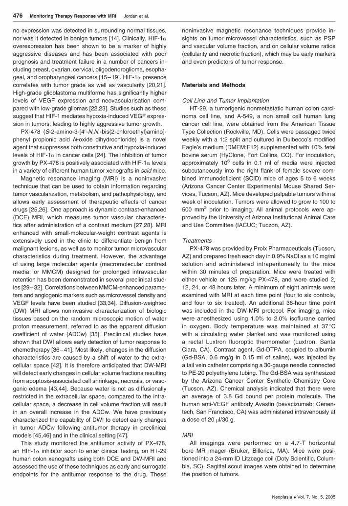
nial tumor models [4649] ADC maps from representative
animals at different times posttherapy are shown in Figure 1
Changes in mean tumor ADCw values over time posttreat-
ment are presented in Figure 2 No change in ADC distri-
bution was observed in sham-treated animals (Figure 2)
At early time points (2 and 12 hours) ADCw values were
not significantly different between control and treated
groups A substantial increase in mean relative tumor ADCw
was observed for the treated groups at 24 and 36 hours
posttreatment (945 plusmn 48 P = 005 and 384 plusmn 49
P = 01 respectively) before returning to pretreatment mean
ADCw values by 48 hours posttreatment (nonsignificant
change of 25 plusmn 67 P = 38) ROIs defining the tumor
were used to generate histograms of tumor ADCw values
ADCw histograms of individual tumors were then summed
for each time point (Figure 2) A right shift in tumor water
diffusion beginning by 24 hours after therapy is shown in
Figure 2 Water diffusibility was still increased by 36 hours
posttreatment and appeared to return to pretreatment values
by the second day after therapy This significant change in
ADC (by 24 hours) occurs sooner than in other reports
Effects of PX-478 on HT-29 Tumor DCE-MRI Parameters
Extravasation of the Gd-BSA was assumed to be describ-
able by a PSP-limited two-compartment model with uni-
directional transport of contrast agent on the timescale of
our DCE-MRI experiments
Parameter maps of lsquolsquopermeabilityrsquorsquo and vascular volume
fraction were created to visualize the heterogeneity of tumor
hemodynamic parameters Heterogeneities in the distribu-
tions of pharmacokinetic parameters have previously been
shown in experimental as well as in human tumors Typical
permeability (P ) and vascular volume fraction (VV) maps
at each time point are shown in Figure 3 Tumors were iden-
tified on proton densityndashweighted images and delineated
by hand-drawn ROIs Tumor vascular PSP is dramatically
decreased in the PX-478 group 2 12 and 24 hours after
treatment in comparison with the control group (Figure 3A)
Figure 1 DW images at a b value of 25 (up) and corresponding diffusion maps (bottom) of an HT-29 tumor-bearing mouse before 24 hours and 48 hours after
PX-478 injection Each image represents an axial slice of the mouse with the tumor area encircled and indicated by an arrow
478 Monitoring Therapy Response with MRI Jordan et al
Neoplasia Vol 7 No 5 2005
This decrease is no longer observed by 48 hours after
treatment Although some individual changes (positive or
negative) in tumor vascular volume fraction were sometimes
observed (Figure 3B 2 and 24 hours posttreatment) the
mean change between groups was not statistically signifi-
cant Hence we conclude that the mechanism underlying the
change in PSP is due to alterations in permeability with little
or no change in surface area because surface area changes
will also be reflected in the vascular volume estimation
Time courses of mean normalized values and mean
VV fraction values are presented in Figure 4 (relative
data) and Table 1 (normalized values) A rapid decrease
in tumor blood vessel permeability was observed within
2 hours after drug administration compared to control
tumors with a mean reduction of 733 plusmn 139 (P = 012)
The decrease in permeability was still 724 plusmn 69 at
12 hours after treatment (P = 003) The effect progressively
decreased in later time points with a mean reduction of
550 plusmn103 (P = 02) at 24 hours posttreatment and a
return to control values at 48 hours (39 plusmn 109 P = 71
not significant) By contrast the vascular volume fraction
of the tumor was not significantly modified at any time
point and remained unchanged between control and
treated tumors
Histogram analyses of these data lose spatial informa-
tion yet retain the distribution of values for quantitative
analyses Figure 5 shows histogram data summed for all
animals in each group Control tumors at each time point
(filled bars in each plot) were characterized by hetero-
geneous and broad distributions of lsquolsquopermeabilityrsquorsquo values
at all time points In contrast treated tumors showed more
homogeneous and narrow histograms centered around
much lower values at 2 12 and 24 hours (open bars)
Note that the range of median of the distribution of perme-
ability values returned to control levels at 48 hours These
data can also be further reduced to median values (dashed
vertical lines in each population) which were significantly
decreased in the treated groups 2 12 and 24 hours
after treatment
Effects of Anti-VEGF Antibodies on HT-29 Tumor
DCE Parameters
To assess the ability of the MMCM DCE technique to
detect acute changes after treatment with an antitumor
agent aimed at decreasing VEGF in this tumor model human
anti-VEGF antibody (Avastin) was administered to HT-29
tumor-bearing mice A 750 plusmn 40 decrease in vascular
PSP was observed within an hour of injection of the anti-
body (P lt 0001) similar to the changes observed 2 and
12 hours after PX-478 administration (Figure 6A Table 1)
The anti-VEGF antibody treatment also induced a signifi-
cant 315 plusmn 26 (P = 023) decrease in vascular volume
fraction unlike treatment with PX-478 (Figure 6A Table 1)
Hence in this case the PSP changes may not be entirely
due to permeability and may also involve alterations in the
vascular surface area
Figure 2 Top Full time course of average tumor ADCw following PX-478 administration (control mice full line treated mice dotted line) A significant increase in
average tumor ADCw is observed at 24 and 36 hours posttreatment Bottom Summed ADCw histograms of control (filled bars) and treated tumors (open bars) at
each time point A right shift in tumor ADCw is observed at 24 and 48 hours posttreatment
Monitoring Therapy Response with MRI Jordan et al 479
Neoplasia Vol 7 No 5 2005
Effects of PX-478 on A-549 Tumor DCE Parameters
A-549 non small cell lung tumors are resistant to PX-
478 (Antitumor Studies section and Ref [24]) and were
therefore used as negative controls for the DCE-MRI proto-
col No significant change was observed for either tumor
permeability or vascular volume fraction (Figure 6B Table 1)
These data suggest that the changes observed on HT-29
xenografts after administration of PX-478 are connected to
the sensitivity of this tumor model to the drug Notably the
untreated PSP values of A-549 tumors were lower than the
control values obtained in HT-29 tumors suggesting that
baseline PSP values may be prognostic for the antitumor
effects of PX-478 although further investigation is required
Antitumor Effect of PX-478 on HT-29 and A-549 Xenografts
HIF-1a Staining and VEGF Detection
HT-29 colon cancer xenografts exhibited staining for
HIF-1a whereas A-549 non small cell lung cancer xeno-
grafts showed very little staining (Figure 7) The A-549 lung
cancer xenografts showed no growth inhibition when treated
with PX-478 (100 mgkg ip) daily for 5 days whereas the
HT-29 colon cancer xenografts exhibited a tumor growth
delay of 16 days with a calculated log cell kill of 16 (P lt 05)
The lack of responsiveness to PX-478 by A-549 tumors is
probably due to the lack of HIF-1a expression in these
tumors compared to HT-29 xenografts (Figure 7) The lower
permeability observed is probably explained by the lower
expression of VEGF-A an HIF-1 target gene Levels of
VEGF-A are also markedly lowered in A-549 tumors versus
HT-29 tumors (5012 plusmn 1209 vs 181 plusmn 023 pgmg P = 012
MannndashWhitney rank sum test) as measured by ELISA
Discussion
The activity of PX-478 an inhibitor of HIF-1a in experimental
tumors was evaluated on HT-29 human colon xenografts
using both DCE and DW-MRI PX-478 induced a substantial
reduction in tumor blood vessel permeability as early as
2 hours after a single dose of 125 mgkg which persisted
until 24 hours posttreatment and had returned to control
values by 48 hours The tumor vascular volume fraction
was not significantly altered over the same time course
Although the time course of response was different for
diffusion MRI tumor ADCw was also shown to be an early
marker of tumor response No change in tumor ADCw could
be observed at very early time points but a significant in-
crease was shown 24 and 36 hours after treatment having
returned to control values by 48 hours posttreatment
Tumor vessel permeability to MMCM has been used
in the preclinical setting to assess the efficacy of differ-
ent antiangiogenic therapies [2730323450] MMCM-
enhanced MRI has been demonstrated to be capable of
monitoring the direct antivascular effects of anti-VEGF anti-
body treatment in xenografts [51ndash53] Changes in tumor
vascular parameters have been measured by DCE-MRI
using clinically approved small molecule contrast agents in
Figure 3 (A) Permeability maps of tumors 2 12 24 and 48 hours after either vehicle (control) or drug (PX-478) injection Each image represents an axial slice
of the mouse with the tumor area encircled A substantial reduction in tumor vascular permeability is observed as soon as 2 hours after PX-478 injection and until
24 hours in comparison with the control situation This is no longer observed by 48 hours after treatment (B) Vascular volume fraction (VV) maps of tumors 2 12
24 and 48 hours after either vehicle (control) or drug (PX-478) injection Each image represents an axial slice of the mouse with the tumor area encircled Some
individual positive or negative changes can be observed but these were not significant between groups
480 Monitoring Therapy Response with MRI Jordan et al
Neoplasia Vol 7 No 5 2005
animal human tumor xenograft models following treatment
with the small molecule VEGF receptor tyrosine kinase
inhibitors ZD6474 [54] and PTK787ZK222584 [5556] and
anti-VEGF antibody [57ndash59] DCE-MRI studies in patients
with colon cancer receiving PTK 787ZK222584 as part of
Phase I trials while showing heterogeneity in tumor vascular
response have shown a significant correlation between
tumor perfusion and the dose of PTK 787ZK222584 with
patients with stable disease having a significantly greater
reduction in the transfer constant Ktrans
which is related to
flow permeability and vascular surface area [55] Patients
receiving anti-VEGF antibody as part of Phase I trial have
also exhibited a reduction in tumor Ktrans
measured by DCE-
MRI after the first treatment [60] In the present study we
observed acute changes within an hour following anti-
VEGF antibody therapy using the large molecular contrast
agent Gd-BSA This suggests that the reductions in vas-
cular permeability parameters measured by DCE-MRI were
related to changes in tumor VEGF levels In this context PX-
478 has been shown to decrease both HIF-1a and VEGF
staining in HT-29 tumors [24] However the time course for
the decrease in HIF-1a and VEGF was different from the
changes in PSP measured by DCE-MRI In our previous
study both HIF-1a and VEGF decreased within 2 hours
and returned to control values by 8 hours after treatment
In contrast in the current study the vascular permeability
estimated from MMCM kinetics was still reduced 24 hours
after treatment Also Avastin led to changes in both vas-
cular volume and PSP whereas PX-478 affected only PSP
which is interpreted to be due to permeability changes alone
The differences between these responses are unknown
but may also indicate that the effect of PX-478 on hemo-
dynamics is not mediated through VEGF However it also
remains possible that the hemodynamics is affected by local
concentrations or threshold values of VEGF and these
cannot yet be measured In patients increased VEGF ex-
pression has been correlated with the progression of colon
carcinoma [61] and with the development of colon cancer
metastasis [62] In node-negative primary colon cancer
elevated tumor VEGF has been correlated with decreased
patient survival [63] Also increased tumor VEGF expres-
sion has been associated with increased tumor angiogenesis
and metastasis of human gastric cancer [64] However the
estimation of VEGF levels is now more controversial as an
accurate marker of therapeutic efficacy Clinical studies
focused on the relation between angiogenic markers (micro-
vascular density or VEGF levels) and quantitative DCE-MRI
enhancement data have shown mixed results [3334]
Su et al [33] concluded that the lack of correlation could
be partly due to the inability of DCE-MRI with low-molecular-
weight agents to reveal the true vascular function within
the tumor Bhujwalla et al [34] recently described the
Figure 4 Full time course of average vascular permeability (A) and vascular
volume fraction (B) following administration of PX-478 (control mice full line
treated mice dotted line) Blood vessel permeability was estimated from the
slope of the enhancement curves and tumor vascular volume (VV) fraction
was estimated from the ordinate A significant reduction in permeability is
observed 2 12 and 24 hours after treatment with PX-478 whereas no
changes are observed in the VV fraction
Table 1 Absolute Values of DCE-MRI Enhancement Parameters after Treatment with PX-478 or Avastin
Tumor Model Tx 1 hr Post-Tx 2 hr Post-Tx 12 hr Post-Tx 24 hr Post-Tx 48 hr Post-Tx
nP (104) VVf () nP (104) VVf () nP (104) VVf () nP (104) VVf () nP (104) VVf ()
HT-29 Control 065 plusmn 004 64 plusmn 07 062 plusmn 007 65 plusmn 12 062 plusmn 006 60 plusmn 05 062 plusmn 001 60 plusmn 08 060 plusmn 001 58 plusmn 09
PX-478 nd 017 plusmn 009 75 plusmn 18 017 plusmn 004 57 plusmn 03 028 plusmn 006 66 plusmn 08 062 plusmn 005 66 plusmn 07
Avastin 016 plusmn 003 44 plusmn 02 nd
A-549 Control nd 035 plusmn 001 63 plusmn 04 nd
PX-478 034 plusmn 001 60 plusmn 07
Normalized permeability (nP) and vascular volume fraction (VVf) values (mean plusmn SEM) for control (carrier injection) PX-478 (125 mgkg ip) and Avastin (20 l
30 g iv) groups Note that the permeability is significantly decreased 2 12 and 24 hours after treatment with PX-478 and within 1 hour after treatment with the
anti-VEGF antibody Avastin and that the VVf is only affected by Avastin
P lt 05 relative to the control group (t-tests)
P lt 01 relative to the control group (t-tests)
Monitoring Therapy Response with MRI Jordan et al 481
Neoplasia Vol 7 No 5 2005
antiangiogenic effect of the fumagillin derivative TNP-470
by MMCM DCE-MRI They observed a heterogeneous re-
sponse with some regions of decreased PSP and some
regions with increased PSP values resulting in an apparent
lack of overall response based on the average value of
tumor PSP whereas ELISA assays detected an increase
of tumor VEGF DCE-MRI was shown to be a more reliable
marker by taking into account tumor heterogeneity Our
results suggest that DCE-MRI might be a more sensitive
measure of functional tumor permeability or that perme-
ability factors other than VEGF might be involved in the
response to PX-478
Importantly a lack of change in the PSP of A-549 tumors
between control and treated tumors was observed in this
study This correlates well with the inability of PX-478 to
induce growth delays in A-549 tumors In this case the lack
of an effect may be due to the lower baseline PSP values
in A-549 compared to HT-29 tumors It also implies that the
effect of this drug on vascular parameters is mediated
through the tumor cells themselves and not the host vascu-
lature which was the same in both tumor settings
It has been suggested in the past that DCE-MRI could be
used to monitor clinical response to anti-VEGF and inhibition
of angiogenesis [54ndash5765] The current findings suggest
that DCE-MRI may also be useful to assess the response to
inhibition of HIF-1 However it should be acknowledged that
the current study used MMCM which are currently unavail-
able in a clinical setting These results emphasize the need
to develop MMCM for monitoring antivascular therapies Our
finding that a tumor with low HIF-1a staining which was not
responsive to antindashHIF-1 therapy also had a very low vas-
cular PSP as measured by MMCM DCE-MRI suggests that
DCE-MRI may also be useful clinically for screening and
preselecting patients for therapy with antindashHIF-1 and other
antiangiogenic therapies
DW-MRI is able to detect early changes in the morpho-
logy and physiology of tissues after antineoplastic therapies
An increase in tumor ADCw could result from changes in cell
membrane permeability cell shrinkage or vasogenic edema
Vasogenic edema results from high vessel permeability that
results in the movement of osmotically active proteins and
associated water to the interstitium This mechanism is
unlikely to be the cause of increased ADC in the current
system because vessel permeability decreased in response
to PX-478 Both of these are associated with cell death and
result in the modification of the intracellular to extracellular
water populations ratio [43] Parameters such as cell density
and necrotic fraction have indeed been monitored with
diffusion MRI [4966] In this study we observed an increase
in tumor ADCw that is consistent with other studies using
other tumors and drugs [37ndash41444667] In these studies
Figure 5 Summed permeability histograms of control (open n = 4) and treated tumors (plain n = 4) at each time point Note that the median (dotted line) of the
treated tumors is lower than the median value of the controls It is progressively shifted to the median of the controls over time and is back at control values
48 hours posttreatment
Figure 6 (A) Relative change in HT-29 tumor vascular permeability and
vascular volume fraction 1 hour after treatment with anti-VEGF antibody
(Avastin) A significant reduction in permeability as well as in VV fraction is
observed with this positive control (B) Relative change in A-549 tumor
(resistant to the antitumor activity of PX-478 negative control) vascular
permeability and vascular volume fraction 2 hours after treatment with
PX-478 No significant change is observed in DCE parameters
482 Monitoring Therapy Response with MRI Jordan et al
Neoplasia Vol 7 No 5 2005
an increase in ADCw is correlated with ultimate tumor re-
sponse whether by apoptosis or other means of cell death
Notably the current data document the earliest significant
increase in chemotherapy-induced ADCw Previous reports
have indicated that the earliest significance was not reached
until 48 hours following therapy [3639]
The combination of dynamic and DWMRI in the follow-up
of chemotherapy has been used in the past [6869] and has
been proven to be of good predictive value for therapy
outcome in patients with primary rectal carcinoma [70] In
this study the acquisition of both DW and DCE images in a
single protocol on the same animal allowed us to coregister
these data and compare the two techniques We define the
dynamic range (DR) as the maximum change relative to the
variance of controls For these studies the DR was higher
for DW-MRI (maximum effect at 24 hours DR = 87) than
for DCE-MRI (maximum effect at 2 hours DR = 32) Tumor
ADCw was thereby shown to be a sensitive and early marker
of tumor response in this study Nonetheless the DCE-MRI
response preceded the diffusion response and opened up
the possibility of monitoring acute effects of drug in vivo The
combination of the two techniques gives unique insights
into the complex response of HT-29 tumors to PX-478 by
showing very early changes in vascular permeability fol-
lowed by large changes in cellularity Considering the mag-
nitude of response of HT-29 xenografts to PX-478 observed
with early and sensitive markers we can speculate that
noninvasive monitoring of PX-478 by DCE andor diffusion
MRI will be of particular interest in the clinic
Acknowledgements
We gratefully acknowledge Z Bhujwalla (Johns Hopkins)
for providing us with the protocol for the preparation and
characterization of albumin-Gd(DTPA) We thank Christine
Howison for research animal care and preparation for MRI
studies tumor inoculation and treatment administration We
would also like to acknowledge Brenda Baggett for cell
culture support Wendy R Tate for technical assistance
with ELISA Gillian Payne-Murietta and Bethany Skovan for
the experimental mouse shared service and Merry Warner
for organizational support
References[1] Hoeckel M Schlenger K Aral B Mitza M Schaffer U and Vaupel P
(1996) Association between tumour hypoxia and malignant pro-
gression in advanced cancer of the uterine cervix Cancer Res 56
4509ndash4515
[2] Moulder J and Rockwell S (1987) Tumor hypoxia its impact on cancer
therapy Cancer Metastasis Rev 5 313ndash341
[3] Nordsmark M Hoyer M Keller J Nielson OS Jensen OM and
Overgaard J (1996) The relationship between tumor oxygenation
Figure 7 HIF-1a levels and antitumor activity of PX-478 in HT-29 human colon cancer and A-549 non small cell lung cancer xenografts in SCID mice Male SCID
mice were injected subcutaneously with (A) 107 HT-29 human colon cancer cells or (B) A-549 non small cell lung cancer cells The HT-29 tumors were allowed to
grow to 400 mm3 and the A-549 tumors to 360 mm3 and treatment was begun with (o) vehicle alone or (n) PX-478 at 80 mgkg ip daily for 5 days for HT-29
xenografts and 100 mgkg ip daily for 5 days for A-549 xenografts Treatment times are shown by arrows The upper panels show typical immunohistochemical
staining for HIF-1a in the untreated tumor xenografts at the start of the study The lower panels show tumor xenograft growth curves There were eight mice in each
group and bars are SE
Monitoring Therapy Response with MRI Jordan et al 483
Neoplasia Vol 7 No 5 2005
and cell proliferation in human soft tissue sarcomas Int J Radiat
Oncol Biol Phys 35 701ndash708
[4] Goonewardene TI Sowter HM and Harris AL (2002) Hypoxia-induced
pathways in breast cancer Micro Res Tech 59 41ndash48
[5] Chen J Zhao S Nakada K Kuge Y Tamaki N Okada F Wang J
Shindo M Higashino F Takeda K Asaka M Katoh H Sugiyama T
Hosokawa M and Kobayashi M (2003) Dominant-negative hypoxia-
inducible factor-1a reduces tumorigenicity of pancreatic cancer cells
through the suppression of glucose metabolism Am J Pathol 162
1283ndash1291
[6] Hanahan D and Folkman J (1996) Patterns and emerging mechanisms
of the angiogenic switch during tumorigenesis Cell 86 353ndash364
[7] Semenza GL (2000) HIF-1 mediator of physiological and pathophysio-
logical responses to hypoxia J Am Phys Soc 88 1474ndash1480
[8] Powis G and Kirkpatrick L (2004) HIF-1a as a cancer drug target Mol
Cancer Ther 3 647ndash654
[9] Wang G Dong Z Xu G Yang Z Shou C Wang N and Liu T (1998)
The effect of antibody against vascular endothelial growth factor on
tumor growth and metastasis J Cancer Res Clin Oncol 124 615ndash620
[10] Chilov D Camneish G Kvietikova I Ziegler U Gassmann M and
Wenger RH (1999) Induction and nuclear translocation of hypoxia-
inducible factor-1 (HIF-1) heterodimerization with ARNT is not neces-
sary for nuclear accumulation of HIF-1a J Cell Sci 112 1203ndash1212
[11] Minchenko A Salceda S and Bauer T (1994) Hypoxia regulatory ele-
ments of the human vascular endothelial growth factor gene Cell Mol
Biol Res 40 35ndash39
[12] Kallio PJ Wilson WJ OrsquoBrien S Makino Y and Poellinger L (1999)
Regulation of the hypoxia-inducible factor-1a by the ubiquitin ndash
proteosome pathway J Biol Chem 274 6519ndash6525
[13] Huang TT Yasunami M Carlson EJ Gillespie AM Reaume AG
Hoffman EK Chan PH Scott RW and Epstein CJ (1997) Super-
oxide-mediated cytotoxicity in superoxide dismutasendashdeficient fetal
fibroblasts Arch Biochem Biophys 344 424ndash432
[14] Zhong H De Marzo AM Laughner E Lim M Hilton DA Zagzag D
Buechler P Isaacs WB Semenza GL and Simons JW (1999) Over-
expression of hypoxia-inducible factor 1 alpha in common human can-
cers and metastases Cancer Res 59 5830ndash5835
[15] Giatromanolaki A Koukourakis M Sivridis E Turley H and Talks K
(2001) Relation of hypoxia inducible factor 1 alpha and 2 alpha in
operable non-small cell lung cancer to angiogenic molecular profile of
tumors and survival Br J Cancer 85 881ndash890
[16] Birner P Schindl M Obermair A Breitenecker G and Oberhuber G
(2001) Expression of hypoxia-inducible factor-1 alpha in epithelial
ovarian tumors its impact on prognosis and on response to chemo-
therapy Clin Cancer Res 7 1661ndash1668
[17] Bos R van der Groep P Greijer AE Shvarts A Meijer S Pinedo HM
Semenza GL van Diest PJ and van der Wall E (2003) Levels of
hypoxia-inducible factor-1alpha independently predict prognosis in
patients with lymph node negative breast carcinoma Cancer 97
1573ndash1581
[18] Aebersold DM Burri P Beer KT Laissue J Djonov V Greiner RH and
Semenza GL (2001) Expression of hypoxia-inducible factor-1alpha a
novel predictive and prognostic parameter in the radiotherapy of oro-
pharyngeal cancer Cancer Res 61 2911ndash2916
[19] Birner P Schindl M Obermair A Plank C Breitenecker G and
Oberhuber G (2000) Over-expression of hypoxia-inducible factor-
1alpha is a marker for an unfavorable prognosis in early-stage in-
vasive cervical cancer Cancer Res 60 4693ndash4696
[20] Semenza GL (2000) Hypoxia clonal selection and the role of HIF-1 in
tumor progression Crit Rev Biochem Mol Biol 35 71ndash103
[21] Zagzag D Zhong H Scalzetti J Laughner E Simons J Semenza G
(2000) Expression of hypoxia-inducible factor-1 alpha in brain tu-
mors association with angiogenesis invasion and progression Cancer
88 2606ndash2618
[22] Pietsch T Valter MM Wolf HK von Deimling A Huang HJ Cavenee
WK and Wiestler OD (1997) Expression and distribution of vascular
endothelial growth factor protein in human brain tumors Acta Neuro-
pathol 93 109ndash117
[23] Takano S Yoshii Y Kondo S Suzuki H Maruno T Shirai S and Nose
T (1996) Concentration of vascular endothelial growth factor in the
serum and tumor tissue of brain tumor patients Cancer Res 56
2185ndash2190
[24] Welsh S Williams R Kirkpatrick L Paine-Murrieta G and Powis G
(2004) Antitumor activity and pharmacodynamic properties of PX-478
an inhibitor of hypoxia-inducible factor-1alpha Mol Cancer Ther 3 (3)
233ndash244
[25] Gillies RJ Bhujwalla ZM Evelhoch J Garwood M Neeman M
Robinson SP Sotak CH and Van Der Sanden B (2000) Applica-
tions of magnetic resonance in model systems tumor biology and
physiology Neoplasia 2 (1ndash2) 139ndash151
[26] Evelhoch JL Gillies RJ Karczmar GS Koutcher JA Maxwell RJ
Nalcioglu O Raghunand N Ronen SM Ross BD and Swartz HM
(2000) Applications of magnetic resonance in model systems cancer
therapeutics Neoplasia 2 (1ndash2) 152ndash165
[27] Padhani AR (2002) Dynamic contrast-enhanced MRI in clinical on-
cology current status and future directions J Magn Reson Imaging 16
(4) 407ndash422
[28] Knopp MV Giesel FL Marcos H von Tengg-Kobligk H and Choyke P
(2001) Dynamic contrast-enhanced magnetic resonance imaging in
oncology Top Magn Reson Imaging 12 (4) 301ndash308
[29] Brasch R Pham C Shames D Roberts T van Dijke K van Bruggen N
Mann J Ostrowitzki S and Melnyk O (1997) Assessing tumor angio-
genesis using macromolecular MR imaging contrast media J Magn
Reson Imaging 7 (1) 68ndash74
[30] Demsar F Roberts TP Schwickert HC Shames DM van Dijke CF
Mann JS Saeed M and Brasch RC (1997) A MRI spatial mapping
technique for microvascular permeability and tissue blood volume
based on macromolecular contrast agent distribution Magn Reson
Med 37 (2) 236ndash242
[31] Gossmann A Okuhata Y Shames DM Helbich TH Roberts TP
Wendland MF Huber S and Brasch RC (1999) Prostate cancer tumor
grade differentiation with dynamic contrast-enhanced MR imaging in
the rat comparison of macromolecular and small-molecular contrast
mediamdashpreliminary experience Radiology 213 (1) 265ndash272
[32] Daldrup H Shames DM Wendland M Okuhata Y Link TM Rosenau
W Lu Y and Brasch RC (1998) Correlation of dynamic contrast-
enhanced MR imaging with histologic tumor grade comparison of macro-
molecular and small-molecular contrast media AJR Am J Roentgenol
171 (4) 941ndash949
[33] Su MY Cheung YC Fruehauf JP Yu H Nalcioglu O Mechetner E
Kyshtoobayeva A Chen SC Hsueh S McLaren CE and Wan YL
(2003) Correlation of dynamic contrast enhancement MRI para-
meters with microvessel density and VEGF for assessment of angio-
genesis in breast cancer J Magn Reson Imaging 18 (4) 467ndash477
[34] Bhujwalla ZM Artemov D Natarajan K Solaiyappan M Kollars P and
Kristjansen PE (2003) Reduction of vascular and permeable regions
in solid tumors detected by macromolecular contrast magnetic reso-
nance imaging after treatment with antiangiogenic agent TNP-470 Clin
Cancer Res 9 (1) 355ndash362
[35] Bammer R (2003) Basic principles of diffusion-weighted imaging Eur J
Radiol 45 (3) 169ndash184
[36] Zhao M Pipe JG Bonnett J and Evelhoch JL (1996) Early detection
of treatment response by diffusion-weighted 1H-NMR spectroscopy in
a murine tumour in vivo Br J Cancer 73 (1) 61ndash64
[37] Hakumaki JM Poptani H Puumalainen AM Loimas S Paljarvi LA
Yla-Herttuala S and Kauppinen RA (1998) Quantitative 1H nuclear
magnetic resonance diffusion spectroscopy of BT4C rat glioma during
thymidine kinase ndashmediated gene therapy in vivo identification of
apoptotic response Cancer Res 58 (17) 3791ndash3799
[38] Poptani H Puumalainen AM Grohn OH Loimas S Kainulainen R
Yla-Herttuala S and Kauppinen RA (1998) Monitoring thymidine ki-
nase and ganciclovir-induced changes in rat malignant glioma in vivo
by nuclear magnetic resonance imaging Cancer Gene Ther 5 (2)
101ndash109
[39] Chenevert TL Stegman LD Taylor JM Robertson PL Greenberg HS
Rehemtulla A and Ross BD (2000) Diffusion magnetic resonance
imaging an early surrogate marker of therapeutic efficacy in brain tu-
mors J Natl Cancer Inst 92 (24) 2029ndash2036
[40] Mardor Y Roth Y Lidar Z Jonas T Pfeffer R Maier SE Faibel M
Nass D Hadani M Orenstein A Cohen JS and Ram Z (2001) Moni-
toring response to convection-enhanced taxol delivery in brain tumor
patients using diffusion-weighted magnetic resonance imaging Cancer
Res 61 (13) 4971ndash4973
[41] Mardor Y Roth Y Ochershvilli A Spiegelmann R Tichler T Daniels D
Maier SE Nissim O Ram Z Baram J Orenstein A and Pfeffer R
(2004) Pretreatment prediction of brain tumors response to radiation
therapy using high b-value diffusion-weighted MRI Neoplasia 6 (2)
136ndash142
[42] Norris DG (2001) The effects of microscopic tissue parameters on
the diffusion weighted magnetic resonance imaging experiment NMR
Biomed 14 (2) 77ndash93
[43] Kauppinen RA (2002) Monitoring cytotoxic tumour treatment response
by diffusion magnetic resonance imaging and proton spectroscopy
NMR Biomed 15 (1) 6ndash17
484 Monitoring Therapy Response with MRI Jordan et al
Neoplasia Vol 7 No 5 2005
[44] Ross BD Moffat BA Lawrence TS Mukherji SK Gebarski SS Quint DJ
Johnson TD Junck L Robertson PL Muraszko KM Dong Q Meyer CR
Bland PH McConville P Geng H Rehemtulla A and Chenevert TL
(2003) Evaluation of cancer therapy using diffusionmagnetic resonance
imaging Mol Cancer Ther 2 (6) 581ndash587
[45] Galons JP Altbach MI Paine-Murrieta GD Taylor CW and Gillies RJ
(1999) Early increases in breast tumor xenograft water mobility in re-
sponse to paclitaxel therapy detected by non-invasive diffusion mag-
netic resonance imaging Neoplasia 1 (2) 113ndash117
[46] Jennings D Hatton BN Guo J Galons JP Trouard TP Raghunand
N Marshall J and Gillies RJ (2002) Early response of prostate carci-
noma xenografts to docetaxel chemotherapy monitored with diffusion
MRI Neoplasia 4 (3) 255ndash262
[47] Theilmann RJ Borders R Trouard TP Xia G Outwater E Ranger-
Mooreb J Gillies RJ and Stopeck A (2005) Changes in water mobility
measured by diffusion MRI predict response of metastatic breast can-
cer to chemotherapy Neoplasia (in press)
[48] Trouard TP Theilmann RJ Altbach MI and Gmitro AF (1999)
High-resolution diffusion imaging with DIFRAD-FSE (diffusion-
weighted radial acquisition of data with fast spin-echo) MRI Magn
Reson Med 42 (1) 11ndash18
[49] Lyng H Haraldseth O and Rofstad EK (2000) Measurement of cell
density and necrotic fraction in human melanoma xenografts by diffu-
sion weighted magnetic resonance imaging Magn Reson Med 43 (6)
828ndash836
[50] Marzola P Degrassi A Calderan L Farace P Crescimanno C Nicolato
E Giusti A Pesenti E Terron A Sbarbati A Abrams T Murray L and
Osculati F (2004) In vivo assessment of antiangiogenic activity of
SU6668 in an experimental colon carcinoma model Clin Cancer Res
10 739ndash750
[51] Pham CD Roberts TP van Bruggen N Melnyk O Mann J Ferrara N
Cohen RL and Brasch RC (1998) Magnetic resonance imaging de-
tects suppression of tumor vascular permeability after administration
of antibody to vascular endothelial growth factor Cancer Invest 16 (4)
225ndash230
[52] Roberts TP Turetschek K Preda A Novikov V Moeglich M Shames
DM Brasch RC and Weinmann HJ (2002) Tumor microvascular
changes to anti-angiogenic treatment assessed by MR contrast media
of different molecular weights Acad Radiol 9 S511ndashS513
[53] Turetschek K Preda A Floyd E Shames DM Novikov V Roberts TP
Wood JM Fu Y Carter WO and Brasch RC (2002) MRI monitoring of
tumor response to a novel VEGF tyrosine kinase inhibitor in an experi-
mental breast cancer model Acad Radiol 2 S519ndashS520
[54] Checkley D Tessier JJ Kendrew J Waterton JC and Wedge SR
(2003) Use of dynamic contrast-enhanced MRI to evaluate acute
treatment with ZD6474 a VEGF signalling inhibitor in PC-3 prostate
tumours Br J Cancer 89 (10) 1889ndash1895
[55] Morgan B Thomas AL Drevs J Hennig J Buchert M Jivan A
Horsfield MA Mross K Ball HA Lee L Mietlowski W Fuxuis S Unger
C OrsquoBryne K Henry A Cherryman GR Laurent D Dugan M Marme D
and Steward WP (2003) Dynamic contrast-enhanced magnetic reso-
nance imaging as a biomarker for the pharmacological response of
PTK787ZK 222584 an inhibitor of the vascular endothelial growth
factor receptor tyrosine kinases in patients with advanced colorectal
cancer and liver metastases results from two phase I studies J Clin
Oncol 21 (21) 3955ndash3964
[56] Turetschek K Preda A Floyd E Shames DM Novikov V Roberts TP
Wood JM Fu Y Carter WO and Brasch RC (2003) MRI monitoring of
tumor response following angiogenesis inhibition in an experimental
human breast cancer model Eur J Nucl Med Mol Imaging 30 (3)
448ndash455
[57] Gossmann A Helbich TH Kuriyama N Ostrowitzki S Roberts TP
Brasch RC van Bruggen N Wendland MF Israel MA and Brasch
RC (2002) Dynamic contrast-enhanced magnetic resonance imaging
as a surrogate marker of tumor response to anti-angiogenic therapy
in a xenograft model of glioblastoma multiforme J Magn Reson
Imaging 15 (3) 233ndash240
[58] Kiessling F Farhan N Lichy MP Vosseler S Heilmann M Krix M
Bohlen P Miller DW Mueller MM Semmler W Fusenig NE and
Delorme S (2004) Dynamic contrast-enhanced magnetic resonance
imaging rapidly indicates vessel regression in human squamous cell
carcinomas grown in nude mice caused by VEGF receptor 2 blockade
with DC101 Neoplasia 6 (3) 213ndash223
[59] Turetschek K Preda A Novikov V Brasch RC Weinmann HJ
Wunderbaldinger P and Roberts TP (2004) Tumor microvascular
changes in antiangiogenic treatment assessment by magnetic reso-
nance contrast media of different molecular weights J Magn Reson
Imaging 20 (1) 138ndash144
[60] Jayson GC Zweit J Jackson A Mulatero C Julyan P Ranson M
Broughton LWagstaff J Hakannson L GroenewegenG Bailey J Smith
N Hastings D Lawrance J Haroon H Ward T McGown AT Tang M
Levitt D Marreaud S Lehmann FF Herold M and Zwierzina H (2002)
Molecular imaging and biological evaluation of HuMV833 anti-VEGF
antibody implications for trial design of antiangiogenic antibodies J Natl
Cancer Inst 94 (19) 1484ndash1493
[61] Wong MP Cheung N Yuen ST Leung SY and Chung LP (1999)
Vascular endothelial growth factor is upregulated in early premalignant
stage of colorectal tumour progression Int J Cancer 81 845ndash850
[62] OkamotoKOshikaY FukushimaY YoshimuraMOhnishi Y Tokunaga
T Hashimoto T Hatanaka H Tomii Y Yoshida Y Miura S Tsuchida T
Kijima H Yamazaki H Nakamura M and Ueyama Y (1999) Inhibition
of liver metastasis of colon cancer by in vivo administration of anti-
vascular endothelial growth factor antibody Oncol Rep 6 (3) 553ndash556
[63] Wang GL Jiang BH and Rue EA (1995) Hypoxia-inducible factor I is
a basic ndashhelix ndash loopndashhelix ndashPAS heterodimer regulated by cellular O2
tension Proc Natl Acad Sci USA 92 5510ndash5514
[64] Yonemura Y Endo Y Fujita H Fishida S Ninomiya I Bandou E
Taniguchi K Miwa K Ohoyama S Sugiyama K and Sasaki T (1999)
Role of vascular endothelial growth factor C expression in the develop-
ment of lymph node metastasis in gastric cancer Clin Cancer Res 5
1823ndash1829
[65] Thomas AL Morgan B Drevs J Unger C Wiedenmann B Vanhoefer
U Laurent D Dugan M and Steward WP (2003) Vascular endothelial
growth factor receptor tyrosine kinase inhibitors PTK787ZK 222584
Semin Oncol 30 (3) 32ndash38
[66] Helmer KG Meiler MR Sotak CH and Petruccelli JD (2003) Com-
parison of the return-to-the-origin probability and the apparent diffusion
coefficient of water as indicators of necrosis in RIF-1 tumors Magn
Reson Med 49 (3) 468ndash478
[67] Stegman LD Rehemtulla A Hamstra DA Rice DJ Jonas SJ Stout KL
Chenevert TL and Ross BD (2000) Diffusion MRI detects early events
in the response of a glioma model to the yeast cytosine deaminase
gene therapy strategy Gene Ther 7 (12) 1005ndash1010
[68] Beauregard DA Pedley RB Hill SA and Brindle KM (2002) Differential
sensitivity of two adenocarcinoma xenografts to the anti-vascular drugs
combretastatin A4 phosphate and 56-dimethylxanthenone-4-acetic
acid assessed using MRI and MRS NMR Biomed 15 (2) 99ndash105
[69] Sinha S and Sinha U (2002) Functional magnetic resonance of human
breast tumors diffusion and perfusion imaging Ann N Y Acad Sci 980
95ndash115
[70] DeVries AF Kremser C Hein PA Griebel J Krezcy A Ofner D Pfeiffer
KP Lukas P and Judmaier W (2003) Tumor microcirculation and
diffusion predict therapy outcome for primary rectal carcinoma Int J
Radiat Oncol Biol Phys 15 56 (4) 958ndash965
Monitoring Therapy Response with MRI Jordan et al 485
Neoplasia Vol 7 No 5 2005
no expression was detected in surrounding normal tissues
nor was it detected in benign tumors [14] Clinically HIF-1a
overexpression has been shown to be a marker of highly
aggressive diseases and has been associated with poor
prognosis and treatment failure in a number of cancers in-
cluding breast ovarian cervical oligodendroglioma esopha-
geal and oropharyngeal cancers [15ndash19] HIF-1a presence
correlates with tumor grade as well as vascularity [2021]
High-grade glioblastoma multiforme has significantly higher
levels of VEGF expression and neovascularisation com-
pared with low-grade gliomas [2223] Studies such as these
suggest that HIF-1 mediates hypoxia-induced VEGF expres-
sion in tumors leading to highly aggressive tumor growth
PX-478 (S-2-amino-3-[4V-NN-bis(2-chloroethyl)amino]-
phenyl propionic acid N-oxide dihydrochloride) is a novel
agent that suppresses both constitutive and hypoxia-induced
levels of HIF-1a in cancer cells [24] The inhibition of tumor
growth by PX-478 is positively associated with HIF-1a levels
in a variety of different human tumor xenografts in scidmice
Magnetic resonance imaging (MRI) is a noninvasive
technique that can be used to obtain information regarding
tumor vascularization metabolism and pathophysiology and
allows early assessment of therapeutic effects of cancer
drugs [2526] One approach is dynamic contrast-enhanced
(DCE) MRI which measures tumor vascular characteris-
tics after administration of a contrast medium [2728] MRI
enhanced with small-molecular-weight contrast agents is
extensively used in the clinic to differentiate benign from
malignant lesions as well as to monitor tumor microvascular
characteristics during treatment However the advantage
of using large molecular agents (macromolecular contrast
media or MMCM) designed for prolonged intravascular
retention has been demonstrated in several preclinical stud-
ies [29ndash32] Correlations betweenMMCM-enhancedparame-
ters and angiogenic markers such as microvessel density and
VEGF levels have been studied [3334] Diffusion-weighted
(DW) MRI allows noninvasive characterization of biologic
tissues based on the random microscopic motion of water
proton measurement referred to as the apparent diffusion
coefficient of water (ADCw) [35] Preclinical studies have
shown that DWI allows early detection of tumor response to
chemotherapy [36ndash41] Most likely changes in the diffusion
characteristics are caused by a shift of water to the extra-
cellular space [42] It is therefore anticipated that DW-MRI
will detect early changes in cellular volume fractions resulting
from apoptosis-associated cell shrinkage necrosis or vaso-
genic edema [4344] Because water is not as diffusionally
restricted in the extracellular space compared to the intra-
cellular space a decrease in cell volume fraction will result
in an overall increase in the ADCw We have previously
characterized the capability of DWI to detect early changes
in tumor ADCw following antitumor therapy in preclinical
models [4546] and in the clinical setting [47]
This study monitored the antitumor activity of PX-478
an HIF-1a inhibitor soon to enter clinical testing on HT-29
human colon xenografts using both DCE and DW-MRI and
assessed the use of these techniques as early and surrogate
endpoints for the antitumor response to the drug These
noninvasive magnetic resonance techniques provide in-
sights on tumor microvessel characteristics such as PSP
and vascular volume fraction and on cellular volume ratios
(cellularity and necrotic fraction) which may be early markers
and even predictors of tumor response
Materials and Methods
Cell Line and Tumor Implantation
HT-29 a tumorigenic nonmetastatic human colon carci-
noma cell line and A-549 a non small cell human lung
cancer cell line were obtained from the American Tissue
Type Collection (Rockville MD) Cells were passaged twice
weekly with a 12 split and cultured in Dulbeccorsquos modified
Eaglersquos medium (DMEMF12) supplemented with 10 fetal
bovine serum (HyClone Fort Collins CO) For inoculation
approximately 106 cells in 01 ml of media were injected
subcutaneously into the right flank of female severe com-
bined immunodeficient (SCID) mice of ages 5 to 6 weeks
(Arizona Cancer Center Experimental Mouse Shared Ser-
vices Tucson AZ) Mice developed palpable tumors within a
week of inoculation Tumors were allowed to grow to 100 to
500 mm3 prior to imaging All animal protocols were ap-
proved by the University of Arizona Institutional Animal Care
and Use Committee (IACUC Tuczon AZ)
Treatments
PX-478 was provided by Prolx Pharmaceuticals (Tucson
AZ) and prepared fresh each day in 09NaCl as a 10mgml
solution and administered intraperitoneally to the mice
within 30 minutes of preparation Mice were treated with
either vehicle or 125 mgkg PX-478 and were studied 2
12 24 or 48 hours later A minimum of eight animals were
examined with MRI at each time point (four to six controls
and four to six treated) An additional 36-hour time point
was included in the DW-MRI protocol For imaging mice
were anesthesized using 10 to 20 isoflurane carried
in oxygen Body temperature was maintained at 37jCwith a circulating water blanket and was monitored using
a rectal Luxtron fluoroptic thermometer (Luxtron Santa
Clara CA) Contrast agent Gd-DTPA coupled to albumin
(Gd-BSA 06 mgg in 015 ml of saline) was injected by
a tail vein catheter comprising a 30-gauge needle connected
to PE-20 polyethylene tubing The Gd-BSA was synthesized
by the Arizona Cancer Center Synthetic Chemistry Core
(Tucson AZ) Chemical analysis indicated that there were
an average of 38 Gd bound per protein molecule The
human anti-VEGF antibody Avastin (bevacizumab Genen-
tech San Francisco CA) was administered intravenously at
a dose of 20 ml30 g
MRI
All imagings were performed on a 47-T horizontal
bore MR imager (Bruker Billerica MA) Mice were posi-
tioned into a 24-mm ID Litzcage coil (Doty Scientific Colum-
bia SC) Sagittal scout images were obtained to determine
the position of tumors
476 Monitoring Therapy Response with MRI Jordan et al
Neoplasia Vol 7 No 5 2005
DW-MRI methodology Contiguous axial 20-mm slices
covering the entire tumor were imaged as per the follow-
ing protocol DW images were obtained using the DIFRAD
sequence [48] with the acquisition parameters TR = 2 sec-
onds TE = 36 milliseconds D = 13 milliseconds d = 5 milli-
seconds matrix size = 128 128 and FOV = 4 4 cm
where d and D represent the duration and separation of dif-
fusion gradients respectively At each slice location images
were obtained at three b values (25 500 and 950 secmm2)
with a time resolution of 13 minutes for a complete data set
The b value is equal to c2Gd2d2(D(d3)) where Gd is the
strength of the diffusion weighting gradient and c is the
gyromagnetic ratio for protons Images were reconstructed
using a filtered backprojection algorithm of magnitude data
to minimize motion artifacts ADCw maps were generated
by fitting the three b values to the Stejskal-Tanner equa-
tion S = S0ebADCw where S0 is the signal intensity in the
absence of diffusion weighting and S is the signal intensity
with diffusion weighting ADCw maps were analyzed using
programs written in Interactive Data Language (Research
Systems Boulder CO) Hand-drawn regions of interest
(ROIs) corresponding to tumor localized on the scout scans
were cloned onto the ADCw maps and ADCw distribution
histograms were obtained for each tumor For each time
point (2 12 24 36 and 48 hours after vehicle or PX-478
injection) two groups (one control and one treated) of four
to six mice were imaged In addition four mice were moni-
tored over the full time course independently of the DCE-
MRI protocol to confirm the pattern observed on separate
groups of mice
DCE-MRI acquisition and analysis Contiguous axial 20-mm
slices covering the entire tumor as well as a slice over the
kidneys were imaged in the following protocol A proton
densityndashweighted (TR = 8 seconds TE = 59 milliseconds
NA = 2 and FOV = 4 4 cm) and a T1-weighted spin-echo
image (TR = 300milliseconds TE = 59 milliseconds NA = 8
and FOV = 4 4 cm) were collected prior to injection of con-
trast A dynamic series of spin-echo images (TR = 300 milli-
seconds TE = 59 milliseconds NA = 4 FOV = 4 4 cm and
NR = 19) were collected over 45 minutes with the contrast
agent solution being injected during repetitions 2 to 5
Signal enhancement in the DCE data was converted
to albuminndashGd-DTPA concentration using the relaxivity of
108 lg s measured in vitro at 37jC This can be converted to
648 mM albuminsec assuming a MW of 60 kDa Enhance-
ment was converted to concentration by assuming a linear
relationship between Gd concentration and relaxation rate
enhancement These [albuminndashGd-DTPA] versus time data
were fitted to a straight line for each pixel to obtain a slope
(related to vascular permeability times the vascular surface
area PSP) and y-axis intercept (related to the vascular
volume) In the absence of vascular volume changes the
PSP is referred to simply as lsquolsquopermeabilityrsquorsquo
The vascular volume (VV) parameter measured in tumor
pixels was normalized to the mean value obtained in an ROI
placed on a muscle in the same animal and multiplied
by 5 (fVV fraction of the muscle) to convert it to the
vascular volume fraction of the tumor To be able to compare
values between different mice the slope parameter was
normalized for Gd dose as follows for each mouse The
mean slope parameter calculated from pixels falling within
the vena cava was used to normalize the slope determined
in the tumor The vena cava was identified using a hand-
drawn ROI of approximately 5 to 10 pixels Data analysis
was performed using programs written in Interactive Data
Language (Research Systems)
Antitumor Studies
The doses of PX-478 used for antitumor studies were
80 mgkg daily for 5 days for the HT-29 colon cancer
xenograft mice and 100 mgkg daily for 5 days for the
A-549 lung cancer xenograft mice There were eight mice
in each group Tumor volume was measured twice weekly
until the tumor reached 2000 mm3 or became necrotic
at which point the animals were euthanized Orthogonal
tumor diameters (dshort and dlong) were measured twice
weekly with electronic calipers and converted to volume
by the formula volume = (dshort)2(dlong)2 Log10 cell kill was
calculated by the formula log10 cell kill = (tumor growth
delay [day]) (tumor doubling time [day] 332) One-way
analysis of variance using the general linear model was
used to test for the effect of treatment on tumor growth rate
and growth delay
HIF-1a Immunohistochemistry
Paraffin-embedded tumor sections were heated at
60jC for 30 minutes and rehydrated through xylene and
graded alcohols Antigen retrieval was at 40 minutes at pH
90 for HIF-1a The slides were blocked for 30 minutes in
4 milk 1 goat serum and 01 thimerosal in phosphate-
buffered saline (PBS) After blocking the slides were
processed using a Ventana Medical Systems ES autoslide
stainer Endogenous peroxidase activity was quenched
using a hydrogen peroxidendashbased inhibitor (DAB Basic
Detection Kit Ventana Medical Systems Tucson AZ)
and endogenous biotin blocked using an AB Blocking Kit
(Ventana Medical Systems) The slides were incubated
for 32 minutes at 42jC with the mouse monoclonal
antihuman HIF-1a (Transduction Laboratories Lexington
KY) at 10 gml A biotinylated universal secondary anti-
body which recognized mouse IgGIgM was applied fol-
lowed by horseradish peroxidasendashconjugated avidin DAB
hydrogen peroxide and a copper enhancer The slides
were dehydrated through graded alcohols toluene and
xylene and coverslipped using Vectamount (Vector Labo-
ratories Burlingame CA) HIF-1a staining was normalized
to the staining of an onslide control of hypoxic HT-29 colon
cancer cells
VEGF Detection
Plasma was collected into EDTA tubes and tumors
were removed and immediately snap-frozen in liquid nitro-
gen Tumors were then placed in buffer (10 mM TrisHCl
pH 74 and 100 mM NaCl) and homogenized using
a PowerGen 125 (Fisher Scientific Pittsburg PA) The
Monitoring Therapy Response with MRI Jordan et al 477
Neoplasia Vol 7 No 5 2005
suspension was then centrifuged twice at 8000g at 4jCfor 15 minutes Protein was quantitated in a supernatant
using the Pierce (Rockford IL) BCA assay VEGF levels
were quantitated in plasma and tumor lysates using both
human (hVEGF) and mouse VEGF (mVEGF) ELISA (RampD
Systems Minneapolis MN) according to the manufac-
turerrsquos instructions
Statistical Analysis
Data are presented as the mean and standard error of
the mean (SEM) Two-tailed Studentrsquos t tests ANOVA or
MannndashWhitney rank sum tests were used where appropriate
P lt 05 was considered to be statistically significant
Results
Effect of PX-478 on HT-29 Tumor ADCw
DW-MRI was used to detect the early response of HT-29
tumor xenografts to the antitumor agent PX-478 A single
gradient direction was used in this study because previous
studies have shown the absence of anisotropy in extracra-
nial tumor models [4649] ADC maps from representative
animals at different times posttherapy are shown in Figure 1
Changes in mean tumor ADCw values over time posttreat-
ment are presented in Figure 2 No change in ADC distri-
bution was observed in sham-treated animals (Figure 2)
At early time points (2 and 12 hours) ADCw values were
not significantly different between control and treated
groups A substantial increase in mean relative tumor ADCw
was observed for the treated groups at 24 and 36 hours
posttreatment (945 plusmn 48 P = 005 and 384 plusmn 49
P = 01 respectively) before returning to pretreatment mean
ADCw values by 48 hours posttreatment (nonsignificant
change of 25 plusmn 67 P = 38) ROIs defining the tumor
were used to generate histograms of tumor ADCw values
ADCw histograms of individual tumors were then summed
for each time point (Figure 2) A right shift in tumor water
diffusion beginning by 24 hours after therapy is shown in
Figure 2 Water diffusibility was still increased by 36 hours
posttreatment and appeared to return to pretreatment values
by the second day after therapy This significant change in
ADC (by 24 hours) occurs sooner than in other reports
Effects of PX-478 on HT-29 Tumor DCE-MRI Parameters
Extravasation of the Gd-BSA was assumed to be describ-
able by a PSP-limited two-compartment model with uni-
directional transport of contrast agent on the timescale of
our DCE-MRI experiments
Parameter maps of lsquolsquopermeabilityrsquorsquo and vascular volume
fraction were created to visualize the heterogeneity of tumor
hemodynamic parameters Heterogeneities in the distribu-
tions of pharmacokinetic parameters have previously been
shown in experimental as well as in human tumors Typical
permeability (P ) and vascular volume fraction (VV) maps
at each time point are shown in Figure 3 Tumors were iden-
tified on proton densityndashweighted images and delineated
by hand-drawn ROIs Tumor vascular PSP is dramatically
decreased in the PX-478 group 2 12 and 24 hours after
treatment in comparison with the control group (Figure 3A)
Figure 1 DW images at a b value of 25 (up) and corresponding diffusion maps (bottom) of an HT-29 tumor-bearing mouse before 24 hours and 48 hours after
PX-478 injection Each image represents an axial slice of the mouse with the tumor area encircled and indicated by an arrow
478 Monitoring Therapy Response with MRI Jordan et al
Neoplasia Vol 7 No 5 2005
This decrease is no longer observed by 48 hours after
treatment Although some individual changes (positive or
negative) in tumor vascular volume fraction were sometimes
observed (Figure 3B 2 and 24 hours posttreatment) the
mean change between groups was not statistically signifi-
cant Hence we conclude that the mechanism underlying the
change in PSP is due to alterations in permeability with little
or no change in surface area because surface area changes
will also be reflected in the vascular volume estimation
Time courses of mean normalized values and mean
VV fraction values are presented in Figure 4 (relative
data) and Table 1 (normalized values) A rapid decrease
in tumor blood vessel permeability was observed within
2 hours after drug administration compared to control
tumors with a mean reduction of 733 plusmn 139 (P = 012)
The decrease in permeability was still 724 plusmn 69 at
12 hours after treatment (P = 003) The effect progressively
decreased in later time points with a mean reduction of
550 plusmn103 (P = 02) at 24 hours posttreatment and a
return to control values at 48 hours (39 plusmn 109 P = 71
not significant) By contrast the vascular volume fraction
of the tumor was not significantly modified at any time
point and remained unchanged between control and
treated tumors
Histogram analyses of these data lose spatial informa-
tion yet retain the distribution of values for quantitative
analyses Figure 5 shows histogram data summed for all
animals in each group Control tumors at each time point
(filled bars in each plot) were characterized by hetero-
geneous and broad distributions of lsquolsquopermeabilityrsquorsquo values
at all time points In contrast treated tumors showed more
homogeneous and narrow histograms centered around
much lower values at 2 12 and 24 hours (open bars)
Note that the range of median of the distribution of perme-
ability values returned to control levels at 48 hours These
data can also be further reduced to median values (dashed
vertical lines in each population) which were significantly
decreased in the treated groups 2 12 and 24 hours
after treatment
Effects of Anti-VEGF Antibodies on HT-29 Tumor
DCE Parameters
To assess the ability of the MMCM DCE technique to
detect acute changes after treatment with an antitumor
agent aimed at decreasing VEGF in this tumor model human
anti-VEGF antibody (Avastin) was administered to HT-29
tumor-bearing mice A 750 plusmn 40 decrease in vascular
PSP was observed within an hour of injection of the anti-
body (P lt 0001) similar to the changes observed 2 and
12 hours after PX-478 administration (Figure 6A Table 1)
The anti-VEGF antibody treatment also induced a signifi-
cant 315 plusmn 26 (P = 023) decrease in vascular volume
fraction unlike treatment with PX-478 (Figure 6A Table 1)
Hence in this case the PSP changes may not be entirely
due to permeability and may also involve alterations in the
vascular surface area
Figure 2 Top Full time course of average tumor ADCw following PX-478 administration (control mice full line treated mice dotted line) A significant increase in
average tumor ADCw is observed at 24 and 36 hours posttreatment Bottom Summed ADCw histograms of control (filled bars) and treated tumors (open bars) at
each time point A right shift in tumor ADCw is observed at 24 and 48 hours posttreatment
Monitoring Therapy Response with MRI Jordan et al 479
Neoplasia Vol 7 No 5 2005
Effects of PX-478 on A-549 Tumor DCE Parameters
A-549 non small cell lung tumors are resistant to PX-
478 (Antitumor Studies section and Ref [24]) and were
therefore used as negative controls for the DCE-MRI proto-
col No significant change was observed for either tumor
permeability or vascular volume fraction (Figure 6B Table 1)
These data suggest that the changes observed on HT-29
xenografts after administration of PX-478 are connected to
the sensitivity of this tumor model to the drug Notably the
untreated PSP values of A-549 tumors were lower than the
control values obtained in HT-29 tumors suggesting that
baseline PSP values may be prognostic for the antitumor
effects of PX-478 although further investigation is required
Antitumor Effect of PX-478 on HT-29 and A-549 Xenografts
HIF-1a Staining and VEGF Detection
HT-29 colon cancer xenografts exhibited staining for
HIF-1a whereas A-549 non small cell lung cancer xeno-
grafts showed very little staining (Figure 7) The A-549 lung
cancer xenografts showed no growth inhibition when treated
with PX-478 (100 mgkg ip) daily for 5 days whereas the
HT-29 colon cancer xenografts exhibited a tumor growth
delay of 16 days with a calculated log cell kill of 16 (P lt 05)
The lack of responsiveness to PX-478 by A-549 tumors is
probably due to the lack of HIF-1a expression in these
tumors compared to HT-29 xenografts (Figure 7) The lower
permeability observed is probably explained by the lower
expression of VEGF-A an HIF-1 target gene Levels of
VEGF-A are also markedly lowered in A-549 tumors versus
HT-29 tumors (5012 plusmn 1209 vs 181 plusmn 023 pgmg P = 012
MannndashWhitney rank sum test) as measured by ELISA
Discussion
The activity of PX-478 an inhibitor of HIF-1a in experimental
tumors was evaluated on HT-29 human colon xenografts
using both DCE and DW-MRI PX-478 induced a substantial
reduction in tumor blood vessel permeability as early as
2 hours after a single dose of 125 mgkg which persisted
until 24 hours posttreatment and had returned to control
values by 48 hours The tumor vascular volume fraction
was not significantly altered over the same time course
Although the time course of response was different for
diffusion MRI tumor ADCw was also shown to be an early
marker of tumor response No change in tumor ADCw could
be observed at very early time points but a significant in-
crease was shown 24 and 36 hours after treatment having
returned to control values by 48 hours posttreatment
Tumor vessel permeability to MMCM has been used
in the preclinical setting to assess the efficacy of differ-
ent antiangiogenic therapies [2730323450] MMCM-
enhanced MRI has been demonstrated to be capable of
monitoring the direct antivascular effects of anti-VEGF anti-
body treatment in xenografts [51ndash53] Changes in tumor
vascular parameters have been measured by DCE-MRI
using clinically approved small molecule contrast agents in
Figure 3 (A) Permeability maps of tumors 2 12 24 and 48 hours after either vehicle (control) or drug (PX-478) injection Each image represents an axial slice
of the mouse with the tumor area encircled A substantial reduction in tumor vascular permeability is observed as soon as 2 hours after PX-478 injection and until
24 hours in comparison with the control situation This is no longer observed by 48 hours after treatment (B) Vascular volume fraction (VV) maps of tumors 2 12
24 and 48 hours after either vehicle (control) or drug (PX-478) injection Each image represents an axial slice of the mouse with the tumor area encircled Some
individual positive or negative changes can be observed but these were not significant between groups
480 Monitoring Therapy Response with MRI Jordan et al
Neoplasia Vol 7 No 5 2005
animal human tumor xenograft models following treatment
with the small molecule VEGF receptor tyrosine kinase
inhibitors ZD6474 [54] and PTK787ZK222584 [5556] and
anti-VEGF antibody [57ndash59] DCE-MRI studies in patients
with colon cancer receiving PTK 787ZK222584 as part of
Phase I trials while showing heterogeneity in tumor vascular
response have shown a significant correlation between
tumor perfusion and the dose of PTK 787ZK222584 with
patients with stable disease having a significantly greater
reduction in the transfer constant Ktrans
which is related to
flow permeability and vascular surface area [55] Patients
receiving anti-VEGF antibody as part of Phase I trial have
also exhibited a reduction in tumor Ktrans
measured by DCE-
MRI after the first treatment [60] In the present study we
observed acute changes within an hour following anti-
VEGF antibody therapy using the large molecular contrast
agent Gd-BSA This suggests that the reductions in vas-
cular permeability parameters measured by DCE-MRI were
related to changes in tumor VEGF levels In this context PX-
478 has been shown to decrease both HIF-1a and VEGF
staining in HT-29 tumors [24] However the time course for
the decrease in HIF-1a and VEGF was different from the
changes in PSP measured by DCE-MRI In our previous
study both HIF-1a and VEGF decreased within 2 hours
and returned to control values by 8 hours after treatment
In contrast in the current study the vascular permeability
estimated from MMCM kinetics was still reduced 24 hours
after treatment Also Avastin led to changes in both vas-
cular volume and PSP whereas PX-478 affected only PSP
which is interpreted to be due to permeability changes alone
The differences between these responses are unknown
but may also indicate that the effect of PX-478 on hemo-
dynamics is not mediated through VEGF However it also
remains possible that the hemodynamics is affected by local
concentrations or threshold values of VEGF and these
cannot yet be measured In patients increased VEGF ex-
pression has been correlated with the progression of colon
carcinoma [61] and with the development of colon cancer
metastasis [62] In node-negative primary colon cancer
elevated tumor VEGF has been correlated with decreased
patient survival [63] Also increased tumor VEGF expres-
sion has been associated with increased tumor angiogenesis
and metastasis of human gastric cancer [64] However the
estimation of VEGF levels is now more controversial as an
accurate marker of therapeutic efficacy Clinical studies
focused on the relation between angiogenic markers (micro-
vascular density or VEGF levels) and quantitative DCE-MRI
enhancement data have shown mixed results [3334]
Su et al [33] concluded that the lack of correlation could
be partly due to the inability of DCE-MRI with low-molecular-
weight agents to reveal the true vascular function within
the tumor Bhujwalla et al [34] recently described the
Figure 4 Full time course of average vascular permeability (A) and vascular
volume fraction (B) following administration of PX-478 (control mice full line
treated mice dotted line) Blood vessel permeability was estimated from the
slope of the enhancement curves and tumor vascular volume (VV) fraction
was estimated from the ordinate A significant reduction in permeability is
observed 2 12 and 24 hours after treatment with PX-478 whereas no
changes are observed in the VV fraction
Table 1 Absolute Values of DCE-MRI Enhancement Parameters after Treatment with PX-478 or Avastin
Tumor Model Tx 1 hr Post-Tx 2 hr Post-Tx 12 hr Post-Tx 24 hr Post-Tx 48 hr Post-Tx
nP (104) VVf () nP (104) VVf () nP (104) VVf () nP (104) VVf () nP (104) VVf ()
HT-29 Control 065 plusmn 004 64 plusmn 07 062 plusmn 007 65 plusmn 12 062 plusmn 006 60 plusmn 05 062 plusmn 001 60 plusmn 08 060 plusmn 001 58 plusmn 09
PX-478 nd 017 plusmn 009 75 plusmn 18 017 plusmn 004 57 plusmn 03 028 plusmn 006 66 plusmn 08 062 plusmn 005 66 plusmn 07
Avastin 016 plusmn 003 44 plusmn 02 nd
A-549 Control nd 035 plusmn 001 63 plusmn 04 nd
PX-478 034 plusmn 001 60 plusmn 07
Normalized permeability (nP) and vascular volume fraction (VVf) values (mean plusmn SEM) for control (carrier injection) PX-478 (125 mgkg ip) and Avastin (20 l
30 g iv) groups Note that the permeability is significantly decreased 2 12 and 24 hours after treatment with PX-478 and within 1 hour after treatment with the
anti-VEGF antibody Avastin and that the VVf is only affected by Avastin
P lt 05 relative to the control group (t-tests)
P lt 01 relative to the control group (t-tests)
Monitoring Therapy Response with MRI Jordan et al 481
Neoplasia Vol 7 No 5 2005
antiangiogenic effect of the fumagillin derivative TNP-470
by MMCM DCE-MRI They observed a heterogeneous re-
sponse with some regions of decreased PSP and some
regions with increased PSP values resulting in an apparent
lack of overall response based on the average value of
tumor PSP whereas ELISA assays detected an increase
of tumor VEGF DCE-MRI was shown to be a more reliable
marker by taking into account tumor heterogeneity Our
results suggest that DCE-MRI might be a more sensitive
measure of functional tumor permeability or that perme-
ability factors other than VEGF might be involved in the
response to PX-478
Importantly a lack of change in the PSP of A-549 tumors
between control and treated tumors was observed in this
study This correlates well with the inability of PX-478 to
induce growth delays in A-549 tumors In this case the lack
of an effect may be due to the lower baseline PSP values
in A-549 compared to HT-29 tumors It also implies that the
effect of this drug on vascular parameters is mediated
through the tumor cells themselves and not the host vascu-
lature which was the same in both tumor settings
It has been suggested in the past that DCE-MRI could be
used to monitor clinical response to anti-VEGF and inhibition
of angiogenesis [54ndash5765] The current findings suggest
that DCE-MRI may also be useful to assess the response to
inhibition of HIF-1 However it should be acknowledged that
the current study used MMCM which are currently unavail-
able in a clinical setting These results emphasize the need
to develop MMCM for monitoring antivascular therapies Our
finding that a tumor with low HIF-1a staining which was not
responsive to antindashHIF-1 therapy also had a very low vas-
cular PSP as measured by MMCM DCE-MRI suggests that
DCE-MRI may also be useful clinically for screening and
preselecting patients for therapy with antindashHIF-1 and other
antiangiogenic therapies
DW-MRI is able to detect early changes in the morpho-
logy and physiology of tissues after antineoplastic therapies
An increase in tumor ADCw could result from changes in cell
membrane permeability cell shrinkage or vasogenic edema
Vasogenic edema results from high vessel permeability that
results in the movement of osmotically active proteins and
associated water to the interstitium This mechanism is
unlikely to be the cause of increased ADC in the current
system because vessel permeability decreased in response
to PX-478 Both of these are associated with cell death and
result in the modification of the intracellular to extracellular
water populations ratio [43] Parameters such as cell density
and necrotic fraction have indeed been monitored with
diffusion MRI [4966] In this study we observed an increase
in tumor ADCw that is consistent with other studies using
other tumors and drugs [37ndash41444667] In these studies
Figure 5 Summed permeability histograms of control (open n = 4) and treated tumors (plain n = 4) at each time point Note that the median (dotted line) of the
treated tumors is lower than the median value of the controls It is progressively shifted to the median of the controls over time and is back at control values
48 hours posttreatment
Figure 6 (A) Relative change in HT-29 tumor vascular permeability and
vascular volume fraction 1 hour after treatment with anti-VEGF antibody
(Avastin) A significant reduction in permeability as well as in VV fraction is
observed with this positive control (B) Relative change in A-549 tumor
(resistant to the antitumor activity of PX-478 negative control) vascular
permeability and vascular volume fraction 2 hours after treatment with
PX-478 No significant change is observed in DCE parameters
482 Monitoring Therapy Response with MRI Jordan et al
Neoplasia Vol 7 No 5 2005
an increase in ADCw is correlated with ultimate tumor re-
sponse whether by apoptosis or other means of cell death
Notably the current data document the earliest significant
increase in chemotherapy-induced ADCw Previous reports
have indicated that the earliest significance was not reached
until 48 hours following therapy [3639]
The combination of dynamic and DWMRI in the follow-up
of chemotherapy has been used in the past [6869] and has
been proven to be of good predictive value for therapy
outcome in patients with primary rectal carcinoma [70] In
this study the acquisition of both DW and DCE images in a
single protocol on the same animal allowed us to coregister
these data and compare the two techniques We define the
dynamic range (DR) as the maximum change relative to the
variance of controls For these studies the DR was higher
for DW-MRI (maximum effect at 24 hours DR = 87) than
for DCE-MRI (maximum effect at 2 hours DR = 32) Tumor
ADCw was thereby shown to be a sensitive and early marker
of tumor response in this study Nonetheless the DCE-MRI
response preceded the diffusion response and opened up
the possibility of monitoring acute effects of drug in vivo The
combination of the two techniques gives unique insights
into the complex response of HT-29 tumors to PX-478 by
showing very early changes in vascular permeability fol-
lowed by large changes in cellularity Considering the mag-
nitude of response of HT-29 xenografts to PX-478 observed
with early and sensitive markers we can speculate that
noninvasive monitoring of PX-478 by DCE andor diffusion
MRI will be of particular interest in the clinic
Acknowledgements
We gratefully acknowledge Z Bhujwalla (Johns Hopkins)
for providing us with the protocol for the preparation and
characterization of albumin-Gd(DTPA) We thank Christine
Howison for research animal care and preparation for MRI
studies tumor inoculation and treatment administration We
would also like to acknowledge Brenda Baggett for cell
culture support Wendy R Tate for technical assistance
with ELISA Gillian Payne-Murietta and Bethany Skovan for
the experimental mouse shared service and Merry Warner
for organizational support
References[1] Hoeckel M Schlenger K Aral B Mitza M Schaffer U and Vaupel P
(1996) Association between tumour hypoxia and malignant pro-
gression in advanced cancer of the uterine cervix Cancer Res 56
4509ndash4515
[2] Moulder J and Rockwell S (1987) Tumor hypoxia its impact on cancer
therapy Cancer Metastasis Rev 5 313ndash341
[3] Nordsmark M Hoyer M Keller J Nielson OS Jensen OM and
Overgaard J (1996) The relationship between tumor oxygenation
Figure 7 HIF-1a levels and antitumor activity of PX-478 in HT-29 human colon cancer and A-549 non small cell lung cancer xenografts in SCID mice Male SCID
mice were injected subcutaneously with (A) 107 HT-29 human colon cancer cells or (B) A-549 non small cell lung cancer cells The HT-29 tumors were allowed to
grow to 400 mm3 and the A-549 tumors to 360 mm3 and treatment was begun with (o) vehicle alone or (n) PX-478 at 80 mgkg ip daily for 5 days for HT-29
xenografts and 100 mgkg ip daily for 5 days for A-549 xenografts Treatment times are shown by arrows The upper panels show typical immunohistochemical
staining for HIF-1a in the untreated tumor xenografts at the start of the study The lower panels show tumor xenograft growth curves There were eight mice in each
group and bars are SE
Monitoring Therapy Response with MRI Jordan et al 483
Neoplasia Vol 7 No 5 2005
and cell proliferation in human soft tissue sarcomas Int J Radiat
Oncol Biol Phys 35 701ndash708
[4] Goonewardene TI Sowter HM and Harris AL (2002) Hypoxia-induced
pathways in breast cancer Micro Res Tech 59 41ndash48
[5] Chen J Zhao S Nakada K Kuge Y Tamaki N Okada F Wang J
Shindo M Higashino F Takeda K Asaka M Katoh H Sugiyama T
Hosokawa M and Kobayashi M (2003) Dominant-negative hypoxia-
inducible factor-1a reduces tumorigenicity of pancreatic cancer cells
through the suppression of glucose metabolism Am J Pathol 162
1283ndash1291
[6] Hanahan D and Folkman J (1996) Patterns and emerging mechanisms
of the angiogenic switch during tumorigenesis Cell 86 353ndash364
[7] Semenza GL (2000) HIF-1 mediator of physiological and pathophysio-
logical responses to hypoxia J Am Phys Soc 88 1474ndash1480
[8] Powis G and Kirkpatrick L (2004) HIF-1a as a cancer drug target Mol
Cancer Ther 3 647ndash654
[9] Wang G Dong Z Xu G Yang Z Shou C Wang N and Liu T (1998)
The effect of antibody against vascular endothelial growth factor on
tumor growth and metastasis J Cancer Res Clin Oncol 124 615ndash620
[10] Chilov D Camneish G Kvietikova I Ziegler U Gassmann M and
Wenger RH (1999) Induction and nuclear translocation of hypoxia-
inducible factor-1 (HIF-1) heterodimerization with ARNT is not neces-
sary for nuclear accumulation of HIF-1a J Cell Sci 112 1203ndash1212
[11] Minchenko A Salceda S and Bauer T (1994) Hypoxia regulatory ele-
ments of the human vascular endothelial growth factor gene Cell Mol
Biol Res 40 35ndash39
[12] Kallio PJ Wilson WJ OrsquoBrien S Makino Y and Poellinger L (1999)
Regulation of the hypoxia-inducible factor-1a by the ubiquitin ndash
proteosome pathway J Biol Chem 274 6519ndash6525
[13] Huang TT Yasunami M Carlson EJ Gillespie AM Reaume AG
Hoffman EK Chan PH Scott RW and Epstein CJ (1997) Super-
oxide-mediated cytotoxicity in superoxide dismutasendashdeficient fetal
fibroblasts Arch Biochem Biophys 344 424ndash432
[14] Zhong H De Marzo AM Laughner E Lim M Hilton DA Zagzag D
Buechler P Isaacs WB Semenza GL and Simons JW (1999) Over-
expression of hypoxia-inducible factor 1 alpha in common human can-
cers and metastases Cancer Res 59 5830ndash5835
[15] Giatromanolaki A Koukourakis M Sivridis E Turley H and Talks K
(2001) Relation of hypoxia inducible factor 1 alpha and 2 alpha in
operable non-small cell lung cancer to angiogenic molecular profile of
tumors and survival Br J Cancer 85 881ndash890
[16] Birner P Schindl M Obermair A Breitenecker G and Oberhuber G
(2001) Expression of hypoxia-inducible factor-1 alpha in epithelial
ovarian tumors its impact on prognosis and on response to chemo-
therapy Clin Cancer Res 7 1661ndash1668
[17] Bos R van der Groep P Greijer AE Shvarts A Meijer S Pinedo HM
Semenza GL van Diest PJ and van der Wall E (2003) Levels of
hypoxia-inducible factor-1alpha independently predict prognosis in
patients with lymph node negative breast carcinoma Cancer 97
1573ndash1581
[18] Aebersold DM Burri P Beer KT Laissue J Djonov V Greiner RH and
Semenza GL (2001) Expression of hypoxia-inducible factor-1alpha a
novel predictive and prognostic parameter in the radiotherapy of oro-
pharyngeal cancer Cancer Res 61 2911ndash2916
[19] Birner P Schindl M Obermair A Plank C Breitenecker G and
Oberhuber G (2000) Over-expression of hypoxia-inducible factor-
1alpha is a marker for an unfavorable prognosis in early-stage in-
vasive cervical cancer Cancer Res 60 4693ndash4696
[20] Semenza GL (2000) Hypoxia clonal selection and the role of HIF-1 in
tumor progression Crit Rev Biochem Mol Biol 35 71ndash103
[21] Zagzag D Zhong H Scalzetti J Laughner E Simons J Semenza G
(2000) Expression of hypoxia-inducible factor-1 alpha in brain tu-
mors association with angiogenesis invasion and progression Cancer
88 2606ndash2618
[22] Pietsch T Valter MM Wolf HK von Deimling A Huang HJ Cavenee
WK and Wiestler OD (1997) Expression and distribution of vascular
endothelial growth factor protein in human brain tumors Acta Neuro-
pathol 93 109ndash117
[23] Takano S Yoshii Y Kondo S Suzuki H Maruno T Shirai S and Nose
T (1996) Concentration of vascular endothelial growth factor in the
serum and tumor tissue of brain tumor patients Cancer Res 56
2185ndash2190
[24] Welsh S Williams R Kirkpatrick L Paine-Murrieta G and Powis G
(2004) Antitumor activity and pharmacodynamic properties of PX-478
an inhibitor of hypoxia-inducible factor-1alpha Mol Cancer Ther 3 (3)
233ndash244
[25] Gillies RJ Bhujwalla ZM Evelhoch J Garwood M Neeman M
Robinson SP Sotak CH and Van Der Sanden B (2000) Applica-
tions of magnetic resonance in model systems tumor biology and
physiology Neoplasia 2 (1ndash2) 139ndash151
[26] Evelhoch JL Gillies RJ Karczmar GS Koutcher JA Maxwell RJ
Nalcioglu O Raghunand N Ronen SM Ross BD and Swartz HM
(2000) Applications of magnetic resonance in model systems cancer
therapeutics Neoplasia 2 (1ndash2) 152ndash165
[27] Padhani AR (2002) Dynamic contrast-enhanced MRI in clinical on-
cology current status and future directions J Magn Reson Imaging 16
(4) 407ndash422
[28] Knopp MV Giesel FL Marcos H von Tengg-Kobligk H and Choyke P
(2001) Dynamic contrast-enhanced magnetic resonance imaging in
oncology Top Magn Reson Imaging 12 (4) 301ndash308
[29] Brasch R Pham C Shames D Roberts T van Dijke K van Bruggen N
Mann J Ostrowitzki S and Melnyk O (1997) Assessing tumor angio-
genesis using macromolecular MR imaging contrast media J Magn
Reson Imaging 7 (1) 68ndash74
[30] Demsar F Roberts TP Schwickert HC Shames DM van Dijke CF
Mann JS Saeed M and Brasch RC (1997) A MRI spatial mapping
technique for microvascular permeability and tissue blood volume
based on macromolecular contrast agent distribution Magn Reson
Med 37 (2) 236ndash242
[31] Gossmann A Okuhata Y Shames DM Helbich TH Roberts TP
Wendland MF Huber S and Brasch RC (1999) Prostate cancer tumor
grade differentiation with dynamic contrast-enhanced MR imaging in
the rat comparison of macromolecular and small-molecular contrast
mediamdashpreliminary experience Radiology 213 (1) 265ndash272
[32] Daldrup H Shames DM Wendland M Okuhata Y Link TM Rosenau
W Lu Y and Brasch RC (1998) Correlation of dynamic contrast-
enhanced MR imaging with histologic tumor grade comparison of macro-
molecular and small-molecular contrast media AJR Am J Roentgenol
171 (4) 941ndash949
[33] Su MY Cheung YC Fruehauf JP Yu H Nalcioglu O Mechetner E
Kyshtoobayeva A Chen SC Hsueh S McLaren CE and Wan YL
(2003) Correlation of dynamic contrast enhancement MRI para-
meters with microvessel density and VEGF for assessment of angio-
genesis in breast cancer J Magn Reson Imaging 18 (4) 467ndash477
[34] Bhujwalla ZM Artemov D Natarajan K Solaiyappan M Kollars P and
Kristjansen PE (2003) Reduction of vascular and permeable regions
in solid tumors detected by macromolecular contrast magnetic reso-
nance imaging after treatment with antiangiogenic agent TNP-470 Clin
Cancer Res 9 (1) 355ndash362
[35] Bammer R (2003) Basic principles of diffusion-weighted imaging Eur J
Radiol 45 (3) 169ndash184
[36] Zhao M Pipe JG Bonnett J and Evelhoch JL (1996) Early detection
of treatment response by diffusion-weighted 1H-NMR spectroscopy in
a murine tumour in vivo Br J Cancer 73 (1) 61ndash64
[37] Hakumaki JM Poptani H Puumalainen AM Loimas S Paljarvi LA
Yla-Herttuala S and Kauppinen RA (1998) Quantitative 1H nuclear
magnetic resonance diffusion spectroscopy of BT4C rat glioma during
thymidine kinase ndashmediated gene therapy in vivo identification of
apoptotic response Cancer Res 58 (17) 3791ndash3799
[38] Poptani H Puumalainen AM Grohn OH Loimas S Kainulainen R
Yla-Herttuala S and Kauppinen RA (1998) Monitoring thymidine ki-
nase and ganciclovir-induced changes in rat malignant glioma in vivo
by nuclear magnetic resonance imaging Cancer Gene Ther 5 (2)
101ndash109
[39] Chenevert TL Stegman LD Taylor JM Robertson PL Greenberg HS
Rehemtulla A and Ross BD (2000) Diffusion magnetic resonance
imaging an early surrogate marker of therapeutic efficacy in brain tu-
mors J Natl Cancer Inst 92 (24) 2029ndash2036
[40] Mardor Y Roth Y Lidar Z Jonas T Pfeffer R Maier SE Faibel M
Nass D Hadani M Orenstein A Cohen JS and Ram Z (2001) Moni-
toring response to convection-enhanced taxol delivery in brain tumor
patients using diffusion-weighted magnetic resonance imaging Cancer
Res 61 (13) 4971ndash4973
[41] Mardor Y Roth Y Ochershvilli A Spiegelmann R Tichler T Daniels D
Maier SE Nissim O Ram Z Baram J Orenstein A and Pfeffer R
(2004) Pretreatment prediction of brain tumors response to radiation
therapy using high b-value diffusion-weighted MRI Neoplasia 6 (2)
136ndash142
[42] Norris DG (2001) The effects of microscopic tissue parameters on
the diffusion weighted magnetic resonance imaging experiment NMR
Biomed 14 (2) 77ndash93
[43] Kauppinen RA (2002) Monitoring cytotoxic tumour treatment response
by diffusion magnetic resonance imaging and proton spectroscopy
NMR Biomed 15 (1) 6ndash17
484 Monitoring Therapy Response with MRI Jordan et al
Neoplasia Vol 7 No 5 2005
[44] Ross BD Moffat BA Lawrence TS Mukherji SK Gebarski SS Quint DJ
Johnson TD Junck L Robertson PL Muraszko KM Dong Q Meyer CR
Bland PH McConville P Geng H Rehemtulla A and Chenevert TL
(2003) Evaluation of cancer therapy using diffusionmagnetic resonance
imaging Mol Cancer Ther 2 (6) 581ndash587
[45] Galons JP Altbach MI Paine-Murrieta GD Taylor CW and Gillies RJ
(1999) Early increases in breast tumor xenograft water mobility in re-
sponse to paclitaxel therapy detected by non-invasive diffusion mag-
netic resonance imaging Neoplasia 1 (2) 113ndash117
[46] Jennings D Hatton BN Guo J Galons JP Trouard TP Raghunand
N Marshall J and Gillies RJ (2002) Early response of prostate carci-
noma xenografts to docetaxel chemotherapy monitored with diffusion
MRI Neoplasia 4 (3) 255ndash262
[47] Theilmann RJ Borders R Trouard TP Xia G Outwater E Ranger-
Mooreb J Gillies RJ and Stopeck A (2005) Changes in water mobility
measured by diffusion MRI predict response of metastatic breast can-
cer to chemotherapy Neoplasia (in press)
[48] Trouard TP Theilmann RJ Altbach MI and Gmitro AF (1999)
High-resolution diffusion imaging with DIFRAD-FSE (diffusion-
weighted radial acquisition of data with fast spin-echo) MRI Magn
Reson Med 42 (1) 11ndash18
[49] Lyng H Haraldseth O and Rofstad EK (2000) Measurement of cell
density and necrotic fraction in human melanoma xenografts by diffu-
sion weighted magnetic resonance imaging Magn Reson Med 43 (6)
828ndash836
[50] Marzola P Degrassi A Calderan L Farace P Crescimanno C Nicolato
E Giusti A Pesenti E Terron A Sbarbati A Abrams T Murray L and
Osculati F (2004) In vivo assessment of antiangiogenic activity of
SU6668 in an experimental colon carcinoma model Clin Cancer Res
10 739ndash750
[51] Pham CD Roberts TP van Bruggen N Melnyk O Mann J Ferrara N
Cohen RL and Brasch RC (1998) Magnetic resonance imaging de-
tects suppression of tumor vascular permeability after administration
of antibody to vascular endothelial growth factor Cancer Invest 16 (4)
225ndash230
[52] Roberts TP Turetschek K Preda A Novikov V Moeglich M Shames
DM Brasch RC and Weinmann HJ (2002) Tumor microvascular
changes to anti-angiogenic treatment assessed by MR contrast media
of different molecular weights Acad Radiol 9 S511ndashS513
[53] Turetschek K Preda A Floyd E Shames DM Novikov V Roberts TP
Wood JM Fu Y Carter WO and Brasch RC (2002) MRI monitoring of
tumor response to a novel VEGF tyrosine kinase inhibitor in an experi-
mental breast cancer model Acad Radiol 2 S519ndashS520
[54] Checkley D Tessier JJ Kendrew J Waterton JC and Wedge SR
(2003) Use of dynamic contrast-enhanced MRI to evaluate acute
treatment with ZD6474 a VEGF signalling inhibitor in PC-3 prostate
tumours Br J Cancer 89 (10) 1889ndash1895
[55] Morgan B Thomas AL Drevs J Hennig J Buchert M Jivan A
Horsfield MA Mross K Ball HA Lee L Mietlowski W Fuxuis S Unger
C OrsquoBryne K Henry A Cherryman GR Laurent D Dugan M Marme D
and Steward WP (2003) Dynamic contrast-enhanced magnetic reso-
nance imaging as a biomarker for the pharmacological response of
PTK787ZK 222584 an inhibitor of the vascular endothelial growth
factor receptor tyrosine kinases in patients with advanced colorectal
cancer and liver metastases results from two phase I studies J Clin
Oncol 21 (21) 3955ndash3964
[56] Turetschek K Preda A Floyd E Shames DM Novikov V Roberts TP
Wood JM Fu Y Carter WO and Brasch RC (2003) MRI monitoring of
tumor response following angiogenesis inhibition in an experimental
human breast cancer model Eur J Nucl Med Mol Imaging 30 (3)
448ndash455
[57] Gossmann A Helbich TH Kuriyama N Ostrowitzki S Roberts TP
Brasch RC van Bruggen N Wendland MF Israel MA and Brasch
RC (2002) Dynamic contrast-enhanced magnetic resonance imaging
as a surrogate marker of tumor response to anti-angiogenic therapy
in a xenograft model of glioblastoma multiforme J Magn Reson
Imaging 15 (3) 233ndash240
[58] Kiessling F Farhan N Lichy MP Vosseler S Heilmann M Krix M
Bohlen P Miller DW Mueller MM Semmler W Fusenig NE and
Delorme S (2004) Dynamic contrast-enhanced magnetic resonance
imaging rapidly indicates vessel regression in human squamous cell
carcinomas grown in nude mice caused by VEGF receptor 2 blockade
with DC101 Neoplasia 6 (3) 213ndash223
[59] Turetschek K Preda A Novikov V Brasch RC Weinmann HJ
Wunderbaldinger P and Roberts TP (2004) Tumor microvascular
changes in antiangiogenic treatment assessment by magnetic reso-
nance contrast media of different molecular weights J Magn Reson
Imaging 20 (1) 138ndash144
[60] Jayson GC Zweit J Jackson A Mulatero C Julyan P Ranson M
Broughton LWagstaff J Hakannson L GroenewegenG Bailey J Smith
N Hastings D Lawrance J Haroon H Ward T McGown AT Tang M
Levitt D Marreaud S Lehmann FF Herold M and Zwierzina H (2002)
Molecular imaging and biological evaluation of HuMV833 anti-VEGF
antibody implications for trial design of antiangiogenic antibodies J Natl
Cancer Inst 94 (19) 1484ndash1493
[61] Wong MP Cheung N Yuen ST Leung SY and Chung LP (1999)
Vascular endothelial growth factor is upregulated in early premalignant
stage of colorectal tumour progression Int J Cancer 81 845ndash850
[62] OkamotoKOshikaY FukushimaY YoshimuraMOhnishi Y Tokunaga
T Hashimoto T Hatanaka H Tomii Y Yoshida Y Miura S Tsuchida T
Kijima H Yamazaki H Nakamura M and Ueyama Y (1999) Inhibition
of liver metastasis of colon cancer by in vivo administration of anti-
vascular endothelial growth factor antibody Oncol Rep 6 (3) 553ndash556
[63] Wang GL Jiang BH and Rue EA (1995) Hypoxia-inducible factor I is
a basic ndashhelix ndash loopndashhelix ndashPAS heterodimer regulated by cellular O2
tension Proc Natl Acad Sci USA 92 5510ndash5514
[64] Yonemura Y Endo Y Fujita H Fishida S Ninomiya I Bandou E
Taniguchi K Miwa K Ohoyama S Sugiyama K and Sasaki T (1999)
Role of vascular endothelial growth factor C expression in the develop-
ment of lymph node metastasis in gastric cancer Clin Cancer Res 5
1823ndash1829
[65] Thomas AL Morgan B Drevs J Unger C Wiedenmann B Vanhoefer
U Laurent D Dugan M and Steward WP (2003) Vascular endothelial
growth factor receptor tyrosine kinase inhibitors PTK787ZK 222584
Semin Oncol 30 (3) 32ndash38
[66] Helmer KG Meiler MR Sotak CH and Petruccelli JD (2003) Com-
parison of the return-to-the-origin probability and the apparent diffusion
coefficient of water as indicators of necrosis in RIF-1 tumors Magn
Reson Med 49 (3) 468ndash478
[67] Stegman LD Rehemtulla A Hamstra DA Rice DJ Jonas SJ Stout KL
Chenevert TL and Ross BD (2000) Diffusion MRI detects early events
in the response of a glioma model to the yeast cytosine deaminase
gene therapy strategy Gene Ther 7 (12) 1005ndash1010
[68] Beauregard DA Pedley RB Hill SA and Brindle KM (2002) Differential
sensitivity of two adenocarcinoma xenografts to the anti-vascular drugs
combretastatin A4 phosphate and 56-dimethylxanthenone-4-acetic
acid assessed using MRI and MRS NMR Biomed 15 (2) 99ndash105
[69] Sinha S and Sinha U (2002) Functional magnetic resonance of human
breast tumors diffusion and perfusion imaging Ann N Y Acad Sci 980
95ndash115
[70] DeVries AF Kremser C Hein PA Griebel J Krezcy A Ofner D Pfeiffer
KP Lukas P and Judmaier W (2003) Tumor microcirculation and
diffusion predict therapy outcome for primary rectal carcinoma Int J
Radiat Oncol Biol Phys 15 56 (4) 958ndash965
Monitoring Therapy Response with MRI Jordan et al 485
Neoplasia Vol 7 No 5 2005
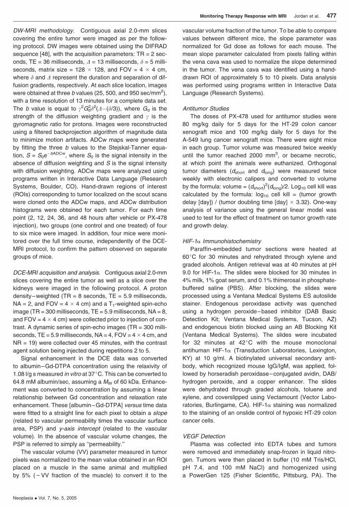