dynamics expression of osr40c1 gene and growth of...
TRANSCRIPT

BIODIVERSITAS ISSN: 1412-033X Volume 18, Number 2, April 2017 E-ISSN: 2085-4722 Pages: 801-808 DOI: 10.13057/biodiv/d180252
Dynamics expression of Osr40c1 gene and growth of maize (Zea mays) calluses in responding to salt stress
TRIONO BAGUS SAPUTRO♥, NUR FADLILLATUS SHOLIHAH, DINI ERMAVITALINI Department of Biology, Faculty of Mathematics and Natural Sciences, Institut Teknologi Sepuluh Nopember. Jl. Arif Rahman Hakim, Keputih, Sukolilo,
Surabaya 60111, East Java, Indonesia. Tel. +62-31-592-7939, Fax.: 62-31-592-3411, ♥email: [email protected], [email protected]
Manuscript received: 31 August 2016. Revision accepted: 27 March 2017.
Abstract. Saputro TB, Sholihah NF, Ermavitalini D. 2017. Dynamics expression of Osr40c1 gene and growth of maize (Zea mays) calluses in responding to salt stress. Biodiversitas 18: 801-808. Maize (Zea mays L) is one of the most planted cereal plants worldwide. The demand of maize plant is increased over the years since its various usage in industry. Unfortunately, the production rate does not meet the demands caused by the significant reduction of plantation area for housing and the others building. The marginal saline soil area can be a promising area to grow the maize although contains a high amount of salt. The high concentration of salt can significantly reduce the plant growth and productivity. The aims of this study were to determine the tolerant level of Manding and Bluto varieties against salt stress. There were two main stages on this research. First, callus induction through in vitro culture (MS0 + 2,4-D 3 mg/L), then the calluses were subcultured on the selection medium (MS0 + 2,4-D 3 mg/L + NaCl concentrations (0, 2500, 5000 and 7500 mg/L)). Second, Q-PCR analysis was used to observe the differences of gene expression level. The result shows that NaCl stress can alter the color of callus, from yellow to brown. The percentage of surviving callus was 100% in both varieties, although the callus weight decreased along with the increment of NaCl concentrations. In 7500 mg/L, Manding has 0.023 g in fresh weight, while Bluto 0,027 gr. Interestingly, the expression of Osr40c1 gene showed different level between those two varieties. Manding had only 0.82 fold, while Bluto had 4.86 folds compared with the control. Taken together, Bluto has a better performance in high content of NaCl.
Keywords: Gene expression, in vitro selection, NaCl stress, Zea mays
INTRODUCTION
Maize or corn (Zea mays L.) belongs to the family of Graminaceae (Iriany et al. 2008), which is one of main foods and essential commodities in agribusiness (Kuruseng and Farid 2009). The yield of maize is important in improving the agricultural economy in the world (Balkrishna and Shankarrao 2013). Department of Agriculture (2005) explained that in the next 20 years, the total usage of maize is expected to continue to increase. On the other hand, the conversion of agricultural land into residential area causes these needs will be difficult to fulfill. The problem can be overcome by planted maize in marginal land including saline soils that are the largest marginal land in Indonesia.
Saline soils in the world include “salt marshes” in dry regions and tidal areas; both can be found in the subtropical and tropical regions. It is estimated that between 400-900 million hectares of land in the world has a problem of salinity. Since Indonesia is an archipelago country, it has many shorelines that have a great potential to extend the area of agriculture. However, the main challenge of this soils will be the high content of salt.
Salinity is one of the main limiting factors which capable of causing a decrement in growth and productivity of plant species (Flowers 2004). Suwignyo et al. (2009) reported that salinity has a negative effect on several parameters including plant height, leaf number, leaf area, dry weight, root length and chlorophyll content on three varieties of maize, namely Arjuna, Bhisma, and
Sukmaraga. High concentration of salt can affect plant physiology, such as increases respiration rate, ion toxicity, changes in plant growth, mineral distribution, disrupting membrane permeability and decreases the photosynthetic rate (Ashraf and Foolad 2007).
Based on this reasons, it is necessary to analyze the tolerance level of local Indonesian maize against salinity stress. Two local maize were used in this research were Manding and Bluto, both varieties were showed the ability to stand in Madura island which is well known for its high content of salt. Local maize has a great demand, especially for food, because it is more durable in storage periods parameter (Arifin et al. 2010).
The aims of this research were to observe the tolerant level of Manding and Bluto clones. Badami and Amzeri (2011) explained that in vitro selection is one of the effective technique to select the varieties maize that tolerant to salinity stress with appropriate selection media. Through in vitro selection, it will be able to obtain varieties that have superior tolerant properties to salinity condition without any others environmental factor that affects the result (Rosas et al. 2003). The usage of tissue culture can help to be more focus on the physiological and biochemical mechanism without any environmental influence. Plant culture through in vitro culture is proper and reliable technique compared to traditional propagation methods because it leads to the production of disease and virus-free plants. Moreover, it allows the production of a high number of plants, in a short period and limited propagation space. In addition, rapid multiplication rate of plants that are

B IODIVERSITAS 18 (2): 801-808, April 2017
802
difficult to propagate conventionally can be easily achieved through in vitro culture (Al- Ajlouni et al. 2012). In addition, this research also analyzed the expression levels of salt-responsive genes. Salt-responsive gene is a group of an inducible gene that will be expressed when the plant is exposed to high salt concentration. The aims of this research are to evaluate the response of plant's growth, the resistance level of callus and the level of gene expression of Manding and Bluto varieties against salinity stress.
MATERIALS AND METHODS
Experimental design This research was conducted using completely
randomized design (CRD) with three replication. While the observation was made during 28 days for callus induction and 28 days for in vitro selection.
Sterilization of explant The kernels of maize were used as explants. The entries
used in this research are local varieties from Madura island including Manding and Bluto. For surface sterilization of explants, the kernels were washed in running water for 15 minutes, then soaked in a soap solution (active compounds: sodium alkyl benzene sulfonate 12%, sodium lauryl ether sulfate 5%, 0.75% cocoamido-propyl betaine, sodium salicylate 0.5%) and stirred for 30 minutes. Furthermore, the explants were soaked in an 2 grams/L of antifungal that solved in distilled double distilled water (active compounds: propinep 70%) for 30 min, and then followed by soaking in 5.25% NaOCl solution for 5 min, 70% alcohol for 10 min, and finally rinsed in sterile aquadest 3 times.
Callus induction MS0 Medium supplemented with The induction of callus
used medium MS0 (Murashige and Skoog) added with PGR (plant growth regulator) 2,4-D 3 mg/L were utilized to induced callus. First, the explants that have been surface sterilized were split into two parts, with each part contain an embryo. Then, the explants inoculated into culture bottles jar/chamber that contains medium. After inoculation process, the explants were incubated in the culture room in the dark conditions for 28 days at 25oC± 2oC (Balkrishna and Shankarrao 2013), and every two weeks, the explants were subcultured into a new medium with the same composition.
In vitro selection The formed calluses were subcultured into selection
medium MS0 + 2,4-D 3 mg/L + NaCl (0, 2500, 5000 and 7500 mg/L) and incubated for 28 days in a culture room with dark conditions. In this research, the selection of local maize was conducted through in vitro culture.
RNA extraction RNA extraction was conducted after the callus treated
with NaCl for 28 days. RNA minikit (plant) (GeneAid)
were utilized to extract the total RNA. Total RNA extraction began by cutting the fresh or frozen callus as much as 80-100 mg. Only callus from non-treated with NaCl 0 mg/L (control) and surviving callus from NaCl 7500 mg/L. were used as a material for RNA extraction. The sample was grinded in liquid nitrogen to obtain a fine powder and transferred into 1.5 ml microcentrifuge tubes. Then followed by a series of steps based on kit procedure. RNA extraction can be used immediately and stored at-80°C. The quantity of RNA was determined by using Scientific TM Thermo NanoDrop 2000, and the quality was tested by an electrophoresis method. The electrophoresis was running on 0.8% agarose gel for 20 minutes 100 volt.
Quantitative Real Time-PCR (qRT-PCR) The extraction of RNA for qRT-PCR used KAPA
SYBR® FAST One-Step qRT-PCR Kit and the procedures were performed by the kit. In this study, Osr40c1 gene was used as the target gene (accession number: X95402) and the actin gene (accession number: NM_0011136991) as a reference gene; then they were searched for the sequences obtained from NCBI (The National Center for Biotechnology Information). For Osr40c1 gene sequences, it was used a forward primer: 5'-AAG AGC TTC CGC CGC TGC ATT-3' and a reverse primer: 5'-TTC AAG CAC TGG TTG TCG CCC-3', while for an actin gene sequences, it was used a forward primer: 5'-TCG TGC CTG ACT TTG GTG ACG-3' and a reverse primer: 5'-GTC GTG GTG AAG GAG TAA CCC-3'. PCR reaction used a Qiagen Rotor-Gene. Furthermore, the amplification could be observed in the chart that appears as a result of the accumulation of fluorescence of the probe (marker). Then, the data of Quantitative PCR results were analyzed by using a relative calculation of 2-∆∆CT method to measure the difference of gene expression level in the treatment of NaCl 0 mg/L and NaCl 7500 mg/L.
Observation of parameters Morphology of callus. The parameters of callus
morphology consisted of color and texture of callus. The color of callus was generally white, yellow, purple, green and brown to blackish (Santoso and Nursandi 2003). Meanwhile, the texture of callus could be distinguished by a textured-compact callus (non-friable), friable and intermediate callus (a blend of compact callus and friable callus) (Sugiyarto and Kuswandi 2014).
Percentage of surviving callus. The observation of percentage of surviving callus was conducted with the following formula (Htwe et al. (2011):
% surviving callus = No. of tolerant callus x 100% No. of inoculated callus
Callus weight gain. Callus weight gain was measured by weighing the fresh callus weight before selection (initial weight) and after the selection of callus (final weight). The data of callus weight can be simply obtained by applying this following formula:
Callus weight gain = final growth-initial growth
SAPUTRO et al. – The growth of maize calluses on salt stress
803
Analysis of gene expression. Data were analyzed by a quantitative PCR with 2-∆∆CT method. The 2-∆∆CT method was performed after amplification process at Q-PCR process completed. The 2-∆∆CT method is an easy way to analyze the relative changes in gene expression as the results of real-time amplification using RT-PCR. To get the value of the targeted gene of mRNA expression levels, the necessary steps to calculate is formulated as follows as follows (Kenneth and Thomas 2001):
Normalization of CT (target gene) to CT (reference gene) ΔCT (cal.) = CT (calib. target)-CT (calib. reference) ΔCT (exp.) = CT (exp. target)-CT (exp. reference) Normalization of ΔCT (exp. sample) to ΔCT (calib. sample) ΔΔCT = ΔCT (exp. sample)-ΔCT
(calib. sample) Calculate the ratio of gene expression The ratio of expression = 2-∆∆C
T Note: CT = Threshold cycle, calib = calibrator, exp =
experimental
Data analysis The observation data of morphological callus, the
surviving callus percentage and the level of gene expression were analyzed qualitatively. The data of callus weight parameter was analyzed using ANOVA (analysis of variance) with one factor at a confidence level of 95%. If ANOVA test results showed “p-value” ≤ 0.05, it would be continued to a Duncan test at 5% significance level by using SPSS Statistic Software 24.0 version.
RESULTS AND DISCUSSION
Effect of variation of NaCl concentrations on callus morphology
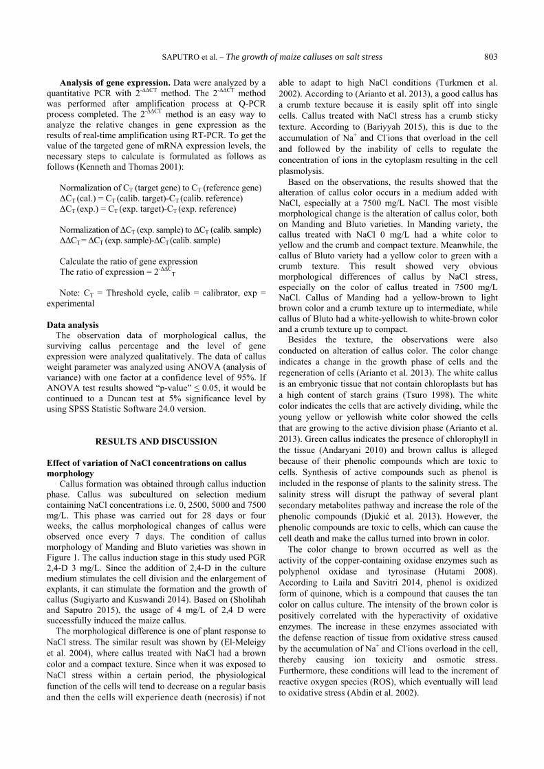
Callus formation was obtained through callus induction phase. Callus was subcultured on selection medium containing NaCl concentrations i.e. 0, 2500, 5000 and 7500 mg/L. This phase was carried out for 28 days or four weeks, the callus morphological changes of callus were observed once every 7 days. The condition of callus morphology of Manding and Bluto varieties was shown in Figure 1. The callus induction stage in this study used PGR 2,4-D 3 mg/L. Since the addition of 2,4-D in the culture medium stimulates the cell division and the enlargement of explants, it can stimulate the formation and the growth of callus (Sugiyarto and Kuswandi 2014). Based on (Sholihah and Saputro 2015), the usage of 4 mg/L of 2,4 D were successfully induced the maize callus.
The morphological difference is one of plant response to NaCl stress. The similar result was shown by (El-Meleigy et al. 2004), where callus treated with NaCl had a brown color and a compact texture. Since when it was exposed to NaCl stress within a certain period, the physiological function of the cells will tend to decrease on a regular basis and then the cells will experience death (necrosis) if not
able to adapt to high NaCl conditions (Turkmen et al. 2002). According to (Arianto et al. 2013), a good callus has a crumb texture because it is easily split off into single cells. Callus treated with NaCl stress has a crumb sticky texture. According to (Bariyyah 2015), this is due to the accumulation of Na+ and Cl-ions that overload in the cell and followed by the inability of cells to regulate the concentration of ions in the cytoplasm resulting in the cell plasmolysis.
Based on the observations, the results showed that the alteration of callus color occurs in a medium added with NaCl, especially at a 7500 mg/L NaCl. The most visible morphological change is the alteration of callus color, both on Manding and Bluto varieties. In Manding variety, the callus treated with NaCl 0 mg/L had a white color to yellow and the crumb and compact texture. Meanwhile, the callus of Bluto variety had a yellow color to green with a crumb texture. This result showed very obvious morphological differences of callus by NaCl stress, especially on the color of callus treated in 7500 mg/L NaCl. Callus of Manding had a yellow-brown to light brown color and a crumb texture up to intermediate, while callus of Bluto had a white-yellowish to white-brown color and a crumb texture up to compact.
Besides the texture, the observations were also conducted on alteration of callus color. The color change indicates a change in the growth phase of cells and the regeneration of cells (Arianto et al. 2013). The white callus is an embryonic tissue that not contain chloroplasts but has a high content of starch grains (Tsuro 1998). The white color indicates the cells that are actively dividing, while the young yellow or yellowish white color showed the cells that are growing to the active division phase (Arianto et al. 2013). Green callus indicates the presence of chlorophyll in the tissue (Andaryani 2010) and brown callus is alleged because of their phenolic compounds which are toxic to cells. Synthesis of active compounds such as phenol is included in the response of plants to the salinity stress. The salinity stress will disrupt the pathway of several plant secondary metabolites pathway and increase the role of the phenolic compounds (Djukić et al. 2013). However, the phenolic compounds are toxic to cells, which can cause the cell death and make the callus turned into brown in color.
The color change to brown occurred as well as the activity of the copper-containing oxidase enzymes such as polyphenol oxidase and tyrosinase (Hutami 2008). According to Laila and Savitri 2014, phenol is oxidized form of quinone, which is a compound that causes the tan color on callus culture. The intensity of the brown color is positively correlated with the hyperactivity of oxidative enzymes. The increase in these enzymes associated with the defense reaction of tissue from oxidative stress caused by the accumulation of Na+ and Cl-ions overload in the cell, thereby causing ion toxicity and osmotic stress. Furthermore, these conditions will lead to the increment of reactive oxygen species (ROS), which eventually will lead to oxidative stress (Abdin et al. 2002).

B IODIVERSITAS 18 (2): 801-808, April 2017
804
Figure 1. The callus morphology of Manding variety (left) and Bluto variety (right) at different NaCl concentrations. Note: A. 0 mg/L, B. 2500 mg/L, C. 5000 mg/L, and D. 7500 mg/L
Table 1. The effect of NaCl concentration variation to the percentage of surviving callus on Manding and Bluto varieties.
Varieties
The percentage of surviving callus (%) in NaCl concentration variation
0 mg/L 2500 mg/L
5000 mg/L
7500 mg/L
Manding 100 100 100 100
Bluto 100 100 100 100
The result shows that the greenish callus turns into
yellow. This condition indicates a decrease of chlorophyll content in the callus cells. The salinity stress may affect the reduction in chlorophyll content due to the increased activity of the enzyme chlorophyllase caused by reducing absorption of Mg and Fe ions that are involved in the formation of chloroplasts (Al Shorafa et al. 2014). Mg2+ ion also acts as an enzyme cofactor and plays an important role in the export process of photosynthesis, and can prevent the degradation of chlorophyll by increasing the activity of RuBP carboxylase oxygenase enzyme (Ramezani et al. 2011).
Effect of variation in NaCl stress concentrations on percentage of surviving callus
The details percentage of surviving callus incubated for 28 days in various NaCl concentrations was shown in Table 1. In this study, callus was able to stand and survive to any treatment. This condition was demonstrated by the percentage of surviving callus reaches 100% in each treatment.
Callus was able to survive in the medium with the treatment of NaCl in tested concentrations i.e 0, 2500, 5000 and 7500 mg/L in 28 days through the defense and tolerance mechanisms with different response to each callus. Nonetheless, it was still possible the salinity stress may cause a reduction in growth rate and the mortality callus tissue when exposed over a month (Queiros et al. 2007). Summart et al. 2010 stated that the salinity stress with a particular concentration leads the slow growth of growth.
Several adaptation mechanisms will be developed by the plant in high NaCl condition. Betaine, free amino acids, soluble carbohydrates, and proline are several well-known compounds that protect the plant against salinity (Al-Shorafa et al. 2014). Such compounds will provide the protection to plants from the effects of stress through the different mechanisms. Furthermore, the mechanism can be single or a combination of cellular osmotic balance, detoxification of reactive oxygen species maintains the membrane integrity and stabilizes the enzyme or protein (Ramezani et al. 2011).
Plants that exposed with high salinity showed the accumulation of proline. Proline is a signaling molecule/ regulatory, which can activate the response to salinity stress adaptation process, as well as an osmoregulators (Ashraf and Ooraj 2006). Soluble carbohydrate will also accumulate when a decrease in the level of CO2 assimilation in drought stress, which has a role in balancing the osmotic regulation, it also acts as a metabolic signal of NaCl stress treatment (Chaves et al. 2003). In addition, the cell will produce antioxidants and enzymes such as superoxide dismutase (SOD), APX (ascorbate peroxidase)

SAPUTRO et al. – The growth of maize calluses on salt stress
805
and catalase that act to recover callus from the effects of osmotic stress Quesada et al. 2003). Superoxide dismutase (SOD) system is the first defense in tackling the damage caused by AOS to catalyze O2 into H2O2. Ascorbate peroxidase (APX) plays an important role in the detoxification of H2O2. APX is contained in the chloroplast stroma, the thylakoid membrane, micro entities, the cytosol, and mitochondria. Catalase is an enzyme that serves to catalyze the decomposition of H2O2 into water and oxygen (Rao and Prabhavathi 2012).
Some tolerance mechanisms explained, can help plants to survive on NaCl stress. As reported by (Bariyyah 2015), that the criteria of resistant callus to NaCl stress is the callus that capable of living on a saline medium, callus color is white to yellow-brown, has a friable structure and able to grow in enlargement and cell division, thereby affects the volume of callus and the percentage of callus proliferation.
Effect of variation of NaCl concentration on callus weight gain
The callus weight was calculated from the difference between the weight of callus before subjected to selection medium and callus weight after 28 days of incubation. The influence of NaCl concentration on callus weight was analyzed using One-Way ANOVA test. If the results of ANOVA test showed a significant effect (the Significance ≤0.05), it proceed to a Duncan test with a confidence level of 95% (α = 0.05). The effect of NaCl concentration variation on weight gain callus of Manding and Bluto varieties were shown in Table 2.
Based on the results of ANOVA analysis, the variations of NaCl concentration were significantly affected to the callus weight gain of Manding with the significance of 0.053 (>0.05). Meanwhile, on Bluto variety, the variations of NaCl concentration had a significant impact on the callus weight gain with the significance value 0.001 (<0.05). As shown in Table 2, the higher concentration of NaCl was followed by callus weight loss. The lowest weight gain callus was obtained at the highest NaCl concentration treatment, i.e. 7500 mg/L. Both Manding and Bluto, they had the same callus weight at 7500 mg/L NaCl stress. The callus weight of Manding variety in the highest concentration of NaCl was decreased to 93% compared with controls, whereas the Bluto callus decreased to 87% compared with control. The details of callus weight decline can be seen in Figure 2.
Based on the ANOVA analysis, the result of callus weight measurement shows not significant. The increasing concentration of NaCl was not followed by a decrease in callus weight significantly. The phenomenon caused by the level of NaCl stress tolerance depends on the genotype. Thus, the different callus on the medium with the same concentration will have a different response (Summart et al. 2010). The decrement of callus weight was inclined following the increment of NaCl concentration on each variety. Sholihah and Saputro (2015) report the same results, the salinity stress decreased the callus weight of Manding variety up to 83% compared to control.
Untreated callus with NaCl (control) had a greater
callus weight compared with a NaCl treated-stress. The fresh weight of large callus without NaCl treatment caused by the optimum absorption of water through the cells. The fresh weight depends on the speed of cells to divide and multiply to be callus (Rahayu et al. 2003). Meanwhile, the decrease of callus weight gain can occur because of salinity stress, the imbalance absorption of water and nutrients, and the inhibition of metabolism due to the imbalance disorder of ion and osmotic effect. The decline in the growth of callus is a common phenomenon that occurs when the plant was exposed to salinity.
Callus requires more energy to held a normal metabolism and causing the decrease of growth as the final impact (Ubudiyah 2013; Yunita et al. 2014). The energy generated seems to be more widely used to set the adjustment of osmotic pressure and it will have a negative impact on the cell mass (Babu 2007). Moreover, callus weight loss can be caused by a water deficit in plant cells. Water shortage phenomenon due to the accumulation of NaCl on the planting media. The high concentration of NaCl in media leads to the decrement of water potential in media that will drive water out of the cell. Hence, in plants, it will suffer a water deficit and a lower turgor pressure. The impact of water deficit will have a significant influence on the growth of callus since water is a major factor in the physiological processes of plants (Li et al. 2006). Table 2. The effect of NaCl concentration variations on callus weight gain on Manding and Bluto varieties
Varieties NaCl concentration (mg/L)
Callus weight (gram)
Manding 0 0.360 2500 0.120 5000 0.030 7500 0.023
Bluto 0 0.207 2500 0.053 5000 0.043 7500 0.027
Note: The values followed by the same letter in the same column of each variety showed no significant difference in a Duncan test with a significance level α = 0.05
Figure 2. Interaction of NaCl concentration effect to callus weight gain of Manding and Bluto varieties

B IODIVERSITAS 18 (2): 801-808, April 2017
806
Table 3. Osr40c1 gene expression levels of qPCR analysis results
Varieties Ct Ct Value ∆Ct (Ex.) ∆Ct (Con.) ∆∆Ct Expression Fold Change (2^ (-∆∆Ct))
Manding TE 21.33 1.02 - 0.28 0.823 HE 20.31 TC 19.84 - 0.74 HC
19.10
Bluto TE 18.55 -1.76 - -2.28 4.857 HE 20.31 TC 19.62 - 0.52 HC 19.10
Note: TE = Target gene experimental), HE = housekeeping gene experimental, TC = target gene control, HC = housekeeping gene control
Salinity stress can also decrease the levels of K+ and Ca2
+, while Na+ ions increase. It had been reported previously that K+ and Ca2
+ ions play an important role in maintaining osmotic balance also regulate the opening and closing of stomata (Hirschi 2004). According to (Summart et al. 2010), the high content of Na+ ions in cells inhibits decision K+ leads to an increased ratio of Na+/K+, so the balance of ions in the cell will be disturbed and damage the structure and function of membrane integrity. The excess of Na+ and Cl- contents in the extracellular can also inhibit the absorption of nitrogen assimilation of nitrate (NO3) which essential for plant growth (Yuniati 2004). The accumulation of Na+ and Cl-ions is overload in the cell causing ion toxicity and osmotic stress. Furthermore, these conditions will lead to an increase in a reactive oxygen species (ROS), which eventually will lead to oxidative stress (Abdin et al. 2002). Oxidative stress may cause the death of cells (necrotic) because of not able to adapt to NaCl stress conditions (Turkmen et al. 2002), which can cause a weight loss of callus on NaCl stress.
Analysis of salt responsive gene expression (Osr40c1) There were two approaches to the analysis of gene
expression levels in qPCR results. Those two approaches are absolute and relative calculations (Kenneth and Thomas 2001). In this experiment, we used a relative calculation to measure the gene expression rate. The relative calculation was conducted by simple comparison between the expression level of the target gene with housekeeping gene, both in callus treated with NaCl (test) or callus without treated NaCl (control). The callus analyzed its gene expression is only the callus treated with 7500 mg/L NaCl and callus without treated (NaCl 0 mg/L). The calculation of gene expression used qPCR results data in the form of CT value (threshold cycle) which was a point in time or cycle where the fluorescence of the target PCR amplification was first detected (Wong and Juan 2005), then CT value obtained was analyzed by using the 2-∆∆CT method. The target genes analyzed included Osr40c1 and actin as a housekeeping gene. The analytical results were shown in Table 3.
Based on the calculation results in Table 3 showed that the Osr40c1 gene expression level of Manding variety
treated with NaCl as much as 0.82 fold compared without NaCl. The Osr40c1 gene expression level on a Bluto variety treated with NaCl increased as much as 4.86 folds compared without NaCl. The level of gene expression indicated that the salinity-responsive gene in callus would be increased to reduce the impact of NaCl stress as a defensive response. The expression of salinity-responsive genes will overcome the effects of stress due to salinity, so the plants can adapt and survive.
The level of Osr40c1 gene expression in callus of Bluto variety was higher than in callus of Manding variety. The level of gene expression of several inducible genes shows the survival rate of the plant to adapt to high salinity environment. The level of gene expression depends on the duration of stressing time, the stress level and the degree of severity of crops due to stress (Moons et al. 1997). The expression of Osr40c1 gene will increase the resistance to stand in NaCl stress.
Osr40c1 gene is one of the salinity-responsive genes. Besides Osr40c1, other salinity-responsive genes are SOS gene (salt sensitive overlay), NHX, AVP, PAR, AAPK, PKS3, P5CS, OTS, IMTI, MT1D and others (Munns 2002). Osr40c1 gene is an osmotic stress-responsive gene which can be expressed on salinity stress, induced by the accumulation of ABA (abscisic acid) hormone and translated into proteins putative r40c1 (Osr40c1), and has a molecular weight of 40 kDa (Dooki et al. 2006). Osr40c1 protein can be found in the plasma membrane of young rice roots gripped in salinity (Huang et al. 2006).
Osr40c1 gene is included Osr40 gene family which is responsive genes to ABA and salinity stress first characterized by (Moons et al. 1995). In a report of Riccardi et al. (1998), it also identified a protein of the same type in maize treated in water shortages and associated with the dehydration tolerance mechanisms (Riccardi et al. 2004). This gene acts to ameliorate the effect of osmotic stress, such as detoxification of ROS (Veeranagamallaiah et al. 2008). It can also reduce the process of wilting. This indicates that this gene as the osmoregulation gene and unresponsive to the impact of the gripped in the long term. The protein of Osr40c1 also can regulate the prevention of loss of water in the cell and the rigidity of the cell wall (Dooki et al. 2006).

SAPUTRO et al. – The growth of maize calluses on salt stress
807
A saline condition in the growing media is a result of the accumulation of salt in a high concentration, such as NaCl. NaCl salt in excessive planting medium will cause a decrease in soil water potential and the water potential in higher plants will trigger the release of water from plant tissue to outside soil or growing media, so the plants lose the turgor pressure and water deficit. It is due to the high concentration of Na+ and Cl-ions will disrupt the membrane permeability, resulting in Na+ and Cl-ions can enter into a tissue of cells. The accumulation of Na+ and Cl-ions overload in the cell will give a signal of salinity stress in plants (Salisbury and Ross 1995).
Salinity stress may induce the activation of many genes in ABA biosynthesis. ABA is one of the phytohormones that has a key role in plant defense activity against many environmental stresses. Chinnusamy et al. (2004) reported that several genes that involved in ABA synthesis were activated during the salt treatment. Further accumulation of ABA will be able to induce an osmotic stress-responsive gene expression, i.e. through the ABA-dependent pathway, such as the DRE and Osr40c1 genes. In addition, those induced the expression of genes that not responsive to salinity through the ABA-dependent pathway, called the ABA-independent pathways, such as SOS and MYC (Tuteja 2007).
All in all, it can be concluded the performance of callus were affected by NaCl stress in both varieties Manding and Bluto. In control, the callus performance was dominated by a white color and a crumb texture, while in NaCl treatment dominated by a brown color and a crumb texture. The percentage of surviving callus were 100% in each treatment in both varieties. The callus weight on both varieties decreases in line with an increment of NaCl. Callus of Manding variety was declined as much 93% and Bluto variety was 87% compared with control. A big differentiation in the level of Osr40c1 gene expression occurred on both varieties. The level of gene expression in Manding variety was 0.82 fold compared with control. Furthermore, the level of Osr40c1 gene expression on Bluto variety was 4.86 folds compared with control. Based on the observations of some parameters, it showed that the resistance level of Manding and Bluto to the high concentration of NaCl was different. Bluto variety showed more tolerant than Manding variety.
ACKNOWLEDGEMENTS
Authors thank Laboratory of Plant Bioscience and Technology, Institut Teknologi Sepuluh Nopember, Surabaya, Indonesia for supporting the equipment.
REFERENCES
Abdin MZ, Rehman RU, Israr M et al. 2002. Abiotic stress related genes and their role in conferring resistance in plant. Indian J Biotechnol 1: 225-24.
Al-Ajlouni Z, Ajlouni M, Shatnawi M, Shibli R, Makhdmeh I, Abu-Romman S, Al-Ghazawi Al. 2012. Callus induction, plant regeneration, and growth on barley (Hordeum vulgare L.). South Western J Hort Biol Environ 3 (1): 25-39.
Al-Shorafa W, Mahadeen A, Al-Absi K. 2014. Evaluation for salt stress tolerance in two strawberry cultivars. Am J Agric Biol Sci 9 (3): 334-341.
Andaryani S. 2010. Studies Using Various Concentrations of BAP and 2,4-D on Callus Induction (Jatropha curcas L.) In Vitro. [Thesis]. Universitas Sebelas Maret, Surakarta. [Indonesian]
Arianto B, Zainuddin, Mirni UB. 2013. Callus induction of two clones of Sulawesi cacao (Theobroma cacao L.) in various concentrations of 2.4-dichlorophenoxyacetic acid in vitro. Agrotekbis 1 (3): 211-220. [Indonesian]
Arifin Z, Istiqomah N, Fatmawati. 2010. Development of local varieties Sumenep maize. Proceedings of National Seminary of Cereals Crops. Cereal Crops Research Institute, Maros, 27-28 Juli 2010. [Indonesian]
Ashraf M, Foolad MR. 2007. Improving plant abiotic stress resistance by exogenous application of osmoprotectants glycine betaine and proline. Environ Exp Bot 59: 206-216.
Ashraf M, Orooj A. 2006. Salt stress effects on growth, ion accumulation and seed oil concentration in an arid zone traditional medicinal plant ajwain (Trachyspermum ammi [L.] Sprague). J Arid Environ 64: 209-220.
Babu S. 2007. Effect of salt stress in the selection of salt tolerant hybrids in rice under in vitro and in vivo condition. Asian J Plant Sci 6 (1): 137-142.
Badami K, Amzeri A. 2011. Identification of drought tolerant somaclonal variants in maize population of in vitro selection with PEG. Agrovigo 4 (1): 7-13.
Balkrishna RA, Shankarrao SS. 2013. In vitro screening and molecular genetic markers associated with salt tolerance in maize. Afr J Biotechnol 12 (27): 4251-4255.
Bariyyah K. 2015. The effect of NaCl on callus of sugarcane varieties Bululawang. Agroekotek J 7 (1): 1-5. [Indonesian]
Chaves MM, Maroco JP, Pereira JS. 2003. Understanding plant response to drought: from genes to the whole plant. Funct Plant Biol 30: 239-264.
Chinnusamy V, Schumaker K, Zhu JK. 2004. Molecular genetic perspectives on cross-talk and specificity in abiotic stress signaling in plants. J Exp Bot 5: 225-236.
Department of Agriculture. 2005. Prospects and direction of maize agribusiness development. Agency for Agricultural Research and Development, Department of Agriculture, Jakarta. [Indonesian]
Djukić JM, Nemanja S, Svetlana R et al. 2013. Differential response of three contrasting peas (Pisum arvense, P. sativum and P. fulvum) species to salt stress: assessment of variation in antioxidative defense and miRNA expression. Aust J Crop Sci 7 (13): 2145-2153.
Dooki AD, Mayer PF, Askari H et al. 2006. Proteomic responses of rice young panicles to salinity. Proteomics 6: 6498-6507.
El-Meleigy AE, Gabr MF, Fouad H et al. 2004. Responses to NaCl salinity of tomato cultivated and breeding lines differing in salt tolerance in callus cultures. Int J Agric Biol 6 (1): 1560-8530.
Flowers TJ. 2004. Improving crop salt tolerance. J Exp Bot 396 (55): 307-319.
Hirschi D. 2004. The calcium conundrum, both versatile nutrient and specific signal. Plant Physiol 136: 2438-2442.
Htwe NN, Mahmood M, Ling HC et al. 2011. Responses of some selected Malaysian rice genotypes to callus induction under in vitro salt stress. Afr J Biotechnol 10 (3): 350-362.
Huang F, Fulda S, Hagermann M et al. 2006. Proteomic screening of salt-stress-induced changes in plasma membranes of Synechocystis sp. strain PCC6803. Proteomics 6: 910-920.
Hutami S. 2008. Review: Browning in tissue culture. J AgroBigen 4 (2): 83-88. [Indonesian]
Iriany RN, Yasin MHG, Andi TM. 2008. The origin, history, evolution and taxonomy of maize. Cereal Crops Research Institute, Maros. [Indonesian]
Kenneth JL, Thomas DS. 2001. Analysis of relative gene expression data using real-time quantitative PCR and the 2-∆∆CT method. Methods 25: 402-408.
Kuruseng MA, Farid M. 2009. Analysis of the heritability of maize resistant salinity and drought result of mutations induced by gamma rays. Jurnal Agrisistem 5 (1): 30-39.
Laila FN, Savitri, ES. 2014. The production of secondary metabolites steviosida in stevia (Stevia rebaudiana Bert. M.) callus culture with the addition of 2,4-D and PEG (Polyethylene Glycol) 6000 on MS medium. El-Hayah 4 (2): 57-65. [Indonesian]

B IODIVERSITAS 18 (2): 801-808, April 2017
808
Li R, Guo P, Baum M et al. 2006. Evaluation of chlorophyll content and fluorescence parameters as indicators of drought tolerance in Barley. Agr Sci China 5 (10): 751-757.
Moons A, Bauw G, Prinsen E et al. 1995. Molecular and physiological responses to abscisic acid and salts in roots of salt-sensitive and salt-tolerant Indica rice varieties. Plant Physiol 107: 177-186.
Moons A, Els P, Guy B et al. 1997. Antagonistic effects of abscisic acid and jasmonates on salt stress-inducible transcripts in rice roots. The Plant Cell 9: 2243-2259.
Munns R. 2002. Comparative physiology of salt and water stress. Plant Cell Environ 25: 239-250.
Queiros F, Fidalgo F, Santos I et al. 2007. In vitro selection of salt tolerant cell lines in Solanum tuberosum L. Biol Plantarum 51 (4): 728-734.
Quesada V, Ponce MR, Micol JL. 2003. Genetic analysis of salt-tolerant mutant in Arabidopsis thaliana. Genetic 154: 421-436.
Rahayu B, Solichatun, Anggarwulan E. 2003. Effect of 2,4-dichlorophenoxyacetic acid (2,4-D) on the formation and growth of callus and flavonoid content on Acalypha indica L. callus cultures. Biofarmasi 1 (1): 1-6. [Indonesian]
Ramezani E, Sepanlou MG, Badi AN. 2011. The effect of salinity on the growth, morphology and physiology of Echium amoenum Fisch. & Mey. Afr J Biotechnol 10 (44): 8765-8773.
Rao S, Prabhavathi P. 2012. Biotechnology-molecular studies and novel applications for improved quality of human life. InTech, Shanghai.
Riccardi F, Gazeau P, Jacquemot MP et al. 2004. Deciphering genetic variations of proteome responses to water deficit in maize leaves. Plant Physiol Biochem 42: 1003-1011.
Riccardi F, Gazeau P, Vienne D et al. 1998. Protein changes in response to progressive water deficit in maize: quantitative variation and polypeptide identification. Plant Physiol 117: 1253-1263.
Rosas HG, Garcia SS, Reyes GR et al. 2003. Preliminary results on in vitro selection for tolerance to chloride excess in avocado. Revista Chapingo Serie Horticulture 9 (1): 39-43.
Salisbury FB, Ross CW. 1995. Plant Physiology. 4th ed. Wadsworth Publishing Company, California.
Santoso U, Nursandi F. 2003. Plant tissue culture. Pusbitan University of Muhammadiyah Malang, Malang. [Indonesian]
Sholihah NF, Saputro TB. 2015. In vitro selection of maize (Zea mays L.) varieties Talango and Manding to Salinity Stress. Jurnal Sains dan Seni ITS 4 (1): E60-E63. [Indonesian]
Sugiyarto L, Kuswandi PC. 2014. Anredera codifolia L. leaf callus induction in the development of traditional medicinal plants. Jurnal Sains Dasar 3 (1): 56-60. [Indonesian]
Summart J, Thanonkeo P, Panichajakul S et al. 2010. Effect of salt stress on growth, inorganic ion and proline accumulation in Thai aromatic rice, Khao Dawk Mali 105, callus culture. Afr J Biotechnol 9 (2): 145-152.
Suwignyo RA, Hayati R, Mardiyanto. 2009. Effect of early low salinity treatment on growth and salinity tolerant maize. Sriwijaya University, Palembang.
Tsuro M, Koda M, Inoue M. 1998. Comparative effect of different types of cytokinin for shoot formation and plant regeneration in leaf-derived callus of lavender (Lavandula vera D.C.). Kyoto Prefectural University, Japan.
Turkmen O, Sensoy S, Erdal I et al. 2002. Effect of calcium on the emergence and seedling of tomatoes grown in salty growing media conditions. J Agric Sci 12: 53-57.
Tuteja N. 2007. Mechanisms of high salinity tolerance in plants. Methods in Enzymol 428: 419-439.
Ubudiyah IWA. 2013. Response of several varieties of rice (Oryza sativa L.) callus in salinity stress conditions (NaCl). [Thesis]. Institute of Teknologi Sepuluh Nopember, Surabaya. [Indonesian]
Veeranagamallaiah G, Jyothsnakumari G, Thippeswamy M et al. 2008. Proteomic analysis of salt stress responses in foxtail millet (Setaria italica L. cv. Prasad) seedlings. Plant Sci 175: 631-641.
Wong ML, Medrano JF. 2005. Real-time PCR for mRNA quantitation. BioTechniques 39: 75-85.
Yuniati R. 2004. Screening strains of soybean (Glycine max (L.) Merrill) that NaCl tolerant for cultivation on marginal land. Makara Sains 8 (1): 21-24. [Indonesian]
Yunita R, Khumaida N, Sopandie D et al. 2014. Growth and regeneration of rice (Oryza sativa L.) callus in salt medium. Biosci Res 11 (1): 4-9.