dynamics of cell division (frontiers in molecular biology)
TRANSCRIPT


Dynamics of Cell Division

Frontiers in Molecular BiologySERIES EDITORS
B. D. HamesDepartment of Biochemistry
and Molecular BiologyUniversity of Leeds, Leeds LS2 9JT, UK
D. M. GloverCancer Research Campaign LaboratoriesDepartment of Anatomy and Physiology
University of Dundee, Dundee DD1 4HN, UK
TITLES IN THE SERIES
1. Human RetrovirusesBryan R, Cullen
2. Steroid Hormone ActionMalcolm G. Parker
3. Mechanisms of Protein FoldingRoger H. Pain
4. Molecular GlycobiologyMinoru Fukuda and Ole Hindsgaul
5. Protein Kinases]im Woodgett
6. RNA-Protein InteractionsKyoshi Nagai and Iain W. Mattaj
7. DNA-Protein: Structural InteractionsDavid M. J. Lilley
8. Mobile Genetic ElementsDavid J. Sherratt
9. Chromatin Structure and Gene ExpressionSarah C. R. Elgin
10. Cell Cycle ControlChris Hutchinson and D. M. Glover
11. Molecular Immunology (Second Edition)B. David Hames and David M. Glover
12. Eukaryotic Gene TranscriptionStephen Goodbourn
13. Molecular Biology of Parasitic ProtozoaDeborah F. Smith and Marilyn Parsons
14. Molecular Genetics of PhotosynthesisBertil Andersson, A. Hugh Sailer, and James Barber
15. Eukaryotic DNA Replication/. Julian Blow
16. Protein TargetingStella M. Hurtley
17. Eukaryotic mRNA ProcessingAdrian Kramer
18. Genomic ImprintingWolfReik and Azim Surani
19. Oncogenes and Tumour SuppressorsGordon Peters and Karen Vousden
20. Dynamics of Cell DivisionSharyn A. Endow and David M. Glover

Dynamics ofCell Division
EDITED BY
Sharyn A. EndowDepartment of Microbiology
Duke University Medical CenterDurham, North Carolina, USA
and
David M. GloverCRC Laboratories
Department of Anatomy and PhysiologyUniversity of Dundee, Dundee, UK
Oxford New York TokyoOXFORD UNIVERSITY PRESS
1998

Oxford University Press, Great Clarendon Street, Oxford OX2 6DP
Oxford New 'YorkAthens Auckland Bangkok Bogota Bombay Buenos Aires Calcutta
Cape Town Chennai Dar es Salaam Delhi Florence Hong Kong IstanbulKarachi Kuala Lumpur Madrid Melbourne Mexico City Mumbai
Nairobi Paris Sao Paolo Singapore Taipei Tokyo Toronto Warsaw
and associated companies inBerlin Ibadan
Oxford is a trade mark of Oxford University Press
Published in the United Statesby Oxford University Press Inc., New York
© Oxford University Press, 1998
All rights reserved. No part of this publication may bereproduced, stored in a retrieval system, or transmitted, in any
form or by any means, without the prior permission in writing of OxfordUniversity Press. Within the UK, exceptions are allowed in respect of anyfair dealing for the purpose of research or private study, or criticism or
review, as permitted under the Copyright, Designs and Patents Act, 1988, orin the case of reprographic reproduction in accordance with the terms of
licences issued by the Copyright Licensing Agency. Enquiries concerningreproduction outside those terms and in other countries should be sent tothe Rights Department, Oxford University Press, at the address above.
This book is sold subject to the condition that it shall not,by way of trade or otherwise, be lent, re-sold, hired out, or otherwise
circulated without the publisher's prior consent in any form of bindingor cover other than that in which it is published and without a similar
condition including this condition being imposedon the subsequent purchaser.
A catalogue record for this book is available from the British Library
Library of Congress Cataloging in Publication Data(Data applied for)
ISBN 0 19 963683 4 (Hbk)ISBN 0 19 963682 6 (Pbk)
Typeset by Footnote Graphics, Warminster, WiltsPrinted in Great Britain by Bath Press Ltd., Bath

Preface
The major events in the cytology of cell division were described over a century ago byearly cell biologists. Only recently have molecular studies begun to have a majorimpact on our comprehension of the regulation, dynamics, and mechanics of theprocess. Although recent advances have shed considerable light upon the topic, thenew findings raise many questions for the future. In the past decade we have seen aquantum leap in our understanding of the processes that regulate progression of thecell through different stages of the cell cycle. The transitions from one phase of thecycle to the next depend upon the sequential activity of cyclin dependent kinases. Tomaintain the fidelity of replication and segregation of genetic information, a numberof surveillance mechanisms have evolved to ensure that a cell does not progress to thenext phase of the cycle before the previous one has been completed. In the context ofcell division, it is crucial to ensure that DNA has been completely replicated and anydamage repaired before the cell enters mitosis, and there must also be mechanismsthat ensure the correct alignment of chromosomes at metaphase, before sisterchromatids segregate from one another.
We begin the book with an overview of the regulation of cell division by Yu,Duronio, and Sullivan. Entry into mitosis requires the activation of the major mitotickinase, p34cdc2, which is though to have a wide range of substrates. Breakdown of thenuclear envelope, chromosome condensation, and changes in microtubule dynamicsall require the activity of this enzyme, which in many cases acts directly uponsubstrates that are major components of the cytoskeleton. In the second chapter,Miller and Forbes discuss how nuclear envelope breakdown and reassembly occur ascells progress through mitosis. The function and regulation of spindle pole bodies andcentrosomes are reviewed in Chapter 3 by Hagan, Gull, and Glover. These organellesundergo their own cyclical duplication and division cycle that must be coordinatedwith the nuclear cycle and the overall phase of the cell cycle. The duplication andseparation of these bodies is central to the organization of microtubules into bipolarspindles, and thus to ensure correct chromatid segregation in mitosis and meiosis. Theprocess of spindle assembly and chromosome behaviour is the topic examined byVernos and Karsenti. These authors consider how the 'dynamics of microtubuleassembly and disassembly are coordinated with, and facilitated by, microtubule-associated motor proteins during formation of the mitotic spindle. They emphasizethat a bipolar structure can arise through the intrinsic properties of microtubules andmotor proteins alone. The following chapter by Sullivan discusses centromeres andkinetochores, and how these vary from yeast to metazoans. Not only the structure ofthese chromosomal regions is considered, but also the mechanisms by which sisterchromatid cohesion is achieved and kinetochore function is regulated during mitosis.The ends of chromosomes present a different set of problems, particularly during

vi PREFACE
S-phase. Telomeres and the mechanisms by which the ends of chromosomes arereplicated are the subjects of the chapter by Hughes and Lundblad. Exciting recentwork has led to the identification of the telomerase catalytic subunit in both yeast andhuman cells. The generation of gametes during meiosis is an area of cell divisionabout which we understand relatively little. The chapter by Karpen and Endowfocuses on the special behaviour of the meiotic chromosomes and the dynamics of themeiotic spindle. Two divisions occur during meiosis without an intervening S-phase.The divisions must be integrated into a highly specialized programme of cellulardifferentiation and must accommodate the exchange of genetic material betweenmaternal and paternal homologues. Perhaps most important within the context of ourbook, there must be mechanisms to ensure that homologous chromosomes segregatefrom each other without separation of sister chromatids in the first division, incontrast to the second division in which sisters undergo segregation. In thinkingabout cell division, we must not forget the partitioning of cytoplasm and organellesbetween the daughers. This topic is explored by Shima and Warren. Many pressingquestions still remain in the area of cytoplasmic inheritance, in particular those thatconcern the biogenesis and partitioning of membranous organelles like the Golgiapparatus. The final chapter by Goldberg and colleagues concerns cytokinesis, thefinal event of cell division. The chapter addresses how the site of the cleavage furrowis determined, how initiation of furrow formation is regulated, and the mechanisms ofcontractile ring assembly and function.
Reviewing the enormous literature for the chapters has been a tremendous task forthe authors. We can both speak from personal experience in saying that the insightsthat have come from writing the chapters that we co-authored were well worth theexpended effort. We hope that this is a sentiment felt by the other authors. Ourcollective efforts should make it easier for others to share the excitement that we allfeel about this field. We greatly appreciate the efforts of all the authors. We thankthem for their participation in the project, for completing their chapters in a timelyfashion, for revising their chapters and bringing them up to date for the book, and fortheir patience with the editorial process.
Durham, NCDundee, ScotlandNovember 1997
S.E.D.G.

Contents
Plate section falls between pages 222 and 223
List of contributors
Abbreviations
1. Cell cycle checkpoints: safe passage through mitosisKRISHNA R. YU, ROBERT J. DURONIO, AND WILLIAM SULLIVAN
1. Introduction2. Activation of the mitotic CDK controls entry into mitosis
2.1 Activation of mitotic CDK requires an association with cyclin2.2 The APC mediates cyclin degradation and sister chromosome separation2.3 Proteins that negatively regulate CDK activity2.4 Post-translational phosphorylation regulates mitotic CDK activity
3. Cell cycle checkpoints3.1 Mutational analysis identifies RAD9 as a DNA damage checkpoint3.2 Checkpoints monitor many cellular events and involve signal
transduction pathways that link delays in the cell cycle to repairprocesses
3.3 Ambiguities in the concept of cell cycle checkpoints4. Lessons from budding yeast: the role of checkpoints in monitoring
the completion of S phase and DNA damage4.1 Synthetic lethal screens provide an efficient means of identifying
additional checkpoint mutations4.2 Detecting DNA damage4.3 Monitoring completion of S phase4.4 Signal transduction4.5 p53 is a mammalian checkpoint gene that functions during Gj-S
and G2-M4.6 The mammalian p53 gene is involved in a spindle assembly checkpoint4.7 Arresting the cell cycle4.8 Adaptation releases checkpoint-induced arrest
5. The role of checkpoints in monitoring spindle assembly5.1 Genetic identification of the spindle assembly checkpoint5.2 Spindle checkpoints monitor the state of the kinetochore
XV
xvii
1
1
23455
57
99
10
10111213
14151617
171819

viii CONTENTS
5.3 Tension is monitored by the spindle assembly checkpoint 205.4 Molecular changes at the kinetochore in response to tension 205.5 In some cells, free kinetochores rather than tension activate the spindle
assembly checkpoint 225.6 The MAD and BUB genes are involved in different steps of the spindle
assembly checkpoint 225.7 MAP kinase is required for the spindle assembly checkpoint in cell
cycle extracts 245.8 The APC may be the target of the spindle assembly checkpoint 24
6. The role of checkpoints in the initial embryonic cell cycles 246.1 Relative timing of mitotic events may be the primary mechanism
maintaining fidelity of division in early Xenopus embryos 266.2 Checkpoint control mechanisms are present but not activated in early
Xenopus embryos 266.3 The syncytial Drosophila nuclear cycles exhibit a number of dependency
relationshiops 276.4 A DNA replication/DNA damage checkpoint may operate during the
late syncytial divisions of Drosophila 286.5 In the syncytial Drosophila embryo, checkpoints link delays in the
cell cycle to nuclear elimination 30
7. Future directions 30
References 31
2. Mitotic changes in the nuclear envelope 42BRIAN R. MILLER and DOUGLASS J. FORBES
1. Overview 42
2. The nuclear lamina and membrane 422.1 Changes to the nuclear lamina at mitosis 422.2 Lamin-associated proteins 442.3 Regulation of nuclear membrane dynamics by phosphorylation 442.4 Disassembly of the lamina and nuclear membrane 45
3. Nuclear pore complexes 464. Reassembly of the nuclear envelope 475. Nuclear pore assembly 506. Future directions 53References 54

CONTENTS ix
3. Poles apart? Spindle pole bodies and centrosomesdiffer in ultrastructure yet their function andregulation are conserved 57IAIN M. HAGAN, KEITH GULL, and DAVID M. GLOVER
1. Introduction 57
2. Ultrastructure of the spindle poles 582.1 The spindle pole bodies of fungi 582.2 Animal cell centrosomes 62
3. Components of polar MTOCs and their function 663.1 The gammasome 663.2 Units of self-assembly in the budding yeast SPB 703.3 Yeast SPB components identified through genetic screening 743.4 Components of the SPB in S. pombe 753.5 Components of animal cell centrosomes 75
4. Duplication cycles 774.1 SPB duplication and separation in S. cerevisiae 774.2 The centrosome cycle: maturation of the centriole 794.3 Co-ordination of the centrosome cycle with the cell cycle 804.4 Centrosome separation 82
5. Conclusions and perspectives 85Acknowledgements 86References 86
4. Microtubule dynamics, molecular motors, andchromosome behavior 97ISABELLE VERNOS and ERIC KARSENTI
1. Introduction 972. Microtubule dynamics during the cell cycle 98
2.1 Microtubule dynamics in vitro 992.2 Microtubule dynamics in vivo 1012.3 Regulation of microtubule dynamics during the cell cycle 101
3. Chromosome movements during mitosis 1033.1 Microtubule dynamics at kinetochores and chromosome movements 1063.2 The role of chromosome arms in chromosome movements 108

x CONTENTS
4. Role of kinetochores and chromosome arms in spindle assembly 1104.1 Role of kinetochores in spindle assembly 1104.2 Role of chromosome arms in spindle assembly 1114.3 Molecular basis of the effect of chromosome arms on spindle assembly 112
5. The importance of motor localizations in spindle assembly andfunction 1145.1 Targeting by stereospecific interactions 1155.2 Control of targeting by phosphorylation 115
6. Conclusion 117
References 118
5. A moveable feast: the centromere-kinetochorecomplex in cell division 124KEVIN F. SULLIVAN
1. Introduction 124
2. Centromere structure 1252.1 S. cerevisiae 1262.2 S.pombe 1292.3 Metazoan centromeres 1322.4 Centromere proteins 1362.5 Chromatin structure and the epigenetic centromere 140
3. Microtubule binding and motor function 1423.1 A multitude of motors 1423.2 Kinetochores and microtubule dynamics 1453.3 What is the primary kinetochore-microtubule contact? 146
4. Chromatid cohesion 1474.1 DNA topoisomerase II 1474.2 Centromeric cohesion proteins 1484.3 Proteolysis and chromatin cohesion 149
5. Regulatory properties of centromeres 1495.1 Mechanoregulation by kinetochores 1495.2 Kinetochore structure as a process 150
6. Conclusions and perspectives 151
Acknowledgements 152
References 152

CONTENTS xi
6. Telomeres: structure, synthesis, and cell cycleregulation 164TIMOTHY R. HUGHES and VICTORIA LUNDBLAD
1. Introduction 1642. Telomerase 165
2.1 The RNA subunit of telomerase 1662.2 Protein subunits of telomerase 1692.3 Telomerase biochemistry 172
3. Coordination of telomere replication with semi-conservativeDNA replication 1763.1 The consequence of leading and lagging strand DNA synthesis for
linear chromosomes 1773.2 Is telomerase action coordinated with the primary replication
machinery? 1804. Telomeric chromatin and telomere-binding proteins 182
4.1 Proteins that bind double-stranded telomeric DNA 1834.2 The yeast Rapl protein recruits a silencing complex 1854.3 Proteins that bind single-stranded telomeric DNA 1864.4 Regulation of telomeric proteins 1884.5 Other proteins that act at the telomere 189
5. Telomeres and telomerase regulation in mammals: the telomerehypotheses of cancer and aging 189
6. Alternative pathways for telomere maintenance 1917. Future perspectives 192References 193
7. Meiosis: chromosome behavior and spindle dynamics 203GARY H. KARPEN and SHARYN A. ENDOW
1. General features of meiosis 2032. Chromosome pairing, synapsis, and movement 206
2.1 Homolog synapsis and disjunction 2062.2 Non-recombinant chromosomes disjoin normally in meiosis I 2102.3 Chromosome segregation in meiosis 218
3. Spindle assembly and dynamics 2223.1 Meiotic spindle structure 222

xii CONTENTS
3.2 Assembly of a bipolar meiotic spindle 2233.3 Meiotic spindle dynamics 2303.4 Cell cycle regulation of the meiotic divisions 232
4. Conclusions and future prospects 235
Acknowledgements 236
References 236
8. Inheritance of the cytoplasm during cell division 248DAVID T. SHIMA and GRAHAM WARREN
1. Introduction 248
2. Defining inheritance 248
3. Biogenesis 2503.1 Clues from disassembly/reassembly of the Golgi apparatus 2513.2 Growth and division of the Golgi stack 2543.3 Templated growth: the budding yeast vacuole 256
4. Partitioning of the cytoplasm 2574.1 Ultrastructural view of Golgi partitioning 2594.2 Cell-free systems 2594.3 Analysis of Golgi partitioning using green fluorescent protein 2614.4 Semi-ordered partitioning 263
5. Conclusions and future directions 263References 264
9. Cytokinesis 270MICHAEL L. GOLDBERG, KRISTIN C. GUNSALUS, ROGER E. KARESS, andFRED CHANG
1. Introduction 270
2. How do cells know where to place the cleavage furrow? 2702.1 Cleavage site determination in animal cells 2702.2 Division site determination in Schizosaccharomyces pombe 2762.3 Division site determination in S. cerevisiae 2792.4 Is a unified view of the control of contractile ring location possible? 281
3. How do cells know when to begin cleavage? 2823.1 When is the signal transmitted? 2823.2 Cytokinesis and cell cycle controls 283

CONTENTS xiii
4. What events link cytokinetic initiation signals with elaboration ofthe contractilering? 2864.1 Calcium in cytokinetic signal transduction 2874.2 The role of Rho-family G proteins in cytokinesis 287
5. How is the contractile ring assembled and how does it function? 2925.1 Actin at the cleavage furrow 2935.2 The role of myosin in cytokinesis 2975.3 The role of septins in cytokinesis 3025.4 Other cytoskeletal proteins at the cleavage furrow 3045.5 Processes related to cytokinesis 304
6. What do we still need to leant about cytokinesis? 305References 306
Index 317

This page intentionally left blank

Contributors
FRED CHANGDepartment of Microbiology, Columbia University, 701 W. 168th Street, New York,NY 10032, USA.
ROBERT J. DURONIOLineberger Comprehensive Cancer Center, Program in Molecular Biology andBiotechnology, and Department of Biology, University of North Carolina, Chapel Hill,NC 27599-3280, USA.
SHARYN A. ENDOW
Department of Microbiology, Duke University Medical Center, Durham, NC 27710, USA.
DOUGLASS J. FORBES
Department of Biology, University of California at San Diego, 9500 Gilman Drive, LaJolla, CA 92093-0347, USA.
DAVID M. GLOVER
CRC Laboratories, Department of Anatomy and Physiology, University of Dundee,Dundee DD1 4HN, UK.
MICHAEL L. GOLDBERGSection of Genetics and Development, 425 Biotechnology Building, Cornell University,Ithaca, NY 14853-2703, USA.
KEITH GULLSchool of Biological Sciences, University of Manchester, Stopford Building, OxfordRoad, Manchester M13 9PT, UK.
KRISTIN C. GUNSALUSSection of Genetics and Development, 425 Biotechnology Building, Cornell University,Ithaca, NY 14853-2703, USA.
IAIN M. HAGANSchool of Biological Sciences, University of Manchester, Stopford Building, OxfordRoad, Manchester M13 9PT, UK.
TIMOTHY R. HUGHESProgram in Cell and Molecular Biology, Baylor College of Medicine, 1 Baylor Plaza,Houston, TX 77030, USA.
ROGER E. KARESSCentre de Genetique Moleculaire, Avenue de la Terrasse, 91198 Gif sur Yvette, France.

xvi | CONTRIBUTORS
GARY H. KARPENMBVL, The Salk Institute, 10010 North Torrey Pines Road, La Jolla, CA 92037, USA.
ERIC KARSENTI
Department of Cell Biology, EMBL, Meyerhofstrasse 1, D-69117 Heidelberg, Germany.
VICTORIA LUNDBLADDepartment of Molecular and Human Genetics, and Program in Cell and MolecularBiology, Baylor College of Medicine, 1 Baylor Plaza, Houston, TX 77030, USA.
BRIAN R. MILLERDepartment of Biology, University of California at San Diego, 9500 Gilman Drive, LaJolla, CA 92093-0347, USA.
DAVID T. SHIMAICRF, Lincoln's Inn Fields, London WC2A 3PX, UK.
KEVIN F. SULLIVAN
Department of Cell Biology, The Scripps Research Institute, 10550 North Torrey PinesRoad, La Jolla, CA 92037, USA.
WILLIAM SULLIVANSinsheimer Laboratories, Department of Biology, University of California, Santa Cruz,CA 95064, USA.
ISABELLE VERNOSDepartment of Cell Biology, EMBL, Meyerhofstrasse 1, D-69117 Heidelberg, Germany.
GRAHAM WARRENICRF, Lincoln's Inn Fields, London WC2A 3PX, UK.
KRISTINA R. YUSinsheimer Laboratories, Department of Biology, University of California, Santa Cruz,CA 95064, USA.

Abbreviations
ADFAPCARSBAPTABFACAKGDICDKCENPCHOCSFDSBELCEMSERERMFISHGAPGFPIGSINCENPIP3KLPLAPLBRLTRMAPMBCMCAKMHCMLCKMMSMPFMTOCNEMNSFNuMAORF
actin depolymerizing factoranaphase-promoting complexautonomously replicating sequencel,2-Ws(2-aminophenoxy)ethane-N,N,N',N' -tetraacetic acidbrefeldin Acyclin-activating complexCDK inhibitorcyclin-dependent kinasecentromere proteinChinese hamster ovarycytostatic factordouble-strand breakessential light chain (of myosin)ethyl methane sulfonateendoplasmic reticulumezrin / radixin / moesinfluorescent in situ hybridizationGTPase-activating proteingreen fluorescent proteinintergenic spacerinner centromere proteininositol triphosphatekinesin-like protein (or kinesin-related protein)lamin-associated proteinlamin B receptorlong terminal repeatmitogen-activated protein, or microtubule-associated proteinmethylbenzimidazole-2-yl-carbamate (or methyl 2-benzimidazole carbonate)mitotic centromere-associated kinesinmyosin heavy chainmyosin light chain kinasemethyl methanesulfonatematuration- (or mitosis-)promoting factormicrotubule organizing centreN-ethylmaleimideNEM-sensitive fusion protein (or NEM-sensitive factor)nuclear mitotic apparatus (associated) proteinopen reading frame

xviii ABBREVIATIONS
PCBPCNAPCRPEST
PHPIPIP2
RLCRNPSCSMC
SNARESPBTPETERT
pericentriolar materialproliferating cell nuclear antigenpolymerase chain reactionsequences rich in Pro (P), Glu (E), Ser (S), and Thr (T) which are thought totarget the proteins containing them for rapid intracellular degradationpleckstrin homologyphosphoinositidephosphatidylinositol-4,5-bisphosphateregulatory light chain (of myosin)ribonucleoproteinsynaptonemal complexstable maintenance of chromosomes proteins—proteins needed for mitoticchromosome condensationsoluble NSF-attachment protein receptorspindle pole bodytelomere position effecttelomerase reverse transcriptase

1 Cell cycle checkpoints: safepassage through mitosisKRISHNA R. YU, ROBERT J. DURONIO, and WILLIAM SULLIVAN
1. IntroductionMitosis results in the production of two daughter cells containing identical geneticcomplements. Achieving this requires a carefully orchestrated series of nuclear andcytoplasmic events leading to the accurate replication and segregation of thechromosomes. A powerful combination of molecular genetics, cell biology, andbiochemistry have led to the isolation of key structural, enzymatic, and regulatorycomponents governing these events. One of the most gratifying aspects of thisresearch has been the realization that many of these components are highlyconserved throughout the phyla. Therefore many of the lessons learned from onesystem will probably apply to others.
The major transitions in the cell cycle are driven by the successive activation of afamily of cyclin-dependent kinases (CDKs) (1-4). The CDKs are structurally relatedand their activity requires a physical association with cyclin. Regulation of CDKactivity occurs through post-translational modifications and through associationswith a conserved family of activating cyclins and a family of CDK inhibitors. Entryinto mitosis occurs through the activation of a universal mitotic CDK (also known asCdc2 and p34). Activation occurs in a step wise fashion: first by its association withcyclin and then through the progressive alteration of the phosphorylation states ofkey residues. The activated mitotic CDK initiates a diverse array of cytoplasmic andnuclear events driving the cell into mitosis. The mitotic CDK also activates theanaphase-promoting complex (APC), a ubiquitin ligase responsible for the degrada-tion of cyclin and other inhibitors of anaphase (5). This allows the cell to progressthrough anaphase, forming two daughter cells.
For the most part, these events satisfactorily explain the order and timelyprogression of events necessary for entry into and exit from mitosis. However, iferrors occur or individual steps in the cell cycle are delayed, it is likely that additionalregulatory mechanisms are required for the proper progression of the cell cycle. Itwas through addressing this issue that the concept of cell cycle checkpoints wasdeveloped (6). Checkpoints increase the fidelity of the cell cycle by monitoring theaccurate completion of specific cellular events. If an event in the cell cycle is not

2 CELL CYCLE CHECKPOINTS: SAFE PASSAGE THROUGH MITOSIS
completed or is improperly completed, checkpoints delay progression of the cellcycle to provide time for repair or completion of the event. This inhibitoryphenomenon was initially observed in a classic series of cell fusion experiments. If acell in S phase was fused with a cell in G2, the G2 nucleus delayed entry into mitosisuntil the second nucleus had completed S phase (7). This suggested that cells whichhave not completed S phase produce a diffusable inhibitor of mitosis. Mutationalanalysis in the budding yeast, Saccharomyces cerevisiae, led to the first explicitdescription of the concept of cell cycle checkpoints and the first identification of acheckpoint gene (8, 9). These genetic studies provided the conceptual frameworkwhich led to the identification of a number of checkpoints monitoring many eventsthroughout the cell cycle. Checkpoints have been identified in many phyla in bothgermline and somatic cells and are probably universal components of the cell cycle.In this chapter, we review recent work on how cell cycle checkpoints guide cells intoand out of mitosis. In addition, we discuss the role of cell cycle checkpoints inmaintaining the fidelity of the synchronous rapid divisions observed during earlyembryogenisis in many higher eukaryotes. This review is not intended to becomprehensive. Instead we highlight studies that illustrate concepts and issuescentral to the field.
2. Activation of the mitotic CDK controls entry intomitosisEntry into mitosis involves a dramatic reorganization of the nucleus and cytoplasm.Centrosomes migrate to opposite poles of the nucleus and establish the micro-tubule organizing centers and spindle orientation. Microtubule arrays undergo adramatic reorganization to produce the bipolar mitotic spindle. Also occurring at thistime is the breakdown of the nuclear envelope and disassembly of the sheetlikenetwork of lamins lining the inner nuclear membrane. One of the most dramaticmitotic events is condensation of the chromosomes and their alignment along themetaphase plate.
It is clear that phosphorylation plays a key role in the cellular reorganization thataccompanies entry into mitosis. As a cell enters mitosis, the total amount of protein-bound phosphate increases. Much of this increase is probably due to the activation ofmitotic CDK (1). Activated mitotic CDK initiates many of the nuclear and cyto-plasmic rearrangements described above. For example, this complex phosphorylatesthe nuclear lamins which result in their disassembly at mitosis (10) (see Chapter 2for a detailed account). In addition, mitotic CDK-directed phosphorylation is re-sponsible for altering microtubule polymerization dynamics and the dramaticreorganization of microtubules into a spindle as the cell enters mitosis (11, 12), asdescribed in Chapter 4. However, most of the in vivo substrates remain elusive andlittle is known about the mechanisms that provide substrate specificity to theactivated mitotic CDK (13).

ACTIVATION OF THE MITOTIC CDK CONTROLS ENTRY INTO MITOSIS 3
2.1 Activation of mitotic CDK requires an association withcyclinThere is a wealth of information concerning the mechanisms that modulate theactivity of mitotic CDK and other CDKs (14). Phosphorylation and dephosphory-lation of key residues and specific protein associations are the primary mechanismsof regulating CDK activity (4). As the name implies, activation of CDKs requires aphysical association with cyclin (Fig. 1). Cyclins are a large family of proteinsoriginally identified because their abundance oscillates with the cell cycle (15). Eachcyclin maintains a cell-cycle-specific pattern of accumulation and rapid proteolysis.Cyclins are generally classified as G1, S, or M based on the timing of their peakconcentrations and association with specific CDKs (1,16). The orderly progression ofkey transitions throughout the cell cycle is defined by the phase-specific accumula-tion of specific cyclin-CDK complexes. Cyclin accumulation is the rate-limiting stepin key phase transitions in many cell cycles. For example, in Xenopus laevis extractsthe accumulation of cyclin B is the rate-limiting step in the activation of mitotic CDKand entry into mitosis (17). In addition, Drosophila melanogaster embryos exhibitsignificant delays in the cortical syncytial cycles when levels of cyclin B are reduced.These delays are more severe when levels of both cyclin A and cyclin B are reduced(18). These studies provide in vivo evidence that cyclin A and B levels control thetiming of the syncytial cycles in the Drosophila embryo. However, cyclin is not therate-limiting component controlling entry into mitosis in all cell cycles. In Drosophilacycle 14 embryos, entry into mitosis is controlled by expression of string (19, 20), a
Fig. 1 The G2-M transition in S. pombe. The conserved mitotic CDK (p34cdc2) is activated through a physicalassociation with cyclin B followed by series of steps which modify the phosphorylation state of residues Thrl4and TyrlS. The Weel kinase and Cdc25 phosphatase are key enzymes influencing the phosphorylation state ofthese residues. Mitotic CDK activation also requires phosphorylation on Thrl61.

4 | CELL CYCLE CHECKPOINTS: SAFE PASSAGE THROUGH MITOSIS
gene that encodes a homolog of Schizosaccharomyces pombe cdc25, a phosphatase thattargets inhibitory mitotic CDK phosphates.
2.2 The APC mediates cyclin degradation and sisterchromosome separationIn addition to regulating events required for entry into mitosis, the mitotic CDK-cyclin B complex activates a pathway leading to the ubiquitin-mediated proteolysisof cyclin B and thus its own inactivation. Proteolysis is dependent on the presence ofa conserved 9 amino acid-terminal domain (D-box) that targets cyclin B forubiquitination and subsequent proteolysis (21-25). The timing of cyclin B destructionis controlled by the late metaphase activation of a multiprotein complex known asthe anaphase promoting complex (APC) (Fig. 2) (5). Through an as yet undefinedpathway, mitotic CDK-cyclin B activates this complex during late metaphase (26). Inaddition to targeting cyclin for destruction, the APC also promotes initiation ofanaphase by targeting the proteolysis of proteins required for sister chromosomecohesion. Support for this latter activity comes from the observation that un-degradable cyclins result in a telophase arrest in which sister chromosomes havecompletely separated (27, 28). In contrast, disruption of APC activity preventsseparation of the sister chromosomes and entry into anaphase (5, 27, 29).
Fig. 2 A model for the role of anaphase promoting complex (APC) in driving exit from metaphase. Active mitoticCDK initiates a number of mitotic events including APC activation. APC is a large ubiquitin-ligase protein complexthat inactivates the mitotic CDK by targeting cyclin B proteolysis. APC also acts earlier to promote sisterchromosome separation through ubiquitin-dependent proteolysis.

CELL CYCLE CHECKPOINTS 5
2.3 Proteins that negatively regulate CDK activityThe CDK inhibitors (CDIs) are an emerging class of cell cycle regulatory proteins. Inresponse to external environmental cues and internal signals these proteins associatewith CDKs to inhibit their activity. When activated by alpha factor, S. cerevisiae Far 1protein binds to and inhibits the activity of mitotic CDK-Cln2 (a Gj cyclin). Thisproduces a G1 arrest (30). Another S. cerevisiae protein, Sic 1, binds to and inhibits an Sphase cyclin-CDK complex. Sic 1 may be involved in preventing inappropriaterounds of DNA synthesis (31-33). Well known mammalian GDIs include p15, p16,p21, and p27 (34-39). p21 is a target of p53, a gene which is often mutated in humantumors (40). Interestingly p15 and p16 act by preventing the formation of theCDK-cyclin complex (34, 41). The chromosomal region to which both of these genesmap in humans is often deleted in individuals with hereditary melanoma (42).
2.4 Post-translational phosphorylation regulates mitoticCDK activityIn addition to cyclin association, CDK activation requires specific phosphorylationand dephosphorylation of key residues. The ATP-binding site of S. pombe mitoticCDK contains threonine and tyrosine at residues 14 and 15 respectively.Phosphorylation of one or both of these residues maintains the mitotic CDK-cyclincomplex in an inactive state (Fig. 1). In addition, activity of mitotic CDK requiresphosphorylation at Thrl61 (1, 14). This residue may influence the binding of thesubstrate to the CDK kinase domain. The enzyme responsible for the phosphoryla-tion of threonine 161 has been historically referred to as cyclin-activating kinase(CAK). CAK activity is associated with the Cdk7-cyclin H complex in highereukaryotes. However, the S. cerevisiae homolog of cdk7-cyclin H does not phosphory-late Thrl61 in vitro or in vivo (43). Recent genetic and biochemical evidence suggeststhat Cakl/Civl is the S. cerevisiae kinase responsible for phosphorylating Thrl61 invivo (44, 45). It remains to be seen if a Cakl/Civl homolog is found in highereukaryotes and if there are additional classes of physiologically important CAKs.
In S. pombe, the conserved phosphatase, Cdc25, and kinases, Weel and Mikl, actantagonistically to influence the phosphorylation state of Thrl4 and TyrlS (46, 47).Increasing the dosage of Cdc25 relative to Weel results in a shortened G2 andpremature entry into mitosis. Increasing the dosage of Weel relative to Cdc25 has theopposite effect: entry into mitosis is delayed. It may be that as a cell normallyprogresses through G2, the increase in the Cdc25/Weel ratio regulates the timing ofentry into mitosis.
3. Cell cycle checkpointsThe initiation of many events in the cell cycle depends on the proper completion of aprevious event. For example, in many cells treatment with hydroxyurea, a potent

6 | CELL CYCLE CHECKPOINTS: SAFE PASSAGE THROUGH MITOSIS
inhibitor of DNA replication, results in a G2 delay and failure to progress intomitosis. These studies demonstrate that entry into mitosis depends on completion ofDNA synthesis (8, 48). The most extensive description of dependency relationshipsexists for S. cerevisiae and S. pombe cell cycles. In these organisms, large numbers ofmutations have been isolated that cause an initially asynchronous population of cellsto arrest at a specific point in the cell cycle. Analysis of these cell division cycle (cdc)mutations enabled the major cytoplasmic and nuclear events of the cell cycle to beplaced in one of a series of dependent pathways (49-51).
As described by Hartwell and Weinert (6), these dependency relationships may beeither the result of intrinsic substrate-product relationships or may be established byadding external feedback controls enforcing the dependency relationships (Fig. 3). Inthe former, the product of one step in the pathway serves as a substrate for the next.Therefore if the first step does not occur or is improperly executed, the next stepcannot be initiated. Alternatively, surveillance mechanisms extrinsic to the processmonitor the accurate completion of each step. If a step fails or is inaccuratelycompleted, the surveillance mechanism prevents the initiation of the next step. Inthis case, elimination of the surveillance mechanism eliminates the dependencyrelationship but does not affect the actual process. In contrast, if the dependencyrelationship is a consequence of a substrate-product relationship, it is unlikely thatthe dependency can be relieved without disrupting the process itself. Thereforefinding conditions (mutations or drugs) that relieve the dependency relationship andallow cell cycle progression provides strong evidence that it is enforced by afeedback mechanism. These negative feedback controls are referred to as cell cyclecheckpoints.
In many cells, exposure to low, non-lethal doses of X-irradiation produces anarrest in G2 and cells do not enter metaphase. This demonstrates that entry intomitosis requires undamaged DNA (52, 53). In the presence of caffeine, entry intomitosis no longer requires intact DNA (54-56). The caffeine-mediated relief of thisdependency relationship demonstrates that it is due to a cell cycle checkpoint ratherthan a substrate-product relationship.
Fig. 3 Normal rates of DNA polymerization rates depend on undamaged DNA. It may be that the lesionsphysically prevent progression of the DNA polymerase (A, substrate product-mediated delay). Alternatively (B), thelesions may activate feedback controls that slow the polymerization rate (checkpoint-dependent delay).Identifying a condition that relieves the dependency relationship (C) demonstrates that it is the result of anegative feedback mechanism. These feedback mechanisms are called cell cycle checkpoints.

CELL CYCLE CHECKPOINTS | 7
Delays, as well as arrests, in the cell cycle, indicate the presence of a dependencyrelationship (Fig. 3). For example, studies in mammalian cells demonstrate that DNAdamage significantly reduces the rate of DNA replication (57-59). This is a directconsequence of decreasing the frequency of replicon initiation and slowing the rateof strand elongation by DNA polymerase (58). This delay demonstrates that the rateof DNA synthesis depends on undamaged DNA. It is reasonable to assume that thisdependency is due to a substrate-product relationship; that is, the slowing of DNAreplication is a direct result of the replication machinery navigating lesions in theDNA. However, S. cerevisiae mutants have been identified that fail to slow DNAreplication in response to methylmethanesulfonate (MMS) induced damage (59). Asthese mutations relieve the dependency relationship, this indicates that the slowingof S-phase is also enforced by a cell cycle checkpoint.
In mitotic cell cycles, migration of sister centrosomes to opposite nuclear polesprecedes spindle formation. In Drosophila embryos, mutations that disruptseparation of the centrosomes also disrupt spindle formation (60). This indicates thatspindle formation is dependent on proper centrosome separation. The identificationof a condition that relieves this dependency relationship is required to demonstratethat this dependency relationship is a consequence of a cell cycle checkpoint ratherthan a substrate-product relationship. To date such a condition has not beenidentified. This is not unexpected since it is reasonable to assume that centrosomeseparation is a necessary first step in the formation of a spindle. However, one cannotconclude that this is the result of a substrate-product relationship. In fact, there are anumber of instances in other organisms in which functional spindles form in theabsence of centrosomes (61-67). Only by finding a condition that eliminates thedependency relationship, is it possible to conclude that it is the result of a checkpointrather than a substrate-product relationship.
3.1 Mutational analysis identifies RAD9 as a DNA damagecheckpointThe first gene shown to be involved in a cell cycle checkpoint was originallyrecovered because of its sensitivity to X-irradiation (6). In S. cerevisiae, as with othercells, X-irradiation produces a delay in G2. If this delay is the result of a cell cyclecheckpoint, two classes of X-irradiation-sensitive mutations are expected: those thatare defective in the enzymatic machinery required for repair and those that aredefective in the checkpoint-induced delay which provides time for repair to occur(Fig. 4). Among the large collection of X-irradiation-sensitive mutants isolated in S.cerevisiae, both classes have been identified (6, 68). For those in the first class, theDNA damage checkpoint is intact and they exhibit a G2-M delay in response to DNAdamage, but repair does not occur. The latter class of mutants have an intact DNArepair system but fail to delay at G2-M in response to X-irradiation (8, 67). rad9 wasthe first of the latter class of mutants to be identified (6). The X-irradiation sensitivityof rad9 mutants results from progression through mitosis with damaged DNA. Thisleads to inviable aneuploid daughter cells.

8 CELL CYCLE CHECKPOINTS: SAFE PASSAGE THROUGH MITOSIS
Fig. 4 Identifying DNA damage checkpoint mutations. Checkpoints, by delaying entry into metaphase, providetime for repair (compare unirradiated cells (A) with X-irradiated cells (B)). Consequently, there should be twoclasses of X-irradiation-sensitive mutations: those that are defective in the repair response (C) and those that aredefective in the checkpoint response (D). These classes are distinguished because repair mutations, but notcheckpoint mutations, cause a delay in response to X-irradiation. The boxes indicate the outcome for eachsituation.
This phenotype indicates that the primary function of the RAD9 gene product is todelay the cell in G2-M to provide time for repair of damaged DNA beforeprogressing into mitosis. A prediction of this model is that slowing progressionthrough G2-M should reduce the sensitivity to X-irradiation in rad9 cells. In S.cerevisiae, this experiment is readily performed because spindle formation beginsdirectly after the completion of S phase. Consequently, exposing yeast cells tomethylbenzimidazole-2-yl-carbamate (MBC), a drug that depolymerizes micro-tubules, dramatically slows progression through G2-M. If rad9 cells are treated withMBC prior to X-irradiation and are maintained in the drug for an extended periodthereafter, the sensitivity to X-irradiation is greatly diminished. This indicates that iftime is provided, the damaged DNA is repaired in rad9 cells (6).
Given that RAD9 is primarily required for the DNA damage checkpoint, it is notexpected to be an essential gene. Null alleles of rad9 are viable, but they exhibit a 20-fold increase in chromosome loss (6). The high rate of chromosome loss, however,indicates that checkpoints are required during normal growth to maintain cell cyclefidelity. This reflects the fact that cells occasionally require extra time to repair lesionsthat inevitably occur during S phase and normal progression through the cell cycle.RAD9 is also involved in DNA damage checkpoints operating during G1 and S (69,70). Although the RAD9 checkpoint operates throughout the cell cycle, it is specificfor DNA damage and is not involved in the well-documented DNA synthesis andspindle assembly checkpoints.

CELL CYCLE CHECKPOINTS | 9
3.2 Checkpoints monitor many cellular events and involvesignal transduction pathways that link delays in the cellcycle to repair processesCheckpoints have been identified at all major transitions in the cell cycle. Theymonitor a diverse array of events including cell size, chromosome condensation,DNA replication, DNA integrity, and spindle assembly (71). Checkpoints monitoringthe latter three have been intensively investigated through mutational analysis(72-76). The mechanisms employed by checkpoints to monitor cell cycle events andinduce arrest in the cell cycle are not well understood, although it is clear that signaltransduction processes are involved (77). Checkpoints require sensors to respond tosignals generated from an incomplete or improperly completed event in the cellcycle. Signal transduction processes amplify this signal and activate effectors thatmediate cell cycle arrest. The CDKs responsible for driving the major transitions inthe cell are a likely target of the checkpoints. As described previously, CDK activity isregulated by diverse mechanisms and the various array of cell cycle checkpoints mayact through these mechanisms.
3.3 Ambiguities in the concept of cell cycle checkpointsIdentifying a mutation that eliminates a dependency relationship indicates that themutated gene is involved in a cell cycle checkpoint. This operational definition hasprovided a valuable framework for genetically identifying components involved inmonitoring cell cycle progression. However, it has also led to some confusionbecause mutations in the enzymatic machinery driving major cell cycle transitionshave been identified that also eliminate cell cycle dependency relationships (15). Forexample, certain mutations in mitotic CDK eliminate the dependency of M phase ona previously completed S phase (78). In addition, mutations in DNA replicationenzymes disrupt the DNA synthesis checkpoint (79). While the behavior of thesemutations is in accord with the operational definition of cell cycle checkpoints, theyare not in accord with the conceptual definition of a surveillance system thatmonitors but is extrinsic to the basic events of the cell cycle.
In general, cell cycle checkpoints are thought to monitor the cell cycle but areconsidered unnecessary for completion of an event or a repair process. The relief ofdependence criteria has served to identify many genes involved in these checkpoints.However, for some potential cell cycle checkpoint genes a more stringent criterionhas been applied: not only do mutations in these genes relieve a dependencyrelationship, but they can be rescued by slowing the cell cycle to provide time forrepair or completion of an event (8, 78). Rescuing the mutant phenotype solely byproviding additional time clearly demonstrates that the gene is not involved in theprocesses required for repair or completion of an event. Although it is not alwayspractical to apply this criterion, when it is successfully applied it clearly establishesthat these genes are involved in cell cycle checkpoints.

10 CELL CYCLE CHECKPOINTS: SAFE PASSAGE THROUGH MITOSIS
4. Lessons from budding yeast: the role of checkpointsin monitoring the completion of S phase and DNAdamageThe cdc mutations disrupt specific events in the cell cycle and produce a phase-specific cell cycle arrest. These provide a useful set of tools to test both the phase andsignal specificity of a given cell cycle checkpoint. For example, CDC9 encodes a DNAligase; at restrictive temperatures, cdc9 mutants produce unligated Okazakifragments and arrest in late S-G2. Even in this arrested state, the mutant cells maintainviability for hours. The unligated DNA fragments produced at the restrictivetemperature in cdc9 mutants activate a cell cycle checkpoint that arrests the cell inS-G2. This idea is confirmed by the observation that cdc9-rad9 double mutants fail toarrest when placed at a restrictive temperature (80). Unlike the arrested cells, thesedouble mutants rapidly lose viability. This is probably a consequence of progressingthrough mitosis with damaged DNA. The double mutant demonstrates that the cdc9arrest is dependent on the RAD9 checkpoint. This also indicates that the RAD9-dependent checkpoint operates during S-G2 in response the DNA damage.
Analysis of a series of cdc-rad9 double mutants strengthens the notion that theRAD9-dependent checkpoint responds specifically to DNA damage. Of 12 cdcmutants tested, four were dependent on the RAD9 checkpoint for their arrest. Threeof these four encode known DNA replication enzymes (DNA ligase, DNApolymerase I, and DNA polymerase III) (81-85). In addition, the cell cycle arrestinduced by hydroxyurea, a potent inhibitor of S phase, is not dependent on RAD9.Therefore incomplete DNA replication does not activate the RAD9-dependentcheckpoint. Functional RAD9 also is not required for the spindle assemblycheckpoint (6). Taken together, these data indicate that RAD9 functions during lateS-G2 in a DNA damage, but not a DNA replication checkpoint.
Screening the existing collection of radiation-sensitive mutations resulted in theidentification of a second cell cycle checkpoint mutation rad17 (80). rad17-cdc doublemutants exhibit a pattern of phenotypes identical to that of the rad9-cdc doublemutants. This indicates that both genes are involved in the same DNA damagecheckpoint.
4.1 Synthetic lethal screens provide an efficient means ofidentifying additional checkpoint mutationsThe synthetic lethal phenotype of specific rad9-cdc double mutants led to thedevelopment of a general strategy for isolating additional checkpoint mutants. TheCDC13 gene encodes a protein that binds to and protects the telomere fromdegradation and facilitates telomerase loading (86). cdclS mutations result in theaccumulation of single-stranded telomeric DNA (86). This accumulation activatesthe arrest induced by a DNA damage checkpoint. As with other cdc mutant cells,

LESSONS FROM BUDDING YEAST 11
these remain viable for hours in this arrested condition. Without a functionalcheckpoint, these cells fail to arrest and die as a consequence of progressing throughmitosis with damaged telomeres.
EMS screens for new checkpoint mutations were performed by isolating mutantsthat resulted in lethality in the absence of CDC13 function (87). The screen identifiedfour additional cell cycle checkpoint genes: MEC1 (mitosis entry checkpoint),MEC2/RAD53, MEC3, and RAD24. None of these mutations delayed G2 whenexposed to X-irradiation and all exhibited an increased sensitivity to X-irradiation.Since the failed G2 delay was an unselected phenotype, this provided strongevidence that the mutations disrupted genes involved in a cell cycle checkpoint.
Similar screens based on synthetic lethality and drug sensitivity have identified Sphase and G2 checkpoint mutations in S. pombe and spindle assembly checkpoints inS. cerevisiae (88-91). Although these screens involve different aspects of the cell cycle,they are all based on the common principle that by delaying the cell cycle, check-points provide time for repair and increase the tolerance of a cell to both internallyand externally induced damage.
4.2 Detecting DNA damageAlthough the specific signals responsible for activating DNA damage cell cyclecheckpoints have not been identified, properties of these signals have been defined.The DNA damage checkpoint is activated by UV and X-irradiation, but not by drugsthat inhibit DNA replication (76). That is, it is activated by signals specific todamaged rather than unreplicated DNA. The checkpoint is extremely sensitive andis activated by a single double-strand break in the S. cerevisiae genome (92, 93). Thesignals generated from DNA damage are capable of eliciting a checkpoint responsethroughout the cell cycle. For example, the checkpoint genes RAD9, RAD17, andRAD24 monitor the state of the DNA during G1-S and G2-M (92).
Of the identified checkpoint genes, RAD9, RAD17, RAD24, and MEC3 appear to bemost directly involved in monitoring signals generated from DNA damage (Table 1).These genes are required for the DNA damage but not the DNA replicationcheckpoint (80, 87). In addition, they influence the processing of damaged DNA. Asdescribed above, cdc13 mutations disrupt the stability of telomeric DNA and exhibita G2-M checkpoint-induced arrest. While arrested, these mutations accumulatetelomeric single-stranded DNA. If rad24, rad17, and mec3 are maintained in a cdc13background, the single-stranded telomeric DNA accumulates much more slowly(72). In contrast, rad9-cdc13 double mutants accumulate single-stranded DNA morerapidly. In accord with these results RAD24 encodes a protein with some homologyto Rfc, a protein that binds gapped DNA, and RAD17 encodes a putative exonuclease(72, 94). These results suggest that these genes may play a role in processing andrepair of damaged DNA in addition to their checkpoint function. Damaged DNA isprocessed by multiple pathways: modified and crosslinked bases are often repairedby an excision-based repair process while breaks are repaired by a recombination-based process (68, 95). This processing may be required to generate signals

12 CELL CYCLE CHECKPOINTS: SAFE PASSAGE THROUGH MITOSIS
Table 1 Checkpoint genes
Gene
BUB1
BUB2BUB3chkl/rad27DUN1
MAD1
MAD2
MAD3MEC1
MEC2MEC3RAD53MPS1
Pol2
Pole
RAD9RAD17
RAD24
Organism
S. cerevisiaeS. cerevisiaeS. cerevisiaeS. pombeS. cerevisiaeS. cerevisiaeS. cerevisiae
S. cerevisiaeS. cerevisiae
S. cerevisiaeS. cerevisiaeS. pombeS. cerevisiaeS. cerevisiae
S. cerevisiae
S. cerevisiaeS. cerevisiae
S. cerevisiae
Checkpointposition
G2/MG2/MG2/MG2
SG2/MG2/M
G2/MG1/S, S, S/M, G2M
G1/S, S, S/M, G2/MG2/MG 1 / S , S,S/M, G2/MG2/MS
S/M
G1/S, G2/MG1/S, G2/M
G1/S, G2/M
Function
protein kinase
protein/lipid kinase
protein kinase
protein kinase
DNA polymerasesubunitDNA polymerase with3'->5' exonuclease
3'->5' exonuclease
weak homology toRFC
Homotogs
mBUB (mouse)
grp (Drosophila)
hsMAD2 (human)XM/tD2(Xenopus)
rad3(S. pombe)ATM (human)ME/41 (Drosophila)
cdsl* (S. pombe)
cds20* (S. pombe)
radl* (S. pombe)rec(U, maydis)radlT* (S. pombe)
References
15515515590, 135, 186, 19319433, 155, 168, 169
3,1553, 155, 168, 169
878787, 101-103, 19515079
79
6, 9, 69, 79, 10272, 80, 94, 197
72,87,197
recognized by DNA damage checkpoints. In addition, activating a given checkpointmay require a specific form of processed DNA.
4.3 Monitoring completion of S phaseIn S. cerevisiae, DNA polymerase II is a multiprotein complex required for DNAreplication. The largest member of this complex is the 256 kDa polymerase encodedby POL2 (96). Genetic analysis demonstrates that this protein possesses an N-terminal domain required for polymerase activity and a separable C-terminaldomain required for the S phase checkpoint (79). Mutations have been identified thatdisrupt each of these functions separately.
The C-terminal domain of this polymerase is also required for complex formationwith other proteins (97). Multicopy suppressor screens of C-terminal domainmutations identified the Dpbll protein (98). Dpbll is homologous to the product ofthe fission yeast RAD4/CUT5 checkpoint gene and associates with DNA polymeraseII during replication (99). Null alleles of Dpbll demonstrate that it is essential. Inaddition, temperature-sensitive alleles of Dpbll also demonstrate that the gene isrequired for an S phase checkpoint.

LESSONS FROM BUDDING YEAST 13
Rfc5, a component in the small subunit of the DNA replication factor C complex,was identified in a screen designed to identify genes that interact with the S phasecheckpoint gene, MEC2 (100). Replication factor C binds gapped DNA and recruitsproliferating cell nuclear antigen (PCNA) (101). This DNA-protein complex recruitspolymerases e and 8 to form a functional replication complex. A temperature-sensitive allele of the RFC gene, rfc5-l, was recovered because it is suppressed byoverexpression of the DNA-damage gene, MEC2. At the restrictive temperature, rfc5mutations do not complete replication. In addition, they fail to arrest or delay in G2
and progress into mitosis with incompletely replicated DNA. This results inaneuploidy and loss of viability. As with DNA polymerase II and Dpbll, the RFCgene product is involved in DNA replication and in the checkpoint that monitorscompletion of DNA replication. These studies demonstrate that DNA polymerase isa key component of the S phase checkpoint (102).
4.4 Signal transductionThe properties of mutations in S. cerevisiae MEC1 and RAD53 genes indicate that theyact as central components in the signal transduction pathway leading to checkpoint-induced cell cycle arrest (Fig. 5). mecl and rad53 mutations disrupt both DNAdamage and DNA replication checkpoints operating throughout the cell cycle (87).Both are required for the expression of DUN1, a protein necessary for thetranscriptional response that normally accompanies checkpoint activation (102).MEC1 is also required for a meiotic cell cycle checkpoint (87). These studiesdemonstrate that MEC1 and DUN1 process signals derived from multiple cellcheckpoints. Consistent with their role in a centralized signal transduction process,both are essential protein kinases. In addition, they function downstream of POL2and RAD9, genes that are required early in the checkpoint response to monitorsignals from improperly replicated DNA (102).
MEC2 and RAD53 are also required for a checkpoint that operates during S phasein response to DNA damage (103). In S. cerevisiae, exposure to low doses of the DNAdamaging agent MMS results in a six-fold decrease in the rate of S phase. This DNAdamage-induced reduction in the rate of S phase has also been observed inmammalian cells. As described previously, in mecl and rad53 mutants, the rate of Sphase is not slowed in response to DNA damage.
MEC1 encodes a phosphatidylinositol-3 kinase and has Drosophila and mam-malian homologs (104, 105). Mutations in the human homolog, A-T (ataxiatelangiectasia), behave as autosomal recessives. Homozygotes experience symptomsthat include progressive neurodegeneration, permanently dilated blood vessels andan elevated occurrence of cancerous tumors. There is also evidence suggesting thatheterozygotes at the A-T locus have a slightly elevated risk of breast cancer (106). A-Tmammalian cell lines lack Gj and G2 DNA damage checkpoints (107). In addition,they fail to slow progression of S phase in response to DNA damage. G2 and S phaseDNA damage checkpoints require MEC1 and it is probably required for the G1 DNA

14 CELL CYCLE CHECKPOINTS: SAFE PASSAGE THROUGH MITOSIS
Fig. 5 Outline of the S. cerevisiae DNA damage and replication checkpoint signal transduction pathway. Thegenes indicated in bold (POL2, MEC1, RAD53) are essential. Common pathways are denoted by solid, striped ordashed lines. See Table 1 for references. In S. cerevisiae, G2 and M phase events temporally overlap. In additionto participating in the phase-specific mitotic arrest, some of these genes are also required for meiotic checkpointfunction and the transcriptional induction that accompanies checkpoint activation. Mecl and Rad53 act ascentral processors in the DNA damage and replication checkpoint response.
damage checkpoint as well. Therefore the Mecl and A-T proteins are likely to befunctional as well as structural homologs.
4.5 p53 is a mammalian checkpoint gene that functionsduring G1-S and G2-MIn addition to A-T, the mammalian p53 and p21 genes are also required for the G1DNA damage checkpoint (108,109). Cells lacking p53 are severely compromised intheir ability to arrest in G1 in response to DNA damage, while the G1 arrest is onlymildly compromised in cells lacking p21. This suggests that the mammalian G1 arrestinvolves multiple pathways and that p53 may be required for a number of thesepathways. This is readily explained by the fact that the p53 gene encodes a tran-scription factor that may regulate a battery of genes involved in the G1 checkpoint(110). p53 exhibits rapid turnover, possibly in response to ubiquitin-dependentproteolysis (111). In response to DNA damage, both the activity and stability of thep53 protein are increased by mechanisms that are not well understood (108,112). Incells lacking A-T, the activation of p53 in response to DNA damage is attenuated,indicating that A-T is an upstream regulator of p53 (113, 114). Like A-T, p53 isfrequently mutated in human cancers, indicating that both are essential formaintaining genome stability (115-117). As well as inducing a repair response, p53 isrequired for DNA-damage induced apoptosis (114,118,119).

LESSONS FROM BUDDING YEAST 15
There is mounting evidence that p53 functions during G2 and M as well as G1-S.Mouse embryonic fibroblasts lacking p53 are much more sensitive to low doses ofcaffeine, a potent inhibitor of the G2-M DNA damage checkpoint, than geneticallymatched wild-type controls (120). This indicates that p53 is involved in the G2-MDNA damage response. Further support is provided from studies demonstratingthat immortalized Li Fraumeni fibroblasts lacking p53 exhibit a defective G2 DNAdamage checkpoint response (121). Temperature-sensitive alleles of p53 have alsobeen used to demonstrate that p53 may regulate the cell cycle at G2-M and as well asGj-S (122). Driving p53 expression with an inducible promoter can arrest cells atG2-M (123). p53 undergoes a G2-M cyclin-dependent phosphorylation that alters itsDNA-binding activity (124). Concluding that p53 is involved in a G2 DNA damagecheckpoint must be tempered by the possibility that this may be a secondary effect ofincreased genetic instability of cells lacking p53. As will be described below, p53 isalso required for the spindle assembly checkpoint.
The p21 gene is a target of the p53 transcription factor. The p21 protein is aninhibitor of the Cdk2 and Cdk4 cyclin-dependent kinases required to drive cells intoS phase. p21 is activated by and requires p53 (40). Embryonic cells derived from p21null mice are defective in their ability to arrest in G1 in response to DNA damage(109). However, unlike p53 knockouts they do not develop tumors, nor are theydefective in thymocyte apoptosis or the spindle checkpoint. It is likely that othertargets of the p53 transcription factor operate during G2 and M.
4.6 The mammalian p53 gene is involved in a spindleassembly checkpointStudies demonstrating that loss of p53 activity often leads to polyploidy suggeststhat it may be involved in checkpoints operating during metaphase as well as Gj(110, 125, 126). To test this idea directly, wild-type and p53-deficient cells wereexposed to low doses of the microtubule inhibitors nocodazole and colcemid (127).Due to activation of the spindle assembly checkpoint, wild-type cells delay inmetaphase directly after exposure to nocodazole and exhibit a dramaticallyincreased mitotic index. Cells lacking p53 exhibit only a small increase in the mitoticindex in response to nocodazole, indicating that the spindle assembly checkpoint iscompromised in these cells. Consequently, after 44 h exposure to nocodazole, none ofthe wild-type cells exhibited an 8N DNA content while 44% of the p53 minus cellsexhibited an 8N content. Cells lacking p53 also undergo unregulated centrosomeduplication (128). Thirty per cent of the cells derived from mouse embryonic fibro-blasts contained from three to ten centrosomes. Often these additional centrosomesnucleate microtubules that associate with chromosomes and result in their mis-segregation. Whether the abnormal centrosome duplication is a direct consequenceof a defective spindle assembly checkpoint in cells lacking p53 remains to bedetermined.

16 CELL CYCLE CHECKPOINTS: SAFE PASSAGE THROUGH MITOSIS
4.7 Arresting the cell cycleSince checkpoints delay the cell cycle, core cell cycle regulators such as mitotic CDKare probably the targets of checkpoint-activated signal transduction pathways.Nevertheless, a number of experiments indicate that mitotic CDK is not the target ofDNA damage and S phase checkpoints in S. cerevisiae (129,130). Mitotic CDK activityis high in cells arrested due to the activation of an S phase or DNA damagecheckpoint. In addition, extensive targeted mutagenesis screens of mitotic CDK inS. cerevisiae failed to generate mutations disrupted in the S phase checkpoint.
In many organisms, activation of mitotic CDK during the G2-M transition dependson dephosphorylation of a conserved tyrosine (Tyrl9 in S. cerevisiae). However, the Sphase and DNA damage checkpoints remain intact when the equivalent residue ismutated. These experiments indicate that in S. cerevisiae the checkpoint-inducedarrest is not achieved by influencing mitotic CDK activity. This may be a con-sequence of the fact that in S. cerevisiae, the DNA damage checkpoints prevent exitfrom rather than entry into metaphase. In this organism there is no clear distinctionbetween G2 and M phases. Spindle assembly and other mitotic events begin in G2
directly after S phase. This cytological observation explains why inhibitors of micro-tubule assembly alleviate the X-irradiation sensitivity of DNA damage checkpoints(6). These inhibitors activate the spindle assembly checkpoint and slow progressionthrough G2-M. This provides time for repair of DNA damage in spite of the absenceof a functional DNA damage checkpoint. Consequently, genes that regulate exit frommetaphase are the probable targets of the DNA damage checkpoints in S. cerevisiae.
Recent studies indicate that the product of the PDS gene may mediate thecheckpoint-induced mitotic arrest. This gene has been identified as the most down-stream element in the DNA damage and spindle assembly checkpoint pathways inbudding yeast (131). pds mutants were originally discovered because of theirsensitivity to microtubule inhibitors and their lack of a spindle assembly checkpoint(132,133). Gamma irradiation studies demonstrate that Pdsl is also necessary for theG2-M DNA damage checkpoint. In addition, there is evidence that Pdsl is a target ofthe APC (131). Pdsl is ubiquitinated and degraded during the onset of anaphase inan APC-dependent manner. Nondegradable forms of Pds prevent exit from mitosis.These studies implicate Pds rather than Cdc2 as a target of cell cycle checkpoints inS. cerevisiae.
In S. pombe there is strong evidence that the G2 DNA damage and replicationcheckpoints arrest the cell cycle by inhibiting the activity of Cdc2. The G2 arrestmediated by the DNA replication checkpoint requires an intact inhibitory TyrlSphosphorylation site on Cdc2 (134). Disrupting this site results in the loss of the DNAreplication checkpoint. In accord with this result, mutations that disrupt normalCdc25 control of Cdc2 activity (overexpression of cdc25 for instance) also eliminatethe dependence of mitosis on DNA synthesis (134).
chkl/rad27 has proven to be a key gene in the pathway linking the activation of theDNA damage checkpoint to cell cycle arrest in S. pombe (135). A number ofobservations are consistent with the hypothesis that Chkl/Rad27 mediates its

THE ROLE OF CHECKPOINTS IN MONITORING SPINDLE ASSEMBLY 17
checkpoint function by inhibiting Cdc2 activity in response to DNA damage (90,119). Recent work suggests that Chkl acting through Weel induces a G2 arrest bymaintaining Cdc2 in a tyrosine-inhibited form. Overexpression of Chkl in un-damaged cells produces a G2 arrest with Cdc2 in a tyrosine-inhibited form. Inaddition, overexpression of Chkl has no effect in cells lacking Weel kinase activity.Further support that Weel is a target of the Chkl kinase comes from studiesdemonstrating that Chkl phosphorylates Weel in vitro and that Weel ishyperphosphorylated in cells delayed in G2 by exposure to UV irradiation or byoverexpression of Chkl (136). Furthermore, Chkl activity may be responsible forCdc25 phosphatase inhibition (137). Taken together, these studies strongly implicateCdc2 tyrosine phosphorylation in the Chkl mediated DNA-damage checkpoint.
4.8 Adaptation releases checkpoint-induced arrestIrreparable DNA damage results in prolonged exposure to checkpoint-activatingsignals. Eventually, the cells habituate and are released from their checkpoint-inducedarrest. This phenomenon, known as adaptation, is observed in many cell cyclecheckpoints. Without it, cells suffering irreparable damage would remain arrestedand die. Adaptation may have evolved because it allows cell cycle progression in spiteof damage. This provides an opportunity, although slight, for the cells to weather thedamage and survive. Although little is known about the molecular basis of adaptation,mutations that affect this process have been identified (73).
5. The role of checkpoints in monitoring spindleassemblyThe mitotic spindle is an extremely complex and dynamic microtubule-basedstructure and its proper assembly is essential for the accurate segregation of sisterchromatids. Sister centrosome separation marks the initiation of spindle assemblyand determines the orientation of the bipolar spindle. The mature spindle consists ofthree sets of microtubules originating from each centrosome (138, 139). Polarmicrotubules extend from each centrosome and overlap in the middle of the spindle.These are responsible for spindle stability and separation of spindle poles duringanaphase. Kinetochore microtubules extend from the centrosomes to the centro-meres of each chromosome. They attach to a defined region of the centromere knownas the kinetochore and play a key role in segregating sister chromatids to opposingspindle poles. Astral microtubules radiate from each centrosome into the surround-ing cytoplasm and are involved in centrosome separation and spindle orientationwithin the cell.
Much of spindle assembly relies on the phenomenon that slowly growing andrapidly shrinking populations of microtubules co-exist simultaneously in the cell asdescribed in detail in Chapter 4. In addition, individual microtubules frequentlyswitch from growing to shrinking. This behavior is known as dynamic instability

18 | CELL CYCLE CHECKPOINTS: SAFE PASSAGE THROUGH MITOSIS
(140). As a cell progresses into mitosis, the rate at which a microtubule switches fromgrowing to shrinking increases (and the rate at which a microtubule switches fromshrinking to growing decreases) (141). This creates an extremely dynamic micro-tubule population and facilitates the reorganization of the interphase microtubulearray into a spindle. Spindle formation is achieved by the fact that growingmicrotubules are stabilized by associations with a kinetochore or other microtubules.Microtubule-based motor proteins also play a role in spindle formation andseparation of the sister chromosomes during anaphase (142,143).
Accurate segregation of sister chromosomes requires that each chromosome isproperly attached to a microtubule. The kinetochore plays a key role in this process.Kinetochores are large protein complexes existing within the region of the chromo-some known as the centromere (144). Centromeres and kinetochores are discussed infurther detail in Chapter 5. The kinetochore provides a number of functions duringmetaphase, including microtubule capture, alignment and balancing of thechromosomes on the metaphase plate, and segregation and poleward movement ofsister chromosomes during anaphase (145). Given these diverse tasks, it is notsurprising that it is a complex organelle. Kinesin-like proteins, cytoplasmic dyneins,other microtubule-associated proteins, phosphatases, and kinases are concentratedat the kinetochore. The motor proteins appear to convert the energy of ATPhydrolysis and microtubule depolymerization into chromosome movement (146).The CENP-E motor protein is a fundamental component of the kinetochore. Injectionof polyclonal antibodies against the CENP-E protein disrupts the depolymerization-driven movement of the chromosomes (147). These experiments suggest that CENP-E is the motor protein involved in coupling chromosome movement to microtubuledepolymerization.
In most cells, exposure to drugs that disrupt microtubule polymerization preventsexit from metaphase (148, 149). Tubulin mutations that compromise spindlestructure also produce a similar mitotic arrest. These studies demonstrate that exitfrom mitosis depends on a properly assembled spindle and may be the result of a cellcycle checkpoint.
5.1 Genetic identification of the spindle assembly checkpointUsing a rationale similar to that used for the identification of DNA damagecheckpoints, screens for mutants sensitive to microtubule depolymerization drugswere employed to identify genes included in the spindle assembly checkpoint.Under normal conditions a dividing cell does not require a spindle assemblycheckpoint because the time required to inactivate mitotic CDK is longer than thetime required to assemble a spindle. However if spindle assembly is slowed byexposing the cell to low doses of a microtubule-depolymerizing drug, a checkpoint isrequired to prevent exit from mitosis before the spindle is properly assembled.Through this approach, two independent screens isolated six non-essential spindleassembly checkpoint mutants: madl, mad2, mad3, bubl, bub2, and bub3 (88, 89). Thedrug-induced lethality is a consequence of cells progressing into anaphase in the

THE ROLE OF CHECKPOINTS IN MONITORING SPINDLE ASSEMBLY | 19
absence of a completely formed spindle. This produces an increased frequency ofnondisjunction, chromosome loss, and inviable aneuploid daughter cells. Even in theabsence of microtubule inhibitors, the cells bearing these mutations exhibit anincreased rate of chromosome loss. This is expected as normally dividing cellsoccasionally require extra time for spindle assembly and establishment of theappropriate kinetochore-microtubule associations.
As with other cell cycle mutants, mad and bub mutants are primarily defective intheir ability to delay progression through G2-M in response to slowed spindleassembly. Therefore, providing an alternative means of slowing progression throughG2-M should eliminate the sensitivity of these mutations to microtubule inhibitors.In S. cerevisiae, this is achieved by the addition of hydroxyurea which inhibits DNAreplication and activates an S phase checkpoint. As predicted, the sensitivity of themad mutants to microtubule depolymerizing drugs is relieved by exposing the cellsto low doses of hydroxyurea (92).
Another spindle assembly checkpoint mutant, mpsl, was originally recovered as amember of a class of mutants that disrupts spindle pole formation (150). Mpsl alsofunctions as a spindle assembly checkpoint; it fails to cause arrest in metaphase in thepresence of the microtubule inhibitor nocodazole.
The identification of mutants that disrupt the spindle assembly checkpointprovides a means of rapidly determining which aspects of spindle assembly arebeing monitored (73). A mutation or condition that disrupts spindle assembly andresults in a checkpoint-dependent metaphase arrest indicates that the processdisrupted is monitored by the checkpoint. This approach shows that the checkpointmonitors a variety of aspects of spindle formation including chromosome number,centromeric DNA, centrosome duplication, microtubule polymerization, kineto-chores, and microtubule motors (88, 89, 149-155). As found for the DNA damagecheckpoint, different aspects of spindle assembly may be monitored. For example,the state of the centrosome and the state of the microtubules may be monitoredindependently by distinct spindle assembly checkpoints.
5.2 Spindle checkpoints monitor the state of the kinetochoreThe conclusion that proper spindle-kinetochore interactions are required for cells toprogress into anaphase is supported by live observations in mammalian tissueculture cells. Although there is considerable cell-to-cell variability in the timerequired for all the kinetochores to become properly attached to spindles, the intervalfrom spindle attachment of the last free kinetochore to the initiation of anaphase isrelatively constant (156). This fits with a model in which the spindle assemblycheckpoint is activated by negative signals produced by free kinetochores. This ideais supported by analysis of mutations and reagents that compromise kinetochorefunction (157,158). Mutations in the S. cerevisiae Ctf kinetochore protein activate thespindle assembly checkpoint and cause a delay in metaphase (159). Injection of anti-centromeric antibodies derived from human autoimmune sera disrupt kinetochoreassembly and delay progression through mitosis (160, 161). Similar studies were

20 CELL CYCLE CHECKPOINTS: SAFE PASSAGE THROUGH MITOSIS
performed using antibodies directed against CENP-C, a component of the innerkinetochore (162). The injected antibodies severely disrupt and reduce kinetochoresize. These cells arrest in metaphase, indicating the presence of a checkpointmonitoring kinetochore integrity.
The kinetochore can be completely eliminated through laser ablation. Surprisingly,this procedure does not elicit an anaphase delay. This clearly demonstrates that themetaphase arrest is a consequence of negative rather than positive signals producedby the kinetochore. A complete removal of the kinetochore renders it undetectable bythe spindle assembly checkpoint (163).
5.3 Tension is monitored by the spindle assembly checkpointMicromanipulation studies in spermatocytes of praying mantids indicate that thespindle assembly checkpoint monitors tension on the kinetochores (Fig. 6) (164). Thespermatocytes of praying mantids manage an XXY sex chromosome constitution byforming a trivalent in which the two X chromosomes pair and segregate from thesingle Y chromosome. Occasionally the trivalent breaks down because an Xchromosome prematurely detaches from the Y, generating an unpaired X. Thesemeiocytes never progress into anaphase. If the free X is placed under tension throughmicromanipulation, the meiocyte progresses into anaphase. These observationssupport a model in which a spindle assembly checkpoint monitors the tension ateach kinetochore. A single kinotechore not under tension is sufficient to activate thecheckpoint and prevent exit from metaphase.
5.4 Molecular changes at the kinetochore in responseto tensionThese studies indicate that tension alters kinetochore composition and/or con-formation and that the spindle assembly checkpoint monitors these alterations.Immunofluorescence analysis using the 3F3 antibody, which recognizes a subset ofkinetochore proteins only when they are phosphorylated, has provided insight intothe molecular nature of this phenomenon (165). These kinetochore proteins aredephosphorylated on chromosomes that have aligned and properly attached to thespindle. The kinetochore proteins in chromosomes that have not attached to thespindle remain phosphorylated. Microinjection of the 3F3 antibody inhibits kineto-chore dephosphorylation and delays entry into anaphase (166). Therefore phos-phorylation and dephosphorylation of these kinetochore proteins is correlatedwith the presence and absence of the signal that activates the spindle assemblycheckpoint.
Phosphorylation of kinetochore proteins is directly correlated with tension (167).Experiments in grasshopper spermatocytes demonstrate that detachment of achromosome from the spindle through micromanipulation results in phosphoryla-tion of kinetochore proteins. Conversely, applying tension to an unattached chromo-some through micromanipulation results in dephosphorylation of the kinetochore

THE ROLE OF CHECKPOINTS IN MONITORING SPINDLE ASSEMBLY | 21
Fig. 6 The spindle assembly checkpoint monitors tension in the spermatocytes of praying mantids. Normallyeach arm of the Y chromosome pairs with an X chromosome, forming a trivalent in which each kinetochore isunder tension. When this occurs, the cells proceed directly into anaphase. Occasionally X-Y pairing isprematurely disrupted, resulting in a free X chromosome no longer under tension. These cells remain arrested inmetaphase. If the kinetochore of the free X chromosome is placed under tension through micromanipulation, thecell is release from the metaphase arrest and proceeds into anaphase.
proteins. Tension-sensitive proteins may exist within the kinetochore that regulatethe phosphorylation state of these kinetochore proteins (167). The tension-mediateddephosphorylation of these proteins is necessary for release from the spindleassembly checkpoint.
Male grasshoppers contain only a single X chromosome and thus during meiosisthe chromosome is unpaired and not under tension. As anaphase is not delayed in

22 CELL CYCLE CHECKPOINTS: SAFE PASSAGE THROUGH MITOSIS
these meiocytes, the unpaired chromosome must not be eliciting the spindleassembly checkpoint. Significantly, the X chromosome kinetochore does not reactwith the 3F3 antibody, indicating that its kinetochore proteins remain dephosphory-lated whether or not they are under tension (167). Consequently, the unpaired Xchromosome fails to activate the spindle assembly checkpoint. The observation thatthese exceptional unpaired X chromosomes also exhibit an exceptional 3F3kinetochore staining further supports the notion that the checkpoint is monitoringthe state of phosphorylation of kinetochore proteins.
Human and Xenopus homologs of the MAD2 protein exhibit a localization patternsimilar to that of the 3F3 epitope. They localize to the kinetochores as the chromo-somes are condensing, but localization is no longer observed once the chromosomesare attached to the spindle (168, 169). Depletion of MAD2 from Xenopus embryoextracts inactivates the spindle assembly checkpoint (170). Since Xenopus MAD2 isnot phosphorylated, the relationship between Xenopus MAD2 and the 3F3phosphoepitope is unclear.
In the Drosophila oocyte, tension is employed in a reciprocal manner to that of thegrasshopper spermatocyte and is required to prevent premature entry into meiosis(Fig. 7). Analyzing the behavior of specialized compound chromosomes in theDrosophila oocyte demonstrates that the tension generated from physical exchange ofhomologs is required for the programmed metaphase arrest of meiosis I (171). It hasbeen suggested that this distinct response to tension may be a consequence of the factthat progression through female meiosis, but not male meiosis, includes aprogrammed metaphase arrest (171).
5.5 In some cells, free kinetochores rather than tensionactivate the spindle assembly checkpointTension is not the primary inducer of the spindle assembly checkpoint in all cells. Ptkcells in which all but one of the kineochores are attached to the metaphase spindlemaintain an active spindle assembly checkpoint. Laser ablation of this single un-attached kinetochore inactivates the checkpoint and the cells proceed into anaphaseeven though the sister of the unattached kinetochore is no longer under tension (163).These studies indicate that it is the presence of an unattached kinetochore, ratherthan the lack of tension, that activates the spindle assembly checkpoint.
5.6 The MAD and BUB genes are involved in different steps ofthe spindle assembly checkpointTo prevent entry into anaphase, a spindle checkpoint requires sensors to detect animproperly formed spindle, a signal transduction pathway, and finally a target thatimpinges upon the enzymes driving the cell cycle. The mad and bub spindlecheckpoint mutants identify components in these processes and epistasis analysisindicates that they function in a common pathway (155). The finding that Madl

THE ROLE OF CHECKPOINTS IN MONITORING SPINDLE ASSEMBLY 23
Rg. 7 In the Drosophila oocyte, the tension produced by recombination between homologues is required for theprogrammed meiosis I metaphase arrest (A). Oocytes bearing a set of compound chromosomes in whichhomologous recombination does not produce kinetochore tension fail to arrest in metaphase (B). Replacing thecompound X chromosome with a pair of normal X chromosomes restores the metaphase arrest becauserecombination now produces tension at the kinetochores (C). The fourth chromosome does not undergohomologous recombination.
is hyperphosphorylated when the spindle assembly checkpoint is induced pro-vides a means of partially ordering the action of spindle checkpoint genes (3).MSP1, MAD2, BUB1, and BUBS, but not MAD3 and BUB2, are requiredfor hyperphosphorylation of Madl (155). These studies indicate that MAD2, BUB1,and BUBS act upstream of MAD1 while BUB2 and MADS act downstream or inparallel to MADL
M.PS1 encodes a protein kinase that may be responsible for the in vivo phos-phorylation of Madl. mps1 mutations disrupt spindle pole body duplication duringG! and also disrupt the spindle assembly checkpoint (150, 172). Overexpression ofMpsl results in hyperphosphorylation of Madl and a mitotic arrest that is de-pendent on an intact spindle assembly checkpoint. This indicates that MPS1functions in the same MAD and BUB spindle-dependent pathway and acts upstreamofMADl(73).

24 | CELL CYCLE CHECKPOINTS: SAFE PASSAGE THROUGH MITOSIS
5.7 MAP kinase is required for the spindle assemblycheckpoint in cell cycle extractsMitogen-activated protein (MAP) kinases are a family of proteins that are activatedby a wide range of cellular signals and relay them via phosphorylation to otherproteins. Recently, a member of this family of signaling proteins has proven to beessential for the spindle assembly checkpoint in Xenopus egg extracts (170,173,174).Minshull et al. found that a Xenopus MAP kinase, p44ERK2, is transiently activated incycling egg extracts. When treated with MAP kinase phosphatase, these extracts inwhich the spindle assembly checkpoint has been activated, failed to arrest in thepresence of the microtubule inhibitor nocadazole (170). Furthermore, extracts thatare immunodepleted of MAP kinase fail to arrest in response to microtubuledepolymerization, but introduction of recombinant MAP kinase to these extractsrestores the spindle assembly checkpoint (174). Immunofluorescence studies localizethe MAP kinase to the mitotic spindle during prometaphase and metaphase,suggesting that it is involved in monitoring the state of the mitotic spindle and/orassociated proteins (173). Other proposed roles for MAP kinase in the spindleassembly checkpoint include regulation of cyclin destruction machinery, orconversion of cyclins to non-destructable forms (173).
5.8 The APC may be the target of the spindle assemblycheckpointAs described above, a great deal is known about the protein complexes that normallydrive cells into anaphase and it is likely that the spindle assembly checkpointinfluences their activity to prevent entry into anaphase. Passage through anaphaseand entry into Ga is driven by the activation of a previously described large proteincomplex known as the APC. APC mediates sister chromatid separation andinactivation of CDC2. Through ubiquitin-mediated proteolysis, activated APCdrives sister chromatid separation and cyclin B degradation. Activation of thespindle assembly checkpoint prevents cyclin degradation and sister chromatidseparation. Therefore, the spindle assembly checkpoint may arrest the cell inmetaphase by inhibiting APC activity. This model is in accord with the observationthat mutations disrupting APC activity are epistatic to spindle assembly checkpointmutations (74).
6. The role of checkpoints in the initial embryonic cellcyclesThe eggs of many higher eukaryotes are endowed with large volumes of maternallysupplied cytoplasm. Consequently early embryogenesis is characterized by cleavagedivisions which divide the cytoplasm into successively smaller units. This allowsfor rapid, synchronous divisions alternating between M and S phases without

THE ROLE OF CHECKPOINTS IN THE INITIAL EMBRYONIC CELL CYCLES 25
measurable G1 and G2 phases. For example, Xenopus embryos undergo 12synchronous cleavage divisions each approximately 30 min in length. The cell cycleslengthen, exhibit gap phases, and become asynchronous after the twelfth division.This dramatic alteration in the regulation of the cell cycle, known as the mid-blastulatransition, is activated by an increase in the ratio of nuclei to volume of cytoplasm(Table 2) (175,176).
In Drosophila and other insects not only do these initial divisions occur without cellgrowth, they are syncytial, that is they occur without cytokinesis (177). The earlyDrosophila embryo undergoes 13 rapid synchronous nuclear divisions withextremely reduced gap phases and cellularizes during interphase of nuclear cycle 14.These divisions occur in waves that initiate near the poles of the embryo. Nuclearcycles 1-10 occur in as little as 8 min, then during nuclear cycles 11-14, the cyclesgradually lengthen to 25 min.
The extent to which cell cycle checkpoints maintain the fidelity of these earlydivisions in Xenopus and Drosophila is unclear (8, 178). Many checkpoints operateduring gap phases which have been effectively eliminated in early embyrogenesis. Inaddition, checkpoint-induced delays would disrupt the synchrony of these divisioncycles. This suggests either that the operation of checkpoints is modified or that other
Table 2 Estimates of the ratio of DNA (pg) to cytoplasm (ul) in Xenopusembryos. In Xenopus embryos, checkpoints are activated around the700-800 cell stage (cycles 10 and 11) when the DNAxytoplasm ratiois above 2000.
Cycle
1
2
3
4
5
6
7
8
9
10
11
12
# Nuclei
12
4
8
16
32
64
128
256
512
1024
2048
DNA content(PS)
6.4
12.8
25.6
51.2
102.4
204.8
409.6
819.2
1638.4
3276.8
6553.6
13107.2
DNA/totalcytoplasm (pg/ul)1
3.6
7.1
14.2
28.4
56.9
113.8
227.6
455.1
910.2
1820.4
3640.9
7281.8
Assuming the egg is a sphere of 1.5 mm in diameter, the volume is 1.8 p.1 and the DNAcontent is 6.4 pg/cell.

26 CELL CYCLE CHECKPOINTS: SAFE PASSAGE THROUGH MITOSIS
mechanisms are employed to maintain fidelity during these early embryonicdivisions.
6.1 Relative timing of mitotic events may be the primarymechanism maintaining fidelity of division in earlyXenopus embryosIn Xenopus, the mitotic cycles do not slow down until the mid-blastula transitionduring nuclear cycle 12. In addition, during these initial cycles entry into mitosisdoes not depend on completely replicated DNA and entry into anaphase doesdepend on a properly assembled spindle (179-181). X-irradiation does not affect therate of progression through these initial cell cycles. This indicates that cell cyclecheckpoints are not active at this time and fidelity must be maintained throughalternative mechanisms. It is likely that the relative timing of basic cell cycle events isthe primary mechanism by which fidelity is maintained during the initial Xenopusdivisions (93). During these early cycles, the time required to drive the cells intomitosis is longer than the time required to complete S phase. Therefore, undernormal conditions, embryos enter mitosis with fully replicated DNA. Relying onrelative timing, however, leaves embryos vulnerable to X-irradiation and othertreatments that extend the time required to properly complete events of the cell cycle.
As the replication machinery is titered out with increasing nuclear concentrationduring the later cycles in Xenopus embryos, S phase slows down. Relative timingis no longer an effective mechanism of ensuring that S phase is completed beforethe cell enters mitosis. Consequently, checkpoints are required during these latercycles.
6.2 Checkpoint control mechanisms are present but notactivated in early Xenopus embryosThe lack of dependency relationships in the early Xenopus embryo could result fromcheckpoints not being present, or being present but not being activated. Studiesusing Xenopus embryonic cytoplasmic extracts support the latter model (182).Extracts derived from Xenopus eggs cycle between S and M phases. In these extracts,the length of S phase, but not M phase, increases with increasing nuclear density. Inaddition, the dependency of mitosis on properly replicated DNA occurs only whensperm nuclei are added to the extract. These results suggest the presence of a DNAreplication checkpoint that requires a critical concentration of unreplicated DNA foractivation. Entry into mitosis is prevented by inactivation of the Cdc2-cyclincomplex through post-translational modification, presumably phosphorylation(182). As found in somatic cells, caffeine relieves this dependency relationship.
These results are in accordance with in vivo studies in which Xenopus embryos areinjected with the DNA synthesis inhibitor aphidicolin, which delays progression ofthe cell cycle only after the embryo contains more than 700-800 cells (after nuclear

THE ROLE OF CHECKPOINTS IN THE INITIAL EMBRYONIC CELL CYCLES 27
cycle 10) suggesting that threshold concentrations of nuclei are required forcheckpoint activation (Fig. 8) (182). Analogous studies demonstrate that a spindleassembly checkpoint is also present in early Xenopus embryos but requires athreshold concentration of nuclei for activation (170).
6.3 The syncytial Drosophila nuclear cycles exhibit a number ofdependency relationshipsEarly Drosophila development begins with a series of synchronous syncytial nucleardivisions (177). These divisions are rapid and alternate between M and S phases withextremely abbreviated G1 and G2 phases. Nuclear cycles 1-8 occur in the interior ofthe embryo. During nuclear cycles 9 and 10, the majority of the nuclei migrate to thecortex. Once at the cortex, they undergo four more rounds of synchronous divisionsbefore cellularizing during interphase of nuclear cycle 14 (Table 3).
During division cycles 2-7, cell cycle oscillations do not occur in cyclin levels, Cdc2phosphorylation, or Cdc2 activity (18). It has been proposed that during these earlycycles only localized pools of cyclin closely associated with each spindle or nucleusare degraded. Cycles 8-13 exhibit more conventional oscillations in cyclin abundanceand Cdc2 activity. During nuclear cycle 14, entry into mitosis is controlled by zygotictranscription of string, a homolog of the S. pombe gene cdc25 (20).
These cortical syncytial divisions maintain a surprising number of dependencyrelationships. Treating syncytial embryos with drugs that disrupt spindle formationproduces a metaphase arrest (183). Altered chromosome structure extends meta-phase and delays entry into anaphase (178). Exposing embryos to aphidicolin, aDNA synthesis inhibitor, slows the nuclear cycles (184). Centrosome duplicationalso depends on DNA synthesis (185). X-irradiation delays progression throughmetaphase (186).
It is possible that the regulation of the Drosophila syncytial divisions is similar tothat of the early Xenopus embryo: checkpoints are present but require a criticalconcentration of nuclei to become activated. The dependency relationships describedabove occur during the cortical syncytial divisions when nuclei exist in a monolayerjust beneath the plasma membrane. This uniform distribution effectively increasesthe nucleus:cytoplasm ratio at the cortex and may be sufficient to activatecheckpoints enforcing dependency relationships.
However, unpublished observations indicate that even before nuclear migrationin the Drosophila embryo, disruption of the spindle arrests the nuclei in mitosis (J.Corbin and W. Sullivan, unpublished data). This demonstrates that the Drosophilaspindle assembly checkpoint operates even at low nucleus:cytoplasm ratios. Similarnucleusrcytoplasm ratios would not activate the spindle assembly checkpoint inXenopus embryos. The ratio of each nucleus and its sphere of tightly associatedcytoplasm may be the critical ratio controlling progression through the division cyclein the Drosophila embryo. The observation that individual nuclei delay initiation ofanaphase during the syncytial Drosophila divisions supports the view that regulationof the division cycle occurs at the level of a single nucleus (178). In addition,

28 CELL CYCLE CHECKPOINTS: SAFE PASSAGE THROUGH MITOSIS
Table 3 Estimates of the ratio of DNA (pg) to cytoplasm (jxl) in Drosophila embryos. After nuclearmigration (nuclear cycle 10) in the Drosophila embryo, the nuclei reside in a monolayer directlybeneath the plasma membrane. This disruption effectively increases the DNA:cytoplasm ratio.Consequently, we calculated the nucleus:cortical cytoplasm ratio (the ratio of nuclei to the ~ 5 txmlayer of yolk-free cytoplasm that exists at the periphery of the embryo). Approximately 30 nuclei failto reach the cortex at nuclear cycle 10. In Drosophila, a value of 2000 for the DNA:cytoplasm ratiois not reached unless one considers the DNA:cortical cytoplasm ratio.
Cycle
1
2
3
4
5
6
7
8
9
10
11
12
13
# Nuclei
1
2
4
8
16
32
64
128
256
480
960
1920
3840
DNA content(pg)
0.32
0.64
1.28
2.56
5.12
10.24
20.48
40.96
81.92
153.60
307.20
614.40
1228.80
DNA/totalcytoplasm (pg/ul)1
0.35 X 10²
0.70
1.39
2.78
5.57
11.13
22.26
44.52
89.04
167.00
333.91
667.83
1335.65
DNA/corticalcytoplasm (pg/ul)2
731.43 X 10³
1462.86
2925.71
5851.43
1Assuming the egg is an ellipsoid 450 x 200 (um, the egg volume is 0.92 x 10⁻² ul and the diploid DNA content is 0.32 pg.2Assuming the cortical cytoplasm extends 5 (um in from the surface, the cortical volume is 0.021 x 10⁻² ul.
chromosome loss during the early divisions results in individual nuclei losingdivision synchrony with neighboring nuclei (187).
6.4 A DNA replication / DNA damage checkpoint mayoperate during the late syncytial divisions ofDrosophila
Interphase increases in length during the late syncytial divisions of Drosophila (188).This is thought to be a direct consequence of the increase in time required tocomplete DNA replication, as maternally supplied replication factors are pro-gressively depleted by an exponentially increasing population of nuclei. As S phaselengthens with each division cycle, feedback controls may be required to prevententry into mitosis until DNA replication is completed (Fig. 8).
A number of experiments suggest that the Drosophila maternally supplied geneproduct Grp is required for this feedback control. In normal Drosophila embryos,

THE ROLE OF CHECKPOINTS IN THE INITIAL EMBRYONIC CELL CYCLES | 29
Fig. 8 (A) In the initial embryonic cycles, relative timing ensures that DNA replication is completed before the cellenters mitosis. (B) Later, DNA replication slows down and relative timing no longer solves the completionproblem. (C) Eventually, DNA replication checkpoints may be activated to delay entry into mitosis until DNAreplication is complete. The Drosophila grp gene, a homologue of S. pombe chkl, may be required for thischeckpoint function during the syncytial cortical divisions.
interphase becomes progressively longer during the late syncytial cycles. Thislengthening of interphase does not occur in grp-derived embryos (189). Embryosderived from grp mutant mothers are specifically disrupted during the late syn-cytial cycles when there is a dramatic increase in the length of interphase andpresumably S phase (190). X-irradiation of normal syncytial embryos results in sistertelophase nuclei snapping back and fusing with one another. This is probably aconsequence of dicentric bridges formed from damaged chromosomesinappropriately progressing through mitosis. Unirradiated grp-derived embryosexhibit a similar snap-back and fusion of sister telophase nuclei during nuclear cycle12. These fusions are probably a consequence of nuclei in grp-derived embryosprogressing into mitosis with incompletely replicated DNA. In accordance with thismodel, grp-derived embryos exhibit a five-fold increase in DNA damage relative tonormal embryos (186).
Molecular and biochemical analyses also support a model in which Grp functionsin a developmentally regulated interphase checkpoint during the late syncytialdivisions. Grp encodes a serine/threonine kinase with extensive homology to thechk1/rad27 gene of S. pombe (186). chk1 /rad27 is a G2 DNA-damage checkpoint gene,as previously mentioned. A number of studies indicate that chkl inhibits Cdc2activity in response to DNA damage (135-137). Overexpression of Chkl inundamaged cells produces a G2 arrest with Cdc2 in the tyrosine-inhibited form.There is evidence that Grp may induce an interphase delay during the late syncytialdivisions by modulating the levels of tyrosine phosphorylation on Cdc2. Wild-typeembryos accumulate high levels of an inhibited phosphorylated isoform of Cdc2during interphase of nuclear cycle 14. This does not occur in grp-derived embryos(186). Whether this effect is direct or the consequence of a failure to progress throughnormal developmental transitions that occur at this time remains to be determined.As in Xenopus embryos, the Grp checkpoint is probably present and functionalthroughout the early divisions but it may require a threshold concentration ofunreplicated DNA for activation.

30 CELL CYCLE CHECKPOINTS: SAFE PASSAGE THROUGH MITOSIS
6.5 In the syncytial Drosophila embryo, checkpoints link delaysin the cell cycle to nuclear eliminationThe above studies indicate that at least some cell cycle checkpoints function duringthe rapid synchronous divisions of early embryogenesis in both Xenopus andDrosophila. This result is paradoxical because the delays induced by cell cyclecheckpoints would disrupt the synchrony of these early divisions and thus disruptearly development. This issue was addressed by examining the effects of anabnormally long and rearranged Drosophila chromosome on the embryonic syncytialdivisions (178). This chromosome dramatically increases the error rate of thesyncytial embryonic divisions but has little effect on the error rate of the later larvalneuroblast divisions. Live analysis demonstrates that in embryos bearing this longchromosome individual nuclei in the syncytial population of dividing nuclei oftendelay initiation of anaphase. The telophase products of nuclei delayed in enteringanaphase recede into the yolk and are effectively removed from the dividingpopulation of syncytial nuclei. Many studies demonstrate that the products of abroad range of division errors are eliminated in a similar fashion (60, 191, 192). Inmany cases, removal is preceded by a delay in anaphase. These delays suggest thepresence of a checkpoint operating during the cortical syncytial divisions. However,unlike many somatic cell cycle checkpoints which link delays to repair processes,these checkpoints link delays to elimination processes. This results in the increase ofthe fidelity of the population of dividing nuclei by culling abnormal nuclei. Thisprocess is analogous to the checkpoint-induced apoptic response observed inmammalian systems (172).
7. Future directionsCheckpoints are central to maintaining the fidelity of the eukaryotic cell cycle. Theidentification and characterization of a number of checkpoint components haveestablished the outlines of the signal transduction pathways by which they operate.However, much remains unknown about many aspects of these pathways. In no casehas the signal responsible for activating a checkpoint been defined at the molecularlevel. For example, although it is clear that the spindle assembly checkpoint monitorskinetochore tension, the kinetochore proteins involved in the checkpoint responseare only just beginning to be identified. Identifying and understanding at themolecular level how these proteins respond to tension is certain to be a focus offuture investigation. In addition, many steps in the checkpoint signal transductionpathways remain to be defined and the targets producing cell cycle arrest remainelusive.
In response to irreparable damage, a process known as adaptation occurs; thecheckpoint-induced arrest is overridden and the cell inappropriately progressesthrough the cycle. This process is largely unexplored. Little is known about thegenetic basis of adaptation and the factors that control the timing of the adaptive

REFERENCES | 31
response. In addition, the molecular basis of release from the checkpoint-induced cellcycle arrest remains to be determined.
Mutational analysis of checkpoints demonstrates that cell cycle checkpoints areessential for maintaining division fidelity in higher eukaryotes. Disruption ofmammalian cell cycle checkpoints often leads to tumorigenesis. Checkpoint genes,such as p53, maintain fidelity of large populations of cells by either inducing anarrest and repair response or an apoptotic elimination response. Little is knownabout the environmental and genetic factors influencing which alternative is chosen.In addition, the relationship between these two responses has not been determined.For example, it is not known whether the cell cycle arrest and apoptotic pathways areindependent processes or whether they represent branchpoints of a commonpathway.
Given the role of cell cycle checkpoints in maintaining division fidelity inmammals, it will be important to identify those checkpoint genes that arefunctionally as well as structurally conserved as these are likely to be involved incore aspects of the checkpoint process. Analysis of these conserved checkpointcomponents in model genetic organisms will provide the most direct insight intotheir role in mammals.
Higher eukaryotic development is quite resistant to both externally and internallygenerated damage. It may be that developmental mechanisms formally equivalent tocell cycle checkpoints are responsible for this resilience. In response to an incompleteor improperly executed developmental event, the organism may arrest developmentand properly complete the event before proceeding to the next developmental stage.These developmental checkpoints may also exhibit a number of other propertiessimilar to cell cycle checkpoints. For example, they may only be required underconditions of unusual stress. Progress in this area will require the identification andcharacterization of key components of this process. It is likely that the generalstrategies developed for the genetic analysis of yeast cell cycle checkpoints will serveas an excellent guide in this new area of research.
References1. Nigg, E. A. (1995) Cyclin-dependent protein kinases: key regulators of the eukaryotic cell
cycle. BioEssays, 17, 471.2. King, R. W., Deshaies, R. }., Peters, ]., and Kirschner, M. W. (1996) How proteolysis
drives the cell cycle. Science, 274,1652.3. Murray, A. W. and Hardwick, K. G. (1995) Madlp, a phosphoprotein component of the
spindle assembly checkpoint in budding yeast. /. Cell Biol., 131,709.4. Lew, D. J. and Kornbluth, S. (1996) Regulatory roles of cyclin dependent kinase
phosphorylation in cell cycle control. Curr. Opin. Cell Biol., 8,795.5. King, R. W., Peters, J., Tugendreich, S., Rolfe, M., Heiter, P., and Kirschner, M. W. (1995)
A 20S complex containing CDC27 and CDC16 catalyzes the mitosis-specific conjugationof ubiquitin to cyclin B. Cell, 81,279.
6. Weinert, T. A. and Hartwell, L. H. (1988) The RAD9 gene controls the cell cycle responseto DNA damage in Saccharomyces cerevisiae. Science, 241,317.

32 CELL CYCLE CHECKPOINTS: SAFE PASSAGE THROUGH MITOSIS
7. Rao, P. N. and Johnson, R. T. (1970) Mammalian cell fusion: studies on the regulation ofDNA synthesis and mitosis. Nature, 225,159.
8. Hartwell, L. H. and Weinert, T. A. (1989) Checkpoints—controls that ensure the order ofcell cycle events. Science, 246,629.
9. Weinert, T. A. and Hartwell, L. H. (1990) Characterization of RAD9 of Saccharomycescerevisiae and evidence that its function acts post-translationally in cell cycle arrest aferDNA damage. Mol. Cell. Biol., 10, 6554.
10. Peter, M, Nakagawa, J., Doree, M., Labbe, J. C, and Nigg, E. A. (1990) In vitrodisassembly of the nuclear lamina and M-phase specific phosphorylation of lamins bycdc2 kinase. Cell, 61,591.
11. Verde, F., Labbe, J., Doree, M., and Karsenti, E. (1990) Regulation of microtubuledynamics by cdc2 protein kinase in cell-free extracts of Xenopus eggs. Nature, 343, 233.
12. Verde, F., Dogterom, M., Stelzer, E., Karsenti, E., and Leibler, S. (1992) Control ofmicrotubule dynamics and length by cyclin A- and cyclin B-dependent kinases inXenopus egg extracts. J. Cell Biol., 118, 1097.
13. Kellogg, D. R. and Murray, A. W. (1995) NAP1 acts with Clb2 to perform mitoticfunctions and to suppress polar bud growth in budding yeast. /. Cell Biol., 130,675.
14. Morgan, D. O. (1995) Principles of CDK regulation. Nature, 374,131.15. Evans, T., Rosenthal, E. T., Yongblom, J., Distel, D., and Hunt, T. (1983) Cyclin: a protein
specified by maternal mRNA in sea urchin eggs that is destroyed at each cleavagedivision. Cell, 33, 389.
16. Nasmyth, K. (1996) Putting the cell cycle in order. Science, 274,1643.17. Murray, A. W. and Kirschner, M. W. (1989) Cyclin synthesis drives the early embryonic
cell cycle. Nature, 339, 275.18. Edgar, B. A., Sprenger, P., Duronio, R. J., Leopold, P., and O'Farrell, P. H. (1994) Distinct
molecular mechanism regulates cell cycle timing at successive stages of Drosophilaembryogenisis. Genes Dev., 8, 440.
19. Edgar, B. A. and O'Farrell, P. H. (1989) Genetic control of cell division patterns in theDrosophila embryo. Cell, 57, 177.
20. Edgar, B. A. and O'Farrell, P. H. (1990) The three postblastoderm cell cycles in Drosophilaare regulated in G2 by String. Cell, 62, 469.
21. Glotzer, M., Murray, A. W., and Kirschner, M. W. (1991) Cyclin is degraded by theubiquitin pathway. Nature, 349, 132.
22. Kobayashi, H., Stewart, E., Poon, R., Adamczewski, J. P., Gannon, J., and Hunt, T. (1992)Identification of the domains in cyclin A required for binding to, and activation of,p34cdc2 and p32cdk2 protein kinase subunits. Mol. Biol. Cell, 3, 1279.
23. Amon, A., Irniger, S., and Nasmyth, K. (1994) Closing the cell cycle circle in yeast-G2
cylin proteolysis initiated at mitosis persists until the activation of G1 cyclins in the nextcycle. Cell, 7, 993.
24. King, R. W. Glotzer, M., and Kirschner, M. W. (1996) Mutagenic analysis of thedestruction signal of mitotic cyclins and structural characterization of ubiquitinatedintermediaries. Mol. Biol. Cell, 7, 1343.
25. Klotzbucher, A., Stewart, E., Harrison, D., and Hunt, T. (1996) The destruction box ofcyclin A allows B-type cyclins to be ubiquitinated but not efficiently destroyed. EMBO J.,15, 3053.
26. Sudakin, V. (1995) The cyclosome, a large complex containing cyclin-selective ubiquitinligase activity, targets cyclins for destruction at the end of mitosis. Mol. Biol. Cell, 6,185.

REFERENCES | 33
27. Holloway, S. L., Glotzer, M., King, R. W., and Murray, A. W. (1993) Anaphase is initiated byproteolysis rather than by the inactivation of maturation promoting factor. Cell, 73,1393.
28. Surana, U., Amon, A., Dowzer, C, McGrew, J., Byers, B., and Nasmyth, K. (1993)Destruction of the CDC28 CLB mitotic kinase is not required for the metaphase toanaphase transition in budding yeast. EMBO J., 12,1969.
29. Irniger, S., Piatti, S., Michaelis, C., and Nasmyth, K. (1995) Genes involved in sister chro-matid separation are needed for B-type cyclin proteolysis in budding yeast. Cell, 81, 269.
30. Peter, M. and Herskowitz, I. (1994) Direct inhibition of the yeast cyclin-dependent kinaseCdc28-Clnby Farl. Science, 265, 1228.
31. Mendenhall, M. D. (1993) An inhibitor of p34cdc28 protein kinase activity fromSaccharomyces cerevisiae. Science, 259,216.
32. Schwob, E., Bohm, T., Mendenhall, M. D., and Nasmyth, K. (1994) The B-type cyclinkinase inhibitor p40s1Cl controls the G1 to S transition in S. cerevisiae. Cell, 79, 233.
33. Donovan, J. D., Toyn, J. H., Johnson, A. L., and Johnston, L. H. (1994) p40SDB2S, a putativeCDK inhibitor, has a role in the M/Gi transition in Saccharomyces cerevisiae. Genes Dev., 8,1640.
34. Hannon, G. J. and Beach, D. (1994) p1511NK4B is a potential effector of TGF-p-induced cellcycle arrest. Nature, 371, 257.
35. Xiong, Y., Hannon, G. J., Shang, H., Casso, D., Kobayashi, R., and Beach, D. (1993) p21 isa universal inhibitor of cyclin kinases. Nature, 366, 701.
36. Harper, J. W., Adami, G. R., Wei, N., Keyomarsi, K., and Elledge, S. J. (1993) The p21 Cdk-interacting protein Cipl is a potent inhibitor of G! cyclin-dependent kinases. Cell, 75, 805.
37. Zhang, H., Hannon, G. J., and Beach, D. (1994) p21-containing cyclin kinases exist in bothactive and inactive states. Genes Dev., 8,1750.
38. Toyoshirna, H. and Hunter, T. (1994) p27, a novel inhibitor of G: cyclin-Cdk proteinkinase activity, is related to p21. Cell, 78,67.
39. Polyak, K. (1994) p27(KIPl), a cyclin-CDK inhibitor and a potential mediator ofextracellular antimitogenic signals. Cell, 78,59.
40. el-Deiry, W. S., Tokino, T., Velculescu, V. E., Levy, D. B., Parsons, A., Trent, J. M., Lin, D.,Mercer, W. E., Kinzler, K. W., and Vogelstein, B. (1993) WAF1, a potential mediator ofp53 tumor supression. Cell, 75,817.
41. Serrano, M., Hannon, G. J., and Beach, D. (1993) A new regulatory motif in cell-cyclecontrol causing specific inhibition of cyclin D/CDK4. Nature, 366, 704.
42. Kamb, A., Gruis, N. A., Weaver-Feldhaus, J., Liu, Q., Harshman, K., and Tavtigaian, S. V.(1994) A cell cycle regulator potentially involved in the genesis of many tumor types.Science, 264,436.
43. Cismowski, M. J., Laff, G. M., Solomon, M. J., and Reed, S. I. (1995) KIN28 encodes a C-terminal kinase that controls mRNA transcription in Saccharomyces cerevisiae but lackscyclin-dependent kinase-activating kinase (CAK) activity. Mol. Cell. Biol., 15,2983.
44. Espinoza, F. H., Farrell, A., Erdjument-Bromage, H., Tempst, P., and Morgan, D. O.(1996) A cyclin-dependent kinase-activating kinase (CAK) in budding yeast unrelated tovertebrate CAK. Science, 273,1714.
45. Kaldis, P., Sutton, A., and Solomon, M. J. (1996) The Cdk-activating kinase (CAK) frombudding yeast. Cell, 86,553.
46. Russell, P. and Nurse, P. (1986) cdc25+ functions as an inducer in the mitotic control offission yeast. Cell, 45,143.
47. Russell, P. and Nurse, P. (1987) Negative regulation of mitosis by weel+, a geneencoding a protein kinase homolog. Cell, 49,559.

34 CELL CYCLE CHECKPOINTS: SAFE PASSAGE THROUGH MITOSIS
48. Murray, A. W. (1992) Creative blocks—cell-cycle checkpoints and feedback controls.Nature, 359,599.
49. Pringle, J. R. and Hartwell, L. H. (1981) In Molecular biology of the yeast Saccharomyces: lifecycle and inheritance. Strathern, J. N., Jones, E., and Broach, J. (ed.). Cold Spring HarborLaboratory Press, Cold Spring Harbor, NY, Vol. 1, p. 97.
50. Hirano, T. and Yanagida, M. (1989) In Molecular and cell biology of yeasts. Walton, E. F.(ed.). (Blackie, Glasgow), p. 223.
51. Hartwell, L. H., Culotti, J., Pringle, J. R., and Reid, B. J. (1974) Genetic control of the celldivision cycle in yeast. Science, 183,46.
52. Brunborg, G. and Williamson, D. H. (1978) The relevance of the nuclear division cycle toradiosensitivity in yeast. Mol. Gen. Genet., 162,277.
53. Burns, V. W. (1956) X-ray induced division delay of individual yeast cells. Radiat. Res., 4,394.54. Busse, P. M., Bose, S. K., Jones, R. W., and Tolmack, L. J. (1978) The action of caffeine on
X-irradiated HeLa cells. Radiat. Res., 76,292.55. Schlegal, R. and Pardee, A. B. (1986) Caffeine-induced uncoupling of mitosis from the
completion of DNA replication in mammalian cells. Science, 232,1264.56. Tolmach, L. J., Jones, R. W., and Busse, P. M. (1977) Delayed inhibition of DNA synthesis.
Radiat. Res., 71, 653.57. Lamer, J. M., Lee, H., and Hamlin, J. L. (1994) Radiation effects on DNA synthesis in a
denned chromosomal replicon. Mol. Biol. Cell, 14,1901.58. Painter, R. B. and Young, B. R. (1980) Radiosensitivity in ataxia telangiectasia: a new
explanation. Proc. Natl. Acad. Sci. USA., 77,7315.59. Young, B. R. and Painter, R. B. (1989) Radioresistant DNA synthesis and human genetic
diseases. Human Genet, 82,113.60. Sullivan, W., Minden, J. M., and Alberts, B. M. (1990) daughterless-abo-like, a Drosophila
maternal-effect mutation that exhibits abnormal centrosome separation during the lateblastoderm divisions. Development, 110,311.
61. Heald, R., Tournebize, R., Blank, T., Sandalzopoulos, R., Becker, P., Hyman, A., andKarsenti, E. (1996) Self-organization of microtubules into bipolar spindles aroundartificial chromosomes in Xenopus egg extracts. Nature, 382,420.
62. Church, K., Nicklas, R. B., and Lin, H. P. (1986) Micromanipulated bivalents can trigger mini-spindle formation in Drosophila melanogaster spermatocyte cytoplasm. J. Cell Biol., 103,2765.
63. Dietz, R. (1966) The dispensability of the centrioles in the spermatocyce divisions of Palesferruginea (Nematocera). Heredity, 19,161.
64. Karsenti, E., Newport, J., Hubble, R., and Kirschner, M. (1984) Interconversion ofmetaphase and interphase microtubule arrays as studied by the injection of centrosomesand nuclei into Xenopus eggs. J. Cell Biol., 98,1730.
65. Steffen, W., Fuge, H., Dietz, R., Bastmeyer, M., and Muller, G. (1986) Aster-free spindlepoles in insect spermatocytes: evidence for chromosome-induced spindle formation. /.Cell Biol., 102,1679.
66. Theurkauf, W. E. and Hawkey, R. S. (1992) Meiotic spindle assembly in Drosophilafemales: behavior of nonexchange chromosomes and the effects of mutations in the nodkinesin-like protein. J. Cell Biol., 116,1167.
67. Debec, A., Szollosi, A., and Szollosi, D. (1982) A Drosophila melanogaster cell line lackingcentriole. Biol. Cell, 44,7.
68. Haynes, R. H. and Kunz, B. A. (1981) In Molecular biology of the yeast Saccharomyces.Strathern, J., Jones, E., and Broach, J. (ed.). Cold Spring Harbor Laboratory Press, ColdSpring Harbor, NY, p. 371.

REFERENCES | 35
69. Siede, W., Friedberg, A. S., and Friedberg, E. C. (1993) RAD9-dependent G1 arrest definesa second checkpoint for damaged DNA in the cell cycle of Saccharomyces cerevisiae. Proc.Nail. Acad. Set. USA, 90,7985.
70. Siede, W., Friedberg, A. S., Dianova, I., and Friedberg, E. C. (1994) Characterization of Gjcheckpoint control in the yeast Saccharomyces cerevisiae following exposure to DNA-damaging agents. Genetics, 138,271.
71. Elledge, S. J. (1996) Cell cycle checkpoints: preventing an identity crisis. Science, 274,1664.
72. Lydall, D. and Weinert, T. (1995) Yeast checkpoint genes in DNA damage processing:implications for repair and arrest. Science, 270,1488.
73. Rudner, A. D. and Murray, A. W. (1996) The spindle assembly checkpoint in yeast. Curr.Opin. Cell Biol., 8, 773.
74. Wells, W. A. (1996) The spindle-assembly checkpoint: aiming for a perfect mitosis, everytime. Trends Cell Biol., 6,288.
75. D'Urso, G. and Nurse, P. (1995) Checkpoints in the cell cycle of fission yeast. Curr. Opin.Genet. Dev., 5,12.
76. Stewart, E. and Enoch, T. (1996) S-phase and DNA-damage checkpoints—a tale of twoyeasts. Curr. Opin. Cell Biol., 8,781.
77. Hartwell, L. H. and Kastan, M. B. (1994) Cell cycle control and cancer. Science, 266,1821.78. Gould, K. L. and Nurse, P. (1989) Tyrosine phosphorylation of the fission yeast cdc2+
protein kinase regulates entry into mitosis. Nature, 342,39.79. Navas, T. A., Zhou, Z., and Elledge, S. J. (1995) DNA polymerase e links the DNA
replication machinery to the S phase checkpoint. Cell, 80,29.80. Weinert, T. A. and Hartwell, L. H. (1993) Cell cycle arrest of cdc mutants and specificity
of the RAD9 checkpoint. Genetics, 134,63.81. Carson, M. J. (1987) CDC17, the structural gene for DNA polymerase I of yeast: mitotic
hyper-recombination and effects on telomere metabolism. University of Washington,Seattle, WA.
82. Johnston, L. J. and Nasmyth, K. A. (1978) Saccharomyces cerevisiae cell cycle mutant cdc9 isdefective in DNA ligase. Nature, 274,891.
83. Sclafani, R. A. and Fangman, W. L. (1984) Yeast gene CDC8 encodes thymidylate kinaseand is complemented by herpes thymidine kinase gene TK. Proc. Natl. Acad. Sci. USA, 81,5821.
84. Johnston, L. M., Snyder, M., Chang, L. M., Davis, R. W., and Campbell, J. L. (1985)Isolation of the gene encoding yeast DNA polymerase I. Cell, 43,369.
85. Boulet, A. M., Simon, M., Faye, G., Baur, G. A., and Burgers, P. M. J. (1989) Structure andfunction of the Saccharomyces cerevisiae CDC2 gene encoding the large subunit of DNApolymerase III. EMBOJ., 8,1849.
86. Garvik, B., Carson, M., and Hartwell, L. (1995) Single-stranded DNA arising at telomeresin cdc!3 mutants may constitute a specific signal for the RAD9 checkpoint. Mol. Cell. Biol.,15, 6128.
87. Weinert, T. A., Kiser, G. L., and Hartwell, L. H. (1994) Mitotic checkpoint genes inbudding yeast and the dependence of mitosis on DNA replication and repair. Genes Dev.,8,652.
88. Li, R. and Murray, A. W. (1991) Feedback control of mitosis in budding yeast. Cell, 68,519.
89. Hoyt, A. M., Totis, L. and Roberts, T. B. (1991) S. cerevisiae genes required for cell cyclearrest in response to loss of microtubule function. Cell, 66,507.

36 | CELL CYCLE CHECKPOINTS: SAFE PASSAGE THROUGH MITOSIS
90. Al-Khodairy, F., Fotou, Ev Sheldrick, K. S., Griffiths, D. J. F., Lehmann, A. R., and Carr,A. M. (1994) Identification and characterization of new elements involved in checkpointand feedback controls in fission yeast. Mol. Biol. Cell, 5,147.
91. Al-Khodairy, F. and Carr, A. M. (1992) DNA repair mutants denning G2 checkpointpathways in Schizosaccharomyces pombe. EMBO J., 11,1343.
92. Lydall, D. and Weinert, T. (1996) From DNA damage to cell cycle arrest and suicide: abudding yeast perspective. Curr. Biol., 6,4.
93. Murray, A. and Hunt, T. (1993) The cell cycle: an introduction. W. H. Freeman, New York.94. Siede, W., Nusspaumer, G., Portillo, V., Rodriquez, R., and Friedbert, E. C. (1996)
Cloning and characterization of RAD17, a gene controlling cell cycle responses to DNAdamage in Saccharomyces cerevisiae. Nucleic Acids Res., 24,1669.
95. Freidberg, E. C., Walker, G. C., and Siede, W. (1995) DNA repair and mutagenesis. ASMPress, Washington, DC.
96. Hamatake, R. K., Hasegawa, H., Clark, A. B., Bebenek, K., Kunkel, T. A., and Sugino, A.(1996) Purification and characterization of DNA polymerase II from the yeastSaccharomyces cerevisiae. Identification of the catalytic core and a possible holoenzymeform of the enzyme. /. Biol. Chem., 265,4072.
97. Morrison, A., Araki, H., Clark, A. B., Hamatake, R. K., and Sugino, A. (1990) A thirdessential DNA polymerase in S. cerevisiae. Cell, 62,1143.
98. Araki, H., Ropp, P. A., Johnson, A. L., Morrison, A., and Sugino, A. (1992) DNApolymerase II, the probable homolog of mammalian DNA polymerase epsilon,replicates chromosomal DNA in the yeast Saccharomyces cerevisiae. EMBO J., 11, 733.
99. Saka, Y. and Yanagida, M. (1993) Fission yeast cut5+, required for S-phase onset and M-phase restraint, is identical to the radiation-damage repair gene rad4+. Cell, 74,383.
100. Sugimoto, K., Shimomura, T., Hashimoto, K., Araki, H., Sugino, A., and Matsumoto, K.(1996) Rfc5, a small subunit of replication factor C complex, couples DNA replicationand mitosis in budding yeast. Proc. Natl. Acad. Sci. USA, 93,7048.
101. Mossi, R., Jonsson, Z. O., Allen, B. L., Hardin, S. H., and Hubscher, U. (1997) Replicationfactor C interacts with the C-terminal side of proliferating cell nuclear antigen. /. Biol.Chem., 272,1769.
102. Navas, T. A., Sanchez, Y., and Elledge, S. J. (1996) RAD9 and DNA polymerase e formparallel sensory branches for transducing the DNA damage checkpoint signal inSaccharomyces cerevisiae. Genes Deo., 10,2632.
103. Paulovich, A. G. and Hartwell, L. H. (1995) A checkpoint regulates the rate ofprogression through S-phase in S. cerevisiae in response to DNA damage. Cell, 82,841.
104. Hari, K. L., Santerre, A., Sekesky, J. J., McKim, K. S., Boyd, J. B., and Hawley, R. S. (1995)The mei-41 gene of D. melanogaster is a structural and functional homolog of the humanataxia telangiectasia gene. Cell, 82,815.
105. Pecker, L, Auraham, K. B., Gilbert, D. J., Savitsky, K., Rotman, G., Harnik, A., Fukao, T.,Schrock, E., Hirotsune, S., Tagle, D. A., et al. (1996) Identification and chromosomallocalization of Atm, the mouse homolog of the ataxia-telangiectasia gene. Genomics, 35,39.
106. Lehmann, A. R. and Carr, A. M. (1995) The ataxia-telangiectasia gene: a link betweencheckpoint controls, neurodegeneration and cancer. Trends Genet., 11,375.
107. Meyn, S. M. (1995) Ataxia-telangiectasia and cellular responses to DNA damage. CancerRes., 55, 5991.
108. Kastan, M. B., Onyekwere, O., Sidransky, D., Vogelstein, B., and Craig, R. W. (1991)Participation of p53 protein in the cellular response to DNA damage. Cancer Res., 51,6304.

REFERENCES 37
109. Deng, C, Zhang, P., Harper, J. W., Elledge, S. }., and Leder, P. (1995) Mice lacking p21CIP1/WAF1 undergo normal development, but are defective in Gl checkpoint control.Cell, 82, 675.
110. Livingstone, L. R., White, A., Sprouse, J., Livanos, E., Jacks, T., and Tlsty, T. D. (1992)Altered cell cycle arrest and gene amplification potential accompany loss of wild-typep53. Cell, 70,923.
111. Scheffher, M., Huibregtse, J. M., Vierstra, R. D., and Rowley, P. M. (1993) The HPV-16 E6and E6-AP complex functions as a ubiquitin-protein ligase in the ubiquitination of p53.Cell, 75,495.
112. Ko, L. J. and Prives, C. (1996) p53: puzzle and paradigm. Genes Dev., 10,1054.113. Kastan, M. B., Zhan, Q., el-Deiry, W. S., Carrier, F., Jacks, T., Walsh, W. V., Plunkett, B. S.,
Vogelstein, B., and Fornace, A. J. J. (1992) A mammalian cell cycle checkpoint pathwayutilizing p53 and CADD45 is defective in ataxia telangiectasia. Cell, 71,587.
114. Lu, X. and Lane, D. P. (1993) Differential induction of transcripionally active p53following UV or ionizing radiation: defects in chromosome instability syndrome? Cell,75, 765.
115. Nigro, J. M., Baker, S. J., Preisinger, A. C., Jessup, J. M., Hostetter, R., Cleary, K., Bigner, S.H., Davidson, N., Baylin, S., Devilee, P., et al. (1989) Mutations in the p53 gene occur indiverse human tumour types. Nature, 342, 705.
116. Levine, A. J., Momand, J., and Finlay, C. A. (1991) The p53 tumor suppressor gene.Nature, 351,453.
117. Greenblatt, M. S., Bennett, W. P., Hollstein, M., and Harris, C. C. (1994) Mutations in thep53 tumor suppressor gene: clues to cancer etiology and molecular pathogenesis. CancerRes., 54,4855.
118. Lowe, S. W., Schmitt, E. M., Smith, S. W., Osborne, B. A., and Jacks, T. (1993) p53 isrequired for radiation-induced apoptosis in mouse thymocytes. Nature, 362,847.
119. Clarke, A. R., Purdie, C. A., Harrision, D. J., Morris, R. G., Bird, C. C., Hooper, M. L., andWyllie, A. H. (1993) Thymocyte apoptosis induced by p53-dependent and independentpathways. Nature, 362,849.
120. Powell, S. N., DeFrank, J. S., Connell, P., Eogan, M., Preffer, F., Dombkowski, D., Tang,W., and Friend, S. (1995) Differential sensitivity of p53 (-) and p53 (+) cells to caffeine-induced radiosensitization and override of G2 delay. Cancer Res., 55,1643.
121. Rong, S., Donehower, L. A., Hansen, M. F., Strong, L., Tainsky, M., Jeffers, M., Resau, J.H., Hudson, E., Tsarfaty, I., and Vande Woude, G. F. (1995) Met proto-oncogene productis overexpressed in tumors of P53-deficient mice and tumors of Li-Fraumeni patients.Cancer Res., 55, 1963.
122. Stewart, N., Hicks, G. G., Paraskevas, F., and Mowat, M. (1995) Evidence for a second cellcycle block at G2/M by p53. Oncogene, 10,109.
123. Agarwal, M. L., Agarwal, A., Taylor, W. R., and Stark, G. R. (1995) p53 controls both theG2/M and the G1 cell cycle checkpoints and mediates reversible growth arrest in humanfibroblasts. Proc. Natl. Acad. Sci. USA, 92,8493.
124. Wang, Y. and Prives, C. (1995) Increased and altered DNA binding of human p53 by Sand G2/M but not G1 cyclin-dependent kinases. Nature, 376,88.
125. Harvey, M., Sands, A. T., Weiss, R. S., Hegi, M. E., Wiseman, R. W., Pantazis, P.,Giovanella, B. C., Tainsky, M. A., Bradley, A., and Donehower, L. A. (1993) In vitro growthcharacteristics of embryo fibroblasts isolated from p53-deficient mice. Oncogene, 8,2457.
126. Metz, T., Harris, A. W., and Adams, J. M. (1995) Absence of p53 allows directimmortalization of hematopoietic cells by the myc and raf oncogenes. Cell, 82,29.

38 CELL CYCLE CHECKPOINTS: SAFE PASSAGE THROUGH MITOSIS
127. Cross, S. M., Sanchez, C. A., Morgan, C. A., Schimke, M. K., Ramel, S., Idzerda, R. L.,Raskind, W. H., and Reid, B. J, (1995) A p53-dependent mouse spindle checkpoint.Science, 267,1353.
128. Fukasawa, K., Choi, T., Kuriyama, R., Rulong, S., and Vande Woude, G. F. (1996)Abnormal centrosome amplification in the absence of p53. Science, 271,1744.
129. Amon, A., Surana, U., Muroff, I., and Nasmyth, K. (1992) Regulation of p34CDC2S
tyrosine phoshorylation is not required for entry into mitosis in S. cerevisiae. Nature, 355,368.
130. Sorger, P. K. and Murray, A. W. (1992) S-phase feedback control in budding yeastindependent of tyrosine phosphorylation of p34afc28 Nature, 355,365.
131. Cohen-Fix, O., Peters, J. M., Kirschner, M. W., and Koshland, D. (1996) Anaphaseinitiation in Saccharomyces cerevisiae is controlled by the APC-dependent degradation ofthe anaphase inhibitor Pdslp. Genes Dev., 10,3081.
132. Yamamoto, A., Guacci, V., and Koshland, D. (1996) Pdslp is required for faithfulexecution of anaphase in the yeast, Saccharomyces cerevisiae. J. Cell Biol., 133,85.
133. Yamamoto, A., Guacci, V., and Koshland, D. (1996) Pdslp, an inhibitor of anaphase inbudding yeast, plays a critical role in the APC and checkpoint pathway(s). J. Cell Biol.,133, 99.
134. Enoch, T. and Nurse, P. (1990) Mutation of fission yeast cell cycle control genes abolishesdependence of mitosis on DNA replication. Cell, 60, 665.
135. Walworth, N., Davey, S., and Beach, D. (1993) Fission yeast chkl protein kinase links therad checkpoint pathway to cdc2. Nature, 363,368.
136. O'Connell, M. K., Raleigh, J. M., Verkade, H. M., and Nurse, P. (1997) Chkl is a weelkinase in the G2 DNA damage checkpoint inhibiting cdc2 by Y15 phosphorylation.£MBO/.,16,545.
137. Rhind, N., Furnari, B., and Russell, P. (1997) Cdc2 tyrosine phosphorylation is requiredfor the DNA damage checkpoint in fission yeast. Genes Dev., 11,504.
138. Masuda, H. (1995) The formation and the functioning of yeast mitotic spindles.BioEssays, 17,45.
139. Sobel, S. G. (1997) Mitosis and the spindle pole body in Saccharomyces cerevisiae. J. Exp.Zoo/., 277,120.
140. Mitchison, T. and Kirschner, M. (1984) Microtubule assembly nucleated by isolatedcentrosomes. Nature, 312,232.
141. Belmont, L. D., Hyman, A. A., Sawin, K. E., and Mitchison, T. J. (1990) Real-timevisualization of cell cycle-dependent changes in microtubule dynamics in cytoplasmicextracts. Cell, 62,579.
142. Sawin, K. E. and Endow, S. A. (1993) Meiosis, mitosis and microtubule motors. BioEssays,6,399.
143. Bloom, G. S. and Endow, S. A. (1995) Motor proteins 1: kinesins. Protein Profile, 2,1105.144. Yen, T. J. and Schaar, B. T. (1996) Kinetochore function: molecular motors, switches and
gates. Curr. Opin. Cell Biol., 8,381.145. Hyman, A. A. and Sorger, P. K. (1995) Structure and function of kinetochores in budding
yeast. Annu. Rev. Cell Dev. Biol., 11,471.146. Desai, A. and Mitchison, T. J. (1995) A new role for motor proteins as couplers to
depolymerizing microtubules. /. Cell Biol., 128,1.147. Lombillo, V. A., Nislow, C., Yen, T. J., Gelfand, V. L, and Mclntosh, J. A. (1995)
Antibodies to the kinesin motor domain and CENP-E inhibit microtubuledepolymerization-dependent motion of chromosomes in vitro.J. Cell Biol., 128,107.

REFERENCES 39
148. Reider, C. L. and Palazzo, R. E. (1992) Colcemid and the mitotic cell cycle. /. Cell ScL, 102,387.
149. Guenette, S., Magendantz, M., and Solomon, F. (1995) Suppression of a conditionalmutation in alpha tubulinby overexpression of two checkpoint genes. J. Cell ScL, 108,1195.
150. Weiss, E. and Winey, M. (1996) The Saccharomyces cerevisiae spindle pole bodyduplication gene MPS1 is part of a mitotic checkpoint. /. Cell Biol., 132, 111.
151. Wells, W. A. E. and Murray, A. W. (1996) Aberrantly segregating centromeres activatethe spindle assembly checkpoint in budding yeast. J. Cell Biol., 133,75.
152. Neff, M. W. and Burke, D. J. (1992) A delay in the Saccharomyces cerevisiae cell cycle that isdependent upon mitotic checkpoints. Mol. Cell. Biol., 12,3857.
153. Wang, Y. and Burke, D. J. (1995) Checkpoint genes required to delay cell division inresponse to nocodazole respond to impaired kinetochore function in the yeastSaccharomyces cerevisiae. Mol. Cell. Biol., 15,6838.
154. Pangilinan, F. and Spencer, F. (1996) Abnormal kinetochore structure activates thespindle assembly checkpoint in budding yeast. Mol. Biol. Cell, 7,1195.
155. Hardwick, K. G., Weiss, E., Luca, F. C., Winey, M., and Murray, A. W. (1996) Activationof the budding yeast spindle assembly checkpoint without mitotic spindle disruption.Science, 273,953.
156. Rieder, C. L., Shultz, A., Cole, R., and Sluder, G. (1994) Anaphase onset in vertebratesomatic cells is controlled by a checkpoint that monitors sister kinetochore attachment tothe spindle. J. Cell Biol., 127,1301.
157. Hegemann, J. H. and Fleig, U. N. (1993) The centromere of budding yeast. BioEssays, 15,451.
158. Lechner, J. and Oritz, J. (1996) The Saccharomyces cerevisiae kinetochore. FEBS Lett., 389,70.159. Spencer, F. and Hieter, P. (1992) Centromere DNA mutations induce a mitotic delay in S.
cerevisiae. Proc. Natl. Acad. Sci. USA, 89,8908.160. Bernat, R. L., Borisy, G. G., Rothfield, N. F., and Earnshaw, W. C. (1990) Injection of
anticentromere antibodies in interphase disrupts events required for chromosomemovement at mitosis. J. Cell Biol, 111, 1519.
161. Yen, T. J., Compton, D. A., Wise, D., Zinkowski, R. P., Brinkley, B. R., Earnshaw, W. C.,and Cleveland, D. W. (1991) CENP-E, a novel human centromere-associated proteinrequired for progression of metaphase to anaphase. EMBO J., 10,1245.
162. Tomkiel, J., Cooke, C. A., Saitoh, H., Bernat, R. L., and Earnshaw, W. C. (1994) CENP-C isrequired for maintaining proper kinetochore size and for a timely transition to anaphase.J. Cell Biol., 125,531.
163. Reider, C. L., Cole, R. W., Khodjakov, A., and Sluder, G. (1995) The checkpoint delayinganaphase in response to chromosome monoorientation is mediated by an inhibitorysignal produced by unattached kinetochores. /. Cell Biol., 130,941.
164. Nicklas, B. R. and Li, X. (1995) Mitotic forces control a cell-cycle checkpoint. Nature, 373,630.
165. Gorbsky, G. J. and Ricketts, W. A. (1993) Differential expression of a phosphoepitope atthe kinetochores of moving chromosomes. J. Cell Biol., 122, 1311.
166. Campbell, M. S. and Gorbsky, G. J. (1995) Microinjection of mitotic cells with the 3F3/2anti-phosphoepitope antibody delays the onset of anaphase. J. Cell Biol., 129,1195.
167. Nicklas, B. R., Ward, S. C., and Gorbsky, G. J. (1995) Kinetochore chemistry is sensitive totension and may link mitotic forces to a cell cycle checkpoint. J. Cell Biol., 130,929.
168. Li, Y. and Benzra, R. (1996) Identification of a human mitotic checkpoint gene: hsMAD2.Science, 274,246.

40 CELL CYCLE CHECKPOINTS: SAFE PASSAGE THROUGH MITOSIS
169. Chen, R., Waters, J. C, Salmon, E. D., and Murray, A. W. (1996) Association of spindleassembly checkpoint component XMAD2 with unattached kinetochores. Science, 274,242.
170. Minshull, J., Sun, H., Tonks, N. K., and Murray, A. W. (1994) A MAP kinase-dependentspindle assembly checkpoint in Xenopus egg extracts. Cell, 79,475.
171. Jang, J. K., Messina, L., Erdman, M. B., Arbel, T., and Hawley, R. S. (1995) Induction ofmetaphase arrest in Drosophila oocytes by chiasma-based kinetochore tension. Science,268,1917.
172. Winey, M., Goetsch, L., Baum, P., and Byers, B. (1991) MPS1 and MPS2: novel yeastgenes defining distinct steps of spindle pole body duplication. J. Cell Biol., 114, 745.
173. Wang, X. M., Zhai, Y., and Ferrell, J. E. J. (1997) A role for mitogen-activated proteinkinase in the spindle assembly checkpoint in XTC cells. J. Cell Biol., 137,433.
174. Takenaka, K., Gotoh, Y., and Nishida, E. (1997) MAP kinase is required for the spindleassembly checkpoint but is dispensable for the normal M phase entry and exit in Xenopusegg cell cycle extracts. J. Cell Biol., 136,1091.
175. Newport, J. W. and Kirschner, M. W. (1982) A major developmental transition in earlyXenopus embryos. I. Characterization and timing of cellular changes at the midblastulastage. Cell, 30,675.
176. Newport, J. W. and Kirschner, M. W. (1982) A major developmental transition in earlyXenopus embryos. II. Control of the onset of transcription. Cell, 30,687.
177. Foe, V., Odell, G. M., and Alberts, B. A. (1993) In The Development of Drosophilamelanogaster. Bate, M. and Martinez-Arias, A. (ed.). Cold Spring Harbor LaboratoryPress, Cold Spring Harbor, NY, Vol. 1, p. 149.
178. Sullivan, W., Daily, D. R., Fogarty, P., Yook, K. J., and Pimpinelli, S. (1993) Delays inanaphase initiation occur in individual nuclei of the syncytial Drosophila embryo. Mol.Biol. Cell, 4,885.
179. Kimelman, D., Kirschner, M., and Scherson, T. (1987) The events of the midblastulatransition in Xenopus are regulated by changes in the cell cycle. Cell, 48,399.
180. Gerhart, J., Wu, M., and Kirschner, M. (1984) Cell cycle dynamics of an M-phase-specificcytoplasmic factor in Xenopus laevis oocytes and eggs. /. Cell Biol., 98,1247.
181. Hara, K., Tydeman, P., and Kirschner, M. (1980) A cytoplasmic clock with the sameperiod as the division cycle in Xenopus eggs. Proc. Natl. Acad. Sci. USA, 77,462.
182. Dasso, M. and Newport, J. W. (1990) Completion of DNA replication is monitored by afeedback system that controls the initiation of mitosis in vitro: studies in Xenopus. Cell, 61,811.
183. Zalokar, M. and Erk, I. (1976) Division and migration of nuclei during earlyembryogenesis of Drosophila melanogaster. J. Microbiol. Cell, 25,97.
184. Raff, J. W. and Glover, D. M. (1988) Nuclear and cytoplasmic mitotic cycles continue inDrosophila embryos in which DNA synthesis is inhibited with aphidicolin. /. Cell Biol.,107,2009.
185. Debec, A., Kalpin, R. F., Daily, D. R., McCallum, P. D., Rothwell, W. F., and Sullivan, W.(1996) Live analysis of free centrosomes in normal and aphidicolin-treated Drosophilaembryos. J. Cell Biol., 134,103.
186. Fogarty, P., Campbell, S. D., Abu-Shumays, R., De Saint Phalle, B., Yu, K. R., Uy, G. L.,Goldberg, M. L., and Sullivan, W. (1997) grp, a Drosophila gene with homology to the S.pombe chkl/rad27 checkpoint gene, is required for the fidelity of the late syncytialdivisions. Curr. Biol., 7,418.
187. Zusman, S. B. and Wieschaus, E. (1987) A cell marker system and mosaic patterns duringearly embryonic development in Drosophila melanogaster. Genetics, 115,725.

REFERENCES | 41
188. Edgar, B. A., Kiehle, C. P., and Schubiger, G. (1996) Cell cycle control by the nucleo-cytoplasmic ratio in early Drosophila development. Cell, 44,365.
189. Sibon, O. C., Stevenson, V. A., and Theurkauf, W. E. (1997) DNA-replication checkpointcontrol at the Drosophila midblastula transition. Nature, 388,93.
190. Fogarty, P., Kalpin, R. F., and Sullivan, W. (1994) The Drosophila maternal effect mutationgrapes causes a metaphase arrest at nuclear cycle 13. Development, 120,2131.
191. Minden, J. S., Agard, D. A., Sedat, J. W., and Alberts, B. M. (1989) Direct cell lineageanalysis in Drosophila melanogasterby time lapse three-dimensional optical microscopy ofliving embryos. J. Cell Biol., 109,505.
192. Sullivan, W., Fogarty, P., and Theurkauf, W. (1993) Mutations affecting the cytoskeletalorganization of syncytial Drosophila embryos. Development, 118,1245.
193. Walworth, N. C. and Bernards, R. (1996) rad-dependent response of the chkl -encodedprotein kinase at the DNA damage checkpoint. Science, 271,353.
194. Zhou, Z. and Elledge, S. J. (1993) DUN1 encodes a protein kinase that controls the DNAdamage response in yeast. Cell, 75,1119.
195. Murakami, H. and Okayama, H. (1995) A kinase from fission yeast responsible forblocking mitosis in S phase. Nature, 374,817.
196. Onel, K., Koff, A., Bennett, R. L., Unrau, P., and Holloman, W. K. (1996) The REC1 geneof Ustilago maydis, which encodes a 3'—>5' exonuclease, couples DNA repair andcompletion of DNA synthesis to a mitotic checkpoint. Genetics, 143,165.
197. Griffiths, D. J. F., Garbet, N. C., McCready, S., Lehmann, A. R., and Carr, A. M. (1995)Fission yeast rad17: a homologue of budding yeast RAD24 that shares regions ofsequence similarity with DNA polymerase accessory proteins. EMBO J., 14,5812.
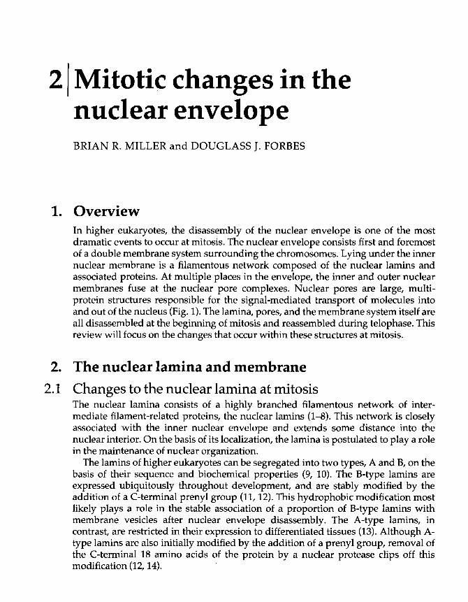
2 Mitotic changes in thenuclear envelopeBRIAN R. MILLER and DOUGLASS J. FORBES
1. OverviewIn higher eukaryotes, the disassembly of the nuclear envelope is one of the mostdramatic events to occur at mitosis. The nuclear envelope consists first and foremostof a double membrane system surrounding the chromosomes. Lying under the innernuclear membrane is a filamentous network composed of the nuclear lamins andassociated proteins. At multiple places in the envelope, the inner and outer nuclearmembranes fuse at the nuclear pore complexes. Nuclear pores are large, multi-protein structures responsible for the signal-mediated transport of molecules intoand out of the nucleus (Fig. 1). The lamina, pores, and the membrane system itself areall disassembled at the beginning of mitosis and reassembled during telophase. Thisreview will focus on the changes that occur within these structures at mitosis.
2. The nuclear lamina and membrane2.1 Changes to the nuclear lamina at mitosis
The nuclear lamina consists of a highly branched filamentous network of inter-mediate filament-related proteins, the nuclear lamins (1-8). This network is closelyassociated with the inner nuclear envelope and extends some distance into thenuclear interior. On the basis of its localization, the lamina is postulated to play a rolein the maintenance of nuclear organization.
The lamins of higher eukaryotes can be segregated into two types, A and B, on thebasis of their sequence and biochemical properties (9, 10). The B-type lamins areexpressed ubiquitously throughout development, and are stably modified by theaddition of a C-terminal prenyl group (11,12). This hydrophobic modification mostlikely plays a role in the stable association of a proportion of B-type lamins withmembrane vesicles after nuclear envelope disassembly. The A-type lamins, incontrast, are restricted in their expression to differentiated tissues (13). Although A-type lamins are also initially modified by the addition of a prenyl group, removal ofthe C-terminal 18 amino acids of the protein by a nuclear protease clips off thismodification (12,14).

THE NUCLEAR LAMINA AND MEMBRANE 43
Fig. 1 Diagram of a section though the nuclear envelope. The outer nuclear membrane (OMM) is contiguous withthe rough ER (RER). Nuclear pore complexes (NPC) are present at specialized junctions between the outer andinner nuclear membranes. Just beneath the inner nuclear membrane lies the lamina, an intermediate filament-like network assembled from lamin A and B heterodimer. Two integral membrane proteins of the inner nuclearmembrane are shown: p54/p58 and a lamin-associated protein, LAP. These two polypeptides make contactswith both the lamina and chromatin.
In addition to a structural role, the lamins also appear to be involved in themaintenance of chromatin structure and DNA replication (15-20). Interest-ingly, mutation of the gene for l amin D0 (the B-type lamin in this organism) inUrosopliila resulted in a severe phenotype affecting survival, locomotion, andfertility (21). The observed cellular phenotype included incomplete nuclearenvelopes, nuclear pore clustering, and the appearance of cytoplasmic annulatelamellae, indicating that lamin D(1 is essential for normal nuclear envelopeassembly in Drosophila (21).
At an early stage in mitosis, the lamina disappears before nuclear membranebreakdown (1, 2, 22). During prophase, both A- and B-type lamins become phos-phorylated, and this phosphorylation coincides exactly with the depolymerization ofthe nuclear lamina (23, 24). Each type of lamin contains multiple copies of theconsensus sequence for phosphorylation by the mitotic kinase, cdc2/cyclin B. It isthought that the level of polymerization of the lamins is directly regulated byphosphorylation. Indeed, purified lamins that have been assembled into paracrystalsand short filaments can be solubilized by the addition of either mitotic cytosol orpurified cdc2/cyclin B kinase (23). In addition, mitotic phosphorylation of the laminscan be blocked by mutation of the serine residues at the cdc2/cyclin B consensus sites

44 MITOTIC CHANGES IN THE NUCLEAR ENVELOPE
(25). When transfected into fibroblasts, incorporation of the mutant laminpolypeptides into the nuclear lamina blocks normal mitotic disassembly (25). It istherefore believed that the mitotic disassembly of the lamina is directly regulated bythe presence of active cdc2/cyclin B kinase.
2.2 Lamin-associated proteinsA number of proteins of the nuclear envelope have been identified as either lamin-binding proteins or lamina-associated proteins. One of the first to be identified was amolecule in avian and human cells called p54/p58 (it has also been termed lamin Breceptor, or LBR). Surprisingly, sequence analysis revealed that p54/p58 was highlyhomologous to a number of sterol biosynthetic enzymes (26-28). Whether or notp54/p58 is specifically a receptor for lamin B remains controversial; however,p54/p58 is localized to the inner nuclear envelope and has been shown to be able tobind both to chromatin and to chromatin-associated proteins (Fig. 1; 29). Thissuggests that the p54/p58 protein may play a number of different roles within thecell. The p54/p58 protein is phosphorylated at mitosis by a tightly associated kinase,and phosphorylation modifies the ability of p54 / p58 to associate with both DNA andlamin B in vitro (30,31).
In addition to p54/p58, a number of integral membrane lamin-associated proteins(LAPs) have been identified by monoclonal antibodies raised against nuclear laminapreparations (32-34). Three of these proteins, LAP1A to LAP1C, are generated byalternative splicing of a single gene (35). The different isoforms differ in their affinityfor lamins and chromatin, and are expressed in a tissue-specific manner. A fourthprotein, LAP2, is also an integral membrane protein capable of binding chromatinand lamin B (33,34). The binding of LAP2 to these ligands is abolished when LAP2 isphosphorylated by mitotic cytosol (33). This suggests that the role of LAP2 may be tocoordinate binding of nuclear membrane vesicles to the lamina and the chromatin ina cell-cycle dependent manner.
2.3 Regulation of nuclear membrane dynamics byphosphorylationCell-free nuclear assembly assays have provided a number of insights into theprocess of envelope assembly. A simplified form of a Xenopus nuclear reconstitutionassay revealed that binding of nuclear envelope precursor vesicles to chromatin canoccur in the complete absence of cytosol (36, 37). When vesicle-bound chromatin isthen incubated in interphase egg cytosol in the presence of the phosphatase inhibitorokadaic acid, the bound vesicles are released; they are not released if a kinaseinhibitor is included. This indicated that the binding of nuclear envelope vesicles ismediated by a kinase/phosphatase system. Vesicle release could also be achieved bythe direct addition of mitotic cytosol (converted to a mitotic state by the addition ofcdc2/cyclin B) (36). Fractionation of the cytosol revealed that the mitotic membrane-

THE NUCLEAR LAMINA AND MEMBRANE | 45
release kinase is distinct from cdc2/cyclin B, but this kinase is activated bycdc2/cyclin B at mitosis (38).
2.4 Disassembly of the lamina and nuclear membraneSince both the state of assembly of the lamina and the association of the membranewith chromatin are regulated by active cdc2/cyclin B, it would seem likely thatvesiculation of the envelope would occur at the same time as lamina breakdown.However, at least experimentally, the two events can be temporally separated usingin vitro assays. For example, in mitotic Xenopus extracts, vesiculation of the nuclearmembranes requires a stoichiometric component or components: vesiculation can becompletely eliminated by the addition of a large excess of nuclei to the extract (39).Since the outer nuclear membrane is contiguous with and can perform all thebiochemical functions of the endoplasmic reticulum (ER), it is possible that thecoatamer proteins involved in ER vesicle trafficking are also involved in nuclearenvelope breakdown. Excess nuclei would titrate out the soluble pool of coatamerproteins, specifically blocking the mitotic membrane vesiculation. In contrast, in thesame in vitro system, the mitotic disassembly of the lamina (as assayed by the releaseof soluble lamin proteins) appeared to require enzymatic components only.Specifically, addition of a large excess of nuclei to a mitotic extract did not blocklamin solubilization, and the rate of lamin removal reached a steady state level (39).In vivo, during meiosis in both frogs and chickens, the lamins are solubilized andreleased from nuclei which still contain intact envelopes and nuclear pore complexes(40). This suggests that the disassembly of the nuclear envelope is preceded by thedepolymerization of the lamina, a separation supported by the in vitro results.
The fate of the nuclear membrane during mitosis is somewhat unclear. One modelis that the nuclear envelope is disassembled into membrane vesicles which retain anidentity distinct from the rest of the ER. In support of this model, mitotic vesiclescompetent for nuclear assembly can be isolated from Xenopus eggs and can bepartially separated from the bulk of vesicles containing ER markers (41). In addition,a number of studies have reported that there are multiple classes of mitotic vesiclesderived from the nuclear envelope (42-^46). These studies suggest that the nuclearmembrane is fragmented into domain-specific vesicles during mitosis.
Much of the evidence for multiple classes of nuclear envelope precursor vesicles isderived from in vitro studies using cell extracts (42, 43). Recently, different resultswere obtained when the dynamics of a LBR-GFP (lamin B receptor-green fluor-escent protein) fusion protein were followed in living cells. In this system, theLBR-GFP-labelled nuclear envelope appeared to physically retract into the ERnetwork at mitosis, without obvious vesiculation of either structure (47). Anotherstudy used antibodies to both inner nuclear envelope and ER membrane proteinsand followed their distribution throughout the cell cycle; this study reached identicalconclusions (48). These studies are consistent with a model where specific integralmembrane proteins are immobilized to the inner nuclear membrane by interactionwith intranuclear ligands such as the lamins or chromatin, now defining a 'nuclear'

46 MITOTIC CHANGES IN THE NUCLEAR ENVELOPE
membrane domain distinct from the adjacent membrane comprising the bulk of theER. Reversal of these interactions at mitosis would allow the nuclear membraneproteins and membrane to flow back into the ER.
The discrepancy between the LBR-GFP results and the studies discussed above onvesicularization at mitosis remain to be resolved. One way in which the disparateresults could be reconciled is in realizing that the in vitro reconstitution extracts arederived from mechanically disrupted cells. The ER network, which in Xenopus eggscontains large stored amounts of integral nuclear membrane proteins, wouldprobably become fragmented into many small vesicles (or microsomes) upon celllysis. A subset of the vesicles, those containing integral nuclear membrane proteinscapable of binding to chromatin, might well be separable from vesicles notcontaining nuclear membrane proteins, creating multiple classes of vesicles uponseparation. Similarly, lysis of tissue culture cells at mitosis would also createmicrosomes. In vivo, the reticular network containing ER and nuclear membraneproteins would remain intact. Integral nuclear membrane proteins would show anaffinity for the chromatin and/or attached lamins in interphase. Thus, during timesof nuclear assembly, these integral membrane proteins would draw a portion of theER membrane to the chromatin, thereby initiating formation of a nuclear envelope.The proteins would lose that affinity at mitosis and the nuclear membranes woulddetach from the chromatin and lamins and retract into the ER, thereby disassemblingthe nucleus.
3. Nuclear pore complexesThe nuclear pore complex, composed of approximately 1000 proteins, is assembledin the nuclear envelope and forms a junction between the outer and inner nuclearmembranes. The membrane at the border of the pores has been termed the poremembrane (Fig. 2; 49-52). At an early step in mitosis the pore complexes are removedfrom the envelope. Indeed, a series of electron microscopic studies reveal that thepores are removed from the nuclear envelope before nuclear membrane breakdown,leaving holes in the remaining intact nucleus (53-55). As is the case for the lamins,mitotic phosphorylation of pore proteins is thought to play a role in disassembly ofthe pore complexes. A number of nucleoporins have been shown to becomehyperphosphorylated at mitosis (56,57). Moreover, two nucleoporins can be directlyphosphorylated by purified cdc2/cyclin B kinase in vitro (57).
Structurally, the nuclear pore consists of two eight-fold symmetric rings located oneither side of the nuclear membranes (Fig. 2). These rings bracket a third ring ofspoke elements, which are thought to contain at their hub a central transporter (58,59). A set of eight short filaments extend from the ring on the cytoplasmic side of thepore, the cytoplasmic filaments. Conversely, a set of long thin filaments terminatingin a ring, the nuclear basket, extends 1000 A from the nuclear side of the pore. Atpresent, it is not known whether these structures are removed from the pore atmitosis in an ordered, sequential fashion, or are simultaneously disassembled.
One intriguing aspect of disassembly is that the nuclear pore does not appear to

RE ASSEMBLY OF THE NUCLEAR ENVELOPE 47
Fig. 2 The nuclear pore complex mediates macromolecular transport across the nuclear envelope. This large(~125 MDa) structure is composed of three eight-fold symmetric rings. Eight short, thick filaments project fromthe cytoplasmic face of the pore, and are thought to provide docking sites for nuclear import substrates. Thenuclear basket is composed of eight thin filaments joined to a small ring approximately 1000 A inside thenucleoplasm. The pore is anchored in the nuclear envelope by integral membrane pore proteins. These areaswhere the inner and outer nuclear membranes are joined has been called the pore membrane.
disassemble into completely monomeric polypeptides. A number of nucleoporinshave been shown to exist in relatively large heteromeric complexes when un-assembled in interphase Xenopus eggs (<1000 kDa), although these complexes aremuch smaller than a complete nuclear pore (~120,000 kDa). The observed mitoticphosphorylation of individual subunits of these complexes does not result indetectable changes to the size or composition of the complexes (57,60). These proteincomplexes appear to represent individual modular portions of the nuclear pore. Themitotic phosphorylation of certain individual pore proteins may well serve todisassemble a nuclear pore into its constituent modules, rather than into individualmonomeric pore proteins.
4. Reassembly of the nuclear envelopeThe nuclear envelope is completely disassembled during the early phases of mitosis.However, by the end of mitosis, two complete and functional nuclear membranesmust be correctly assembled around each of the daughter nuclei. The exactmechanism by which this occurs is still somewhat of a mystery, even more so nowthat the mechanism of nuclear membrane disassembly is in question. One possibilityis that lamins act to guide membrane vesicles containing nuclear envelope proteinsto the telophase chromosomes. A fraction of lamin B proteins remains associatedwith membrane vesicles during mitosis (at least in somatic cells), and lamin B hasbeen found capable of binding to chromatin. Lamin B-binding would thus provide asimple mechanism for the specific recruitment of nuclear envelope precursor vesicles

48 | MITOTIC CHANGES IN THE NUCLEAR ENVELOPE
to chromosomes. A-type lamins have also been reported to bind to metaphasechromosomes (61). Chromatin-bound lamin A could provide docking sites for laminB- or LAP-containing vesicles (Fig. 3A). In support of such a model, it was reportedthat immunodepletion of A-type lamins from Chinese hamster ovary (CHO) extractsinhibited in vitro nuclear formation by almost two-thirds. Depletion of lamin B fromthe CHO extracts inhibited nuclear formation by roughly one-third, suggestingpossible additive contribution of lamin function in nuclear assembly (60). Similarresults have been reported in studies using extracts prepared from Drosophilaembryos and Xenopus eggs (62).
There are, however, several reports suggesting that lamin removal from Xenopusextracts did not block nuclear formation. Extracts depleted of B-type lamin Lm
formed small, fragile nuclei that were incapable of DNA replication (15, 18). Thesedata would suggest that lamins do not play an obligatory role in nuclear membraneassembly. Instead, formation of a lamina may be necessary for maintenance ofnormal nuclear function after assembly of the envelope. One caveat of these studiesis that while the antibody used removed all of lamin Lin, the major species of lamin inthe Xenopus eggs, it did not completely remove all lamin subtypes from the extracts.A small percentage of the lamins in the egg extract (estimated at 5-10% of the total) isof the membrane-bound Ln subtype, and would not have been removed by theantibodies used in these reports (63,64). It is therefore possible that the small amountof B-type LII lamins left in the extract are both necessary and sufficient for nuclearmembrane vesicle binding to chromatin in vitro.
Additional confusion on the role of lamins in the targeting of the vesicles tochromatin comes from immunofluorescence studies. Experiments examining thetiming of the association of integral nuclear membrane proteins to chromosomesduring nuclear reassembly revealed that two of the proteins of the inner nuclearenvelope, p54/58 and LAP2, associate with chromosomes well before detectablelamin B association at telophase (33,44). It may be possible that p54/58 and LAP2 arerecognizing metaphase chromatin-associated lamin A bound to the chromosomes(61). If so, then the two proteins could be responsible for binding nuclear membraneto the chromosomes, and lamin B would later accumulate at the interface of themembrane and chromatin (Fig. 3B). The timing of the association of lamins withchromatin needs to be carefully addressed.
Finally, it is experimentally clear that vesicles can be targeted to chromatin in theabsence of cytosolic factors in the Xenopus system (37, 41-43). Again, it is possiblethat the very small amount of membrane-bound lamins (in Xenopus eggs the vastmajority of B-type lamins are soluble at interphase) are responsible for this. It is
Fig. 3 Models for nuclear envelope assembly. At the end of mitosis, the disassembled nuclear envelope mustreform around daughter chromosomes. Multiple models for envelope reformation have been proposed. (A)Targeting by lamins. In this model, chromatin-bound lamin A serves as a docking site for nuclear envelopeprecursor vesicles carrying membrane-bound lamin B. (B) Targeting by integral nuclear envelope membraneproteins (INMP). Integral membrane proteins such as one of the LAPs may target vesicles to the chromatin, withlamin association as a secondary event. In this and the preceding model, all of the nuclear membrane is suppliedby a single, specific vesicular precursor. (C) Multiple vesicular precursors to the nuclear envelope. This model

RF ASSEMBLY OF THE NUCLEAR ENVELOPE 49
contains at least two classes of nuclear envelope precursor. One class provides recognition and binds chromatinby either of ttie above models. The chromatin-targeted vesicles are then recognized by other vesicles containingthe rest of the components of the nuclear membrane system. (D) Reformation of the envelope from the ER. Inthis model, during mitosis, the nuclear membranes are retracted into the ER, and the integral membrane proteinsof the inner nuclear envelope mix with those of the ER. To reform the nuclear envelope, recognition of thechromatin by either membrane-bound larnins or by an integral membrane protein such as one of the LAPs occursand causes the chromatin to become enveloped by membrane.

50 | MITOTIC CHANGES IN THE NUCLEAR ENVELOPE
possible that there are multiple classes of vesicles, each with its own chromosome-targeting protein, involved in nuclear formation. There have been a number ofreports of multiple classes of nuclear vesicle precursors (42-44). In perhaps theclearest demonstration of this, it has been shown that the association of the p54/58membrane proteins with chromatin occurs long before the association of the integralmembrane nuclear pore protein gp210 (44). The complete assembly of a functionalnuclear envelope in vitro may require contributions from multiple vesicularprecursors. Some of the vesicles could be targeted to the chromatin by lamins, someby LAPs or other membrane proteins. After initial enclosure of the nuclear envelope,growth of the nucleus could then proceed by vesicle-vesicle interactions (Figure 3C)or, if the model where nuclear membranes are retracted into the ER at mitosis is true,then some integral membrane proteins such as the LAPs may return early, bindingthe membrane to the chromatin and establishing a nuclear envelope. Later, integralmembrane pore proteins could diffuse through the plane of the ER membranes to thenucleus, and be induced to stay there by protein-protein interactions (Fig. 3D). Thesemodels are summarized in Fig. 3.
5. Nuclear pore assemblyAt some point during envelope assembly, the nuclear pore complexes mustassemble, fusing both the inner and outer nuclear membranes at the pore. Exactlyhow this occurs is still largely unknown. One model was that pore assembly occurson the surface of chromatin and is simultaneous with vesicle fusion. Assembly ofpores between vesicles, followed by vesicle-vesicle fusion, would guarantee theinsertion of the pores across the double membrane. Certain electron microscopicevidence supported this possibility (65). However, this appears not to be the case. Invivo, new pores assemble into pre-existing, intact nuclear membranes at S phase(reviewed in 49, 52, 66). In addition, morphologically normal nuclear pores can beassembled in membranous organelles called annulate lamellae, both in vivo and invitro, in the absence of any chromatin substrate (67, 68).
In vitro, nuclear pores assemble only into a pre-existing double nuclear membrane(69). In the absence of cytosol, the membrane vesicles derived from egg extracts arecapable of binding to chromatin, as stated above (37, 70,71). When ATP and GTP areadded, vesicle-vesicle fusion occurs, generating a nuclear intermediate containingpatches of flattened double membrane interspersed with chromatin-bound, unfusedvesicles (Fig. 4a). Upon the addition of cytosol to the intermediate, nuclear pores canassemble. Interestingly, the pores only assemble in the regions of flattened doublemembrane, not between vesicles. Thus, pore assembly obligatorily requires a pre-existing double nuclear membrane (69).
Although the pore formation observed above occurred only in pre-existingpatches of double nuclear membrane, it was possible that pore formation wasinitiated on the surface of the chromatin, and the pores then inserted into themembrane in a second step. This chromatin seeding mechanism was ruled out by

NUCLEAR PORE ASSEMBLY 51
Rg. 4 Steps in nuclear pore assembly, as revealed by chemical inhibitors. (A) Two separate membrane fusionevents are required for pore assembly. In the absence of cytosol or nucleotides. membrane vesicles can bind tothe chromatin (1). Addition of ATP and GTP allows a limited amount of vesicle-vesicle fusion along the surface ofthe chromatin, resulting in the formation of small patches of flattened double membrane (2). This fusion event isblocked by either treatment of the vesicles with NEM: or the inclusion of the non-hydrolyzable GTP analog GTP--/Sin the reaction. Addition of cytosol to the reaction promotes the assembly of pore complexes into the regions offlattened double membranes (pore membrane fusion) (3). The pore assembly initiated by cytosol is blocked bythe addition of GTP-yS, (B) Nuclear membrane assembly can occur in the absence of pore assembly. When acomplete nuclear reconstitution assay is mixed with the Ca2+ chelator BAPTA, the resulting nuclei are surroundedby a complete, double nuclear membrane system devoid of nuclear pore complexes (4), demonstrating thatBAPTA blocks a step in pore assembly. Replacement of the BAPTA-containing cytosol with fresh cytosol allowsassembly of functional nuclear pore complexes even in the presence Of GTP--vS (5), indicating that the BAPTA-sensitive step in pore assembly is downstream from both GTP--yS-sensitive steps in pore assembly. (C) Summaryof the steps in pore assembly revealed by the experiments in (A) and (B).
utilizing the finding that, in the presence of the Ca2+ chelator BAPTA, pore-freenuclei containing complete double nuclear membranes surrounding the chrurnatincould be formed (69). BAPTA had been shown previously to slow vesicle fusion andto block nuclear growth in this system (72, 73). When the pore-free nuclei wereincubated in fresh cytosol lacking BAPTA, numerous functional nuclear pores wereassembled in the nuclear membranes (Fig. 4B; 69). Thus, pore assembly occurs afterdouble membrane assembly and is presumably initiated by a factor present in the

52 ) MITOTIC CHANGES IN THE NUCLEAR ENVELOPE
Fig. 5 Model for changes to the nuclear envelope at mitosis. Shown are two possible pathways for nuclearenvelope breakdown and reassembly during mitosis. In both models, phosphorylation of nuclear envelopeproteins, initiated by cdc2/cyclin B, leads to the disassembly of the nuclear pore complexes, the nuclear lamina,and release of the lamin-associated proteins from the chromatin. In the model on the left, the nuclear envelope isfragmented into specific vesicles, distinct from the putative vesicles formed from intracellular organelles such asthe ER at mitosis. During telophase, reduction in the levels of mitotic kinases allows for the rebinding of nuclearvesicular precursors to the chromatin. Bound vesicles fuse and flatten on to the surface of the chromatin,forming the double nuclear membrane system. Nuclear pores reassemble into the nuclear envelope and the restof the nuclear constituents are re-imported from the cytoplasm. An alternative model is shown as the right-handpathway. As in the model above, disassembly of the pores and lamina is initiated by cdc2/cyclin B. Instead offragmentation into specific vesicles, the nuclear envelope is retracted into and mixes with the ER membranesystem, which also remains intact. Reduction in cdc2/cyclin B activity allows chromatin recognition by nuclearenvelope proteins, and the chromatin once again becomes enveloped by membrane. As in the first model,regions of double nuclear envelope serve as targets for nuclear pore assembly.

FUTURE DIRECTIONS 53
soluble cytosol. Intriguingly, sequential blocks in the pore assembly process could beidentified through the use of the inhibitors N-ethylmaleimide (NEM), CTP-^S, andBAPTA (66, 69; Fig. 4C). These results indicated that the block in pore formationcaused by BAPTA was downstream of two GTP--yS-sensitive steps in nuclear poreassembly. Exactly how the nuclear pores are assembled into the nuclear envelope,and what specifies this membrane for pore insertion remain questions for futurestudy.
From the studies reviewed here, a model for nuclear envelope assembly can beproposed (Fig. 5). The initial step probably involves the specific recognition ofchromatin by nuclear envelope precursors, either in the form of specific vesicles, orderived from the ER. This recognition may be mediated either by lamin B, or by theLAPs, p54/58, or by some as yet undiscovered protein. It is possible that there are infact multiple classes of nuclear envelope precursors, each using a different targetingprotein. After binding, the membranes fuse and flatten on to the chromatin, creatingthe double nuclear membrane system. The nuclear membranes then serve as a targetfor nuclear pore assembly, initiated by a soluble cytosolic protein. At the site of afuture nuclear pore, the inner and outer nuclear membranes fuse and the 1000estimated proteins comprising the structure of the pore assemble. Once the nucleibecome completely surrounded by a closed nuclear envelope containing nuclearpores, nuclear import can occur through the newly assembled pores. The solublelamins are presumably then transported through the pores and subsequentlyassembled into the nuclear lamina, giving rise to a functional interphase nuclearenvelope.
6. Future directionsThe lamins are obviously extremely important in the assembly and function of anormal nucleus. What remains to be clarified are the exact role of the lamins in theassembly of the nuclear envelope, and also the interaction of the lamins with othercomponents of the envelope.
The kinase that reverses nuclear membrane binding to chromatin and its targetsremain to be identified at the molecular level. Potential targets of this kinase areLAP1A-C and LAP2, as mitotic phosphorylation of these proteins blocks theirbinding to chromatin. Whether these proteins are the sole molecules responsible fortargeting of the nuclear membranes to chromatin remains to be determined. One keyquestion is the identity of the membranous precursors to the nuclear envelope. Arethere multiple classes, each with its own targeting mechanism? If so, how are theygenerated during mitosis? Or does the nuclear envelope retreat into the ER networkduring mitosis?
Exactly how nuclear pore complexes are assembled into the nuclear enveloperemains an intriguing problem. What soluble and integral membrane proteins arerequired for the fusion of the inner and outer nuclear envelope? Do these proteins,despite the fact that at least some of them reside within the lumen of the ER, resemble

54 MITOTIC CHANGES IN THE NUCLEAR ENVELOPE
the cytosolic factors such as NSF (N-ethylmaleimide sensitive factor) involved in therecognition and fusion of transport vesicles with their target organelles in thesecretory pathway? Once the pore membrane is formed, does the pore self-assemblefrom its constituent soluble components, or does assembly require a scaffold orchaperone? Finally, is disassembly of the pore merely the reverse of the assemblypathway, or are distinct mechanisms employed?
At present, quite a bit is known about the molecular details of mitotic changes tocertain individual molecular constituents of the nuclear envelope, such as the lamins.At the ultrastructural level, the morphological changes that occur during nuclearenvelope breakdown and reassembly are also well characterized. What is sorelyneeded are the connections between the two, and these will remain as fundamentalareas of enquiry for some time to come.
References1. Gerace, L. and Blobel, G. (1980) Cell, 19,277.2. Gerace, L. and Blobel, G. (1982) Cold Spring Harbor Symp. Quant. Biol., 46,967.3. Krohne, G., Franke, W. W., Ely, S., D'Arcy, A. and Jost, E. (1978) Cytobiologie, 18,22.4. Moir, R. D. and Goldman, R. D. (1993) Curr. Opin, Cell Biol., 5,408.5. Franke, W. W., Scheer, U., Krohne, G., and Jarasch, E. D. (1981) /. Cell Biol., 91,39s.6. Benavente, R., Krohne, G., Schmidt-Zachmann, M. S., Hugle, B., and Franke, W. W. (1984)
/. Cell Set., Suppl. 1,161.7. Krohne, G. and Benavente, R. (1986) Exp. Cell Res., 162,1.8. Benavente, R., Krohne, G., and Franke, W. W. (1985) Cell, 41,177.9. McKeon, F. D., Kirschner, M. W., and Caput, D. (1986) Nature, 319,463.
10. Fisher, D. Z., Chaudhary, N., and Blobel, G. (1986) Proc. Natl. Acad. Sci. USA, 83,6450.11. Wolda, S. L. and Glomset, J. A. (1988) /. Biol. Chem., 263,5997.12. Vorburger, K., Kitten, G. T., and Nigg, E. A. (1989) EMBO J., 8,4007.13. Lehner, C. F., Stick, R., Eppenberger, H. M., and Nigg, E. A. (1987) J. Cell Biol., 105,577.14. Beck, L. A., Hosick, T. J., and Sinensky, M. (1988) J. Cell Biol., 107,1307.15. Newport, J. W., Wilson, K. L., and Dunphy, W. G. (1990) J. Cell Biol., Ill, 2247.16. Hutchison, C. J., Bridger, J. M., Cox, L. S., and Kill, I. R. (1994) J. Cell Sci., 107,3259.17. Hutchison, C. J., Brill, D., Cox, R., Gilbert, J., Kill, L, and Ford, C. C. (1989) J. Cell Sci., Suppl.
12,197.18. Meier, J., Campbell, K. H., Ford, C. C., Stick, R., and Hutchison, C. J. (1991) J. Cell Sci., 98,
271.19. Jenkins, H., Whitfield, W. C., Goldberg, M. W., Allen, T. D., and Hutchison, C. J. (1995)
Acta Biochim. Polon., 42,133.20. Spann, T. P., Moir, R. D., Goldman, A. E., Stick, R., and Goldman, R. D. (1997) J. Cell Biol.,
136,1201.21. Lenz-Bohme, B., Wismar, K., Fuchs, S., Reifegerste, R., Buchner, E., Betz, H., and Schmitt,
B. (1997)J. Cell Biol., 137,1001.22. Stick, R. and Schwarz, H. (1983) Cell, 33,949.23. Ward, G. E. and Kirschner, M. W. (1990) Cell, 61,561.24. Peter, M., Heitlinger, E., Haner, M., Aebi, U., and Nigg, E. A. (1991) EMBO J., 10,1535.

REFERENCES | 55
25. Heald, R. and McKeon, F. (1990) Cell, 61,579.26. Smith, S. and Blobel, G. (1994) Proc. Natl. Acad. Sci. USA, 91,10124.27. Worman, H. ]., Yuan, J., Blobel, G., and Georgatos, S. D. (1988) Proc. Natl. Acad. Sci. USA,
85,8531.28. Worman, H. J., Evans, C. D., and Blobel, G. (1990) J. Cell Biol., Ill, 1535.29. Pyrpasopoulou, A., Meier, J., Maison, C., Simos, G., and Georgatos, S. D. (1996) EMBO J.,
15,7108.30. Simos, G. and Georgatos, S. D. (1992) EMBO J., 11,4027.31. Nikolakaki, E., Simos, G., Georgatos, S. D., and Giannakouros, T. (1996) J. Biol Chem., 271,
8365.32. Senior, A. and Gerace, L. (1988) /. Cell Biol, 107,2029.33. Foisner, R. and Gerace, L. (1993) Cell, 73,1267.34. Gerace, L. and Foisner, R. (1994) Trends Cell Biol, 4,127.35. Martin, L., Crimaudo, C., and Gerace, L. (1995) J. Biol Chem., 270,8822.36. Pfaller, R., Smythe, C., and Newport, J. W. (1991) Cell, 65,209.37. Newport, J. W. and Dunphy, W. (1992) /. Cell Biol, 116, 295.38. Pfaller, R. and Newport, J. (1995) J. Biol. Chem., 270,19066.39. Newport, J. W. and Spann, T. (1987) Cell, 48,219.40. Stick, R., Angres, B., Lehner, C. F., and Nigg, E. A. (1988) J. Cell Biol, 107,397.41. Wilson, K. L. and Newport, J. W. (1988) /. Cell Biol, 107,57.42. Vigers, G. P. and Lohka, M. J. (1992) /. Cell Set., 102,273.43. Vigers, G. P. and Lohka, M. J. (1991) J. Cell Biol, 112,545.44. Chaudhary, N. and Courvalin, J. C. (1993) J. Cell Biol, 122,295.45. Collas, P., Courvalin, J. C. and Poccia, D. (1996) J. Cell Biol, 135,1715.46. Collas, P. and Poccia, D. (1995) Dev. Biol., 169,123.47. Ellenberg, J., Siggia, E. D., Moreira, J. E., Smith, C. L., Presley, J. F., Worman, H. J., and
Lippincott-Schwartz, J. (1997) /. Cell Biol, 138,1193.48. Yang, L., Guan, T., and Gerace, L. (1997) /. Cell Biol, 137,1199.49. Forbes, D. J. (1992) Annu. Rev. Cell Biol, 8,495.50. Gerace, L. (1992) Curr. Opin. Cell Biol, 4,637.51. Wiese, C. and Wilson, K. L. (1993) Curr. Opin. Cell Biol, 5,387.52. Davis, L. (1995) Annu. Rev. Biochem., 64,865.53. Maul, G. G. (1977) /. Cell Biol, 74,492.54. Maul, G. G. and Deaven, L. (1977) J. Cell Biol, 73,748.55. Maul, G. G., Maul, H. M., Scogna, J. E., Lieberman, G. S., Stein, G. S., Hsu, B. Y.-L., and
Borun, T. W. (1972) J. Cell Biol, 55,433.56. Favreau, C., Worman, H. J., Wozniak, R. W., Frappier, T., and Courvalin, C. (1996)
Biochemistry, 35,8035.57. Macaulay, C., Meier, E., and Forbes, D. J. (1995) J. Biol. Chem., 270,254.58. Akey, C. W. and Radermacher, M. (1993) /. Cell Biol, 122,1.59. Hinshaw, J. E., Carragher, B. O., and Milligan, R. A. (1992) Cell, 69,1133.60. Burke, B. and Gerace, L. (1986) Cell, 44,639.61. Glass, J. R. and Gerace, L. (1990) /. Cell Biol, III, 1047.62. Smith, D. E. and Fisher, P. A. (1989) J. Cell Biol, 108,255.63. Lourim, D. and Krohne, G. (1993) J. Cell Biol, 123,501.64. Lourim, D. and Krohne, G. (1994) Trends Cell Biol, 4,314.65. Sheehan, M. A., Mills, A. D., Sleeman, A. M., Laskey, R. A., and Blow, J. J. (1988) /. Cell
Biol, 106,1.

56 | MITOTIC CHANGES IN THE NUCLEAR ENVELOPE
66. Macaulay, C. and Forbes, D. J. (1996) Cell Dev. Eiol, 7,475.67. Dabauvalle, M. C., Loos, K., Merkert, H., and Scheer, U. (1991) /. Cell Biol., 112,1073.68. Meier, E., Miller, B., and Forbes, D. J. (1995) J. Cell Biol, 129,1459.69. Macaulay, C. and Forbes, D. J. (1996) J. Cell BioL, 132,5.70. Boman, A. L., Taylor, T. C., Melancon, P., and Wilson, K. L. (1992) Nature, 358,512.71. Boman, A. L., Delannoy, M. R., and Wilson, K. L. (1992) /. Cell BioL, 116,281.72. Sullivan, K. M., Busa, W. B., and Wilson, K. L. (1993) Cell, 73,1411.73. Sullivan, K. M. and Wilson, K. L. (1994) Cell Calcium, 16,314.

3 Poles apart? Spindle polebodies and centrosomesdiffer in ultrastructure yettheir function andregulation are conservedIAIN M. HAGAN, KEITH GULL, and DAVID M. GLOVER
1. IntroductionOne of the most overpowering images in biology has to be the simplicity andsymmetry of the apparatus that forms in order to segregate the eukaryotic genomebefore cell division, the spindle. Nothing illustrates more graphically the complexityof structure that can be generated by regulating the function of biological polymers.The essential bipolarity of the spindle requires the function of specializedmicrotubule organizing centres (MTOCs) at its poles, termed centrosomes in animalcells and spindle pole bodies (SPBs) in fungi. These specialized organelles differmarkedly in structure from one class of organisms to another. Notably, they are notfound in plant cells, and the meiotic spindles of flies, frogs and mammals becomeorganized without centrosomes at their poles. In eggs the chromosomes themselvesappear to act as MTOCs and initiate the nucleation of microtubules, which sub-sequently become focused at the spindle poles through the concerted action ofmicrotubule motor proteins. The role of the chromosomes and nuclear proteins forthe organization of spindles has been emphasized by a number of recent studiesperformed in vitro with egg extracts. Magnetic beads coated with plasmid DNA havebeen shown to facilitate the formation of a bipolar spindle in Xenopus egg extracts (ref1; also discussed in Chapter 4). Waters and Salmon have reviewed this evidence, andsuggested that there may be an alternative form of spindle assembly in cells thatappear to lack focused centrosomes (2). In such cells microtubules are envisaged tobind to and thus be stabilized by chromosomes, whilst the action of mitotic motorssuch as chromosomally located chromokinesins (plus-end-directed motors) maywork in concert with minus-end-directed motors which focus these microtubules to

58 POLES APART
produce a bipolar metaphase spindle with centrally located chromosomes (2). Whilstthe mechanisms for the formation and maintenance of spindles in oocytes perhapsrepresent an extreme example of a self-assembly system, similar features are likely tounderlie many of the aspects of spindle function in those cases where MTOCs arenucleating the microtubules at the two poles. The overall outcome is a universalorganization of the half-spindle with individually dynamic microtubules having apolarity of minus ends at the MTOC and plus ends distal, either at chromosomes, orinteracting with microtubules from the other half spindle, organelles or othercytoskeletal components. Given the ability of the microtubules to self-organize insuch a dramatic way in both mitotic (1) and interphase cells (3, 4), the two mitoticMTOCs may serve a moderating function and act as a dominant determinant toensure that only two poles are formed and that these two poles remain stablethroughout division.
In the century that has elapsed since the centrosome was first described andnamed, there has been a focus on morphological documentation of the extensivevariety of form amongst diverse spindle types and in various species (5). The fieldthen developed into one of molecular taxonomy in a process of trying to identify thecomponents of the spindle poles (6). We are now at the dawning of a particularlyexciting time in which the combined powers of genetics, biochemistry, and cellbiology are being harnessed in a convergent and highly productive fashion toprovide considerable insight into some key aspects of pole organization andfunction.
We will attempt to highlight some of the studies on the structure of the polarMTOC and its function most pertinent to understanding its role in genomesegregation. Constraints of space lead us to refer to recent more comprehensivereviews for a more complete picture of the MTOC per se (6-11). We will concentrateon data obtained in studies of some key model organisms to exemplify the behaviourof the major types of polar MTOC.
2. Ultrastructure of the spindle poles2.1 The spindle pole bodies of fungi
There is a bewildering array of different MTOC forms in eukaryotic microorganisms(5). However, the genetic malleability of three model systems, Saccharomycescerevisiae, Schizosaccharomyces pombe, and Aspergillus nidulans, has resulted in theseorganisms receiving most attention in recent years. Of these by far the most is knownabout the MTOC at the pole of the budding yeast (S. cerevisiae) spindle, the SPB.Indeed, at the molecular level, the budding yeast SPB is the best characterized MTOCof any system and its continued study is likely to define some of the ground rules forthe analysis of higher eukaryotic MTOCs, as much by its differences from othersystems, as by its similarities.
The budding yeast SPB is a multi-layered structure that remains embedded in thenuclear envelope throughout the cell cycle (Figs 1 and 2A). A dense central plaque

ULTRASTRUCTURE OF THE SPlNDLE POLES 59
Rg. 1 Electron micrographs illustrating the differences between the S. cerevisiae and S. pombe spindle polebodies (SPBs) which are represented schematically in Figs 2 and 3. (A) S, cerevisiae SPB residing within thenuclear envelope (NE). (B-D) S. pombe SPB at different stages of the cell cycle. During interphase the main bodyof the SPB sits on the outer face of an apparently contiguous nuclear envelope. The arrowhead indicates a regionof dark amorphous -y-tubuiin-containing material directly underneath the nuclear envelope under the SPB, Uponcommitment to mitosis, the SPB inserts into a newly opened fenestra within the nuclear envelope. Duringanaphase (B) the nuclear envelope closes and the number of microtuPules decreases. The limits of the nuclearmemPrane are indicated Py two arrows in panels C and D. The arrowhead in panel D indicates electron-densematerial bound to the nuclear membrane. Scale bars represent 100 nm. Panel A is reproduced from The Journalof Cell Biology, 1996,132, 887 (ref, 80} by copyright permission of The Rockefeller University Press and (B)-(D)from Molecular Biology of the Cell, 1997, 8, 1461 (ref. 17) with permission of the American Society for CellBiology.

60 | POLES APART
Fig. 2 A cartoon depicting SPB duplication in budding yeast. The SPB resides within the nuclear envelope(depicted by the thin black line curving around either side of the SPB) throughout the cell cycle. The cartoon isdrawn with the cytoplasmic face uppermost and the nuclear face below the nuclear envelope. The single half-bridge-bearing SPB (A) gains a satellite structure (B) before a new SPB appears at the end of a larger full-bridgestructure (C). Microtubules are nucleated from the cytoplasmic face of the bridge (C) until it is split to produce twoseparate, half-bridge-bearing SPBs (D).
with a somewhat less dense centre is flanked by two nuclear and three cytoplasmicplaques (12-15). A structure referred to as the half-bridge is associated with themembrane on one side of the SPB.
The synthesis of a new SPB is conservative and initiated from a small amorphousstructure at the end of the half-bridge referred to as the satellite (Fig. 2B). The resultis two SPBs which are connected by a bridge (Fig, 2C). The development of thesatellite into a mature SPB is presumably very rapid as no intermediates have beenrecorded. The bridge nucleates microtubules from its cytoplasmic face until it breakstowards the end of S phase after which cytoplasmic microtubules are only nucleatedfrom the cytoplasmic plaque and not the half bridge (ref. 12; Fig, 2D). Microtubulesextend from the nuclear face of the SPB throughout the cell cycle and differ fromthose found in most eukaryotes as their ends are sealed by a dome-shaped capstructure (16).
In contrast to the budding yeast SPB, the main part of the SPB in both fission yeast(S, pombe) and A. nidulans sits on the outside of the nuclear membrane in interphaseand inserts into the membrane upon commitment to mitosis (5,17; Figs 1 and 3), Themethod of SPB duplication in fission yeast also appears to differ from that in buddingyeast as the single interphase SPB splits into two daughter SPBs which then growfrom half this final unitary size to create two full-sized SPBs (17). What role thebridge plays in this duplication is unclear, but it is present throughout and the twodaughter SPBs end up connected by a bridge structure (Fig. 3C). Upon mitoticcommitment the nuclear membrane opens beneath the SPB and the SPB drops into

ULTRASTRUCI'URE OF THE SPINDLE POLES 61
Fig. 3 A cartoon depicting SPB duplication in fission yeast. The double black line in each panel represents thenuclear envelope. The cytoplasm is above this line and the nucleoplasm below it. Arrows indicate temporalprogression. (A) A single large, half-bridge-bearing SPB resides on the outside of the nuclear envelope aboveamorphous material on the nuclear side of the membrane (grey bar). (B) SPB duplication in late G? sees areduction in size of the two SPBs which lie either side of the bridge. These SPBsgrow in size(C) until commitmentto mitosis when the nuclear envelope breaks down beneath the paired SPBs which slot into the newly formed gap(D). Cytoplasmic microtubules are associated with the cytoplasmic face of the SPB throughout interphase.Nuclear, spindle microtubules are nucleated from the nucleoplasmic face of the SPB upon insertion into themembrane. Cytoplasmic microtubules are nucleated by and run tangentally to the SPB after spindle formation butbefore the metaphase-anaphase transition (E). After this transition (F) the nuclear envelope starts to close andthe number of spindle microtubules decreases. Drawn after ref. 17.
the gap within the membrane and nucleates microtubules. The process of SPB re-exclusion is initiated at the metaphase-anaphase transition and results in a virtualsealing of the membrane in the presence of late spindles (Figs 3F and ID). Duringinterphase, filamentous structures are seen within the membrane and an electron-dense region is found directly beneath the membrane (Fig. IB), as is -y-tubulin,strongly suggesting a structural link across the nuclear envelope in interphase.Moreover, centromeres are continuously associated with the interphase SPB region(18), confirming that the functional unit does extend across the apparently con-tiguous membrane. The SPB-centromere interaction during interphase in fissionyeast probably underlies the role of the SPB in directing nuclear positioning (19). Asthe NdclOp component of the budding yeast kinetochore is associated with the SPBin interphase (20), SPB-centromere interactions and co-ordinated nuclear position-ing may be a common feature of fungal systems.

62 POLES APART
2.2 Animal cell centrosomesThe centrosomes of animal cells have many functions: (a) they provide the intrinsicdominant control of the number of major sites of microtubule nucleation within thecytoplasm, one in interphase, two in the bipolar mitotic spindle; (b) they modulatethe number of microtubules nucleated during the cell cycle (21); and (c) they controlthe structure of the nucleated microtubules, their polarity and the cellular position ofthe microtubule array. Thus, by means of the intrinsic properties of polarity andposition, both of itself and of the subtended microtubule array, the centrosome isable to influence many of the major events occurring within the eukaryotic cell.These include the directed movement of vesicles, the positioning of membranousorganelles, and the processes of mitosis and cytokinesis. These complex, almostholistic properties enabled the importance of the centromere to be recognized as farback as the nineteenth century (21), and yet our understanding of its biochemistryand molecular cell biology is only recently beginning to emerge.
An alternative view of the animal cell centrosome is as a unitary conglomerate ofthree MTOCs that define the interphase microtubule array, the mitotic spindle, and,in the case of motile cells, the axonemal microtubules (22). Studies of microtubulenucleation/organization sites in eukaryotic microbes provide instructive insightto understanding the animal cell centrosome as in many eukaryotic microbes,particularly protozoa and algae, there is often invariant control over the number andcontent of microtubule arrays nucleated by MTOCs. Each of these three componentMTOCs may either be found at a single site, as in many somatic animal cells, or bedispersed to three different sites, exemplified for instance by the trypanosome cell(Fig. 4) (23). In contrast, the microtubule arrays seen in the amoeboid cell of Physarumsuggest that the interphase centrosome of these cells exists in a highly organizedstate, which intriguingly is not so apparent at mitosis (24,25).
2.2.1 CentriolesThe classical description of the animal cell centrosome with the advent of electronmicroscopy was of a pair of centrioles surrounded by a rather amorphous cloud ofelectron-dense pericentriolar material (PCM). Conceptually, we can attribute specificcharacteristics to each of these two component parts. Firstly, much evidence suggeststhat microtubule nucleation and anchoring of both cytoplasmic and spindlemicrotubules is, in the main, a function of the PCM (26). A pair of centrioles normallylies at the heart of the animal cell centrosome (8). We know that the two centrioleswithin the centrosome are non-identical and that the mature centriole is bothultrastructurally and functionally related to the basal body that acts as the MTOCfor the axonemal microtubules of cilia and flagella. Figure 5 represents a schematicview of the centriole pair within a centrosome. The centriole cylinder (~ 0.2 |xmdiameter X 0.4 (Jim long) is composed of nine triplet microtubules with the outertubule of the triplet (tubule C) being shorter than tubules A (inner) and B (middle).The interlinked triplets adopt a slightly twisted organization to form the cylinder-like structure or centriole 'pinwheel'. One centriole is defined as mature by its

ULTRASTRUCTURE Or THE SPINDLE POLES ; 63
Fig. 4 Examples of grouped and dispersed MTOCs in different systems. The mammalian cell centrosorne is aform of grouped MTOC in that it nucleates interphase and mitotic microtubules (A, B) as well as those of theaxoneme (the primary cilium in A). The trypanosome cell, in contrast, has dispersed and separate MTOCs forcytoplasrnic microtubules and flagellar axonemal microtubules (both present at interphase and mitosis, C and D),In addition it has a separate MTOC for the intranuclear mitotic spindle.
functional capacity to subtend a primary (immotile) cilium. It also bears a set ofappendages and satellites that distinguish it from its immature partner. Otherstructural features define the immature centriole, for instance, its internal cartwheelstructure and orthogonal relationship to the mature centriole. The centrioleduplication cycle is discussed in relation to centrosorne duplication and separationbelow in Sections 4,2 and 4.3.
2.2.2 Relationship of the centriole to the ciliumThe relationship of the centriole to the basal bodies is probably most easHyappreciated in microbial systems such as the amoebo-flagellate stages of organisms

64 POLES APART
Fig. 5 Schematic view of the centriole pair in animal cells. The two centrioles differ in structure: the maturecentriole carries appendages and satellites that the immature centriole lacks. The diagram also illustrates the'right-angle rule' whereby the long axis of the immature centriole intersects the mature centriole, but the long axisof the mature centriole does not intersect the immature centriole. Also shown is the connection between the twoproximal ends of the centrioles. The lower part of the figure shows, in a simplified manner, the internal structuresof the centriole as viewed from the outside at each end. The cartwheel structure in the proximal part of theimmature centriole is typical of a growing centriole, whilst the distal end of both centrioles contains a dense ringand sometimes an incomplete outer tubule in the microtubule triplet. The proximal ends exhibit discreteconnections between A and C tubules. Adapted from ref. 8 with permission of Elsevier Science.

ULTRASTRUCTURE OF THE SPINDLE POLES | 65
such as Physarum. The centrosomal area of the amoebal form possesses two non-identical centrioles, which, when this cell undergoes the reversible transformation toa flagellate, immediately become the basal bodies and act as the MTOC for the twonon-identical flagella (25, 27). In animal cells, centriole duplication and maturationwithin the cell cycle ensures that the early interphase centrosome has one mature andone immature centriole. The potential of the mature centriole to act as an MTOC foran axoneme is also seen during G1 phase when in many animal cells it forms animmotile, primary cilium. In many microbial systems and in the somatic cells ofanimals there is a close spatial, temporal, and numerical relationship between exist-ing centrosomal centrioles and the formation of new centrosomal procentrioles.These replication relationships may well be maintained during differentiation of themonoflagellated sperm cells. However, during ciliogenesis in multiciliated epithelialcells, these tight relationships between replication and maturation have to becircumvented. The centriole precursors of the basal bodies of the 200-300 cilia ofthese differentiated cells assemble close to a mass of electron-dense material, thefibrogranular complex (28).
2.2.3 PCM, microtubule nucleation, and releaseIn contrast to the very precise description of the centrioles within the centrosome, wehave had, until recently, very little information on the substructure of the peri-centriolar material. In vitro experiments on isolated centrosomes clearly showed thatthe major microtubule nucleating potential of the centrosome was associated withthe PCM (26). However, until recently even high-resolution electron microscopy hasrevealed little substructure in the PCM (see Section 3.1).
Kimble and Kuriyama (29) suggested that one may expect three levels oforganization: (a) a single nucleating element at the minus end of the microtubule, (b)clusters or centres of such nucleating elements, and (c) the organization of many suchcentres within the pericentriolar material. Their suggestion of different levels oforganization does not necessitate that individual microtubules are permanentlyanchored. In fact many observations suggest that microtubule nucleation, anchoring,attachment, and detachment may all be part of a dynamic process. Vorobjev andNadezhdina (30) considered the possibility that microtubules may be nucleated andthen released from individual sites within a centrosome.
Data from Belmont et al. describing the release of individual microtubules fromcentrosomes within a mitotic microtubule aster in Xenopus egg extracts (31) and theexistence of a microtubule-severing protein, katanin, in a region just outside the PCMof centrosomes throughout the cell cycle of sea urchin cells (32) suggest that theconcept of nucleation and then release may extend to the spindle pole centrosome.Indeed, microtubules are released en masse from the Dictyostelium interphase centro-some upon commitment to mitosis and from sea urchin poles at telophase (33-36).Such characteristics also provide a logical mechanism to account for the ability of -y-tubulin-nucleated microtubules to depolymerize at the pole after nucleation duringmetaphase flux (37) (see Section 3.1.2).

66 POLES APART
3. Components of polar MTOCs and their functionA substantive body of information is now accumulating about structural com-ponents of the yeast SPBs. This contrasts with a relative dearth of information aboutthe molecular constitution of the animal cell centrosome. One difficulty is that,although over the years many molecules have been described as being associatedwith centrosomes, there has often been no means of assessing the functionality ofsuch associations. In a sense, the animal cell centrosome can act as a central railwaystation served by a vast microtubular railway network. Many proteins arrive at the'station' as 'passengers' of a variety of 'locomotive' proteins. Nevertheless, positionalinformation in respect of the classification of a protein as a centrosomal componenthas remained based on a central tenet that it is found in this site by immuno-fluorescence and immunoelectron microscopy and that this assumption remainseven following the depolymerization of microtubules using drugs. Sometimes thispositional information can be supported by biochemical or genetic evidence to linkits function to its location at this site. However, in classifying any protein as acomponent of the centrosome one has to rehearse the caveat that this label may notindicate an exclusive location of the total cellular population of such molecules to thiscentrosomal site. The result is a rapidly lengthening list of proteins with some formof centrosomal location, the catalogue of which has been extensively reviewed (6,11,29). In most cases we only have information on position at the centrosome and lackinformation on likely function.
The first good structural identity of a molecular complex with microtubule-organizing properties in the PCM has been provided by the characterization of the'gammasome', which probably satisfies the criteria for the first level of organizationof the PCM as defined by Kimble and Kuriyama (29), namely the microtubulenucleation element.
3.1 The gammasomeIn a few cases, some general principles are emerging about fundamental componentsof the MTOCs that unify the function of the diverse structures of the yeast SPBs andthe animal cell centrosome. Nowhere has this been more apparent than in ourrapidly expanding understanding of the function of "y-tubulin. The gene for thisprotein was first identified through a suppressor mutation in an A. nidulans colonyon an agar plate that could compensate for the hyperstable microtubules in a benA33(3-tubulin mutant (38, 39). It was not long before antibodies had been raised againstthe molecule and used to show that it was located on the spindle poles (40) and thatthe protein was also present in the centrosomes of higher eukaryotes (41, 42). Itwas subsequently shown that microinjection of anti-'y-tubulin antibodies blocksmicrotubule nucleation in mammalian cells (43) and that •y-tubulin is part of a largemultiprotein complex, the gammasome (44-46).
It is now established that -y-tubulin is concentrated at MTOCs in many different

COMPONENTS OF POLAR MTOCs AND THEIR FUNCTION 67
organisms and a key role for -y-tubulin in microtubule nucleation has been mostclearly illustrated by the disruption to the MTOCs in -y-tubulin mutants of A.nidulans, S. pombe, S. cerevisiae, and Drosophila melanogaster (47-51). Given its locationat the MTOC, low abundance of the protein and its requirement for microtubulenucleation, Oakley and colleagues (52) proposed that a ring of -y-tubulin within thecentrosome could serve as a template for microtubule formation (44, 53,54).
Genes encoding two -y-tubulin isoforms are known in Drosophila. One polypeptideencoded by the 23C gene is expressed in a variety of somatic tissues anddevelopmental stages. Mutations in this gene dramatically disrupt the size and shapeof the centrosome and decrease the numbers of microtubules that it nucleates (50).The second polypeptide is only detected in oocytes and embryos and mutations in itsgene, located at 37C, disrupt the structure and function of the female meiotic spindle(51). This is a particularly instructive finding since this meiotic spindle in Drosophilais anastral, lacks a structurally recognizable centrosome, and shows no obviouslocalization of centrosomal antigens, including -y-tubulin, by immunofluorescencetechniques (see Chapter 7). In the formation of the meiotic spindle, microtubules areinitially nucleated by association with chromatin and then bundled and focused intothe poles by the action of kinesin-like motor proteins such as Nod and Ned (55-59).The process was thought not to require specific centrosomal functions. Although theprecise role of -Y-tubulin in organizing these spindles is not clear, the phenotype ofthis mutant provides a dramatic illustration that inability to visualize a specificlocation of a molecule should not be taken as evidence for lack of function in aparticular process.
There is now abundant evidence that may support the view of -y-tubulin as amicrotubule template during nucleation as -y-tubulin is part of the large 25Sgammasome, in embryonic and somatic animal cells. The complex was first isolatedfrom Xenopus oocyte extracts and was visualized by electron microscopy as an openring structure with a diameter of 25-28 nm (44). This particular complex includes sixextra proteins including a- and B-tubulin and proteins of 195,133,109, and 75 kDa.Interestingly, none of the molecular weights corresponded to the 60 and 190 kDacentrosome-associated proteins, CP60 and CP190, which have been shown to co-purify with -y-tubulin from Drosophila embryos (60). Importantly, combined use ofimmuno-electron microscopy and electron microscope tomography has shown thepresence of ring-like structures reminiscent of gammasomes in isolated Drosophilacentrosomes (45, 61). Convincing arguments for these rings being functionalnucleators came from the demonstration that -y-tubulin complexes were close to theminus ends of microtubules nucleated in vitro by these isolated centrosomes.Recently, Vogel et al. (46) used similar technology to study highly purified centro-somes from Spisula oocytes, and again detected ring structures on the centrosomes.Biochemical analyses of the component proteins of the Spisula gammasome indicatean unusual stoichiometric ratio of a-tubulin:(3-tubulin (being around 1:5 rather thanthe expected 1:1), suggesting that centrosomes may contain p-tubulin that iscomplexed with proteins other than a-tubulin—the prime possibility being that B/ycomplexes occur at the nucleating site (minus end) of microtubules (46).

68 POLES APART
3.1.2 Models for microtubule nucleation by the gammasomeTwo main models for this centrosomal microtubule nucleation complex have beenproposed (44, 52, 62; reviewed in 63) (Fig. 6). Oakley (52) and Zheng et al. (44)essentially suggest a model whereby proteins in the nucleation complex provide amolecular scaffold on which •y-tubulin subunits align in a helix. In the simplest formof this model, a tubulin heterodimer would then pair directly with each -y-subunit,providing a template for the first elongation step of microtubule assembly (Fig. 6). Inan alternative view, Erickson and Stoffler (62) suggest that -y-tubulin ring complexes,perhaps influenced by other proteins, form curved protofilaments and that these-y-protofilaments interact with both a- and p-tubulin subunits to initiate microtubulenucleation (Fig. 6). This latter mode of action would be more consistent with recentmodels for microtubule growth in which microtubules polymerize as long sheetswhich subsequently curl up upon GTP hydrolysis, thus the microtubule is notextending as a uniform tube but by side association into a sheet which then becomesa uniform tube (64). The model of Erickson and Stoffler is also more consistent withthe depolymerization of microtubules at the pole during metaphase flux (37).Without invoking severing of every microtubule it is difficult to reconcile suchbehaviour with the ring nucleation model of Oakley (52) and Zheng et al. (44) if•y-tubulin stays on the end of the microtubule throughout division.
3.1.3 A -y-tubulin variant in an organism with closed microtubule endsThe -y-tubulins of A. nidulans and S. pombe show great similarity to those of highersystems. In fact, human -y-tubulin can substitute for the fission yeast molecule (65).Budding yeast was, for a long time, in the strange position of being one of the mostextensively studied systems and yet the one in which -y-tubulin could not be found.This failure challenged the models for "/-tubulin function in microtubule nucleationderived from studies in other systems. The cause of this discrepancy becameapparent when the S. cerevisiae TUB4 sequence was identified through the yeastgenome sequencing project; its predicted amino acid sequence exhibits no more than35-40% homology to other -y-tubulins (66). This contrasts with the usual identities ofaround 65% between -y-tubulins from different species and explained why the low-stringency hybridization and PCR approaches, which had been successful inidentifying virtually all of the other -y-tubulin genes in other systems, had notidentified TUB4. The divergence of TUB4 was further highlighted by the inability ofhuman or Xenopus -y-tubulin to substitute for its essential function and, conversely,the inability of TUB4 to substitute for the function of the fission yeast "y-tubulin gene,gtbl+(49,67).
The analysis of the budding yeast -y-tubulin gene and its interacting genes serves toillustrate the power and incisive nature of genetic approaches in yeast in dissectingbiological processes at the molecular level. As anticipated, immunogold labellingwith anti-Tub4p antibodies showed that Tub4p is localized to the inner and outerplaques of the SPB (49). Conditional tub4 mutants exhibit disorganized non-functional spindles with less than the normal complement of microtubules (49, 66,

COMPONENTS OF POLAR MTOCS AND THEIR FUNCTION 69
Fig. 6 Illustration of models for the nucleation of microtubules by the rtubulin complex at the MTOC (section3.1.2). (A) In the first model the proteins in the ytubulin complex interact with the MTOC and provide a scaffold(grey helix) on which 13 y-tubulin subunits are aligned. u-Tubulin interacts directly with the ytubulin and sodetermines the polarity of the rnicrotubule. (B) In the second model, ytubulin forms a protofilament terminatingin a helical structure. The polarity of the ytubulin units may be determined by accessory proteins which may alsoprovide the contact sites with proteins of the MTOC. The ytubulin protofilament functions as stable seeds uponwhich additional u and p heterodimer tubulin subunits assemble. The extended microtubule sheet is then closedto a cylinder of n and B neterodimers when it contains 13 protofllaments. Adapted from ref. 63 with permission ofthe Company of Biologists, Ltd,

70 POLES APART
67). Spc98p was identified as interacting with Tub4p, as its overexpression rescued aconditional tub4.1 mutant. SPC98 encodes a 90 kDa protein identified by Rout andKilmartin (13) as a structural component of the SPB (see Section 3.2 below). It wasshown to be an essential gene and its product localizes to the same parts of the SPB asTub4p (13, 68). A search for plasmids which, when present in multiple copies, couldrescue the conditional spc98.2 allele, identified a further essential SPB component,Spc97p (69). Extensive genetic and biochemical data convincingly showed thatSpc97p, Tub4p, and Spc98p exist in a complex that contains more than one moleculeof Tub4p and differs in its sedimentation coefficient from that of the Xenopusgammasome in being 6S rather than 24S (63, 69). These differences in -y-tubulincomplex composition could provide a molecular basis to account for the closedcapping of microtubule ends at the yeast SPB which contrasts with the openstructures in other systems (63; see Fig. 6).
3.2 Units of self-assembly in the budding yeast SPBAn emerging theme from the analysis of the budding yeast SPB is of a number ofmolecules which appear to form discrete, functional, modular groupings that areable to direct self-assembly into specific structural units. As long as some aspect ofthe process is regulated to define limits, accurate duplication of the organelle occursin a manner analogous to the assembly of viral coat protein complexes. The cascadeof self-assembly of interacting units is initiated and maintained by the biochemicalproperties of the components themselves. This eccentric, yet attractive, view of SPBassembly may also apply to centriole assembly and the wider process of centrosomeduplication in animal cells.
Despite the considerable hurdles placed by the low copy number of SPBcomponents and ignorance of their biochemical characteristics, pioneering work byRout and Kilmartin identified three spindle components. This was achieved byraising a bank of monoclonal antibodies against highly enriched SPB fractions (13).Using immunogold electron microscopy techniques, they localized moleculesidentified by these antibodies to different parts of the spindle: a 110 kDa antigen tothe inner plaque, a 90 kDa antigen to both the inner and outer plaques (section 3.1.2),and an 80 kDa antigen adjacent to both the SPBs and the kinetochores (Fig. 7).
With the exception of the tubulins, the 110 kDa antigen is the most studied spindlepole component. Its gene, SPC110, was identified by three further routes after itsinitial description by Rout and Kilmartin (13): a screen for insoluble nuclear antigens(70), identification of mutants that compensate for the calmodulin mutation cmdl.l(68), and a screen of an expression bank for calmodulin-binding proteins (71). Thepredicted amino acid sequence of the SPC110 gene suggests a defined domainstructure with a globular N-terminus separated from a calmodulin-binding C-terminal domain by a long coiled-coil region (68, 71-73). In an elegant study,Kilmartin et al. demonstrated that while strains lacking the entire gene are inviable,those containing a deletion of virtually the entire coiled-coil domain survive, and thespacing between the site of microtubule nucleation and the main body of the SPB is

COMPONENTS OF POL ARMTOCS AND THEIR FUNCTION | 71
Rg. 7 A cartoon depicting the structure and composition of the budding yeast SPB. The relative location of differentcomponents to the different structural features of the SPB is presented. For further details see Section 3.2.
reduced in a manner that correlates with the amount of coil deleted, leading to theproposition that the coiled coil provides a spacing function which must serve somenon-essential, but important, purpose (72). It may impart a degree of flexibility tomicrotubule attachment, thus enhancing the efficiency of microtubule interactionsduring the initial stages of spindle formation when the adjacent SPBs both have to re-orientate through 90° to face each other (72). It was later shown that the N-terminaldomain lies at the inner plaque and the C-terminal domain resides, as doescalmodulin, at the central plaque (49, 73, 74).
A wealth of genetic and biochemical evidence demonstrates that correct inter-

72 POLES APART
action between calmodulin and SpcllOp is vital for the correct function of the full-length molecule (68, 72, 73, 75, 76). Deletion of the C-terminal region containing thecalmodulin binding site suppresses the lethality of the calmodulin mutant cmdl.l(68). Mutations in the calmoduiin gene define four internal complementation groupsinvolved in different cellular processes: polarized growth, spindle formation, andtwo non-cell-cycle events (77). It would thus seem that the role of calmodulin inspindle formation is mediated through its interaction with SpcllOp (71, 73, 74, 78,79).
One interpretation of these findings is that calmodulin overcomes a block thatprevents SpcllOp incorporation into the SPB. This wrould be consistent with thephenotype of an spclW mutation which is unable to interact with calmodulin at therestrictive temperature (76). In a particular genetic background, cells harbouring thismutation accumulate large, amorphous intranuclear aggregates which are capable ofnucleating microtubules (73, 74, 76). Furthermore, the overexpression of either full-length or internally truncated SpcllOp resulted in the accumulation of remarkablepolygonal nuclear structures containing SpcllOp and calmodulin (73) (Fig, 8). Suchcomplexes could be as large as 0.5 |j,m in diameter within the 2,5 |xm diameternucleus.
Fig. 8 Self-assembly of excess Spc llOp into polygonal structures within the nucleus, [A) Normal orientation ofSpcllOp as it extends with its calmodulin binding C-terminus towards the central plaque and its N-termmus at theinner plaque. (B) a electron micrograph of a nuclear SpcllOp polymer; (C) and (D)the structure of this polyhedronas deduced from immunoelectron analysis of purified polymers. Scale bar represents 100 nm. Adapted from TheEMBO Journal, 1996,15, 4592 (ref. 73) by permission of Oxford University Press.

COMPONENTS OF POLAR MTUCS AND THEIR FUNCTION 73
The ability of Spel10p and calmodulin to assemble with such order and symmetrydemonstrates that the underlying architecture of SPB modules probably lies withinthe primary amino acid sequence of the molecules themselves. The ability of themutant aggregates to nucleate microtubules (74) suggests they may be able to recruitthe Spc98p/Spc97p/Tub4p microtubule nucleating complex, either directly orthrough as yet unidentified intermediaries.
SPC42 encodes another example of an SPB component with striking self-assemblyproperties (15, 80). This protein is a component of the central plaque which ispredicted to contain a central coilcd-coil domain and is required for correct SPBduplication. SPC42 overexpression produces a large polymeric disc composedentirely of phosphorylated Spc42p which extends from the side of the central plaquealong the cytoplasmic face of the nuclear membrane (Fig. 9). The endoplasmicreticulum appears to associate with this novel structure. Electron microscopetomography of frozen hydrated preparations of the Spc42p raft reveal a remarkablyorganized crystalline array with hexagonal symmetry (15). This has been interpretedas representing three Spc42p dimers at each density maximum of the hexagon.Furthermore, comparison of a low resolution image of these crystalline structures
Fig. 9 Overproduction of Spc42p results in an extension of a Spc42 crystalline array from the central plaque alongthe outer face of the nuclear envelope. Multiple nuclear microtubules extend downwards intothe nucleoplasmfromthe region below the main body of the SPB and not from the region underlying the large Spc42p crystalline array.Reproduced from The Journal of Cell Biology, 1996,132, 887 (ref. 80} by copyright permission of The RockefellerUniversity Press. Bar represents 100 nm.

74 POLES APART
with structural information obtained in the same way from heparin-extracted SPBssuggest that one layer of the SPB exhibits identical geometry to the Spc42p raft,suggesting that the core of the SPB is a highly organized crystal lattice of Spc42pupon which further modules assemble to form the SPB (15).
3.3 Yeast SPB components identified through geneticscreeningAt the heart of the genetic approach lies the ability to identify the role of proteinsbased solely upon the consequences of loss of their normal function. We willexemplify the approach by outlining studies that help to understand the participationof Karlp and CdcSlp in the 'half-bridge' component of the budding yeast SPB.
The KAR1 gene was identified through mutants that block nuclear fusion duringmating (81). CDC31, on the other hand, was identified in a screen for cell divisioncycle mutants (82). Karlp contains a domain that is essential for karyogamy, anotherthat is required for both viability, SPB localization and association with CdcSlp, anda C-terminal region which is hydrophobic and directs fusion proteins to the nuclearperiphery, suggesting that it acts as a nuclear membrane anchoring sequence (14,83-85).
Strains lacking Karlp or CdcSlp block cell cycle progression with an enlargedsingle SPB with an indistinct half-bridge and lacking the satellite structure (86, 87)(Fig. 10). Early studies used fusions of Karlp with B-galactosidase (84, 85) to showthat Karlp localized to the outer face of the SPB upon overexpression and thatCdcSlp co-localized at the outer face. Later immunolocalization of the wild-typemolecule at the light and electron microscope level showed that this localization wasartefactual and that both CdcSlp and Karlp are components of the bridge and half-bridge (14, 88). CdcSlp requires Karlp to localize to the SPB but there is no suchreciprocal requirement (14). However, dominant cdcBl mutations (89) bypass arequirement for Karlp (85) and direct SPB localization in the absence of Karlp. Thissuggests the presence of an additional component in the complex to which thesedominant mutant forms of CdcSlp can bind in the absence of Karlp function. Stepstowards the identification of such molecules have been taken by Rose and co-workers (89) who propose that they may be encoded by suppressors of the karlmutant such as the DSK2 gene. Schiebel and colleagues (14) have also reportedpreliminary data suggesting that a 30 kDa protein can be cross-linked to CdcSlp. Itseems that Karlp and CdcSlp are required at a discrete stage of the cell cycle forduplication of the SPB rather than growth, since mutations in both genes lead to theabsence of a satellite and the timing of the requirement for cdcSlp function coincideswith satellite emergence (90). Given the nucleation of microtubules by the bridgestructure before SPB separation, the CdcSlp/Karlp complex may interact at somelevel with the Tub4p complex.
The overall picture that emerges from studies of the SPB in budding yeast is one offunctional interdependency between different modular units. This is apparent not

COMPONENTS OF POLAR MTOCS AND THEIR FUNCTION 75
only from the striking oligomerization of SpcllOp and Spc42p, but also from theobservation that mutation of Spel10p can lead to complete instability and dis-appearance of the entire SPB under certain conditions (73). The startling images ofheparin-extracted SPBs suggest that a crystalline array of Spc42 nucleates asymmetry of form that extends throughout the SPB that is gradually lost as oneworks one's way to the flexible microtubule attachment sites. This concept furtherenhances the view of the SPB as the product of a remarkably simple feat of assemblyof interdependent substructures (15). Because a diploid SPB is larger than a haploidSPB (12), the identification of mutants where the relationship between nuclear andSPB size is abnormal may provide a key to understanding the quantum nature ofMTOC duplication in eukaryotes. The potential of yeast genetics to uncoverfurther aspects of the basic principles of MTOC organization and function is all tooapparent.
3.4 Components of the SPB in S. pombeThe list of SPB components in fission yeast is far shorter than that amassed forbudding yeast. It extends to "y-tubulin, Sadl, Cutl2, and calmodulin (48, 91-93). Inaddition, the kinesin-related molecule Cut7, p34cdc2/cyclin, and the proteinphosphatase-related molecule Disl have all been shown to associate with or lieadjacent to the SPB at some time during the life cycle (18, 94-97). The 58 kDa proteinencoded by the sadl+ gene is required for the correct formation of the mitotic spindle(91, 98). It has an acidic region near its N-terminus which is followed by a stretch of19 hydrophobic amino acids that may, as has been suggested for the C-terminalstretch of amino acids in Karlp, direct nuclear membrane interactions. Loss of cut!2function results in monopolar spindle formation and surprisingly semi-dominantmutants bypass the requirement for the MPF activator, Cdc25 (92).
3.5 Components of animal cell centrosomesIn the absence of genetics, animal cell centrosomal proteins have often been recog-nized via antibodies raised to isolated centrosomes or spindles or by the use of auto-immune antisera. Use of such antibodies has facilitated the characterization ofcentrosomal proteins such as pericentrin (99), CHO2 (100), and the centriolar protein,cenexin (8). The list of proteins that show some form of centrosomal location (giventhe caveats rehearsed above in Section 3.0) have been extensively reviewed (6,11,29),Some components of the vertebrate cell centrosome have been discovered viamolecular studies of eukaryotic microbes; specific examples include -y-tubulin(Section 3.1) and centrin. The budding yeast gene CDC32 described in Section 3.3encodes centrin (101). This calcium-binding, calmodulin-related protein was firstfound in the flagellar apparatus of the unicellular alga Chlamydomonas reinhardtiiwhere it provides a contractile function which may be directly related to its role incalcium-induced flagellar excision (9,102). Its role in the centrosome, where it locatesto the pericentriolar matrix, satellites, and striated roots of centrioles, is less clear. As

76 POLES APART
with a number of other such proteins it is apparent that there is a large pool of non-centrosomal centrin within cells (103). The fibrous material connecting the centriolesof isolated centrosomes contracts upon the addition of calcium ions (104) and it maybe that centrin or a related molecule (105) plays some role in this process. Calciumspikes have been recorded during mitosis (106) but how these relate to the activity ofcalcium-binding proteins at the pole is also still not clear. The presence of centrin andcalmodulin (see Section 3.2) at spindle poles suggest that at some level MTOCfunction may be regulated by calcium ions (9).
Pericentrin, which has a widespread distribution (99), was characterized using the5051 autoimmune serum which was used extensively as a cytological marker for thecentrosome. The 218 kDa protein is not abundant in cells: it has been calculated thatthere are around 500 molecules per centrosome. Micro-injection of anti-pericentrinantibodies into mouse oocytes resulted in serious abnormalities in spindle formation(99). Pericentrin may be a critical component of the pericentriolar matrix importantfor microtubule nucleation and so may be involved in gammasome recruitment.
Studies of other proteins such as NuMA (nuclear mitotic apparatus protein) (107)show that the form and structure of the centrosome may be influenced not only bynucleating microtubules but also by the action of molecules on those microtubules.NuMA is phosphorylated and released from the interphase nucleus on breakdown ofthe envelope at mitosis and becomes associated with spindle microtubules (108).Comparison of immunofluorescence given by anti-pericentrin and anti-NuMAantibodies suggests that NuMA does not become a component of the centrosomeitself but rather is associated with the emerging cloud of microtubules concentratingat the mitotic spindle poles. Immunodepletion experiments show that NuMA forms acomplex with cytoplasmic dynein and dynactin, so tethering microtubules (109).Such studies are illustrative of the likely dynamics at both interphase and mitoticspindle centrosomes, leading again to concepts of some functions being essentiallydirectly mediated by components of the centrosome (nucleation) whilst others(microtubule movements and cross-linking) (54) are mediated by components thatcome to congregate at the centrosome by means of their activity. The importance ofthis is to understand that the form, structure and perhaps composition of thecentrosome in any particular cell type will be a summation of the balance betweenthese activities at any one particular point in time.
We have already encountered an excellent example to illustrate the importance ofthis balance in the structure of the centrosome in Drosophila -y-tubulin mutants(Section 3.1). In this case centrosomal components adopt extended amorphousmorphologies simply because they cannot nucleate microtubules (50). This suggeststhat the very spherical nature of the centrosome itself is due to the presence ofnucleated microtubules. So dependent is the centrosome upon the activity of motorproteins that, when microtubules are depolymerized in mitotic cells and thenallowed to regrow, multiple asters form, indicating a dispersal of PCM (110). Takingthese arguments to their logical conclusion it may be that the apparent flattening ofthe PCM sphere during telophase may be due to alteration of the forces acting on thepole by the motor proteins at this stage of mitosis (see Section 4.2.3).

DUPLICATION CYCLES | 77
4, Duplication cycles4.1 SPB duplication and separation in S. cerevisiae
The formation of a second SPB is, along with bud emergence, one of the firstlandmarks of commitment to the cell cycle (Fig. 2). Conditional mutants defective inbudding yeast cdkl, Cdc28p, arrest as unbudded cells with a single SPB and asatellite-bearing half-bridge (Fig, 10). The ordering of events in the yeast cell cycle iscontrolled by the successive activation of the Cdc28p protein kinase through itsinteractions with different regulatory, cyclin subunits encoded by the CLN and CLBgenes (for a detailed review of the budding yeast cell cycle see ref. 111). The precisecombination that is active at a particular stage is further determined by proteinstability and a specific Cdc28p/Clbp inhibitor protein. cdc4 mutants arrestmarginally after the cdc28 arrest point, with high Clnlp- and Cln2p-associatedCdc28p kinase activity due to an inability to degrade the Cdc28p Clb5/6p kinase
Fig. 10 The genetics of SPB duplication in budding yeast. The SPB resides within the nuclear envelope (depicted bythe thin black line curving around either side of the SPB) throughout the cell cycle. The cartoon is drawn with thecytoplasmic face uppermost and the nuclear face below the nuclear envelope. The single half-bridge-bearing SPBgains a satellite structure before a new SPB appears at the end of a larger full bridge structure. Microtubules arenucleated from the cytoplasmic face of the bridge until it is split to produce two separate, half-bridge-bearing SPBs.The protein names indicate arrest points for defects in the reapective proteins, whilst the grey arrows show novelmorphologies which arise from defects in the indicated gene products. The relationship between the nueleation ofmicrotubules from the bridge and the timing of action of Cdc28p/Clbp complexes, Cdc2Sp Tyrl9 mutants and thecin8kip1 and tub4.1 checkpoint arrests awaits clarification but is drawn herewith microtubule nueleation occurringbefore all of the blocks.

78 POLES APART
inhibitor, Siclp. These cells have duplicated SPBs connected by a full bridgestructure (12, 111). Thus Cdc28p/Clnp activity is required to initiate SPBduplication. A Cdc28p/Clbp kinase activity is responsible for the breakdown of thebridge that connects newly duplicated SPBs as strains lacking CLB1 to CLB4 arrestwith duplicated but unseparated SPBs. This artificial situation may be mimickingthe inhibition of Cdc28p activity via the reversible phosphorylation of the tyrosineresidue at position 19 (Thrl9) in the ATP-binding domain of Cdc28p. Eithermutation of this residue to glutamate or induction of a kinase capable ofphosphorylating Thrl9 on wild-type Cdc28p, results in an identical phenotype(112). Thrl9 phosphorylation has been implicated in the checkpoint controls thatlink cell cycle arrest to incorrect bud emergence (113) and chromosome alignment(114,115). It may also be involved in checkpoint pathways that sense spindle statusas a tub4 allele and cin8/kipl double mutants all arrest cell cycle progression withthe two SPBs connected by an intact bridge (67, 116, 117). Interestingly, themicrotubules emanating from the SPBs in both clbl-4 deletants and El9 strains areangled towards each other as if attempting interdigitation. In contrast, microtubulesin cdc4 mutants show perpendicular extension from the SPB. This suggests thatmitotic microtubule motor activity is active in db deletants, but not at the cdc4 arrestpoint (112,118).
Several genes have now been identified in the budding yeast that are required forSPB duplication (reviewed in ref. 119). Loss of function of such genes results in thecell having only a single functional SPB and so leads to mitotic arrest with amonopolar spindle. The spindle checkpoint genes are responsible for mediating thismitotic arrest, and so, in the presence of a second mutation in such a checkpoint gene,cells undergo a monopolar mitosis to double their ploidy. MPS1 and MPS2 wereidentified in a visual screen for temperature-sensitive mutants which blockedspindle formation with a monopolar spindle (120). The MPS1 gene encodes a proteinkinase that is required both for SPB duplication and the checkpoint pathway thatarrests cell cycle progression if there are defects in spindle structure (121-123). Aswith kar1 and cdc31, the mps1.1 mutant arrests with a large spindle pole body.However, it differs from these other mutations in being required after, rather thanbefore, cell cycle arrest at START by alpha factor and having a prominent half-bridgeas in cdc28 mutant or alpha factor arrested cells (12,120).
mps2.1 and ndc1 mutants exhibit identical phenotypes to each other and direct theformation of a new SPB which is small and malformed and nucleates cytoplasmic butnot nuclear microtubules (120,124,125). However, whereas in the mps2.1 mutant thedefective SPB fails to insert into the nuclear membrane, in the ndc1.1 and ndc1.4mutants the defective SPB is associated with the nuclear membrane. The similarmorphology of the defective SPBs, the fact that the predicted amino acid sequence ofthe Ndcl protein contains six or seven potential membrane-spanning domains, andthe ability of the molecule to associate with the nuclear periphery uponoverexpression suggest that Mps2p and Ndclp may be both involved in integrationof the SPB into the nuclear membrane.

DUPLICATION CYCLES 79
4.2 The centrosome cycle: maturation of the centrioleThe intimate connection between the centrosome cycle and the cell cycle inmetazoans was perhaps stated most eloquently by Mazia et al. (126) as a result of thefinding that when mitosis was prolonged in sea urchin zygotes by one of severalmethods, the two spindle poles split to give four poles that could not double beforethe next mitosis, resulting in the formation of four monopolar spindles at division.This led to the idea that each centrosome has two determinants, and that in the seaurchin egg these normally split into their two component parts at telophase.Prolonging prometaphase did not affect this time of splitting, and yet duplication ofthe determinants did not occur until the cell had passed through the metaphase-anaphase transition. Subsequently, Sluder and Rieder's (127) ultrastructuralexamination of sea urchin eggs having undergone this treatment indicated thatthose centrosomes capable only of forming monopolar structures have a singlecentriole, in contrast to those with the ability to duplicate, which have twocentrioles.
Duplication of the centrioles is semi-conservative with each daughter cellreceiving (via the spindle pole centrosome) a mature and an immature centriole (128)(Fig. 11), the immature centriole being generated in the previous cell cycle and themature centriole at least one cell cycle before that. The duplication of centriolesusually begins during S phase. The newly formed procentrioles elongate to generatefull-length centrioles during S to late G2 phase. At this stage, a cell will contain twomother and two daughter centrioles, but only one of the mother centrioles is mature.Maturation is accompanied by gaining, at the G2-M transition, reactivity to anantibody recognizing a 96 kDa protein, cenexin (8). In mammalian cells, theformation of the procentrioles does not appear to depend upon DNA replication and,following a 48 h block to DNA synthesis, the centriole cycle will continue to thefollowing mitosis (129,130). However, Balczon and co-workers (131) have shown inCHO cells that centrioles and centrosomes have an ability to continue replication thatis dependent upon the continued presence of growth factors in the medium.
Thus, during interphase and mitosis there are very precise events of duplication,morphogenesis, and reorientation of the centriolar components at the heart of thecentrosome. It is as yet unclear how these events relate to cell cycle related changes inpericentriolar material morphology which also exhibits changes during progressionthrough interphase, undergoing transitions from spherical to plate-like to sphericalduring various stages of mitosis (summarized in ref. 29). The increased nucleatingcapacity of the centrosome at mitosis (129,132) correlates with a decondensation ofthe pericentriolar material (133). When present, the role of the centrioles within thecentrosome is still unclear. However, the capacity of a centrosome as an efficientcellular organizer appears to correlate with the number of centrioles it contains (134).This has led to the idea of a role for centrioles as organizing sites for the aggregationof pericentriolar material in centriole-containing centrosomes (8).

80 POLES APART
Fig. 11 The modifications to the centriole in the mammalian cell cycle. The Gt cell has a mature centriole and animmature one (the mature centriole can subtend the primary cilium). Centriole duplication occurs around S phase,but acquisition of cenexin and centriole maturation does not occur until mitosis. G1 G2 and S phase represent thedifferent stages of interphase whilst G0 indicates a quiescent or differentiated state. Reproduced from The Journalof Cell Biology, 1995,130, 919 by copyright permission of The Rockefeller University Press.
4.3 Co-ordination of the centrosome cycle with the cellcycleThe tendency of centrosomes to attempt to continue their own division cyclesfollowing perturbation of the cell cycle was evident from Mazia's early work. Theextent to which this can occur is nevertheless impressive. When aphidicolin is usedto block DNA synthesis in sea urchin embryos, centrosomes continue to replicate(135). Similar results were reported by Raff and Glover (136) who, working onsyncytial Drosophila embryos, observed an increase in the ratio of centrosomes tonuclei after aphidicolin injection, although the degree to which centrosomes cancontinue to replicate in this system has recently been questioned by Debec and co-workers (137) from real-time observations. Additional rounds of centrosome

DUPLICATION CYCLES | 81
duplication can occur in embryonic systems in the total absence of DNA. When seaurchin eggs are physically enucleated to leave only a single centrosome in the cell, itundergoes duplication as seen by a geometric increase in the numbers of asters toeight or more per cell (138). Sluder and his colleagues (139) have also observedcontinued centrosome duplication after the inhibition of protein synthesis in seaurchin zygotes, a phenomenon also reported by Card and co-workers (140) inXenopus embryos. These experiments indicate an independence of the centrosomecycle from the mitotic cycles that require protein synthesis to regenerate mitoticcyclins. This lack of a requirement for protein synthesis also reflects the fact that mostembryos contain a stockpile of maternally derived proteins that include centrosomalcomponents. In cultured cells, there is no maternal stockpile of proteins, and proteinsynthesis is required in late Ga phase to permit procentriole synthesis (141).
The above experiments point to an independence of the centrosome cycle fromother cell cycle events in embryonic systems. This uncoupling affects not only thecentrosome cycle, but also other aspects of mitosis. Raff and Glover's (136)experiments, for example, showed that after blocking DNA synthesis, there werecontinued cycles of chromosome condensation, nuclear envelope breakdown, andcortical cytoplasmic contractions. This is indicative of the lack of certain of the cellcycle checkpoint controls at this developmental stage to prevent mitoticprogression in the presence of incompletely replicated DNA. Such checkpointcontrols come into play at later developmental stages. The syncytial Drosophilaembryo is not entirely devoid of checkpoint controls, however, as DNA damagebrought about by X-irradiation will induce a mitotic delay. In the Drosophilaembryos derived from females carrying the grapes mutation, however, X-irradiation leads to a metaphase block (142, 143). The grapes gene encodes ahomologue of the fission yeast checkpoint kinase gene chkl, which functions toprevent yeast cells with damaged DNA from entering mitosis (144) (discussed indetail in Chapter 1). It will be of interest to examine centrosome behaviour withrespect to this checkpoint.
Much remains to be discovered about how centrosome behaviour is co-ordinatedwith the checkpoints that monitor cell cycle progression. In mammalian cells thetumour suppressor protein p53 plays two main roles in preventing cell cycleprogression in response to DNA damage (145). First of all, it provides a checkpointcontrol that reversibly arrests the cell at the G1-S transition to allow time for DNArepair. Secondly, if the damage is extensive, it may induce the cell death pathway.The suggestion of additional checkpoint roles to influence centrosome behaviourcomes from a number of observations including the recent finding that fibroblastsderived from mouse embryos that lack p53 often fail in centrosome assembly and cangenerate multiple centrosomes in a single cell cycle (146,147). Moreover, a separatestudy has shown that p53-deficient fibroblasts are also deficient in the spindleassembly checkpoint control, and fail to arrest cell cycle progression in the presenceof microtubule-depolymerizing drugs (148). Perhaps these observations go someway to explaining the spindle abnormalities that were first described to be associatedwith cancer cells in the last century.

82 POLES APART
4.4 Centrosome separation4.4.1 Regulation by protein phosphorylation
polo-like kinasesWhereas the conserved protein kinase p34cdc2 brings about the major changes tocellular organization upon entry into mitosis, it has recently become clear thatanother group of conserved kinases act to regulate specific cytoskeletal events ascells progress through the mitotic cycle. These are the 'polo-like kinases', named afterthe polo gene of Drosophila (reviewed in 149,150). Mutations in polo result in a varietyof spindle abnormalities which include the formation of disorganized centrosomesand monopolar spindles (151,152). Strikingly, disruption of the homologous gene offission yeast, plol+, also prevents spindle pole body separation, leading to theformation of a monopolar structure (153) (Fig. 12). Furthermore, centrosomeseparation is prevented by injection of antibodies to the human polo-like kinase intocultured HeLa cells (150). Thus in spite of the huge differences in structure of themetazoan centrosome and the yeast SPB, their separation to initiate the formation ofa bipolar spindle would appear to be under the control of a conserved protein kinase.This is not the only role of the polo-like kinases, however, and, at least in the yeasts,there is a requirement for the enzyme for cell division (discussed in Chapter 9). In S.pombe, plol + is required to initiate both the formation of an actin ring and thedeposition of septal material in cytokinesis (153), and in S. cerevisiae, the polo-likekinase homolgue, Cdc5, is required for late events of cell division, mutants arrestingin a late anaphase-like state with the daughter cell failing to separate. Strikingly, theoverexpression of plo1+ in fission yeast will drive the whole process of septation incells arrested at any stage of the cell cycle. Thus the Plol kinase can overcome thenormal dependence of septation upon the completion of mitosis. This suggests that itis near the top of the regulatory cascade downstream of p34cdc2 that triggers celldivision and would be consistent with the localization of both plkl and p34cdc2 to thepole in animal cells (154-157). In animal cells the process that positions the cleavagefurrow is dependent upon the position of the spindle mid-body and, in someorganisms, upon the sites of the asters (see Chapter 9). Although it is fanciful toimagine that polo-like kinases might play a role in regulating such a process, this hasnot been demonstrated. Another mystery surrounding the polo-like kinases emergesfrom the purification of a Xenopus polo-like kinase homologue as an enzyme that willphosphorylate and so activate cdc25 (158). It remains to be seen whether this is a meansof sustaining the mitotic state rather than a means of initiating entry into mitosis.
4.4.2 Other regulatory phosphorylation eventsWhereas the polo-like kinases appear to have a number of functions in addition toregulating centrosome behaviour, another conserved kinase in Drosophila encodedby the aurora gene seems to be required solely for centrosome separation. Nullmutants at this locus develop circular arrays of chromosomes around centrosomesthat nucleate monopolar spindles (159). A budding yeast homologue, Ipll, is re-quired for normal chromosome segregation (160). Moreover, mammalian

DUPLICATION CYCLES 83
Fig. 12 Mutant phenotypes of polo-like kinases in different organisms. In humans, monopolar spindles areobserved when anti-Plk 1 antibodies are injected into cultured cells. Reproduced from The Journal of Cell Biology,1996, 135, 1681 (ref. 149) by copyright permission of The Rockefeller University Press.
homologues of this kinase have now been identified and found to localize tocentrosomes (161; P. Donovan, personal communication). The substrates for thekinases of either of these two families remain to be determined. It is conceivable,however, that the enzymes phosphorylate and thereby activate molecules directlyinvolved in the mechanics of centrosome separation.
4.4.3 Phosphorylation and other methods of regulationIt is clear that phosphorylation cascades must play a key role in the regulation ofspindle formation and centrosome activity, although the precise pathways remain tobe deciphered. In addition to the indirect evidence provided by genetic definition ofso many protein kinases as spindle formation molecules, direct evidence forenhanced phosphorylation of the poles upon commitment to mitosis is provided byenhanced reactivity of mitotic poles to MPM2, a monoclonal antibody thatrecognizes a phospho-epitope in mitotic cells (162). Furthermore, other proteinkinases—such as LK6 in Drosophila—have been directly localized to the centrosomes(163) and protein phosphatases such as PPX1 have also been localized to the pole ofmammalian spindles (164) and so are prime candidates for dephosphorylating thesemitotic substrates after they have executed their appropriate function.

84 POLES APART
In addition to phosphorylation it is likely that other forms of regulation may help todetermine pole maturation and activity. A number of observations suggest that heatshock proteins are centrosome-associated, suggesting that protein folding may beanother mechanism by which centrosome activity is routinely modulated (165). Atwhat level this could be occurring is not clear but -y-tubulin is known to be complexedwith the chaperone-like Tcp proteins, the Cdc37 protein, which is required for activityof the budding yeast SPB maturation kinase, and Mpslp, a member of the Hsp90family (166). This is one of several areas that merits closer attention. A further areathat may well assume increasing significance is protein degradation. BimA, which isa component of the APC complex that targets cyclin for destruction at themetaphase-anaphase transition, has been localized to the SPB in A. nidulans (167).
4.4.4 The mechanics of centrosome separationThe principal motile force for centrosome separation is likely to be supplied througha microtubule-based mechanism. Force is mediated upon microtubular structuresthrough molecular motors of the dynein and kinesin-like protein families. Severalfamilies of kinesin-like proteins seem to be required for centrosome separation, ofwhich perhaps the best studied is the bimC family named after the A. nidulans gene.Mutations in this gene and in the related fission yeast gene cut7+ both prevent theinterdigitation of astral microtubules to form a spindle (98, 168). The Cut7 protein isassociated with or adjacent to the SPBs throughout mitosis, but also associates withthe mid-zone of the spindle in anaphase (94). Spindle association with a tendency toconcentrate near the poles has also been described for the metazoan homologues ofthese plus-end-directed motors (169, 170), the loss of function of which has beenstudied using antibodies or through mutation. The immunodepletion of thehomologue Eg5 from Xenopus egg extracts reduces the extent of spindle formation(169, 171). Similarly, the microinjection of antibodies against human Eg5 intocultured human cells leads to a mitotic arrest with monopolar spindles (170).Mutations in the Drosophila gene Klp61F encoding a related protein also appear toprevent centrosome separation (172). However, centrosome behaviour can also beaffected in a more complicated manner in some mutant alleles of Klp61F, resulting inthe formation of bipolar monastral spindles that only have organized centrosomes atone pole (173). This ability of spindle poles to organize in the absence of an organizedcentrosome is not fully understood. It is discussed by Vernos and Karsenti in somedetail in Chapter 4, and is thought to relate to the ability of opposing motor activitiesto be able to focus microtubules nucleated by chromosomes. However, as wediscussed in section 3.1, the absence of any obvious pattern of immunolocalization ofan antigen cannot be taken as evidence for the lack of function. Experiments byGaglio and co-workers (174) explore the role of Eg5 in some detail both in vivo and invitro. They show that injecting anti-Eg5 antibodies resulted in the extension of thecentrosome into a ring-like structure at the MTOC of the monopolar spindles. Theyalso show loss of function of cytoplasmic dynein results in the loss of microtubule-organizing ability. This leads to the hypothesis that the plus-end-directed motoractivity of Eg5 antagonizes the minus-end-directed activity of dynein to organize the

CONCLUSIONS AND PERSPECTIVES | 85
pole. It is unlikely that Eg5 homologues are the only kinesin-like proteins that play arole in centrosome separation. Boleti et al. (175) have demonstrated a role for anotherkinesin-like protein, Xklp2, in this process, and it seems likely that the co-operationof several motors is required to maintain the integrity of the spindle structure per se,and to form and position it within the cell.
Reider and colleagues (176) have shown that centrosome separation is notdependent upon microtubule interdigitation in newt lung cells, suggesting someother mode of pole separation. A likely candidate could be the actin/myosin system,but its role is less clear. Such a role is implicit from the phenotype of mutations in thetwinstar gene of Drosophila that affect both centrosome migration and cytokinesis.twinstar encodes a member of the cofilin family of small actin-severing proteins; inthe absence of its function, large actin aggregates accumulate in association withcentrosomes (177). The mechanisms whereby these disruptions to the actincytoskeleton affect centrosome migration are unclear, although a role for the actincytoskeleton in tethering microtubules to establish spindle orientation has beenproposed in both yeasts and nematodes (10, 178).
5. Conclusions and perspectivesThe elucidation of the function of the SPB or centrosome through an understandingof its components has been hampered by the biochemical challenges of working withmolecules present in low amounts in the cell. However, a reliance upon immuno-logical approaches has been justly rewarded. Such long-term investments may nowbe superseded by the application of novel approaches such as mass spectroscopy forprotein sequencing and peptide identification from gel slices containing proteins ofinterest. Genetic approaches which are less constrained by problems of quantity assome of the novel biochemical approaches will continue to play a major role in theidentification of the molecular components of eukaryotic centrosomes. However aprotein is identified, there is considerable anticipation that a number of genomesequencing projects currently underway will enable the rapid identification ofhomologues in different species. There will be an unprecedented opportunity tomove between different experimental organisms to exploit particular advantages ofdifferent systems.
Progress will be aided by technical advances in cell biology such as the recentapplication of fluorescent markers for studying the subcellular behaviour ofmolecules in real time. This may be achieved either by tagging genes with a sequencethat encodes the jellyfish green fluorescent protein (179), or by the micro-injection oflabelled components directly into living cells (e.g. 137, 180). These approaches willenable analysis of the centrosome cycle in real time in a number of different organ-isms and equate these events with other landmark events of division. A powerfulcomplement to the in vivo studies will be provided by the parallel development of invitro systems for centrosome duplication and activation assays that will enablefunctional studies by depletion and addition of specific molecules (181-185).However, even taking the potential of these developments into consideration, our

86 POLES APART
ability to accomplish the goal of understanding the duplication cycle and function ofspindle pole MTOCs will be a test of ingenuity.
Many questions remain unanswered, and no doubt many problems still lieundisturbed. How is the replication cycle of the SPB controlled? What is the signalthat initiates its duplication and finalises its completion? What determines the finalsize of the SPB? What constitutes the duplication mechanism of either the SPB or thecentriole? How is the replication and function of the PCM influenced by thecentrioles? How is the size of the replicating centrioles and PCM regulated? Howmany motor proteins are present in the SPB or centrosome, how do they co-operate atdifferent stages of the cycle, and how is their role in centrosome separation andspindle function regulated? What is the relationship between the regulation ofcentrosome separation and cytokinesis? What regulates the positioning ofcentrosomes within a cell prior to mitosis? To what extent does the spindle pole act asan organizing centre in terms of regulating cell cycle progression?
We are at a turning point in cell biology. The opportunities to understand thiscrucial organelle, first described over a century ago, lie within our grasp.
AcknowledgementsI.H. and D.G. are supported by the Cancer Research Campaign and K.G. by theWellcome Trust. We would like to reiterate our apologies for the selective nature ofthis review and the necessity to ignore a considerable body of work due to theconstraints of space.
References1. Heald, R., Tournebize, R., Blank, T., Sandaltzopoulos, R., Becker, P., Hyman, A., and
Karsenti, E. (1996) Self-organization of microtubules into bipolar spindles aroundartificial chromosomes in Xenopus egg extracts. Nature, 382, 420.
2. Waters, J. C. and Salmon, E. D. (1997) Pathways of spindle assembly. Curr. Opin. CellBiol., 9, 37.
3. McNiven, M. A., Wang, M., and Porter, K. R. (1988) Microtubule polarity and thedirection of pigment transport reverse simultaneously in surgically severedmelanophore arms. Cell, 37, 753.
4. Rodionov, V. I. and Borisy, G. G. (1997) Self-centering activity of cytoplasm. Nature, 386,170.
5. Heath, I. B. (1981) Nucleus-associated organelles in fungi. Int. Rev. Cytol., 69, 191.6. Kalt, A. and Schliwa, M. (1993) Molecular components of the centrosome. Trends Cell
Biol., 3, 118.7. Balczon, R. (1996) The centrosome in animal cells and its functional homologs in plant
and yeast cells. Int. Rev. Cytol, 169, 25.8. Lange, B. M. H. and Gull, K. (1996) Structure and function of the centriole in animal-
cells—progress and questions. Trends Cell Biol., 6, 348.9. Salisbury, J. L. (1995) Centrin, centrosomes, and mitotic spindle poles. Curr. Opin. Cell
Biol., 7, 39.

REFERENCES | 87
10. Gonczy, P. and Hyman, A. A. (1996) Cortical domains and the mechanisms ofasymmetric cell division. Trends Cell Biol., 6, 382.
11. Zinovkina, A. and Nadezhdina, E. S. (1996) Centrosomal proteins: a review. Biochemistry(Moscow), 61, 955.
12. Byers, B. and Goetsch, L. (1974) Duplication of spindle plaques and integration of theyeast cell cycle. Cold Spring Harbor Symp. Quant. Biol., 38, 123.
13. Rout, M. P. and Kilmartin, J. V. (1990) Components of the yeast spindle and spindle polebody. J. Cell Biol., 111, 1913.
14. Spang, A., Courtney, I., Grein, K., Matzner, M., and Schiebel, E. (1995) The Cdc31p-binding protein Karlp is a component of the half-bridge of the yeast spindle pole body./. Cell Biol., 128, 863.
15. Bullit, E., Rout, M. P., Kilmartin, J. V., and Akey, C. W. (1997) The yeast spindle polebody is assembled around a central crystal of Spc42. Cell, 89, 1077.
16. Byers, B., Shriver, K., and Goetsch, L. (1978) The role of spindle pole bodies and modifiedmicrotubule ends in the initiation of microtubule assembly in Saccharomyces cerevisiae.J. Cell Sci., 30, 331.
17. Ding, R., West, R. R., Morphew, M., and McIntosh, J. R. (1997) The spindle pole body ofSchizosaccharomyces pombe enters and leaves the nuclear envelope as the cell cycleproceeds. Mol. Biol. Cell, 8, 1461.
18. Funabiki, H., Hagan, I., Uzawa, S., and Yanagida, M. (1993) Cell cycle-dependent specificpositioning and clustering of centromeres and telomeres in fission yeast. /. Cell Biol., 121,961.
19. Hagan, I. and Yanagida, M. (1997) Evidence for cell cycle-specific, spindle pole bodymediated, nuclear positioning in the fission yeast Schizosaccharomyces pombe. ]. Cell Sci.,110, 1851.
20. Goh, P. Y. and Kilmartin, J. V. (1993) Ndc10—a gene involved in chromosomesegregation in Saccharomyces cerevisiae. J. Cell Biol., 121, 503.
21. Kellogg, D. R., Moritz, M., and Alberts, B. M. (1994) The centrosome and cellular-organization. Annu. Rev. Biochem., 63, 639.
22. Mignot, J.-P. (1996) The centrosomal big bang: from a unique central organelle towards aconstellation of MTOCs. Biol. Cell, 86, 81.
23. Sherwin, T. and Gull, K. (1989) The cell-division cycle of Trypanosoma brucei brucei—timing of event markers and cytoskeletal modulations. Phil. Trans. Royal Soc. Lond., 323,573.
24. Havercroft, J. C., Quinlan, R. A., and Gull, K. (1981) Characterization of a microtubuleorganizing center from Physarum polycephalum myxamoebae. /. Ultrastruct. Res., 74, 313.
25. Wright, M., Albertini, C., Planques, V., Salles, L, Ducommon, B., Gely, C., Aklhavan-Niaki, H., Moisand, A., and Oustrin, M. L. (1988) Microtubule cytoskeleton andmorphogenesis in the amoebae of the myxomycete Physarum polycephalum. Biol. Cell, 63,239.
26. Gould, R. R. and Borisy, G. G. (1977) The pericentriolar material in Chinese hamsterovary cells nucleates microtubule formation. /. Cell Biol., 73, 601.
27. Havercroft, J. C., Roobol, A., and Gull, K. (1981) The microtubule-organizing center ofmyxamoebae of Physarum polycephalum. J. Cell Biol., 91, A328.
28. Dirksen, E. R. (1991) Centriole and basal body formation during ciliogenesis revisited.Biol. Cell, 72, 31.
29. Kimble, M. and Kuriyama, R. (1992) Functional components of microtubule-organizingcenters. Int. Rev. Cytol., 136, 1.

88 POLES APART
30. Vorobjev, I. A. and Nadezhdina, E. S. (1987) The centrosome and its role in theorganization of microtubules. Int. Rev. Cytol., 106, 227.
31. Belmont, L. D., Hyman, A. A., Sawin, K. E., and Mitchison, T. J. (1990) Real-timevisualization of cell-cycle dependent changes in microtubule dynamics in cytoplasmicextracts. Cell, 62, 579.
32. McNally, F. J., Okawa, K., Iwamatsu, A., and Vale, R. D. (1996) Katanin, the microtubule-severing ATPase, is concentrated at centrosomes. /. Cell Sci., 109, 561.
33. Bestor, T. H. and Schatten, F. (1982) Configurations of microtubules in artificiallyactivated eggs of the sea urchin Lytechinus variegatus. Exp. Cell Res., 141, 71.
34. Balczon, R., Schatten, H., and Schatten, G. (1983) Microtubule-containing detergent-extracted cytoskeletons during sea urchin fertilization and division. /. Cell Biol., 97, A254.
35. Hollenbeck, P. J. and Cande, W. Z. (1985) Microtubule distribution and reorganization inthe first cell cycle of fertilized eggs of Lytechinus pictus. Eur. J. Cell. Biol., 37, 140.
36. Kitanishi-Yumura, T. and Fukui, Y. (1987) Reorganisation of microtubules duringmitosis in Dictyostelium—dissociation from MTOC and selective assembly disassemblyin situ. Cell Motil. Cytoskel, 8, 106.
37. Mitchison, T. J. (1989) Polewards microtubule flux in the mitotic spindle—evidence fromphotoactivation of fluorescence. /. Cell Biol., 109, 637.
38. Clifford, F., Wiel, C, Oakley, E., and Oakley, B. R. (1986) Isolation of mip (microtubule-interacting protein) mutations of Aspergillus nidulans. Mol. Cell. Biol., 6, 2963.
39. Oakley, C. E. and Oakley, B. R. (1989) Identification of y-tubulin, a new member of thetubulin superfamily encoded by mipA gene of Aspergillus nidulans. Nature, 338, 662.
40. Oakley, B. R., Oakley, C. E., Yoon, Y. S., and Jung, M. K. (1990) •y-tubulin is a componentof the spindle pole body that is essential for microtubule function in Aspergillus nidulans.Cell, 61, 1289.
41. Zheng, Y., Jung, M. K., and Oakley, B. R. (1991) y-tubulin is present in Drosophilamelanogaster and Homo sapiens and is associated with the centrosome. Cell, 65, 817.
42. Stearns, T., Evens, L., and Kirschner, M. (1991) y-tubulin is a highly conservedcomponent of the centrosome. Cell, 65, 825.
43. Joshi, H. C., Palacios, M. J., McNamara, L., and Cleveland, D. W. (1992) y-tubulin is acentrosomal protein required for cell cycle-dependent microtubule nucleation. Nature,356, 80.
44. Zheng, Y., Wong, M. L., Alberts, B., and Mitchison, T. (1995) Nucleation of microtubuleassembly by a y-tubulin-containing ring complex. Nature, 378, 578.
45. Moritz, M., Braunfeld, M. B., Sedat, J. W., Alberts, B., and Agard, D. A. (1995)Microtubule nucleation by -y-tubulin-containing rings in the centrosome. Nature, 378,638.
46. Vogel, J. M., Stearns, T., Rieder, C. L., and Palazzo, R. E. (1997) Centrosomes isolatedfrom Spisula solidissima oocytes contain rings and an unusual stoichiometric ratio of a/btubulin. /. Cell Biol., 137, 193.
47. Martin, M. A., Osmani, S. A., and Oakley, B. R. (1997) The role of y-tubulin in mitoticspindle formation and cell cycle progression in Aspergillus nidulans. J. Cell Sci., 110, 623.
48. Horio, T., Uzawa, S., Jung, M. K., Oakley, B. R., Tanaka, K., and Yanagida, M. (1991) Thefission yeast y-tubulin is essential for mitosis and is localized at microtubule organizingcenters. /. Cell Sci., 99, 693.
49. Spang, A., Geissler, S., Grein, K., and Schiebel, E. (1996) y-tubulin-like Tub4p ofSaccharomyces cerevisiae is associated with the spindle pole body substructures thatorganize microtubules and is required for mitotic spindle formation. /. Cell Biol., 134, 429.

REFERENCES 89
50. Sunkel, C. E., Gomes, R., Sampaio, P., Perdigao, J., and Gonzales, C. (1995) -y-tubulin isrequired for the structure and function of the microtubule organising centre in Drosophilaneuroblasts. EMBOJ., 14, 28.
51. Tavosanis, G., Llamazares, S., Goulielmos, G., and Gonzalez, C. (1997) Essential role for-y-tubulin in the acentriolar female meiotic spindle of Drosophila. EMBO J., 16, 1809.
52. Oakley, B. R. (1992) -y-Tubulin: the microtubule organizer? Trends Cell Biol., 2, 1.53. Oakley, B. R. (1995) Cell biology—a nice ring to the centrosome. Nature, 378, 555.54. Raff. J. W. (1996) Centrosomes and microtubules—wedded with a ring. Trends Cell Biol,
6, 248.55. Theurkauf, W. E. and Hawley, R. S. (1992) Meiotic spindle assembly in Drosophila
females—behavior of nonexchange chromosomes and the effects of mutations in the nodkinesin-like protein. /. Cell Biol., 116, 1167.
56. Hatsumi, M. and Endow, S. A. (1992) Mutants of the microtubule motor protein,nonclaret disjunctional, affect spindle structure and chromosome movement in meiosisand mitotis. J. Cell Sci., 101, 547.
57. Hatsumi, M. and Endow, S. A. (1992) The Drosophila ncd microtubule motor protein isspindle-associated in meiotic and mitotic cells. /. Cell Sci., 103, 1013.
58. Matthies, H. J. G., McDonald, H. B., Goldstein, L. S. B., and Theurkauf, W. E. (1996)Anastral meiotic spindle morphogenesis—role of the non-claret disjunctional kinesin-like protein. /. Cell Biol., 134, 455.
59. Endow, S. A. and Komma, D. J. (1996) Centrosome and spindle function of the DrosophilaNcd microtubule motor visualized in live embryos using Ncd-GFP fusion proteins./. Cell Sci., 109, 2429.
60. Raff, J. W., Kellogg, D. R., and Alberts, B. M. (1993) Drosophila -y-tubulin is part of acomplex containing two previously identified centrosomal MAPS. /. Cell Biol., 121, 823.
61. Moritz, M., Braunfeld, M. B., Fung, J. C., Sedat, J. W., Alberts, B. M., and Agard, D. A.(1995) 3-Dimensional structural characterization of centrosomes from early Drosophilaembryos. /. Cell Biol., 130, 1149.
62. Erickson, H. P. and Stoffler, D. (1996) Protofilaments and rings, two conformations of thetubulin family conserved from bacterial FtsZ to a/b and y-tubulin. /. Cell Biol., 135, 5.
63. Pereira, G. and Schiebel, E. (1997) Centrosome-microtubule nucleation—commentary. /.Cell Sci., 110, 295.
64. Chretien, D., Fuller, S. D., and Karsenti, E. (1995) Structure of growing microtubuleends—2-dimensional sheets close into tubes at variable rates. /. Cell Biol., 129, 1311.
65. Horio, T. and Oakley, B. R. (1994) Human -y-tubulin functions in fission yeast. /. Cell Biol.,126, 1465.
66. Sobel, S. G. and Snyder, M. (1995) Highly divergent -y-tubulin gene is essential for cell-growth and proper microtubule organization in Saccharomyces cerevisiae. J. Cell Biol., 131,1775.
67. Marschall, L. G., Jeng, R. L., Mulholland, J., and Stearns, T. (1996) Analysis of Tub4p, ayeast •y-tubulin-like protein—implications for microtubule-organizing center function. /.Cell Biol., 134, 443.
68. Geissler, S., Pereira, G., Spang, A., Knop, M., Soues, S., Kilmartin, J., and Schiebel, E. (1996)The spindle pole body component Spc98p interacts with the -y-tubulin-like Tub4p ofSaccharomyces cerevisiae at the sites of microtubule attachment. EMBOJ., 15, 3899.
69. Knop, M., Pereira, G., Geissler, S., Grein, K., and Schiebel, E. (1997) The spindle pole bodycomponent Spc97p interacts with the -y-tubulin of Saccharomyces cerevisiae and functions inmicrotubule organization and spindle pole body duplication. EMBO J. 16, 1550.

90 POLES APART
70. Mirzayan, C., Copeland, C. S., and Snyder, M. (1992) The nufl gene encodes an essentialcoiled-coil related protein that is a potential component of the yeast nucleoskeleton.J. Cell Biol., 116, 1319.
71. Stirling, D. A., Welch, K. A., and Stark, M. J. R. (1994) Interaction with calmodulin isrequired for the function of SpcllOp, an essential component of the yeast spindle polebody. EMBOJ., 13, 4329.
72. Kilmartin, J. V., Dyos, S. L., Kershaw, D., and Finch, J. T. (1993) A spacer protein in theSaccharomyces cerevisiae spindle pole body whose transcript is cell-cycle-regulated. J. CellBiol., 123, 1175.
73. Kilmartin, J. V. and Goh, P. Y. (1996) Spc110p—assembly properties and role in theconnection of nuclear microtubules to the yeast spindle pole body. EMBO J., 15, 4592.
74. Sundberg, H. A., Goetsch, L., Byers, B., and Davis, T. N. (1996) Role of calmodulin andSpc110p interaction in the proper assembly of spindle pole body components. /. Cell Biol.,133, 111.
75. Stirling, D. A., Welch, K. A., and Stark, M. J. R. (1994) Interaction with calmodulin isrequired for the function of Spc110p, an essential component of the yeast spindle polebody. EMBOJ., 13, 4329.
76. Stirling, D. A., Rayner, T. F., Prescott, A. R., and Stark, M. J. R. (1996) Mutations whichblock the binding of calmodulin to Spc110p cause multiple mitotic defects. /. Cell Sci.,109, 1297.
77. Ohya, Y. and Botstein, D. (1994) Diverse essential functions revealed by complementingyeast calmodulin mutants. Science, 263, 963.
78. Sun, G. H., Hirata, Y., Ohya, Y., and Anraku, Y. (1992) Mutations in yeast calmodulincause defects in spindle pole body functions and nuclear integrity. /. Cell Biol., 119, 1625.
79. Spang, A., Grein, K., and Schiebel, E. (1996) The spacer protein Spc110p targetscalmodulin to the central plaque of the yeast spindle pole body. /. Cell Sci., 109,2229.
80. Donaldson, A. D. and Kilmartin, J. V. (1996) Spc42p: a phosphorylated component of theS. cerevisiae spindle pole body (SPB) with an essential function during SPB duplication. J.Cell Biol., 132, 887.
81. Conde, J. and Fink, G. R. (1976) A mutant of Saccharomyces cerevisiae defective for nuclearfusion. Proc. Natl. Acad. Sci. USA., 73, 3615.
82. Pringle, J. R. and Hartwell, L. H. (1981) The Saccharomyces cerevisiae cell cycle. In TheMolecular Biology of the Yeast Saccharomyces: Life Cycle and Inheritance. Strathern, J. N.,Jones, E. W., and Broach, J. R. (ed.). Cold Spring Harbor Laboratory Press, Cold SpringHarbor, NY, p. 97.
83. Vallen, E. A., Hiller, M. A., Scherson, T. Y., and Rose, M. D. (1992) Separate domains ofKarl mediate distinct functions in mitosis and nuclear fusion. /. Cell Biol., 117, 1277.
84. Vallen, E. A., Scherson, T. Y., Roberts, T., Vanzee, K., and Rose, M. D. (1992) Asymmetricmitotic segregation of the yeast spindle pole body. Cell, 69, 505.
85. Biggins, S. and Rose, M. D. (1994) Direct interaction between yeast spindle pole bodycomponents—Karlp is required for Cdc31p localization to the spindle pole body. /. CellBiol., 125, 843.
86. Byers, B. (1981) Cytology of the yeast life cycle. In The Molecular Biology of the YeastSaccharomyces cerevisiae—Life Cycle and Inheritance. Strathern, J. N., Jones, E. W., andBroach, J. R. (ed.). Cold Spring Harbor Laboratory Press, Cold Spring Harbor, NY, p. 59.
87. Byers, B. (1981) Multiple roles of the spindle pole bodies in the life cycle of Saccharomycescerevisiae. In Molecular Genetics in Yeast. Wettstein, D. V., Stenderup, A., Kieland-Brandt,M., and Friis, J. (ed.). Alfred Benzon Symposium, Munksgaad, Copenhagen, p. 119.

REFERENCES | 91
88. Spang, A., Courtney, I., Fackler, U., Matzner, M., and Schiebel, E. (1993) The calcium-binding protein Cdc31 of Saccharomyces cerevisiae is a component of the half-bridge of thespindle pole body. J. Cell Biol., 123, 405.
89. Vallen, E. A., Ho, W., Winey, M., and Rose, M. D. (1994) Genetic interactions betweencdc31 and karl, two genes required for duplication of the microtubule-organizing centerin Saccharomyces cerevisiae. Genetics, 137, 407.
90. Winey, M., Baum, P., Goetsch, L., and Byers, B. (1991) Genetic determinants ofspindle pole body duplication in budding yeast. Cold Spring Harbor Symp. Quant. Biol.,56, 705.
91. Hagan, I. and Yanagida, M. (1995) The product of the spindle formation gene sadl+
associates with the fission yeast spindle pole body and is essential for viability. /. CellBiol., 129, 1033.
92. Bridge, A. J., Morphew, M., Barlett, R., and Hagan, I. M. (1998) The fission yeast SPBcomponent Cutl2 links bipolar spindle formation to mitotic control. Genes Dev., 12, inpress.
93. Moser, M. J., Flory, M. R., and Davis, T. N. (1997) Calmodulin localizes to the spindlepole body of Schizosaccharomyces pombe and performs an essential function inchromosome segregation. /. Cell Sci., 110, 1805.
94. Hagan, I. and Yanagida, M. (1992) Kinesin-related Cut7 protein associates with mitoticand meiotic spindles in fission yeast. Nature, 356, 74.
95. Alfa, C. E., Ducommun, B., Beach, D., and Hyams, J. S. (1990) Distinct nuclear andspindle pole body populations of cyclin-cdc2 in fission yeast. Nature, 347,680.
96. Nabeshima, K., Kurooka, H., Takeuchi, M., Kinoshita, K., Nakaseko, Y., and Yanagida,M. (1995) p93(Disl), which is required for sister-chromatid separation, is a novelmicrotubule and spindle pole body-associating protein phosphorylated at the cdc2target sites. Genes Dev., 9, 1572.
97. Chikashige, Y., Ding, D. Q., Funabiki, H., Haraguchi, T., Mashiko, S., Yanagida, M., andHiraoka, Y. (1994) Telomere-led premeiotic chromosome movement in fission yeast.Science, 264, 270.
98. Hagan, I. and Yanagida, M. (1990) Novel potential mitotic motor protein encoded by thefission yeast cut7+ gene. Nature, 347, 563.
99. Doxsey, S. J., Stein, P., Evans, L., Calarco, P. D., and Kirschner, M. (1994) Pericentrin,a highly conserved centrosome protein involved in microtubule organization. Cell, 76,639.
100. Kuriyama, R., Kofron, M., Essner, R., Kato, T., Dragas-Granoic, S., Omoto, C. K., andKhodjakov, A. (1995) Characterization of a minus end-directed kinesin-like motorprotein from cultured mammalian cells. J. Cell Biol., 129, 1049.
101. Baum, P., Furlong, C., and Byers, B. (1986) Yeast gene required for spindle pole bodyduplication—homology of its product with Ca2+-binding proteins. Proc. Natl. Acad. Sci.USA, 83, 5512.
102. Sanders, M. A. and Salisbury, J. L. (1994) Centrin plays an essential role in microtubulesevering during flagellar excision in Chlamydomonas reinhardtii. J. Cell Biol., 124, 795.
103. Paoletti, A., Moudjou, M., Paintrand, M., Salisbury, J. L., and Bornens, M. (1996) Most ofcentrin in animal cells is not centrosome-associated and centrosomal centrin is confinedto the distal lumen of centrioles. J. Cell Sci., 109, 3089.
104. Paintrand, M., Moudjou, M., Delacroix, H., and Bornens, M. (1992) Centrosomeorganization and centriole architecture—their sensitivity to divalent cations. /. Struct.Biol., 108, 107.

92 POLES APART
105. Moudjou, M., Paintrand, M., Vigues, B., and Bornens, M. (1991) A human centrosomalprotein is immunologically related to basal body-associated proteins from lowereukaryotes and is involved in the nucleation of microtubules. /. Cell Biol., 115, 129.
106. Kono, T., Jones, K. T., Bos-Mikich, A., Whittingham, D. G., and Carroll, J. (1996) A cellcycle associated change in Ca2+ releasing activity leads to the generation of Ca2+
transients in mouse embryos during the first mitotic division. J. Cell Biol., 132, 915.107. Cleveland, D. W. (1995) NuMA—a protein involved in nuclear structure, spindle
assembly, and nuclear re-formation. Trends Cell Biol., 5, 60.108. Sparks, C. A., Fey, E. G., Vidair, C. A., and Doxsey, S. J. (1995) Phosphorylation of NuMA
occurs during nuclear breakdown and not mitotic spindle assembly. J. Cell Sci., 108, 3389.109. Merdes, A., Ramyar, K., and Cleveland, D. W. (1995) The nuclear mitotic apparatus
protein (NuMA) is essential for the formation of mitotic spindles in frog egg extracts.Mol. Biol. Cell., 6, 2111.
110. Sager, P. R., Rothfield, N. L., Oliver, J. M., and Berlin, R. D. (1986) A novel mitotic spindlepole component that originates from the cytoplasm during prophase. J. Cell Biol., 103,1863.
111. Nasmyth, K. (1996) At the heart of the budding yeast cell cycle. Trends Genet., 12, 405.112. Lim, H. H., Goh, P. Y., and Surana, U. (1996) Spindle pole body separation in
Saccharomyces cerevisiae requires dephosphorylation of the ryrosine-19 residue of Cdc28.Mol. Cell. Biol., 16, 6385.
113. Lew, D. J. and Reed, S. I. (1993) Morphogenesis in the yeast cell cycle—regulation byCdc28 and cyclins. J. Cell Biol., 120, 1305.
114. Wang, Y. C. and Burke, D. J. (1997) Cdc55p, the B-type regulatory subunit of proteinphosphatase 2A, has multiple functions in mitosis and is required for thekinetochore/spindle checkpoint in Saccharomyces cerevisiae. Mol. Cell. Biol., 17, 620.
115. Minshull, J., Straight, A., Rudner, A. D., Dernburg, A. F., Belmont, A., and Murray, A. W.(1996) Protein phosphatase 2a regulates MPF activity and sister-chromatid cohesion inbudding yeast. Curr. Biol, 6, 1609.
116. Roof, D. M., Meluh, P. B., and Rose, M. D. (1992) Kinesin-related proteins required forassembly of the mitotic spindle. J. Cell Biol., 118,95.
117. Hoyt, M. A., He, L., Loo, K. K., and Saunders, W. S. (1992) Two Saccharomyces cerevisiaekinesin-related gene products required for mitotic spindle assembly. J. Cell Biol., 118,109.
118. Fitch, I., Dahmann, C, Surana, U., Amon, A., Nasmyth, K., Goetsch, L., Byers, B., andFutcher, B. (1992) Characterization of four B-type cyclin genes of the budding yeastSaccharomyces cerevisiae. Mol. Biol. Cell, 3,805.
119. Winey, M. and Byers, B. (1993) Assembly and functions of the spindle pole body inbudding yeast. Trends Genet., 9, 300.
120. Winey, M., Goetsch, L., Baum, P., and Byers, B. (1991) MPS1 and MPS2—novel yeastgenes defining distinct steps of spindle pole body duplication. /. Cell Biol., 114, 745.
121. Lauze, E., Stoelcker, B., Luca, F. C., Weiss, E., Schutz, A. R., and Winey, M. (1995) Yeastspindle pole body duplication gene mpsl encodes an essential dual-specificity proteinkinase. EMBOJ., 14,1655.
122. Weiss, E. and Winey, M. (1996) The Saccharomyces cerevisiae spindle pole bodyduplication gene mpsl is part of a mitotic checkpoint. J. Cell Biol., 132, 111.
123. Hardwick, K. G., Weiss, E., Luca, F. C., Winey, M., and Murray, A. W. (1996) Activationof the budding yeast spindle assembly checkpoint without mitotic spindle disruption.Science, 273, 953.

REFERENCES | 93
124. Thomas, J. H. and Botstein, D. (1986) A gene required for the separation of chromosomeson the spindle apparatus in yeast. Cell, 44, 65.
125. Winey, M., Hoyt, M. A., Chan, C, Goetsch, L., Botstein, D., and Byers, B. (1993) Ndcl—anuclear periphery component required for yeast spindle pole body duplication. /. CellBiol., 122, 743.
126. Mazia, D., Harris, P. J., and Bibring, T. (1960) The multiplicity of mitotic centres and thetime course of their duplication and separation. /. Biophys. Biochem. Cytol., 7,1.
127. Sluder, G. and Rieder, C. L. (1985) Centriole number and the reproductive capacity ofspindle poles. J. Cell Biol., 100, 887.
128. Kochanski, R. S. and Borisy, G. G. (1990) Mode of centriole duplication and distribution./. Cell Biol., 110, 1599.
129. Kuriyama, R. and Borisy, G. G. (1981) Microtubule-nucleating activity of centrosomes inChinese hamster ovary cells is independent of the centriole cycle but coupled to themitotic cycle. J. Cell Biol., 91,822.
130. Kuriyama, R. and Borisy, G. G. (1981) Centriole cycle in Chinese hamster ovary cells asdetermined by whole-mount electron microscopy. /. Cell Biol., 91,814.
131. Balczon, R., Bao, L. M., Zimmer, W. E., Brown, K., Zinkowski, R. P., and Brinkley, B. R.(1995) Dissociation of centrosome replication events from cycles of DNA synthesis andmitotic division in hydroxyurea-arrested Chinese hamster ovary cells. J. Cell Biol., 130,105.
132. Snyder, J. A. and Mclntosh, J. R. (1975) Initiation and growth of microtubules frommitotic centres in lysed mammalian cells. J. Cell Biol., 67, 744.
133. Rieder, C. L. and Borisy, G. G. (1982) The centrosome cycle in Ptk2 cells—asymmetricdistribution and structural changes in the pericentriolar material. Biol. Cell, 44,117.
134. Sluder, G. and Rieder, C. L. (1996) Controls for centrosome reproduction in animalcells—issues and recent observations. Cell Motil. Cytoskel., 33,1.
135. Sluder, G. and Lewis, K. (1987) Relationship between nuclear DNA synthesis andcentrosome reproduction in sea urchin eggs. J. Exp. Zool., 244, 89.
136. Raff, J. W. and Glover, D. M. (1988) Nuclear and cytoplasmic mitotic cycles continue inDrosophila embryos in which DNA synthesis is inhibited with aphidicolin. J. Cell Biol.,107, 2009.
137. Debec, A., Kalpin, R. F., Daily, D. R., McCallum, P. D., Rothwell, W. F., and Sullivan, W.(1996) Live analysis of free centrosomes in normal and aphidicolin-treated Drosophiliaembryos. J. Cell Biol., 134, 103.
138. Sluder, G., Miller, F. J., and Rieder, C. L. (1986) The reproduction of centrosomes—nuclear versus cytoplasmic controls. J. Cell Biol., 103, 1873.
139. Sluder, G., Miller, F. J., Cole, R., and Rieder, C. L. (1990) Protein synthesis and the cell-cycle—centrosome reproduction in sea-urchin eggs is not under translational control. /.Cell Biol., 110, 2025.
140. Card, D. L., Hafezi, S., Zhang, T., and Doxsey, S. J. (1990) Centrosome duplicationcontinues in cycloheximide-treated Xenopus blastulas in the absence of a detectable cellcycle. J. Cell Biol., 110, 2033.
141. Phillips, S. G. and Rattner, }. B. (1976) Dependence of centriole formation on proteinsynthesis. J. Cell Biol., 70, 9.
142. Fogarty, P., Campbell, S. D., Abu-Shumays, R., de Saint Phalle, B., Yu, K. R., Uy, G. L.,Goldberg, M. L., and Sullivan, W. (1997) The Drosophila grapes gene is related tocheckpoint gene chkl/rad27 and is required for late syncytial division fidelity. Curr. Biol.,7,418.

94 POLES APART
143. Ody, C, Stevenson, C. A., and Theurkauf, W. E. (1997) DNA replication checkpointcontrol at the Drosophila midblastula transition. Nature, 388, 93.
144. Carr, A. M. (1995) DNA structure checkpoints in fission yeast. Semin. Cell Biol, 6, 65.145. Levine, A. J. (1997) p53, the cellular gatekeeper for growth and division. Cell, 88, 323.146. Fukasawa, K., Choi, T., Kuriyama, R., Rulong, S., and Vande Woude, G. F. (1996)
Abnormal centrosome amplification in the absence of p53. Science, 271, 1744.147. Levine, D. S., Sanchez, C. A., Rabinovitch, P. A., and Reid, B. J. (1991) Formation of the
tetraploid intermediate is associated with the development of cells with more than fourcentrioles in the elastase simian virus 40 tumour antigen transgenic mouse model ofpancreatic cancer. Proc. Natl. Acad. Sci. USA, 88, 6427.
148. Cross, S. M., Sanchez, C. A., Morgan, C. A., Schimke, M. K., Ramel, S., Idzerda, R. L.,Raskind, W. H., and Reid, B. J. (1995) A p53-dependent mouse spindle checkpoint.Science, 267, 1353.
149. Glover, D. M., Ohkura, H., and Tavares, A. (1996) Polo kinase: the choreographer of themitotic stage? J. Cell Biol., 135, 1681.
150. Lane, H. A. and Nigg, E. A. (1997) Cell-cycle control: POLO-like kinases join the outercircle. Trends Cell Biol., 7, 63.
151. Sunkel, C. E. and Glover, D. M. (1988) Polo, a mitotic mutant of Drosophila displayingabnormal spindle poles. J. Cell Sci., 89,25.
152. Llamazares, S., Moreira, A., Tavares, A., Girdham, C., Spruce, B. A., Gonzalez, C.,Karess, R. E., Glover, D. M., and Sunkel, C. E. (1991) Polo encodes a protein-kinasehomolog required for mitosis in Drosophila. Genes Dev., 5, 2153.
153. Ohkura, H., Hagan, I. M., and Glover, D. M. (1995) The conserved Schizosaccharomycespombe kinase plol, required to form a bipolar spindle, the actin ring, and septum, candrive septum formation in G(l) and G(2) cells. Genes Dev., 9, 1059.
154. Golsteyn, R. M., Mundt, K. E., Fry, A. M., and Nigg, E. A. (1995) Cell-cycle regulation ofthe activity and subcellular localization of plkl, a human protein-kinase implicated inmitotic spindle function. J. Cell Biol., 129, 1617.
155. Bailly, E., Dore'e, M., Nurse, P., and Bornens, M. (1989) p34cdc2 is located in both nucleusand cytoplasm—part is centrosomally associated at G2/M and enters vesicles atanaphase. EMBOJ., 8, 3985.
156. Pockwinse, S. M., Krockmalnic, G., Doxsey, S. J., Nickerson, J., Lian, J. B., van Wijnen, A.J., Stein, J. L., Stein, G. S., and Penman, S. (1997) Cell cycle independent interaction ofCDC2 with the centrosome, which is associated with the nuclear matrix-intermediatefilament scaffold. Proc. Natl. Acad. Sci. USA., 94, 3022.
157. Riabowol, K., Draetta, G., Brizuela, L., Vandre, D., and Beach, D. (1989) The cdc2 kinaseis a nuclear protein that is essential for mitosis in mammalian cells. Cell, 57,393.
158. Kumagai, A. and Dunphy, W. G. (1996) Purification and molecular cloning of plxl, acdc25-regulatory kinase from Xenopus egg extracts. Science, 273, 1377.
159. Glover, D. M., Leibowitz, M. H., McLean, D. A., and Parry, H. (1995) Mutations in auroraprevent centrosome separation leading to the formation of monopolar spindles. Cell, 81,95.
160. Francisco, L. and Chan, C. S. M. (1994) Regulation of yeast chromosome segregation byIP11 protein kinase and type-1 protein phosphatase. Cell. Mol. Biol. Res., 40, 207.
161. Kimura, M., Kotani, S., Hattori, T., Simi, N., Yoshioka, T., Todokoro, K., and Okano, Y.(1997) Cell cycle-dependent expression and spindle pole localisation of a novel humanprotein kinase, aik, related to aurora of Drosophila and yeast IP11. J. Biol. Chem., 272,13766.

REFERENCES | 95
162. Vandre, D. D., Davis, F. M., Rao, P. N., and Borisy, G. G. (1984) Phosphoproteins arecomponents of mitotic microtubule organizing centers. Proc. Natl. Acad Sci. USA, 81,4439.
163. Kidd, D. and Raff, J. W. (1997) LK6, a short lived protein kinase in Drosophila that canassociate with microtubules and centrosomes. J. Cell Sci., 110, 209.
164. Brewis, N. D., Street, A. J., Prescott, A. R., and Cohen, P. T. W. (1993) PPX, a novel proteinserine/threonine phosphatase localized to centrosomes. EMBOJ., 12,987.
165. Liang, P. and MacRae, T. H. (1997) Molecular chaperones and the cytoskeleton. J. CellSci., 110, 1431.
166. Schutz, A. R., Giddings, T. H., Steiner, E., and Winey, M. (1997) The yeast CDC37 geneinteracts with MPS1 and is required for proper execution of spindle pole bodyduplication. J. Cell Biol., 136,969.
167. Mirabito, P. M. and Morris, N. R. (1993) BimA, a TPR-containing protein required formitosis, localizes to the spindle pole body in Aspergillus nidulans. J. Cell Biol., 120, 959.
168. Enos, A. P. and Morris, N. R. (1990) Mutation of a gene that encodes a kinesin-likeprotein blocks nuclear division in Aspergillus nidulans. Cell, 60, 1019.
169. Sawin, K. E., Leguellec, K., Philippe, M., and Mitchison, T. J. (1992) Mitotic spindleorganization by a plus-end-directed microtubule motor. Nature, 359, 540.
170. Blangy, A., Lane, H. A., d'Herin, P., Harper, M., Kress, M., and Nigg, E. A. (1995)Phosphorylation by p34cdc2 regulates spindle association of human Eg5, a kinesin-relatedmotor essential for bipolar spindle formation in vivo. Cell, 83, 1159.
171. Sawin, K. E. and Mitchison, T. J. (1994) Microtubule flux in mitosis is independent ofchromosomes, centrosomes, and antiparallel microtubules. Mol. Biol. Cell, 5, 217.
172. Heck, M. M. S., Pereira, A., Pesavento, P., Yannoni, Y., Spradling, A. C, and Goldstein, L.S. B. (1993) The kinesin-like protein KLP61F is essential for mitosis in Drosophila. J. CellBiol., 123, 665.
173. Wilson, P. G., Fuller, M. T., and Borisy, G. G. (1997) Monastral bipolar spindles:implications for dynamic centrosome organization. J. Cell Sci., 110, 451.
174. Gaglio, T., Saredi, A., Bingham, J. B., Hasbani, M. J., Gill, S. R., Schroer, T. A., andCompton, D. A. (1996) Opposing motor activities are required for the organization of themammalian mitotic spindle pole. J. Cell Biol., 135, 399.
175. Boleti, H., Karsenti, E., and Vernos, I. (1996) Xklp2, a novel Xenopus centrosomal kinesin-like protein required for centrosome separation during mitosis. Cell, 84, 49.
176. Waters, J. C., Cole, R. W., and Rieder, C. L. (1993) The force-producing mechanism forcentrosome separation during spindle formation in vertebrates is intrinsic to each aster.J. Cell Biol., 122, 361.
177. Gunsalus, K. C., Bonaccorsi, S., Williams, E., Verni, F., Gatti, M., and Goldberg, M. L.(1995) Mutations in twinstar, a Drosophila gene encoding a cofilin/ADF homologue,results in defects in centrosome migration and cytokinesis. J. Cell Biol., 131, 1243.
178. Palmer, R. E., Sullivan, D. S., Huffaker, T., and Koshland, D. (1992) Role of astralmicrotubules and actin in spindle orientation and migration in the budding yeast,Saccharomyces cerevisiae. J. Cell Biol., 119,583.
179. Cubitt, A. B., Heim, R., Adams, S. R., Boyd, A. E., Gross, L. A., and Tsien, R. Y. (1995)Understanding, improving and using green fluorescent proteins. Trends Biochem. Sci., 20,448.
180. Paddy, M. R., Saumweber, H., Agard, D. A., and Sedat, J. W. (1996) Time resolved in vivostudies of mitotic formation and nuclear lamina breakdown in Drosophila early embryos.J. Cell Sci., 109, 591.

96 POLES APART
181. Stearns, T. and Kirschner, M. (1994) In vitro reconstitution of centrosome assembly andfunction—the central role of "y-tubulin. Cell, 76, 623.
182. Felix, M.-A., Antony, C, Wright, M., and Maro, B. (1994) Centrosome assembly in vitro:role of -y-tubulin recruitment in Xenopus sperm aster formation. J. Cell Biol., 124, 19.
183. Palazzo, R. E., Vaisberg, E., Cole, R. W., and Rieder, C. L. (1992) Centriole duplication inlysates of Spisula solidissima oocytes. Science, 256, 219.
184. Masuda, H. and Shibata, T. (1996) Role of "y-tubulin in mitosis-specific microtubulenucleation from the Schizosaccharomyces pombe spindle pole body. J. Cell Sci., 109,165.
185. Masuda, H., Sevik, M., and Cande, W. Z. (1992) In vitro microtubule-nucleating activityof spindle pole bodies in fission yeast Schizosaccharomyces pombe: cell cycle-dependentactivation in Xenopus cell-free extracts. J. Cell Biol., 117, 1055.

4 Microtubule dynamics,molecular motors, andchromosome behaviorISABELLE VERNOS and ERIC KARSENTI
1. IntroductionPerhaps because of its aesthetically pleasing and exquisite geometrical shape, themitotic spindle has fascinated researchers for more than a hundred years. In 1896,Wilson (see ref. 1) had already described accurately the shape of the spindle and theprincipal phases of mitosis. He described how two centrosomes moved around thenucleus, how fibers grew between the migrating centrosomes and how chromo-somes eventually aligned on the 'equatorial plate' of the mitotic spindle. It wasalready clear that chromosome segregation took place on the fibers of the spindleduring anaphase, providing the two daughter cells with equivalent chromosomenumbers.
We now know that these fibers are microtubules and that it is their arrangementinto two antiparallel arrays that defines the shape and bipolarity of the spindle(Fig. 1). This bipolar arrangement ensures that chromatids are segregated to twoopposite poles to form two daughter cells. Positioning of the chromatids on themetaphase plate before their segregation is also very important because this is themechanism by which each daughter cell inherits only one of the two chromatidsfrom each chromosome during cell division (Fig. 1).
Assembly of the bipolar spindle and the positioning of chromosomes on themetaphase plate require precise control of the length and orientation ofmicrotubules. This involves a tight regulation of their dynamics, both globally in thecytoplasm and locally around the chromosomes, a regulation of microtubulenucleation, and the action of motors, both cytoplasmic and chromosome-bound(2-5). Most of these events are probably regulated by phosphorylation-dephosphorylation reactions under the control of the master mitotic kinase, thecyclin-B-dependent kinase (6). In fact, this enzyme probably activates a network ofkinases and phosphatases that determines the shape of the spindle by modulatingprecisely microtubule dynamics and the activity of motors. This enzymatic networkseems to behave as a sort of 'coordinator' of the mitotic dance. In the light of the

98 MICROTUBULE DYNAMICS, MOLECULAR MOTORS, AND CHROMOSOME BEHAVIOR
Fig. 1 Schematic representation of microtubule organization during the cell cycle. Centrosomes are representedas grey circles and kinetoetiores as black circles. In the interphase cell, microtubules irradiate from a centrallylocated centrosome towards the cell periphery. During mitosis the microtubule interphase networkdisassembles. Two centrosomes nucleate dynamic microtubules which start to interact with condensedchromosomes. The two antiparallel microtubule arrays overlap near the chromosomes. Metaphase ischaracterized by the alignment of chromosomes on the equatorial plate. At anaphase sister chromatids migrateto opposite poles and the spindle elongates, increasing the distance between the separating chromaticts.
recent work done in this field, we will examine in this chapter how the dynamicproperties of microtubules, the activity of motors, and phosphorylation-dephosphorylation reactions generate a functional mitotic spindle that can segregatechromosomes fa i thful ly .
2. Microtubule dynamics during the cell cycleWhen cells enter mitosis, there is a dramatic change in microtubule dynamics whichis associated with the assembly of the mitotic spindle. The half-life of individualmicrotubules changes from about 10 min in the interphase network to seconds innon-kinetochorc microtubules (7-9). Since the discovery of dynamic instability (10),we know that microtubules exist in dynamic equilibrium with tubulin subunits,growing and shrinking by addition or loss of tubulin dimers from the ends of themicrotubules (11). Individual microtubules switch stochastically between phases of

MiCROTUBULE DYNAMICS DURING THE CELL CYCLE 99
Fig. 2 Microtubule dynamic instability, (A) The upper microtubule is growing. GTP-tubulin subunits areincorporated at its plus end, which has an open sheet conformation. The tower microtubule is depolymerizing,GDP subunits are released. The microtubule plus end has protofilaments that are curling. The switch ofmicrotubules from the growing to the shrinking state is called 'catastrophe1, while the transition between theshrinking and the growing states is called 'rescue'. (B) Schematic representation of microtubules nucleated at acentrosome. At a given time some microtubules are growing while others are shrinking. Microtubules can switchbetween periods of growing and shrinking. The centrosome is represented by a circle containing two blackcentrioles. Microtubules in the shrinking state are represented by an open box correspond ing to the GDP-tubulinlattice. Microtubules in the growing state have a black box at the plus end corresponding to GTP-tubulin.
slow growth and fast shrinkage so that in a microtubule population some will begrowing and some shrinking (12). Microtubule dynamics is therefore defined by therate of growth, the rate of shrinkage, the transition frequencies between growing andshrinking (catastrophe) and shrinking to growing (rescue) (Fig. 2). During theinterphase-mitosis transition, microtubule dynamics is increased through anincrease in catastrophe rate (13, 14). The molecular analysis of the regulation ofmicrotubule dynamics during entry into mitosis is just beginning. Before describingrecent discoveries that shed some light on this interesting problem we will brieflysummarize the present knowledge of the nature of microtubule dynamics in puretubulin solutions.
2.1 Microtubule dynamics in vitroIn vitro, the growth of microtubules can be nucleated by the addition of centrosomesto pure tubulin. Under these conditions, the wall of the microtubules is formed of 13protofilaments (Fig. 3) (15, 16). Microtubules elongate by addition of tubulin subunitsto the plus ends of the protofilaments, and polymerization appears to be a diffusion-limited reaction (17). Each tubulin dimer brings two molecules of GTP into the lattice,one of which is rapidly hydrolyzed (18, 19). GTP hydrolysis is not required for

100 MICROTUBULE DYNAMICS, MOLECULAR MOTORS, ANDCHKOMOSOME BEHAVIOR
Fig. 3 Cryo-eiectron microscopy of vitrified microtubules. (A) Growing microtubules have an outwardly curvedsheet at their plus end which closes to form the microtubule cylinder (photo courtesy of Denis Chretien). (B, C)Depolymerizing microtubules disassemble by release of small oligomers that curl from the plus end (photoscourtesy of Thomas Muller-Reichert).
assembly but is instead essential to produce unstable microtubules (17, 20), Althoughit is not yet entirely clear why microtubules undergo catastrophes stochastically,recent experiments indicate that a few GTP-like subunits at the end of a microtubuleare sufficient to stabilize it against de-polymerization (21), Interestingly, the structureof microtubule ends is different during growing and shrinking phases (Fig. 3).Microtubules grow by the elongation of sheets of slightly curved protofilaments thatclose into a tube further down the lattice. They shrink by losing bits of protofilamentsthat curl over as they 'peel' off from the microtubule lattice (Fig. 3) (22-24). Acatastrophe occurs therefore when the structure of microtubule ends switches from asheet to curled protofilaments. It seems that CTP hydrolysis changes theconformation of the tubulin subunit from straight to curled (25-27). So, it may be thatwhen, by chance, all subunits at the tip of a growing microtubule are in a GDP form,lateral interactions between protofilaments become weak because of the curled shapeof the subunits and the protofilaments start to peel off. It has been proposed that GTP

MICROTUBULE DYNAMICS DURING THE CELL CYCLE 101
hydrolysis is forced by closure of the tube into a cylinder, thereby suggesting that thestochastic occurrence of catastrophes is due to the stochastic closure of sheets intotubes (3, 28). Microtubules start to grow off centrosomes when the tubulin con-centration is higher than 7 uM. Catastrophes are observed below 12-15 uM. At 20 uM,roughly the free tubulin concentration in the cell cytoplasm, microtubules grow atabout 2 (um/min and catastrophes never occur (29).
2.2 Microtubule dynamics in vivoIn vivo, microtubules grow about five times faster than in vitro for an equivalentconcentration of tubulin (9, 13, 29-31). This stimulation of growth rate is due tomicrotubule-associated proteins (MAPs) which bind to the wall of the microtubuleand it can indeed be mimicked in vitro by addition of MAPs to pure tubulin (32-35).Two kinds of MAPs have been identified: thermostable MAPs, like MAP4 (36, 37)and Xenopus XMAP230 (32, 38, 39) suppress catastrophes and moderately increasethe growth rate whereas another Xenopus MAP (XMAP 210) (35, 40) does notsuppress catastrophes but increases the growth rate dramatically. In contrast to whathappens in pure tubulin solutions, the high growth rate observed in vivo isaccompanied by a high catastrophe frequency. This suggests that other factors maybe involved in microtubule destabilization. Two destabilizing factors, XKCM1 andOP18/stathmin, have been identified recently. XKCM1 is a Xenopus kinesin-likeprotein (KLP) (41) homologous to the human kinetochore protein MCAK (42).OP18/stathmin is a phosphoprotein present at elevated levels in many cancer cells(43—46). Immunodepletion of either protein from mitotic Xenopus egg extracts resultsin an increased microtubule length (41, 47). It was also shown that spindle assemblyin XKCMl-depleted extracts is abnormal, with centrally localized chromatin fromwhich long microtubules emanate. Analysis of the effect of immunodepletion ofXKCM1 or Opl8/stathmin on individual microtubule dynamics showed that theseproteins increase the catastrophe rate without affecting the other parameters ofdynamic instability. Moreover, immunodepletion of both factors is additive,suggesting that they act independently on microtubule dynamics. It is not yet clearhow these factors work at the molecular level. Opl8/ stathmin may work by bindingto the tubulin dimer in solution and affecting its properties for assembly. XKCM1may 'walk' towards the microtubule plus ends and trigger a catastrophe by forcingthe splaying apart of protofilaments that is observed in shrinking microtubules (22,23,41). Therefore, in vivo, microtubule dynamics is determined and governed by thetubulin concentration present in the cell cytoplasm, the nature and concentration ofMAPs which stabilize and increase the growth rate and the activity of factors thatdestabilize microtubules (Fig. 4).
2.3 Regulation of microtubule dynamics during the cell cycleHow can these parameters be modulated in such a way as to generate a mitotic spindleduring mitosis? First, how are the dynamics increased during the interphase-mitosis

102 MICROTUBULE DYNAMICS, MOLECULAR MOTORS, AND CHROMOSOME BEHAVIOR
Fig. 4 Speculative view of the role of XKCM1, OP18/stathmin and MAPs in the regulation of microtubuledynamics. The upper microtubule is growing. Dephosphorylated MAPs (like XMAP230) attach to the microtubulewall and stabilize it. Others (like XMAP210) also bind to the microtubule wall and increase its growth rate.QPlS/stathmin is a soluble cytoplasmic factor that binds to tubulin subunits. By moving towards themicrotubuie's plus end, XKCM1 promotes microtubule depolymerization, perhaps by pulling the protofilamentsapart. The lower microtubule depolymerizes. MAPs like XMAP230 are phosphorylated and released from themicrotubule wall. OP18/stathmin binds to tubulin dimers, lowering the amount of free tubulin available forpolymerization. The net result is microtubule depolymerization.

CHROMOSOME MOVEMENTS DURING MITOSIS | 103
transition? It is now clear that protein phosphorylation dependent on cdc2 kinaseactivity is responsible for the increased catastrophe rate observed in mitosis (13, 31,48). Stabilizing factors such as XMAP230 and MAP4 appear to be inactivated by acdc2-dependent phosphorylation during mitosis (32, 38, 39, 49, 50). On the contrary,XMAP210, which promotes the high growth rate observed in vivo, is probably notinactivated during mitosis since if anything, there is an increase in growth rate.Destabilizing factors like OP18/stathmin and XKCM1 may also be regulated duringmitosis but we still know very little about this. OP18/stathmin becomes highlyphosphorylated during mitosis, suggesting that phosphorylation activates itsdestabilizing activity (51). Overexpression of the wild-type protein, or the proteinmutated in the phosphorylation sites by cell transfection, results in microtubuledestabilization in interphase. The mutant protein destabilizes the spindle micro-tubules, but, unexpectedly the wild-type protein does not, suggesting that mitoticphosphorylation of OP18/stathmin inhibits its activity (51). This does not fit with theobservation that depletion of OP18/stathmin from mitotic extracts results in anincrease in microtubule length (47) and further studies are necessary to understandthe regulation of the activity of this protein (Fig. 5).
Overall, one could propose the following scenario to explain the change in micro-tubule dynamics that takes place during the interphase to mitotic transition (Fig. 4):MAPs, like XMAP230, stabilize microtubules in interphase by binding along theirlength and keep the catastrophe frequency at a relatively low level. During mitosis,these MAPs are phosphorylated, their affinity for microtubules is reduced and thecatastrophe frequency is increased. MAPs of the type of XMAP210 that generate ahigh growth rate both in interphase and mitosis, do not participate in the change inmicrotubule dynamics upon entry in mitosis. How the activity of destabilizingfactors like OP18/stathmin and XKCM1 is modulated between interphase andmitosis is still unclear. However, some factor of this type must function duringmitosis to explain the high microtubule dynamics observed. This scenario is stillhighly speculative but it should be possible to test it in the next few years.
The regulation of microtubule dynamics during the interphase to mitotictransition may have at least two functions. First the highly dynamic microtubulesnucleated in mitotic cells result in microtubule populations with defined steady-statelengths (31, 52). This certainly plays an important role in the determination of the sizeof the mitotic spindle. Second it provides a possibility for the differential stabilizationof a subset of microtubules in specific cellular domains (11). This can be achieved bycapture and stabilization of microtubules at specific sites and / or by local modulationof the activity of MAPs and destabilizing factors. Microtubule dynamics is alsocoupled to chromosome movements as will be discussed in the next section.
3. Chromosome movements during mitosisFollowing nuclear envelope breakdown at prometaphase, chromosomes are releasedin the cytoplasm and start to interact with astral microtubules (53). The first clearchromosome movements occur when microtubules emanating from the two

104 MICROTUBUEF DYNAMICS. MOLECULAR MOTORS, AND CHROMOSOME BEHAVIOR
Fig. 5 Overexpression of OP18/stathmin in culture cells. Wild-type OP18/stathmir, or the protein mutated atphosphorylation sites, was overexpressed by cell transfection. The four phosphorylation sites and theirphosphorylation state is shown for every case. Mutated phosphorylated sites are represented by a star. (A) Innon-transfected cells, OP18/stathmin is not phosphorylated in interphase. and microtubules are long andrelatively stable. In mitosis, OP18/stathmin is phosphorylated and dynamic microtubules are organized into amitotic spindle. (B) Overexpression of wild-type Opl8/stathmin induces the shortening of microtubules duringinterphase but the mitotic spindle can assemble normally during mitosis, (C) Overexpression of OP18/stathminmutated at two phosphorylation sites induces the shortening of microtubules during interphase. Microtubulesremain very short during mitosis and a mitotic spindle cannot assemble. Adapted from ref. 51.
centrosomes hit a kinetochore and become attached to it (54, 55). For centrally locatedchromosomes, each kinetochore becomes attached to microtubules nucleated atthe closest pole, resulting in a rapid bi-orientation. The chromosomes which arcclose to one pole become mono-oriented because only one kinetochore interactswith and captures the microtubules emanating from that pole. While the chromo-some slides along these microtubules towards the pole, more microtubulesmake contact with the kinetochore resulting in the formation of a kinetochorefiber. Mono-oriented chromosomes capture microtubules emanating from theopposite pole at the free kinetochore and become bioriented. Chromosomes start tooscillate, switching rapidly between movements towards and away from the poles(56). These oscillations culminate in the alignment of chromosomes on the rnetaphaseplate (Fig. 6).

CHROMOSOME MOVEMENTS DURING MITOSIS 105
Fig. 6 Chromosome capture and oscillations in the initial phases of mitosis. Only one chromosome is shown forsimplicity. (A) A condensed chromosome interacts at the kinetochore with microtubules emanating from theclosest pole and moves towards it. (B) Polar microtubules emanating from that pole interact with chromosomesarms and push them away from the pole. (C) The movement back towards the equator favors the interaction ofmicrotuhules emanating from the opposite pole with the sister kinetochore. (D) Chromosomes oscillate betweenthe pole and the equator and eventually congress at the equator of the spindle at metaphase. In this diagram,the forces acting on the chromosome are represented by black arrows.
To explain how chromosomes move between the equator and the poles, Ostergrenin 1951 proposed a 'traction fiber model' (57), In this model, the force that moves akinetochore away from its associated pole is generated by a force exerted at its sisterkinetochore pulling it towards the opposite pole. Congression occurs because thestrength of the pulling force is proportional to the length of the kinetochore fiber. Inmetaphase both kinetochore fibers have the same length and thus the antagonisticpulling forces are equal. Several observations have challenged this model, inparticular the following: a kinetochore can move away from its pole even when itssister kinetochore is unattached or absent (58); severing one of the two kinetochorefibers at metaphase does not significantly shift the chromosome towards the oppositepole (59, 60; see also ref, 61 for more references). These observations indicate that thekinetochore fiber itself is not exerting the force that moves kinetochores. Finally thecentromere is stretched poleward during chromosome poleward motion while it isflattened or indented during chromosome movement away from the pole (62). Theseobservations favour the idea that the forces moving kinetochores duringprometaphase are produced at the kinetochore itself and not along the kinetochorefiber.

106 | MICROTUBULE DYNAMICS, MOLECULAR MOTORS, AND CHROMOSOME BEHAVIOR
3.1 Microtubule dynamics at kinetochores and chromosomemovementsSkibbens and co-workers (63) observed that kinetochores switch autonomously andabruptly between phases of constant movement towards one pole and phases ofconstant movement away from that pole. They called this property 'directionalinstability'. Two important factors affect kinetochore directional instability, namelythe assembly and disassembly of the kinetochore fibers and the tension produced atthe kinetochores.
For vertebrate tissue cells, movement of kinetochores away from the pole iscoupled to microtubule assembly while poleward movement is coupled to micro-tubule disassembly. Laser photobleaching and severing experiments have shownthat microtubule assembly and disassembly occurs at the kinetochore (64-68). Thisraises the question of how microtubule dynamics is regulated at the kinetochore andhow kinetochores follow dynamic microtubule ends. The coordination of chromo-some movement with microtubule assembly and disassembly at kinetochores mayinvolve kinetochore motors as suggested by the assay of Lombillo et al. (69). Theyshowed that beads coated with pure plus-end-directed motors can attach to the plusends of microtubules emanating from Tetrahymena pellicles and remain attached tothem when microtubules are induced to depolymerize by tubulin dilution,mimicking a minus-end movement. This assay mimics in some ways the attachmentof a kinetochore to a microtubule depolymerizing end and shows that a motor canfunction in this process. Two kinesin-like proteins, CENP-E and MCAK/XKCM1,have been localized at kinetochores. CENP-E has an N-terminal motor domain and islocalized in the corona region of kinetochores (70). MCAK has a central motordomain and is localized throughout the centromere region and between thekinetochore plates of isolated CHO chromosomes (42) (Fig. 7). Antibodies againstCENP-E block microtubule disassembly-dependent chromosome motion suggestingthat this protein could function in vivo by tethering chromosomes to depolymerizingmicrotubules. In contrast, antibodies against MCAK do not block chromosomemotion under the same conditions (71). Thus, CENP-E is the only motor for whichthere is experimental evidence indicating that it could coordinate chromosomemovements with microtubule assembly and disassembly. However, the structuralorganization of MCAK and its location suggest that it could also participate in thecoordination of microtubule dynamics with chromosome movements although thereis no functional experiment supporting this idea. Interestingly, however, a homologof MCAK, called XKCM1, has been shown to increase the catastrophe frequency ofmicrotubules in Xenopus egg extracts (41). This supports the idea that motors likeMCAK could regulate microtubule dynamics at the kinetochore. The other minus-end-directed kinetochore motor known is dynein. It is localized in the corona regionof kinetochores. Given its directionality of movement, it is tempting to suggest that itis involved in the poleward movement of chromosomes although, again, there is nofunctional experiment to support this idea.
The other parameter that could regulate kinetochore 'directional instability' is the

CHROMOSOME MOVEMENTS DURING MITOSIS 107
Fig. 7 Localization and potential functions of kinetochore motors. In this drawing the chromosome is moving tothe right. On the left, a kinetochore microtubule is growing (with a sheet extension at its end) and on the right akinetochore microtubule is shrinking (with coiled oligomers at Its end). Dynein may move towards the minus endof the depolymerizing microtubule. CENP-E could tether growing and shrinking microtubules to the kinetochore.MCAK/XKCM1 may be localized between the kinetochore plaques as suggested by a recent paper (42) and maybe involved in coordinating microtubule dynamics at the kinetochore.
physical tension that occurs at kinetochores. Tension has been proposed to regulatethe kinetochore switch by indicating to chromosomes their position on the spindle(72, 73). It has also been proposed that phosphorylation of kinetochore componentsmight control the activity of motor proteins. Since tension has been shown to affectthe phosphorylation level of kinetochore proteins directly, it is tempting to speculate

108 MICROTUBULE DYNAMICS, MOLECULAR MOTORS, AND CHROMOSOME BEHAVIOR
that it could control the activity of kinetochore motors and thus the directionality ofmovements of kinetochores.
3.2 The role of chromosome arms in chromosome movementsA number of observations indicate that kinetochore motility alone is not sufficient toexplain chromosome behavior. The oscillations can indeed cover substantial distances,persist throughout all stages and continue on mono-oriented chromosomes (inwhich only one kinetochore is attached to one pole) (61). Chromosome fragmentslacking kinetochores are actively transported away from the closest pole (at 2um/min) indicating that there is another mechanism involved in chromosomemovement away from the pole (74). This mechanism seems to be dependent on thedynamic astral microtubules and on the size of the chromosomes. Severing the armsof a mono-oriented chromosome results in the movement of the kinetochore-containing fragment towards the pole (75). Its final position depends on the size ofthe chromosome surface. There is also a direct correlation between the number ofastral microtubules interacting with a mono-oriented chromosome and itskinetochore-to-pole distance (58). Thus, in addition to the forces exerted atkinetochores, moving them towards and away from the poles, there are other forcesproduced by half-spindle microtubule arrays that push chromosomes away from thepole. These forces have been called 'polar ejection forces'. It has been proposed thatthese forces are produced by growing microtubules pushing on chromosome arms(61). Although this may be part of the story, recent findings indicate that motors arealso almost certainly at play. Two distinct types of KLPs have been localized onchromosome arms: Nod (76) and Xklpl/chromokinesin (77, 78). The phenotype ofnod mutations in Drosphila indicated that it is required for the proper positioning ofnon-exchange chromosomes between the poles and the equator of the spindle(79-81). This suggested that it could be involved in polar ejection forces, an idea thathas been reinforced by the finding that Nod is associated with meiotic chromosomearms. It would still be essential to know the motility properties of Nod, in particularits direction of movement, in order to confirm its function. Xenopus Xklpl andchicken chromokinesin are also associated with chromosome arms (77, 78) (see Fig.12 for Xklpl localization). Antisense and in vitro spindle assembly experiments haveshown that Xklpl is required for chromosome positioning and spindle bipolaritystabilization. Xklpl may function by capturing and stabilizing non-kinetochoremicrotubules on the surface of the chromosome arms. By moving towardsmicrotubule plus ends, it may contribute to the polar ejection forces. In addition, bystabilizing microtubules around the chromosomes it may favor the establishment ofinteractions between antiparallel microtubules (Fig. 8).
In summary, chromosome oscillations in prometaphase seem to be achievedthrough a balance of forces produced both at the kinetochores and by astralmicrotubules. Poleward movements are produced by pulling forces exerted at thekinetochore. The movements away from the poles seem to be produced by acombination of motor activities present at the kinetochores and of ejection forces

CHROMOSOME MOVEMENTS DURING MITOSIS 109
Fig. 8 Xklpl function in spindle assembly. (A) Xhlpl present on chromosome arms captures astral microtubules.Although its direction of movement has not been determined yet, it probably moves towards microtubule plus-ends {as does the related KIF4). This tends to push chromosomes away from the poles and contributes to thecongression of chromosomes on the metaphase plate. By maintaining non-kinetochore microtubules along thesurface of chromosomes, Xklpl favors interactions between antiparallel microtubules, allowing other plus-end-directed motors to cross-link these microtubules and thereby stabilize spindle bipolarity. (B) In the absence offunctional Xhlpl, only kinetochore microtubules connect poles to chromosomes. Although plus- and minus-end-directed motors are present at kinetochores, the lack of a constant strong force pushing chromosome armstowards the spindle equator results in constant oscillation of chromosomes that never congress on themetaphase plate. The lack of stabilization of microtubules on the chromosome surface results in failure toestablish stable antiparalell interactions and eventually in spindle disassembly.
produced by the polar array of microtubules pushing on the chromosome arms (61),However, the recent finding that a symmetric bipolar spindle assembles aroundchromatin beads lacking kinetochores demonstrates that the positioning ofchromosomes on the metaphase plate does not require kinetochore pushing forces(82). The coordination between microtubule dynamics and chromosome movementsseems to be achieved at least in part by kinetochore motors, but the activity of motorsassociated with chromosome arms may also participate in this regulation.
The movement of chromosomes during anaphase A may occur by a change in thebalance of forces produced by plus- and minus-end-directed kinetochore motors, bythe inhibition of the activity of plus-end motors associated with chromosome arms orby the force produced by depolymerization of kinetochore microtubules. In thiscontext, it is interesting to note that CENP-E is still localized on kinetochores during

110 MICROTUBULE DYNAMICS, MOLECULAR MOTORS, AND CHROMOSOME BEHAVIOR
anaphase A and thus could tether chromosomes to shortening anaphase kinetochoremicrotubules (83).
4. Role of kinetochores and chromosome arms inspindle assemblyAt the same time as chromosomes interact with microtubules and move to becomepositioned on the metaphase plate, microtubules are reorganized into the bipolararray that constitutes the mitotic spindle. Since the beginning of the century,chromosomes were thought to be fairly passive in the process of spindle assembly(84). Centrosomes were considered to be the 'division organ of the cell' that deter-mined the assembly and bipolarity of the mitotic spindle. In fact, being based on thesimple observation of chromosome and centrosome movements during mitosis inanimal cells, this conclusion was justified. However, several observations challengedthis conclusion very early on as spindles form in the absence of centrosomes in plantsand during meiosis (see Chapter 7 for a detailed discussion of meiotic spindleassembly). More recent experiments pointed out the possibility that chromosomescould induce spindle assembly in the absence of centrosomes and that spindlebipolarity could arise in the absence of the cues provided by centrosomes andkinetochores (85,86). In other words, it became apparent that chromosomes were notonly transported as 'passive corpses' by the fibers of the mitotic spindle, as suggestedby Mazia (84), but that they first instructed in some way the assembly of theapparatus that would in the end distribute them to the two daughter cells. What kindof activity associated with chromosomes could drive spindle assembly?
4.1 Role of kinetochores in spindle assemblyIt was first thought that the only part of the chromosome that played a role in spindleassembly was the kinetochore. Although kinetochores were initially thought of aselements strictly involved in chromosomal movements as described in the previoussection, their potential role in spindle assembly was formulated recently (11). It wasproposed that kinetochores could capture and stabilize some of the growing andshrinking astral microtubules (Fig. 9). In this way, the kinetochores would favor theestablishment of spindle bipolarity by stabilizing two antiparallel arrays ofmicrotubules originating from the two centrosomes. This view was supported by thefollowing experiments. First, in Chinese hamster ovary cells, kinetochores andcentrosomes can assemble a bipolar spindle on their own after kinetochores areexperimentally detached from the bulk of the chromosome arms (87). Second, inechinoderm egg and newt lung cells, in the absence of chromosomes, centrosomesnucleate two separate asters instead of a bipolar spindle (88, 89); although thisexperiment was taken as a demonstration of the need of kinetochores to stabilize thespindle, it may mean also that chromosome arms do so. If this were true, one wouldhave to look for additional chromosomal factors affecting spindle assembly. Until

ROLE OF KINETOCHORRS AND CHROMOSOME ARMS IN SPINDLE ASSEMBLY 111
Fig. 9 Search and capture model for spindle assembly. In this model, spindle bipolarity is determined by thepresence of two centrosomes and two kinetochores, (A) Dynamic microtubules emanating from the twocentrosomes positioned at opposite sides of the chromosome grow and shrink in all directions. (B) Eventuallyone of them hits a kinetochore and becomes captured. (C) In the same way, its sister kinetochore capturesmicrotubules emanating from the opposite pole. A bipolar spindle is formed.
now, none of the kinetochore proteins has been implicated in the process of spindleassembly.
4.2 Role of chromosome arms in spindle assemblyThe ability of non-kinetochore chromatin to promote spindle assembly and organ-ization in the absence of kinetochores was supported by experimental evidence.When phage or bacterial DNA is injected into Xenopus eggs, fully functionalchromatin assembles by recruitment of stored components. In interphase eggs, thischromatin forms nuclei and in mitotic eggs, it induces a local growth of microtubulesthat reorganize into bipolar-like arrays (85, 86), A similar result is obtained whenmagnetic beads coated with fragments of plasmid DNA are used instead of phageDNA (Plate 4) (82). Since neither the phage nor the plasmid DNA have centromeresequences, the chromatin effect is not due to the capture (or stabilization) ofmicrotubules by kinetochores. In this case, the principles ruling spindle assembly

112 | MICROTUBULE DYNAMICS, MOLECULAR MOTORS, AND CHROMOSOME BEHAVIOR
must be different from those proposed by Kirschner and Mitchison in 1986 (11). Themechanism might be one of randomly oriented growth followed by the self-organization of linear polymers into a bipolar array under the influence of mitoticchromatin, perhaps the chromosome arms.
Another approach was used by Zhang and Nicklas (90) to examine thecontribution of chromosomes and kinetochores in spindle assembly in meioticgrasshoper spermatocytes. Using glass microneedles they could play with thenumber and position of chromosomes of different size in the cell. The first conclusionfrom their experiments is that chromosomes indeed have a direct effect on themicrotubule distribution in the spindle and on the total mass of microtubules (theeffect of chromosomes on microtubule density had already been observed by Sawinand Mitchison (91) during in vitro spindle assembly). This effect is dependent on thesize of the chromosome rather than on the number of kinetochores present. Indeed,'a single large chromosome with two kinetochores has a greater impact than threesmaller ones with six kinetochores' (90) (Fig. 10). This result indicates again thatchromosome arms and not kinetochores play a direct role in spindle assembly. Oneapparently contradictory result is that a spindle can be maintained when all thechromosomes are removed from the cell although with a reduced microtubuledensity. Since the chromosomes were removed from an already formed metaphasespindle this result means that chromosomes are not absolutely required for themaintenance of the spindle but does not question their role during spindle assembly.The stability of these chromosome-less spindles may come from the activity ofmotors in different areas of the spindle, in particular those that cross-link antiparallelmicrotubules at the equator. Chromosomes appear to exert a strong effect on thestability of astral microtubules. When the nuclear envelope is ruptured artificiallyduring prophase, a spindle is formed prematurely (92). This strong effect is probablyalso responsible for the disassembly of the spindle when the only chromosome left ispositioned in the cytoplasm away from the spindle, by perturbing the conditions formicrotubule assembly in the cytoplasm (Fig. 10). These results suggest thatchromosomes act on microtubules from a distance and that this is essential fortriggering spindle assembly. This effect may not be necessary for spindle stability, atleast for some time.
4.3 Molecular basis of the effect of chromosome arms onspindle assemblyAll these observations raise the question of the molecular mechanism by whichchromosome arms affect the assembly of a bipolar spindle. They might do so in twoways. First, the chromosome arms may create a micro-environment in thesurrounding cytoplasm more favorable to microtubule growth than in the rest of thecytoplasm. Second, the arms may physically capture microtubules.
That mitotic chromatin may favor microtubule assembly in its vicinity was firstproposed by Karsenti et al. in 1984 (85,86) at the same time as dynamic instability wasdiscovered. The subsequent discovery that the catastrophe frequency increased

ROLF OF KINETOCHORES AND CHROMOSOME ARMS IN SPINDLE ASSEMBLY 113
Fig. 10 Role of kinetochore and chromosome arms in spindle assembly. (A) Drawing of a meiotic grasshopperspermatocyte at metaphase. Bioriented chromosomes are aligned at the metaphase plate. Normal cells contain11 bivalent chromosomes and an X chromosome. IB) All chromosomes except one have been removedexperimentally from the cell. Centrosomes have been moved away from the original spindle which thendisassembles. The single chromosome left in the cell is placed close to one centrosome. A new spindle isformed that shows four times more microtubules in the half-spinctle containing the chromosome. (C) To evaluatethe role of kmetochores and chromosome mass in spindle assembly, three small chromosomes containing sixkinetochores were positioned at one spindle pole while a single large chromosome containing two kinetochoreswas positioned at the other pole. In this way one half-spindle contains more kinetochores while the othercontains more chromosome mass. The spindle half containing more chromosome mass shows a highermierotubule density than the spindle half containing more kinetochores.
during mitosis by a cdc2 kinasc-dependent phosphorylation event led to thesuggestion that the local effect of chroma tin on microtubule dynamics could be due toa modulation of phosphorylation reactions around the chromosomes, perhapsthrough the localization of a phosphatase on chromosome arms (93). The idea is asfollows: upon entry into mitosis, microtubules become highly dynamic and shortbecause of the activation of the cdc2 kinase. In the same time, the chromosomes

114 | MICROTUBULE DYNAMICS, MOLECULAR MOTORS, AND CHROMOSOME BEHAVIOR
condense and start to dephosphorylate locally substrates of the cdc2 kinase thatincrease microtubule dynamics when phosphorylated. The result would be a lowercatastrophe rate and therefore longer microtubules around the chromosomes. Thisincrease in relative stability of microtubules would then allow their interaction withkinetochores and chromosome arms. Although there is no firm evidence yet for such amechanism, the fact that microtubules are stabilized around chromosomes withoutmaking physical contact with them has been documented recently by Dogterom et al.(94). They showed that in low-speed Xenopus meiotic extracts, chromatin exerts ashort-range stabilization effect on microtubules. This local stabilization is broughtabout by an effect on three of the four parameters that define microtubule dynamics: thecatastrophe frequency is reduced, the rescue frequency is increased and the growthvelocity is decreased. This happens fairly close to the chromatin. A longer range effecthas also been observed. In this case, it is only the catastrophe frequency that is reduced.Dogterom et al. called this effect a 'long range guidance' on microtubules.
Once microtubules have been attracted by the chromosome 'perfume', they mustbe captured by their arms. Motors of the chromokinesin family appear to be excellentcandidates for fulfilling this task. Indeed, inactivation of the function of Xklpl inXenopus egg extracts results in a lack of stabilization of spindle bipolarity (77). Itseems that, in this case, the only part of the chromosomes that can capturemicrotubules are the kinetochores. Interestingly, this appears to be insufficient tostabilize bipolarity and spindle integrity as chromosomes are lost from theabnormally formed spindles at a high rate (Fig. 8). Positioning of chromosomes onthe metaphase plate appears also to result mainly from the action of the plus-end-directed motor Xklpl. Indeed, chromatin beads are always localized in the center ofthe spindles although kinetochores are absent (82).
In summary, it seems that chromosomes play an important role in directingspindle assembly. They first attract microtubule plus ends, then microtubules arecaptured and further stabilized by kinetochores and motors present on chromosomearms. Although kinetochores do not appear to be required for spindle assembly, theyare probably essential for chromosomal movements in anaphase and chromatidsegregation. Therefore, chromosome movements and spindle assembly are tightlyinterrelated. Chromosomes play an important role in governing both the assembly ofthe apparatus that moves them around and their own movements.
5. The importance of motor localizations in spindleassembly and functionThe localization of motors to specific domains of the spindle is crucial for theirfunction in spindle assembly (Fig. 11) and chromosome segregation. Motors arelocalized at the poles of the spindle, in the overlap zone, on kinetochores and onchromosome arms (Fig. 12) (2, 5). These localizations are essential since they allowthe motors respectively, to organize the poles (dynein), to separate them(Xklp2/BimC family), to sort antiparallel microtubule arrays (CHOI, Cin8, Kipl), to

THE IMPORTANCE OF MOTOR LOCALIZATIONS IN SPINDLE ASSEMBLY 115
establish bipolarity (BimC/CHOl/Xklpl), to position chromosomes on the meta-phase plate (kinetochore motors, Nod and Xklpl) and to segregate them in anaphase(CENP-E, dynein?) (95, 96). We know that localization is determined in part byspecific targeting sequences and by accessory proteins interacting often with the tailof the motors. Moreover, phosphorylation reactions seem to play an important rolein the temporal and spatial control of motor localization.
5.1 Targeting by stereospecific interactionsWe are just beginning to identify sequences involved in the targeting of mitoticmotor proteins to specific cellular domains. Nod has a specific sequence involved inits targeting to DNA (97). Other motors, like Xklpl and Xklp2, have interestingsequences in their tails that may be responsible for their localization, but there is notyet a definitive proof of their importance in targeting (77, 98). Xklpl has a zinc fingermotif in its tail that may be important for the localization but we still do not know itsfunction. The nuclear localization signals in its stalk are probably responsible for itstargeting to the nucleus in interphase. Xklp2 has a leucine zipper in the tail and thishas recently been shown to be essential for targeting the protein to the spindle poles(T. Wittmann, H. Boleti, C. Antony, E. Karsenti, and I. Vernos, submitted).
The localization of motors may also be regulated by their association with specificsubunits. For instance, KAR3 localization at spindle pole bodies and on spindlemicrotubules is dependent on its interaction with CIK1 (99).
The structure of the motor is also very important for its localization and function.One can imagine that the motors that are targeted to specific domains of the spindleare dimeric with a recognition signal in the tail. Once bound to their target site, theywill move the element containing the site along a microtubule (i.e., Xklpl will movechromosomes and Xklp2 will move centrosomes or microtubule minus ends).However, multimeric motors may not be targeted to specific sites, but localized byvirtue of their mode of interaction with microtubules. For example, motors of theBimC family are tetrameric (100). They could organize microtubules by cross-linkingthem and moving along two adjacent microtubules towards their plus ends (Fig. 11).This mode of action does not require specific targeting. Dynein also seems toorganize spindle poles by cross-linking adjacent microtubules and moving towardstheir minus ends (82,101). Interestingly, dynein must be targeted to something thathelps it to stay associated with microtubules in order to function in spindle poleorganization. This involves the dynactin complex (102). Indeed, the addition ofantibodies against the intermediate chain of dynein displaces the dynein heavy chainfrom spindles (103) and dynamitin disrupts spindle poles (104). It is not yet clear towhat dynein is targeted through the intermediate chain and dynactin: microtubulesor an unknown complex involved in spindle pole organization.
5.2 Control of targeting by phosphorylationSome studies suggest that phosphorylation by the cdc2 kinase during mitosis canalter or determine the localization of some KLPs. Xenopus Eg5 has a consensus site for

116 , MICROTUBULE DYNAMICS, MOLECULAR MOTORS, AND CHROMOSOME BEHAVIOR
Fig. 11 Possible role of BimC family members in spindle assembly. Kinesin-like proteins from the BimCsubfamily move towards microtubule plus ends. They are tetrameric molecules with two motor domains at eachend of a stalk region and thus could cross-link microtubules throughout the spindle by moving between adjacentmicrotubules from the poles to the equator
phosphorylation by cdc2 in the tail. Mutations at this site inhibit the localization ofthe protein to the mitotic spindle. Both the N- and C-terminal regions of the proteinare required for this localization (105, 106). Since Eg5 belongs to the BimC family andis a tctramer, it is possible that it has to tetramcrize to be functional and thus tolocalize on spindle microtubules. It is not clear yet whether phosphorylation of thetail is directly involved in the targeting or if it affects the conformation of the proteinor its level of oligomerization.
Another KLP regulated by phosphorylation, CENP-E, is relocalized from thekinetochores to the spindle midzone during anaphase. CENP-E has a second micro-tubule binding site in the C-terminal tail domain with a consensus phosphorylationsite for cdc2. Phosphorylation of this site inhibits binding of CENP-E to microtubules(107). This result suggests that phosphorylation inhibits CENP-E microtubulebinding during mitosis and dephosphorylation allows its relocalization to thespindle midzone during anaphase. Since many mitotic motors have consensus sitesfor phosphorylation by cdc2, phosphorylation could be a general mechanism forregulating the binding to microtubules and thus the localization and function ofmotors.

CONCLUSION 117
Fig. 12 Xklpl localization in an anaphase Xenopus cell. The brighter staining at the poles and the center of thespindle corresponds to Xklpl localized on chromosomes and the spindle equator, respectively (see cover).
6. ConclusionChromosomes are segregated faithfully on a microtubular apparatus, the mitoticspindle, which assembles once every cell cycle. Chromosome segregation requireschromosomal movements towards two cellular domains that will form the twodaughter cells so that each of them inherits the proper genetic information. This ispossible because microtubules in the mitotic spindle have a bipolar organization.Although it was first thought that bipolarity was determined by the number ofcentrosomcs and kinetochores in one cell (one pair of each), it became clear recentlythat bipolarity is the most stable configuration that microtubules take aroundchromosomes during mitosis. This is due to the local regulation of microtubule

118 | MICROTUBULE DYNAMICS, MOLECULAR MOTORS, AND CHROMOSOME BEHAVIOR
dynamics by chromosomes and the activity of plus- and minus-end-directed motors,in particular motors localized on chromosome arms.
Chromosomes play an important role in spindle assembly. In fact, they govern theassembly of this apparatus in the same time as they are moved by it. Chromosomemovements involve a tight coordination between microtubule dynamics at kineto-chores and chromosome arms, and motor activities. We still do not understand verywell how this coordination is achieved. Phosphorylation reactions may be involvedsince it has been observed that tension on kinetochore fibers affects the phosphory-lation level of kinetochore proteins. In fact, both the assembly of the spindle andchromosome movements are probably coordinated by phosphorylation reactionscontrolling microtubule dynamics and the activity of motors around chromosomes.
The mitotic spindle appears therefore as a sophisticated self-assembly machine inwhich structural components and motors are under the control of a regulatorynetwork made of kinases and phosphatases. This network, by controlling the spatialdynamics and motility of microtubules, coordinates the assembly of the spindle.Spatial information comes from the localization of enzymes and motors on thechromosomes which provide the initial asymmetry responsible for the establishmentof bipolarity.
References1. Wilson, E. B. (1966) The cell in development and inheritance. In The Sources of Science. H.
Woolf (ed.). Johnson Reprint Corporation, New York, Vol. 30, p. 45.2. Waters, J. C. and Salmon, E. D. (1997) Pathways of spindle assembly. Curr. Opin. Cell
Bibl., 9, 37.3. Hyman, A. A. and Karsenti, E. (1996) Morphogenetic properties of microtubules and
mitotic spindle assembly. Cell, 84, 401.4. McNally, F. J. (1996) Modulation of microtubule dynamics during the cell cycle. Curr.
Opin. Cell Bid., 8, 23.5. Vernos, I. and Karsenti, E. (1996) Motors involved in spindle assembly and chromosome
segregation. Curr. Opin. Cell Biol, 8, 4.6. Murray, A. and Hunt, T. (ed.) (1993) The Cell Cycle: an Introduction. Freeman, New York.7. Cassimeris, L. U. (1993) Regulation of microtubule dynamics. Cell Motil. Cytoskel, 26,
275.8. Zhai, Y., Kronebusch, P. J., and Borisy, G. G. (1995) Kinetochore microtubule dynamics
and the metaphase-anaphase transition. J. Cell Biol., 131, 721.9. Zhai, Y., Kronebusch, P. J., Simon, P. M., and Borisy, G. G. (1996). Microtubule dynamics
at the G2/M transition: abrupt breakdown of cytoplasmic microtubules at nuclearenvelope breakdown and implications for spindle morphogenesis. J. Cell Biol., 135, 201.
10. Mitchison, T. and Kirschner, M. (1984) Dynamic instability of microtubule growth.Nature, 312, 237.
11. Kirschner, M. and Mitchison,. T. (1986) Beyond self-assembly: from microtubules tomorphogenesis. Cell, 45, 329.
12. Walker, R. A., O'Brien, E. T., Pryer, N. K., Sobeiro, M. F., Voter, W. A., Erickson, H. P.,and Salmon, E. D. (1988) Dynamic instability of individual microtubules analyzed byvideo light microscopy: rate constants and transition frequencies. J. Cell Biol., 107, 1437.

REFERENCES 119
13. Belmont, L. D., Hyman, A. A., Sawin, K. E., and Mitchison, T. J. (1990) Real-timevisualization of cell cycle-dependent changes in mlcrotubule dynamics in cytoplasmicextracts. Cell, 62,579.
14. Verde, F., Dogterom, M., Stelzer, E., Karsenti, E., and Leibler, S. (1992) Control ofmicrotubule dynamics and length by cyclin A and cyclin B dependent kinases in Xenopusegg extracts. J. Cell Biol, 118,1097.
15. Amos, L. A. and Klug, A. (1974) Arrangement of subunits in flagellar microtubules.J. Cell Sci., 14, 523.
16. Evans, L., Mitchison, T. J., and Kirschner, M. W. (1985) Influence of the centrosome onthe structure of nucleated microtubules. J. Cell Bid., 100, 1185.
17. Erickson, H. P. and O'Brien, E. T. (1992) Microtubule dynamic instability and GTPhydrolysis. Annu. Rev. Biophys. Biomol. Struct., 21,145.
18. Carlier, M. F. (1989) Role of nucleotide hydrolysis in the dynamics of actin filaments andmicrotubules. Int. Rev. Cytol, 115, 139.
19. Purich, D. L. and Kristoffersen, D. (1984) Microtubule assembly: a review of progress,principles, and perspectives. Adv. Protein Chem., 36, 133.
20. Hyman, A. A., Salser, S., Drechsel, D. N., Unwin, N., and Mitchison, T. J. (1992) Role ofGTP hydrolysis in microtubule dynamics: information from a slowly hydrolyzableanalogue, GMP-CPP. Mol. Biol. Cell, 3, 1155.
21. Drechsel, D. N. and Kirschner, M. W. (1994) The minimum GTP cap required to stabilizemicrotubules. Curr. Biol., 4,1053.
22. Chretien, D., Fuller, S. D. and Karsenti, E. (1995) Structure of growing microtubule ends:two-dimensional sheets close into tubes at variable rates. J. Cell Biol., 129, 1311.
23. Mandelkow, E. M., Mandelkow, E., and Milligan, R. A. (1991) Microtubule dynamicsand microtubule caps: a time-resolved cryo-electron microscopy study. J. Cell Biol., 114,977.
24. Simon, J. R. and Salmon, E. D. (1990) The structure of microtubule ends during theelongation and Shortening phases of dynamic instability examined by negative-stainelectron microscopy. J. Cell Sci., 96, 571.
25. Arnal, I. and Wade, R. H. (1995) How does taxol stabilize microtubules? Curr. Biol., 5, 900.26. Hyman, A. A., Chretien, D., Arnal, L, and Wade, R. H. (1995) Structural changes
accompanying GTP hydrolysis in microtubules: information from a slowly hydrolyzableanalogue guanylyl-(a,p)-methylene-diphosphonate. J. Cell Biol., 128, 117.
27. Mickey, B. and Howard, J. (1995) Rigidity of microtubules is increased by stabilizingagents. J. Cell Biol., 130, 909.
28. Wade, R. H. and Hyman, A. A. (1997) Microtubule structure and dynamics. Curr. Opin.Cell Biol., 9, 12.
29. Simon, J. R., Parsons, S. F., and Salmon, E. D. (1992) Buffer conditions and non-tubulinfactors critically affect the microtubule dynamic instability of sea urchin egg tubulin. CellMotil.Cytoskel., 21, 1.
30. Sammak, P. J. and Borisy, G. G. (1988) Direct observation of microtubule dynamics inliving cells. Nature, 332,724.
31. Verde, F., Dogterom, M., Stelzer, E., Karsenti, E., and Leibler, S. (1992) Control ofmicrotubule dynamics and length by cyclin A- and cyclin B-dependent kinases inXenopus egg extracts. J. Cell Biol., 118, 1097.
32. Andersen, S., Buendia, B., Dominguez, J. E., Sawyer, A., and Karsenti, E. (1994) Effect onmicrotubule dynamics of XMAP230, a microtubule-associated protein present inXenopus laevis eggs and dividing cells. J. Cell Biol., 127, 1289.

120 MICROTUBULE DYNAMICS, MOLECULAR MOTORS, AND CHROMOSOME BEHAVIOR
33. Drechsel, D. N., Hyman, A. A., Cobb, M., and Kirschner, M. W. (1992) Modulation ofdynamic instability by the microtubule-associated protein tau. Mol. Biol. Cell, 3, 1141.
34. Fryer, N. K., Walker, R. A., Skeen, V. P., Bourns, B. D., Soboeiro, M. F., and Salmon, E. D.(1992) Brain microtubule-associated proteins modulate dynamic instability in vitro. J. CellSci., 103, 965.
35. Vasquez, R. J., Card, D. L., and Cassimeris, L. (1994) XMAP from Xenopus eggs promotesrapid plus end assembly of microtubules and rapid microtubule polymer turnover. /.Cell Biol., 127, 985.
36. Chapin, S. J. and Bulinski, J. C. (1991) Non-neuronal 210 X 103 Mr microtubule-associatedprotein (MAP4) contains a domain homologous to the microtubule-binding domains ofneuronal MAP2 and tau. J. Cell Sci., 98,27.
37. Aizawa, H., Emori, Y., Murofushi, H., Kawasaki, H., Sakai, H., and Suzuki, K. (1990)Molecular cloning of a ubiquitously distributed microtubule-associated protein with Mr
190,000. J. Biol. Chem., 265, 13849.38. Faruki, S. and Karsenti, E. (1994) Purification of microtubule proteins from Xenopus egg
extracts: identification of a 230 K MAP4-like protein. Cell Motil. Cytoskel., 28, 108.39. Shiina, N., Moriguchi, T., Ohta, K., Gotoh, Y., and Nishida, E. (1992) Regulation of a
major microtubule-associated protein by MPF and MAP kinase. EMBO J., 11, 3977.40. Card, D. and Kirschner, M. (1987) A microtubule-associated protein from Xenopus eggs
that specifically promotes assembly at the plus end. J. Cell Biol., 105, 2203.41. Walczak, C. E., Mitchison, T. J., and Desai, A. (1996) XKCM1: a Xenopus kinesin-related
protein that regulates microtubule dynamics during mitotic spindle assembly. Cell, 84, 37.42. Wordeman, L. and Mitchison, T. J. (1995) Identification and partial characterization of
mitotic centromere-associated kinesin, a kinesin-related protein that associates withcentromeres during mitosis. J. Cell Biol., 128, 95.
43. Braverman, R., Bhattacharya, B., Feuerstein, N., and Cooper, H. L. (1986) Identificationand characterization of the nonphosphorylated precursor of pp17, a phosphoproteinassociated with phorbol ester induction of growth arrest and monocytic differentiationin HL-60 promyelocytic leukaemia cells. J. Biol. Chem., 261,14342.
44. Schubart, U. K., Alago, W. J., and Danoff, A. (1987) Properties of p19, a novel cAMP-dependent protein kinase substrate protein purified from bovine brain. J. Biol. Chem.,262, 11871.
45. Hanash, S. M., Strahler, J. R., Kuick, R., Chu, E. H., and Nichols, D. (1988) Identificationof a polypeptide associated with the malignant phenotype in acute leukaemia. /. Biol.Chem., 263, 12813.
46. Sobel, A., Boutterin, M. C, Beretta, L., Chneiweiss, H., Doye, V., and Peyro-Saint-Paul,H. (1989) Intracellular substrates for extracellular signaling: characterization of aubiquitous, neuron-enriched phosphoprotein (stathmin). J. Cell Biol., 264, 3765.
47. Belmont, L. D. and Mitchison, T. J. (1996) Identification of a protein that interacts withtubulin dimers and increases the catastrophe rate of microtubules. Cell, 84,623.
48. Verde, F., Labbe, J. C., Dor6e, M., and Karsenti, E. (1990) Regulation of microtubuledynamics by cdc2 protein kinase in cell-free extracts of Xenopus eggs. Nature, 343, 233.
49. Faruki, S., Dorfe, M., and Karsenti, E. (1992) cdc2 kinase-induced destabilization ofMAP2-coated microtubules in Xenopus egg extracts. J. Cell Sci., 101, 69.
50. Ookata, K., Hisanaga, S., Bulinski, J. C., Murofushi, H., Aizawa, H., Itoh, T. J., Hotani, H.,Okumura, E., Tachibarna, K., and Kashimoto, T. (1995) Cyclin B interaction withmicrotubule-associated protein 4 (MAP4) targets p34cdc2 kinase to microtubules and is apotential regulator of M-phase microtubule dynamics. J. Cell Biol., 128, 849.

REFERENCES | 121
51. Marklund, U., Larsson, N., Melander Gradin, H., Brattsand, G., and Gullberg, M. (1996)Oncoprotein 18 is a phosphorylation-responsive regulator of microtubule dynamics.EMBO J., 15, 5290.
52. Dogterom, M. and Leibler, S. (1993) Physical aspects of the growth and regulation ofmicrotubule structures. Phys. Rev. Lett., 70, 1347.
53. Rieder, C. L. (1991) Mitosis: towards a molecular understanding of chromosomebehavior. Curr. Opin. Cell Biol., 3,59.
54. Hayden, J. H., Bowser, S. S., and Rieder, C. L. (1990) Kinetochores capture astralmicrotubules during chromosome attachment to the mitotic spindle: direct visualizationin live newt lung cells. /. Cell Biol, 111, 1039.
55. Merdes, A. and De Mey, J. (1990) The mechanism of kinetochore-spindle attachment andpolewards movement analyzed in PtK2 cells at the prophase-prometaphase transition.Eur. J. Cell Biol., 53, 313.
56. Murray, A. W. and Mitchison, T. J. (1994) Kinetochores pass the IQ test. Curr. Biol., 4,38.
57. Ostergren, G. (1951) The mechanism of co-orientation in bivalents and mulnvalents. Thetheory of orientation by pulling. Hereditas, 37,85.
58. Cassimeris, L., Rieder, C. L., and Salmon, E. D. (1994) Microtubule assembly andkinetochore directional instability in vertebrate monopolar spindles: implications for themechanism of chromosome congression. /. Cell Sci., 107,285.
59. Nicklas, R. B. (1989) The motor for poleward chromosome movement in anaphase is inor near the kinetochore. J. Cell Biol., 109, 2245.
60. Czaban, B. B., Forer, A., and Bajer, A. S. (1993) Ultraviolet microbeam irradiation ofchromosomal spindle fibers in Haemanthus katherinae endosperm. I. Behavior of theirradiated region. J. Cell Sci., 105, 571.
61. Rieder, C. L. and Salmon, E. D. (1994) Motile kinetochores and polar ejection forcesdictate chromosome position on the vertebrate mitotic spindle. J. Cell Biol., 124,223.
62. Waters, J. C., Skibbens, R. V., and Salmon, E. D. (1996) Oscillating mitotic newt lung cellkinetochores are, on average, under tension and rarely push. J. Cell Sci., 109, 2823.
63. Skibbens, R. V., Skeen, V. P., and Salmon, E. D. (1993) Directional instability ofkinetochore motility during chromosome congression and segregation in mitotic newtlung cells: a push-pull mechanism. /. Cell Biol., 122, 859.
64. Mitchison, T., Evans, L., Schulze, E., and Kirschner, M. (1986) Sites of microtubuleassembly and disassembly in the mitotic spindle. Cell, 45,515.
65. Centonze, V. E. and Borisy, G. G. (1991) Pole-to-chromosome movements induced atmetaphase: sites of microtubule disassembly. /. Cell Sci., 100, 205.
66. Cassimeris, L. and Salmon, E. D. (1991) Kinetochore microtubules shorten by loss ofsubunits at the kinetochores of prometaphase chromosomes. /. Cell Sci., 98, 151.
67. Gorbsky, G. J., Sammak, P. J., and Borisy, G. G. (1987) Chromosomes move poleward inanaphase along stationary microtubules that coordinately disassemble from theirkinetochore ends. /. Cell Biol., 104,9.
68. Mitchison, T. J. (1989) Polewards microtubule flux in the mitotic spindle: evidence fromphotoacnvation of fluorescence. /. Cell Biol., 109, 637.
69. Lombillo, V. A., Stewart, R. J., and Mclntosh, J. R. (1995) Minus-end-directed motionof kinesin-coated microspheres driven by microtubule depolymerization. Nature, 373,161.
70. Yen, T. J., Li, G., Schaar, B. T., Szilak, L, and Cleveland, D. W. (1992) CENP-E is a putativekinetochore motor that accumulates just before mitosis. Nature, 359,536.

122 MICROTUBULE DYNAMICS, MOLECULAR MOTORS, AND CHROMOSOME BEHAVIOR
71. Lombillo, V. A., Nislow, C, Yen, T. J., Gelfand, V. I., and Mclntosh, R. (1995) Antibodiesto the kinesin motor domain and CENP-E inhibit microtubule depolymerization-dependent motion of chromosomes in vitro. J. Cell Biol., 128, 107.
72. Nicklas, R. B., Ward, S. C., and Gorbsky, G. J. (1995) Kinetochore chemistry is sensitive totension and may link mitotic forces to a cell cycle checkpoint. /. Cell Biol., 130,929.
73. Ault, J. G. and Nicklas, R. B. (1989) Tension, microtubule rearrangements, and the properdistribution of chromosomes in mitosis. Chromosoma, 98, 33.
74. Rieder, C. L., Davison, E. A., Jensen, L. C., Cassimeris, L., and Salmon, E. D. (1986)Oscillatory movements of mono-oriented chromosomes and their position relative to thespindle pole result from the ejection properties of the aster and half spindle. /. Cell Biol.,103, 581.
75. Ault, J. G., DeMarco, A. J., Salmon, E. D., and Rieder, C. L. (1991) Studies on the ejectionproperties of asters: astral microtubule turnover influences the oscillatory behavior andpositioning of monooriented chromosomes. /. Cell Sci., 99, 701.
76. Afshar, K., Barton, N. R., Hawley, R. S., and Goldstein, L. S. B. (1995) DNA bindingand meiotic chromosomal localisation of the Drosophila Nod kinesin-like protein. Cell, 81,129.
77. Vernos, I., Raats, J., Hirano, T., Heasman, J., Karsenti, E., and Wylie, C. (1995) Xklpl, achromosomal Xenopus kinesin-like protein essential for spindle organization andchromosome positioning. Cell, 81, 117.
78. Wang, S.-Z. and Adler, R. (1995) Chromokinesin: a DNA-binding, kinesin-like nuclearprotein. /. Cell Biol., 128, 761.
79. Theurkauf, W. E. and Hawley, R. S. (1992) Meiotic spindle assembly in Drosophilafemales: behavior of non-exchange chromosomes and the effects of mutations in the nodkinesin-like protein. J. Cell Biol., 116, 1167.
80. Carpenter, A. T. C. (1991) Distributive segregation: motors in the polar wind? Cell, 64,885.
81. Zhang, P., Knowles, B. A., Goldstein, L. S. B., and Hawley, R. S. (1990) A kinesin-likeprotein required for distributive chromosome segregation in Drosophila. Cell, 62, 1053.
82. Heald, R., Tournebize, R., Blank, T., Sandaltzopoulos, R., Becker, P., Hyman, A., andKarsenti, E. (1996) Self-organization of microtubules into bipolar spindles aroundartificial chromosomes in Xenopus egg extracts. Nature, 382, 420.
83. Brown, K. D., Wood, K. W., and Cleveland, D. W. (1996) The kinesin-like protein CENP-E is kinetochore-associated throughout poleward chromosome segregation duringanaphase A. /. Cell Sci., 109, 961.
84. Mazia, D. (1984) Centrosomes and mitotic poles. Exp. Cell Res., 153, 1.85. Karsenti, E., Newport, J., and Kirschner, M. (1984) Respective roles of centrosomes and
chromatin in the conversion of microtubule arrays from interphase to metaphase. /. CellBiol., 99, 47S.
86. Karsenti, E., Newport, J., Hubble, R., and Kirschner, M. (1984) Interconversion ofmetaphase and interphase microtubule arrays, as studied by the injection of centrosomesand nuclei into Xenopus eggs. /. Cell Biol., 98, 1730.
87. Brinkley, B. R., Zinkowski, R. P., Mollon, W. L., Davis, F. M., Pisegna, M. A., Pershouse,M., and Rao, P. N. (1988) Movement and segregation of kinetochores experimentallydetached from mammalian chromosomes. Nature, 336, 251.
88. Picard, A., Harricane, M. C., Labbe, J. C., and Doree, M. (1988) Germinal vesiclecomponents are not required for the cell-cycle oscillator of the early starfish embryo. Dev.Biol., 128,121.

REFERENCES | 123
89. Sluder, G. and Rieder, C. L. (1985) Centriole number and the reproductive capacity ofspindle poles. /. Cell Biol., 100, 887.
90. Zhang, D. and Nicklas, R. B. (1995) The impact of chromosomes and centrosomes onspindle assembly as observed in living cells. J. Cell Biol., 129, 1287.
91. Sawin, K. E. and Mitchison, T. J. (1991) Mitotic spindle assembly by two differentpathways in vitro. ]. Cell Biol., 112, 925.
92. Zhang, D. and Nicklas, R. B. (1995) Chromosomes initiate spindle assembly uponexperimental dissolution of the nuclear envelope in grasshopper spermatocytes. J. CellBiol., 131, 1125.
93. Karsenti, E., Verde, F., and Felix, M. A. (1991) Role of type 1 and type 2A proteinphosphatases in the cell cycle. Adv. Protein Phosph., 6, 453.
94. Dogterom, M., Felix, M. A., Guet, C. C, and Leibler, S. (1996) Influence of M-phasechromatin on the anisotropy of microtubule asters. /. Cell Biol., 133, 125.
95. Karsenti, E., Boleti, H., and Vernos, I. (1996) The role of microtubule dependent motorsin centrosome movements and spindle pole organization during mitosis. Semin. CellBiol., 7, 367.
96. Barton, N. R. and Goldstein, L. S. B. (1996) Going mobile: microtubule motors andchromosome segregation. Proc. Natl. Acad. Sci. USA, 93, 1735.
97. Afshar, K., Scholey, J., and Hawley, R. S. (1995) Identification of the chromosomelocalization domain of the Drosophila Nod kinesin-like protein. /. Cell Biol., 131, 833.
98. Boleti, H., Karsenti, E., and Vemos, I. (1996) Xklp2, a novel Xenopus centrosomal kinesin-like protein required for centrosome separation during mitosis. Cell, 84,49.
99. Page, B. D., Satterwhite, L. L., Rose, M. D., and Snyder, M. (1994) Localization of theKAR3 kinesin heavy chain related protein requires the Cikl interacting protein. J. CellBiol., 124, 507.
100. Kashina, A. S., Baskin, R. J., Cole, D. G., Wedaman, K. P., Saxton, W. M., and Scholey, J.M. (1996) A bipolar kinesin. Nature, 379, 270.
101. Verde, F., Berrez, J.-M., Antony, C., and Karsenti, E. (1991) Taxol-induced microtubuleasters in mitotic extracts of Xenopus eggs: requirement for phosphorylated factors andcytoplasmic dynein. /. Cell Biol., 112, 1177.
102. Schroer, T. A. (1996) Structure and function of dynactin. Semin. Cell. Dev. Biol., 7, 321.103. Heald, R., Tournebize, R., Habermann, A., Karsenti, E., and Hyman, A. (1997) Spindle
assembly in Xenopus egg extracts: respective roles of centrosomes and microtubule self-organization. /. Cell Biol., 138, 615.
104. Echeverri, C. J., Paschal, B. M., Vaughan, K. T., and Vallee, R. B. (1996) Molecularcharacterization of 50 kD subunit of dynactin reveals function for the complex inchromosome alignment and spindle organization during mitosis. /. Cell Biol., 132, 617.
105. Sawin, K. E. and Mitchison, T. J. (1995) Mutations in the kinesin-like protein Eg5disrupting localization to the mitotic spindle. Proc. Natl. Acad. Sci. USA, 92, 4289.
106. Blangy, A., Lane, H. A., d'Herin, P., Harper, M., Kress, M., and Nigg, E. A. (1995)Phosphorylation by p34cdc2 regulates spindle association of human eg5, a kinesin-relatedmotor essential for bipolar spindle formation in vivo. Cell, 83, 1159.
107. Liao, H., Li, G., and Yen, T. J. (1994) Mitotic regulation of microtubule cross-linkingactivity of CENP-E kinetochore protein. Science, 265,394.

5 A moveable feast: thecentromere-kinetochorecomplex in celldivisionKEVIN F. SULLIVAN
1. IntroductionThe goal of cell division is to transmit the genome from one generation to the next.This occurs in two essential steps: the genome is first replicated in S phase and thenduring mitosis the two copies are separated and transported into separate cyto-plasmic domains destined to become new cells. This transport function is specifiedprimarily by the centromeres, specialized nucleoprotein domains present in a singlecopy on each chromosome (1). Centromeres direct assembly of the machinery formicrotubule binding and motor activities, known as the kinetochore, at the surface ofeach sister chromatid, maintain cohesion between sister chromatids, and possessregulatory elements that integrate chromosome motility and spindle function withcell cycle control pathways. During mitosis the kinetochores interact with spindlemicrotubules first to bind the spindle in prometaphase, then to achieve the crucialbipolar orientation in metaphase and finally to drive poleward movement inanaphase. A parallel system of regulatory elements located within the centromeremonitors kinetochore attachment and communicates with the spindle to controlanaphase onset. On the other side of the anaphase switch, centromeres very probablyfunction as targets for regulated proteolysis of the 'glue' that holds sister chromatidstogether.
It is clear now that centromeres, once thought to be a passive 'handle' for micro-tubule binding, are sophisticated motile devices that integrate multiple forms ofpropulsion with control circuitry that regulates both local and global features of themitotic mechanism. Mazia's widely quoted quip is that the chromosomes are likethe corpse at a funeral—the reason for the gathering but an inactive participant inthe proceedings. Mitosis, then, must be akin to Finnegans Wake, where the corpsesuddenly wakes to join the party. Other chapters in this volume detail the

CENTROMERE STRUCTURE | 125
mechanisms of microtubule motor functions and sister chromatid cohesion. In thischapter, I will focus on how these functions are specified by the centromere and howthey interact within the replicated chromosome and with the regulatory machineryof cell division to ensure transmission of the genome.
2. Centromere structureWhat is a centromere? At the genetic level it is a kind of chromosomal singularity, anode of suppressed recombination to which genes are attached left and right forminga linkage group. At the cytological level the centromere is familiar as the primaryconstriction of a mitotic chromosome, a densely packed heterochromatic domaincapped by the trilaminar kinetochorc that binds to microtubules in the spindle.Ultimately, the centromere is a chromosomal locus in which DNA assembles into aunique multicomponent nucleoprotein complex that integrates microtubule bindingand spindle mechanics with the chromatin fiber (Fig, 1). Most of what we knowabout the structure of centromeres has come from one of four key systems, eachcontributing information at a different scale of chromosome structure and lendingcomplementary perspectives on how centromeres are built and how they work.Studies in budding yeast (Saccharomyces cerevisiae) have provided the most completedescription of a centromere at the molecular level, detailing key protein-DNAinteractions required to assemble their compact (125 bp) point centromere. The large(40-100 kb) centromeres of Schizosaccharomyces pombe furnish unique insight into therole of chromatin structure and epigenetic features of centromeres. Drosophila has
Fig. 1 Organization of the centrornere-kinetochore complex. Indian muntjac chromosomes stained with anti-centromere antibodies show the position of centromere/kinetochore proteins at the primary constriction (left). Anelectron micrograph of the centromere /kinetochore of a mitotic chromosome from the green alga Oedogon/um(center) reveals the elaboration of the Kinetochore as a discrete complex of plates organized at the surface of thecentric heterochro matin, as diagrammed on the right. Micrograph courtesy of Dr M. J. Schibler (196), reproducedby permission of Springer-Verlag.

126 A MOVEABLE FEAST
recently provided the first detailed definition of DNA sequence requirements forcentromere function in a rnulticellular organism as well as genetic 'handles' onseveral important centromere functions. Among vertebrates, human centromeres arcthe best understood at the level of DNA and associated proteins. While the mostdaunting in size at 0.5-5 Mb, the identification of a group of centromere proteins ashuman autoantigans, coupled with structural insight gleaned from adventitiouslypresented centromere derivatives identified in clinical cytogenetics laboratories, hasprovided important insight into their functional organization.
2.1 S. cerevisiaeBudding yeast has the simplest centromeres known and currently provides the mostdirect view of the interface between centromeric DNA and the microtubule at themolecular level. The compact S. cerevisiae centromere comprises only about 125 bp ofDNA, which nucleates the assembly of multisubunit protein complexes which, inturn, carry out the dynamic functions of centromeres in mitosis (for review see ref. 2).Sequence homology between different S. cerevisiae centromeres, as well as functionalanalyses, demonstrate three functional domains of CEN DNA: CDEI (8 bp), CDEII,(78-86 bp), and CDEIII (26 bp) (Fig. 2). CDEI and CDEJ11 both function as bindingsites for sequence-specific DNA-binding proteins (3, 4). CDEII, in contrast, is con-served not in DNA sequence but rather in DNA composition and length, suggesting
Fig. 2 The budding yeast centromere. The 125 bp consensus determined from each of the 16 centromeres of S.cerevisiae is diagrammed with consensus sequences for CDEI and CDEIII shown at the top. Centromere proteinsand putative centromere proteins and their interactions are diagrammed below. The stoichiornetry and relativepositions of CBF3 components are taken from ref. 2. Dashed lines indicate genetic interactions between Mif2p,CBF1 and CBF3 components, and predicted interaction with CDEII. Proteins that are predicted to interact withyeast centromere elements are marked with a '?'. The inset illustrates schematically the highly orderedchromatin surrounding the centromere. Adapted from ref. 2.

CENTROMERE STRUCTURE | 127
that it functions at the level of DNA conformation or folding. The entire centromereis packaged into a nuclease-resistant particle flanked by arrays of highly phasednucleosomes (5). Assembling a functional centromere in S. cerevisiae is thus mediatedby a hierarchy of interactions: local protein-DNA interactions that fix proteins tospecific sites on the DNA, higher order interactions required to fold the centromerelocus into a discrete complex, and supramolecular interactions at the level ofchromatin that order the chromosomal region around the centromeric nucleoproteincomplex.
2.1.1 CDEIII and CBF3: the essential interfaceCDEIII is the most critical DNA sequence element for centromere function in thebudding yeast, providing the DNA attachment site for the microtubule binding andmotility components of the kinetochore. A 26 bp partially palindromic sequence,mutational analysis has shown that single site mutations in CDEIII can abolish CENactivity, pointing to direct DNA sequence recognition at CDEIII as a key mechanismfor centromere function(6). This was confirmed by affinity purification of a CDEIIIDNA-binding activity, yielding a multisubunit protein complex designated CBF3that bridges CDEIII DNA to microtubules in vitro (4, 7, 8). CBF3 contains four distinctpolypeptides tightly associated in a complex estimated at 800 kDa (9). Cbf3b/Cep3p,a 68 kDa zinc finger protein, is the best candidate for the DNA-binding subunit ofCBF3 (10). It has no DNA-binding activity on its own, but requires assembly of three,possibly four, heterologous subunits for interaction with CDEIII in vitro (9). Thisunusual requirement is in contrast to many protein-DNA interactions duringtranscription, where sequence-specific proteins can bind DNA on their own and thenrecruit other factors into a transcription complex (11). One suggestion is that thecomplex assembly required for CBF3 DNA binding is a fail-safe mechanism toprevent promiscuous kinetochore formation and the catastrophe of dicentricchromosomes (9). The functions of two other proteins of the complex, Cbf3a/Cbf2p/NdclOp and Cbf3c/Ctfl3p, are unknown although Cbf3a possesses apotential GTPase motif and appears to self-assemble to form small oligomers (12).
Purified CBF3 can recruit microtubule-binding and motor activities to immobil-ized CDEIII DNA sequences in vitro (see Section 3.3; 8, 9). A potential component ofthe microtubule-binding activity was identified as a low affinity CDEIII-bindingprotein (13). Designated Cbf5p, this protein is essential, binds to microtubules in vitroand acts as a high copy suppressor of Cbf2/Ndc10. Surprisingly, Cbf5p is a highlyconserved nucleolar protein with prokaryotic homologs (14). While the relationshipof CbfSp to centromere function remains to be clarified, it should be noted thatanimal centromeres are known to associate with nucleoli at the cytological level andCENP-C, a human centromere protein, also interacts with nucleolar components(15,16).
A fourth intrinsic subunit of CBF3 was recently identified that establishes animportant link between the kinetochore and the cell cycle regulatory apparatus (17).SKP1 was originally identified in human cells as a potential regulator of cyclinA-CDK2 S-phase kinase (18). It appears to be a versatile adaptor molecule that links

128 A MOVEABLE FEAST
a variety of cell cycle-regulated proteins to the ubiquitin-dependent proteolysispathway (18). The discovery that Skplp is an intrinsic kinetochore protein requiredfor assembly of an active CBF3 complex suggests a direct interaction betweenkinetochore function and cell cycle regulation (9, 17). Whether Skplp provides anentry point for the proteosome or acts as an adaptor between other molecules atkinetochores remains to be determined. However, the demonstration that CbfSbp/Cep3p binds to a ubiquitin-conjugating enzyme, UBC9, strongly reinforces the ideathat the kinetochore is subject to ubiquitin-mediated proteolysis (19). Both Skplp andUBC9 are highly conserved across evolution. These proteins illustrate thefundamental relationship between the kinetochore and the machinery of cell cycleregulation that has rapidly expanded into one of the most important concepts incentromere function (see Chapter 1 and Section 5).
2.1.2 CDEIandCeplp/CBPlCDEI also functions as a binding site for a sequence-specific DNA-binding protein,Ceplp/CBPl, a classic basic helix-loop-helix protein (3). The role of Ceplp atcentromeres is enigmatic; deletion of this element results in a relatively minordecrease in CEN activity (20) while it also functions as a transcriptional regulator atnon-centromeric sites (21). Ceplp may act by facilitating aspects of chromatinassembly at centromeres as well as promoters, reflecting a general requirement formodulators of chromosome structure for assembly of nucleoprotein complexes on tochromatin-bound DNA (22, 23).
2.1.3 CDEII and the specialized centromeric nucleosomeCDEII plays a different role at the centromere than CDEI or III—its function dependsnot on DNA sequence, but rather on DNA composition and, it is thought, con-formation (24, 25). Centromeres are wrapped in a nuclease-resistant core spanningsome 250 bp of DNA which is flanked by highly phased nucleosome arrays (5). Thisstructure is necessary for centromere function, since transcription across a centro-mere abolishes the protected core and chromosome stability along with it (26).Although no CDEII-specific DNA-binding proteins have been unambiguouslyidentified, a search for genes that can stabilize a minichromosome bearing a mutantCDEII resulted in isolation of a homolog of the core nucleosomal protein histone H3,dubbed CSE4 (27). Cse4p is a chromatin protein required for mitosis and, while notyet experimentally demonstrated to reside at yeast centromeres, it is the yeasthomolog of a human kinetochore protein, CENP-A. Coupled with the observationsthat histones H4 and H2b are required for centromere assembly (28) and that histoneH4 interacts genetically with CSE4 to provide an essential mitotic function (29), thesedata suggest a model in which a modified nucleosome forms the core structure ofthe yeast kinetochore (see Section 2.4.1).
2.1.4 Putting it all together: the integrated nucleoprotein complexAvailable evidence indicates that the known kinetochore components of yeast do notthemselves bind directly to microtubules, but rather recruit microtubule-binding

CENTROMERE STRUCTURE 129
components and, ultimately, motor proteins to the surface of the chromosome (8).Why is this chromosome-spindle interface so complex? Why not simply mountmotor proteins with DNA-binding activity—such as Nod (30)—directly on tocentromeric DNA? The answer may lie in the mechanical features required of thecentromere, which must withstand forces (at least in animal cells) up to 104 timesthose necessary to drag a chromosome through the medium (31) and provide a stablemount for tension-sensitive devices used to monitor chromosome attachment to thespindle. A primary requirement of the nucleoprotein component of the centromere isto provide a fail-safe grip on the DNA fiber.
The product of the M.IF2 gene has the properties of an integrator that acts to fusethe centromeric nucleoprotein complex into a mechanically stable unit (23, 32). Mif2phas convincing homology with mammalian CENP-C, which is co-localized in theinner kinetochore plate with CENP-A (33, 34). Mif2p also contains an AT hook motiffound among proteins that bind to A+T-rich DNA, leading to speculation that it maybind the CDEII element of the centromere (32). Genetic analyses also reveal inter-actions with Ceplp/CBFl and components of the CBF3 complex (23). In particular,Ceplp/CBFl—which is dispensable for nearly normal chromosome segregation—shows synthetic lethality with temperature-sensitive mutations in M/F2 and also actsas a dosage-dependent suppressor of such mutations. This has been interpreted toshow that Mif2p and Ceplp/CBFl interact at the centromere and that Ceplp/CBFlfunctions to stabilize a higher order protein-DNA complex (23). Mif2p in contactwith Ceplp and CBF3 components would also be situated to bind the interveningAT-rich CDEII segment. Thus, like TFIID complexes, which have been proposed toassemble a modified nucleosome-like core particle to recruit or stabilize tran-scriptional components at promoters (35, 36), the budding yeast kinetochore is con-figured as a highly ordered three-dimensional nucleoprotein complex that organizesthe appropriate binding and enzymatic components onto the chromatin fiber toeffect the mechanical functions of mitosis (23).
2.2 S.pombeThe centromeres of fission yeast are much larger and more complex than those ofbudding yeast (Fig. 3; reviewed in ref. 37). They are 40-100 kb in length, almost threeorders of magnitude larger than their budding yeast counterparts (38, 39). Unlike inS. cerevisiae, centromere function in S. pombe is not specified by a unique cis-actingDNA sequence. Rather, activation of a functional centromere is the result of inter-actions between centromere-promoting DNA sequences that fold this chromosomaldomain into a unique large-scale configuration. As with budding yeast, the centro-mere adopts a specific chromatin organization which, while structurally differentfrom the budding yeast model, is an absolute requirement for centromere function.The enigmatic relationship between DNA structure and centromere function inS. pombe provides a good model for thinking about centromere structure inmulticellular species.

130 ! A MOVEABLE FEAST
Fig. 3 The fission yeast centromere. The centromeres of each of the three chromosomes of S. pombe areorganized as complex inverted repeats made up of individual repeat elements (B, B', J, K, K', K", L, !_' and M)flanking a central core element present once in each centromere. Arrowheads indicate the orientation of K-typerepeats within each repeat unit ensemble. A functional centromere that shows immediate activation ofcentromere activity requires only a K-type repeat and central core sequences, shown to the right of cen2. Thelarge-scale inverted repeat structure of wild-type centromeres is not necessary for centromere function onminichramosomes. Two minimal cen2 constructs illustrate the orientation-independent nature of the K-typerepeat as a centromere enhancer. A comparable construct from cen3, shown to the right, displays a requirementfor a stochastic activation step revealing a strong epigenetic component required for centromere function in S.pombe. Adapted from the data of Clarke and co-workers (45, 47, 56).
2.2.1 DNA structureEach of the three S, pombe centromeres is organized roughly as a large inverted repeatinterrupted by a chromosome-specific central core DNA segment 4-7 kb in length(Fig. 3). The inverted repeat motifs have a complex structure, consisting of individualrepeat sequence elements (K, L, B, J and M in Fig. 3) which themselves are arrayedinto chromosome-specific repeating units (40, 41). The precise arrangement ofsequences is highly variable, differing among the three chromosomes and exhibitingpolymorphism among laboratory strains (42). The central core regions of centromeres1 and 3 (ccl and cc3) share significant homology while cc2 is unique (43). They arepackaged into a distinctive type of chromatin characterised by irregular nucleosomalspacing, in contrast to the flanking repeat regions which behave like bulk chromatin(43, 44). Molecular dissection reveals that the inverted repeat organization is notessential for centromere function, nor are most of the individual repeat elements (41,45). A minimal centromere can be constructed from a central core and one K-typerepeat sequence, comprising —12 kb of DNA (45). However, both the central coredomain and the K-type repeats can tolerate internal deletions across their length,indicating functional redundancy within these key sequences (45, 46).
2.2.2 Chromatin structure—distant relationsThe unique packaging of central core DNA is strictly correlated with centromerefunction—the same sequences are packaged into normal chromatin when

CENTROMERE STRUCTURE 131
Fig. 4 The S. pombe centromere enhancer affects chromatin structure at a distance. The orientation anddistance-in dependent activity of the K-type repeat in association with the central core of cen2(cc2) reveals large-scale folding of the chromosomal domain containing the centromere. An inactive centromere derivative showsnormal chromatin structure within the central core element (left). In contrast, an active centromere bearing the K-type repeat possesses the disordered chromatin configuration observed for wild-type centromeres (right). Theactive chromatin configuration is induced without alteration of intervening chromatin and is insensitive to theorientation and distance of the K-type repeat, suggesting a folding or looping of DNA at the centromere. Triangleslinking the K-type repeat and the central core illustrate the proposed DNA-binding proteins, perhaps ABP1/CBP1,that link the enhancer to the central core.
propagated in budding yeast or when present on inactive centromere derivatives(44). The central core chromatin signature is found only with a functional K-typerepeat, but the orientation and distance between the two DNA elements isunimportant (45, 47). This behaviour is reminiscent of transcriptional enhancersand thus the K-type repeat has been designated a 'centromere enhancer' (45, 46),Central core differentiation occurs without any changes in the chromatin structureof intervening sequences and so the K-type repeat probably acts by looping backover several kilobases to interact directly with the central core (Fig, 4), The picturethat emerges is that the centromere of S. pombe is organized as a large three-dimensional nuclcoprotcin complex spanning several kilobases of DNA and thatthis organization is crucial for presenting microtubule-binding sites on thechromosome surface.
Heterochromatin is a hallmark feature of animal centromeres and this has beenfound true of S. pombe using elegant genetic approaches. Heterochromatin istranscriptionally inactive and genes placed next to it exhibit unstable or variegatedgene expression, a phenomenon extensively studied in Drosophila as position-effectvariegation (for review see ref, 48). Reporter genes placed within the S. pombecentromeres also exhibit position-dependent variegation and disruption ofchromatin structure associated with the inactive state (49). The variegated phenotypehas allowed analysis of genes involved in establishing centromeric chromatinstructure, revealing that genes previously identified as necessary for silencinginactive mating loci are also required for normal centromere function (50-52). Inparticular, Swi6p, a chromodomain protein previously known only for its effects ontranscription, is co-localized with centromeres throughout the S. pombe cell cycle (51).Identifying the heterochromatic nature of S, pombe centromeres has provided an

132 I A MOVEABLE FEAST
important entry site for analysis of centromere assembly and its relationship togeneral chromosomal architecture.
2.2.3 ABP1/CBP1Despite the degenerate behavior of S. pombe centromere DNA sequences, they doappear to contain discrete cis-acting sequences that act as binding sites for centro-meric proteins (45, 46, 53). Genetic and biochemical experiments aimed at identifyingcentromere proteins have converged on a DNA-binding protein, ABPl/CBPlp, thatbears significant homology to a human centromere DNA-binding protein, CENP-B(53). Deletion or overexpression of ABPlp/CBPlp results in mitotic chromosomeinstability and it binds with high affinity to multiple sites within cc2. It wasindependently identified as an autonomously replicating sequence (ARS)-bindingprotein, raising the possibility that it performs a common role in assembly offunctionally distinct nucleoprotein complexes (54). ABPlp/CBPlp joins a family ofCENP-B-related proteins that bear homology to transposases, but are predicted to beinactive in transposition (55). One possibility is that these proteins use part of thetransposase mechanism to induce unique DNA conformations; indeed, humanCENP-B bends its target DNA through an angle of ~70° (K. F. Sullivan, unpublisheddata).
2.2.4 Centromere activation and epigenetic behaviorWork with S. pombe has established that there is a strong epigenetic component ofcentromere function (56); this was not anticipated from molecular analysis of buddingyeast centromeres. The paradigm for centromere function derived from the buddingyeast centromere is a transformation assay in which presumptive centromeresequences are placed on a plasmid vector and directly assayed for mitotic stability inthe absence of selection (57). A class of S. pombe centromere derivatives have beenidentified that fail to show centromere behavior directly after transformation, butrandomly acquire it through a stochastic time-dependent process (56). Centromereactivation occurs in the absence of any detectable change in the DNA sequence andDNA from a strain harboring an activated centromere recapitulates the behavior of theparental DNA in the transformation assay, requiring a time-dependent activationevent for function. An activated centromere does, however, possess a functionalchromatin configuration at the central core. Centromere activation requires thepresence of a centromere enhancer, a K-type repeat, and is thought to be a result of thestochastic acquisition of an appropriate chromatin and higher order structure by asub-optimal centromere (46). Once activated, such centromeres are stable indefinitely,pointing to presence of a self-replicating structure that is associated with, but notstrictly templated by, centromere DNA. This is a key concept for trying to understandthe complex structure and behaviour of mammalian centromeres.
2.3 Metazoan centromeresIdentification of functional centromere DNA elements in metazoan species remains a

CENTROMERE STRUCTURE 133
work in progress that has yet to reveal distinct centromere-determining sequencescomparable to those found in fungal chromosomes. The emerging theme, derivedfrom experiments primarily in Drosophila (58) and humans (59, 60), is that large arraysof repetitive satellite DNA, spanning from —250 kb to >5 Mb of chromosomal DNA,promote formation of centromeres although the mechanisms involved are not yetunderstood. Nevertheless, progress is being made rapidly and unravelling thecomplex issues of centromere structure promises to provide new insight intochromosome structure and function in addition to the practical potential for buildingartificial mammalian chromosomes.
2,3.1 DrosophilaCentromeres of Drosophila arc cytologically similar to those of vertebrate chromo-somes, consisting of large heterochromatin domains with a trilaminar kinetochore(61). An elegant tour de force combining classical X-ray-induced chromosomefragmentation techniques with modern approaches for mapping large DNAsegments has recently led to the definition of a 420 kb DNA segment that is necessaryand sufficient for complete centromere activity in flies (58, 62). This element, reducedfrom a 1 Mb heterochromatic region of a Drosophila X chromosome-derived mini-chromosome, consists of simple satellite DNA that is punctuated by 'islands' ofcomplex DNA containing transposon-likc sequences (Fig. 5) (62). The relationship of
Fig. 5 Definition of a metazoan centromere: Drosophila. A 1.3 Mb Drosophita minichromosome, Dp1187, wasthe starting point for dissecting a functional centromere. It consists largely of simple satellite elementsdesignated by their buoyant density in CsCI; the 1.672 satellite sequence, AATAT, is shown. Islands of complexsequence are interspersed within the 1 Mb segment of heterochromatic satellite DNA (Tahiti, Moorea, and BoraBora). Two derivatives used for mapping experiments and the smallest regions defined as fully functionalcentromeres are illustrated. The minimal centromere is diagrammed below. It contains the complex Bora Borasequence flanked by 200 kb of simple satellite, which can reside on either side of Bora Bora, Adapted from thedata of Karpen and coworkers (58, 62).

134 A MOVEABLE FEAST
these sequence elements to centromere function is anything but simple, however, asthey are neither conserved at the centromeres of other Drosophila chromosomes norare they uniquely localized in the centromere (G. Karpen, personal communication).Further, isolation of an apparently acentric derivative of this chromosome, whichlacks the essential 420 kb DNA segment yet has partial centromere activity in maleflies, again calls into question the basic idea of DNA sequence-directed centromereand kinetochore formation (58).
Drosophila is uniquely suited for investigating the role of heterochromatin incentromere function, as many genes identified as suppressors [Su(var) ] or enhancers[E(var)] of position-effect variegation have a role in regulating heterochromatinstructure or assembly. One of these, HP1 or heterochromatin protein 1, is thearchetypal chromodomain protein and has structural homology with the S. pombeSwi6 protein (63). Like Swi6, HP1 is concentrated in centric heterochromatin andmutations in the gene cause mitotic abnormalities, although this may be due to ageneral role in chromosome condensation rather than centromere function per se(64). Nonetheless, investigating the relationships between the (var) genes, whichaffect epigenetic states of genes subject to position-effect variegation, and Drosophilacentromeres is likely to lead to important insight into epigenetic propagation of thecentromere. A more detailed discussion of the structure and meiotic segregationproperties of Drosophila heterochromatin is provided in Chapter 7.
2.3.2 HumansVertebrate centromeres are similar to those of Drosophila, consisting of large arrays oftandemly repeated satellite DNA tightly packaged into heterochromatin, but theydiffer in the details of DNA organization (Fig. 6; 65). The predominant DNAsequence of human centromeres, alpha satellite or alphoid DNA, is a complexsatellite sequence consisting of a 171 bp repeating unit that forms tandem arrays of1500 to > 30,000 copies (66, 67). The alpha satellite family has a complex structure,both within chromosomes and across the genome, as individual monomer unitsdiffering by as much as 40% in sequence are arranged in higher order repeat unitscontaining from two to >16 monomers which are then tandemly iterated to formlarge alpha satellite arrays. Individual chromosomes or small chromosome familieseach have characteristic alpha satellite DNA sequences and higher order repeatingunits, such that chromosome-specific DNA probes can be generated from clonedalpha satellite sequences (68). The chromosome-specific sequence families are notuniform across the centromeres of most chromosomes, however, but can showinternal heterogeneity within arrays (69), be interrupted by non-alpha satellitesequences (70) or coexist with other distinctive alpha satellite sequence families(71). Further, significant sequence variation and array size polymorphism ofchromosome-specific alpha satellite arrays within the human population completethe picture of alpha satellite as a bewilderingly diverse and evolutionarily dynamicDNA sequence component (72, 73). The structural organization of complexcentromeric satellite DNA in other species follows the broad outlines described foralpha satellite but their sequences, with the exception of the binding site for CENP-B

CENTROMERE STRUCTURE 135
Fig. 6 Human centromere DNA organization. The hierarchical structure of alpha satellite DNA is illustrated in (A).Monomers are arranged in higher-order repeat units containing from two to more than 16 monomer units.Monomer sequence variants are arranged in characteristic order, designated by letters in the higher-order repeat(this example does not show a specific higher-order repeat). Higher-order repeats are then iterated in directtandem repeats several hundred kilobases to several megabases in length. Two human centromeres that havemapped at high resolution are shown in (B). The Y centromere mapped by Tyler-Smith and coworkers (192-194)is shown at the top. Elements designated by numbers of base pairs correspond to simple satellite elements. A9.75 Mb region encompassing the centromere of chromosome 10 has been mapped from expressed sequenceson both arms using a combination of YAC clones and pulsed field restriction mapping (195).
described below, diverge rapidly from one another, showing detectable homologyonly among closely related species (74, 75).
As for S. pombe and Drosophila, available evidence indicates that alpha satelliteDNA docs not directly specify centromere formation but rather facilitates it in somemanner. Examination of natural centromere derivatives isolated from humanpopulations reveals that alpha satellite DNA is neither necessary nor sufficient tospecify centromere function. Stable dicentric chromosomes have been identified withan inactive centromere that nonetheless has alpha satellite DNA, forms a constrictionin mitosis and binds to CENP-B (34, 76), Conversely, functional centromeres that lackdetectable alpha satellite DNA have been identified on several rearranged chromo-

136 | A MOVEABLE FEAST
some derivatives (77-79). Nevertheless, alpha satellite DNA has been implicated as afunctional element of the Y chromosome centromere by analysis of naturallyoccurring rearrangements (80) or deletions generated by telomere integration (59). Inaddition, experimental integration of alpha satellite sequences into chromosomes bytransfection produces some features of the centromere, including formation of aconstriction and reaction with human anti-centromere antibodies (81, 82). Theseintegration sites did not generate functional centromeres but rather interfered withchromosome movement in anaphase by failing to separate properly. More recently, ithas been possible to generate stable microchromosomes in cultured cells bytransfection of a mixture containing alpha satellite, telomeres and total genomicDNA, resulting in apparent de novo formation of centromeres on the exogenous alphasatellite DNA (60). The available data provide evidence that alpha satellite by itselfcan specify several features of the centromere and is a functional component ofnormal centromeres. Activating this centromere-forming potential, however,requires additional components, as discussed below (83).
2.4 Centromere proteinsDespite the complexity of their DNA organization, a good deal is known about theprotein components of animal cell centromeres, spurred primarily by the discoveryand characterization of human centromere proteins as autoantigens recognized bysera of patients with scleroderma (CREST) (84, 85). The centromere proteins (CENPs)are chromatin-associated structural components of the centromere and playimportant roles in organizing satellite DNA into a functional state competent tonucleate assembly of a kinetochore in mitosis. The CREST sera have been used astools to identify novel centromere proteins by biochemical methods (86) and spurredthe productive search for additional centromere-related autoantigens (87). Coupledwith genetic approaches in Drosophila that are beginning to identify centromere-associated proteins based on functional criteria (88, 89) the components required toassemble an animal cell centromere are being uncovered at a rapid pace.
2.4.1 Centromeric chromatin: beginning to make CENPsCENP-B was the first centromere protein cloned from any species and functions as asequence-specific alpha satellite DNA-binding protein (90, 91). A highly conservedsingle copy gene in mammalian cells, CENP-B has a distinctive N-terminal DNA-binding domain that is functional as a monomer, but also associates to formhomodimer complexes capable of binding two DNA molecules in vitro (92, 93).CENP-B has been proposed to function as a cross-linker for higher order assembly ofcentromeric DNA because the 17 bp CENP-B box sequence, the DNA-binding site forCENP-B, is iterated with high frequency in alphoid DNA and CENP-B is co-localizedwith alpha satellite throughout the inner heterochromatic domain of the centromere(93-95). The significance of CENP-B for centromere function is, nevertheless, deeplyenigmatic. CENP-B box sequences are conserved in non-human satellite DNAs that

CENTROMERE STRUCTURE | 137
share no other homology with primate alpha satellite (75, 96). However, the CENP-Bbox cannot be detected in the African green monkey, although it has highly relatedcentromeric alpha satellite sequences as well as CENP-B (97,98). In addition, inactivecentromeres found on stable dicentric chromosomes bind to CENP-B but display nocentromere function. Why would a DNA-binding protein be highly conserved(>90% identity among mammals) if its DNA-binding target is dispensable forfunction? It has been proposed that CENP-B may function to promote recombinationwithin alpha satellite arrays, based on sequence similarity to transposase-likeproteins, thus operating at the level of chromosomal evolution without a specificfunction in mitosis or meiosis (99). Perhaps the identification of ABPlp/CBPlp infission yeast, with its clear homology to CENP-B, will provide insight into thefunction of CENP-B-like molecules.
A more direct link to centromere function is seen with the other two foundingmembers of the CENP family, CENPs A and C. CENP-A is a centromere-specificform of the core nucleosomal protein histone H3 and is thought to be the mammalianhomolog of yeast Cse4p (100, 101). Biochemical analyses reveal that CENP-A isfound in a nucleosome-like particle (102). These data provide evidence for thestriking conclusion that the centromere is differentiated from the chromosome armsat the most fundamental level of chromosome organization, that of the nucleosome.CENP-A can target to centromeres in non-human cells, demonstrating that itrecognizes a conserved feature of mammalian centromeres (101). Although CENP-Ahas a unique N-terminal domain unrelated to that of histone H3, this does not specifyits distinctive assembly properties; rather, DNA-binding and self-assemblystructures within the histone H3 homology domain are necessary for centromericlocalization of CENP-A (101, 103). CENP-A is co-localized at the inner kinetochoreplate with CENP-C, where it is found only in association with active centromeres(34). Recently, it has been possible to directly isolate human kinetochore DNA forthe first time by immunoprecipitation of CENP-A chromatin, demonstratingunambiguously that alpha satellite DNA is kinetochore-associated DNA in humancells (104). Taken together, these results demonstrate that the foundation of thekinetochore, the inner plate, is made from a distinctive form of chromatin.
CENP-C was the first protein identified that tracks with centromere functionrather than sequence (76). It is a basic ~110 kDa protein located in the innerkinetochore plate at the level of immunoelectron microscopy, and is essential fornormal kinetochore function in human cells (33, 105). Inhibition of CENP-C byantibody microinjection results in reduced kinetochore size and mitotic delay,demonstrating a limiting function for CENP-C in kinetochore replication. Thisexperiment also underscores the relationship between kinetochore assembly andmitotic checkpoint control discussed in Chapter 1 and Section 5. Like CENP-A,CENP-C can assemble at centromeres in a variety of vertebrate species and thus isrecognizing some common feature related to kinetochore structure (106). CENP-Chas a general DNA-binding activity and the sequences necessary for targetingCENP-C to centromeres are found adjacent to this DNA binding element in thecentral region of the molecule (106, 107). CENP-C bears sequence similarity with

138 A MOVEABLE FEAST
budding yeast Mif2p, discussed above. Intriguingly, mutations in the Mif2phomology domain disrupt the centromeric targeting of CENP-C (106).
2.4.2 A universal kinetochore nucleoprotein complex?The inner kinetochore plate is the site where centromeric DNA organizes thekinetochore; exhaustive immunocytochemical analysis at the ultrastructural levelhas failed to reveal DNA in the outer plate (108). The presence of CENP-A andCENP-C at the inner kinetochore plate in animal centromeres and yeast Cse4p andMif2p in the core nucleoprotein particle of the budding yeast centromere raises astriking parallel between yeast and human kinetochores. Because of the vastdifferences in scale and organization, many have thought the two centromeres tobe unrecognizably divergent. These results indicate that a similar set of moleculesis found at the interface between chromatin and the force-producing apparatus ofthe kinetochore in both species. Why are these components specifically conserved?
We propose a model in which a complex of CENP-C with a CENP-A nucleosome(or Mif2p with Cse4p) provides the anchor that stably binds the apparatus of thekinetochore directly to the chromatin fiber (Fig. 7). As discussed above, the structureand genetic interactions of Mif2p point toward multiple contacts with kinetochoreproteins and with DNA. The pairwise conservation of Mif2p and Cse4p homologssuggests that these two proteins may interact in human kinetochores. CENP-C isproposed to bind asymmetrically on the 'side' of a CENP-A nucleosome, within thegyre of the superhelical DNA path, as proposed for linker histones (109), and also toCEN DNA across the DNA path, sandwiching the DNA within a tight proteincomplex. Linker histones are potent inhibitors of nucleosome sliding on DNA(110, 111). Accordingly, CENP-C/Mif2p could act to provide improved mechanicalstability, providing a static linkage to the chromatic fiber. In the yeast centromere,Mif2p is envisioned as integrating a complex of DNA-binding proteins onto the corenucleoprotein particle and these, in turn, function as the spindle interface. The DNA-binding function of CBF3 is necessary to place the complex on a specific DNA locus.It is possible that this recognition function has been replaced by specific protein-protein interactions as centromeres evolved, essentially passing this function up inthe hierarchy of nucleoprotein interactions and uncoupling centromere functionfrom DNA sequence per se. This model would account for the lack of DNA sequenceconservation among centromeres and the strict correlation between centromerefunction and the presence of CENP-A/CENP-C (34). It could also explain thepersistence of function in kinetochore fragments that has led to the repeated subunitmodel of the kinetochore, as inner plate chromatin fragments would be predicted toretain kinetochore activity (112, 113). The inner kinetochore plate is proposed tofunction essentially as a large two-dimensional array of unit kinetochore particles,each one tightly riveted to the DNA strand, displaying key protein-proteininteraction domains required to recruit functional components of the kinetochoreonto the surface of the chromosome (104).

CENTROMERE STRUCTURE 139
Fig. 7 A model for a conserved centromeric nucIeoprotein core. (A) CENP-A and CENP-C are proposed tocollaborate in const ruction of a uniquely stable nucleosome derivative. The model supposes that CENP-C binds toCENP-A nucleosomes in a manner akin to linker histones (168), interacting with CENP-A-specific amino acidresidues exposed on the 'side' of the nucleosome within the DNA gyre. A second interaction with the DNAwrapped around the nucleosome serves to pin the DNA in a stable configuration. The third requirement is that thecomplex has specific protein-protein interaction sites used to recruit other kinetochore proteins. (B) A model thatcombines the geometry of the nucleosome with the DNA sequence and protein organization of the Pudding yeastcentromere is shown. Wrapping CEN DNA around a nucleosome would place CDEI and CDEIII on the same face ofthe nucleosome, within about 20° of each other (10). Mif2p could contact both CBF1 and CBF3 bound to theseelements as well as the adjacent CDEH DNA sequences. A third type of contact with Cse4p completes the model,linking the centromeric nucleoprotein complex into a tightly integrated structure. (C) The inner kinetochore plateis proposed to consist of a tightly packed array of CENP-A/CENP-C nucleosome derivatives. Alpha satellite DNAdoes in fact exist in phased nueleosomal arrays at the inner kinetochore plate (104). By recruiting microtubule-binding components the inner kinetochore plate creates a field of potential microtubule-binding sites at thechromosome surface, manifested as the outer kinetochore plate.
2.4.3 Kinetochore proteinsOur knowledge of the molecular anatomy of the kinetochore is expanding asnumerous proteins, old and new, are being found there (1). Several of these areregulatory proteins and are discussed in Section 5. Others, for example NuMA(114, 115), INCENPs (116) and topoisomerase 11 (117), are transient residents thatredistribute to the kinetochore from other sites and are then shed during or aftermitosis, often at anaphase (118). In the case of INCENPs, this appears to be amechanism for 'measuring' the midplanc of the mitotic cell for placement of thecytokinetic furrow (119). A recently discovered autoantigen, CENP-F, is achromosomal protein that relocates to centromeres in G2 and is rapidly degraded atthe end of mitosis (87, 120). CENP-F is remarkable for its size (367 kDa) and astructure that resembles the SMC family of proteins, with two extended alpha-helical

140 A MOVEABLE FEAST
coiled-coil domains and a putative nucleotide-binding loop. CENP-F is a likelycandidate for a scaffolding protein that spans the distance between the chromatin ofthe inner plate and proteins of the outer kinetochore plates although its ultra-structural distribution remains to be determined. The SMC proteins are involved in awide variety of chromosomal functions and have been proposed to function asintranuclear motors (121), suggesting a potential mechanical role for CENP-F atkinetochore.
The ZW10 (zeste-white 10) protein of Drosophila is a recently identified centromerecomponent, necessary for normal mitotic chromosome distribution, that shows aunique pattern of redistribution during mitosis (89, 122). Entering the nuclear zoneafter nuclear envelope breakdown, ZW10 concentrates on centromeres in pro-metaphase. When chromosomes achieve bipolar attachment to the spindle, ZW10redistributes toward the poles along filaments thought to be spindle microtubules.At anaphase onset it rapidly redistributes to the centromeres of separatingchromatids or homologs in meiosis. Homologs of ZW10 are present in a number ofspecies, including worms and mammals, suggesting that it has an importantconserved function in mitosis. ZW10 has been proposed to be a component of thetension-sensitive regulatory machinery of the kinetochore, perhaps reporting bipolarattachment to the spindle poles for reasons yet unknown (89).
The burgeoning population of the centromere-kinetochore complex presumablyreflects the complexity of events, structural, mechanical and regulatory, that areintegrated at the kinetochore to accomplish the foolproof distribution of chromo-somes between generations. Beyond identification of these components anddemonstration that cells need them to divide, understanding their biochemicalactivities and the interactions they provide for centromere function will be asignificant challenge for cell biologists in the years ahead.
2.5 Chromatin structure and the epigenetic centromereThe properties of centromere DNA from fission yeast, flies and humans fail tosupport the deterministic paradigm established by S. cerevisiae in which cis-actingDNA sequences bind to trans-acting sequence-specific DNA-binding proteins tonucleate centromere/kinetochore assembly. If centromere formation is not specifiedat the DNA sequence level, then where? The view that is emerging is that centromerefunction is specified as an epigenetic feature of the chromosome, dependent not somuch on the underlying DNA, but on heritable structures built on to the DNA (34,49, 56, 58, 83). The most likely candidate is the chromatin structure of the centromereitself, although covalent modification of the DNA, by methylation for example, couldplay a role in some systems (123).
Epigenetic control of centromere formation is clearly established in two experi-mental examples provided above. S. pombe reveals a requirement for an epigeneticevent in activation of latent centromere sequences (46, 56). Establishment of anacentric chromosome derivative in Drosophila apparently involves the transfer ofcentromere function into euchromatic DNA sequences not normally associated with

CENTROMERE STRUCTURE 141
Fig. 8 Neocentromere formation in Drosophila A proposed pathway for generation of a neocentromere inDrosophila. Minichromosome derivative y238 was originally isolated as an inversion derivative of Dp 8-23 whichplaced centric heterochromatin adjacent to a normally euchromatic DNA segment. A plausible pathway forneocentromere activation involves spreading of the heterochromatic protein organization into adjacenteuchromatic DNA. A subsequent deletion event severed the left portion of the chromosome, removing the originalcentromere and revealing a surprisingly robust centromere that lacks any original centromere DNA sequences.From reference (58).
centromere activity (G. Karpen, personal communication; 58). Formation of this'neocentromere' was a multistep event involving first the inversion of thecentromere, placing it cis to the presumptive neocentromere, followed by a deletionthat removed the centric heterochromatin entirely (Fig. 8). By analogy with a welldocumented feature of position-effect variegation (124), one explanation forformation of this neocentromere is that an essential centromeric chromatin structurespread into a normally euchromatic DNTA segment while it was juxtaposed to thecentromere.
The stability of neocentromeres may reflect a ternplating phenomenon thatpropagates chromatin structure through the chromatin assembly pathway or otherprotein assembly events. Coupled with the variability of DNA sequences capable ofsupporting centromere formation discussed above, the phenomenon of centromereactivation points to a model for the centromere as a self-propagating nucleoproteincomplex that templates its own replication rather than relying on cis-acting DNAsequences. In this scenario, the centromere may be more similar to the centriolcs andspindle pole bodies—whose replication requires a pre-existing structure as atemplate—than they are to the DNA sequence-directed assembly of transcriptionfactor complexes that have so far provided the major model for understanding howthe chromosome integrates DNA sequence with function.
What is the molecular basis for the epigenetic centromere? What is being placed on-to the DNA that propagates with high fidelity once a centromere is activated? A de factoepigenetic mark on human centromeres is provided by CENP-A and CENP-C asdiscussed above. But do they actively contribute to specifying a chromosomal locus asa functional centromere? CENP-A assembles onto alpha satellite DNA through amechanism that requires its differentiated DNA contact surfaces (103). A significant

142 A MOVEABLE FEAST
additional requirement, however, is that CENP-A expression is uncoupled from that ofhistone H3; CENP-A is expressed in the late S/G2 phase of the cell cycle, after mostbulk histone expression has occurred (103). These observations suggest that replicationof the CENP-A chromatin occurs through a mechanism that is distinct, at least in part,from that of bulk chromatin, pointing toward a discrete molecular process that couldform the basis for epigenetic propagation of the centromere.
3. Microtubule binding and motor functionMicrotubule binding to the kinetochore is the essence of the mitotic mechanism, sinceit serves simultaneously to attach the chromosomes to the spindle and to generatethe key forces that move the chromosomes. The mature kinetochore fiber is boundwith microtubule ends buried in the outer plate of the kinetochore and yetmicrotubule assembly dynamics persist throughout mitosis. The assembly anddisassembly of tubulin occurs only at microtubule ends, requiring a kinetochoreattachment mechanism dynamic enough to permit subunit exchange. The attach-ment is highly stable, however, because once chromosomes establish bipolarattachment to spindle fibers they rarely detach. It has become clear that microtubule-dependent motor proteins are important elements of the microtubule-kinetochoreinterface. What is not as clear is how these proteins contribute to microtubulebinding or whether additional microtubule-binding proteins exist at the kinetochoreto capture microtubules in a motor-independent fashion. One of the importantoutstanding questions in mitosis is: what is the source of energy for chromosomemovement in anaphase? Is it the ATPase activity of motor proteins or the GTPaseactivity of tubulin that stores energy in the microtubule lattice, or a combination ofthe two? The relationship between microtubule dynamics, chromosome movementsand motors is discussed in depth in Chapter 4. In this section, I will try to present acentromere's eye view of microtubule binding and motoring.
3.1 A multitude of motorsBarely a decade ago, the idea that the kinetochore was a force-producing 'organelle'on the chromosome was a revolutionary idea (125, 126). The questions currentlybeing asked revolve around which motor is doing what and when: at least threedistinct motor-related proteins have been shown to reside at the centromere/kinetochore in animal cells and it is not clear that these represent all the motileactivities associated with the centromere (127).
3.1.1 DyneinA fraction of cytoplasmic dynein can be detected at centromeres by immuno-cytochemical methods (128, 129). This minus-end-directed motor protein is the mostlikely candidate for one of the earliest interactions between microtubules and the

MICROTUBULE BINDING AND MOTOR FUNCTION 143
kinetochore (130). During prometaphase, the initial attachment of kinetochores tomicrotubules occurs as a lateral association with the wall of the microtubule andresults in rapid poleward motility, the correct directionality for a dynein-basedmovement. A role for dynein in prometaphase microtubule attachment is bolsteredby an experiment in which the 50 kDa subunit of the dynactin complex, a multi-subunit protein complex bound to cytoplasmic dynein, was overexpressed, resultingin the disruption of dynactin complexes within the cell (131). The phenotype of thesecells was mitotic arrest in a prometaphase-like state, with condensed chromosomesloosely associated with abnormally structured spindles. While the ultrastructure ofmicrotubule-kinetochore attachment was not examined in this experiment, thephenotype is consistent with a role for dynein in catalyzing the attachment ofmicrotubules to kinetochores. It is not clear whether kinetochore-associated dyneinhas additional motility functions at later stages of mitosis in animal cells, but in yeastdynein functions to place the spindle in its proper orientation during mitosis ratherthan in chromosome movement per se (132, 133).
3.1.2 CENP-EThe first candidate for a kinetochore-specific motor protein was CENP-E, a 312 kDakinesin-related protein that is necessary for proper execution of mitosis (134, 135).CENP-E is located in the fibrous corona/outer kinetochore plate and is one of theearliest known mitosis-specific proteins to associate with kinetochores duringprometaphase (X. Yao and D. W. Cleveland, in press). Unlike cytoplasmic dynein,the function of CENP-E is almost certainly restricted to chromosome motility: it doesnot accumulate until G2 and is rapidly degraded at the end of mitosis (135, 136).CENP-E distribution is dynamic during mitosis: it is associated with kinetochoresuntil anaphase onset, when it begins to relocate to the spindle midzone (134). Afraction of CENP-E does, however, remain associated with kinetochores throughoutanaphase A, where it could play a role in moving chromosomes to the poles (137).The precise function of CENP-E as well as the directionality of its putative motoractivity remain to be firmly established. Antibody microinjection experiments resultin arrest at or prior to metaphase, indicating that CENP-E could be involved inchromosome congression or necessary for the metaphase-anaphase transition(134,138 One key activity of CENP-E is the ability to bind stably to the track withmicrotubule ends under conditions of microtubule disassembly, thus possibly actingas a kinetochore-microtubule linker harnessing microtubule lattice energy formovement (139). CENP-E purified from HeLa cells is associated with a -900 kDaparticle that possesses minus-end-directed microtubule motor activity whichcould be used in chromosome congression, anaphase segregation or both (140).Surprisingly, however, bacterially expressed CENP-E exhibits plus-end-directedmotility as determined in standard microtubule gliding assays (K. Wood and D.Cleveland, personal communication). This finding raises the possibility that CENP-Eexists as part of a bi-directional motor complex in HeLa cells. The alternative, thatCENP-E is itself a bi-directional motor protein, would represent an unprecedentedfinding in the field of molecular motors. The range of biological properties exhibited

144 A MOVEABLE FEAST
by CENP-E remains to be fully elucidated, but it is clear that this protein forms animportant link between the kinetochore and the microtubules of the spindle.
3.1.3 XKCM-1 and MCAKA distinctive microtubule catastrophe-inducing activity has been ascribed to arecently discovered kinesin-related protein of Xenopus, XKCM-1, that is concentratedat centromeres and also localizes to spindle poles and along microtubules (141),Biochemical depletion and add-back experiments demonstrate that XKCM-1 isessential for normal spindle assembly in Xenopus extracts: in the absence of XKCM-1,nucleated microtubule arrays grow to enormous lengths (>50 um), effectivelyinhibiting normal spindle morphogenesis. Analysis of microtubule assembly inXenapus extracts revealed that the frequency of microtubule catastrophe, thede-polymerization phase of microtubule dynamic instability, was reduced four-foldafter depletion of XKCM-1. Taken together, these results suggest that XKCM-1functions at least in part as a regulator of microtubule length and dynamic instability.An attractive model for kinetochore minus end motility can be built fromconsideration of the combined activities of XKCM-1 and CENP-E (141) (Fig, 9). Aplus-end-directcd motility of XKCM-1 [proposed on the basis of relatedness to the
Fig. 9 Functional and molecular organization of the kinetochore. A schematic depiction of the kinetochore isshown in (A), illustrating the collar-like structure of the kinetochore and the proposed segregation of differentmolecular components at different depths within the kinetochore. A model for induced de polymerization-coupledmotility mediated by XKCM-1 in conjunction with CENP-E is shown in (B). XKCM-1 is shown binding the tip of themicrotubule, where it induces microtubuie catastrophe and conversion to the shrinking phase of dynamicinstability. CENP-E maintains a stable attachment to the microtubule, tracking the shrinking plus end andtranslocating the chromosome toward the microtubule minus end, located at the spindle pole. From reference141,

MICROTUBULE BINDING AND MOTOR FUNCTION | 145
known plus-end motor KIF2 (142)] could localize XKCM-1 at kinetochore-boundmicrotubule plus ends. Induction of catastrophe, presumably through a con-formational change induced in the sensitive cap region of the microtubule, wouldthrow the microtubule into the shortening phase of dynamic instability. CENP-E atthe kinetochore would maintain association with the depolymerizing end, resultingin an induced poleward motility. These coordinated activities would be not unlikethe two feet of a skateboard rider: one (XKCM-1) pushes off against the substrate, inthis case by inducing depolymerization, while the other (CENP-E) hangs on tocapture the motility so induced. One remarkable aspect of this model is that thepresumptive ATPase activities of the two motors need not be directly involved inpoleward motility. Rather, they capture the energy of tubulin GTPase activity storedin the microtubule lattice.
XKCM-1 is most closely related to a chromosomal kinesin family protein frommammalian cells: MCAK or mitotic centromere-associated kinesin, originallyidentified in CHO cells (143). Like CENP-E, MCAK becomes localized to centromeresin mitosis. Unlike CENP-E, however, it localizes to the inner heterochromaticdomain of the centromere, between the two sister kinetochores. This would be anappropriate place for a microtubule-depolymerizing activity, if MCAK shares thatproperty with its sister protein from Xenopus. However, the conventional wisdom isthat microtubules bind and terminate very near the outer plate of the kinetochore,since that is what is observed in most electron micrographs of kinetochores (144). Theunexpected localization of MCAK suggests that there may be functional interactionswith microtubules that extend deep into the centromere.
3.2 Kinetochores and microtubule dynamicsOnce attached to the spindle, movement of chromosomes is tightly coupled tomicrotubule assembly and disassembly (reviewed in 145, 146). The continualoscillations that chromosomes undergo during congression and metaphase providede facto evidence that tubulin polymerization and depolymerization continue atkinetochore-bound microtubule ends (147). It has long been known that kinetochoresaffect the dynamic behavior of microtubules to which they are bound (148-150).The sign of this effect—whether kinetochores stabilize or destabilize boundmicrotubules—is dependent on assay methods as well as on the stage of mitosis: inlife they probably do both (149,151,152; for review see ref. 145 and Chapter 4). Asmeasured by resistance to depolymerization induced by cold (148,153) or antimitoticdrugs (154), the kinetochore stabilizes microtubules relative to unbound micro-tubules in the spindle. Net assembly at the kinetochore-bound plus ends ofmicrotubules nevertheless occurs throughout mitosis since fluorescence markingexperiments reveal continual poleward flow of tubulin through kinetochore fibers ina process referred to as flux (155) or treadmilling (156). Careful analysis of thedynamic behavior of kinetochore microtubules reveals that it is subject to cell cycleregulation which may promote the stability of the attachment during anaphase (152).In addition, chromosome oscillations require that the 20-50 microtubules comprising

146 I A MOVEABLE FEAST
a kinetochore fiber coordinately switch between growing or shortening phasesabruptly within 1 s or less (147, 157). These considerations point to the idea that thekinetochore has a sophisticated machinery for manipulating microtubule ends todirect chromosome movement.
3.3 What is the primary kinetochore-microtubule contact?A critical question that remains regarding the mechanism of microtubule attachmentto kinetochores is whether kinetochores have specific non-motor proteins that bindmicrotubules or whether the binding is mediated solely by motor protein inter-actions. An important clue comes from analysis of requirements for budding yeastCBF3 binding to microtubules (8). CBF3 preparations bound to CDEIII DNA onbeads exhibit motility on microtubules (7). CBF3 was therefore thought to containintrinsic motor activities, raising for the first time the hypothesis that the primaryinterface between kinetochores and microtubules is through motor proteins (7).Subsequently it was shown that CBF3 does not contain an integral motor, but recruitsan extrinsic motor protein, possibly the kinesin-related Kar3 (158). Through carefuldissection of CBF3-DNA complex binding to microtubules in vitro, an extrinsicactivity that is required for binding to microtubules has been identified (8). CBF3alone cannot bind microtubules, but it recruits additional components that can beeluted from CBF3 complexes without disrupting DNA-binding activity. However,complexes that were competent to bind to microtubules did not have motile activity.The data are suggestive of a model in which the microtubule binding and motoractivities associated with CBF3 are distinct from one another, pointing to a motor-independent mechanism for microtubule binding. Certainly an active motor is notrequired.
While the structure and components of the microtubule-binding apparatus of thekinetochore remain to be elucidated, the available evidence places some constraintson how it is designed (for a lucid discussion see ref. 145). Microtubules bind end-onto the kinetochore once they are stably attached (144, 159). Because of the require-ment to maintain tubulin exchange at the end, most authors draw the kinetochore asa collar-like structure containing multiple microtubule 'receptors' that reach out totouch the microtubule wall near the plus end (Fig. 9; 145, 151). Two features arepredicted for the binding complex: (i) that the activation energy for binding is lowenough to allow rapid association and dissociation, enabling dynamic attachment,and (ii) that there be a sufficient number of such sites that the probability of completedetachment from the microtubule wall is low; i.e., the aggregate binding energysums to a high affinity for the microtubule (145). The kinetochore corona fibercomplex, a fuzzy surface layer of fine fibrillar material extending ~250 nm awayfrom the kinetochore outer plate (160), is a prime suspect for the multivalentmicrotubule binding 'collar'. Alternatively or in addition, similar components couldreside in the kinetochore outer plate or central region. Indeed, it has been proposedthat components with differing functional effects on microtubule binding or stabilitycould be located at different depths within the kinetochore (146, 151). CENP-E and

CHROMATID COHESION 147
MCAK are an example of this type of segregation, as discussed above. The distancebetween the outer plate and the surface of the inner plate of the kinetochore isapproximately 50-75 nm at metaphase (144), roughly five to ten tubulin dimers thick,which provides ample opportunity for multiple associations along the wall of themicrotubule. In addition to lateral association within the corona or outer region ofthe kinetochore, it is possible that binding may occur directly on the terminal tubulinmolecules at the end of the microtubule. Since there are 13 protofilaments that can bebound, it is possible to conceive of a truly terminal binding complex that couldmaintain attachment while allowing assembly and disassembly to occur (145).Although a complete understanding of the kinetochore-microtubule interface mustawait high-resolution ultrastructural analysis, by mapping the biochemical pro-perties of kinetochore-associated proteins such as CENP-E and XKCM-1 onto thekinetochore, a functional understanding of this complex device is beginning toemerge.
4. Chromatid cohesionThe cohesion between sister chromatids, and between homologous chromosomesduring meiosis I, provides a mechanical linkage that is essential for accuratechromosome distribution. It ensures that the two sister chromatids will orienttoward opposing poles of the spindle (161) and also acts as a physical barrier thatprevents progression to anaphase until checkpoint controls have been relieved andthe spindle is able to accurately deliver its precious cargo to two new cells. It is clearthat the chromatids are joined all along their length throughout the early stages ofmitosis, but the evidence also points toward a distinctive form of cohesion at thecentromere. Premature separation of centromeres prior to the anaphase transition isa hallmark of Roberts' syndrome and leads to high levels of chromosome mis-segregation, resulting in aneuploidy (162, 163). Conversely, the familiar X shape ofmitotic chromosomes after treatment of cells with antimitotic drugs reveals thatcentromeres can remain paired after the arms of the chromosome have fullyseparated. Mechanistically, centromere cohesion appears to involve both DNAintercatenation as well as specific 'anaphase glue' proteins, while the proteolyticevent(s) necessary for severing the link is one of the key regulatory points of mitosis.
4.1 DNA topoisomerase IIDNA topoisomerases perform essential functions in mitosis, as revealed in yeastwhere topoisomerase II mutations result in failure of chromatid separation andchromosome breakage (164). Both topoisomerase I and topoisomerase II, a majorstructural component of the mitotic chromosome scaffold, are necessary forchromosome condensation in prophase (165, 166). Topoisomerase II is also essentialfor anaphase chromatid separation (167, 168). This was first demonstrated in fissionyeast by constructing a strain containing complementary temperature-sensitivetopoisomerase II (top2) and cold-sensitive b-tubulin (nda3) genes (166). Chromo-

148 A MOVEABLE FEAST
somes condense at the permissive temperature for temperature-sensitive top2 in theabsence of spindle formation, but fail to separate after a shift to the nonpermissivetemperature that activates |3-tubulin-dependent spindle formation while inactivatingtopoisomerase II. Taken together, these results strongly point towards DNAintercatenation as one component of the sister chromatid adhesion mechanism.
Does topoisomerase II have a specific function at the centromere? A recent surveyof topoisomerase II distribution in several mammalian species revealed thattopoisomerase II is concentrated in the centromere and kinetochore during mitosis,temporally associated with onset of centromeric heterochromatin condensation andpersistent until the metaphase-anaphase transition (117). Any functional inter-pretations of these data amount to 'guilt by localization', but they are clearlysuggestive of a role for topoisomerase II in centromere cohesion. Another potentialdistinction between centromeric and arm cohesion is revealed by analysis ofmutations in the Drosophila barren (barr) gene, which encodes a protein that binds toand modulates the function of topoisomerase II (169). In ban mutants, centromeresmove apart at the metaphase-anaphase transition while sister chromatids remainattached along their lengths. Thus, topoisomerase II probably plays a direct role inseparation of centromeres and this may be regulated through a mechanism that is atleast partially distinct from that of the chromosome arms.
4.2 Centromeric cohesion proteinsThree genes have been identified in Drosophila that appear to function specifically inchromatid cohesion at centromeres (88, 170, 171). One of these, meiS332, encodes ameiosis-specific centromere protein that presumably links sister chromatids togetheruntil anaphase II (88), as discussed in Chapter 7. The other two genes, pimples (171)and three rows (170), are required for chromatid separation in mitosis. The Pimplesprotein is unstable and is rapidly degraded at the metaphase-anaphase transition(171). Mutants accumulate polyploid chromosome complexes joined at theircentromeres in metaphase after one or more rounds of defective mitosis, a phenotypesimilar to that of Three rows. Although it has not been shown directly that Pimples isa centromere protein, the apparently normal separation of chromosome arms inpimples mutants points to the centromere as its target of action. One possible scenariois that Pimples may promote cohesion directly through protein-protein interactions,acting as a glue to bind the chromatids together. Experimental introduction of lacoperator arrays into yeast chromosomes, coupled with lac represser expression,results in chromatid cohesion at these sites through self-assembly of lac repressortetramers, demonstrating that protein-protein interactions are sufficient to maintainsister chromatid cohesion during anaphase (172). Another possibility is that Pimplesor Three rows act as centromere-specific modulators of topoisomerase II, acomplement to the chromosome arm-associated ban gene product. These proteinsmay represent founders of a set of proteins that cooperate to form and regulate thecohesive structure that joins the centromere into an integral structural unit duringmitosis.

REGULATORY PROPERTIES OF CENTROMERES | 149
4.3 Proteolysis and chromatid cohesionAnalysis of anaphase chromatid separation in vitro led to the important finding thatthe onset of anaphase requires a proteolytic event that occurs independently of cyclindegradation (173, 174). Although the actual targets of this ubiquitin-dependentproteolysis have not been identified, association of the ubiquitin-dependentproteolysis pathway with the budding yeast kinetochore discussed above suggests afunctional relationship between centromeres and the proteolytic mechanisms ofmitosis (9, 17, 19). A second suggestive link with proteolytic pathways is revealed bythe discovery of a yeast gene, SMT3, that acts as a suppressor of mutations in theMIF2 gene (P. Meluh, personal communication). SMT3 encodes a ubiquitin-likeprotein and although any link between SMT3 and proteolysis remains purelyspeculative, the theme of ubiquitin/ubiquitin-like modification of centromereproteins provides support for the hypothesis that proteolysis within centromeres isdirectly involved in sister chromatid separation. Clearly, proteolytic events mediatedby the anaphase-promoting complex (APC) or cyclosome are the key to getting cellsout of mitosis (175). Linking APC proteolysis to specific chromosomal andcentromeric proteins is an exciting prospect that should be fulfilled in the near future,
5. Regulatory properties of centromeresA remarkable discovery of the past few years has been that centromeres function assophisticated signal processing centers, regulating cell cycle progression throughcheckpoint controls that sense the disposition of the chromosomes and their properassembly on the spindle (176-178). The noteworthy feature of these systems is thatthey appear, at least in part, to be mechanosensory pathways that convert tensionexerted across the centromere into biological signals that affect global features ofmitotic progression as well as local features of individual kinetochores and spindlefibers. Through their regulatory elements, centromeres act as guidance systems thatcoordinate and report the activities of the chromosomes throughout the critical phaseof mitosis leading to the onset of anaphase. At anaphase they switch roles from signalgenerators to receptors as APC-mediated proteolysis severs the link between sisterchromatids, unleashing them for transport to the spindle poles and the nextgeneration. The role of centromeres in cell cycle regulation is detailed in Chapter 1 ofthis volume so I will make just a few comments here regarding the incorporation ofregulatory mechanisms into the structure of the centromere-kinetochore complex.
5.1 Mechanoregulation by kinetochoresSeveral protein components of the spindle assembly checkpoint and the mechano-sensory regulation circuit described in Chapter 1 are structurally associated with thekinetochore. The vertebrate homologs of MAD2 (179, 180) and BUB1 (181) have bothbeen found to reside at centromeres during mitosis. The 3F3 phosphoepitopeassociated with tension-dependent regulation has been localized to the central region

150 I A MOVEABLE FEAST
between the inner and outer kinetochore plates (182, 183). These important re-sults demonstrate that key checkpoint components are in fact structural elementsof the centromere and that the spindle assembly/anaphase checkpoint is one ofthe most highly conserved features of centromere function. An exciting corollary ofthese findings is that yeast genetics and vertebrate cytology can now directlycomplement each other in dissecting a critical function of the centromere-kinetochore complex.
The anaphase or spindle assembly checkpoint operates on a global level, buttension also regulates key events at the local level of each kinetochore. Tension is akey factor in stabilizing microtubule binding to the kinetochore, which may be ameans of allowing a chromosome to quit and try again if it is having troublecapturing the second pole (184-186). The force-generating mechanisms of thekinetochore also appear to be responsive to tension (147, 187). During prometaphaseand metaphase sister kinetochores are engaged in a tug-of-war as each generatesforce primarily toward one pole (157, 188). As a result chromosomes show nearlycontinuous oscillations between the poles, stretching centromeric chromatin askinetochores alternate between driving poleward and remaining passive (147, 187,189). When kinetochore vectors are correlated with the degree of tension reported bychromatin stretch, there is a correlation between stretching and coordinatedkinetochore motion (147, 157). These experiments led to the proposal that thecentromere functions as a molecular tensiometer, providing feedback to kinetochore-associated motility components that allow integration of sister kinetochores forcoordinated chromosome movement (147, 187).
Taken together, these experiments show that the cell relies on the mechanicalconnection between sister centromeres for several aspects of spindle assembly, forceproduction and control. Surprisingly, kinetochores (190) and even the chromosomes(191) are dispensable for the construction of bipolar spindles and anaphase-likechanges in microtubule dynamics. The spindle apparently consists of severalindependent programs of morphogenesis and dynamics. Nevertheless, it is clear thatintegrating these different components, or subroutines, of the mitotic apparatus intoan effective program for chromosome distribution requires the mechanicalintegration afforded by the tension-bearing components of the centromere.
5.2 Kinetochore structure as a processAs the 3F3 antibody documents the changing state of protein phosphorylation in thekinetochore, evidence is mounting that the protein composition of the kinetochorechanges as mitosis proceeds. Vertebrate Mad2p and Bublp are tightly localizedto kinetochores during prophase and much of prometaphase, but disappear bymetaphase (179-181). In both cases, disassembly of the spindle with antimitoticagents results in sustained accumulation of checkpoint proteins at the centromere,suggesting that the association and dissociation of centromere-associated regulatoryproteins represent orchestrated events dependent on spindle function. Coupled withexperiments discussed previously, in which proteins like CENP-F, the INCENPs

CONCLUSIONS AND PERSPECTIVES 151
and ZW10 come and go at the centromere, it becomes clear that thecentromere-kinetochore complex is a highly dynamic structure whose compositionevolves as mitosis proceeds.
6. Conclusions and perspectivesOur concept of what centromeres are and how they function has evolved a great dealin the past decade. Once viewed as passive attachment sites like the handles on asuitcase, centromeres are now understood to occupy, quite literally, a centralposition in the structure and regulation of the mitotic spindle. While a great deal hasbeen learned about certain aspects of centromere structure and function, we are nowin a position to refine some of the questions necessary for solving the problem of howchromosomes are moved during cell division at a molecular level.
What is the basis for centromere assembly? Biological assembly is understood toproceed through a series of discrete molecular recognition events that are required toestablish the network of protein-protein and protein-nucleic acid interactions that knita structure together. The ultimate paradigm for this is the assembly of T-even phage.With the identification of unique cis-acting DNA sequences sufficient for centromerefunction in budding yeast, it was anticipated that centromere formation in other specieswould exhibit the same principle: a key protein-DNA binding interaction encoded inthe sequence of centromeric DNA that initiates centromere assembly. It seems almostcertain that specification of a centromere in higher cells is only remotely related to theunderlying DNA sequence and depends much more on the pre-existing nucleoproteinassembly on the chromatin fiber and the functional history of the centromere.Nevertheless, centromere assembly is a highly regulated and specific process; it isexpected that a discrete set of molecular interactions occurs to nucleate formation of afunctional centromere at a unique site on the chromosome. Identifying these importantmolecular recognition events, when they occur and how they are regulated, stands asone of the foremost challenges for understanding the complex centromeres of animalcells. Unraveling this assembly process will undoubtedly require a more completeunderstanding of chromosomal assembly mechanisms—how chromatin is replicatedand remodeled for specific functions. In turn, as diverse processes such as transcriptionand DNA replication are being found to share common features of construction andarchitecture, we can expect that efforts to understand centromeres will illuminateshared principles of chromosome metabolism (23).
Mitosis and meiosis are, ultimately, mechanical transport processes in which thecell lugs chromosomes back and forth and around on the spindle until everything isin order, whereupon chromosomes disjoin and move to the poles. The forcesinvolved are large and sufficient to stretch the tightly packed heterochromatin of thecentromere to twice or more its original dimensions, and yet the link betweenmicrotubules and the chromosome is perfectly stable. The conundrum of a dynamicand responsive microtubule linkage that keeps ends free for exchange with solubletubulin, while at the same time bound tenaciously to the chromosome will be animportant one to solve. What is the pathway leading from the chromatin fiber to the

152 A MOVEABLE FEAST
microtubule surface? What accounts for its tensile strength? How does microtubule-generated work feed back to control motility and structure? As experiments aredesigned to answer these and associated questions it will be important to bear inmind that mitotic inhibition may occur as a result of loss of function or by stimulatingcheckpoint arrest through disruption of proper assembly. Similarly, it will bedifficult to dissect the contributions of microtubule-dependent motor function fromthose of microtubule assembly dynamics in force production, since microtubuleassembly and disassembly are obligately coupled to changes in chromosomeposition within the spindle. Indeed, microtubule end-dependent functions ofproteins like CENP-E and XKCM-1 indicate that it may be important to broadlyrethink our concept of cytoskeletal ATPases as 'filament walking' proteins.
The strength and mechanics of the microtubule-chromatin connection form thebasis for one of the most remarkable signaling systems yet discovered within the cell:the spindle assembly checkpoint. Mounted within the kinetochore are a group ofmolecules that interface directly with the mechanical situation of each chromosomeduring prometaphase and metaphase movements, and translate molecular tensioninto biological signals that speak directly to the engine driving the cell cycle. Themechanisms that the kinetochore uses to accomplish this translation step fromphysical strain to biological information will not only be fascinating to unravel butshould shed light on basic biomechanical design principles at the molecular level. Inaddition, the spindle assembly checkpoint pathway may prove to be a fertile site fortherapeutic intervention in disease states characterized by aneuploidy, includingoncogenesis, fertility and developmental disorders.
Through combined approaches of genetics, molecular biology, biochemistry andcytology, we have reached a sophisticated level of understanding of how centro-meres function to produce chromosome movement and have established a clearpathway for future discovery. What was once considered a moribund lump of junkDNA has turned into one of the exciting puzzles of molecular cell biology, a veritablefeast of experimental opportunities that promises to turn out new surprises inmolecular design and assembly as we elucidate the mechanics of the process bywhich the cell makes the step from one generation to the next.
AcknowledgementsThis paper is dedicated to the memory of F. George Klier and his unique enthusiasmfor biological structure. Thanks to Don Cleveland, Gary Karpen, Omid Vafa andother members of the San Diego Centromere Club and also to Pamela Meluh forvaluable discussions and for sharing data. A special thanks to Richard Shelby and ofcourse to the Editors, Sharyn Endow and David Glover, for their extraordinarypatience. Work in the author's laboratory is supported by a grant from the NIH.
References1. Pluta, A. F., Mackay, A. M., Ainsztein, A. M., Goldberg, I. G., and Earnshaw, W. C. (1995)
The centromere: hub of chromosomal activities. Science, 270,1591.

REFERENCES 153
2. Lechner, J. and Ortiz, J. (19%) The Saccharomyces cerevisiae kinetochore. FEES Lett, 389,70.
3. Cai, M. and Davis, R. W. (1990) Yeast centromere binding protein CBF1, of the helix-loop-helix protein family, is required for chromosome stability and methionineprototrophy. Cell, 61, 437.
4. Lechner, J. and Carbon, J. (1991) A 240 kd multisubunit protein complex, CBF3, is a majorcomponent of the budding yeast centromere. Cell, 64, 717.
5. Bloom, K. S. and Carbon, J. (1982) Yeast centromere DNA is in a unique and highlyordered structure in chromosomes and small circular minichromosomes. Cell, 29, 305.
6. McGrew, J., Diehl, B., and Fitzgerald-Hayes, M. (1986) Single base-pair mutations incentromere element III cause aberrant chromosome segregation in Saccharomycescerevisiae. Mol. Cell Biol., 6, 530.
7. Hyman, A. A., Middleton, K., Centola, M., Mitchison, T. J., and Carbon, J. (1992)Microtubule-motor activity of a yeast centromere-binding protein complex. Nature, 359,533.
8. Sorger, P. K., Severin, F. F., and Hyman, A. A. (1994) Factors required for the binding ofreassembled yeast kinetochores to microtubules in vitro. J. Cell Biol., 127, 995.
9. Stemmann, O. and Lechner, J. (1996) The Saccharomyces cerevisiae kinetochore contains acyclin-CDK complexing homologue, as identified by in vitro reconstitution. EMBO /., 15,3611.
10. Lechner, J. (1994) A zinc finger protein, essential for chromosome segregation,constitutes a putative DNA binding subunit of the Saccharomyces cerevisiae kinetochorecomplex, Cbf3. EMBO J., 13, 5203.
11. Mitchell, P. J. and Tjian, R. (1989) Transcriptional regulation in mammalian cells bysequence-specific DNA binding proteins. Science, 245, 371.
12. Jiang, W. and Carbon, J. (1993) Molecular analysis of the budding yeast centromere/kinetochore. Cold Spring Harbor Symp. Quant. Biol., 58, 669.
13. Jiang, W., Middleton, K., Yoon, H. J., Fouquet, C, and Carbon, J. (1993) An essentialyeast protein, CBF5p, binds in vitro to centromeres and microtubules. Mol. Cell. Biol., 13,4884.
14. Meier, U. T. and Blobel, G. (1994) NAP57, a mammalian nucleolar protein with aputative homolog in yeast and bacteria. /. Cell Biol., 127, 1505.
15. Moroi, Y., Hartman, A. L., Nakane, P. K., and Tan, E. M. (1981) Distribution ofkinetochore (centromere) antigen in mammalian cell nuclei. /. Cell Biol., 90, 254.
16. Pluta, A. F. and Earnshaw, W. C. (1996) Specific interaction between human kineto-choreprotein CENP-C and nucleolar transcriptional regulator. /. Biol. Chem., 271,18767.
17. Connelly, C. and Hieter, P. (1996) Budding yeast SKP1 encodes an evolutionarilyconserved kinetochore protein required for cell cycle progression. Cell, 86,275.
18. Zhang, H., Kobayashi, R., Galaktionov, K., and Beach, D. (1995) p19skp1 and P45SkP2 areessential elements of the cyclin A-CDK2 S phase kinase. Cell, 82, 915.
19. Jiang, W. and Koltin, Y. (1996) Two-hybrid interaction of a human UBC9 homolog withcentromere proteins of Saccharomyces cerevisiae, Mol. Gen. Genet., 251, 153.
20. Gaudet, A. and Fitzgerald-Hayes, M. (1989) Mutations in CEN3 cause aberrantchromosome segregation during meiosis in Saccharomyces cerevisiae. Genetics, 121, 477.
21. Mellor, J., Jiang, W., Funk, M., Rathjen, J., Barnes, C. A., Hinz, T., Hegemann, J. H., andPhilippsen, P. (1990) CPF1, a yeast protein which functions in centromeres andpromoters. EMBO J., 9, 4017.

154 A MOVEABLE FEAST
22. Masison, D. C, O'Connell, K. F., and Baker, R. E. (1993) Mutational analysis of theSaccharomyces cerevisiae general regulatory factor CP1. Nucleic Acids Res., 21, 4133.
23. Meluh, P. B. and Koshland, D. (1995) Evidence that the MIF2 gene of Saccharomycescerevisiae encodes a centromere protein with homology to the mammalian centromereprotein CENP-C. Mol. Biol. Cell, 6, 793.
24. Bloom, K., Hill, A., Kenna, M., and Saunders, M. (1989) The structure of a primitivekinetochore. Trends Biochemi. Sci., 14, 223.
25. Gaudet, A. and Fitzgerald-Hayes, M. (1987) Alterations in the adenine-plus-thymine-rich region of CEN3 affect centromere function in Saccharomyces cerevisiae. Mol. Cell. Biol.,7, 68.
26. Hill, A. and Bloom, K. (1989) Acquisition and processing of a conditional dicentricchromosome in Saccharomyces cerevisiae. Mol. Cell. Biol., 9, 1368.
27. Staler, S., Keith, K. C., Curnick, K. E., and Fitzgerald-Hayes, M. (1995) A mutation inCSE4, an essential gene encoding a novel chromatin-associated protein in yeast, causeschromosome nondisjunction and cell cycle arrest at mitosis. Genes Dev., 9, 573.
28. Saunders, M. J., Yeh, E., Grunstein, M., and Bloom, K. (1990) Nucleosome depletionalters the chromatin structure of Saccharomyces cerevisiae centromeres. Mol. Cell. Biol., 10,5721.
29. Smith, M. M., Yang, P., Soledad, M., Santisteban, P. W., Boone, Goldstein, A. T., andMegee, P. C. (1996) A novel histone H4 mutant defective in nuclear division and mitoticchromosome transmission. Mol. Cell. Biol, 16, 1017.
30. Afshar, K., Barton, N. R., Hawley, R. S., and Goldstein, L. S. B. (1995) DNA binding andrneiotic chromosomal localization of the Drosophila Nod kinesin-like protin. Cell, 81, 129.
31. Nicklas, R. B. (1983) Measurements of the force produced by the mitotic spindle inanaphase. /. Cell Biol., 97, 542.
32. Brown, M. T., Goetsch, L., and Hartwell, L. H. (1993) MIF2 is required for mitotic spindleintegrity during anaphase spindle elongation in Saccharomyces cerevisiae. J. Cell Biol., 123,387.
33. Saitoh, H., Tomkiel, J., Cooke, C. A., Ratrie, H., Maurer, M., Rothfield, N. F., andEarnshaw, W. C. (1992) CENP-C, an autoantigen in scleroderma, is a component of thehuman inner kinetochore plate. Cell, 70, 115.
34. Warburton, P. E., Cooke, C. A., Bourassa, S., Vafa, O., Sullivan, B. A., Stetten, G., Gimelli,G., Warburton, D., Tyler-Smith, C., Sullivan, K. F., Poirier, G. G., and Earnshaw, W. C.(1997) Immunolocalization of CENP-A suggests a distinct nucleosome structure at theinner kinetochore plate of active centromeres. Curr. Biol., 7, 901.
35. Hoffmann, A., Chiang, C. M., Oelgeschlager, T., Xie, X., Burley, S. K., Nakatani, Y., andRoeder, R. G. (1996) A histone octamer-like structure within TFIID. Nature, 380, 356.
36. Xie, X., Kokubo, T., Cohen, S. L., Mirza, U. A., Hoffmann, A., Chait, B. T., Roeder, R. G.,Nakatani, Y., and Burley, S. K. (1996) Structural similarity between TAFs and theheterotetrameric core of the histone octamer. Nature, 380, 316.
37. Clarke, L. (1990) Centromeres of budding and fission yeasts. Trends Genet., 6, 150.38. Hahnenberger, K. M., Carbon, J., and Clarke, L. (1991) Identification of DNA regions
required for mitotic and meiotic functions within the centromere of Schizosaccharomycespombe chromosome I. Mol. Cell. Biol, 11, 2206.
39. Niwa, O., Matsumoto, O., Chikashige, Y., and Yanagida, M. (1989) Characterization ofSchizosaccharomyces pombe minichromosome deletion derivatives and a functionalallocation of their centromere. EMBOJ., 8, 3045.
40. Chikashige, Y., Kinoshita, N., Nakaseko, Y., Matsumoto, T., Murakami, S., Niwa, O., and

REFERENCES 155
Yanagida, M. (1989) Composite motifs and repeat symmetry in S. pombe centromeres:direct analysis by integration of NotI restriction sites. Cell, 57, 739.
41. Clarke, L. and Baum, M. P. (1990) Functional analysis of a centromere from fission yeast:a role for centromere-specific repeated DNA sequences. Mol. Cell. Biol, 10, 1863.
42. Steiner, N. C, Hahnenberger, K. M., and Clarke, L. (1993) Centromeres of thefission yeast Schizosaccharomyces pombe are highly variable genetic loci. Mol. Cell. Biol,13, 4578.
43. Takahashi, K., Murakami, S., Chikashige, Y., Funabiki, H., Niwa, O., and Yanagida, M.(1992). A low copy number central sequence with strict symmetry and unusualchromatin structure in fission yeast centromere. Mol. Biol. Cell, 3, 819.
44. Polizzi, C. and Clarke, L. (1991) The chromatin structure of centromeres from fissionyeast: differentiation of the central core that correlates with function. /. Cell Biol., 112,191.
45. Baum, M., Ngan, V. K., and Clarke, L. (1994) The centromeric K-type repeat and thecentral core are together sufficient to establish a functional Schizosaccharomyces pombecentromere. Mol. Biol. Cell, 5, 747.
46. Ngan, V. K. and Clarke, L. (1997) The centromere enhancer mediates centromereactivation in Schizosaccharomyces pombe. Mol. Cell. Biol., 17, 3305.
47. Marschall, L. G. and Clarke, L. (1995) A novel a's-acting centromeric DNA elementaffects S. pombe centromeric chromatin structure at a distance. /. Cell Biol., 128, 445.
48. Spradling, A. C. and Karpen, G. H. (1990) Sixty years of mystery. Genetics, 126, 779.49. Allshire, R. C., Javerzat, J. P. Redhead, N. ]., and Cranston, G. (1994) Position effect
variegation at fission yeast centromeres. Cell, 76, 157.50. Allshire, R. C, Nimmo, E. R., Ekwall, K., Javerzat, J. P., and Cranston, G. (1995)
Mutations derepressing silent centromeric domains in fission yeast disrupt chromosomesegregation. Genes Deo., 9, 218.
51. Ekwall, K., Javerzat, J. P., Lorentz, A., Schmidt, H., Cranston, G., and Allshire, R. (1995)The chromodomain protein Swi6: a key component at fission yeast centromeres. Science,269, 1429.
52. Ekwall, K., E. R. Nimmo, Javerzat, J. P., Borgstrom, B., Egel, R., Cranston, G., andAllshire, R. C. (1996) Mutations in the fission yeast silencing factors clr4+ and rik+disrupt the localisation of the chromodomain protein Swi6p and impair centromerefunction. /. Cell Sri., 109, 2637.
53. Halverson, D., Baum, M., Stryker, J., Carbon, J., and Clarke, L. (1997) A centromereDNA-binding protein from fission yeast affects chromosome segregation and hashomology to human CENP-B. J. Cell Biol., 136, 487.
54. Murakami, Y., Huberman, J. A., and Hurwitz, J. (1996) Identification, purification, andmolecular cloning of autonomously replicating sequence-binding protein 1 from fissionyeast Schizosaccharomyces pombe. Proc. Natl. Acad. Sci. USA, 93, 502.
55. Smit, A. F. and Riggs, A. D. (1996) Tiggers and DNA transposon fossils in the humangenome. Proc. Natl. Acad. Sci. USA, 93, 1443.
56. Steiner, N. C. and Clarke, L. (1994) A novel epigenetic effect can alter centromerefunction in fission yeast. Cell, 79, 865.
57. Clarke, L. and Carbon, J. (1980) Isolation of a yeast centromere and construction offunctional small circular chromosomes. Nature, 287, 504.
58. Murphy, T. D. and Karpen, G. H. (1995) Localization of centromere function in aDrosophila minichromosome. Cell, 82,599.
59. Brown, K. E., Barnett, M. A., Burgtorf, C., Shaw, P., Buckle, V. J., and Brown, W. R. (1994)

156 A MOVEABLE FEAST
Dissecting the centromere of the human Y chromosome with cloned telomeric DNA.Human Mol. Genet., 3, 1227.
60. Harrington, J. A., Van Bokkelen, G., Mays, R. W., Gustashaw, K., and Willard, H. F.(1997) Formation of de novo centromeres and construction of first-generation humanartificial microchromosomes. Nature Genet., 15, 345.
61. Goldstein, L. S. B. (1981) Kinetochore structure and its role in chromosome orientationduring the first meiotic division in male D. melanogaster. Cell, 25, 591.
62. Le, M. H., Duricka, D., and Karpen, G. H. (1995) Islands of complex DNA are widespreadin Drosophila centric heterochromatin. Genetics, 141, 283.
63. Clark, R. F. and Elgin, S. C. (1992) Heterochromatin protein 1, a known suppressor ofposition-effect variegation, is highly conserved in Drosophila. Nucleic Acids Res., 20, 6067.
64. Kellum, R. and Alberts, B. M. (1995) Heterochromatin protein 1 is required for correctchromosome segregation in Drosophila embryos. /. Cell Sci., 108, 1419.
65. Tyler-Smith, C. and Willard, H. F. (1993) Mammalian chromosome structure. Curr. Opin.Genet. Dev., 3, 390.
66. Choo, K. H., Vissel, B., Nagy, A., Earle, E., and Kalitsis, P. (1991) A survey of the genomicdistribution of alpha satellite DNA on all the human chromosomes, and derivation of anew consensus sequence. Nucleic Acids Res., 19, 1179.
67. Willard, H. F. (1991) Evolution of alpha satellite. Curr. Opin. Genet. Dev., 1, 509.68. Willard, H. F. (1985) Chromosome-specific organization of human alpha satellite DNA.
Am. ]. Human Genet., 37, 524.69. Warburton, P. E. and Willard, H. F. (1990) Genomic analysis of sequence variation in
tandemly repeated DNA. Evidence for localized homogeneous sequence domainswithin arrays of alpha-satellite DNA. J. Mol. Biol., 216, 3.
70. Antonacri, R., Rocchi, M. Archidiacono, N., and Baldini, A. (1995) Ordered mapping ofthree alpha satellite DNA subsets on human chromosome 22. Chromosome Res., 3, 124.
71. Waye, J. S., England, S. B., and Willard, H. F. (1987) Genomic organization of alphasatellite DNA on human chromosome 7: evidence for two distinct alphoid domains on asingle chromosome. Mol. Cell Biol., 7, 349.
72. Mahtani, M. M. and Willard, H. F. (1990) Pulsed-field gel analysis of alpha-satellite DNAat the human X chromosome centromere: high-frequency polymorphisms and array sizeestimate. Genomics, 7, 607.
73. Warburton, P. E. and Willard, H. F. (1995) Interhomologue sequence variation of alphasatellite DNA from human chromosome 17: evidence for concerted evolution alonghaplotypic lineages. J. Mol. Evol., 41, 1006.
74. Vig, B. K., Latour, D., and Frankovich, J. (1994) Dissociation of minor satellite from thecentromere in mouse. /. Cell Sci., 107, 3091.
75. Wong, A. K., Biddle, F. G., and Rattner, J. B. (1990) The chromosomal distribution of themajor and minor satellite is not conserved in the genus Mus. Chromosoma, 99, 190.
76. Earnshaw, W. C., Ratrie, H., and Stetten, G. (1989) Visualization of centromere proteinsCENP-B and CENP-C on a stable dicentric chromosome in cytological spreads.Chromosoma, 98, 1.
77. Blennow, E., Telenius, H., de Vos, D., Larsson, C., Henriksson, P., Johansson, O., Carter,P., and Nordenskjold, M. (1994) Tetrasomy 15q: two marker chromosomes with nodetectable alpha-satellite DNA. Am. J. Hum. Genet., 54, 877.
78. Bukvic, N., Susca, F., Gentile, M., Tangari, E., lanniruberto, A., and Guanti, G. (1996) Anunusual dicentric Y chromosome with a functional centromere with no detectable alpha-satellite. Human Genet., 97, 453.

REFERENCES | 157
79. Voullaire, L. E., Slater, H. R., Petrovic, V., and Choo, K. H. (1993) A functional markercentromere with no detectable alpha-satellite, satellite III, or CENP-B protein: activationof a latent centromere? Am. J. Human. Genet., 52, 1153.
80. Tyler-Smith, C, Oakey, R. J., Larin, Z., Fisher, R. B., Crocker, M., Affara, N. A., Ferguson-Smith, M. A., Muenke, M., Zuffardi, O., and Jobling, M. A. (1993) Localization of DNAsequences required for human centromere function through an analysis of rearranged Ychromosomes. Nature Genet., 5, 368.
81. Haaf, T., Warburton, P. E., and Willard, H. F. (1992) Integration of human alpha-satelliteDNA into simian chromosomes: centromere protein binding and disruption of normalchromosome segregation. Cell, 70,681.
82. Larin, Z., Fricker, M. D., and Tyler-Smith, C. (1994) De novo formation of several featuresof a centromere following introduction of a Y alphoid YAC into mammalian cells.Human Mol. Genet., 3, 689.
83. Brown, W. and Tyler-Smith, C. (1995) Centromere activation. Trends Genet., 11, 337.84. Earnshaw, W. C. and Rothfield, N. (1985) Identification of a family of human centro-
mere proteins using autoimmune sera from patients with scleroderma. Chromosoma, 91,313.
85. Moroi, Y., Peebles, C., Fritzier, M. J., Steigerwald, J., and Tan, E. M. (1980) Autoantibodyto centromere (kinetochore) in scleroderma sera. Proc. Natl. Acad. Sci. USA, 77, 1627.
86. Compton, D. A., Yen, T. J., and Cleveland, D. W. (1991) Identification of novelcentromere/kinetochore-associated proteins using monoclonal antibodies generatedagainst human mitotic chromosome scaffolds. /. Cell. Biol., 112, 1083.
87. Rattner, J. B., Rao, A., Fritzier, M. J., Valencia, D. W., and Yen, T. J. (1993) CENP-F is a ca400 kDa kinetochore protein that exhibits a cell-cycle dependent localization. Cell Motil.Cytoskel, 26, 214.
88. Kerrebrock, A. W., Moore, D. P., Wu, J. S., and Orr-Weaver, T. L. (1995) Mei-S332, aDrosophila protein required for sister-chromatid cohesion, can localize to meioticcentromere regions. Cell, 83, 247.
89. Williams, B. C., Gatti, M., and Goldberg, M. L. (1996) Bipolar spindle attachments affectredistributions of ZW10, a Drosophila centromere/kinetochore component required foraccurate chromosome segregation. /. Cell Biol., 134, 1127.
90. Earnshaw, W. C., Sullivan, K. F., Machlin, P. S., Cooke, C. A., Kaiser, D. A., Pollard, T. D.,Rothfield, N. F., and Cleveland, D. W. (1987) Molecular cloning of cDNA for CENP-B,the major human centromere autoantigen. /. Cell Biol., 104, 817.
91. Masumoto, H., Masukata, H., Muro, Y., Nozaki, N., and Okazaki, T. (1989) A humancentromere antigen (CENP-B) interacts with a short specific sequence in alphoid DNA, ahuman centromeric satellite. J. Cell Biol., 109, 1963.
92. Sullivan, K. F. and Glass, C. A. (1991) CENP-B is a highly conserved mammalian centromereprotein with homology to the helix-loop helix family of proteins. Chromosoma, 100, 360.
93. Yoda, K., Kitagawa, K., Masumoto, H., Muro, Y., and Okazaki, T. (1992) A humancentromere protein, CENP-B, has a DNA binding domain containing four potentialalpha helices at the NH2 terminus, which is separable from dimerizing activity. /. CellBiol., 119, 1413.
94. Cooke, C. A., Bernat, R. L., and Earnshaw, W. C. (1990) CENP-B: a major humancentromere protein located beneath the kinetochore. /. Cell Biol, 110, 1475.
95. Muro, Y., Masumoto, H., Yoda, K., Nozaki, N., Ohashi, M., and Okazaki, T. (1992)Centromere protein B assembles human centromeric alpha-satellite DNA at the 17-bpsequence, CENP-B box. J. Cell Biol., 116, 585.

158 I A MOVEABLE FEAST
96. Kipling, D., Mitchell, A. R., Masumoto, H., Wilson, H. E., Nicol, L., and Cooke, H. J.(1995) CENP-B binds a novel centromeric sequence in the Asian mouse Mus caroli. Mol.Cell. Biol., 15, 4009.
97. Goldberg, I. G., Sawhney, H., Pluta, A. F., Warburton, P. E., and Earnshaw, W. C.(1996) Surprising deficiency of CENP-B binding sites in African green monkey alpha-satellite DNA: implications for CENP-B function at centromeres. Mol. Cell. Biol, 16,5156.
98. Yoda, K., Nakamura, T., Masumoto, H., Suzuki, N., Kitagawa, K., Nakano, M., Shinjo,A., and Okazaki, T. (1996) Centromere protein B of African green monkey cells: genestructure, cellular expression, and centromeric localization. Mol. Cell. Biol, 16,5169.
99. Kipling, D. and Warburton, P. E. (1997) Centromeres, CENP-B and Tigger too. TrendsGenet., 13, 141.
100. Palmer, D. K., O'Day, K., Trong, H. L., Charbonneau, H., and Margolis, R. L. (1991)Purification of the centromere-specific protein CENP-A and demonstration that it is adistinctive histone. Proc. Natl. Acad. Sci. USA, 88, 3734.
101. Sullivan, K. F., Hechenberger, M., and Masri, K. (1994) Human CENP-A contains ahistone H3 related histone fold domain that is required for targeting to the centromere. /.Cell Biol., 127,581.
102. Palmer, D. K., O'Day, K., Wener, M. H., Andrews, B. S., and Margolis, R. L. (1987) A 17-kD centromere protein (CENP-A) copurifies with nucleosome core particles and withhistones. /. Cell Biol, 104, 805.
103. Shelby, R. D., Vafa, O., and Sullivan, K. F. (1997) Assembly of CENP-A into centromericchromatin requires a cooperative array of nucleosomal DNA contact sites. /. Cell Biol,136, 501.
104. Vafa, O. and Sullivan, K. F. (1997) Chromatin containing CENP-A and alpha-satelliteDNA is a major component of the inner kinetochore plate. Curr. Biol, 7,897.
105. Tomkiel, J., Cooke, C. A., Saitoh, H., Bernat, R. L., and Earnshaw, W. C. (1994) CENP-C isrequired for maintaining proper kinetochore size and for a timely transition to anaphase./. Cell Biol, 125,531.
106. Lanini, L. and McKeon, F. (1995) Domains required for CENP-C assembly at thekinetochore. Mol. Biol. Cell, 6, 1049.
107. Yang, C. H., Tomkiel, J., Saitoh, H., Johnson, D. H., and Earnshaw, W. C. (1996)Identification of overlapping DNA-binding and centromere targeting domains in thehuman kinetochore protein CENP-C. Mol. Cell. Biol, 16,3576.
108. Cooke, C. A., Bazett-Jones, D. P., Earnshaw, W. C., and Rattner, J. B. (1993) MappingDNA within the mammalian kinetochore. /. Cell Biol, 120, 1083.
109. Pruss, D., Bartholomew, B., Persinger, J., Hayes, J., Arents, G., Moudrianakis, E. N., andWolffe, A. P. (1996) An asymmetric model for the nucleosome: binding site for linkerhistones inside the DNA gyres. Science, 274,614.
110. Meerssenman, G., Pennings, S., and Bradbury, E. M. (1992) Mobile nucleosomes—ageneral behavior. EMBOJ., 11,2951.
111. Pennings, S., Meersseman, G., and Bradbury, E. M. (1994) Linker histones HI and H5prevent the mobility of positioned nucleosomes. Proc. Natl. Acad. Sci. USA, 91,10275.
112. Brinkley, B. R., Zinkowski, R. P., Mollon, W. L., Davis, F. M., Pisegna, M. A., Pershouse,M., and Rao, P. N. (1988) Movement and segregation of kinetochores experimentallydetached from mammalian chromosomes. Nature, 336, 251.
113. Zinkowski, R. P., Meyne, J., and Brinkley, B. R. (1991) The centromere-kinetochorecomplex: a repeat subunit model. J. Cell Biol, 113, 1091.

REFERENCES | 159
114. Compton, D. A. and Cleveland, D. W. (1994) NuMA, a nuclear protein involved inmitosis and nuclear reformation. Curr. Opin. Cell Biol., 6, 343.
115. Price, C. M. and Pettijohn, D. E. (1986) Redistribution of the nuclear mitotic apparatusprotein (NuMA) during mitosis and nuclear assembly. Properties of purified NuMAprotein. Exp. Cell Res., 166, 295.
116. Mackay, A. M. and Earnshaw, W. C. (1993) The INCENPs: structural and functionalanalysis of a family of chromosome passenger proteins. Cold Spring Harbor Symp. Quant.Biol., 58, 697.
117. Rattner, J. B., Hendzel, M. J., Furbee, C. S., Muller, M. T., and Bazett-Jones, D. P. (1996)Topoisomerase II alpha is associated with the mammalian centromere in a cell cycle- andspecies-specific manner and is required for proper centromere/kinetochore structure. /.Cell Biol., 134, 1097.
118. Earnshaw, W. C. and Bernat, R. L. (1991) Chromosomal passengers: toward anintegrated view of mitosis. Chromosoma, 100, 139.
119. Eckley, D. M., Ainsztein, A. M., Mackay, A. M., Goldberg, I. G., and Earnshaw, W. C.(1997) Chromosomal proteins and cytokinesis: patterns of cleavage furrow formationand inner centromere protein positioning in mitotic heterokaryons and mid-anaphasecells. J. Cell Biol, 136, 1169.
120. Liao, H., Winkfein, R. J., Mack, G., Rattner, J. B., and Yen, T. J. (1995) CENP-F is a proteinof the nuclear matrix that assembles onto kinetochores at late G2 and is rapidly degradedafter mitosis. /. Cell Biol, 130, 507.
121. Hirano, T., Mitchison, T. J., and Swedlow, J. R. (1995) The SMC family: from chromo-some condensation to dosage compensation. Curr. Opin. Cell Biol, 7, 329.
122. Williams, B. C., Karr, T. L., Montgomery, J. M., and Goldberg, M. L. (1992) The DrosophilaKDzwlO gene product, required for accurate mitotic chromosome segregation, isredistributed at anaphase onset. /. Cell Biol, 118, 759.
123. Mitchell, A. R., Jeppesen, P., Nicol, L., Morrison, H., and Kipling, D. (1996) Epigeneticcontrol of mammalian centromere protein binding: does DNA methylation have a role?/. Cell Sci., 109, 2199.
124. Henikoff, S. (1996) Dosage-dependent modification of position-effect variegation inDrosophila. BioEssays, 18, 401.
125. Gorbsky, G. J., Sammak, P. J., and Borisy, G. G. (1987) Chromosomes move poleward inanaphase along stationary microtubules that coordinately disassemble from theirkinetochore ends. /. Cell Biol, 104, 9.
126. Gorbsky, G. J., Sammak, P. J., and Borisy, G. G. (1988) Microtubule dynamics andchromosome motion visualized in living anaphase cells. J. Cell Biol, 106, 1185.
127. Hyman, A. A. and Mitchison, T. J. (1991) Two different microtubule-based motoractivities with opposite polarities in kinetochores. Nature, 351, 206.
128. Pfarr, C. M., Coue, M., Grissom, P. M., Hays, T. S., Porter, M. E., and Mclntosh, J. R.(1990) Cytoplasmic dynein is localized to kinetochores during mitosis. Nature, 345, 263.
129. Steuer, E. R., Wordeman, L., Schroer, T. A., and Sheetz, M. P. (1990) Localization ofcytoplasmic dynein to mitotic spindles and kinetochores. Nature, 345, 266.
130. Alexander, S. P. and Rieder, C. L. (1991) Chromosome motion during attachment to thevertebrate spindle: initial saltatory-like behavior of chromosomes and quantitativeanalysis of force production by nascent kinetochore fibers. J. Cell Biol, 113, 805.
131. Echeverri, C. J., Paschal, B. M., Vaughan, K. T., and Vallee, R. B. (1996) Molecularcharacterization of the 50-kD subunit of dynactin reveals function for the complex inchromosome alignment and spindle organization during mitosis. /. Cell Biol, 132, 617.

160 A MOVEABLE FEAST
132. Li, Y. Y., Yeh, E., Hays, T., and Bloom, K. (1993) Disruption of mitotic spindle orientationin a yeast dynein mutant. Proc. Natl. Acad. Sd. USA, 90, 10096.
133. Yeh, E., Skibbens, R. V., Cheng, J. W., Salmon, E. D., and Bloom, K. (1995) Spindledynamics and cell cycle regulation of dynein in the budding yeast, Saccharomycescerevisiae. J. Cell Biol, 130, 687.
134. Yen, T. J., Compton, D. A., Wise, D-, Zinkowski, R. P., Brinkley, B. R., Earnshaw, W. C,and Cleveland, D. W. (1991) CENP-E, a novel human centromere-associated proteinrequired for progression from metaphase to anaphase. EMBO J., 10, 1245.
135. Yen, T. J., Li, G., Schaar, B. T., Szilak, I., and Cleveland, D. W. (1992) CENP-E is a putativekinetochore motor that accumulates just before mitosis. Nature, 359, 536.
136. Brown, K. D., Coulson, R. M., Yen, T. J., and Cleveland, D. W. (1994) Cyclin-like accumulation and loss of the putative kinetochore motor CENP-E results fromcoupling continuous synthesis with specific degradation at the end of mitosis. J. Cell Biol.,125, 1303.
137. Brown, K. D., Wood, K. W., and Cleveland, D. W. (1996) The kinesin-like protein CENP-E is kinetochore-associated throughout poleward chromosome segregation duringanaphase-A. J. Cell Sd., 109, 961.
138. Schaar, B. T., Chan, G. K., Maddox, P., Salmon, E. D., and Yen, T. J. (1997) CENP-Efunction at kinetochores is essential for chromosome alignment. J. Cell Biol, 139, 1373.
139. Lombillo, V. A., Nislow, C., Yen, T. }., Gelfand, V. I., and Mclntosh, J. R. (1995)Antibodies to the kinesin motor domain and CENP-E inhibit microtubuledepolymerization-dependent motion of chromosomes in vitro. J. Cell Biol., 128, 107.
140. Thrower, D. A., Jordan, M. A., Schaar, B. T., Yen, T. J., and Wilson, L. (1995) Mitotic HeLacells contain a CENP-E-associated minus end-directed microtubule motor. EMBO /., 14,918.
141. Walczak, C. E., Mitchison, T. J., and Desai, A. (1996) XKCM1: A Xenopus kinesin-relatedprotein that regulates microtubule dynamics during mitotic spindle assembly. Cell, 84,37.
142. Noda, Y., Sato-Yoshitake, R., Kondo, S., Nangaku, M., and Hirokawa, N. (1995) KIF2is a new microtubule-based anterograde motor that transports membranousorganelles distinct from those carried by kinesin heavy chain or KIF3A/B. /. Cell Biol.,129,157.
143. Wordeman, L. and Mitchison, T. J. (1995) Identification and partial characterization ofmitotic centromere-associated kinesin, a kinesin-related protein that associates withcentromeres during mitosis. /. Cell Biol, 128,95.
144. Rieder, C. L. (1982) The formation, structure, and composition of the mammaliankinetochore and kinetochore fiber. Int. Rev. Cytol., 79,1.
145. Inou£, S. and Salmon, E. D. (1995) Force generation by microtubule assembly/disassembly in mitosis and related movements. Mol. Biol. Cell, 6,1619.
146. Mitchison, T. J. (1988) Microtubule dynamics and kinetochore function in mitosis. Annu.Rev. Cell Biol., 4,527.
147. Skibbens, R. V., Skeen, V. P., and Salmon, E. D. (1993) Directional instability ofkinetochore motility during chromosome congression and segregation in mitotic newtlung cells: a push-pull mechanism. J. Cell Biol., 122, 859.
148. Brinkley, B. R. and Cartwright, J. (1975) Cold-labile and cold-stable microtubules in themitotic spindle of mammalian cells. Ann. N. Y. Acad. Sd., 253, 428.
149. Mitchison, T. J. and Kirschner, M. W. (1985) Properties of the kinetochore in vitro. IIMicrotubule capture and ATP-dependent translocation. J. Cell Biol., 101, 766.

REFERENCES 161
150. Mitchison, T. J. and Kirschner, M. W. (1985) Properties of the kinetochore in vitro. IMicrotubule nucleation and tubulin binding. /. Cell Biol, 101, 755.
151. Hyman, A. A. and Mitchison, T. J. (1990) Modulation of microtubule stability bykinetochores in vitro. J. Cell Biol, 110, 1607.
152. Zhai, Y., Kronebusch, P. J., and Borisy, G. G. (1995) Kinetochore microtubule dynamicsand the metaphase-anaphase transition. /. Cell Biol., 131, 721.
153. Rieder, C. L. (1981) The structure of the cold-stable kinetochore fiber in metaphase PtK1cells. Chromosoma, 84, 145.
154. Salmon, E. D., McKeel, E. D., and Hays, T. (1984) Rapid rate of tubulin dissociation frommicrotubules in the mitotic spindle in vivo measured by blocking polymerization withcolchicine. J. Cell Biol, 99,1066.
155. Mitchison, T. J. (1989) Polewards microtubule flux in the mitotic spindle: evidence fromphotoactivation of fluorescence. /. Cell Biol, 109, 637.
156. Margolis, R. L. and Wilson, L. (1978) Opposite end assembly and disassembly ofmicrotubules at steady state in vitro. Cell, 13, 1.
157. Waters, J. C., Skibbens, R. V., and Salmon, E. D. (1996) Oscillating mitotic newt lung cellkinetochores are, on average, under tension and rarely push. J. Cell Sci., 109, 2823.
158. Middleton, K. and Carbon, J. (1994) KAR3-encoded kinesin is a minus-end-directedmotor that functions with centromere binding proteins (CBF3) on an in vitro yeastkinetochore. Proc. Natl. Acad. Sci. USA, 91, 7212.
159. Roos, U. P. (1973) Light and electron microscopy of rat kangaroo cells in mitosis. II.Kinetochore structure and function. Chromosoma, 41, 195.
160. Brinkley, B. R. and Stubblefield, E. (1966) The fine structure of the kinetochore of amammalian cell in vitro. Chromosoma, 19, 28.
161. Miyazaki, W. Y. and Orr-Weaver, T. L. (1994) Sister-chromatid cohesion in mitosis andmeiosis. Annu. Rev. Genet., 28, 167.
162. Jabs, E. W., Tuck-Muller, C. M., Cusano, R., and Rattner, J. B. (1989) Centromereseparation and aneuploidy in human mitotic mutants: Roberts' syndrome. Prog. Clin.Biol. Res., 318, 111.
163. Madan, K., Lindhout, D., and Palan, A. (1987) Premature centromere division (PCD): adominantly inherited cytogenetic anomaly. Human Genet., 77, 193.
164. Uemura, T. and Yanagida, M. (1984) Isolation of type I and II DNA topoisomerasemutants from fission yeast: single and double mutants show different phenotypes in cellgrowth and chromatin organization. EMBO J., 3, 1737.
165. Earnshaw, W. C. and Heck, M. M. (1985) Localization of topoisomerase II in mitoticchromosomes. /. Cell Biol, 100,1716.
166. Uemura, T., Ohkura, H., Adachi, Y., Morino, K., Shiozaki, K., and Yanagida, M. (1987)DNA topoisomerase II is required for condensation and separation of mitoticchromosomes in S. pombe. Cell, 50,917.
167. Downes, C. S., Mullinger, A. M., and Johnson, R. T. (1991) Inhibitors of DNAtopoisomerase II prevent chromatid separation in mammalian cells but do not preventexit from mitosis. Proc. Natl. Acad. Sci. USA, 88,8895.
168. Shamu, C. E. and Murray, A. W. (1992) Sister chromatid separation in frog egg extractsrequires DNA topoisomerase II activity during anaphase. /. Cell Biol, 117,921.
169. Bhat, M. A., Philp, A. V., Glover, D. M., and Bellen, H. J. (1996) Chromatid segregation atanaphase requires the barren product, a novel chromosome-associated protein thatinteracts with topoisomerase II. Cell, 87,1103.
170. Philp, A. V., Axton, J. M., Saunders, R. D., and Glover, D. M. (1994) Mutations in the

162 I AMOVEABLEFEAST
Drosophila melanogaster gene three rows permit aspects of mitosis to continue in theabsence of chromatic! segregation. /. Cell Sci., 107, 1102.
171. Stratmann, R. and Lehner, C. F. (1996) Separation of sister chromatids in mitosis requiresthe Drosophila pimples product, a protein degraded after the metaphase/anaphase transition. Cell, 84, 25.
172. Straight, A. F., Belmont, A. S., Robinett, C. C., and Murray, A. W. (19%) GFP tagging ofbudding yeast chromosomes reveals that protein-protein interactions can mediate sisterchromatid cohesion. Curr. Bid., 6,1599.
173. Holloway, S. L. (1995) Sister chromatid separation in vivo and in vitro. Curr. Opin. Genet.Dev., 5,243.
174. Holloway, S. L., Glotzer, M., King, R. W., and Murray, A. W. (1993) Anaphase is initiatedby proteolysis rather than by the inactivation of maturation-promoting factor. Cell, 73,1393.
175. King, R.-W., Deshaies, R. J., Peters, J. M., and Kirschner, M. W. (1996) How proteolysisdrives the cell cycle. Science, 274,1652.
176. Mclntosh, J. R. (1991) Structural and mechanical control of mitotic progression. ColdSpring Harbor Symp. Quant. Biol., 56, 613.
177. Nicklas, R. B. (1997) How cells get the right chromosomes. Science, 275,632.178. Rudner, A. D. and Murray, A. W. (1996) The spindle assembly checkpoint. Curr. Opin.
Cell Biol., 8, 773.179. Chen, R.-Y., Waters, J. C., Salmon, E. D., and Murray, A. W. (1996) Association of
spindle assembly checkpoint component XMAD2 with unattached kinetochores. Science,274, 242.
180. Li, Y. and Benezra, R. (1996) Identification of a human mitotic checkpoint: hsMAD2.Science, 274, 246.
181. Taylor, S. S. and McKeon, F. (1997) Kinetochore localization of murine Bubl is requiredfor normal mitotic timing and checkpoint response to spindle damage. Cell, 89, 727.
182. Campbell, M. S. and Gorbsky, G. J. (1995) Microinjection of mitotic cells with the 3F3/2anti-phosphoepitope antibody delays the onset of anaphase. J. Cell Biol., 129, 1195.
183. Gorbsky, G. J. and Ricketts, W. A. (1993) Differential expression of a phosphoepitope atthe kinetochores of moving chromosomes. J. Cell Biol., 122, 1311.
184. Ault, J. G. and Nicklas, R. B. (1989) Tension, microtubule rearrangements, and the properdistribution of chromosomes in mitosis. Chromosoma, 98, 33.
185. Nicklas, R. B. and Kubai, D. F. (1985) Microtubules, chromosome movement, andreorientation after chromosomes are detached from the spindle by micromanipulation.Chromosoma, 92,313.
186. Nicklas, R. B. and Ward, S. C. (1994) Elements of error correction in mitosis: microtubulecapture, release, and tension. J. Cell Biol., 126, 1241.
187. Skibbens, R. V., Rieder, C. L., and Salmon, E. D. (1995) Kinetochore motility aftersevering between sister centromeres using laser microsurgery: evidence that kinetochoredirectional instability and position is regulated by tension. /. Cell Sci., 108, 2537.
188. Khodjakov, A. and Rieder, C. L. (1996) Kinetochores moving away from their associatedpole do not exert a significant pushing force on the chromosome. J. Cell Biol, 135, 315.
189. Shelby, R. D., Hahn, K. M., and Sullivan, K. F. (1996) Dynamic elastic behavior of alpha-satellite DNA domains visualized in situ in living human cells. /. Cell Biol., 135, 545.
190. Heald, R., Tournebize, R., Blank, T., Sandaltzopoulos, R., Becker, P., Hyman, A., andKarsenti, E. (19%) Self-organization of microtubules into bipolar spindles aroundartificial chromosomes in Xenopus egg extracts. Nature, 382,420.

REFERENCES | 163
191. Zhang, D. and Nicklas, R. B. (1995) The impact of chromosomes and centrosomes onspindle assembly as observed in living cells. J. Cell Biol., 129, 1287.
192. Cooper, K. F., Fisher, R. B., and Tyler-Smith, C. (1993) Structure of the sequences adjacentto the centromeric alphoid satellite DNA array on the human Y chromosome. J. Mol.Biol., 230, 787.
193. Cooper, K. F., Fisher, R. B., and Tyler-Smith, C. (1993) The major centromeric array ofalphoid satellite DNA on the human Y chromosome is non-palindromic. Human Mol.Genet., 2, 1267.
194. Tyler-Smith, C. and Brown, W. R. (1987) Structure of the major block of alphoid satelliteDNA on the human Y chromosome. /. Mal. Biol., 195, 457.
195. Jackson, M. S., See, C. G., Mulligan, L. M., and Lauffart, B. F. (1996) A 9.75-Mb mapacross the centromere of human chromosome 10. Genomics, 33,258.
196. Schibler, M. J. and Pickett-Heaps, J. D. (1987) The kinetochore fiber structure in theacentric spindles of the green alga Oedogonium. Protoplasma, 137, 29.

6 Telomeres: structure,synthesis, and cell cycleregulationTIMOTHY R. HUGHES and VICTORIA LUNDBLAD
1. IntroductionGenomic integrity depends on a number of different processes affecting chromo-some replication and stability. Telomeres are specialized structures found at the endsof linear chromosomes and are functionally defined based on their properties, whichdiffer from those of ends resulting from chromosome breakage. Studies in Drosophilaand maize from the 1930s and 1940s first demonstrated that broken chromosomesthat lack telomeres are unstable and capable of fusing with other newly broken ends.These observations, showing that telomeres are required for proper linear chromo-some function, have since been confirmed and extended in a number of experimentalsystems. Over the last two decades, advances in our molecular knowledge regardingfeatures of telomeric DNA structure, the identification of a novel DNA polymeraseresponsible for replicating telomeres, and the characterization of numerous otherproteins that interact with telomeres have greatly extended our understanding ofthis essential chromosomal element.
A striking result of these studies is that many features of telomere structure andreplication are conserved across the eukaryotic kingdom. With a few notableexceptions, telomeres consist of tandem repeats of simple G-rich sequences, with theG-rich strand oriented 5' to 3' towards the end of the chromosome. In addition to thissequence conservation, telomeres exhibit a conserved structural feature, in that theG-rich strand extends as a 3' overhang beyond the telomeric duplex region. Theprimary mechanism for replicating chromosomal termini is the enzyme telomerase,which is responsible for elongating the G-rich strand of the telomere; this enzyme is aremarkable RNA-dependent enzyme that employs a novel mode of synthesis toelongate chromosome ends. Recent studies in humans have also suggested thatregulation of telomerase activity in different cell types may play roles in both cellularsenescence and oncogenesis, thereby directing a focus on the synthesis andregulation of this enzyme in multicellular systems.
In addition to telomerase, the telomere also interacts with both duplex and single-

TELOMERASE | 165
strand DNA-binding proteins, which are responsible for forming a non-nucleosomalchromatin structure at the end of the chromosome. Among other roles, these proteinsare presumed to form a protective cap at chromosomal termini. This protectivestructure, specific to the telomere, is thought to help distinguish natural chromosomeends from chromosomal breaks, the latter signaling disruptions in cell cycleprogression. Other recent reviews (1-5) discuss the above issues as well as aspects oftelomere replication and function that are not covered in this chapter.
2. TelomeraseThe enzyme telomerase is the primary mechanism by which most eukaryotes ensurecomplete replication of chromosomal termini, which conventional DNA polymer-ases are unable to duplicate fully (see Section 3.1). Although telomerase is apparentlynot absolutely required for cell division, the function of the enzyme is clearlyimportant in continued cell proliferation (6-10). Current studies on the consequencesof a telomerase deficiency have concentrated primarily on telomere length declineand the resulting effects on proliferative capacity; the possible involvement of thisenzyme in cell cycle checkpoints and/or regulatory interactions with othercomponents of the DNA replication machinery has not yet been addressed in detail(see Section 3).
Initial studies on telomerase were conducted predominantly on enzymes from theciliated protozoa, in large part due to a unique feature of normal ciliate development.Although single-celled, these organisms have two types of nuclei: a transcriptionallysilent diploid micronucleus, responsible for propagating genetic material, and apolygenomic transcriptionally active macronucleus. During the formation of themacronucleus, the micronucleus genome is fragmented into thousands of gene-sizedDNA molecules, requiring the de novo addition of telomeres to allow maintenanceand replication of the newly formed sub-chromosomal molecules (11). As aconsequence, extracts prepared during this period of telomere formation are arelatively rich source of telomerase. Telomerase was first identified from the ciliateTetrahymena thermophila as a biochemical activity capable of adding telomeric repeatsonto the 3' terminus of single-stranded telomeric oligomers (12). Biochemicalcharacterization revealed that enzyme activity was sensitive to treatment withRNase, indicating an essential RNA component (13). Subsequent partial purificationof the telomerase ribonucleoprotein (RNP) led to the identification of a 159nucleotide RNA molecule with an internal sequence CAACCCCAA (14), com-plementary to the d(TTGGGG)n telomeric sequence synthesized by the Tetrahymenaenzyme. The presence of this complementary sequence was the basis for thehypothesis that telomerase, unlike other DNA polymerases, carries the necessaryinformation to dictate the sequence of its product; specifically, this model proposesthat a short region of an RNA subunit of telomerase serves as a template to dictate thesequence of telomeric repeats that are added onto a chromosomal terminus (Fig. 1; 14).A wealth of experimental manipulations of this RNA subunit, first in Tetrahymenaand later in a number of other organisms, has proven that the RNA templating model

166 | TELOMERES: STRUCTURE, SYNTHESIS, AND CELL CYCLE REGULATION
Fig. 1 The RNA subunit of telomerase serves as a template for the telomeric repeat sequence added ontochromosomal termini, reprinted from ref. 14 with permission of the publisher. (A) The telomerase RNA templatehybridizes with telomeric 3' overhangs. (B) Polymerization proceeds to the end of the template. (C) Thesubstrate-enzyme translocates to enable subsequent rounds of elongation, as shown in (D). Reprinted withpermission from Nature, 1989, 337, 331 (ref. 14). Copyright 1989 Macmillan Magazines Ltd.
is correct. Therefore, these early studies led to the proposal that this unusual DNApolymerase functions as a specialized reverse transcriptase.
2.1 The RNA subunit of telomeraseSince the initial characterization of this RNA-dependent enzyme from Tetrahymena,telomerase identified from every additional source has been shown to be a RNP, andthe gene encoding the RNA subunit has now been cloned from over 20 species,including the ciliates, several yeasts, mice and humans. These genes, with onenotable exception discussed below, have been primarily cloned using molecular

TELOMERASE 167
techniques based on the predicted template region of the RNA (8, 9, 15, 16) or cross-hybridization between related species (17, 18); for two sets of ciliate telomeraseRNAs, features such as conserved promoter elements have also contributed to thesemolecular approaches (19, 20). However, the identification of an RNA displaying ashort sequence complementary to telomeric repeats is, in the absence of other data,not a sufficient criterion to prove that the RNA is in fact a telomerase subunit. Thisproblem can be addressed by a functional test to demonstrate that expression in vivoof candidate RNA molecules with mutations in the template region results in thecorresponding mutant telomeric repeat sequence being introduced into chromo-somal termini (6-9). This in vivo demonstration is often aided by designing RNAtemplate substitutions such that a restriction site is introduced into the newlysynthesized telomeric DNA. Mutation of the template region of candidate RNAs hasproven to be a simple and relatively rapid diagnostic test for an RNA component oftelomerase, compared to the proof necessary to demonstrate that a potential proteincandidate is a component of the enzyme (discussed in Section 2.2).
Strikingly, aside from the template region, there is very little conservation at theprimary sequence level among telomerase RNAs from different species. Com-parison of the sequence between two budding yeasts (Saccharomyces cerevisiae andKluveromyces lactis) shows almost no sequence similarity (7, 8), and even the murineand human RNA subunits show only 65% sequence identity (9, 18). Despite this, ithas been possible to construct a conserved secondary structure from telomeraseRNAs from multiple, evolutionarily distant, ciliates (17, 19-21). Although somedetails of the proposed foldings differ somewhat among different ciliate species, acommon feature of all of these structural models is the accessibility of the templateas an unpaired region of RNA (Fig. 2). Consistent with this secondary prediction,the templating domain can pair with oligonucleotides complementary to thisportion of the RNA, as assessed by inhibition of enzyme activity and / or cleavage byRNase H (14-16). The functional role of structural elements other than the templatehas not yet been determined but, by analogy with other RNPs, at least some of thesesecondary features will presumably provide specificity for protein binding andhence formation of the telomerase complex. Whether this structural model for theciliate RNA subunit will extend to telomerase RNAs from other eukaryotes has notyet been determined.
In contrast to the techniques used to isolate most telomerase RNAs, the geneencoding the S. cerevisiae telomerase RNA subunit was recovered by a very differentapproach which may have revealed an additional role at the telomere for thisenzyme. This telomerase RNA gene was unexpectedly identified from a geneticscreen intended to detect factors that influenced transcription of telomere-locatedgenes (7). Previous studies had demonstrated that reporter genes placed next to theG-rich repeats of the telomere exhibited metastable transcriptional repression (22), aprocess termed telomere position effect (TPE). Although classic heterochromatin hasnot been identified in yeast, TPE bears many similarities to the position effectsobserved in heterochromatin in Drosophila, and the phenomenon of TPE can beviewed as a reflection of the non-nucleosomal chromatin structure present at the

168 TELOMERES: STRUCTURE, SYNTHESIS, AND CELL CYCLE REGULATION
Fig. 2 Secondary structure of the Tetrahymena telomerase RNA, reprinted from ref. 63 with permission of thepublisher; this secondary structure is conserved in distantly related ciliates, as described in the text. Thetemplate and alignment regions of the RNA are indicated; note that this section of the RNA is proposed to beaccessible as a single-stranded region. Reprinted with permission from Genes & Development, 1995, 9, 2227(ref. 63). Copyright 1995 Cold Spring Harbor Laboratory Press.
telomere (discussed in Section 4). In a genetic approach to identify factors thatperturb TPE, Singer and Gottschling (7) screened for cDNAs that, when over-expressed, disrupted telomeric silencing. Of the ten genes identified, one gene,called TLC1, encoded a 1.3 kb RNA without open reading frames (ORFs) with a 17nucleotide region previously predicted for the template region of yeast telomeraseRNA (23). Introduction of a restriction site into the template site, and thedemonstration that this site now appeared in telomeres of strains expressing thisRNA, provided proof that TLC1 encoded the yeast telomerase RNA subunit. Theidentification of this telomerase component from a screen for alterations intelomeric silencing also argued that telomerase is not only responsible forreplicating telomeric DNA but may also directly influence telomeric chromatinstructure, possibly as a component of the silencing complex.
The isolation of a telomerase component in a genetically tractable system such as S.cerevisiae also provided an in vivo examination of the phenotype of a telomerasedeficiency. Previous investigation of co-expression of mutant telomerase RNAs inthe presence of the wild-type RNA in Tetrahymena had implicated telomerase in cellproliferation (6). Complete replacement of the wild-type TLC1 gene with a tlcldeletion resulted in a yeast strain that exhibited telomere shortening, as expected, aswell as a decrease in both growth rate and viability (7). The correlation between anabsence of telomerase activity and reduced viability in budding yeast providesstrong parallels to the proposed mechanism by which higher eukaryotes limit theproliferation of somatic tissues (discussed in Section 5).

TELOMERASE 169
2.2 Protein subunits of telomerase2.2.1 Identification of telomerase protein components in model systems
In comparison to the RNA subunit, characterization of protein subunits of telo-merase has until very recently lagged behind that of the RNA subunit, in part due tothe difficulty in proving that potential candidates are in fact protein components ofthe enzyme. Two avenues of investigation have been taken towards solving thisproblem. The first has been a biochemical approach, via purification of thetelomerase complex from a relatively abundant source and characterization ofpolypeptides that co-purify with activity and the RNA subunit. This has led to theidentification of two potential telomerase protein components, 80 and 95 kDa, of theTetrahymena enzyme (24). The larger of the two subunits can be cross-linked totelomeric primers, whereas the 80 kDa protein appears to bind the telomerase RNA.More recently, a similar purification approach has been used to identify twopotential protein subunits of 123 and 43 kDa of telomerase from another ciliate(Euplotes aediculatus), which are present in apparently equal stoichiometry with theRNA subunit (25). The 123 kDa subunit can be cross-linked to telomeric oligo-nucleotides and has other properties that suggest it contains a second site involved inrecognition of telomeric DNA primers by telomerase (26; discussed in more detail inSection 2.3.2). The disparate sizes of the telomerase protein components fromEuplotes and Tetrahymena could be due to the substantial evolutionary distancebetween these two species. However, since it is not yet clear how many proteins are apart of the telomerase complex, it is also possible that non-overlapping sets oftelomerase protein subunits were recovered from these two purification schemes.
An alternative to the biochemical route is a genetic approach, via the identificationof genes which, when mutated, result in phenotypes expected from the eliminationof telomerase activity. This has led to the identification of four genes in S. cerevisiaethat, in addition to TLC1, are required for telomerase function. The first of these is theEST1 gene, long hypothesized to encode a component of telomerase, due to thephenotypes displayed by a strain deleted for ESTl:est1-& strains show a progressivetelomere shortening as well as a so-called senescence phenotype, manifested as asteady decline in cell viability (27). Since, as described above, deletion of a knowncomponent of telomerase (the TLC1 telomerase RNA gene) resulted in the samephenotypes (7), this provided support for the hypothesis that Estl could be acomponent of telomerase, and also indicated that this set of phenotypes could bediagnostic for a telomerase defect. Therefore, using information from the analysis ofestl strains, an expanded screen for mutants of yeast displaying telomere shorteningand cellular senescence was conducted, leading to the identification of threeadditional EST genes (28). Strains mutant for each of these genes resulted in an est-like phenotype; furthermore, epistasis analysis demonstrated that all four EST genesfunctioned in the same pathway for telomere replication as defined by the TLC1gene, suggesting that these genes encode either components of telomerase or factorswhich positively regulate telomerase activity.
Convergence between the biochemical and genetic approaches has come with the

170 TELOMERES: STRUCTURE, SYNTHESIS, AND CELL CYCLE REGULATION
demonstration that the p!23 subunit of the Euplotes enzyme and the EST2 gene fromS. cerevisiae are homologs (10). A striking insight into the function of these twoproteins resulted from the observation that both Est2p and p!23 displayed a set ofmotifs previously shown to comprise the active site of reverse transcriptases.Introduction of missense mutations in highly conserved residues in these motifs inthe Est2 protein abolished telomerase activity in vitro and resulted in an estphenotype in vivo (10, 29), demonstrating that Est2p and p!23 are the catalyticprotein subunits of the yeast and Euplotes telomerase enzymes. This discovery hasprovided a direct mechanistic basis for the early parallels drawn between thetelomerase enzyme and reverse transcriptases, mentioned above. Homologs of thep!23 and Est2 proteins have subsequently been identified in the fission yeast Schizo-saccharomyces pombe and in humans (30, 31), suggesting that a reverse transcriptase-like subunit may be a universal feature of telomerase. Since this is the first example ofa reverse transcriptase with a role that is essential for cellular growth, this invokesintriguing speculations about the evolutionary relationship between telomerase andthe reverse transcriptase enzymes used by cellular parasites such as transposableelements or retroviruses.
The identification of this catalytic subunit in a number of species raises thequestion of why telomerase purified from Tetrahymena did not have a similar reversetranscriptase subunit. One possibility is that this protein subunit was under-represented during the purification of the Tetrahymena telomerase RNP. Alterna-tively, the p80- and p95-containing enzyme may use an active site provided by eitherof these two proteins and therefore not require a p!23/Est2-like component. Thesetwo possibilities can be distinguished either by the identification of a similarhomolog in Tetrahymena or via the demonstration that p80, p95 and the RNA subunitare sufficient to reconstitute Tetrahymena telomerase activity. A second unexplainedpuzzle is the absence of easily identifiable homologs of the Tetrahymena proteins inthe completely sequenced S. cerevisiae genome. Since mammalian homologs of p80have been identified and shown to be associated with telomerase (discussed below),it is unclear why yeast telomerase would lack components that are apparentlyconserved from ciliates to mammals. One explanation could be that the telomeraseholoenzyme consists of a core enzyme with differing sets of accessory proteins indifferent organisms. Alternatively, yeast proteins that perform the equivalentfunction to p80 or p95 may not be readily identified by a comparison at the primaryamino acid sequence level. If this latter explanation is indeed the case, possiblecandidates for functionally equivalent yeast proteins could be the remaining Estproteins.
Unlike the situation in the ciliates, it is relatively simple to ask whether extractsprepared from strains mutated for each EST gene still retain telomerase activity; acomparable in vivo test is not easily performed in the ciliates and thus the in vivoconsequence of the absence of p80 or p95 activity has not yet been determined.Assaying telomerase activity in extracts prepared from each est mutant has shownthat, in contrast to Est2p, the other Est proteins are dispensable for yeast telomeraseactivity in vitro (32, 33). Therefore, although each Est protein is essential in vivo for

TELOMERASE 171
complete telomere replication, only the Est2 protein is required for core enzymaticactivity. However, this does not rule out the possibility that one or more of the Estproteins are components of the enzyme and provide other non-catalytic functions.One approach to determining the roles of the other Est proteins in telomerereplication has been via biochemical investigation of the individual properties ofeach protein. Analysis of the Estl protein has shown that it has the properties of asingle-strand telomere binding factor; binding requires a single-stranded DNAsubstrate with a free 3' terminus and is specific for G-rich telomeric sequences (34).Estlp also associates with the telomerase RNA in vivo (35, 36; T. R. Hughes,unpublished data) and exhibits non-specific RNA-binding activity in vitro (34). Thishas led to the proposal that Estlp functions, via its single-strand DNA-bindingactivity, as a positive regulator of telomerase by directing the enzyme to thechromosomal terminus. However, the current data do not distinguish between Estlpperforming this role as an integral subunit of the enzyme or as a terminus-specificcomponent of telomeric chromatin. Although the RNA interaction would bepredicted for a telomerase subunit, it has not yet been determined whether theassociation of the Estl protein with the telomerase RNA is stoichiometric, as wouldbe expected of two subunits of the same enzyme complex. If Estlp is instead acomponent of telomeric chromatin, RNA binding may be a necessary feature of itstelomerase loading function. Fractionation of yeast telomerase while monitoring thestoichiometry of the associated proteins will be necessary to differentiate betweenthese two models.
Estl is not the only single-strand telomere DNA-binding protein identified inyeast. Cloning of the wild-type EST4 gene revealed that it was identical to thepreviously identified CDC13 gene, which has also been shown to encode a single-strand telomere-binding protein (37, 38). Since other features of the Cdc13 proteinsupport the hypothesis that this protein is a component of telomeric chromatin, amore detailed analysis of the role of CdclSp in telomere function and telomeraseregulation is discussed in Section 4.
2.2.2 Mammalian telomerase subunitsAlthough components of telomerase were first identified in the ciliates and yeast,genes from these lower eukaryotes have now provided the tools for identification ofcomparable genes in mammals. As a result, two components of the human telo-merase core enzyme, the RNA subunit (called hTR; 9) and the reverse transcriptasecatalytic protein (referred to as hTERT, for telomerase reverse franscriptase; 30, 31),have been identified. Consistent with studies in the ciliates and yeast, antisenseexpression directed against the human telomerase RNA leads to telomere shorteningand cell crisis in cell culture (9), arguing that telomerase is required for cellulargrowth in higher eukaryotes as well.
Mammalian homologs of the Tetrahymena p80 protein have also recently beenidentified and shown via immunoprecipitation to be associated with telomerase;however, a stoichiometric association has not yet been demonstrated, leaving openthe possibility that this protein may be telomerase-associated but not an integral

172 TELOMERES: STRUCTURE, SYNTHESIS, AND CELL CYCLE REGULATION
component of the enzyme complex (39,40). Although the mammalian and ciliate p80homologs show extensive sequence similarity in several regions, the mammalianprotein is substantially larger, with N- and C-terminal domains of unknownfunction.
2.3 Telomerase biochemistry2.3.1 Telomerase substrate specificity
The preferred substrate for telomerase in vitro is a single-stranded oligonucleotidecomposed of telomeric repeat sequence. However, the sequence of this primer doesnot need to conform to the telomere repeat of the species from which the enzyme wasidentified; as was first shown for the Tetrahymena enzyme, telomerase shows relaxedsubstrate specificity in vitro, in that the enzyme will efficiently extend telomericprimers from other species (12, 41-44). This loose specificity is also observed in vivo,at least in some organisms; in the yeast S. cerevisiae, linear molecules with telomerictermini from other species can be converted to functional linear chromosomes,although addition of yeast telomeric repeats is an obligatory step in the conversion toa functional telomere (45-48). In contrast, the sequence requirements for telomereformation in human cells are quite stringent and appear to reflect the DNA-bindingspecificity of the human telomeric protein TRF1, rather than the primer specificity oftelomerase (49, 50).
The relaxed substrate specificity also indicates that the only clear requirement fortelomerase primer recognition in vitro is the degree of G-richness. Because G-richprimers have a propensity to form secondary structures, most notably G-quartets(51, 52), it was initially proposed that formation of a G-quartet structure might be aprerequisite for telomerase recognition. However, G-quartets are refractory totelomerase elongation in vitro (53), leaving open the question of whether thesestructures have a role in telomere length regulation in vivo. Telomerase also has anadditional substrate requirement, in that it is unable to extend duplex substrates thathave a blunt 3' terminus, even when such substrates are composed of telomericrepeats; for example, the Euplotes enzyme requires a minimum of a four nucleotide 3'G-rich overhang (25). The inability of telomerase to elongate a blunt substrate in vitrohas implications for how this enzyme functions in vivo, discussed in more detail inSection 3.
2.3.2 Two-site model for telomerase processivityOne consequence of the RNA templating model for elongation of primers bytelomerase (Fig. 1) is that the addition of a single round of telomeric repeatsalternates with translocation along the template, such that the enzyme either pausesor dissociates prior to translocation. For those enzymes which synthesize regulartelomeric repeats, this results in reaction products that display a periodicity whenexamined after denaturing gel electrophoresis (Fig. 3). This pausing pattern can beused as a diagnostic feature when characterizing telomerases from species withregular telomeric repeats. A further characteristic is that the position of the pause

TELOMERASE | 173
Fig. 3 Radioiabeled human telomerase reaction products run on a sequencing gel: reproduced from ref. 42 withpermission of the publisher. The banding pattern reflects pausing or dissociation which occurs whenpolymerization reaches the end of the telomerase RNA template (step 2 in Fig. 1). Elongation by four differentprimers is shown, each of which is circularly permuted by one nucleotide; this results in a shift in the pausingpattern. The sequencing gel is shown with the origin at the right. Reproduced with permission from Cell, 1989,59, 521 (ref. 42). Copyright 1989 Cell Press.
shifts, when comparing elongation products from different telomeric primers whichare the same size but circularly permuted relative to each other; the shift in thebanding pattern is a consequence of how many nucleotides are added during the firstround prior to translocation (Fig. 3), These data indicate that one site for bindingtelomeric primers is provided by the telomerase RNA template region,
In vitro, ciliate and human telomerases can elongate telomeric primers forhundreds of nucleotides (12, 41, 42, 54, 55). In the case of Tctrahymcna, these longproducts have been shown to be the consequence of a processive mode of elongation(54), although under other reaction conditions, the enzyme is highly non-processive(56, 57). A mechanistic basis for telomerase proccssivity has been provided by twolines of evidence suggesting that in addition to the template site, telomerase is alsocapable of interacting with primers at a second site, called the 'anchor site' (Fig. 4).This second site is specific for telomeric or C-rich sequences but is distinct from basepairing that can occur between primer and template, and presumably plays a rolesimilar to the posterior legs of an inchworm in preventing dissociation from primersduring translocation along the template. The first line of evidence supporting asecond site is the demonstration that primer recognition and processivity of thetelomerase enzyme is dependent on the presence of G-rich sequence away from thesubstrate 3' end. In fact, telomerase will extend primers with non-tclomeric ormismatched 3' termini, provided these substrates contain internal or 5' telomericsequence, which can be as far as 36 nucleotides from the 3' end (13, 58, 59), Thiswould not be predicted if base-pairing to the template region were the soledeterminant of substrate specificity.
The second piece of evidence supporting the idea of an anchor site is theobservation that the processivity of the Tctrahymena telomerase is stronglydependent on substrate length (56, 57). A similar dependence of processivity uponsubstrate length is seen for human telomerase (42). Short primers (<10 nucleotides)are much less likely to be extended over multiple translocation rounds, and it hasbeen proposed that this may be due to the inability of short substrates to interact withan anchor site, resulting in more rapid primer dissociation (56). However, kineticanalyses of the reaction products of 10- and 12-mcrs indicate that 5' sequence

174 | TELOMERES: STRUCTURE, SYNTHESIS, AND CELL CYCLE REGULATION
Fig. 4 Schematic representation of the two-site model of telomerase-primer interaction, adapted from ref. 56with permission of the publisher. The anchor site is shown as a shaded circle, corresponding to a protein subunitof the enzyme. The telomeric primer interacts with both the template of the RNA and the proposed anchor site.The looping out of the primer, once bound, is speculative, but could explain the fact that there is not a precisedistance requirement in order for 5' sequences to impact elongation (58). Adapted with permission from Genes& Development, 1993, 7, 1364 (ref. 56). Copyright 1993 Cold Spring Harbor Laboratory Press.
composition not only affects affinity of the primer for the enzyme, but also has animpact on the reaction rate of the catalytic step (57). There is also an apparentdiscrepancy in length requirement; processive elongation requires oligomers of >10bases, yet the presence of G-rich sequence 36 nucleotides away from primer 3' endscan affect efficiency of elongation (56,58). This has led to the idea that, rather than thetelomerase enzyme sliding along the substrate, it "loops ouf the nascent product;therefore the exact distance between the residues interacting between the anchor site

TELOMERASE 175
and the template region may not be as critical as it might otherwise seem (26, 56, 58).As telomerase RNAs do not contain any sequences outside the template region
which can be easily envisioned to promote anchoring to consecutive G residues, thehypothesized anchor site is likely to be a protein/primer interaction. Supporting thisis direct physical evidence of primers interacting with telomerase protein subunits.Telomeric DNA substrates can be cross-linked to the large protein subunit of thepurified E. aediculatus telomerase enzyme, as well as to the telomerase RNA (26). Thecross-links between DNA and protein were 20 and 22 residues away from the 3' endof the telomeric primer. Interestingly, this protein subunit will also cross-link to theduplex portion of partial-duplex structures with 3' overhangs, analogous to naturaltelomere structures, suggesting the possibility that in vivo telomerase can interactwith more than just the terminal single-stranded region of the telomere. The 95 kDaTetrahymena telomerase protein subunit also cross-links to radiolabeled telomericDNA primers (24).
2.3.3 Telomerase processivity in vivoAdditional insights into a telomerase mechanism have come from analysis of thepattern of telomeric repeats produced in vivo by telomerase enzymes that synthesizeirregular telomeric repeats (either as a consequence of experimental manipulation ofthe telomerase RNA or due to naturally occurring RNAs). Analysis of cell lines ofTetrahymena that express two telomerase RNAs with differing templates results ininterspersion of the two telomeric repeat sequences. Since the longest stretch ofhomogeneous repeats is no longer than eight repeats, this argues that processivity isfairly low in vivo (6). The budding yeasts, with telomeres composed of irregulartelomeric repeats, provide a natural situation for monitoring enzyme processivity. InS. cerevisiae, the phenotype of a strain deleted for the TLC1 RNA gene is consistentwith the idea that there is only one telomerase RNA gene (7). Although sequenceanalysis of de novo telomere formation suggests that telomerase can infrequently usethe entire 17 nucleotide template domain (23), the bulk of yeast telomeric DNA iscomposed of degenerate repeats conforming to the consensus sequence G1-3. Thissuggests that the yeast enzyme frequently dissociates while elongating chromosomaltermini, leading to degenerate telomeres (7). Consistent with this, S. cerevisiae activityis highly non-processive in vitro (32, 60). In contrast, comparisons to other buddingyeast species indicate that features which influence enzyme processivity in vivo canbe highly variable. For example, the closely related yeast Saccharomyces castellii hasmuch more regular telomeric repeats and an apparently more processive enzymeactivity under in vitro conditions (32).
2.3.4 Templating and alignment regions of the telomerase RNAIn addition to providing sequence information to be copied on to chromosome ends,the telomerase RNA template base-pairs with and thereby aligns the 3' ends oftelomeric DNA substrates, ensuring that telomeric DNA repeats are added in aregular repeat pattern (Fig. 1). Therefore, a portion of the template region of thetelomerase RNA is not copied into DNA and is more properly referred to as the

176 TELOMERES: STRUCTURE, SYNTHESIS, AND CELL CYCLE REGULATION
alignment region (indicated in Fig. 2). Partial reconstitution experiments with theTetrahymena enzyme and in pffro-transcribed telomerase RNAs with mutant templateregions have defined the templating region as bases 43 to 48, such that mutations inthese residues are incorporated into substrates in vitro (61). Thus, one boundary ofthe template defined by this approach is the same as that inferred from analysis of thereaction products of circularly permuted primers, as discussed above in Section 2.3.1.The remainder of the template, residues 49-51, serves as an alignment region;mutations in these residues are not incorporated under standard conditions in thisassay, although mutations in bases 50 and 51 result in reduced enzyme activity. Theresults of experiments involving expression of mutant RNAs in vivo are generallyconsistent with reconstitution experiments, but have indicated that the templatingboundary may extend to residue 49 (62).
In addition to providing sequence information, the template may be important inthe structure and function of the enzyme active site. Alterations in the templatesequence frequently diminish activity in vivo (61, 63) and although a reduction inactivity could be explained by predicted mismatchings, some template mutationshave striking effects on the function of the enzyme (64). Mutations in both thetemplate and alignment domains of the Tetrahymena enzyme have been shown tocause premature substrate dissociation, as well as loss of fidelity as manifested by thefailure to incorporate nucleotides that are complementary to the templating residues(64). Hence, regions of the template domain of the RNA are crucial to properenzymatic function.
2.3.5 Cleavage of telomeric primersIn addition to the elongation reaction that partially purified telomerase displays withtelomeric primers, the enzyme is also capable of catalyzing primer cleavage, thusremoving nucleotides from the 3' end of certain primer substrates (32, 56, 59). Thistelomerase-associated nucleolytic activity is not a back-reaction (56); rather, itproceeds by an endonucleolytic mechanism, at least in Euplotes (59). In the ciliates,cleavage occurs even in the presence of predicted Watson-Crick base-pairingbetween the telomerase RNA and the telomeric primer, arguing against a role as aproofreading mechanism (56, 59). Nonetheless, the Euplotes enzyme will removenon-telomeric 3' segments of at least 13 bases in order to access and extend internaltelomeric sequence (59), although it is clearly capable of extending non-telomeric 3'ends. Speculated functions for this nuclease activity include facilitation of elongationor processivity (suggested by comparison to RNA polymerase, which has a similarcleavage activity; 56). Another hypothesis is that the nuclease activity simply ensuresthat nucleotides beyond the template are not copied on to chromosome ends (59).
3. Coordination of telomere replication withsemi-conservative DNA replicationThe physical structure at the end of the chromosome is a single-strand extension of

COORDINATION OF TELOMERE REPLICATION WITH SEMI-CONSERVATIVE DNA REPLICATION 177
the G-rich strand, providing two critical functions: it serves as a substrate for furtherelongation by telomerase as well as allowing binding of terminus-specific proteinsthat protect the end. However, a consideration of the properties of telomerase andthe semi-conservative replication machinery demonstrates that our understanding ofhow this terminal chromosomal structure is replicated and maintained is stillincomplete.
3.1 The consequence of leading and lagging strand DNAsynthesis for linear chromosomesThe existence of a specialized replication machinery such as telomerase was firstpredicted due to the inability of conventional DNA polymerases to replicate thelagging strand fully (Fig. 5). As first pointed out by Watson (65) and Olovnikov (66)25 years ago, if replication of linear chromosomes relies solely on the semi-
Fig. 5 The end-replication problem that results from the limitations of the semi-conservative replicationmachinery: because DNA polymerases require primers to initiate synthesis, lagging strand synthesis will beincomplete, leaving a gap on the nascent strand. RNA primers used to initiate synthesis are shown as jaggedlines. Although only one round of replication is shown, repeated rounds of cell division in the absence of amechanism to replicate the terminus fully will result in gradual shortening of chromosomal termini. The parentaltelomere is shown here with a blunt end for purposes of illustration, although telomeres are likely to have 3'overhangs before replication.

178 TELOMERES: STRUCTURE, SYNTHESIS, AND CELL CYCLE REGULATION
conservative replication machinery, the termini will be incompletely replicated. Thisproblem stems from the fact that DNA polymerases require an RNA primer toinitiate strand synthesis. However, once the RNA primer of the terminal Okazakifragment is used and removed (Fig. 5), if there is no mechanism to replenish thesequence information provided by this primer, repeated rounds of semi-con-servative DNA replication would result in gradual degradation of the end of thechromosome. Telomerase provides the necessary enzymatic machinery to preventthis problem, by allowing elongation of the 3' terminus of the strand that templateslagging strand synthesis. Telomerase could act prior to the onset of DNA replicationto elongate this strand, or it could elongate this strand at the completion of laggingstrand replication (Fig. 6, step i); there is currently no clear information as to whethertelomerase acts before or after lagging strand synthesis.
However, Lingner et al. (67) pointed out that the consequence for the leadingstrand terminus is quite different and presents an alternative problem for generating
Fig. 6 Possible intermediates in telomere replication, (i) Extension of an overhanging strand by telomerase couldbe followed by an additional round of lagging strand polymerization in order to avoid sequence loss on daughtermolecules, (ii) A helicase activity could unwind a blunt telomeric end, thereby allowing telomerase to extend aterminus and create a G-rich overhang, (iii) Alternatively, blunt termini could be processed by a C-strand-specificexonuclease; action by this nuclease would permit blunt-ended nascent telomeres to be extended by telomeraseand filled in with a subsequent round of lagging-strand polymerization.

COORDINATION OF TELOMERE REPLICATION WITH SEMI-CONSERVATIVE DNA REPLICATION 179
a fully replicated telomere. Replication of the leading strand is capable of proceedingto the very end of the chromosome (Fig. 5). Although this would, unlike the laggingstrand, not result in any net loss of genetic information, it does not generate a G-rich3' telomeric overhang. The presence of a G-rich single-strand overhang appears to bea conserved feature of eukaryotic telomeres, and has been observed at chromosomaltermini isolated from ciliates, yeast and, more recently, from humans (68-72). Theconsequence of the absence of this structure should be that proteins specific for thetelomeric terminus would be unable to bind, leaving the leading-strand terminusexposed and potentially appearing as a double-strand break. One solution would befor telomerase to regenerate a 3' extension on these molecules once leading-strandsynthesis is complete. However, since telomerase is incapable of elongating a bluntduplex terminus in vitro (13, 25), this model invokes the existence of helicases orother activities that would peel apart the blunt leading strand end, providingtelomerase access (Fig. 6, step ii). No such telomere-specific blunt-end helicases haveyet been identified, although likely candidates might be expected to exhibit a cell-cycle-specific defect (possibly late S phase) when mutated.
A second solution would be to postulate a telomere-specific nuclease that isresponsible for processing a blunt terminus to generate the necessary 3' overhang.This would not only allow binding of terminus-specific telomere proteins, therebycapping the telomere, but would also provide a substrate for telomerase. In thismodel (Fig. 6, step iii), a 5' to 3' exonuclease could act on blunt termini prior totelomerase action, providing an overhanging substrate for telomerase. Nucleaseaction could occur at a time during the cell cycle separate from telomerase action,although the two activities might be coordinated in order to maintain normaltelomere length. For example, telomerase could act prior to DNA replication, apossibility proposed by Lingner et al. (67), to hyperextend the telomere; trimmingand formation of the overhang would follow the completion of DNA synthesis.Alternatively, as proposed in Fig. 6 (step iii), nuclease action on a blunt duplexterminus after leading strand synthesis is complete would then permit telomeraseaccess and formation of a functional telomere.
Several experimental systems provide support for a nuclease activity that acts atthe telomere. Studies of de novo telomere formation in the ciliate Euplotes crassus haveshown that macronuclear-destined molecules initially acquire over-sized telomeres(73, 74). These elongated termini are subsequently pared down to their mature form,consisting of a 28 bp duplex and a 14 nucleotide single-strand extension of the Gstrand. Trimming to form mature telomeres occurs at a specific time duringmacronuclear development as the result of an active processing event that isindependent of bulk DNA replication (74). In the yeast S. cerevisiae, long single-stranded tails are transiently observed at chromosomal termini (70). These single-strand extensions appear only at the end of S phase, and follow activation of areplication origin when assayed on linear plasmids resembling chromosomes (75).Surprisingly, however, the G1-3T tails are generated by a mechanism that does notrequire telomerase action, as G^T tails still appear in a cell-cycle-regulated fashionin tlcl-& strains of yeast that are defective for telomerase activity (76, 77). This

180 TELOMERES: STRUCTURE, SYNTHESIS, AND CELL CYCLE REGULATION
suggests the existence of an exonuclease that acts on nascent termini, providing asubstrate for telomerase via resection of the C strand. Following elongation bytelomerase, the lagging strand polymerase would be capable of filling in the Cstrand. Support for nuclease action at the telomere comes from the observation that,in the absence of the activity of the CdclS protein, which presumably binds thesingle-strand G-rich extension of the telomere (37, 38), there is rapid loss of the Cstrand (78). Whether a telomere-specific nuclease is responsible (76), or whethernucleases that are required for more general processing of DNA damage are used(79), is unclear.
The above models presume that telomerase activity should be restricted to aspecific period of the cell cycle. At least some prior models have assumed thattelomerase action occurs primarily at the end of S phase, after the completion of bulkchromosomal DNA replication. This is conceptually tidy, predicting that telomerereplication is coordinated with the cell cycle and that complete telomere replicationcould serve as a means of signaling that S phase is completed. In lower eukaryotessuch as yeast, which are immortal and need to maintain telomere length indefinitely,the issue of whether telomerase activity is present only during a specific period of thecell cycle has not yet been addressed, although as described above, other activity(s)that process the terminus are regulated. However, initial studies in mammalian celllines which are telomerase-plus have shown that enzyme activity is presentthroughout the cell cycle in actively cycling cells (80, 81). Surprisingly, extensions ofthe G-rich strand on mammalian telomeres are present in both actively cycling cellsas well as normal G0-arrested cells (72), arguing that at least in some cells, bothtelomerase and the G-tail processing activity may be constitutive. This would implythat the factors required for converting blunt termini generated as a consequence ofleading strand synthesis into functional telomeres may be readily available.However, other factors, including components of the telomeric chromatin, areinvolved in telomere length regulation in mammalian cells (82) and may limit accessof telomerase to the telomere.
3.2 Is telomerase action coordinated with the primaryreplication machinery?In addition to the above issues concerning the consequences of semi-conservativereplication on telomere structure and replication, little is known about the potentialcoordination between telomerase activity and bulk DNA replication. These issuesare just starting to be addressed, using experimental approaches in ciliates and yeast.Correlative support for the idea that bulk semi-conservative DNA replication iscoincident with telomerase action is the demonstration in ciliates that a subtraction oftelomerase is concentrated at or near the site of semi-conservative DNA replication(83). In the hypotrichous ciliates, S phase macronuclei are cytologically distinguishedby a morphologically distinctive structure called the replication band. This disc-shaped structure contains the macronuclear DNA replication machinery; the bandinitiates at one end of the macronucleus and migrates towards the other end, with

COORDINATION OF TELOMERE REPLICATION WITH SEMI-CONSERVATIVE DNA REPLICATION 181
DNA synthesis occurring in a zone that moves across the band itself (11, 84). Fangand Cech (83) demonstrated that this structure is preferentially enriched for thetelomerase RNA as assessed by in situ hybridization. The position of the RNA, andpresumably telomerase, is also highly dynamic; early in the migration of thereplication band, the telomerase RNA lies in the front zone of the band, in a positiondistinct from where DNA replication is occurring, whereas at later times the positionof the RNA is coincident with the bulk of the replication band. This temporalpositioning could simply reflect early recruitment of telomerase to the replicationprocess, but also does not rule out the possibility of telomerase action early in DNAreplication.
Other observations indicate that the DNA replication machinery can play a role ininfluencing telomere length, as first evidenced by the demonstration that mutationsin DNA polymerase a (cdc17) result in elongated telomeres (85). Adams and Holm(86) have explored this initial observation by asking whether alterations in othercomponents required for semi-conservative DNA replication also affect telomeremaintenance. Mutations in pol 8 (cdc2), DNA ligase (cdc9), thymidylate synthetase(cdc21), thymidylate kinase (cdcS) and the large subunit of replication factor C(cdc44/rfcl) were examined at permissive and semi-permissive temperatures foreffects on telomere length; only cdc44/rfcl mutants showed a substantial effect, givingrise to elongated telomeres similar to that previously observed for cdc17 mutations. Inaddition, exposure to reagents that disrupt DNA replication, such as hydroxyurea ormethyl methanesulfonate, did not alter telomere length. This suggests that generalperturbations in DNA replication, or extension of S phase, are not sufficient to disrupttelomere length regulation, arguing that the effects of cdc17 and cdc44 on telomerelength regulation might be specific. In the case of cdc17, telomere elongation wasshown to require telomerase, as the introduction of a tlcl mutation abolished cdc17-dependent telomere lengthening, and this double mutant strain eventually showedthe telomere shortening expected of a telomerase-deficient strain (86). Oneexplanation for this phenomenon is that telomere elongation is the result of a moreindirect effect of these mutations; for example, as Adams and Holm (86) proposed,these mutations may result in more nicks and gaps at the telomere, thereby reducingthe binding of telomere-binding proteins that have been proposed to negativelyregulate telomerase action. An alternative possibility is that the effect of cdc44 andcdc17 mutations is a direct consequence of disruption of the replication machinery atthe telomeres; perhaps telomerase is actually part of a larger complex involving thesemi-conservative DNA apparatus. An intriguing observation consistent with such apostulated interaction is the inability to recover cdc44 tlcl double mutant strains (86),suggesting synthetic lethality between defects in the DNA polymerase telomeraseand replication factor C, previously shown to play a role in mediating exchanges atthe replication fork between pol a and pol 8 (87). Now that at least one proteincomponent of yeast telomerase has been cloned, it will be possible to explore thispossibility in more detail at a molecular level.
Analysis of macronuclear telomere formation in the ciliate Euplotes crassus alsoindicates that synthesis of not only the G-rich strand but also the C-rich strand of the

182 TELOMERES: STRUCTURE, SYNTHESIS, AND CELL CYCLE REGULATION
telomere is highly regulated. The over-sized telomeres formed during the earlystages of de novo telomere formation are not only longer by about 50 bp, but alsodisplay more length heterogeneity than mature telomeres. Strikingly, most of thelength heterogeneity is due to variation in the G-strand, whereas the C strand is verytightly controlled with regard to both length and the sequence of the terminalnucleotides, indicating that C-strand synthesis may be a primary factor in telomerelength regulation (88). While the G strand is replicated by telomerase, the otherstrand is presumed to be replicated by the normal semi-conservative replicationmachinery. These over-sized telomeres, like their mature counterparts, have a single-strand extension of the G-rich strand; removal of the RNA primer used to prime C-strand synthesis could be responsible for generating the observed G-strandoverhang. However, the sharply defined length of the C strand implies either precisepositioning of the primer, or tightly regulated removal of the RNA primer (andpossibly adjacent DNA sequence) to generate a sequence-specific terminalnucleotide. Although there is some evidence for a primase with specificity fortelomeric DNA in the ciliate Oxytricha nova (89), this primase, like others, does notshow strict terminal nucleotide specificity. This has led Vermeesch and Price (88) topropose than an accessory factor bound to the G-strand may regulate second strandsynthesis; possible candidates could be either of the two different single-strandtelomere-binding proteins identified in Euplotes (90, 91).
4. Telomeric chromatin and telomere-binding proteinsPhysical analysis of the chromatin structure at the end of the chromosome hasprovided evidence for two general types of telomeric chromatin. In lower eukaryotes(protozoa and fungi), telomeres have a distinctive non-nucleosomal structure thatappears to encompass the entire telomeric repeat DNA tract, often referred to as thetelosome (92-95). In S. cerevisiae, a distinct nuclease-sensitive interface betweentelomeric and adjacent nucleosomal chromatin can be observed, allowing release ofsoluble telomeric chromatin preparations by limited nuclease digestion or directedrestriction enzyme digestion at this boundary (95, 96). DNA encompassed by thetelosome is also refractory to methylation by the Escherichia coli dam methylase, incomparison to other, more accessible, regions of the genome (97), furtherunderscoring the non-nucleosomal character of telomeric chromatin. In contrast, invertebrates, with telomere length ranging from as short as several kilobases up to aslong as >100 kb, the majority of telomeric DNA repeats are packaged into verytightly-spaced nucleosomes (98, 99). The distal portion, however, appears to bepresent in a non-nucleosomal chromatin structure potentially analogous to thatobserved at lower eukaryotic telomeres (99). Although it appears that the histonesare major constituents of telomeric nucleosomal chromatin, rat telomericnucleosomes appear to be more condensed than bulk chromatin and have a morecompact higher order structure (100). Human telomeres are also attached to thenuclear matrix (101) and, like centric heterochromatin, contain hypoacetylatedhistone H4 (102). This may reflect a similarity with yeast telomeres, as histone H3

TELOMERIC CHROMATIN AND TELOMERE-BINDING PROTEINS 183
and H4 N-terminal tails are required for telomeric silencing in yeast (103-105). Otherfactors are also localized to human telomeric chromatin, such as the telomere duplexDNA-binding protein TRF1 (50). The precise distribution of TRF1 is not known, andmay potentially be interspersed along the telomeric tract and participate in formingtight nucleosomal spacing. An alternate possibility (and potentially more likely,given its role in telomere length regulation, discussed below) is that it is part of atelosome-like structure at the very terminus, and that the tight telomeric nuclearchromatin structure has another explanation (106).
Characterization of the individual proteins that comprise telomeric chromatin hasfocused on characterization of two classes of telomere DNA-binding proteins: thosethat bind duplex telomeric DNA and those that bind the single-strand G-richextension. A general conclusion of the studies described below is that both types oftelomeric proteins play important roles in telomere maintenance and/or cell divisionand that these functions may be generally applicable in a wide range of organisms.
4.1 Proteins that bind double-stranded telomeric DNADouble-strand telomeric DNA-binding proteins have been cloned and characterizedin budding yeast (Rapl), fission yeast (Tazl) and mammalian cells (TRF1) (50, 82,107-109). Recent studies on these three proteins have revealed a common role, asnegative regulators of telomere length control.
4.1.1 RaplThe best studied of these telomeric duplex binding proteins is Rapl (Repressor/activator protein) from S. cerevisiae. Raplp is a multifunctional protein which isessential for cell viability. It binds at many other chromosomal locations in additionto telomeres, and depending on the context in which it binds DNA, it can activatetranscription or nucleate transcriptional silencing (for review, see ref. 110). Eachtelomeric tract contains many Rapl binding sites, and Rapl is involved in both theformation of telomeric chromatin and the maintenance and replication of telomeres,via multiple protein-protein interactions (108, 111-115). Experiments from therelated budding yeast K. lactis have led to the proposal that Rapl negativelyregulates telomere length through the formation of a chromatin complex which isinaccessible to telomerase (108). This model stemmed from the observation thatcertain sequence changes in the telomeric repeat sequence of K. lactis, introduced byreprogramming the template region of the telomerase RNA, resulted in explosiveelongation of the telomeres (8). The degree of telomere elongation correlated withloss of K. lactis Rapl-binding ability in vitro, suggesting that alterations in telomericchromatin via the loss of Rapl binding allows unregulated access of telomerase tothe chromosome terminus. In vivo support for this hypothesis is the demonstration ofa synergistic effect of combining a telomerase RNA template mutation with a mutantRapl protein missing its C-terminus: each mutation had only modest effects ontelomere length but in combination produced completely unregulated telomeraseactivity (108).

184 | TELOMERES: STRUCTURE, SYNTHESIS, AND CELL CYCLE REGULATION
The capacity of Rap1 to regulate telomere length was further underscored byrecent studies in which the C-terminal domain of Rap1 was tethered to the proximalend of telomeres by fusion to a heterologous DNA-binding domain (116). The lengthof the terminal telomeric tract decreased in proportion to the number of heterologousbinding sites, indicating that the heterologous binding sites of these fusion moleculeswere counted as part of the telomere. Although the C-terminus of Rapl interacts withSir3 and Sir4 (see below), and tethering these molecules restored silencing to asilencing-defective telomere, the tethered Sir proteins were unable to affect telomerelength, indicating that the role of Rap1 in regulating telomere length is dis-tinguishable from its role in nucleating the silencing complex. A possible bridgebetween Rapl and telomere length control may be the less well characterizedproteins Rifl and Rif2, which were identified on the basis of their interaction withRapl in a two-hybrid screen (117, 118). Cells which lack either Rif1 or Rif2 functionshow a modest increase in telomere length, and the impact on telomere length isadditive when both proteins are absent, arguing that they each independentlyinfluence the telomere length regulation function of Rap1p.
4.1.2 Taz1 and TRF1These observations on Rap1 have recently been generalized by analysis of similartelomeric proteins in S. pombe and mammalian cells. The S. pombe protein Taz1 wasidentified via a one-hybrid screen for proteins capable of binding to S. pombetelomeric sequences linked to a reporter gene (109). A role in telomere lengthregulation was revealed by the demonstration that a strain deleted for the taz1 generesults in a massive increase in telomere length, as well as a disruption of telomericchromatin. A comparable telomeric protein has also been identified in human cells(119). TRF1 was identified initially from HeLa cells as a factor that specifically boundto the human telomeric repeat array TTAGGG in vitro, recognizing duplex but notsingle-stranded telomeric substrates (120). Using indirect immunofluorescence,TRF1 was shown to localize to chromosome ends in human metaphase spreads (50).Direct evidence that TRF1 is a negative regulator of telomere length in mammaliancells came from an examination of the effects of increased dosage of either wild-typeor a dominant negative version of TRF1 (deleted for the Myb domain; see below) onmammalian telomere length (82). Over-expression of wild-type TRF1 caused adecrease in telomere length, whereas displacement of endogenous TRF1 proteinfrom its telomeric location via over-expression of the Myb-deleted derivativeresulted in telomere elongation. These alterations in telomere length were not theconsequence of changes in the level of telomerase activity, leading van Steensel andde Lange (82) to propose that the effects of TRF1 on telomere elongation were theconsequence of a competition between TRF1 and telomerase for interaction at thetelomere, rather than direct inhibition of enzyme activity. TRF1 is the first example ofa mammalian telomeric protein; the parallels between this protein and itscounterparts in model organisms such as S. cerevisiae and S. pombe continues thetheme that many features of telomere function and replication are widely conserved.
One additional point of similarity among these three proteins is that all three

TELOMERIC CHROMATIN AND TELOMERE-BINDING PROTEINS | 185
appear to use the same conserved protein fold to recognize DNA. The structure ofRapl complexed with DNA has shown that this protein uses two subdomains thatare structurally related to both the Myb DNA-binding motifs found in transcriptionfactors and the homeodomain (121; reviewed in 122). Although Rap1 shows littlesequence similarity to Taz1 and TRF1 (nor is a Myb-type domain evident from anexamination of the amino acid sequence of Rap1), the primary amino acid sequenceof Tazl and TRF1 reveals that both exhibit strong homology to the Myb DNAbinding domain (50, 109). Although these two proteins each only have one suchmotif, TRF1 is a dimer in solution and therefore it is likely that both Myb domains areused to recognize and bind DNA, thereby extending the analogy to Rap1.
4.2 The yeast Rapl protein recruits a silencing complexOne role of Rapl at the telomere is to recruit the Sir2/3/4 complex, which, like Rapl,is apparently a major constituent of telomeric chromatin. Rap1 interacts geneticallyand physically with Sir3 and Sir4 through its C-terminus (105, 123, 124). The SIRgenes were originally identified because they are required for the transcriptionalsilencing of yeast mating-type genes (SIR = Silent information regulator) (125-127).Therefore, like Rap1, they are not exclusively localized to telomeric regions of thegenome. However, these proteins appear to play similar roles at different positions inthe genome. For example, telomeres exhibit S/R-dependent silencing similar to thatat the HM loci; i.e., genes placed near telomeres are transcribed only veryinfrequently. The Sir complex apparently acts to inhibit transcription directly,through the interaction of Sir3 and Sir4 proteins with the N-terminal tails of histonesH3 and H4, which are also required for silencing (103-105). In this regard it istempting to speculate that TRF1 and Rap1 may play similar roles in modulating thefunction of histones at telomeres; however, there is currently no evidence fortelomeric silencing at mammalian telomeres similar to that observed in yeast, despiteefforts by several groups to detect such a phenomenon.
As yet, there is also no evidence that transcriptional silencing at the telomeres isimportant to any cellular function; in fact, a transcribed telomere can be maintainedand propagated (128), and the SIR genes are not required for cell viability (127).However, these observations do not rule out a selective advantage. Alternatively,telomeric silencing may simply be a fortuitous consequence of telomeres beingplaced in transcriptionally inactive chromatin, for the reason that they do not usuallyhouse genes. Regardless of its biological significance, telomeric silencing has becomea valuable tool for determining whether various conditions, mutations, or geneticmanipulations affect the structure of telomeric chromatin. In addition to mutations inRAP1 and SIR genes, a number of other mutations and other perturbations influenceTPE (i.e., increase or decrease in the expression of a gene placed near a telomere)(7, 34, 38, 103, 128-133), indicating an impact on telomeric chromatin structure,although in some cases the mechanism remains unknown.
Experiments in which transcription is measured directly in synchronized cellsusing an induction system reveal that telomere-proximal DNA is accessible, and

186 | TELOMERES: STRUCTURE, SYNTHESIS, AND CELL CYCLE REGULATION
presumably telomeric chromatin opened and re-established, at only one point in thecell cycle, G2/M (132). This is consistent with a breakdown and re-establishment oftelomeric chromatin following the end of S phase. It is interesting to note, in light ofthis finding, that perturbations which increase the length of the yeast cell cycleaugment silencing, including telomeric silencing (129). This implies that timerequired for assembly of the silencing complex may be limiting. A more specificmechanism of regulation of telomeric chromatin is suggested by the recent findingthat a deubiquitinating enzyme, UBP3, associates with the Sir complex; deletion ofthe gene encoding this enzyme augmented silencing without disrupting cell growth(133). Therefore ubiquitination, which controls and coordinates many events relatedto the cell cycle, may also regulate telomeric chromatin.
4.3 Proteins that bind single-stranded telomeric DNA4.3.1 Ciliate single-strand telomeric DNA-binding proteins
The most detailed information about telomere end-binding proteins has come fromstudies in the hypotrichous ciliates, particularly from Oxytricha nova. Theseexperiments were initially aided by the large number of telomeres present in theciliate macronucleus, as well as by the extremely tenacious association of theseproteins with telomeric termini, thereby facilitating purification. In Oxytricha, theterminus-specific complex consists of two proteins of 56 kDa (a-subunit) and 41 kDa((3-subunit) bound to the 3' single-strand G-rich extension of the telomere (134, 135).Physical evidence supporting the hypothesis that these proteins form a cap structureto protect the telomere is the resistance of bound telomeric DNA to nucleasedigestion and chemical modification (93, 134, 135). In vitro, recombinant a- and 3-subunits form a complex with telomeric DNA in a cooperative fashion (136). Inisolation, the a-subunit can bind single-stranded telomeric DNA whereas the (B-subunit does not interact specifically with DNA. However, the (3-subunit canpromote the formation of G-quartets in vitro, although the in vivo significance of thisactivity is unclear (137). Euplotes telomeres have a chromatin structure similar to thatobserved in Oxytricha but in contrast, only a single 51 kDa protein with telomericDNA binding activity has been identified (90), with substantial sequence similarities
t o t h e Oxytricha a-subunit; there i s n o evidence f o r a Euplotes counterpr ot e B - s u b u n i t . A n end-binding activity h a s also been identified i n extracts from
Tetrahymena but has not yet been cloned (138).From these biochemical data, it is easy to envision the ciliate telomere-binding
proteins as forming a cap. It is much less clear how they perform what presumablymust be an obligate role in telomere replication. As the half-life of DNA binding ofthese proteins in vivo vastly exceeds the length of the cell cycle (136), it is possible thatthe proteins do not normally dissociate from chromosome ends during DNAreplication, remaining bound to single-stranded DNA. An important observationhas shed some light on how the telomere-binding proteins may behave in the contextof cell division, which is that while DNA bound by the proteins is inaccessible tonuclease, it is readily extended by telomerase and other template-dependent

TELOMERIC CHROMATIN AND TELOMERE-BINDING PROTEINS 187
polymerases in vitro (139). Thus, it may not be necessary for the telomere proteins todissociate in order for chromosome replication to be completed. However, astelomerase elongates telomeric primers in these experiments to a length well beyondthat observed in vivo (139), it also raises the question of what is regulating telomerase,if not the telomere-binding proteins. No double-strand-specific telomeric DNA-binding factors analogous to the yeast Rap1 or human TRF1 proteins have beenidentified in Euplotes or Oxytricha; the extremely short duplex region of the telomerein the hypotrichous ciliates may in fact eliminate the need for duplex-specifictelomere factors. However, there must be a specific length-sensing mechanism inthese species, since both Euplotes and Oxytricha telomeres have precisely determinedtelomere length (68). In Euplotes, one proposed candidate for a length regulator is asecond candidate single-strand telomere-binding protein called rTP. rTP wasidentified based on extensive amino acid identity to the DNA-binding domain ofciliate telomere-binding proteins (91) and presumably also binds telomeric DNA.However, whereas the Euplotes telomere-binding protein is distributed throughoutthe macronucleus, as expected of a protein bound to the bulk of macronucleartelomeres, rTP localization is restricted to the macronuclear replication band,suggesting a specific role for this protein in telomere DNA replication (140). Possiblefunctions include either telomerase recruitment or length regulation.
4.3.2 Single-strand telomeric DNA-binding proteins in budding yeastWith the identification of two candidates for telomeric end-binding proteins in S.cerevisiae, EST1 and CDC13 (34, 37, 38), it is now possible to test hypotheses regardingthe function of these proteins, as budding yeast allows relatively easy in vivoanalysis. The yeast CDC13 gene, which is essential for cell viability, encodes a single-strand telomeric DNA-binding protein (37, 38). Genetic analysis of two distinctclasses of cdc13 mutations has argued that this protein serves two functions whilebound in vivo to the single-strand G-strand terminus. The cdcl3-1ts temperature-sensitive mutant displays a rapid and extensive loss of the C-strand of telomericDNA at the restrictive temperature (78), suggesting either a capping function (37) ora role in regulating access of a nuclease required for G-tail formation (38, 76). Asecond role for CdclSp, in telomerase regulation, has come from the identification ofa novel cdclS mutation, called cdc13-2est, with a phenotype virtually identical to thatof a telomerase-minus strain. Unlike a strain carrying a conditional lethal cdc13-1ts
mutation, the cdc13-2est mutant strain exhibits the senescence and telomere-shortening phenotypes previously observed for a strain deleted for the telomeraseRNA (7, 28, 37). This has led to a model proposing that the Cdcl3 protein regulatestelomerase by mediating, either directly or indirectly, access of telomerase to thechromosomal terminus, with access eliminated by the cdc13-2est mutation (37; Fig. 7).Therefore, in contrast to the negative regulation of telomere length exerted byduplex-binding proteins such as Rap1l, Cdcl3 functions as a positive regulator oftelomerase. Whether these positive and negative regulators act competitively tomodulate telomerase activity or access, or act at different points during the cell cycle,remains unknown.

188 TELOMERES: STRUCTURE, SYNTHESIS, AND CELL CYCLE REGULATION
Fig. 7 A model for the function of Cdc13p at yeast telomeres, adapted from ref. 37 with permission of thepublisher. The model is based on the fact that CdclSp binds single-stranded telomeric oligonucleotides in vitro(37, 38), and plays a role in both telomere protection (68) and length maintenance via telomerase (37). Thenovel esf-like allele of Cdc13 (28, 37) is proposed to define a telomerase-loading function of this protein; thiscould be mediated through a direct interaction between Cdc13 and telomerase, or via an intermediary protein(indicated as I in the figure). Adapted with permission from Science, 1996, 274, 249 (ref. 37). Copyright 1996American Association for the Advancement of Science.
Related to the potential role of Cdcl3 in telomere function is the observation thatconversion of a chromosomal terminus containing a telomere to a double-strandbreak causes cell cycle arrest in G2 (141, 142). However, loss of telomerase is not lethal(7,10) and does not appear to trigger a RAD9-dependent checkpoint (C. Nugent andV. Lundblad, unpublished observations), nor does loss of telomerase result in theloss of the G-rich strand extension observed in late S phase (76, 77). This suggests thatthe critical event at the end of S phase is not telomerase action per se but the formationof a protective structure and its association with capping proteins. Consistent withthis is the fact that cells arrest in late S/G2 in the absence of Cdc13 function (143); thisspecific arrest point also suggests two possibilities. One alternative is that this is theonly period of the cell cycle where loss of telomere protection would signal acheckpoint. However, an alternative is that other proteins mask the terminus duringother stages of the cell cycle, and the Cdc13 protein only contributes during late Sphase/G2. Asking whether Cdc13p, as well as other factors, are constitutivecomponents of the telosome could start to address these questions.
4.4 Regulation of telomeric proteinsThe above data support the idea that telomeric proteins can regulate telomerase, but,so far, little is known about what regulates telomere proteins. Are they boundconstitutively to the telomere and/or are there modifications that regulate theiractivity and hence telomerase access, or even their presence at the telomere? An early

TELOMERES AND TELOMERASE REGULATION IN MAMMALS 189
hint of correlations between cell cycle progression and telomere proteinmodifications has come from analysis of the Oxytricha telomere proteins, shown to bephosphorylated in vivo (144). Technical limitations unique to the ciliate experimentalsystem prevented investigating whether this post-translational modification wascell-cycle-regulated in vivo. However, indirect evidence came from thedemonstration that the p-subunit of the Oxytricha telomere protein complexexhibited cell-cycle dependent phosphorylation in cycling Xenopus egg extracts, withphosphorylation occurring in M-phase and subsequent dephosphorylation duringinterphase. Furthermore, the p-subunit is phosphorylated in vitro by mouse cyclin-dependent kinases (Cdks), consistent with the presence of Cdk consensusrecognition sites in this protein. Since phosphorylation does not appear to affectDNA-binding (136, 144), this implies that other activities of these proteins areregulated in a cell-cycle dependent manner. An intriguing possibility is thatphosphorylation of the telomere-binding proteins regulates telomerase activity,possibly through the protein's ability to form G-quartets, which are refractory totelomerase elongation (53).
4.5 Other proteins that act at the telomereAn additional factor that may be playing a direct physical role in maintainingtelomeric DNA is the heterodimeric Ku protein, which binds DNA ends in asequence-independent manner. This protein complex functions in mammalian cellsin double-strand break repair as well as specialized recombination events such asV(D)J recombination (reviewed in ref. 145). Structurally similar homologs of both Kusubunits have been identified in S. cerevisiae and shown to be responsible for themajor yeast DNA end-binding activity (146, 147), and genetic analysis of yeast strainsdeleted for either gene have also demonstrated an in vivo role in DNA end joiningcomparable to that observed in mammalian cells (147). The additional observationthat yeast strains deleted for either of these Ku homologs also have a telomere-shortening phenotype suggests the possibility that a target of this end-bindingactivity in vivo is the telomere, possibly via protection of the end from exonucleaseaction (148, 149). However, an alternative explanation is that the telomere-shorteningphenotype is the secondary consequence of this somewhat pleiotropic mutant; astrain defective for Ku protein activity is also inviable at high temperatures andshows phenotypes suggesting a defect in the regulation of DNA replication (146).More investigation will be necessary before a definitive role in telomere function canbe attributed to this DNA-binding activity.
5. Telomeres and telomerase regulation in mammals:the telomere hypotheses of cancer and agingMammalian telomeres have received a great deal of attention due to a body ofcorrelative evidence associating telomere length and telomerase activity with

190 | TELOMERES: STRUCTURE, SYNTHESIS, AND CELL CYCLE REGULATION
tumorigenesis and aging. Since a number of reviews have addressed these issues(1, 4, 150-153), this section will only briefly summarize the basic observations andoutstanding questions.
Unicellular immortal organisms such as yeast express telomerase continuouslyand thereby maintain a constant telomere length and unlimited proliferative ability.However, although telomerase is present in human germ-line cells and telomeres areconsequently long, most normal somatic cells in humans are devoid of telomeraseactivity (154), indicating that there are stringent regulatory mechanisms to permitdevelopmental and tissue-specific repression of this enzyme. Normal somatic cellsalso display progressive telomere shortening, which correlates with the limitedreplicative lifespan observed for primary cells in culture (155). This rough inversecorrelation between telomere length and proliferative capacity has led to theproposal that gradual telomere shortening over many successive cell divisions is themechanistic basis or "counting mechanism' that dictates replicative potential (156).Although telomerase is absent and telomere shortening occurs in most non-germ-line cells, the sustained cellular proliferation that is necessary for tumor progressionimplies that either telomerase or some other mechanism must function to restore andmaintain the telomere. Consistent with this expectation, telomerase activity isdetectable in tumor-derived cell lines, and —90% of primary human malignancies arepositive for the enzyme (42, 154, 157, 158). This suggests not only that telomerasemay be useful as a new marker for malignancy but has also led to the proposal thatreactivation of telomerase is crucial for tumor maintenance and progression. Animportant implication of this model is that telomerase could therefore be a criticaltarget for anti-cancer therapies.
The availability of three genes encoding human telomerase components ortelomerase-associated proteins (discussed in Section 2.2.2) has allowed expressionstudies as a means of determining how telomerase activity is regulated. Consistentwith the above model, the human telomerase RNA component, and its murinecounterpart, are up-regulated during tumor progression (9, 159, 160). However, thelevel of the telomerase RNA transcript does not always correlate with the presence orabsence of enzyme activity (30, 31, 159, 161, 162). Initial expression studies of theprotein catalytic subunit indicates that this component may be more tightlyregulated; mRNA levels are low in telomerase-minus cells and tissues and up-regulated during cellular immortalization and in primary human cancers (30, 31). Incontrast, Northern analysis indicates that the mammalian p80 homolog may beubiquitously expressed, including in cell lines which are telomerase-deficient (30, 39,40). If this protein is an integral component of the enzyme, its widespread expressionargues that regulation of telomerase activity is executed at the level of anothercomponent, or possibly via modification of the p80 homolog subunit (40).
However, although many reports suggest that telomeres in immortal cells shortenand then stabilize, telomere length in malignant cells and tissues can be highlyvariable (163, 164). Overall, this suggests that the behavior of telomeres inimmortalized cells may be influenced by numerous variables. The ability oftelomeres to lengthen in the presence of reverse transcriptase inhibitors which inhibit

ALTERNATIVE PATHWAYS FOR TELOMERE MAINTENANCE j 191
telomerase in vitro suggests that there may be non-telomerase mechanisms which canlengthen human telomeres (163), discussed in more detail below in Section 6. Inaddition, several observations suggest that telomerase activity can also be regulatedat levels other than expression of the enzyme. Telomerase activity is present incertain human cell lines such as hematopoietic cell lines (165, 166) which neverthelessundergo telomere shortening (167-169). This argues that other factors must also beinvolved in mediating telomere length control, presumably by regulating telomeraseactivity in vivo or by monitoring access of the enzyme to the chromosomal terminus.Analysis of telomere length in one murine species also does not seem to follow manypredictions of the telomere hypothesis. Telomeres in Mus musculus are very long andvary considerably in length (170-172), despite that fact that this species has a muchshorter lifespan than humans. This extended telomere length could be attributed todetectable levels of telomerase activity present in many mouse tissues (172). Incontrast, the closely related species Mus spretus has much shorter telomeres,comparable to human telomeres (172). In addition, primary fibroblast culturesfrom M. spretus exhibit telomere shortening and are telomerase-minus, whereastelomerase is present and telomere length stabilizes in spontaneous immortalizedclones. Ultimately, the resolution of the role of telomerase in lifespan determinationand/or oncogenesis will await an in vivo determination of the phenotype of mice inwhich enzyme activity has been abolished via the deletion of a gene encoding acomponent of the core enzyme.
All of the above studies on mammalian telomerase regulation have been greatlyfacilitated by the development of a highly sensitive telomerase assay. Although theconventional biochemical assay used to detect ciliate telomerase activity can also beused to monitor activity in human and mouse cell lines (42, 43), a large number ofcells is required, making this technique impractical for screening tissue samples. Thedevelopment of the TRAP assay (telomeric repeat amplification protocol; 154), basedon a PCR amplification step to detect trace amounts of telomerase extension product,has made it possible to detect telomerase activity even in single cells (173).
6. Alternative pathways for telomere maintenanceAlthough telomerase is clearly the primary pathway for maintaining and replicatingthe telomere, a series of reports on budding yeast, Drosophila and mammalian cellshave argued for the existence of one or more telomerase-independent pathways thatcan be used to maintain the telomere; the evidence for these pathways is summarizedbriefly in this section. In Drosophila at least, this so-called alternative pathway is theprimary means by which chromosomal termini are maintained. These observationsraise the possibility that similar mechanisms may be operative at some level in otherorganisms as well. If such pathways can be activated during mammalianoncogenesis, this has substantial implications for the use of telomerase inhibitors asanti-cancer therapeutics.
In budding yeast, elimination of the telomerase-based pathway for telomerereplication results in progressive shortening of telomerase and a resulting reduction

192 | TELOMERES: STRUCTURE, SYNTHESIS, AND CELL CYCLE REGULATION
in growth rate and viability (7, 8,10, 27, 28). However, this does not result in deathof all the cells in the culture; late in the outgrowth of these senescing cultures, asmall sub-population of cells is able to escape the consequence of the absence oftelomerase. These survivors arise as the result of a recombination-mediated bypasspathway for telomerase maintenance, which results in the amplification of bothG-rich and (in the case of S. cerevisiae) sub-telomeric repeat DNA (28, 174, 175).Therefore, in the absence of telomerase, recombination can be used to continuallyreplenish the telomere with fresh telomeric DNA. In mammalian cells, the establish-ment of immortal cell lines in culture is also not always correlated with reactivationof telomerase activity; a variety of immortalization techniques can give rise to asubstantial percentage of cell lines with no detectable enzyme levels (154, 176).Surprisingly, telomeres do not shorten in such immortalized cell lines; instead,telomeres are extremely long, as much as 30 kb longer than those in telomerase-positive cell lines and tissues. Whether the mechanism used to maintain and elongatetelomeres is the same as the recombination-based process described above foryeast has not been determined. The elongated telomeres observed in M. musculusmay also be a consequence of a similar process, although this would argue thatboth telomerase-dependent and telomerase-independent pathways are operatingsimultaneously.
Drosophila termini are maintained by yet another telomerase-independentmechanism, via the continual acquisition of two related non-LTR retrotransposons,TART and HeT-A (177, 178); retrotransposition into termini that are degrading as aconsequence of incomplete semi-conservative replication prevents the eventual lossof proximal, essential genes, thereby fulfilling one of the roles that telomeres arethought to play. However, the absence of traditional G-rich repeat sequences and theG-rich overhang structure, necessary for telomere function in other species, leavesopen the question of how a protective cap is acquired; no telomere end-bindingproteins comparable to those in other species have been identified in Drosophila.However, this retrotransposon-based mechanism for dealing with termini may notbe unique to Drosophila. Two groups have recently shown that double-strand breaksin S. cerevisiae can be repaired via a retrotransposon-dependent process, resulting inthe acquisition of retroelement-encoded fragments at the healed breaks (179, 180).This observation, combined with the natural occurrence of a particular LTR retro-transposon in or near telomeres (181), suggests potential mechanistic connections tothe process of telomere formation in Drosophila.
7. Future perspectivesIn the 12 years since the discovery of the telomerase enzyme, there has been an ever-increasing supply of surprising discoveries in telomere biology. These include themechanistic dissection of the enzyme itself, as well as initial steps towards under-standing the regulation of telomerase activity in vivo. The recent explosion of interestin human telomere length regulation, and its potential implications for cancerbiology, promises to propel continued growth in telomere research. The fact that

REFERENCES 193
virtually all eukaryotes have a similar telomeric DNA structure, coupled withevidence that telomere binding proteins and telomerase enzymes play analogousroles in distantly related species, indicates that advances made in model organismswill continue to be applicable to humans.
The identification of the RNA subunit and the protein catalytic component oftelomerase in a number of different species now provides the basis for a detailedinvestigation of the core enzyme. However, what constitutes a complete telomeraseholoenzyme has not yet been determined in any organism and will continue to be agoal for future studies. In addition, interactions between telomerase, telomere-binding proteins, and telomeric chromatin are only beginning to be understood, andthe coordination of telomere replication with the general DNA replication machineryis almost completely uncharacterized. The presence of G-tails in yeast provides anavenue of approach to addressing the dilemma regarding telomerase and blunt ends;although several models have been proposed, there is presently no clear resolution tothis issue. Finally, it will be of great interest to determine the physiologicalconsequences of mutations in telomerase and telomere-associated proteins inmammals, in order to test thoroughly the intriguing connections between telomerelength maintenance and the dual processes of aging and oncogenesis.
References1. Blackburn, E. H. and Greider, C. W. (1995) Telomeres. Cold Spring Harbor Laboratory
Press, Cold Spring Harbor, NY.2. Blackburn, E. H. (1992) Telomerases. Annu. Rev. Biochem., 61, 113.3. Kipling, D. (1995) The Telomere. Oxford University Press, New York, NY.4. Greider, C. W. (1996) Telomere length regulation. Annu. Rev. Biochem., 65, 337.5. Zakian, V. A. (1996) Structure, function, and replication of Saccharomyces cerevisiae
telomeres. Annu. Rev. Genet, 30, 141.6. Yu, G.-L., Bradley, J. D., Attardi, L. D., and Blackburn, E. H. (1990) In vivo alteration of
telomere sequences and senescence caused by mutated Tetrahymena telomerase RNAs.Nature, 344, 126.
7. Singer, M. S. and Gottschling, D. E. (1994) TLC1: template RNA component ofSaccharomyces cerevisiae telomerase. Science, 266, 404.
8. McEachern, M. J. and Blackburn, E. H. (1995) Runaway telomere elongation caused bytelomerase RNA gene mutations. Nature, 376, 403.
9. Feng, J., Funk, W. D., Wang, S.-S., Weinrich, S. L., Avilion, A. A., Chiu, C.-P., Adams, R.R., Chang, E., Allsopp, R. C., Yu, J. et al. (1995) The RNA component of humantelomerase. Science, 269, 1236.
10. Lingner, J., Hughes, T. R., Shevchenko, A., Mann, M., Lundblad, V., and Cech, T. R.(1997) Reverse transcriptase motifs in the catalytic subunit of telomerase. Science, 276,561.
11. Prescott, D. M. (1994) The DNA of ciliated protozoa. Microbiol. Rev., 58, 233.12. Greider, C. W. and Blackburn, E. H. (1985) Identification of a specific telomere terminal
transferase activity in Tetrahymena extracts. Cell, 43, 405.13. Greider, C. W. and Blackburn, E. H. (1987) The telomere terminal transferase of

194 | TELOMERES: STRUCTURE, SYNTHESIS, AND CELL CYCLE REGULATION
Tetrahymena is a ribonucleoprotein enzyme with two kinds of primer specificity. Cell, 51,887.
14. Greider, C. W. and Blackburn, E. H. (1989). A telomeric sequence in the RNA ofTetrahymena telomerase required for telomere repeat synthesis. Nature, 337, 331.
15. Shippen-Lentz, D. and Blackburn, E. H. (1990) Functional evidence for an RNA templatein telomerase. Science, 247, 546.
16. Melek, M., Davis, B. T., and Shippen, D. E. (1994) Oligonucleotides complementary tothe Oxytricha nova telomerase RNA delineate the template domain and uncover a novelmode of primer utilization. Mol. Cell. Biol, 14, 7827.
17. Romero, D. P. and Blackburn, E. H. (1991) A conserved sevondary structure fortelomerase RNA. Cell, 67, 343.
18. Blasco, M. A., Funk, W., Villeponteau, B., and Greider, C. W. (1995) Functional character-ization and developmental regulation of mouse telomerase RNA. Science, 269, 1267.
19. Lingner, J., Hendrick, L. L., and Cech, T. R. (1994) Telomerase RNAs of different ciliateshave a common secondary structure and a permuted template. Genes Dev., 8, 1984.
20. McCormick-Graham, M. and Romero, D. P. (1996) A single telomerase RNA is sufficientfor the synthesis of variable telomeric DNA repeats in ciliates of the genus Paramecium.Mol. Cell. Biol., 16, 1871.
21. ten Dam, E., van Belkum, A., and Pleij, K. (1991) A conserved pseudoknot in telomeraseRNA. Nucleic Acids Res., 19, 6951.
22. Gottschling, D. E., Aparicio, O. M., Billington, B. L., and Zakian, V. A. (1990) Positioneffect at S. cerevisiae telomeres: reversible repression of Pol II transcription. Cell, 63, 751.
23. Kramer, K. M. and Haber, J. E. (1993) New telomeres in yeast are initiated with a highlyselected subset of TG 1-3 repeats. Genes Dev., 7, 2345.
24. Collins, K., Kobayashi, R., and Greider, C. W. (1995) Purification of Tetrahymenatelomerase and cloning of genes encoding two protein components of the enzyme. Cell,81, 677.
25. Lingner, J. and Cech, T. R. (1996) Purification of telomerase from Euplotes aediculatus:requirement of a primer 3' overhang. Proc. Natl. Acad. Sci. USA, 93, 10712.
26. Hammond, P. W., Lively, T. N., and Cech, T. R. (1997) The anchor site of telomerase fromEuplotes aediculatus revealed by photo-cross-linking to single- and double-stranded DNAprimers. Mol. Cell. Biol., 17, 296.
27. Lundblad, V. and Szostak, J. W. (1989) A mutant with a defect in telomere elongationleads to senescence in yeast. Cell, 57, 633.
28. Lendvay, T. S., Morris, D. K., Sah, J., Balasubramanian, B., and Lundblad, V. (1996)Senescence mutants of Saccharomyces cerevisiae with a defect in telomere replicationidentify three additional EST genes. Genetics, 144, 1399.
29. Counter, C. M., Meyerson, M., Eaton, E. N., and Weinberg, R. A. (1997) The catalyticsubunit of yeast telomerase. Proc. Natl. Acad. Sci. USA, 94, 9202.
30. Nakamura, T. M., Morin, G. B., Chapman, K. B., Weinrich, S. L., Andrews, W. H.,Lingner, J., Harley, C. B., and Cech, T. R. (1997) Telomerase catalytic subunit homologsfrom fission yeast and human. Science, 277, 955.
31. Meyerson, M. Counter, C. M., Eaton, E. N., Ellisen, L. W., Steiner, P., Caddie, S. D.,Ziaugra, L., Beijersbergen, R. L., Davidoff, M. J., Liu, Q. et al. (1997) hEST2, the putativehuman telomerase catalytic subunit gene, is up-regulated in tumor cells and duringimmortalization. Cell, 90, 785.
32. Cohn, M. and Blackburn, E. H. (1995) Telomerase in yeast. Science, 269, 369.33. Lingner, J., Cech, T. R., Hughes, T. R., and Lundblad, V. (1997) Three Ever Shorter

REFERENCES | 195
Telomere (EST) genes are dispensable for in vitro telomerase activity. Proc. Natl. Acad. Sci.USA, 94, 11190.
34. Virta-Pearlman, V., Morris, D. K., and Lundblad, V. (19%) Estl has the properties of asingle-stranded telomere end-binding protein. Genes Dev., 10, 3094.
35. Lin, J.-J. and Zakian, V. A. (1995) An in vitro assay for Saccharomyces telomerase requiresESTl. Cell, 81, 1127.
36. Steiner, B. R., Hidaka, K., and Futcher, B. (1996) Association of the Estl protein withtelomerase activity in yeast. Proc. Natl. Acad. Sci. USA, 93, 2817.
37. Nugent, C. I., Hughes, T. R., Lue, N. F., and Lundblad, V. (1996) CdclS: a single-strandtelomere-DNA binding protein with a dual role in yeast telomere maintenance. Science274, 249.
38. Lin, J.-J. and Zakian, V. A. (1996) The Saccharomyces CDC13 protein is a single-strandTG1_3 telomeric DNA-binding protein in vitro that affects telomere behavior in vivo. Proc.Natl. Acad. Sci. USA, 93, 13760.
39. Harrington, L., McPhail, T., Mar, V., Zhou, W., Oulton, R., Program, A. E., Bass, M. B.,Arruda, I., and Robinson, M. O. (1997) A mammalian telomerase-associated protein.Science, 275, 973.
40. Nakayama, J., Saito, M., Nakamura, H., Matsuura, A., and Ishikawa, F. (1997) TLP1: agene encoding a protein component of mammalian telomerase is a novel member of WDrepeats family. Cell, 88, 875.
41. Shippen-Lentz, D. and Blackburn, E. H. (1989) Telomere terminal transferase activityfrom Euplotes crassus adds large numbers of TTTTGGGG repeats onto telomeric primers.Mol.Cell.Biol., 9, 2761.
42. Morin, G. B. (1989) The human telomere terminal transferase enzyme is aribonucleoprotein that synthesizes TTAGGG repeats. Cell, 59, 521.
43. Prowse, K. R., Avilion, A. A., and Greider, C. W. (1993) Identification of a nonprocessivetelomerase activity from mouse cells. Proc. Natl. Acad. Sci. USA, 90, 1493.
44. Mantell, L. L. and Greider, C. W. (1994) Telomerase activity in germline and embryoniccells of Xenopus. EMBOJ., 13, 3211.
45. Szostak, J. W. and Blackburn, E. H. (1982) Cloning yeast telomeres on linear plasmidvectors. Cell, 29, 245.
46. Pluta, A. F., Dani, G. M., Spear, B. B., and Zakian, V. A. (1984) Elaboration of telomeres inyeast: recognition and modification of termini from Oxytricha macronuclear DNA. Proc.Natl. Acad. Sci. USA, 81, 1475.
47. Brown, W. R. A. (1989) Molecular cloning of human telomeres in yeast. Nature, 338,774.
48. Lustig, A. J. (1992) Hoogsteen G-G base pairing is dispensable for telomere healing inyeast. Nucleic Acids Res., 20, 3021.
49. Hanish, J. P., Yanowitz, J. L., and de Lange, T. (1994) Stringent sequence requirements forthe formation of human telomeres. Proc. Natl. Acad. Sci. USA, 91, 8861.
50. Chong, L., van Steensel, B., Broccoli, D., Erdjument-Bromage, H., Hanish, J., Tempst, P.,and de Lange, T. (1995) A human telomeric protein. Science, 270, 1663.
51. Williamson, J. R., Raghuraman, M. K., and Cech, T. R. (1989) Monovalent cation-inducedstructure of telomeric DNA: the G-quartet model. Cell, 59, 871.
52. Sundquist, W. I. and Klug, A. (1989) Telomeric DNA dimerizes by formation of guaninetetrads between hairpin loops. Nature, 342, 825.
53. Zahler, A. M., Williamson, J. R., Cech, T. R., and Prescott, D. M. (1991) Inhibition oftelomerase by G-quartet DNA structures. Nature, 350,718.

196 | TELOMERES: STRUCTURE, SYNTHESIS, AND CELL CYCLE REGULATION
54. Greider, C. W. (1991) Telomerase is processive. Mol. Cell. Biol, 11, 4572.55. Zahler, A. M. and Prescott, D. M. (1988) Telomere terminal transferase activity in the
hypotrichous ciliate Oxytricha nova and a model for replication of the ends of linear DNAmolecules. Nucleic Acids Res., 16, 6953.
56. Collins, K. and Greider, C. W. (1993) Tetrahymena telomerase catalyzes nucleolyticcleavage and nonprocessive elongation. Genes Dev., 7, 1364.
57. Lee, M. S. and Blackburn, E. H. (1993) Sequence-specific DNA primer effects ontelomerase polymerization activity. Mol. Cell. Biol., 13, 6586.
58. Harrington, L. A. and Greider, C. W. (1991) Telomerase primer specificity andchromosome healing. Nature, 353, 451.
59. Melek, M., Greene, E. C., and Shippen, D. E. (1996) Processing of nontelomeric 3' ends bytelomerase: default template alignment and endonucleolytic cleavage. Mol. Cell. Biol., 16,3437.
60. Prescott, J. and Blackburn, E. H. (1997) Telomerase RNA mutations in Saccharomycescerevisiae alter telomerase action and reveal nonprocessivity in vivo and in vitro. (1997)Genes Dev., 11, 528.
61. Autexier, C. and Greider, C. W. (1994) Functional reconstitution of wild-type and mutantTetrahymena telomerase. Genes Dev., 8, 563.
62. Gilley, D. and Blackburn, E. H. (1996) Specific RNA residue interactions required forenzymatic functions of Tetrahymena telomerase. Mol. Cell. Biol, 16, 66.
63. Autexier, C. and Greider, C. W. (1995) Boundary elements of the Tetrahymena telomeraseRNA template and alignment domains. Genes Dev., 9, 2227.
64. Gilley, D., Lee, M. S., and Blackburn, E. H. (1995) Altering specific telomerase RNAtemplate residues affects active site function. Genes Dev., 9, 2214.
65. Watson, J. D. (1972) Origin of concatemeric T7 DNA. Nature, 239, 197.66. Olovnikov, A. M. (1973) A theory of marginotomy. The incomplete copying of template
margin in enzyme synthesis of polynucleotides and biological significance of thephenomenon. /. Theor. Biol, 41, 181.
67. Lingner, J., Cooper, J. P., and Cech, T. R. (1995) Telomerase and DNA end replication: nolonger a lagging strand problem? Science, 269, 1533.
68. Klobutcher, L. A., Swanton, M. T., Donini, P., and Prescott, D. M. (1981) All gene-sizedDNA molecules in four species of hypotrichs have the same terminal sequence and anunusual 3' terminus. Proc. Natl Acad. Sci. USA, 78, 3015.
69. Henderson, E. R. and Blackburn, E. H. (1989) An overhanging 3' terminus is a conservedfeature of telomeres. Mol. Cell Biol, 9, 345.
70. Wellinger, R. J., Wolf, A. J., and Zakian, V. A. (1993) Saccharomyces telomeres acquiresingle-strand TG1_3 tails late in S phase. Cell, 72, 51.
71. Makarov, V. L., Hirose, Y., and Langmore, J. P. (1997) Long G tails at both ends of humanchromosomes suggest a C strand degradation mechanism for telomere shortening. Cell,88,657.
72. McElligott, R. and Wellinger, R. J. (1997) The terminal DNA structure of mammalianchromosomes. EMBOJ., 16, 3705.
73. Roth, M. and Prescott, D. M. (1985) DNA intermediates and telomere addition duringgenome reorganization in Euplotes crassus. Cell, 41, 411.
74. Vermeesch, J. R., Williams, D., and Price, C. M. (1993) Telomere processing in Euplotes.Nucleic Acids Res., 21, 5366.
75. Wellinger, R. J., Wolf, A. J., and Zakian, V. A. (1993) Origin activation and formation of

REFERENCES 197
single strand TG1_3 tails occur sequentially in late S phase on a yeast linear plasmid. Mol.Cell. Bid., 13, 4057.
76. Wellinger, R. J., Ethier, K., Labrecque, P., and Zakian, V. A. (1996) Evidence for a newstep in telomere maintenance. Cell, 85, 423.
77. Dionne, I. and Wellinger, R. J. (1996) Cell cycle-regulated generation of single-strandedG-rich DNA in the absence of telomerase. Proc. Natl. Acad. Sci. USA, 93, 13902.
78. Garvik, B., Carson, M., and Hartwell, L. (1995) Single-stranded DNA arising at telomeresin cdc13 mutants may constitute a specific signal for the RAD9 checkpoint. Mol. Cell. Biol.,15, 6128.
79. Lydall, D. and Weinert, T. (1995), Yeast checkpoint genes in DNA damage processing:implications for repair and arrest. Science, 270, 1488.
80. Holt, S. E., Wright, W. E., and Shay, J. W. (1996) Regulation of telomerase activity inimmortal cell lines. Mol. Cell. Biol., 16, 2932.
81. Zhu, X., Kumar, R., Mandal, M., Sharma, N., Sharma, H. W., Dhingra, U., Sokoloski, J. A.,Hsiao, R., and Narayanan, R. (1996) Cell cycle-dependent modulation of telomeraseactivity in tumor cells. Proc. Natl. Acad. Sci. USA, 93, 6091.
82. van Steensel, B. and de Lange, T. (1997) Control of telomere length by the humantelomeric protein TRF1. Nature, 385, 740.
83. Fang, G. and Cech, T. R. (1995) Telomerase RNA localized in the replication band andspherical subnuclear organelles in hypotrichous ciliates. J. Cell Biol., 130, 243.
84. Olins, D. E. and Olins, A. L. (1994) The replication band of ciliated protozoa. Int. Rev.Cytol., 153, 137.
85. Carson, M. J. and Hartwell, L. (1985) CDC17: an essential gene that prevents telomereelongation in yeast. Cell, 42, 249.
86. Adams, A. K. and Holm, C. (1996) Specific DNA replication mutations affect telomerelength in Saccharomyces cerevisiae. Mol. Cell. Biol., 16, 4614.
87. Waga, S. and Stillman, B. (1994) Anatomy of a DNA replication fork revealed byreconshrution of SV40 DNA replication in vitro. Nature, 369, 207.
88. Vermeesch, J. R. and Price, C. M. (1994) Telomeric DNA sequence and structurefollowing de novo telomere synthesis in Euplotes crassus. Mol. Cell. Biol., 14, 554.
89. Zahler, A. M. and Prescott, D. M. (1989) DNA primase and the replication of thetelomeres in Oxytricha nova. Nucleic Acids Res., 17, 6299.
90. Price, C. M. (1990) Telomere structure in Euplotes crassus: characterization ofDNA-protein interactions and isolation of a telomere-binding protein. Mol. Cell. Biol., 10,3421.
91. Wang, W., Skopp, R., Scofield, M., and Price, C. M. (1992) Euplotes crassus has multiplegenes encoding telomere-binding proteins and telomere-binding protein homologs.Nucleic Adds Res., 20, 6621.
92. Blackburn, E. H. and Chiou, S. S. (1981) Non-nucleosomal packaging of a tandemlyrepeated DNA sequence at termini of extrachromosomal DNA coding for rRNA inTetrahymena. Proc. Natl. Acad. Sci. USA., 78, 2263.
93. Gottschling, D. E. and Cech, T. R. (1984) Chromatin structure of the molecular endsof Oxytricha macronuclear DNA: phased nucleosomes and a telomeric complex. Cell, 38,501.
94. Budarf, M. L. and Blackburn, E. H. (1986) Chromatin structure of the telomeric regionand 3'-nontranscribed spacer of Tetrahymena ribosomal RNA genes. J. Biol. Chem., 261,363.

198 TELOMERES: STRUCTURE, SYNTHESIS, AND CELL CYCLE REGULATION
95. Wright, J. H., Gottschling, D. E., and Zakian, V. A. (1992) Saccharomyces telomeresassume a non-nucleosomal chromatin structure. Genes Dev., 6, 197.
96. Wright, J. H. and Zakian, V. A. (1995) Protein-DNA interactions in soluble telosomesfrom Saccharomyces cerevisiae. Nucleic Acids Res,, 23, 1454.
97. Gottschling, D. E. (1992) Telomere-proximal DNA in Saccharomyces cerevisiae is refractoryto methyltransferase activity in vivo. Proc. Natl. Acad. Sci. USA, 89, 4062.
98. Macarov, V. L., Lejnine, S., Bedoyan, J., and Langmore, J. P. (1993) Nucleosomalorganization of telomere-specifk chromatin in rat. Cell, 73, 775.
99. Tommerup, H., Dousmanis, A., and de Lange, T. (1994) Unusual chromatin in humantelomeres. Mol Cell Biol., 14, 5777.
100. Bedoyan, J. K., Lejnine, S., Makarov, V. L., and Langmore, J. P. (1996) Condensation of rattelomere-specific nucleosomal arrays containing unusually short DNA repeats andhistone H1. J. Biol, Chem,, 271, 18485.
101. de Lange, T. (1992) Human telomeres are attached to the nuclear matrix. EMBO J,, 11,717.
102. O'Neill, L. P. and Turner, B. M. (1995) Histone H4 acetylation distinguishes codingregions of the human genome from heterochromatin in a differentiation-dependent buttranscription-independent manner. EMBO J., 14, 3946.
103. Aparicio, O. M., Billington, B. L., and Gottschling, D. E. (1991) Modifiers of positioneffect are shared between telomeric and silent mating-type loci in S. cerevisiae. Cell, 66,1279.
104. Hecht, A., Laroche, T., Strahl-Bolsinger, S., Gasser, S. M., and Grunstein, M. (1995)Histone H3 and H4 N-termini interact with SIRS and SIR4 proteins: a molecular modelfor the formation of heterochromatin in yeast. Cell, 80, 583.
105. Hecht, A., Strahl-Bolsinger, S., and Grunstein, M. (1996) Spreading of transcriptionalrepresser SIR3 from telomeric chromatin. Nature, 383, 92.
106. Lejnine, S., Makarov, V. L., and Langmore, J. P. (1995) Conserved nucleoproteinstructure at the ends of vertebrate and invertebrate chromosomes. Proc. Natl. Acad. Sci.USA, 92, 2393.
107. Shore, D. and Nasmyth, K. (1987) Purification and cloning of a DNA binding proteinfrom yeast that binds to both silencer and activator elements. Cell, 51, 721.
108. Krauskopf, A. and Blackburn, E. H. (1996) Control of telomere growth by interactions ofRAP1 with the most distal telomeric repeats. Nature, 383, 354.
109. Cooper, J. P., Nimmo, E. R., Allshire, R. C, and Cech, T. R., Regulation of telomere lengthand function by a Myb-domain protein in fission yeast. Nature, 385, 744.
110. Shore, D. (1994) RAP1: a protean regulator in yeast. Trends Genet., 10, 408.111. Conrad, M. N., Wright, J. H., Wolf, A. J., and Zakian, V. A. (1990) RAP1 protein interacts
with yeast telomeres in vivo: overproduction alters telomere structure and decreaseschromosome stability. Cell, 63, 739.
112. Lustig, A. J., Kurtz, S., and Shore, D. (1990) Involvement of the silencer and UAS bindingprotein RAP1 in regulation of telomere length. Science, 250, 549.
113. Kyrion, G., Boakye, K. A., and Lustig, A. J. (1992) C-terminal truncation of RAP1 resultsin the deregulation of telomere size, stability, and function in Saccharomyces cerevisiae.Mol. Cell. Biol., 12, 5159.
114. Kyrion, G., Liu, K., Liu, C., and Lustig, A. J. (1993) RAP1 and telomere structure regulatetelomere position effects in Saccharomyces cerevisiae. Genes Dev., 7, 1146.
115. Liu, C., Mao, X., and Lustig, A. J. (1994) Mutational analysis defines a C-terminal tail

REFERENCES 199
domain of RAP1 essential for telomeric silencing in Saccharomyces cerevisiae. Genetics, 138,1025.
116. Marcand, S., Gilson, E., and Shore, D. (1997). A protein-counting mechanism fortelomere length regulation in yeast. Science, 275, 5302.
117. Hardy, C. F. J., Sussel, L., and Shore, D. (1992) A RAP1-interacting protein involved intranscriptional silencing and telomere length regulation. Genes Dev., 6, 801.
118. Wotton, D. and Shore, D. (1997) A novel Raplp-interacting factor, Rif2p, cooperates withRiflp to regulate telomere length in Saccharomyces cerevisiae. Genes Dev., 11, 748.
119. Smith, S. and de Lange, T. (1997) TRF1, a mammalian telomeric protein. Trends Genet, 13,21.
120. Zhong, Z., Shiue, L., Kaplan, S., and de Lange, T. (1992) A mammalian factor that bindstelomeric TTAGGG repeats in vitro. Mol. Cell. Biol., 12,4834.
121. Konig, P., Giraldo, R., Chapman, L., and Rhodes, D. (1996) The crystal structure of theDNA-binding domain of yeast RAP1 in complex with telomeric DNA. Cell, 85, 125.
122. Konig, P. and Rhodes, D. (1997) Recognition of telomeric DNA. Trends Biochem. Set., 22,43.
123. Moretti, P., Freeman, K., Coodley, L., and Shore, D. (1994) Evidence that a complex of SIRproteins interacts with the silencer and telomere-binding protein RAP1. Genes Dev., 8,2257.
124. Liu, C. and Lustig, A. J. (1996) Genetic analysis of Raplp/SirSp interactions in telomericand HML silencing in Saccharomyces cerevisiae. Genetics, 143, 81.
125. Rine, J., Strathern, J. N., Hicks, J. B., and Herskowitz, I. (1979) A suppressor of mating-type locus mutations in Saccharomyces cerevisiae: evidence for and identification of crypticmating-type loci. Genetics, 93, 877.
126. Shore, D., Squire, M., and Nasmyth, K. A. (1984) Characterization of two genes requiredfor the position-effect control of yeast mating-type genes. EMBO J., 3, 2817.
127. Ivy, J. M., Klar, A. J. S., and Hicks, J. B. (1986) Cloning and characterization of four SIRgenes from Saccharomyces cerevisiae. Mol. Cell. Biol., 6, 688.
128. Sandell, L. L., Gottschling, D. E., and Zakian, V. A. (1994) Transcription of a yeasttelomere alleviates telomere position effect without affecting chromosome stability. Proc.Natl. Acad. Sci. USA, 91, 12061.
129. Laman, H., Balderes, D., and Shore, D. (1995) Disturbance of normal cell cycleprogression enhances the establishment of transcription silencing in Saccharomycescerevisiae. Mol. Cell. Biol., 7, 3608.
130. Wiley, E. A. and Zakian, V. A. (1995) Extra telomeres, but not internal tracts of telo-meric DNA, reduce transcriptional repression at Saccharomyces telomeres. Genetics, 139,67.
131. Runge, K. W. and Zakian, V. A. (1996) TEL2, an essential gene required for telomerelength regulation and telomere position effect in Saccharomyces cerevisiae. Mol. Cell. Biol,16, 3094.
132. Aparicio, O. M. and Gottschling, D. E. (1994) Overcoming telomeric silencing: a trans-activator competes to establish gene expression in a cell cycle-dependent way. Genes Dev.8, 1133.
133. Moazed, D. and Johnson, A. D. (1996) A deubiquitinating enzyme interacts with SIR4and regulates silencing in S. cerevisiae. Cell, 86, 667.
134. Gottschling, D. E. and Zakian, V. A. (1986) Telomere proteins: specific recognition andprotection of the natural termini of Oxytricha macronuclear DNA. Cell, 47, 195.

200 j TELOMERES: STRUCTURE, SYNTHESIS, AND CELL CYCLE REGULATION
135. Price, C. M. and Cech, T. R. (1987) Telomeric DNA-protein interactions of Oxytrichamacronuclear DNA. Genes Dev., 1, 783.
136. Fang, G. and Cech, T. R. (1993) Oxytricha telomere-binding protein: separable DNA-binding and dimerization domains of the ct-subunit. Genes Dev., 7, 870.
137. Fang, G. and Cech, T. R. (1993) The beta subunit of Oxytricha telomere-binding proteinpromotes G-quartet formation by telomeric DNA. Cell, 74, 875.
138. Sheng, H., Hou, Z., Schierer, T., Dobbs, D. L., and Henderson, E. (1995) Identification andcharacterization of a putative telomere end-binding protein from Tetrahymenathermophila. Mol. Cell. Biol, 15, 1144.
139. Shippen, D. E., Blackburn, E. H., and Price, C. M. (1994) DNA bound by the Oxytrichatelomere protein is accessible to telomerase and other DNA polymerases. Proc. Natl.Acad. Sci. USA, 91, 405.
140. Skopp, R., Wang, W., and Price, C. (1996) rTP: a candidate telomere protein that isassociated with DNA replication. Chromosoma, 105, 82.
141. Sandell, L. L. and Zakian, V. A. (1993) Loss of a yeast telomere: arrest, recovery andchromosome loss. Cell, 75, 729.
142. Bennett, C. B., Westmoreland, T. J., Snipe, J. R., and Resnick, M. A. (1996) A double-stranded break within a yeast artificial chromosome (YAC) containing human DNA canresult in YAC loss, deletion, or cell lethality. Mol Cell. Biol, 16, 4414.
143. Weinert, T. A. and Hartwell, L. H. (1993) Cell cycle arrest of cdc mutants and specificityof the RAD9 checkpoint. Genetics, 134, 63.
144. Hicke, B., Rempel, R., Mailer, J., Swank, R. A., Hamaguchi, J. R., Bradbury, E. M.,Prescott, D. M., and Cech, T. R. (1995) Phosphorylation of the Oxytricha telomere protein:possible cell cycle regulation. Nucleic Acids Res., 23, 1887.
145. Weaver, D. T. (1995) What to do at an end: DNA double-strand-break repair. TrendsGenet., 11,388.
146. Feldmann, H. and Winnacker, E. L. (1993) A putative homologue of the humanautoantigen Ku from Saccharomyces cerevisiae. J. Biol. Chem., 268, 12895.
147. Milne, G. T., Jin, S., Shannon, K. B., and Weaver, D. T. (1996) Mutations in two Kuhomologs define a DNA end-joining repair pathway in Saccharomyces cerevisiae. Mol. Cell.Biol, 16, 4189.
148. Porter, S. E., Greenwell, P. W., Ritchie, K. B., and Petes, T. D. (1996) The DNA-bindingprotein Hdflp (a putative Ku homologue) is required for maintaining normal telomerelength in Saccharomyces cerevisiae. Nucleic Acids Res., 24, 582.
149. Boulton, S. J. and Porter, S. P. (1996). Identification of a Saccharomyces cerevisiae Ku80homologue: roles in DNA double strand break rejoining and in telomeric maintenance.Nucleic Acids Res., 24, 4639.
150. de Lange, T. (1994) Activation of telomerase in a human tumor. Proc. Natl. Acad. Sci.USA, 91, 2882.
151. Harley, C. B., Kim, N. W., Prowse, K. R., Weinrich, S. L., Hirsch, K. S., West, M. D.,Bacchetti, S., Hirte, H. W., Counter, C. M., Greider, C. W., et al. (1994). Telomerase, cellimmortality, and cancer. Cold Spring Harbor Symp. Quant. Biol, 59, 307.
152. Harley, C. B. and Villeponteau, B. (1995) Telomeres and telomerase in aging and cancer.Curr. Opin. Genet. Dev., 5, 249.
153. Autexier, C. and Greider, C. W. (1996) Telomerase and cancer: revisiting the telomerehypothesis. Trends Biochem. Sci., 21, 387.
154. Kim, N. W., Piatyszek, M. A., Prowse, K. R., Harley, C. B., West, M. D., Ho, P. L. C.,

REFERENCES 201
Coviello, G. M., Wright, W. E., Weinrich, S. L., and Shay, J. W. (1994) Specific associationof human telomerase activity with immortal cells and cancer. Science, 266, 2011.
155. Harley, C. B., Futcher, A. B., and Greider, C. W. (1990) Telomeres shorten during aging ofhuman fibroblasts. Nature, 345, 458.
156. Harley, C. B. (1991) Telomere loss: mitotic clock or genetic time bomb? Mutation Res., 256,271.
157. Counter, C. M., Avilion, A. A., LeFeuvre, C. E., Stewart, N. G., Greider, C. W., Harley,C. B., and Bacchetti, S. (1992) Telomere shortening associated with chromosomeinstability is arrested in immortal cells which express telomerase activity. EMBO J., 11,1921.
158. Shay, J. W. and Wright, W. E. (1996) Telomerase activity in human cancer. Curr. Opin.Oncol, 8,66.
159. Blasco, M. A., Rizen, M., Greider, C. W., and Hanahan, D. (1996) Differential regulationof telomerase activity and telomerase RNA during multi-stage tumorigenesis. NatureGenet., 12, 200.
160. Broccoli, D., Godley, L. A., Donehower, L. A., Varmus, H. E., and de Lange, T. (1996)Telomerase activation in mouse mammary tumors: lack of detectable telomere shorten-ing and evidence for regulation of telomerase RNA with cell proliferation. Mol. Cell. Biol.,16, 3765.
161. Avilion, A. A., Piatyszek, M. A., Gupta, J., Shay, J. W., Bacchetti, S., and Greider, C. W.(1996) Human telomerase RNA and telomerase activity in immortal cell lines and tumortissues. Cancer Res., 56, 645.
162. Bestilny, L. J., Brown, C. B., Miura, Y., Robertson, L. D., and Riabowol, K. T. (1996)Selective inhibition of telomerase activity during terminal differentiation of immortalcell lines. Cancer Res., 56, 3796.
163. Strahl, C. and Blackburn, E. H. (1996) Effects of reverse transcriptase inhibitors ontelomere length and telomerase activity in two immortalized human cell lines. Mol. Cell.Biol, 16, 53.
164. Schmitt, H., Blin, N., Zankl, H., and Scherthan, H. (1994) Telomere length variation innormal and malignant human tissues. Genes Chrom. Cancer, 11, 171.
165. Broccoli, D., Young, J. W., and de Lange, T. (1995) Telomerase activity in normal andmalignant hematopoietic cell. Proc. Natl. Acad. Sci. USA, 92, 9082.
166. Counter, C. M., Gupta, J., Harley, C. B., Lever, B., and Bacchetti, S. (1995) Telomeraseactivity in normal leukocytes and in hematological malignancies. Blood, 85, 2315.
167. Cooke, H. J. and Smith, B. A. (1986) Variability at the telomeres of the human X/Ypseudoautosomal region. Cold Spring Harbor Symp. Quant. Biol, 51, 213.
168. Allshire, R. C., Gosden, J. R., Cross, S. H., Cranston, G., Rout, D., Sugawara, N., Szostak,J. W., Fantes, P. A., and Hastie, N. D. (1988) Telomeric repeat from T. thermophila crosshybridizes with human telomeres. Nature, 332, 656.
169. de Lange, T., Shiue, L., Myers, R. M., Cox, D. R., Naylore, S. L., Killery, A. M., andVarmus, H. E. (1990) Structure and variability of human chromosome ends. Mol CellBiol, 10, 518.
170. Kipling, D. and Cooke, H. J. (1990) Hypervariable ultra-long telomeres in mice. Nature,347, 400.
171. Starling, J. A., Maule, J., Hastie, N. D., and Allshire, R. C. (1990) Extensive telomererepeat arrays in mouse are hypervariable. Nucleic Acids Res., 18, 6881.
172. Prowse, K. R. and Greider, C. W. (1995) Developmental and tissue-specific regulation ofmouse telomerase and telomere length. Proc. Natl. Acad. Sci. USA, 92, 4818.

202 | TELOMERES: STRUCTURE, SYNTHESIS, AND CELL CYCLE REGULATION
173. Morrison, S. J., Prowse, K. R., Ho, P., and Weissman, I. L. (1996) Telomerase activity inhematopoietic cells is associated with self-renewal potential. Immunity, 5, 207.
174. Lundblad, V. and Blackburn, E. H. (1993) An alternative pathway for yeast telomeremaintenance rescues est1- senescence. Cell, 73, 347.
175. McEachern, M. J. and Blackburn, E. H. (1996) Cap-prevented recombination betweenterminal telomeric repeat arrays (telomere CPR) maintains telomeres in Kluveromyceslactis lacking telomerase. Genes Dev., 10, 1822.
176. Bryan, T. M., Englezou, A., Gupta, J., Bacchetti, S., and Reddel, R. R. (1995) Telomereelongation in immortal human cells without detectable telomerase activity. EMBO J., 14,4240.
177. Biessmann, H., Mason, J. M., Ferry, K., d'Hulst, M., Valgeirsdottir, K., Traverse, K. L.,and Pardue, M.-L, (1990) Addition of telomere-assoriated HeT DNA sequences "heals"broken chromosome ends in Drosophila. Cell, 61, 663.
178. Levis, R. W., Ganesan, R., Houtchens, K., Tolar, L. A., and Sheen, F. M. (1993)Transposons in place of telomeric repeats at a Drosophila telomere. Cell, 75, 1083.
179. Teng, S.-C, Kim, B., and Gabriel, A. (1996) Retrotransposon reverse-transcriptase-mediated repair of chromosomal breaks. Nature, 383, 641.
180. Moore, J. K. and Haber, J. E. (1996). Capture of retrotransposon DNA at the sites ofchromosomal double-strand breaks. Nature, 383, 644.
181. Zou, S., Wright, D. A., and Voytas, D. F. (1995) The Saccharomyces Ty5 retrotransposonfamily is associated with origins of DNA replication at the telomeres and the silentmating locus HMR. Proc. Natl. Acad. Sd. USA, 92, 920.

7 Meiosis: chromosomebehavior and spindledynamicsGARY H. KARPEN and SHARYN A. ENDOW
1. General features of meiosisMeiosis is an alternative pathway of development that leads to the production ofhaploid cells, usually spores or gametes, for sexual reproduction, rather than theidentical or near identical diploid cells produced by mitosis. The recent discovery ofanalogous proteins that are involved in the regulation of both meiosis and mitosismakes it increasingly apparent that meiosis is closely related to mitosis, and can beregarded as a highly specialized mitotic division. Although meiosis differs frommitosis in several respects, many of these differences are variations of events thatoccur during mitosis. Some, however, are not. Probably the most radical departurefrom mitosis occurs during meiosis I and concerns the behavior of the meioticchromosomes: homologous chromosomes in most organisms undergo pairing(initial alignment of homologs in zygotene), synapsis (intimate association ofhomologs at pachytene) and recombination (physical exchange) during meioticprophase, which are not observed in mitotically dividing cells. These events arefollowed by two successive divisions without an intervening replication, instead ofthe single division that occurs in mitotic cells, resulting in reduction of thechromosomes to the haploid number (Fig. 1).
Synapsis between homologous chromosomes occurs subsequent to DNA syn-thesis and is thought to be mediated by specialized proteins that contribute to, orfacilitate, recombination between the homologs. Some of these proteins have nowbeen identified in yeast and other fungi (1-3), Drosophila (4), vertebrates (5), andhigher plants (6). In some organisms, such as males of Drosophila melanogaster, inwhich meiotic recombination does not occur at a detectable frequency (7), synapsis ofhomologs still takes place but is thought to occur by a different mechanism thanfor recombination (8, 9). Circumstances also exist in which homologs associateappropriately, but recombination is prevented by chromosomal inversions ortranslocations (10). This latter type of pairing and synapsis has been studied mostextensively during meiosis in D. melanogaster females (11,12), and has recently been

204 MEIOSIS: CHROMOSOME BEHAVIOR AND SPINDLE DYNAMICS
Fig. 1 Replication, pairing, and segregation of chromosomes in meiosis. Two homologs are shown (A) prior toand (B) after replication. Pairing of the replicated chromosomes occurs to form bivalents (C), followed bysegregation of the homologs, or half-bivalents during meiosis I. The segregated meiosis I chromosomes (D) eachrepresents the haploid chromosome number. Sister chromatids then segregate from one another in meiosis II toform four haploid meiotic products (E).
referred to as 'achiasmate pairing' (13). Achiasmate pairing of meiotic chromosomes,including pairing in Drosophila males, is discussed in greater detail in the first half ofthis chapter.
Synapsis of homologous chromosomes is followed by the meiotic division(s). Inmost, but not all organisms, the two meiotic divisions occur with segregation of

GENERAL FEATURES OF MEIOSIS 205
homologous chromosomes in meiosis I, followed by segregation of sister chromatidsin meiosis II (Fig. 1). Because the number of chromosomes is reduced to the haploidnumber by the first meiotic division, meiosis I is called the reductional division, andthe meiosis II division, in which the number of chromosomes remains the samebefore and after the division, has been termed the equational division. The meiosis IIdivision has often been compared to mitotic divisions because sister chromatids alsosegregate from one another in mitosis. The second meiotic division in mostorganisms is much more similar to a mitotic division than the first meiotic division,although the nuclear events preceding the meiosis II division differ from those ofmitotic cells, and this is also true of subsequent events. Moreover, the number ofchromosomes that undergoes meiosis II is the haploid number (Fig. 1), rather thanthe diploid number, as in mitosis.
The relative order in which the reductional and equational divisions occur wasthe subject of a 'tedious' controversy among early cytologists at the turn of the nine-teenth century (14). Although the reductional division precedes the equationaldivision during meiosis in most organisms, exceptions are known to exist. Forexample, in organisms such as the heteropterous insects, one or more specificchromosome pairs divide equationally first, then reductionally (14). In some speciesof coccids and lice, reduction to the haploid number of chromosomes occurs inprimordial gonial cells prior to the formation of spermatocytes, and spermatocytesundergo only one maturation division, which is equational (15). These and otherdeviations from the 'normal' sequence of events that occurs in most organismsduring meiosis provide evidence for species-specific adaptations of the meioticpathway of development during evolution. Many of the deviant or variant forms ofmeiosis that are known in various organisms are reviewed in White (15). Thesenaturally occurring exceptions make it difficult to formulate general statementsregarding the events of meiosis.
A further difference between meiosis and mitosis is that haploid spores or gametesare formed following completion of the meiotic divisions, rather than diploiddaughter cells. The haploid cell then mates or fuses with another haploid cell, and theresulting diploid cell undergoes division and developmental events that eventuallylead to the formation of a complete organism. The processes of sporulation andgametogenesis have been described in many organisms, although the genes requiredfor spore or gamete formation have not yet been completely identified in anyorganism. Furthermore, although some of the proteins involved in cell mating orsperm entry into the egg during fertilization have been identified, the cellularproteins that mediate pronuclear conjugation and fusion have not been conclusivelyidentified in any organism.
Finally, progression from DNA synthesis through completion of the meioticdivisions can be delayed or interrupted by extended periods of time. These periodsof arrest can last as long as months or years in oocytes; thus the cell cycle regulationof meiosis differs dramatically from that of mitosis. Again, some of the cell cycleregulatory proteins that control aspects of the meiotic cell cycle have been identifiedin some organisms, but a complete pathway, including identification of the proteins

206 MEIOSIS: CHROMOSOME BEHAVIOR AND SPINDLE DYNAMICS
that activate oocytes following periods of arrest, has not been defined for anyorganism. Regulation of the meiotic cell cycle and the proteins that have beendiscovered to date are reviewed at the end of this chapter.
The broad area of meiosis cannot easily be covered in a chapter of the lengthpresented here. We therefore focus on several aspects of meiosis that have under-gone advances due to recent findings during the past 5 years or so. These include theidentification of proteins that underlie chromosome pairing, synapsis, and move-ment, and facilitate spindle assembly and dynamics. Many of the recent findingshave been made in yeast and Drosophila, but work in other organisms, includingmaize and the mouse, is described where appropriate. We apologize to the manyscientists whose excellent work elucidating meiotic mechanisms could not besummarized here due to space constraints.
2. Chromosome pairing, synapsis, and movementThe proper disjunction of homologous chromosomes in meiosis I involves twogeneral steps: initiation/maintenance of homolog association (pairing and synapsis),and physical segregation of homologs to opposite poles (Fig. 1). Errors in meioticchromosome inheritance (loss or nondisjunction) creates aneuploid germ cells,which produce defective or dead zygotes. The importance of meiotic chromosomeinheritance to humans is demonstrated by reproduction statistics: 25% of all con-ceptions suffer from major chromosome abnormalities that cause spontaneousabortions, miscarriages, or birth defects (e.g., Down's syndrome) (16, 17). Despiterecent advances in this important field, major gaps exist in our understanding of bothchromosome pairing and segregation.
2.1 Homolog synapsis and disjunction2.1.1 The role of recombination in synapsis and disjunction
Eukaryotic genomes are large, and most contain significant amounts of repeatedDNA that is shared among non-homologous chromosomes. How do homologs findeach other and maintain pairing with the appropriate partner, given large amountsof 'competitor' DNA? The initiation, maturation, and maintenance of recombinationevents between homologs play a critical role in ensuring appropriate chromosomesynapsis and disjunction. Significant advances have been made in the last decade inour understanding of the biochemical mechanisms responsible for the initiation andmaturation of reciprocal exchange and gene conversion events, predominantly frominvestigations using the yeast Saccharomyces cerevisiae. Recent exciting findingsregarding meiotic recombination pathways, mechanisms, and proteins will not becovered here; those interested in further reading on these topics should consultrecent reviews (5, 3, 18). Here we will focus only on aspects of this important body ofwork that are relevant to chromosome pairing, synapsis, and disjunction.
The relationship between predominantly euchromatic recombination events and

CHROMOSOME PAIRING, SYNAPSIS, AND MOVEMENT | 207
proper chromosome disjunction was first established by genetic studies in the fruitfly, Drosophila, and was later demonstrated to apply to many other organisms (forreviews see refs 19 and 20). Mutations that reduce or abolish homologous recom-bination cause a significant decrease in the fidelity of homolog disjunction in yeasts,flies, and mammals. In general, exchange is thought to ensure proper disjunctionbecause: (i) homologous recombination events help identify or maintain associationswith the appropriate partner, and (ii) chiasmata (the visible connections betweenrecombined homologs) co-orient centromeres to opposite poles at prometaphase.However, exchange alone is not sufficient to ensure proper homolog disjunction.Recent studies in humans, S. cerevisiae, and Drosophila demonstrate that the location ofexchanges along the chromosome arm determines the efficiency of disjunction(21-23). Exchanges in centromere-proximal or telomere-proximal euchromatic regionsare associated with frequent disjunction failures, unlike exchanges in the centralportion of the arms. Perhaps telomere-proximal events cause bivalents to separateprematurely (before anaphase I), because they interfere with specialized terminalpairing / resolution mechanisms (24,25; see below), or because distal sister cohesion isinsufficient to maintain bivalent stability. Centromere-proximal events may not beresolved before anaphase I, causing the entire bivalent to move to one pole (21-23).Another possibility suggested by these observations is that disjunction is onlyensured by the maturation of exchange events into stable chiasmata (23).
The generation of mature exchange events has also been demonstrated to play animportant role in meiotic cell cycle control. Female oogenesis arrests at themetaphase-anaphase transition in either meiosis I or II, or premeiotically in G1 or G2depending on the organism. Meiosis is usually completed only after fertilization,which can require that the arrested spindle configuration and homolog associationsbe maintained for years or decades. In Drosophila, mutations that severely suppressmeiotic crossing over (e.g., mei-9 and mei-218) cause oocytes to bypass the normalmeiosis I arrest; results of these studies also suggest that even a single chiasma issufficient to arrest the oocyte (26). Elegant studies by Jang et al. (27) used compoundchromosomes in which both homologs were attached to a single centromere(these chromosomes are usually produced by irradiation mutagenesis; ref. 28) todemonstrate that exchange per se is not sufficient to induce meiotic arrest. Instead,chiasmata must join together, or conjoin, chromosomes that contain separatecentromeres attached to opposite poles. Therefore, Jang et al. (27) suggest that meioticarrest is induced by the tension produced when poleward forces (through spindleattachments to the kinetochore or chromosome arms) are counterbalanced by thechiasmata connections (see Section 2.3 and Chapter 1 for an in-depth discussion ofkinetochore tension and cell cycle regulation).
It is clear that the maturation of exchange events helps to ensure properdisjunction and completion of meiosis, but how do appropriate homologs pair andinitiate exchange? What is the nature of homolog pairing and how does it differ frompairing for recombination? Does recombination-based pairing initiate homologpairing, or does general homolog pairing precede and facilitate recombination-basedpairing? In Drosophila, genetic mapping studies with translocations suggest that

208 | MEIOSIS: CHROMOSOME BEHAVIOR AND SPINDLE DYNAMICS
specific boundaries are required for exchange events to occur in the interveningregion (29); there are only four such regions along the entire euchromatic arm of theX chromosome (roughly 25 megabases), and each boundary may correspond to aregion of intercalary heterochromatin (see Section 2.2.1). It is still not known whetherthese sites help to initiate recombination pairing by 'tacking' the homologs together,or act later, in the maintenance or maturation of exchanges.
Studies in S. cerevisiae have demonstrated that double-strand breaks (DSBs) caninitiate recombination, and that the first recombination steps occur before visiblesynapsis of the entire chromosome (30-32). Mutants deficient in meiotic recombin-ation also display reduced chromosome synapsis and condensation (33, 34). Theseobservations suggest that homolog synapsis uses the molecular machinery andmechanisms required for recombination. However, the order of the very firstevents are still unknown—base-pairing could simultaneously initiate exchange andthe physical association of homologs, or DSBs (double-strand breaks) may only beformed after local homologous associations are established (32). Kleckner hasproposed an attractive model, in which multiple, unstable DNA-DNA contacts('kissing') between intact duplexes (paranemic interactions) eventually mature into afew stable, appropriate pairings that are competent to base-pair and recombine(reviewed in ref. 18). In one version of this model, overall chromosome synapsis isdriven simply by chromosome condensation (the visible compaction of DNA thatoccurs in prophase of mitosis and meiosis) around the stable recombination contacts(30), which is consistent with the coincident timing of alignment and condensation(31).
The relevance of the yeast observations and models to other organisms is stillunknown, and in some cases is under dispute (35). In many plant and animal species,condensed chromosomes are not obviously aligned (36-38), and in maize, chromo-some alignment precedes zygotene, the stage at which recombination occurs (39). Wehave a limited understanding of how appropriate chromosome partners are initiallyidentified for recombination pairing in organisms where chromosomes are notvisibly aligned prior to recombination.
2.1.2 The role of telomeres in pairingDo telomeres play a role in initiating pairing in meiosis? Chromosomes in a varietyof organisms display a 'bouquet' structure in prophase of meiosis I, which ischaracterized by clustering of centromeres and telomeres from different chromo-somes (40, 38). In most cases, the bouquets are only observed in early prophase(leptotene/zygotene), which certainly correlates with the timing of early pairingevents. Impressive three-dimensional reconstruction studies of meiosis in maizesuggest that telomeric regions initiate chromosome pairing by using an activechromosome movement mechanism which may involve the cytoskeleton (39). Earlyprophase (prezygotene) chromosomes undergo other significant, visible structuralrearrangements which may facilitate pairing, including partial separation of sisterchromatids and an increase in volume and surface area. In the fission yeast,Schizosaccharomyces pombe, telomeres are clustered at the beginning of meiosis and

CHROMOSOME PAIRING, SYNAPSE, AND MOVEMENT 209
throughout prophase, but not in vegetative nuclei (41). Similarly, elegant fluor-escence in situ hybridization (FISH) analyses of human and mouse meiosisdemonstrate that centromere and telomere movements and associations arecorrelated with the beginning stages of chromosome pairing (42). Recent studies ofthe Ndjl/Taml and UbcD1 genes in S. cerevisiae and Drosophila, respectively, suggestthat telomeres may interfere with normal meiotic synapsis and segregation if theyare not 'processed' appropriately (24, 25, 43); perhaps non-homologous telomericinteractions need to dissociate before proper homologous telomere associations canbe generated, by a passive or active (maize-like) mechanism. For example, theS. cerevisiae NDJ1/TAM1 protein is present only during meiosis, and is localizedto chromosome ends (43). Null mutants display increased homolog nondisjunction,delayed synapsis, and an increase in the number of non-recombinant chromosomes.Recombination per se is not impaired in ndj1/tam1 mutants; only the distribution ofexchanges is altered. These results suggest that telomeres may play a positive role inchromosome synapsis and crossover interference. These are exciting findings;however, these analyses do not demonstrate a role for telomeres in initiating pairing,only that telomeres can interfere with proper resolution of bivalents. In addition,ring chromosomes can recombine at normal frequencies in S. cerevisiae, suggestingthat telomeres may not be essential for pairing in this organism (44). Nevertheless,the possibility that telomeres (or centromeres) provide a common substrate forinitiating homolog pairing in unicellular and multicelluar eukaryotes calls for furtherinvestigations; specifically, genetic evidence is required to demonstrate that thecytologically-visible associations are required to initiate pairing. It is worth notinghere that telomeres play other specialized roles in meiosis. For example, in S. pombe,telomeres from all three chromosome pairs associate, then lead the chromosomes inunusual back-and-forth movements during the 'horse-tail' stage, which precedes anormal centromere-mediated homolog segregation in anaphase (45). The signifi-cance of these intriguing movements is currently unknown.
2.1.3 The synaptonemal complexBy the end of prophase, most chromosomes are synapsed along their length, and inmost (but not all) organisms there is a visible structure called the synaptonemalcomplex (SC), located in the region between the homologs. SC proteins andultrastructure have been characterized in a variety of systems (reviewed in refs 3, 18,and 46). The SC often exhibits a ladderlike structure consisting of a central elementplus flanking lateral elements. Although early investigations led to the suggestionthat the SC was required for pairing, recombination, and synapsis, it is clear from thetiming of events in yeast meiosis (31, 32) that the SC is not required to initiaterecombination in all organisms. Furthermore, the presence of the SC is not sufficientto ensure recombination. In S. cerevisiae, the SC forms in the absence of homologs (inhaploids) (47), while in Drosophila, the SC forms between non-recombinant homologsin female meiosis (48, 49). The accumulated data suggest that the SC is probablynecessary to complete recombination (produce stable chiasmata) and/or control thenumber of crossovers (regulate crossover interference) (3,43).

210 | MEIOSIS: CHROMOSOME BEHAVIOR AND SPINDLE DYNAMICS
In summary, mature exchange events are intimately involved in ensuring normalhomolog pairing and disjunction, but we still do not understand how recombinationpairing is initiated in most organisms. The simple hypothesis that base-pairingassociated with recombination initiates homolog pairing is probably not correctin most organisms, but currently we have too little information to identify whatis responsible for aligning homologs prior to and during recombination. Furtheranalyses are necessary to investigate the exciting suggestion that telomeres may playan important role in initiating pairing, and in resolving bivalents during anaphase I.
2.2 Non-recombinant chromosomes disjoin normally inmeiosis IAlthough the production of complete chiasmata appears to be sufficient to ensureproper disjunction of homologs, in fact, for an individual chromosome, exchange isnot necessary for normal meiosis I pairing and segregation. Non-recombinant(achiasmate) homologs disjoin at high frequency in many different organisms (Fig. 2)(reviewed in ref. 50). Even non-homologous ('heterologous') chromosomes candisjoin from each other when a homologous partner is not present (51, 52), forexample compound X and compound 4 chromosomes disjoin regularly in Drosophilafemales as do non-homologs in a haploid yeast strain (47). In this section we willfocus on recent analyses that have elucidated some of the cis and trans regulators ofachiasmate disjunction.
2.2.1 Centric heterochromatin determines the efficiency of achiasmatepairing in Drosophila femalesIn meiosis I in Drosophila females, 5% of the X chromosome partners and 100% of the4th chromosomes fail to recombine. Nevertheless, non-disjunction frequencies forthe X and 4th chromosomes are less than 0.1% (Fig. 2A) (53, 54). Why are the pairingand segregation of achiasmate chromosomes so efficient? Pioneering studies by Grelldating back to the 1950s described an alternative disjunction system in Drosophilafemales, originally called 'distributive pairing' (reviewed in ref. 54). Partner choice inthe distributive system was originally attributed to similarities in chromosome sizeand shape. More recent cytogenetic analyses suggest that there are two systems toensure the disjunction of achiasmate chromosomes in females (55). This insightfulmodel is based on observed differences between the disjunction of homologous andnon-homologous ('heterologous') chromosomes, when either cfs-acting chromosomeelements or trans-acting genes are altered. First, some trans-acting mutations affectthe disjunction of achiasmate homologs, but not heterologs (aid, Axs, mei-S51).Second, partner choice in the 'heterologous' disjunction system is mediated bydifferences in chromosome size and shape. Heterologous disjunction may notinvolve physical pairing of partners; instead, chromosomes may disjoin bysegregating precociously (Fig. 2A) to the least crowded pole (13), or by moving awayfrom each other along interchromosomal microtubules (56). In contrast, partner

CHROMOSOME PAIRING, SYNAPSIS, AND MOVEMENT 211
Fig. 2 (A) Chiasmate and achiasmate disjunction in Dmsophitafemale meiosis I. An early anaphase preparationis shown. The large chromosomes in the center are positioned near each other and close to the metaphaseplate, typical of chiasmate disjunction. The precocious segregation behavior of the small 4th chromosomes ischaracteristic of achiasmate disjunction (13). Disomic Dp1187 mini-chromosomes segregate to opposite poles;they display precocious segregation, and are closer to the poles than the larger 4th chromosomes. Photographby K. Yook, W. Sullivan, and G.H.K. (B) Heterochromatic homology mediates achiasmate meiotic pairing inDrosophila females. Molecular genetic dissection of the DNA required for achiasmate pairing was performedusing moiecularly defined derivatives of the Dp1187 mini-chromosome. Derivative pairs with 1 Mb of hetero-chromatic homology efficiently disjoin in female meiosis I (1); less heterochromatic homology results inincreased non-disjunction (ND) (2, 3, and 4 differ significantly from 1). Euchromatic homology or similarchromosome size does not result in efficient disjunction (compare 5 with 3 and 6 with 4; see text and ref. 58 fordetails). Box with gray gradient designates the centric heterochromatin; the gradient indicates orientation. Darkgray box is the sub-telomeric heterochro matin, solid black line is the euchromatin, small open circle is theyellow* gene, filled circles are the two rosy* genes (inserted by P element transformation). Reproduced withpermission from Science, 273, 118 (ref, 58), Copyright 1996 American Association for the Advancement ofScience.
choice in the 'homologous' achiasmate system appeared to depend on hetero-chromatic homology. However, the rearranged chromosomes used in these earlystudies could only be characterized cytogenetically (55), so the exact relationshipbetween heterochromatin, pairing, and disjunction could not be determined. A rolefor heterochromatin in meiotic pairing was first suggested by Cershenson in 1940,based on genetic analyses (57). Carpenter's later electron micrograph investigationsof oocytes also suggested that heterochromatic regions are physically paired early inmeiosis (in pachytene) (48, 49).
Recent independent molecular genetic and cytogenetic investigations confirm thatcentric heterochromatin (specifically the heterochromatin located near the centro-mere, also referred to as 'pericentromeric') mediates homologous achiasmate

212 MEIOSIS: CHROMOSOME BEHAVIOR AND SPINDLE DYNAMICS
disjunction, and suggest possible models and mechanisms. First, molecular geneticevidence for the involvement of heterochromatin in pairing was obtained in studiesusing Dp1187, a 'free duplication' of the X chromosome that exists as an extrachromo-somal entity (58). Dp1187 minichromosomes are completely non-recombinant, yetdisjoin frequently via the homologous achiasmate system (Fig. 2). Disjunction ofmolecularly defined deletion derivatives of Dp1187 was monitored genetically, whichdemonstrated that (i) the amount of heterochromatin overlap determined theefficiency of disjunction, (ii) euchromatin did not contribute, and (iii) size similaritywas not sufficient to ensure proper disjunction (Fig. 2B). Heterochromatic deletionsdid not increase non-disjunction by interfering with centromere-mediated segrega-tion during prometaphase or anaphase. For example, many disomic combinationsthat displayed defective disjunction (e.g., pair 3 in Fig. 2B) did not exhibit chromo-some loss. Second, direct localization of chromosome-specific sequences within theoocyte nucleus, by a new method for in situ hybridization, demonstrated that hetero-chromatic regions of homologous achiasmate chromosomes are physically associatedthroughout prophase of meiosis I (Plate 1) (59). In contrast, heterochromatin fromheterologous chromosomes was infrequently paired, as was euchromatin present onhomologs. The studies of Dernburg et al. (59) and Karpen et al. (58) together arguestrongly that centric heterochromatin initiates and/or maintains achiasmate pairingbetween homologs. The minichromosome disjunction studies also suggest thatmultiple heterochromatic pairing elements exist and that they act additively (58).Thus it is likely that all regions of heterochromatin contribute to pairing, rather than afew, specific sites.
Heterochromatin is an enigmatic component of multicellular eukaryotic genomes;it is sparsely populated with genes, inhibits the function of adjacent euchromaticgenes (position effect variegation), replicates late in S phase, and is rich in tandemlyrepeated 'satellite' sequences (reviewed in refs 60-62). How could heterochromatinmediate pairing? Two general classes of models immediately present themselves:DNA base pairing and meiosis-specific pairing proteins. Recombination-like basepairing between heterochromatic regions (Fig. 3A) is an unlikely mechanism; re-ciprocal recombination occurs rarely in heterochromatin (63), suggesting that hetero-chromatin is excluded from base-pair homology searches. Perhaps suppression ofrecombination in heterochromatin has evolved because there are extensive regions ofidentity shared by heterochromatin on non-homologous chromosomes (64, 65),which would certainly confuse a base-by-base homology search. Heterochromaticrecombination events may also 'poison' meiotic segregation by blocking resolution
Fig. 3 Four models for the role of heterochromatin in meiotic pairing. (A) A DNA homology search mechanism.Black bars represent a single DNA strand, duplexes are from separate homologs. Sister chromatids are notshown. The homology search could involve single- or double-strand breaks (not shown), or single-strand loopingwith no breaks. (B) Specialized pairing proteins generate homologous pairing. Here, a combinatorial mechanismis shown, which allows only a few proteins to provide specificity, and to circumvent the potential confusioncaused by the similarity of heterochromatic DNA sequences on non-homologs. Pairing proteins could bind DNA onone homolog, then dimerize (or multimerize) with similar partners on the other homolog. Alternatively, monomericproteins with symmetrical binding sites could facilitate pairing by binding the same DNA on both homologs.

CHROMOSOME PAIRING, SYNAPSIS, AND MOVEMENT 213
Double bars represent sister chromatids. (C) Pairing as an intrinsic property of heterochromatin: cross-homologmultimerization. Multimerization complexes that normally form within the heterochromatin to promote intrinsicheterochromatic functions, such as sister chromatic! cohesion, kinetochore formation, or chrornatin structurecould also form between homologs, providing both specificity and direct pairing contact in meiosis. Double barsrepresent sister chromatids. (D) Pairing as an intrinsic property of heterochromatin: heterochromatic landscape'fit'. DMA and protein structures inherent to heterochromatin could produce a chromosome 'landscape' (blackbackground), which ensures partner recognition and alignment by a 'best fit' mechanism. Double bars representsister ohromatids. Reproduced with permission from Science, 273, 118 (ref. 58). Copyright 1996 AmericanAssociation for the Advancement of Science.

214 | MEIOSIS: CHROMOSOME BEHAVIOR AND SPINDLE DYNAMICS
of linked bivalents, as suggested by studies of the distribution of crossovers in non-disjunctional meioses in flies and humans (21, 22). Alternatively, partner recognitionand pairing could be carried out by meiosis-specific pairing proteins that interactwith identical sequences on each homolog (Fig. 3B). The order of heterochromaticsequences and their corresponding pairing proteins (a combinatorial mechanism)would have to be used to circumvent the problems posed by extensive hetero-chromatic sequence identities present on non-homologs.
It is possible that heterochromatin accomplishes homologous achiasmate pairingby a less specialized mechanism; perhaps intrinsic structural features of hetero-chromatin determine pairing specificity and mediate physical associations (Fig. 3Cand D) (58). In dipteran somatic mitoses, homologs are somatically paired (66-68);even separating sister chromatids are paired during anaphase (Sullivan, W. andKarpen, G., unpublished). The proteins responsible for heterochromatin 'stickiness'in somatic cells (69, 70) could be co-opted to perform the same function in meiosis.Similarly, the physical pairing of monologs in premeiotic, mitotically dividing germcells may simply continue into meiosis. In both mechanisms, best-fit pairing could beaccomplished by cross-homolog multimeric protein complexes (Fig. 3C), or byrecognition of a heterochromatic 'landscape' (Fig. 3D) (58). Similarities exist betweenthe landscape model and Kleckner's 'kissing' model for recombination pairing (18,71) and these warrant further investigation. Perhaps heterochromatin is responsiblefor the initial alignment of homologs destined to recombine, in addition to its role inachiasmate disjunction.
Finally, it is interesting to note that the original hypothesis of Grell, suggesting thatachiasmate chromosomes pair and segregate according to size (11), may play a rolein non-homologous achiasmate segregation in Drosophila females (13, 59). However,the minichromosome derivative analyses conclusively demonstrate that size differ-ential does not play a role in the homologous achiasmate system (58). Chromosomesize also has no effect on the disjunction of non-homologous or homologousachiasmate chromosomes in the yeast S. cerevisiae (72, 73).
2.2.2 Proteins that mediate achiasmate disjunction in Drosophila femalesIdentification of the genes and proteins that affect achiasmate pairing in trans wouldhelp test the models presented above. For example, the specialized meiotic proteinmodel (Fig. 3B) predicts that proteins essential for achiasmate pairing will onlybe utilized during meiosis, and only affect achiasmate chromosome disjunction.However, if achiasmate pairing reflects intrinsic features of heterochromatin (Fig. 3Cand D), genes that control achiasmate pairing will also be involved in hetero-chromatin metabolism or structure in other cell types, e.g., position effect variegation(modified expression of a gene due to its position adjacent to heterochromatin) insomatic cells.
Given the dependence of chromosome segregation on spindle function, it is notsurprising that mutations in proteins that promote spindle assembly and function,and the attachment of chromosomes to the spindle, affect the disjunction ofachiasmate chromosomes (e.g. nod and ncd; see Section 2.3.2 and 3.2.2). What

CHROMOSOME PAIRING, SYNAPSE, AND MOVEMENT | 215
proteins mediate physical pairing? Only four known loci are currently candidates forgenes involved specifically in pairing or segregation of achiasmate homologs, butnone of these genes have yet been demonstrated to play a direct role in homologpairing, or to interact physically or genetically with heterochromatin. Axs, ald, andmwr specifically reduce the disjunction of homologs without affecting heterologs(74-76); mei-S51 displays the same phenotype but also reduces euchromaticrecombination (77). aid also induces high rates of non-disjunction of recombinantchromosomes (74); thus it is possible that aid is involved in the disjunction of allchromosomes, with a greater effect on non-recombinant chromosomes due to theabsence of chiasmata. Axs mutants also display visible spindle defects, but onlywhen non-recombinant chromosomes are present; this suggests that the spindledefects occur in response to aberrant segregation (20). In situ hybridization analysesdemonstrate that heterochromatic regions are paired in all aid and most Axs oocytes(59), suggesting that neither gene is involved in the initiation or maintenance ofpairing. It is more likely that aid and Axs play important roles in the release of pairinginteractions during metaphase (59), or in the physical segregation of homologsduring anaphase. The recently identified mwr mutants have normal spindles andrates of recombination, and have no effect on recombinant chromosomes; thus mwr iscurrently the best candidate pairing protein gene (76). Ultimate dissection of thefunctions of aid, Axs, and mwr requires determination of their protein sequences,biochemical activities, and localizations within cells and chromosomes.
New candidate pairing genes need to be identified and studied. The recentdemonstration that heterochromatin mediates achiasmate pairing suggests anoverlooked source of potential pairing genes—loci known to affect other types ofheterochromatin behavior. Prime examples include the ~130 known dominantsuppressors and enhancers of heterochromatin-induced position effect variegation(78). Minichromosome pairing partners with heterochromatic deletions exhibitintermediate levels of non-disjunction (e.g., pair 3 in Fig. 2B, 25% non-disjunction),and thus are excellent 'sensitized' substrates (79) for genetic screens (58).
2.2.3 Chromosome-specific control of achiasmate disjunction inDrosophila malesNo measurable recombination occurs in meiosis I in Drosophila males, yet allhomologs segregate with high fidelity (8, 80). Surprisingly, neither of the proposedfemale achiasmate disjunction systems appears to be utilized in males (reviewed inref. 4). Instead, male meiotic pairing in Drosophila appears to be controlled bychromosome-specific systems (for detailed review, see ref. 81).
X-Y pairing is mediated by the tandemly repeated 18S/28S ribosomal DNAclusters (the nucleolar organizers) present in the heterochromatin of both chromo-somes. Light (8) and electron (82) microscope analyses demonstrated that the distalregion of the X heterochromatin, the location of the nucleolar organizer, is usuallypaired with the Y chromosome. Genetic evidence supports this view, since Xchromosomes that are deleted for the rDNA cluster (e.g., D f ( 1 ) X - 1 ) disjoin randomlyfrom the Y chromosome and display high frequencies of meiotic drive (i.e., the

216 MEIOSIS: CHROMOSOME BEHAVIOR AND SPINDLE DYNAMICS
deleted X chromosome is recovered in vast excess of the Y chromosome) and sterility(83). The most convincing evidence comes from the demonstration by McKee andKarpen that a single rDNA transgene inserted into Df (1)X-1 partially rescues all threemeiotic phenotypes, and that increasing the number of X-liriked rDNA transgenes totwo results in nearly complete rescue (9). X chromosome context appears to beimportant, since insertions of rDNA in the autosomes did not promote autosome-Ychromosome pairing. McKee has used P-element transformation with deletion con-structs of the rDNA to demonstrate that pairing maps to the 240 bp repeats located inthe intergenic spacer (IGS) (84, 85). It is surprising that only five or six copies of the240 bp repeat are sufficient to produce an obvious stimulation in pairing.
How could such a small region of rDNA act to initiate and/or maintain pairing ofthe much larger X and Y chromosomes (roughly 40 and 45 Mb, respectively)? rDNAis heterochromatic, which on the surface resembles homologous achiasmate pairingin Drosophila females. However, heterochromatic pairing sites are much more diffuseand generally distributed in females (see Section 2.2.1), and extensive arrays of rDNAdo not promote pairing of X-derived minichromosomes with the normal X chromo-some (55). In addition, trans-acting mutations that affect achiasmate disjunction infemales generally have no effect on male meiosis (4). Most likely X-Y pairing inmales uses a different mechanism than achiasmate pairing in females. One attractivemodel is that rDNA from both the X and Y chromosomes produces nucleoli (86), andthat nucleolar fusion mediates pairing by amplifying the information inherent to asmall sequence. A single complete rDNA gene can produce a mini-nucleolus atectopic chromosome locations (87) and mediate X-Y pairing (9). However, IGS-onlyconstructs are not associated with visible mini-nucleoli, yet they are able to promotemeiotic pairing (84, 85). Since the 240 bp IGS repeats contain copies of the highlyactive polymerase I promoter, McKee proposes an alternative model, that tran-scriptional activity may be linked to pairing ability (81). Transcription factors boundto the promoter may be recruited to act as pairing elements, or transiently single-stranded DNA generated during transcription could make the repeated DNAavailable for base pairing. One attractive feature of this model is that it accounts forthe fact that heterochromatin in general does not participate in pairing in males,because most of the heterochromatin is transcriptionally silent.
Although X-Y pairing is restricted to the rDNA in Drosophila, autosomes appear touse multiple euchromatic pairing sites. Studies of chromosome 2 rearrangementsdemonstrate that heterochromatin is excluded from participation in pairing; mostof the euchromatin regions on chromosome 2 individually have a weak pairingcapability, except for a large proximal domain (region 39DE) that includes thehistone locus (88-90). It is unclear how the histone region displays a strong effect onpairing. The tandemly repeated nature of Drosophila histone genes (there are roughly200 copies at this locus) provides a potential mechanistic link with the rDNA repeatsthat mediate X-Y pairing in males, and the involvement of repetitive DNA in femaleachiasmate disjunction. However, tandem satellite repeats are not sufficient for sexchromosome pairing in males (9, 91), and satellite DNA deletions do not affectpairing of any autosome (88). McKee (81) suggests that high rates of histone locus

CHROMOSOME PAIRING, SYNAPSE, AND MOVEMENT 217
transcription may help initiate or maintain meiotic pairing, similar to the mechanismproposed for rDNA-mediated sex chromosome pairing. Transgene studies arenecessary to prove that the histone loci are directly responsible for pairing activity.
As with achiasmate pairing in females, much could be learned by studying theproteins that mediate pairing in male meiosis. Many mutations have been isolatedthat cause high rates of non-disjunctional progeny to be recovered from males.However, none of these genes has been demonstrated to play a role specifically inmeiosis I pairing; in fact, we know very little about their true biological functions.Baker and Carpenter (92) isolated 20 X-linked mutations that specifically reducedX-Y disjunction—unfortunately, all of the mutant phenotypes disappeared afterseveral generations, an intriguing phenomenon that has been reproducedindependently (A. Zitron and R.S. Hawley, personal communication). Similarly, mei-G17 (93) and mei-S8 (94) cause non-disjunction of some but not all chromosomes.Non-disjunction of chromosome 2 alone is elevated in mei-G87 mutants, but this islikely to be caused by defects in maintaining sister chromatid cohesion (see Section2.3.1), rather than homolog pairing.
The identification of chromosome-specific pairing elements and mutations thataffect the disjunction of only a subset of chromosome partners suggests that pairingin male meiosis is accomplished by separate, chromosome-specific mechanisms.However, some mutations affect the disjunction of all Drosophila chromosomes, in-cluding mei-O81 (94), mei-ll, mei-I3 (95), and Dub (96). pal (paternal loss) is generallyclassified as a male meiotic mutation, but the predominant phenotype is chromo-some loss of paternal chromosomes in early embryonic mitoses (97). Thus, the Palprotein probably regulates chromosome transmission in early embryonic divisionsand may be regarded as a paternal-effect mutation, rather than a mutation thatspecifically affects meiosis (K. Owens and B. Wakimoto, personal communication).Dub also affects meiosis I disjunction in females, and has a lethal phenotype,suggesting a more general role in meiosis and mitosis (96). Perhaps pairing initiationis mediated by chromosome-specific mechanisms, whereas later steps, such aspairing maintenance or physical separation in anaphase, are regulated by proteinsand mechanisms shared by different chromosomes, and even by the sexes. Some ofthese genes may also play a role in mitosis.
There is currently much confusion about the nature and function of mutations thatcause meiotic non-disjunction in males, and we are left with no good candidates forproteins that mediate pairing. The cytological defects caused by the availablemutants need to be studied in detail, and the protein products of all these genes needto be identified and localized within meiotic cells. New mutant searches, targeted atgenes specifically involved in pairing, are absolutely necessary for this field toprogress. Biochemical approaches are also feasible. The identification of a short,specific DNA sequence involved in X-Y pairing (the 240 bp repeats) provides aunique opportunity for biochemical isolation of putative pairing proteins. The toolsare available and await exploitation; in the next decade we hope to witnesssignificant progress in our understanding of the mechanisms responsible forchromosome pairing and segregation in male meiosis.

218 MEIOSIS: CHROMOSOME BEHAVIOR AND SPINDLE DYNAMICS
2.3 Chromosome segregation in meiosisWhat forces mediate the physical movement of chromosomes in meiotic anaphases?In Section 3, we will discuss spindle assembly and dynamics in detail. Here, we focuson the chromosome side of segregation in meiosis. Specifically, what are thechromosomal elements and proteins that control physical segregation of homologs inmeiosis I, and sister chromatid separation in meiosis II?
2.3.1 Centromeres and sister chromatid cohesionThe centromeric DNA is associated with the kinetochore, a tri-laminar platestructure that attaches to microtubules and helps direct chromosome movementsalong the spindle in mitosis and meiosis (98). Chapter 5 describes the structures andfunctions of centromeres and kinetochores in detail; here we will only discusscentromere functions that are unique to meiosis.
Replication in both mitosis and meiosis leads to an approximate doubling ofkinetochore size and the presence of joined sister chromatids. However, in a normalmeiosis I, sister chromatids must stay together during anaphase I, and only separate'a la mitosis' in anaphase of meiosis II. Kinetochores and sister chromatids usespecialized mechanisms to accomplish this unique aspect of the reductional division.
First, there is only one functional kinetochore on each half-bivalent early in pro-metaphase of meiosis I, and the mitosis-like arrangement of two sister kinetochores,one on each chromatid, is not visible until late prometaphase, after stable micro-tubule attachments are made and congression of an appropriately oriented bivalentis nearly complete (99). The doubled meiosis I kinetochores are associated with twicethe number of microtubules as in the same stage of meiosis II (100). The sisterkinetochores can act independently in prometaphase of meiosis I, resulting inerroneous attachment of a half-bivalent to opposite poles. These unfavorable bipolarattachments are apparently resolved by an error correction mechanism that involvesrare release and capture events that only cease when appropriate unipolarattachments are achieved (101) (see discussion of tension-sensing mechanisms inSection 2.3.2). Correct attachment of kinetochores to spindle fibers causes a change inthe phosphorylation state of an as yet unidentified kinetochore protein, which can bedetected using an antibody called 3F3/2 (102-104). The molecular components andmechanisms responsible for the unique regulation of kinetochore structure andfunction in meiosis I are completely unknown at this time.
Second, sister chromatid cohesion and separation are regulated in an exquisitemanner during meiosis [see excellent recent reviews (105-107) for details notdiscussed here]. The differences between the regulation of meiotic and mitotic sistercohesion are highlighted by the fact that partial deletions of centromeric DNA in S.pombe, S. cerevisiae and Drosophila cause different levels of meiotic and mitotic sisterchromatid non-disjunction (108-110; Murphy, T. D., Cook, K. and Karpen, G. H.,unpublished). In addition, mutations in genes that specifically affect meiotic sistercohesion have been identified in maize [desynaptic (111)], yeasts [red1 (112) and rec8(113)] and Drosophila [ord and mei-S332 (114, 115)]. The molecular genetic andcytological analyses of the phenotypes of these mutants have been instrumental in

CHROMOSOME PAIRING, SYNAPSIS, AND MOVEMENT 219
elucidating the relationship between sister cohesion and proper bivalent function inmeiosis. Complex, competing forces must be resolved in a coordinated fashion, ornon-disjunction will result. One attractive model is that sister cohesion distal to thesites of exchange is necessary for stabilization of chiasmata and maintenance of theproper alignment of recombinant bivalents during prophase and metaphase ofmeiosis I (23, 116). However, retention of distal sister cohesion becomes a liability inanaphase of meiosis I, because segregation of recombinant bivalents is hindered byany connection between at least one chromatid on each half-bivalent. This situationcannot be resolved by a complete loss of cohesion, because the sisters in each half-bivalent must remain attached until meiosis II anaphase (111, 112, 117).
Genetic and cytological analyses have led to a simple solution to this conundrum:one hypothesis is that chromatid cohesion in the arms is released first, in anaphase ofmeiosis I, but sisters do not separate in the pericentromeric region until anaphaseof meiosis II (107). Release of arm cohesion distal to sites of exchange would allowrecombinant bivalents to separate during anaphase I, while retention of moreproximal cohesion in the pericentromeric region would ensure that each half-bivalent acts as a segregating unit. Orr-Weaver's molecular genetic and cytologicalinvestigations of the Drosophila ord and mei-S332 genes provide direct support for thishypothesis. Premature sister separation occurs in mei-S332 mutant males andfemales, late in anaphase of meiosis I (118). A Mei-S332-green fluorescent protein(GFP) fusion protein localizes to the pericentromeric regions of all Drosophilachromosomes in male meiosis I, and the protein is not visible after the start ofanaphase of meiosis II (114). The mei-S332 mutant phenotype and proteinlocalization are consistent with a role in the maintenance of pericentromeric sistercohesion until meiosis II. By comparison, ord mutants display precocious sisterseparation early in meiosis I, when cohesion must be maintained all along thechromosome arms (117). The reduced recombination seen in ord mutant females isconsistent with a role for this protein in arm cohesion; however, the Ord protein hasnot been localized within chromosomes (115). Mei-S332 and Ord are novel or pioneerproteins without known homologs in other organisms (114, 115), so furtherdissection of their functions is necessary to discover whether their roles in sistercohesion are structural or regulatory.
The molecular mechanics of sister chromatid cohesion and separation have been asource of controversy over the last decade. Mutant analyses in S. cerevisiaedemonstrated that decatenation (removal of the topological intertwining of sisterchromatids produced during replication) by topoisomerase II is required to initiateanaphase separation in mitosis and meiosis, which suggests that catenation may inpart be responsible for the initiation and/or maintenance of sister cohesion (119,120). However, intertwining of sisters is not sufficient to maintain cohesion of an S.cerevisiae mini-chromosome (121), suggesting an alternative model, in which sistercohesion may be accomplished by 'glue' proteins which must be cleaved or removedto allow appropriate sister separation. In fact, analyses in S. cerevisiae demonstratethat the initiation of mitotic anaphase requires ubiquitin-mediated proteolysis(122-124). However, anaphase initiation in mitosis may be mediated by proteolysis

220 | MEIOSIS: CHROMOSOME BEHAVIOR AND SPINDLE DYNAMICS
of a regulatory protein (even one involved in catenation), rather than cleavage of acohesion 'glue' protein. The proteolysis target protein specifically involved in mitoticsister separation is currently unknown, and the role of proteolysis in meiotic sisterseparation has not been evaluated.
The identification of different proteins that affect arm or pericentromeric cohesionin Drosophila provides a possible resolution to the controversy concerning themechanism of sister cohesion and separation. Bickel and Orr-Weaver (107) suggestthat arm cohesion may be mediated by catenation (e.g., through the activity of Ord)and pericentromere cohesion may be accomplished by a 'glue' (e.g., Mei-S332). Thetools are available for direct tests of different models, and the next few years shouldbring advances in our understanding of how meiotic sister cohesion and separationare regulated at the molecular level.
2.3.2 The role of extracentromeric regions in chromosome segregationRecent investigations suggest a fundamental change in our perception of the forcescontrolling chromosome segregation in meiosis and mitosis; these processes requiremore than just spindle microtubule interactions with the kinetochore, and regulationof sister chromatid cohesion and separation. One of the first hints that additionalforces must act on chromosomes came from impressive cytological manipulations oflive mitoses in newt lung cells by Rieder and Salmon (125). Chromosome fragmentslacking a centromere were produced by laser irradiation, and were observed to moveaway from the pole, while the complementary centromere-containing fragmentsmoved towards the pole. These studies demonstrated the existence of anti-polewardforces on chromosome arms, also known as the 'polar wind'. We will refer to thisactivity as an 'anti-poleward force'; however, keep in mind that the mechanismcould involve interchromosomal interactions, or interactions with the near pole('true' anti-poleward forces) or even the opposite pole (contra-poleward forces), withrespect to the kinetochore microtubules. Recent investigations have identified twoclasses of kinesin-related microtubule motor proteins known as chromokinesins,which are likely to participate in the generation of anti-poleward forces in meiosis[Drosophila Nod (126, 127)] and mitosis [Xenopus klpl (128) and chicken Chrkin(129)]. Chromokinesins are distinguished from other members of the kinesin motorfamily (see ref. 130) because they are distributed along chromosome arms. HumanKid (131) and Drosophila Klp38B (132, 133) kinesin-like proteins are also localized tochromosome arms, yet are phylogenetically distinct from the chromokinesins.Antibody depletion of Xenopus klpl in spindle assembly extracts causes defects inmitotic chromosome positioning and bipolar spindle assembly (128). This has led tothe hypothesis that Xklpl stabilizes a population of microtubules that anchors thebipolar state and positions chromosomes on the metaphase plate.
The Drosophila Nod protein is the only chromokinesin currently known to actduring meiosis. Loss-of-function mutations in nod cause high frequencies of non-disjunction and loss of achiasmate chromosomes (see Sections 2.1 and 2.2) duringfemale meiosis (134). Cytological analyses of nod mutant females gave an early hint

CHROMOSOME PAIRING, SYNAPSIS, AND MOVEMENT | 221
that this protein mediated anti-poleward forces; achiasmate chromosomes werepositioned on the spindle to lie closer to the poles, in comparison to recombinantbivalents and achiasmate chromosomes in wild-type females (135). Subsequently, anew method was developed to determine which regions of the chromosome inter-acted genetically with nod to promote normal monosome transmission. Normally,deleting extracentromeric regions or reducing the dosage of inheritance genes (as in amutant/wild-type heterozygote) does not produce defective inheritance pheno-types. However, Dpll87 mini-chromosome deletion derivatives display significantlyincreased loss rates in females heterozygous for loss-of-function alleles of nod(nod/+) (127). The structures of nod-sensitive deletions indicated that multipleregions of Dp1187 interact genetically with nod to promote normal chromosometransmission. The surprising result was that extracentromeric regions are requiredfor the function of this putative microtubule motor. Independent biochemical andcytological studies demonstrated conclusively that the genetic/functional nodinteractions involve direct binding of DNA sequences by Nod protein, and that thereis a strong preference in vitro for binding an AT-rich satellite DNA (126, 136). Themolecular, genetic and cytological data combined to suggest strongly that Nod is achromokinesin that generates an essential anti-poleward force in female meiosis(126, 127), and may carry out a similar but redundant role in mitosis (137). WhetherNod acts as a true microtubule motor, as a 'brake', or as a regulator of another motoris currently unknown.
How could anti-poleward forces acting at extracentromeric regions be critical fornormal meiotic chromosome inheritance? One model proposed for the role of anti-poleward forces in meiotic segregation involves tension-mediated stabilization ofkinetochore-spindle interactions (126, 127). Nicklas has performed elegant studies ofmitotic divisions in live grasshopper and mantid spermatocytes, and argues thatcorrection of unfavourable bipolar attachments requires infrequent release andcapture events that are only stabilized when there is a balanced tension produced byequal kinetochore-mediated forces associated with opposite poles (138). Thistension-sensing mechanism is likely to play an important role in a mitotic andmeiotic 'spindle assembly checkpoint', which monitors the presence of intactchromosome-spindle attachments and signals to the cell that anaphase can safelybegin (139-141) (see Chapter 1). Nicklas' experiments also suggest that tension canact during meiosis to stabilize univalents, and half-bivalents that are conjoined bychiasmata (101, 142, 143). Thus, anti-poleward forces generated on extracentromericregions during meiosis by putative motor proteins like Nod could act indirectly oncentromere function, by maintaining tension on kinetochore microtubules andstabilizing the attachment and reattachment of achiasmate chromosomes to themetaphase spindle (126, 127). One attractive feature of this model is that it canaccount for the high frequency of non-disjunction and low frequency of chromosomeloss observed for nod mutant females (127). The general role of extracentromericregions and chromokinesins in mediating anti-poleward forces and kinetochoretension needs to be evaluated directly for the segregation of exchange chromosomes,in meiosis in other organisms, and in mitosis.

222 MEIOSIS: CHROMOSOME BEHAVIOR AND SPINDLE DYNAMICS
Fig. 4 Ariastral oocyte meiotic spindles. (A) Meiosis I spindle with truncated poles in an oocyte of the copepod,Heterocope. Chromosomal tetrads are segregating to opposite poles. (B) Meiosis II spindle with rounded poles inan oocyte of the marine gastropod mollusk, Triton. Sister chromatids are aligned on the metaphase plate.Reproduced from ref. 14.
3. Spindle assembly and dynamics
3.1 Meiotic spindle structureThe overall structure of meiotic spindles in many organisms differs strikingly frommitotic spindles in the same organisms. In general, these differences are muchgreater for oocyte spindles than those of sperrnatocytes. Spermatocyte spindles ofmany species are much larger than those of somatic cells in the same organisms, butusually closely resemble mitotic spindles in overall structural features. The large sizeof the spermatocyte spindles makes them especially favorable for cytological studies,particularly studies involving micromanipulation of spindles and centrosomes.
In contrast, meiotic spindles of oocytes of many species differ dramatically inshape compared to mitotic spindles and, in many oocytes, lack centrosomes, whichfunction as the microtubule organizing centers for spindle assembly in mostmitotically dividing animal cells. The absence of centrosomes is correlated with theabsence of microtubule asters at the spindle poles, which led early workers to refer tothe acentrosomal spindles as 'anastral' spindles (14). Oocyte anastral spindles canresemble mitotic spindles in overall structure, but the absence of centrosomes andasters at the poles makes the spindle poles appear truncated or rounded (Fig. 4).Anastral spindles were observed by biologists in the late nineteenth century inoocytes of copepods, insects, tunicates, and many vertebrates. The absence of astersand centrosomes was at first attributed to an artefact caused by defective fixation, butthis was excluded by the presence of asters and centrosomes associated with thefertilizing sperm in the same preparations (14).
The meiotic divisions of oocytes of many species occur with the extrusion of polarbodies, which contain the nuclei that do not contribute further to female pronucleusformation. This is not true of Drosophila, however. In Drosophila oocytes, the mciosis I

Plate 1 Physical pairing of heterochroma; r in Drosophia, oocytes. (A) FISH and repeated DNA probes were usedto reveal the locations of heterochromatic regions in .neiosis I. In prophase (left panel), heletochromatic regionsffrom homologs are closely aligned (e.g.. the achiasmate 4th chromosomes, orange), and the heterochromaticdoma n5 of different chromosomes are only loosely associated. By motaphase (right panel) homolgousheterochromat c regions arc their centromeres have separated and are oriented toward opposite poles. (B)Achiasmate X chromosome pairs forma sequence X/FM7. a multiply-inverted balancer chromosome) are clearlyjuxtaposed in the oocyte ir early prophase left panel, extaposed hydrodization signals for each X-specific probe).
not in the somatic foll cell nuclei of the same egg namber right panel, distinctly separated hybridizationsignals for the X and FM7 chromosomes). These studies also demonstrated that the euchromatic histone regionis not paired in pro phase of niciosis I and that heterochromatin of heterologous compound chromosomes is notassociated with each other (sec ref. 59 for examples). Irrages provided by Abby Dernburg. with permission (59).

Plate 2 Meiosis I and in spinnles of D. melanogaster oocytes. Tubulin antibody-stared spindles are shown 'ifed and the DAPI stairs chromosomes in green in Metaphase arosed so adle in a mature oocyle. Thecondensed cromosomes lie in the c e t r e of the taoered spircio. The small chromosomes appear as small.provisions (arrows) at either end of the chromosome mass. (B) Anaphase II spindes in a fertilized eggspindles are aligned in tardon and a a ring-like struture (arrow) with radiating pindlepole horiy. is present between the two central poles. Cms. 10 ,um. Images prov ded by S . A . R .
Plate 3 Despite the loss of Asternal morphology (see Fig. 3), resident proteins of the nutotic Goigi c lustormaintained a polar zed distribution smaller of the Golgi stacK. Confocal irnmu.nofluoi'escence imnges demonstrating a polarized distribution of the C'S-Golgi maiKcr GM130 i.fcd.i tincl a media, /fiv-uis lot: Hi ;-ed version of thegreen fluorescent prctC'-i (green;, in stacks of the interphsse Goigi riuotin .arroiv m lei! panel! and n ••nlofii;c lusters (arrows in right p?neh. Image adapted f 'Of i ! the j. Ci'H Bio!.. 1997. 137. 1211-28 by copyrijJ'Hpermission of the Rockefeller University Press.
Plate 4 Formation of spindlc-s aro-.jnr;DNA eented beads. Magnetic beacscoatcci with plasmid DNA fragments arercubated ir a Xenopus egg extract.Ciiromatin assembles around them andmicrutubules sel f -organize into a nipolarspii-cle. This spiidle assembly processlakes plars ir The absence ofknotochores and centrosomes.

SPINDLE ASSEMBLY AND DYNAMICS 223
and II divisions follow one another in rapid succession and the polar bodies remainin the same cytoplasm as the female pronucleus (144). The meiosis I spindle ofDrosophila oocytes is anastral, but differs in shape from the anastral spindles shownin Fig. 4, appearing narrow and tapered, rather than barrel-shaped (Plate 2A). Themeiosis II spindles of Drosophila oocytes are even more unusual in appearance, withthe two spindles aligned in tandem with one another and a ring-shaped spindle polebody between the two spindles (145, 146) (Plate 2B). Several aspects of meioticspindle assembly differ between oocytes of Drosophila and vertebrates, such as themouse, and between meiosis I and II of Drosophila, reflecting the differences inanastral spindle structure.
In addition to differences in overall spindle structure, spindle fibers of oocytes ofseveral species that have been examined show differences in length compared tothose in mitotic cells. The initial stages of meiosis I spindle assembly in Drosophilaoocytes (135, 147), and of both meiosis I and II spindle assembly in Xenopus oocytes(148), have been reported to involve short microtubules that assemble around thechromosomes, forming a compact mass. The mechanism of attachment of chromo-somes to the spindle during meiosis in these organisms differs from that of mitoticcells, where spindle assembly invariably precedes microtubule capture by thekinetochore (149), chromosome attachment to a spindle fiber (150), and congressionof chromosomes to the metaphase plate. The mechanism by which the narrow,tapered Drosophila meiosis I spindle or the more elongated Xenopus meiosis I and IIspindles are formed from the initial microtubule:chromatin mass is not clear. Thenature of the microtubules in the Drosophila and Xenopus oocyte meiotic spindles isalso not certain—the microtubules appear to be long but have most likely assembledfrom the initially short microtubules. The polarity of these microtubules, that is,the orientation of the dynamic plus ends and more stable minus ends ofthe microtubules relative to the spindle poles, and the location of the microtubuleends with respect to the centromeres of the meiotic chromosomes, still need to bedefined.
3.2 Assembly of a bipolar meiotic spindle3.2.1 Chromosomes can organize a meiosis I spindle
Although the assembly of a bipolar meiotic spindle is often assumed to be similar oridentical to mitotic spindle assembly, it is apparent from several observations thatthe two processes can differ considerably from one another. In several organisms,spindle assembly by chromosomes appears to be a specialized feature of meiosis I.This has been demonstrated in Drosophila primary spermatocytes by detachingindividual chromosomes from the spindle by micromanipulation, monitoring thedetached chromosome by video microscopy, then fixing the cells and examiningthem by serial-sectioning electron microscopy (151). The analysis showed that asmall spindle forms around the detached chromosome, on which the half-bivalentsseparate in anaphase. These ectopic spindles therefore appear to be capable offunctioning in chromosome segregation, although the anaphase separation could be

224 MEIOSIS: CHROMOSOME BEHAVIOR AND SPINDLE DYNAMICS
due to loss of sister chromatid cohesion rather than spindle function. The ability ofmeiosis I chromosomes to assemble a spindle has also been observed in sperma-tocytes of the crane fly, Pales ferruginea, following displacement of the centrosomesfrom the nucleus in late diakinesis (152). Spindles formed around isolated chromo-somes have also been observed in Drosophila oocytes. Chromosomes that becomedetached from the meiosis I or II spindle in mutant oocytes of Drosophila due to lossof function of proteins essential for segregation, such as the Ned or Nod kinesin-related microtubule motor proteins, are often associated with small spindles (135,147). These spindles, too, are apparently functional, as they can be associated withsegregating half-bivalents or sister chromatids (147). The ability of isolated chromo-somes to organize a spindle during meiosis is consistent with the idea that themicrotubulexhromatin complexes that are observed in the initial stages of meiosis Ispindle assembly in Drosophila (135, 147) and Xenopus (148) oocytes constitute thefirst step in organization of the meiosis I spindle in these organisms.
The role of chromosomes in spindle assembly in mitotic cells is not so clear (seealso discussion of the effect of chromatin/chromosomes on spindle assembly inChapter 4). The evidence bearing on this is not extensive, but some of the resultssuggest that mitotic chromosomes are not capable of directing spindle assembly. Inone study, the ability of the chromosomes to organize a spindle was tested in a cellentering mitosis (153). Sea urchin eggs were treated with colcemid prior to fertiliz-ation to block microtubule assembly and pronuclear conjugation, and wereirradiated with ultraviolet light to photo-inactivate the colcemid after fertilization. Abipolar spindle associated with centrosomes formed around the paternal chromo-somes, and the spindle then entered anaphase and underwent a normal division. Thematernal chromosomes were not associated with a spindle, even though they werepresent in the same cytoplasm as the paternal spindle, and, in some cases, werepositioned near the paternal spindle. These observations have led to the conclusionthat chromosomes in mitotically dividing cells require centrosomes for assembly ofspindles. Meiotic chromosomes of the sea urchin have not been demonstrated to becapable of spindle assembly, however, and may require specific motors or otherproteins that are missing from sea urchin oocytes. Thus, the inability of the sea urchinmaternal chromosomes to organize a spindle may reflect a difference in chromatininteractions with microtubules among species, rather than a difference betweenmeiotic and mitotic chromosomes.
The ability of mitotic chromosomes to organize spindles has not yet been properlytested in in vitro spindle assembly systems. The spindle assembly extracts that havebeen developed to date are prepared from Xenopus oocytes, which are meiotic, ratherthan mitotic cells (154, 155). The extracts are made from mature unfertilized oocytes,which are arrested by cytostatic factor (CSF) in metaphase of meiosis II, and treatedwith calcium to inactivate the CSF and allow the extracts to complete meiosis andenter mitosis (155). The calcium-treated extracts are assumed to be mitotic, butprobably more closely resemble meiotic cytoplasm in protein activity and com-position. The findings that spindle assembly in these extracts does not require centro-somes or centromeric DNA sequences, and that chromatin is sufficient to direct

SPINDLE ASSEMBLY AND DYNAMICS 225
spindle assembly, are consistent with previous observations in Drosophila oocytesand spermatocytes that chromosomes are capable of organizing spindles. Spindleshave been shown to assemble around chromatin or DNA-coated beads in thein vitro spindle assembly extracts (155), but have not yet been reported capableof segregating chromosomes. Further functional studies utilizing these in vitro-assembled spindles are needed to determine the role of the centromere in regulatingspindle fiber:chromosome attachments and chromosome movement during thephases of mitosis. Despite the present limited results, the spindle assembly extractsare anticipated to be of enormous utility in dissecting the roles of various com-ponents of the meiotic/mitotic apparatus in spindle assembly.
3.2.2 Nucleation and organization of microtubules for anastral spindleassemblyIn addition to apparent differences in the ability of meiotic and mitotic chromosomesto organize spindles, the mechanisms of spindle assembly and spindle poleformation are fundamentally different in meiotic and mitotic cells. As discussed inSection 3.1, meiotic spindles of oocytes of many species lack centrosomes, which areassociated with almost all animal mitotic spindles and are thought to providemicrotubule nucleating and organizing activity for spindle assembly, as discussed inChapter 3. This raises the questions of how microtubules are nucleated andorganized for spindle assembly and pole formation in anastral spindles. A com-plicating factor in thinking about anastral spindle assembly is that anastral spindlesfall into at least three classes that differ from one another, based on overall structuralconsiderations. These classes can be defined as 'type 1' anastral spindles that containknown centrosomal components, 'type 2' spindles that lack such components, andhybrid anastrahastral spindles. We discuss each of these classes of anastral spindlesin turn.
Type 1 anastral spindlesProbably the best characterized example of a type 1 anastral spindle, one that con-tains known centrosomal components, is the mouse oocyte spindle. A careful studyof the pole regions of the anastral mouse oocyte meiosis I and II spindles by electronmicroscopic analysis of thin-sectioned oocytes (156) showed no centrioles orcentrosomes in the spindles. Instead, dispersed foci of electron-dense material werepresent at the poles of the meiotic spindles, associated with radiating microtubules.The microtubule foci were distributed across the distal ends of the spindlemicrotubules, forming a broad pole region (Fig. 5). The multiple microtubule foci atthe poles have been interpreted as constituting dispersed microtubule organizingcenters, in contrast to the single center present at each pole in mitotic spindles withcentrosomes. In fact, a centrosomal protein, pericentrin, has been localized todispersed patches at the broad pole regions of metaphase II-arrested mouse oocytes(Fig. 5) (157). Anti-pericentrin antibodies have also been shown to disrupt spindleorganization in injected oocytes (157), providing additional support for the idea thatthe microtubule foci at the spindle poles represent dispersed microtubule organizing

226 MEIOSIS: CHROMOSOME BEHAVIOR AND SPINDLE DYNAMICS
Fig. 5 Anastral meiotic spindle of a mouse oocyte showing tubulin and pericentrin localization. (A) Spindlestained with anti-tubulin antibodies. Mote the broad poles of the spindle. (B)The same spindle stained with anti-pericentriri antibodies, showing dispersed localization to several sites at the broad spindle poles. Bar, 40 ^m.Reproduced with permission from Cell., 1994, 76, 639 [ref. 157), Copyright 1994 Cell Press.
centers. In addition to the mouse oocyte meiosis I and II spindles, dispersedmicrotubule foci instead of centrosomes are found associated with the first andsecond mitotic division spindles of the mouse embryo (158).
Nucleationof microtubules for assembly of type 1 anastral spindles with dispersedorganizing centers is likely to occur in a manner similar to that of mitotic spindleswith centrosomes. The microtubule nucleating activity associated with centrosomesof mitotic cells probably resides in ^-tubulin, associated with pericentrin. -y-Tubulinis required for assembly of the mitotic apparatus in vivo (159) and, in a complex withother proteins, can nucleate microtubules in vitro (160), If \-tubulin is the centro-somal microtubule nucleating activity, it seems very likely that the dispersed fociof pericentrin and -y-tubulin (161) at the poles of type 1 anastral spindles functionto nucleate microtubules for spindle assembly, similar to centrosomes of mitoticspindles. The dispersed centers are present within the microtubule array thateventually forms the spindle, however, rather than outside it.
Following microtubule growth, spindle assembly and pole formation of type 1anastral spindles probably occur by a mechanism similar to that of mitotic spindleswith centrosomes, except that the forces required for spindle formation are gener-ated from within the spindle, rather than from outside the spindle. These forces arelikely to be produced by microtubule motors associated with the spindlemicrotubules or localized to the dispersed centers. This is almost certainly the casefor mitotic spindles, except that the motors arc associated with spindle fibers andcentrosomes.

SPINDLE ASSEMBLY AND DYNAMICS 227
Type 2 anastral spindlesIn contrast to type 1 anastral spindles, some anastral spindles ('type 2') are thought tolack known centrosomal proteins, such as pericentrin and -y-tubulin. An example of atype 2 spindle is the meiosis I spindle of Drosophila oocytes, which is narrow andtapered (Plate 2A), rather than broad at the poles like those of the mouse. Thisdifference in shape implies that the mechanism of pole formation, or the distributionof microtubule organizing centers in the Drosophila meiosis I spindle differs from thatof the mouse. Centrosomal proteins, including CP190 (DMAP190), CP60 (DMAP60),and -y-tubulin, have been looked for in Drosophila oocyte meiosis I spindles butnot found (135). How are microtubules nucleated for assembly of these anastralspindles?
One possibility is that microtubule assembly occurs spontaneously, promoted byhigh local tubulin subunit concentrations. Spontaneous assembly of microtubuleswould account for the formation of the many short microtubules observedaggregated with the chromosomes of oocytes of some organisms, but would notaccount for the assembly of these microtubules only near the meiotic chromosomes.An alternative possibility is that binding of tubulin subunits to a substrate such aschromatin might create nucleation centers for microtubule assembly. Microtubulesbind non-specifically to negatively charged molecules, such as chromatin, and this isalso likely to be true of the dimeric a/B-tubulin subunits. The observation ofmicrotubulerchromatin aggregates in the initial stages of meiotic spindle assemblycould be a consequence of charge-charge interactions followed by assembly ofmicrotubules from the tubulin subunits bound to the chromosomes. This hypothesiswould account for the many short microtubules associated with bivalent chromo-somes of oocytes of some organisms by permitting nucleation to occur at manypoints over the surface of the chromosomes.
The mechanism by which the initially short microtubules associated with themeiotic chromosomes undergo transition into the anastral meiosis I spindle ofDrosophila and other oocytes is not known. Microtubule-associated proteins,including motors, may play a role in this process by binding to and stabilizing theshort microtubules, permitting their elongation and growth into the apparently longmicrotubules that comprise the meiotic spindle.
After microtubules have been nucleated, assembly of type 2 anastral spindles isthought to be mediated by minus-end-directed microtubule motors. Current modelsare based on the ability of the minus-end motor, cytoplasmic dynein, to 'focus'microtubules into polar arrays in Xenopus egg extracts (162) and the abnormalspindles observed in Drosophila oocytes mutant for the minus-end Ned motorprotein. The ncd mutant oocytes show spindle pole defects that include diffuse ormultiple poles (Fig. 6) (147, 163, 164), consistent with the idea that the Ncd motor(165, 166) is required for pole formation. Recent observations of spindle assembly inlive wild-type and ncd mutant oocytes have led to the conclusion that Ncd is requiredto stabilize spindle poles in metaphase I-arrested oocytes (167), as well as for initialformation of the bipolar meiotic spindles. The minus-end motor, cytoplasmic dynein,

228 MEIOSIS: CHROMOSOME BEHAVIOR AND SPINDLE DYNAMICS
Rg. 6 Wild-type spindle and multiple, abnormal spindles in an oocyte of the D. simulans claret (ca) mutant. Thedrawings depict (A) a wild-type spindle in a control oocyte, late anaphase of meiosis I, and (B) ca mutant oocyte atlate anaphase of meiosis I, showing chromosomes segregating on an abnormal, multipolar spindle that hasseparated into two components. The D. simulans ca mutant (207) corresponds to the claret non-disjunctionalmutant of D. melanogaster, which is a double mutant of ca and ncd(208, 209). Reproduced from ref. 163.
could also contribute to meiosis I spindle assembly in Drosophila oocytes, althoughevidence for such a role has yet to be reported (168,169).
One model for spindle assembly in type 2 anastral spindles (170) proposes thatmovement of a minus-end-directed motor along microtubules of a cross-linked massis required to focus the microtubule ends into a polar array and keep the minus endstogether. Spindle pole formation and maintenance in Drosophila oocytes requiresboth the motor activity and crosslinking activity of Ned, based on studies of a partialloss-of-function Ned motor (171). The mutant Ned motor, which contains a pointmutation in the putative microtubule-binding region of the motor domain, showedreduced velocity of microtubule gliding and reduced binding to microtubules invitro. The mutant motor was associated with meiotic spindles of mutant oocytes invivo, but the spindles were abnormal with structurally undefined or multiple poles.These observations provide evidence that the minus-end motility of Ned, which isexpected to drive movement of microtubules and attached chromosomes towardsthe spindle poles, is critical for wild-type function. The ability of the Ned motor tocross-link and bundle microtubules is a function of the non-motor 'tail' region of theprotein (172), and is thought to contribute to the ability of Ncd to maintain spindlepoles and spindle structure during the meiotic divisions. Recent studies havedemonstrated a genetic interaction between Ned and the centromeric DNA ofDrosophila (79), suggesting that Ned could mediate spindle assembly by firstinteracting with the centromeres of the meiotic chromosomes and microtubules, thenbundling microtubules associated with the bivalent chromosomes.
Spindle assembly during the meiotic, or spore-forming, divisions of higher plantsinvolves the formation of multipolar spindles as an intermediate step in the assembly

SPINDLE ASSEMBLY AND DYNAMICS 229
of bipolar anastral spindles (14). The similarity of the plant multipolar and bipolarspindles to those observed in Drosophila ncd loss-of-function and wild-type oocytes,respectively, suggests that Ned-like motors function in meiosis in higher plants.Kinesin-related proteins that resemble Ncd in overall structure have been identifiedin higher plants (173-176) and one of these has recently been demonstrated to be aminus-end microtubule motor (177). However, evidence for the involvement of thesekinesin-related proteins in meiotic spindle assembly in higher plants has not yet beenreported.
Hybrid anastral: astral spindlesThe third class of anastral spindles comprises spindles that exhibit characteristicsboth of anastral and astral spindles. Only one member of this class is known so far,the Drosophila oocyte meiosis II spindle. The meiosis II spindle of Drosophila isunusual in structure in that the two spindles that comprise it are tandemly alignedwith one another (Plate 2B), as noted in Section 3.1 above. Recent findings indicatethat the meiosis II spindle of Drosophila oocytes probably undergoes assembly by adifferent mechanism than the meiosis I spindle. A ring- or disk-shaped structure,which consists of foci of tubulin with a radiating array of microtubules, is present inthe region between the two tandem spindles (145, 146) (Plate 2B, arrow). Thisstructure, referred to here as the central spindle pole body, has been suggested tofunction in pole organization for meiosis II (145). The presence of a centrosome-specific protein, CP190, in the central spindle pole body (146) provides evidence thatknown centrosomal components are involved in meiosis II spindle assembly, furthersupporting the idea that the meiosis II division resembles a mitotic division. Itspresence in both unfertilized and fertilized eggs indicates that the CP190 protein isnot contributed by the incoming sperm.
The hybrid nature of the meiosis II spindle of Drosophila oocytes is suggested bydifferent properties of the central and distal poles. The CP190 protein is associatedwith the central spindle pole body, but is not present at the distal poles. How are thedistal poles formed? The two tandem meiosis II spindles of Drosophila oocytesassemble by reorganization of spindle fibers of the meiosis I spindle withoutdisassembly of the meiosis I spindle (178). Thus, the distal spindle poles are likely tobe maintained by the forces originally involved in their formation during meiosis I,that is, through the activity of the Ncd microtubule motor and possibly otherbundling or focusing activities such as cytoplasmic dynein. Ncd localizes tomicrotubules of meiosis II as well as meiosis I spindles (179), and ncd mutant oocytesdisplay spindle abnormalities in meiosis II and I (147, 164), providing further supportfor the idea that Ncd is required for pole formation both in meiosis I and II.
The Drosophila meiosis II spindle thus forms a new class of hybrid anastrahastralspindles in which the central spindle poles, which form de novo for the meiosis IIdivision (178), appear to be organized by an unusual ring- or disk-shaped centro-somal protein-associated structure, the central spindle pole body. The distal spindlepoles, apparently retained from the meiosis I division, fall into the second class ofanastral spindles. The ring- or disk-shaped central spindle pole body associated

230 MEIOSIS: CHROMOSOME BEHAVIOR AND SPINDLE DYNAMICS
with the central poles of the Drosophila meiosis II spindles could be an unusualvariant structure of the -y-tubulin + pericentrin centers, with -y-tubulin providingthe nucleating activity. It remains to be demonstrated, however, that the centralspindle pole body functions in microtubule nucleation and pole organization, andthat -y-tubulin is associated with the central and not the distal poles.
3.3 Meiotic spindle dynamicsFollowing assembly and before undergoing the meiotic divisions, oocyte spindles ofsome organisms undergo a dramatic change in position, completely reorienting withrespect to the axis of the oocyte (Fig. 7). It has been suggested that reorientation of themeiotic spindle is associated with asymmetric or determinative cell divisions (148).The meiotic divisions of most oocytes are asymmetric resulting in two or threepolar bodies located at, or near, the cortex of the oocyte and an oocyte nucleus thatbecomes the female pronucleus. Reorientation of the meiotic spindle perpendicularto the oocyte surface positions the polar nuclei closer to the cortex and the oocytenucleus more internally. This type of reorientation is likely to be a feature common tooocytes of species that form polar bodies during maturation.
Meiotic spindle dynamics were studied in Xenopus using live oocytes injectedwith fluorescein-conjugated tubulin to label spindle microtubules (148). Time-lapselaser scanning confocal microscope analysis showed that meiosis I and II spindleswere positioned parallel to the oocyte surface upon assembly, but reorientedperpendicular to the surface in prometaphase, in alignment with the animal-vegetalaxis (Fig. 7). Reorientation of both meiosis I and II spindles was found to be blockedin oocytes treated during maturation with cytochalasin B, an inhibitor of actinassembly (180), indicating that meiotic spindle rotation in Xenopus is dependent oncortical actin filaments. In Drosophila oocytes, mature meiosis I spindles arepositioned parallel to the oocyte cortex. Studies of live oocytes with spindles labeledin vivo by expression of the Ned motor fused to GFP showed that the metaphase-arrested spindles are in a dynamic state, undergoing slight changes in position andsmall rotational and longitudinal movements (178). Activation of oocytes results in adramatic expansion and elongation of the meiosis I spindle, accompanied by rapidrotational movements of the spindle around its long axis. Reorientation of themeiosis I spindle into a vertical position relative to the cortex occurs soon after oocyteactivation by an acute pivoting movement, accompanied by rapid rotations of thespindle around its long axis (178) (Fig. 7). A similar reorientation of the meioticspindle has been observed in oocytes of the leech, Theromyzon rude (181).
Spindles in mitotically dividing cells can also divide in an orientation differentfrom that of other cells in the same tissue. These mitotic spindle 'reorientations' areassociated with asymmetric cell divisions, and may arise by several differentmechanisms. Orientation of mitotic spindles perpendicular rather than parallel tothe cell surface in Drosophila neuroblasts and epithelial cells of the developing centralnervous system requires the Inscuteable protein and is dependent on the actincytoskeleton (182). Expression of Inscuteable in non-neuronal epithelial cells, in

SPINDLE ASSEMBLY AND DYNAMICS 231
Fig. 7 Spindle reorientation during meiosis in Xenopus and Drosophila oocytes. (A) Meiotic spindle reorientationin Xenopus oocytes. The meiosis I and II spindles are initially parallel to the oocyte cortex (depicted as a line),and during prometaphase rotate into a position perpendicular to the cortex, in alignment with the animal-vegetalaxis. Completion of each meiotic division occurs with the spindle perpendicular to the cortex. The nuclei that donot contribute further to the female pronucleus are extruded as polar bodies. See ref. 148 for further details. (B)Meiotic spindle reorientation in Drosophila oocytes. The meiosis I spindle is parallel to the oocyte surface uponassembly and in metaphase-arrested oocytes. After activation, the spindle expands, extends in length andundergoes rapid rotations around its long axis. An acute pivoting movement reorients the spindle perpendicularto the cortex, followed by completion of the meiosis I and meiosis II divisions. Adapted from The Journal of CellBiology, 1997, 137, 1321-36, (ref. 178) with permission of The Rockefeller University Press.
which the spindles are normally oriented parallel to the cell surface, causes the cellsto divide abnormally with the spindles perpendicular to the cell surface, indicatingthat the Inscuteable protein causes the perpendicular orientation of the spindle. Theasymmetric nature of the Drosophila neuroblast and neuronal epithelial cell divisionsand their dependence on the actin cytoskeleton provide parallels to meiotic

232 | MEIOSIS: CHROMOSOME BEHAVIOR AND SPINDLE DYNAMICS
spindle reorientation in Xenopus and Drosophila oocytes. It is currently unknown ifInscuteable or similar proteins function during spindle reorientation in meiosis.Cytoplasmic dynein mutants have been shown to affect spindle orientation in theasymmetric mitotic divisions of the Drosophila oocyte (169), implying that the minus-end dynein motor orients the spindle at an early stage of oogenesis.
The meiosis I and II divisions follow any changes in orientation that may occur inthe spindle. Meiotic spindles undergo dramatic changes in length and shape duringprogression through the meiosis I and II divisions. These changes can parallel thoseobserved in mitotic spindles, although the basis of meiotic spindle dynamics has notbeen carefully examined and the underlying molecular motors and other forcescould differ from those of mitotic spindles. Time-lapse microscopy of live wild-typeand mutant Drosophila oocytes expressing Ned as a fusion protein with GFP revealsthat the Ned microtubule motor also functions during completion of the meioticdivisions (178). The Ned motor helps maintain bipolar meiotic spindles, facilitatesassembly of the meiosis II spindles, and probably also stabilizes microtubules duringcompletion of the meiotic divisions. The rate of spindle elongation has beendetermined in Xenopus meiotic spindles using time-lapse confocal microscopy (148).The rates of 0.7 jom/min and 1.8 (xm/min for meiosis I and II, respectively, arecomparable to the average rate of anaphase B spindle elongation of 1.7 um/min inmitotically dividing cells of the diatom Stephanopyxis turris (183). The velocityprofiles for elongation of the meiosis I and II spindles did not show clear biphasicmodes, indicating that anaphase A- and B-like phases are probably absent duringthe meiotic divisions of Xenopus oocytes.
3.4 Cell cycle regulation of the meiotic divisionsIn contrast to mitotic divisions and meiotic divisions in spermatocytes, progressionthrough the meiotic divisions in oocytes is usually interrupted by extended periodsof time. The time of arrest with respect to the meiotic cell cycle differs among species.For example, Rana pipiens and Xenopus oocytes, arrested in prophase I, can beinduced by progesterone to re-enter meiosis and undergo the meiosis I division, butare then arrested in metaphase of meiosis II prior to release by fertilization. Incontrast, oocytes of Drosophila are arrested in metaphase of meiosis I and are releasedby ovulation to complete the meiotic divisions and by fertilization to enter mitosis.Meiotic development in mammals is arrested at the dictyotene stage of meioticprophase, which lasts 12 years or longer in humans.
The naturally occurring arrests in the meiotic cell cycle were used more than 25years ago to demonstrate the existence of a cytoplasmic factor present in maturemetaphase II-arrested R. pipiens oocytes that, when injected into immature oocytes,could induce maturation and onset of the meiotic divisions (184). The factor,named maturation promoting factor (MPF), has subsequently been recognized as auniversal regulator of all meiotic and mitotic cell cycles.
The ability to induce re-entry into the meiotic cell cycle in oocytes was also thebasis of functional experiments that identified cyclin as a key cell cycle regulator that

SPINDLE ASSEMBLY AND DYNAMICS | 233
could induce injected Xenopus oocytes to undergo maturation and progressionthrough the meiotic cell cycle (185), like MPF. Although cyclin A was used in theseinitial experiments, it has since been established that cyclin B is the component ofMPF that is essential for regulation of meiosis (186) and mitosis (187, 188). Cyclin Bassociates with a protein kinase, p34cdc2/28 to form MPF, which is activated bydephosphorylation of its p34cdc2/2S subunit by a phosphatase, cdc25 (reviewed inref. 189).
The differences in regulation of meiosis and mitosis predict the existence of cellcycle regulatory proteins that are specific to meiosis—such proteins are nowbeginning to be identified. One of these, a cdc25 homolog that acts in Drosophilameiosis and mitosis (190), Twine, was identified by its ability to complement an S.pombe cdc25 mutant (191, 192) and independently by polymerase chain reaction(PCR) amplification using degenerate primers to conserved regions of the cdc25phosphatases (193). A loss-of-function twine mutant blocks the meiotic divisions inmale Drosophila and causes severe meiotic defects in females (193, 194). The defect infemales has been interpreted as a failure of mature oocytes to arrest in metaphase I(193, 194), followed by continued meiotic divisions (194).
The mutant effect in spermatocytes provides evidence that Twine activity isrequired for the onset of the meiotic divisions, consistent with a mechanism offunction involving activation of an MPF by dephosphorylation, as has been demon-strated for other cdc25 homologs. Twine could act in oocytes to stabilize MPF andmaintain metaphase I arrest (27, 193) by an as yet unknown mechanism. A newsystem for in vitro activation of Drosophila oocytes has been used recently to demon-strate that new protein synthesis does not contribute to maintaining metaphasearrest (195); thus Twine is more likely to act via dephosphorylation of existingproteins. If so, identification of Twine target proteins would help to establish thebiochemical basis of oocyte arrest in Drosophila (see Section 2.1 for a discussion of thephysical basis for arrest), twine and ncd loss-of-function mutants display similardisruptions of oocyte meiotic spindles (194), implying that Ncd or proteins thatregulate Ncd could be direct or indirect targets of Twine activity.
Oocyte arrest in metaphase II in R. pipiens and other amphibia is dependent on aprotein named cytostatic factor (CSF), discovered at the same time as MPF usingcytoplasmic transfer experiments (184). Cytoplasm transferred from a metaphase II-arrested oocyte into one cell of a two-cell embryo caused the injected cell to arrest inmetaphase, but did not affect division of the uninjected cell. CSF is thought to act byinhibiting cyclin proteolysis, causing arrest of the cell cycle (188). Calcium, which isknown to activate oocytes of many species, is believed to inactivate CSF, releasingthe oocytes into the meiotic cell cycle. A component of CSF is now thought to be theproduct of the c-mos proto-oncogene, which had earlier been demonstrated to becapable of inducing activation of MPF and re-entry of oocytes into the meiotic cellcycle (196). Mos functions in mouse oocytes (197), as well as those of R. pipiens andXenopus, to arrest the cell cycle in metaphase II of meiosis (198, 199) throughactivation of MAP kinase (199).
Accumulating evidence indicates that Mos plays a role in meiotic spindle

234 | MEIOSIS: CHROMOSOME BEHAVIOR AND SPINDLE DYNAMICS
assembly. In fixed cells, Mos has been detected associated with the spindle and a Mos'knockout' mouse shows altered oocyte spindle shape and mispositioning of thespindle (200). The spindles in mos-transformed cells that overproduce Mos appearmore meiotic-like than mitotic, with no asters and the spindle poles positioned nearthe cell membrane (201). Finally, Mos has recently been found associated withcondensed metaphase I and II chromosomes in live Xenopus oocytes (K. Swenson,personal communication). This localization provides an intriguing clue that Moscould be a crucial link between the meiotic chromosomes and their ability to nucleatemicrotubules: the Mos kinase activity could cause local changes around thechromosomes in phosphorylation of a microtubule-destabilizing/stabilizing proteinwhich could in turn cause microtubule polymerization around the chromosomes, asdescribed in Chapter 4. This would explain the ability of meiotic chromosomes tonucleate and bind microtubules, while providing a key to the regulation of meioticspindle assembly in oocytes.
Genes have also been identified in Drosophila that function during sperma-togenesis and, when mutant, cause arrest prior to the first meiotic division (202, 203).The products of these genes [always early (aly), cannonball (can), meiosis I arrest (mia),spermatocyte arrest (sa), and pelota (pelo)] have been interpreted to be required forprogression through the G2/M phase of meiosis I. Although these five genes have asimilar mutant phenotype of male sterility caused by arrest prior to the meioticdivisions, the genes are thought to represent different control points in the cell cycle(Fig. 8). The first four genes, aly, can, mia, and sa, act before the degradation of cyclinA, defining a control point in spermatogenesis that is required both for meioticdivision and spermatid differentiation (202). Spermatid differentiation in pelomutants, on the other hand, continues despite the absence of the meiotic divisions,resulting in 4N spermatids in 16-cell cysts instead of 64-cell cysts (203). The effects ofpelo mutants in permitting spermatid differentiation but blocking the meioticdivisions have led to the conclusion that Pelo is required specifically for meioticdivisions. The cytological effects on spermatogenesis of pelo mutants are identical tothose of twine mutants (194, 203), but Pelo is distinct from, and acts downstream fromTwine (Fig. 8), which is also required for onset of the meiotic divisions.
The regulation of the meiosis II cell cycle in oogenesis and spermatogenesis is notwell understood, although genes have been identified that affect both processes. Oneof these genes, roughex (rux), appears to act through cyclin A in premeiotic G2 toregulate meiosis II during spermatogenesis in Drosophila (204). Loss of rux functioncauses entry into an additional division following meiosis II, whereas increased ruxgene dosage results in failure to undergo meiosis II. The effect of loss of rux functioncan be rescued by lowered cyclin A or twine gene dosage. These dosage-dependenteffects have led to the idea that Rux acts on cyclin A, and the cyclin A-p34cdc2/28 kinasethen activates a target protein that persists through meiosis and is required formeiosis II (204). Two Drosophila genes, grauzone (grau) and cortex (cort) that causeoocyte arrest in meiosis II and prevent exit from meiosis have also been identified(205). Further study of these genes in Drosophila and the effects of their homologs inother species should provide a more complete picture of the regulation of meiosis II.

CONCLUSIONS AND FUTURE PROSPECTS 235
Fig. 8 Proposed pathways for spermatid differentiation and meiosis during spermatogenesis in Dmsophila. Thegenes aly, can, mia, and sa are required both for spermatid differentiation and the meiotic divisions, and actupstream of twine and pelo. twine and pelo are both required for onset of the meiotic divisions, but pe/o actsdownstream of twine. Provided by H. White-Cooper and M. Fuller and reproduced from ref. 210 with permission ofthe Company of Biologists, Ltd.
As is evident from the above discussion, major advances in understanding cellcycle regulation have relied on several of the unusual characteristics of the meioticcell cycle, which have contributed to the understanding of cell cycle regulation notonly in meiotic cells, but also in mitotic cells. Further studies of the regulation ofmeiosis in organisms other than amphibia and Drosophila may well reveal variationsin mechanisms that will help in understanding the regulation of differentiated orspecialized mitotic cells, or those undergoing aberrant mitoses.
4. Conclusions and future prospectsSignificant progress has been made in identifying some of the ds-acting DNAelements and trans-acting proteins that regulate spindle assembly/dynamics andchromosome pairing/segregation in meiosis. The idea that meiosis is a specializedmitotic division that can contribute to the understanding of cell division in generalis becoming more widely accepted, based on the advances in understanding theregulation of all cells that have in many cases come from the study of meiotic cells.Further study of meiosis is likely to contribute to knowledge of other basic aspects of

236 MEIOSIS: CHROMOSOME BEHAVIOR AND SPINDLE DYNAMICS
cell biology, including chromosome-microtubule interactions and dynamics, spindlefunction, and the impact of chromosome-chromosome interactions during the cellcycle on gene expression. However, we lack a detailed understanding of meioticchromosome behavior and spindle dynamics, and are especially ignorant about howthe exquisite chromosome and spindle behaviors described here are coordinatelyregulated and integrated. Many questions remain unanswered. For example, howconserved is the role of centric and telomeric heterochromatin in meiotic pairing? Isheterochromatin pairing also a prerequisite for normal recombination pairing in theeuchromatin? How is centromere activity regulated so that homologs segregate inmeiosis I and sister chromatids in meiosis II, and how are extracentromeric anti-poleward forces integrated with kinetochore-based movements? What are the forcesthat drive chromosome movement and spindle elongation in meiotic cells? How arethese forces regulated to permit oocyte arrest, then reactivation?
Identifying the proteins involved in chromosome pairing, centromere function,and spindle function is critical to future investigations of the meiotic process, and todetermine whether the mechanisms and components are conserved amongeukaryotes. Significant progress towards answering these questions in the nearfuture will come predominantly from continued studies of the Drosophila and yeastmodel systems. However, insights gained from studies of meiosis in model systemscan be applied to mammals, thanks to recent technological advances that allowmeiosis to be studied directly in the mouse (206). Investigating the basic biology ofchromosome pairing and segregation promises to help us better understand thefrequent meiotic chromosome disjunction failures in human females, and ultimatelyproduce useful diagnostic tools and treatments.
AcknowledgementsWe thank J. Haber, S. Hawley, T. Orr-Weaver, J. Sekelsky, and K. Swenson forinsightful comments on the manuscript drafts, K. Swenson and M. Fuller forpermission to cite unpublished observations, A. Dernburg and S. Doxsey forpermission to reproduce the images in Plate 1 and Fig. 5, respectively, and H. White-Cooper and M. Fuller for Fig. 8. Work on meiosis in our laboratories is supported byDB1200 (American Cancer Society) and a Pew Scholars Award to G.H.K., andGM46225 from the NIGMS to S.A.E.
References1. Esposito, M. S., Ramirez, R. M., and Bruschi, C. V. (1994) Recombinators, recombinases
and recombination genes of yeasts. Curr. Genet., 25, 1.2. Ogawa, H., Johzuka, K., Nakagawa, T., Leem, S. H., and Hagihara, A. H. (1995)
Functions of the yeast meiotic recombination genes, MRE11 and MRE2. Adv. Biophys., 31,67.
3. Roeder, G. S. (1995) Sex and the single cell: meiosis in yeast. Proc. Natl. Acad. Sti. USA, 92,10450.

REFERENCES 237
4. Orr-Weaver, T. L. (1995) Meiosis in Drosophila: seeing is believing. Proc. Natl. Acad. Sci.USA, 92, 10443.
5. Ashley, T. (1994) Mammalian meiotic recombination: a re-examination. Human Genet.,94,587.
6. Hotta, Y., Furukawa, K., and Tabata, S. (1995) Meiosis specific transcription andfunctional proteins. Adv. Biophys., 31, 101.
7. Morgan, T. H. (1912) Complete linkage in the second chromosome of the male ofDrosophila. Science, 36, 719.
8. Cooper, K. W. (1964) Meiotic conjunctive elements not involving chiasmata. Proc. Natl.Acad. Set. USA, 52, 1248.
9. McKee, B. D. and Karpen, G. H. (1990) Drosophila ribosomal RNA genes function as anX-Y pairing site during male meiosis. Cell, 61, 61.
10. Sturtevant, A. H. and Beadle, G. W. (1936) The relations of inversions in the Xchromosome of Drosophila melanogaster to crossing over and disjunction. Genetics, 21,554.
11. Grell, R. F. (1964) Distributive pairing: the size-dependent mechanism for regularsegregation of the fourth chromosomes in Drosophila melanogaster. Proc. Natl. Acad. Sci.USA, 52, 226.
12. Novitski, E. (1975) Evidence for the single phase pairing theory of meiosis. Genetics, 79,63.
13. Hawley, R. S. and Theurkauf, W. E. (1993) Requiem for distributive segregation:achiasmate segregation in Drosophila females. Trends Genet., 9, 310.
14. Wilson, E. B. (1934) The Cell in Development and Heredity. Macmillan, New York.15. White, M. J. D. (1954) Animal Cytology and Evolution. Cambridge University Press,
Cambridge.16. Hassold, T., Hunt, P. A., and Sherman, S. (1993) Trisomy in humans: incidence, origin
and etiology. Curr. Opin. Genet. Dev., 3, 398.17. Jacobs, P. A. and Hassold, T. J. (1995) The origin of numerical chromosome
abnormalities. Adv. Genet., 33, 101.18. Kleckner, N. (1996) Meiosis: how could it work? Proc. Natl. Acad. Sci. USA, 93, 8167.19. Baker, B. S. and Hall, J. C. (1976) Meiotic mutants: genetic control of meiotic
recombination and chromosome segregation. In The Genetics and Biology of Drosophila.Ashburner, M. and Novitski, E. (ed.). Academic Press, New York, Vol. la, p. 351.
20. Hawley, R. S., McKim, K. S., and Arbel, T. (1993) Meiotic segregation in Drosophilamelanogaster females: molecules, mechanisms, and myths. Annu. Rev. Genet., 27, 281.
21. Koehler, K. E., Boulton, C. L., Collins, H. E., French, R. L., Herman, K. C., Lacefield, S. M.,Madden, L. D., Schuetz, C. D., and Hawley, R. S. (1996) Spontaneous X chromosome MIand Mil nondisjunction events in Drosophila melanogaster oocytes have differentrecombinational histories. Nature Genet., 14, 406.
22. Lamb, N. E., Freeman, S. B., Savage-Austin, A., Pettay, D., Taft, L., Hersey, J., Gu, Y.,Shen, J., Saker, D., May, K. M., et al. (1996) Susceptible chiasmate configurations ofchromosome 21 predispose to non-disjunction in both maternal meiosis I and meiosis II.Nature Genet., 14, 400.
23. Ross, L. O., Maxfield, R., and Dawson, D. (1996) Exchanges are not equally able toenhance meiotic chromosome segregation in yeast. Proc. Natl. Acad. Sci. USA, 93, 4979.
24. Conrad, M. N., Dominguez, A. M., and Dresser, M. E. (1997) Ndjlp, a meiotic telomereprotein required for normal chromosome synapsis and segregation in yeast. Science, 276,1252.

238 MEIOSIS: CHROMOSOME BEHAVIOR AND SPINDLE DYNAMICS
25. Cenci, G., Rawson, R., Belloni, G., Diego, C. H., Tudor, M., Petrucci, R., Goldberg, M. L.,Wasserman, S. A., and Gatti, M. (1997) UbcDl, a Drosophila ubiquitin conjugatingenzyme required for proper telomere behavior. Genes Dev., 11, 863.
26. McKim, K. S., Jang, J. K., Theurkauf, W. E., and Hawley, R. S. (1993) Mechanical basis ofmeiotic metaphase arrest. Nature, 362,364.
27. Jang, J. K., Messina, L., Erdman, M. B., Arbel, T., and Hawley, R. S. (1995) Induction ofmetaphase arrest in Drosophila oocytes by chiasma-based kinetochore tension. Science,268, 1917.
28. Ashburner, M. (1990) Drosophila: A Laboratory Handbook. Cold Spring Harbor LaboratoryPress, Cold Spring Harbor, NY.
29. Hawley, R. S. (1980) Chromosomal sites necessary for normal levels of meioticrecombination in Drosophila melanogaster. I. Evidence for and mapping of the sites.Genetics, 94, 625.
30. Kleckner, N., Padmore, R., and Bishop, D. K. (1991) Meiotic chromosome metabolism:one view. Cold Spring Harbor Symp. Quant. Biol., 56, 729.
31. Scherthan, H., Loidl, J., Schuster, T., and Schweizer, D. (1992) Meiotic chromosomecondensation and pairing in Saccharomyces cerevisiae studied by chromosome painting.Chromosoma, 101, 590.
32. Goyon, C. and Lichten, M. (1993) Timing of molecular events in meiosis in Saccharomycescerevisiae: stable heteroduplex DNA is formed late in meiotic prophase. Mol. Cell. Biol.,13, 373.
33. Ghargava, J., Engebrecht, J., and Roeder, G. S. (1922) The rec102 mutant of yeast isdefective in meiotic recombination and chromosome synapsis. Genetics, 130, 59.
34. Loidl, J., Klein, F., and Scherthan, H. (1994) Homologous pairing is reduced but notabolished in asynaptic mutants of yeast. /. Cell Biol., 125, 1191.
35. Hawley, R. S. and Arbel, T. (1993) Yeast genetics and the fall of the classical view ofmeiosis. Cell, 72, 301.
36. Moens, P. B. (1969) The fine structure of meiotic chromosome pairing in the triploid,Lilium tigrinum. J. Cell Biol., 40, 273.
37. Holm, P. B. (1977) Three-dimensional reconstruction of chromosome pairing during thezygotene stage of meiosis in Lilium longiflorum (Thunb.). Carlsberg Res. Commun., 42, 103.
38. John, B. (1990) Meiosis. Cambridge University Press, Cambridge.39. Dawe, R. K., Sedat, J. W., Agard, D. A., and Cande, W. Z. (1994) Meiotic chromosome
pairing in maize is associated with a novel chromatin organization. Cell, 76, 901.40. von Wettstein, D., Rasmussen, S. W., and Holm, P. B. (1984) The synaptonemal complex
in genetic segregation. Annu. Rev. Genet., 18, 331.41. Scherthan, H., Bahler, J., and Kohli, J. (1994) Dynamics of chromosome organization and
pairing during meiotic prophase in fission yeast. /. Cell Biol., 127, 273.42. Scherthan, H., Weich, S., Schwegler, H., Heyting, C., Harle, M., and Cremer, T. (1996)
Centromere and telomere movements during early meiotic prophase of mouse and manare associated with the onset of chromosome pairing. /. Cell Biol., 134, 1109.
43. Chua, P. R. and Roeder, G. S. (1997) Taml, a telomere-associated meiotic protein,functions in chromosome synapsis and crossover interference. Genes Dev., 11, 1786.
44. Haber, J. E., Thorburn, P. C., and Rogers, D. (1984) Meiotic and mitotic behavior ofdicentric chromosomes in Saccharomyces cerevisiae. Genetics, 106, 185.
45. Chikashige, Y., Ding, D. Q., Funabiki, H., Haraguchi, T., Mashiko, S., Yanagida, M., andHiraoka, Y. (1994) Telomere-led premeiotic chromosome movement in fission yeast.Science, 264,270.

REFERENCES | 239
46. Moens, P. B. (1994) Molecular perspectives of chromosome pairing at meiosis. BioEssays,16,101.
47. Loidl, J., Nairz, K., and Klein, F. (1991) Meiotic chromosome synapsis in a haploid yeast.Chromosoma, 100, 221.
48. Carpenter, A. T. (1975) Electron microscopy of meiosis in Drosophila melanogasterfemales. I. Structure, arrangement, and temporal change of the synaptonemal complex inwild-type. Chromosoma, 51, 157.
49. Carpenter, A. T. (1979) Synaptonemal complex and recombination nodules in wild-typeDrosophila melanogaster females. Genetics, 92, 511.
50. Wolf, K. W. (1994) How meiotic cells deal with non-exchange chromosomes. BioEssays,16, 107.
51. Grell, E. H. (1963) Distributive pairing of compound chromosomes in females ofDrosophila melanogaster. Genetics, 48, 1217.
52. Loidl, J., Scherthan, H., and Kaback, D. B. (1994) Physical association betweennonhomologous chromosomes precedes distributive disjunction in yeast. Proc. Natl.Acad.Sci.USA, 91, 331.
53. Sturtevant, A. H. and Beadle, G. W. (1936) The relations of inversions in the X chromo-somes of Drosophila melanogaster to crossing-over and disjunction. Genetics, 21, 554.
54. Grell, R. F. (1976) Distributive pairing. In The Genetics and Biology of Drosophila.Ashburner, M. and Novitski, E. (ed.). Academic Press, New York, Vol. la, p. 435.
55. Hawley, R. S., Irick, H., Zitron, A. E., Haddox, D. A., Loche, A., New, C, Whitley, M. D.,Arbel, T., Jang, J., McKim, K., and Childs, G. (1993) There are two mechanisms ofachiasmate segregation in Drosophila females, one of which requires heterochromatichomology. Dev. Genet., 13, 440.
56. Carpenter, A. T. (1991) Distributive segregation: motors in the polar wind? Cell, 64, 885.57. Gershenson, S. (1940) The nature of the so-called genetically-inert chromosome regions.
VidenskAkad. Nauk. USSR, 3, 3.58. Karpen, G. H., Le, M. H., and Le, H. (1996) Centric heterochromatin and the efficiency of
achiasmate disjunction in Drosophila female meiosis. Science, 273, 118.59. Dernburg, A. F., Sedat, J. W., and Hawley, R. S. (1996) Direct evidence of a role for
heterochromatin in meiotic chromosome segregation. Cell, 86, 135.60. Karpen, G. H. (1994) Position effect variegation and the hew biology of heterochromatin.
Curr. Opin. Genet. Dev., 4, 281.61. Weiler, K. S. and Wakimoto, B. T. (1995) Heterochromatin and gene expression in
Drosophila. Annu. Rev. Genet., 29, 577.62. Lohe, A. R. and Hilliker, A. J. (1995) Return of the H-word (heterochromatin). Curr. Opin.
Genet. Dev., 5, 746.63. Wurglics, G. and Becker, H. J. (1993) There is no mitotic nor meiotic recombination in the
Drosophila Y-chromosome. Naturwissenschaften, 80, 174.64. Lohe, A. R., Hilliker, A. J., and Roberts, P. A. (1993) Mapping simple repeated DNA
sequences in heterochromatin of Drosophila melanogaster. Genetics, 134,1149.65. Pimpinelli, S., Berloco, M., Fanti, L., Dimitri, P., Bonaccorsi, S., Marchetti, E., Caizzi, R.,
Caggese, C., and Gatti, M. (1995) Transposable elements are stable structuralcomponents of Drosophila melanogaster heterochromatin. Proc. Natl. Acad. Sci. USA, 92,3804.
66. Hiraoka, Y., Dernburg, A. F., Parmelee, S. J., Rykowski, M. C., Agard, D. A., and Sedat, J.W. (1993) The onset of homologous chromosome pairing during Drosophila melanogasterembryogenesis. . Cell Biol., 120, 591.

240 | MEIOSIS: CHROMOSOME BEHAVIOR AND SPINDLE DYNAMICS
67. Lewis, E. B., Knafels, J. D., Mathog, D. R., and Celniker, S. E. (1995) Sequence analysis ofthe cz's-regulatory regions of the bithorax complex of Drosophila. Proc. Natl. Acad. Sci.USA, 92, 8403.
68. Golic, M. M. and Golic, K. G. (1996) A quantitative measure of the mitotic pairing of allelesin Drosophila melanogaster and the influence of structural heterozygosity. Genetics, 143, 385.
69. Dernburg, A. F., Broman, K. W., Fung, J. C, Marshall, W. F., Philips, J., Agard, D. A., andSedat, J. W. (1996) Perturbation of nuclear architecture by long-distance chromosomeinteractions. Cell, 85, 745.
70. Csink, A. K. and Henikoff, S. (1996) Genetic modification of heterochromatic associationand nuclear organization in Drosophila. Nature, 381, 529.
71. Kleckner, N. and Weiner, B. M. (1993) Potential advantages of unstable interactions forpairing of chromosomes in meiotic, somatic, and premeiotic cells. Cold Spring HarborSymp. Quant. Bioi, 58, 553.
72. Guacci, V. and Kaback, D. B. (1991) Distributive disjunction of authentic chromosomes inSaccharomyces cerevisiae. Genetics, 127, 475.
73. Ross, L. O., Rankin, S., Shuster, M. F., and Dawson, D. S. (1996) Effects of homology, sizeand exchange of the meiotic segregation of model chromosomes in Saccharomycescerevisiae. Genetics, 142, 79.
74. O'Tousa, J. (1982) Meiotic chromosome behavior influenced by mutation-altereddisjunction in Drosophila melanogaster females. Genetics, 102, 503.
75. Whyte, W. L., Irick, H., Arbel, T., Yasuda, G., French, R. L., Falk, D. R., and Hawley, R. S.(1993) The genetic analysis of achiasmate segregation in Drosophila melanogaster. III. Thewild-type product of the Axs gene is required for the meiotic segregation of achiasmatehomologs. Genetics, 134, 825.
76. Rasooly, R. (1996) Morewright (mwr), a new meiotic mutant of Drosophila melanogasteraffecting nonexchange chromosome segregation. Genetics, 144, 1725.
77. Robbins, L. G. (1971) Nonexchange alignment: a meiotic process revealed by a syntheticmeiotic mutant of Drosophila melanogaster. Mol. Gen. Genet., 110, 144.
78. Reuter, G. and Spierer, P. (1992) Position effect variegation and chromatin proteins.BioEssays, 14, 605.
79. Cook, K., Murphy, T. D., Nguyen, T., and Karpen, G. H. (1997) Identification of trans-acting genes necessary for centromere function in Drosophila melanogaster usingcentromere-defective minichromosomes. Genetics, 145, 737.
80. Morgan, T. H. (1914) No crossing over in the male of Drosophila of genes in the secondand third pairs of chromosomes. Biol. Bull., 26, 195.
81. McKee, B. D. (1996) The license to pair: identification of meiotic pairing sites inDrosophila. Chromosoma, 105, 135.
82. Ault, J. G., Lin, H. P., and Church, K. (1982) Meiosis in Drosophila melanogaster. IV. Theconjunctive mechanism of the XY bivalent. Chromosoma, 86, 309.
83. McKee, B. D. and Lindsley, D. L. (1987) Inseparability of X heterochromatic functionsresponsible for X:Y pairing, meiotic drive and male fertility in Drosophila melanogastermales. Genetics, 116, 399.
84. McKee, B. D., Habera, L., and Vrana, J. A. (1992) Evidence that intergenic spacer repeatsof Drosophila melanogaster rRNA genes function as X-Y pairing sites in male meiosis, anda general model for achiasmatic pairing. Genetics, 132, 529.
85. Merrill, C. J., Chakravarti, D., Habera, L., Das, S., Eisenhour, L., and McKee, B. D. (1992)Promoter-containing ribosomal DNA fragments function as X-Y meiotic pairing sites inD. melanogaster males. Dev. Genet. 13,468.

REFERENCES | 241
86. Shaw, P. and Jordan, E. (1995) The nucleolus. Annu. Rev. Cell Dev. Biol., 11,93.87. Karpen, G. H., Schaeffer, ]., and Laird, C. D. (1988) A Drosophila rRNA gene located in
euchromann is active in transcription and nucleolus formation. Genes Dev., 2, 1745.88. Yamamoto, M. (1979) Cytological studies of heterochromatin function in the Drosophila
melanogaster male: autosomal meiotic pairing. Chromosoma, 72, 293.89. Hilliker, A. J., Holm, D. G., and Appels, R. (1982) The relationship between hetero-
chromatic homology and meiotic segregation of compound second autosomes duringspermatogenesis in Drosophila melanogaster. Genet. Res., 39, 157.
90. McKee, B. D., Lumsden, S. E., and Das, S. (1993) The distribution of male meiotic pairingsites on chromosome 2 of Drosophila melanogaster: meiotic pairing and segregation of 2-Ytranspositions. Chromosoma, 102, 180.
91. Yamamoto, M. and Miklos, G. L. (1977) Genetic dissection of heterochromatin inDrosophila: the role of basal X heterochromatin in meiotic sex chromosome behaviour.Chromosoma, 60, 283.
92. Baker, B. S. and Carpenter, A. T. (1972) Genetic analysis of sex chromosomal meioticmutants in Drosophila melanogaster. Genetics, 71, 255.
93. Gethmann, R. C. (1974) Meiosis in male Drosophila melanogaster I. Isolation andcharacterization of meiotic mutants affecting second chromosome disjunction. Genetics,78, 1127.
94. Sandier, L., Lindsley, D. L., Nicoletti, B., and Trippa, G. (1968) Mutants affecting meiosisin natural populations of Drosophila melanogaster. Genetics, 60, 525.
95. Ivy, J. M. (1981) Mutations that disrupt meiosis in males of Drosophila melanogaster.Department of Biology, University of California at San Diego, La Jolla, CA, 93 pp.
96. Moore, D. P., Miyazaki, W. Y., Tomkiel, J. E., and Orr-Weaver, T. L. (1994) Double ornothing: a Drosophila mutation affecting meiotic chromosome segregation in bothfemales and males. Genetics, 136, 953.
97. Baker, B. S. (1975) paternal loss (pal): a meiotic mutant in Drosophila melanogaster causingloss of paternal chromosomes. Genetics, 80, 267.
98. Pluta, A. F., Mackay, A. M., Ainsztein, A. M., Goldberg, I. G., and Earnshaw, W. C. (1995)The centromere: hub of chromosomal activities. Science, 270, 1591.
99. Goldstein, L. S. B. (1981) Kinetochore structure and its role in chromosome orientationduring the first meiotic division in male D. melanogaster. Cell, 25, 591.
100. Lin, H. P., Ault, J. G., and Church, K. (1981) Meiosis in Drosophila melanogaster. I.Chromosome identification and kinetochore microtubule numbers during the first andsecond meiotic divisions in males. Chromosoma, 83, 507.
101. Nicklas, R. (1997) How cells get the right chromosomes. Science, 275, 632.102. Gorbsky, G. and Ricketts, W. (1993) Differential expression of a phosphoepitope at the
kinetochores of moving chromosomes. /. Cell Biol., 122, 1311.103. Campbell, M. and Gorbsky, G. (1995) Microinjection of mitotic cells with the 3F3/2 anti-
phosphoepitope antibody delays the onset of anaphase. /. Cell Biol., 129, 1195.104. Nicklas, R. B., Ward, S. C., and Gorbsky, G. J. (1995) Kinetochore chemistry is sensitive to
tension and may link mitotic forces to a cell cycle checkpoint. /. Cell Biol., 130, 929.105. Maguire, M. P. (1993) Sister chromatid association at meiosis. Maydica, 38, 93.106. Miyazaki, W. and Orr-Weaver, T. (1994) Sister-chromatid cohesion in mitosis and
meiosis. Annu. Rev. Genet., 28, 167.107. Bickel, S. E. and Orr-Weaver, T. L. (1996) Holding chromatids together to ensure they go
their separate ways. BioEssays, 18, 293.108. Clarke, L., Baum, M., Marschall, L. G., Ngan, V. K., and Steiner, N. C. (1993) Structure

242 MEIOSIS: CHROMOSOME BEHAVIOR AND SPINDLE DYNAMICS
and function of Schizosaccharomyces pombe centromeres. Cold Spring Harbor Symp. Quant.Biol., 58, 687.
109. Sears, D. D., Hegemann, J. H., Shero, J. Hv and Hieter, P. (1995) Cz's-acting determinantsaffecting centromere function, sister-chromatid cohesion and reciprocal recombinationduring meiosis in Saccharomyces cerevisiae. Genetics, 139, 1159.
110. Murphy, T. D. and Karpen, G. H. (1995) Localization of centromere function in aDrosophila minichromosome. Cell, 82, 599.
111. Maguire, M. P., Paredes, A. M., and Riess, R. W. (1991) The desynaptic mutant of maize asa combined defect of synaptonemal complex and chiasma maintenance. Genome, 34, 879.
112. Rockmill, B. and Roeder, G. S. (1990) Meiosis in asynaptic yeast. Genetics, 126, 563.113. Molnar, M., Bahler, J., Sipiczki, M., and Kohli, J. (1995) The rec8 gene of
Schizosaccharomyces pombe is involved in linear element formation, chromosome pairingand sister-chromatid cohesion during meiosis. Genetics, 141, 61.
114. Kerrebrock, A. W., Moore, D. P., Wu, J. S., and Orr-Weaver, T. L. (1995) Mei-S332, aDrosophila protein required for sister-chromatid cohesion, can localize to meioticcentromere regions. Cell, 83, 247.
115. Bickel, S. E., Wyman, D. W., Miyazaki, W. Y., Moore, D. P., and Orr-Weaver, T. L. (1996)Identification of ORD, a Drosophila protein essential for sister chromatid cohesion. EMBOJ.,15,1451.
116. Maguire, M. P. (1982) The mechanism of chiasma maintenance. A study based uponbehaviour of acentric fragments produced by crossovers in heterozygous paracentricinversions. Cytologia, 47, 699.
117. Mason, J. M. (1976) Orientation disruptor (ord): a recombination-defective and dis-junction-defective meiotic mutant in Drosophila melanogaster. Genetics, 84, 545.
118. Kerrebrock, A. W., Miyazaki, W. Y., Birnby, D., and Orr-Weaver, T. L. (1992) TheDrosophila mei-S332 gene promotes sister-chromatid cohesion in meiosis followingkinetochore differentiation. Genetics, 130, 827.
119. Rose, D., Thomas, W., and Holm, C. (1990) Segregation of recombined chromosomes inmeiosis I requires DNA topoisomerase II. Cell, 60, 1009.
120. Rose, D. and Holm, C. (1993) Meiosis-specific arrest revealed in DNA topoisomerase IImutants. Mol. Cell. Biol., 13, 3445.
121. Koshland, D. and Hartwell, L. (1987) The structure of sister minichromosome DNAbefore anaphase in Saccharomyces cerevisiae. Science, 238, 1713.
122. Holloway, S. L., Glotzer, M., King, R. W., and Murray, A. W. (1993) Anaphase is initiatedby proteolysis rather than by inactivation of maturation-promoting factor. Cell, 73, 1393.
123. Irniger, S., Piatti, S., Michaelis, C., and Nasmyth, K. (1995) Genes involved in sisterchromatid separation are needed for B-type cyclin proteolysis in budding yeast. Cell, 81,269.
124. King, R. W., Peters, J.-M., Tugendreich, S., Rolfe, M., Hieter, P., and Kirschner, M. W.(1995) A 20S complex containing CDC27 and CDC16 catalyzes the mitosis-specificconjugation of ubiquitin to cyclin B. Cell, 81, 279.
125. Rieder, C. L. and Salmon, E. D. (1994) Motile kinetochores and polar ejection forcesdictate chromosome position on the vertebrate mitotic spindle. J. Cell Biol., 124, 223.
126. Afshar, K., Barton, N. R., Hawley, R. S., and Goldstein, L. S. B. (1995) DNA binding andmeiotic chromosomal localization of the Drosophila Nod kinesin-like protein. Cell, 81, 129.
127. Murphy, T. D. and Karpen, G. H. (1995) Interactions between the nod+ kinesin-like geneand extracentromeric sequences are required for transmission of a Drosophilaminichromosome. Cell, 81, 139.

REFERENCES 243
128. Vernos, I., Raats, J., Hirano, T., Heasman, J., Karsenti, E., and Wylie, C. (1995) Xklpl, achromosomal Xenopus kinesin-like protein essential for spindle organization andchromosome positioning. Cell, 81, 117.
129. Wang, S. and Adler, R. (1995) Chromokinesin: a DNA-binding, kinesin-like nuclearprotein. /. Cell Biol., 128, 761.
130. Moore, J. D. and Endow, S. A. (1996) Kinesin proteins: a phylum of motors formicrotubule-based motility. BioEssays, 18, 207.
131. Tokai, N., Fukimoto-Nishiyama, A., Toyoshima, Y., Yonemura, S., Tsukita, S., Inoue, J.,and Yamamota, T. (1996) Kid, a novel kinesin-like DNA binding protein, is localized tochromosomes and the mitotic spindle. EMBO J., 15, 457.
132. Ruden, D. M., Cui, W., Sollars, V., and Alterman, M. (1997) A Drosophila kinesin-likeprotein, Klp38B, functions during meiosis, mitosis, and segmentation. Dev. Biol., 191,284.
133. Molina, I., Baars, S., Brill, J. A., Hales, K. G., Fuller, M. T. and Ripoll, P. (1997) Achromatin-associated kinesin-related protein required for normal mitotic chromosomesegregation in Drosophila. /. Cell Biol., 139, 1361.
134. Zhang, P., Knowles, B. A., Goldstein, L. S. B., and Hawley, R. S. (1990) A kinesin-likeprotein required for distributive chromosome segregation in Drosophila. Cell., 62, 1053.
135. Theurkauf, W. E. and Hawley, R. S. (1992) Meiotic spindle assembly in Drosophilafemales: behavior of nonexchange chromosomes and the effects of mutations in the nodkinesin-like protein. J. Cell Biol., 116, 1167.
136. Afshar, K., Scholey, J., and Hawley, R. S. (1995) Identification of the chromosomelocalization domain of the Drosophila Nod kinesin-like protein. /. Cell Biol., 131, 833.
137. Rasooly, R. S., New, C. M., Zhang, P., Hawley, R. S., and Baker, B. S. (1991) Thelethal(l)TW-6cs mutation of Drosophila melanogaster is a dominant antimorphic allele ofnod and is associated with a single base change in the putative ATP-binding domain.Genetics, 129, 409.
138. Nicklas, R. B. and Ward, S. C. (1994) Elements of error correction in mitosis: microtubulecapture, release, and tension. /. Cell Biol., 126, 1241.
139. Wells, W. A. and Murray, A. W. (1996) Aberrantly segregating centromeres activate thespindle assembly checkpoint in budding yeast. J. Cell Biol., 133, 75.
140. Chen, R. H., Waters, J. C, Salmon, E. D., and Murray, A. W. (1996) Association of spindleassembly checkpoint component XMAD2 with unattached kinetochores. Science, 274,242.
141. Rudner, A. D. and Murray, A. W. (1996) The spindle assembly checkpoint. Curr. Opin.Cell Biol., 8, 773.
142. Nicklas, R. B. and Koch, C. A. (1969) Chromosome manipulation. III. Spindle fibertension and the reorientation of mal-oriented chromosomes. /. Cell Biol., 43, 40.
143. Li, X. and Nicklas, R. B. (1997) Tension-sensitive kinetochore phosphorylation and thechromosome distribution checkpoint in praying mantid spermatocytes. /. Cell Sci., 110, 537.
144. Sonnenblick, B. P. (1950) The early embryology of Drosophila melanogaster. In Biology ofDrosophila. Demerec, M. (ed.). Hafner, New York, p. 62.
145. Puro, J. (1991) Differential mechanisms governing segregation of a univalent in oocytesand spermatocytes of Drosophila melanogaster. Chromosoma, 100, 305.
146. Riparbelli, M. G. and Callaini, G. (1996) Meiotic spindle organization in fertilizedDrosophila oocyte: presence of centrosomal components in the meiotic apparatus. J. CellSci., 109, 911.
147. Hatsumi, M. and Endow, S. A. (1992) Mutants of the microtubule motor protein,

244 | MEIOSIS: CHROMOSOME BEHAVIOR AND SPINDLE DYNAMICS
nonclaret disjunctional, affect spindle structure and chromosome movement in meiosisand mitosis. J. Cell Sci., 101, 547.
148. Card, D. L. (1992) Microtubule organization during maturation of Xenopus oocytes:assembly and rotation of the meiotic spindles. Dev. Biol., 151, 516.
149. Huitorel, P. and Kirschner, M. W. (1988) The polarity and stability of microtubulecapture by the kinetochore. /. Cell Biol., 106, 151.
150. Hayden, J. H., Bowser, S. S., and Rieder, C. L. (1990) Kinetochores capture astralmicrotubules during chromosome attachment to the mitotic spindle: direct visualizationin live newt lung cells. J. Cell Biol., I11, 1039.
151. Church, K., Nicklas, R. B., and Lin, H.-P. P. (1986) Micromanipulated bivalents cantrigger mini-spindle formation in Drosophila melanogaster spermatocyte cytoplasm. J. CellBiol., 103,2765.
152. Steffen, W., Fuge, H., Dietz, R., Bastmeyer, M., and Muller, G. (1986) Aster-free spindlepoles in insect spermatocytes: evidence for chromosome-induced spindle formation? /.Cell Biol., 102, 1679.
153. Sluder, G., Rieder, C. L., and Miller, F. (1985) Experimental separation of pronuclei infertilized sea urchin eggs: chromosomes do not organize a spindle in the absence ofcentrosomes. J. Cell Biol., 100, 897.
154. Sawin, K. E. and Mitchison, T. J. (1991) Mitotic spindle assembly by two differentpathways in vitro. J. Cell Biol., 112, 925.
155. Heald, R., Tournebize, R., Blank, T., Sandalzopoulos, R., Becker, P., Hyman, A., andKarsenti, E. (1996) Self-organization of microtubules into bipolar spindles aroundartificial chromosomes in Xenopus egg extracts. Nature, 382, 420.
156. Szollosi, D., Calarco, P., and Donahue, R. P. (1972) Absence of centrioles in the first andsecond meiotic spindles of mouse oocytes. J. Cell Sci., 11, 521.
157. Doxsey, S. J., Stein, P., Evans, L., Calarco, P. D., and Kirschner, M. (1994) Pericentrin, ahighly conserved centrosome protein involved in microtubule organization. Cell, 76, 639.
158. Calarco-Gillam, P. D., Siebert, M. C., Hubble, R., Mitchison, T., and Kirschner, M. (1983)Centrosome development in early mouse embryos as defined by an autoantibodyagainst pericentriolar material. Cell, 35, 621.
159. Oakley, B. R., Oakley, C. E., Yoon, Y., and Jung, M. K. (1990) -y-Tubulin is a component ofthe spindle pole body that is essential for microtubule function in Aspergillus nidulans.Cell, 61, 1289.
160. Zheng, Y., Wong, M. L., Alberts, B., and Mitchison, T. (1995) Nucleation of microtubuleassembly by a "y-tubulin-containing ring complex. Nature, 378, 578.
161. Gueth-Hallonet, C., Antony, C., Aghion, J., Santa-Maria, A., Lajoie-Mazenc, I., Wright,M., and Maro, B. (1993) •y-Tubulin is present in acentriolar MTOCs during early mousedevelopment. J. Cell Sci., 105, 157.
162. Verde, P., Berrez, J.-M., Antony, C., and Karsenti, E. (1991) Taxol-induced microtubuleasters in mitotic extracts of Xenopus eggs: requirement for phosphorylated factors andcytoplasmic dynein. J. Cell Biol., 112, 1177.
163. Wald, H. (1936) Cytologic studies on the abnormal development of the eggs of the claretmutant type of Drosophila simulans. Genetics, 21, 264.
164. Kimble, M. and Church, K. (1983) Meiosis and early cleavage in Drosophila melanogastereggs: effects of the claret-non-disjunctional mutation. J. Cell Sci., 62, 301.
165. Endow, S. A., Henikoff, S., and Soler-Niedziela, L. (1990) Mediation of meiotic and earlymitotic chromosome segregation in Drosophila by a protein related to kinesin. Nature,345, 81.

REFERENCES 245
166. McDonald, H. B. and Goldstein, L. S. B. (1990) Identification and characterization of agene encoding a kinesin-like protein in Drosophila. Cell, 61,991.
167. Matthies, H. J. G., McDonald, H. B., Goldstein, L. S. B., and Theurkauf, W. E. (1996)Anastral meiotic spindle morphogenesis: role of the non-claret disjunctional kinesin-likeprotein. /. Cell Biol, 134, 455.
168. Rasmusson, K., Serr, M., Gepner, J., Gibbons, L, and Hays, T. S. (1994) A family of dyneingenes in Drosophila melanogaster. Mol. Biol. Cell, 5, 45.
169. McGrail, M. and Hays, T. S. (1997) The microtubule motor cytoplasmic dynein isrequired for spindle orientation during germline cell divisions and oocyte differentiationin Drosophila. Development, 124, 2409.
170. Vernos, I. and Karsenti, E. (1995) Chromosomes take the lead in spindle assembly. TrendsCell Biol., 5,297.
171. Moore, J. D., Song, H., and Endow, S. A. (1996) A point mutation in the microtubule-binding region of the Ned motor protein reduces motor velocity. EMBO J., 15, 3306.
172. Chandra, R., Salmon, E. D., Erickson, H. P., Lockhart, A., and Endow, S. A. (1993)Structural and functional domains of the Drosophila ncd microtubule motor protein. J.Biol. Chem., 268, 9005.
173. Mitsui, H., Yamaguchi-Shinozaki, K., Shinozaki, K., Nishikawa, K., and Takahashi, H.(1993) Identification of a gene family (kat) encoding kinesin-like proteins in Arabidopsisthaliana and the characterization of secondary structure of KatA. Mol. Gen. Genet., 238,362.
174. Mitsui, H., Nakatani, K., Yamaguchi-Shinozaki, K., Shinozaki, K., Nishikawa, K., andTakahashi, H. (1994) Sequencing and characterization of the kinesin-related genes katBand katC of Arabidopsis thaliana. Plant Mol. Biol., 25,865.
175. Reddy, A. S. N., Safadi, F., Narasimhulu, S. B., Golovkin, M., and Hu, X. (1996) A novelplant calmodulin-binding protein with a kinesin heavy chain motor domain. J. Biol.Chem., 271, 7052.
176. Reddy, A. S. N., Narasimhulu, S. B., Safadi, F., and Golovkin, M. (1996) A plant kinesinheavy chain-like protein is calmodulin-binding protein. Plant ]., 10,9.
177. Song, H., Golovkin, M., Reddy, A. S. N., and Endow, S. A. (1997) In vitro motility ofAtKCBP, a calmodulin-binding kinesin protein of Arabidopsis. Proc. Natl. Acad. Sci. USA,94,322.
178. Endow, S. A. and Komma, D. J. (1997) Spindle dynamics during meiosis in Drosophilaoocytes. J. Cell Biol. 137, 1321.
179. Hatsumi, M. and Endow, S. A. (1992) The Drosophila ncd microtubule motor protein isspindle-associated in meiotic and mitotic cells. /. Cell Sci., 103, 1013.
180. Card, D. L., Cha, B.-J., and Roeder, A. D. (1995) F-actin is required for spindle anchoringand rotation in Xenopus oocytes: a re-examination of the effects of cytochalasin B onoocyte maturation. Zygote, 3,17.
181. Fernandez, J., Olea, N., Tellez, V., and Matte, C. (1990) Structure and development of theegg of the glossiphoniid leech Theromyzon rude: reorganization of the fertilized eggduring completion of the first meiotic division. Dev. Biol., 137, 142.
182. Kraut, R., Chia, W., Jan, L. Y., Jan, Y. N., and Knoblich, J. A. (1996) Role of inscuteable inorienting asymmetric cell divisions in Drosophila. Nature, 383,50.
183. McDonald, K. L., Pfister, K., Masuda, H., Wordeman, L., Staiger, C, and Cande, W. Z.(1986) Comparison of spindle elongation in vivo and in vitro in Stephanopyxis turn's. /. CellSci., 5 (Suppl.), 205.
184. Masui, Y. and Markert, C. L. (1971) Cytoplasmic control of nuclear behavior duringmeiotic maturation of frog oocytes. /. Exp. Zoo/., 177,129.

246 | MEIOSIS: CHROMOSOME BEHAVIOR AND SPINDLE DYNAMICS
185. Swenson, K. I., Farrell, K. M., and Ruderman, J. R. (1986) The clam embryo protein cyclinA induces entry into M phase and the resumption of meiosis in Xenopus oocytes. Cell, 47,861.
186. Westendorf, J. M., Swenson, K. I., and Ruderman, J. V. (1989) The role of cyclin B inmeiosis I. /. Cell Biol., 108,1431.
187. Murray, A. W. and Kirschner, M. W. (1989) Cyclin synthesis drives the early embryoniccell cycle. Nature, 339,275.
188. Murray, A. W., Solomon, M. J., and Kirshner, M. W. (1989) The role of cyclin synthesisand degradation in the control of maturation promoting factor activity. Nature, 339,280.
189. Murray, A. and Hunt, T. (1993) The Cell Cycle: An Introduction. W. H. Freeman & Co.,New York.
190. Edgar, B. A. and Datar, S. A. (1996) Zygotic degradation of two maternal Cdc25 mRNAsterminates'Drosophila's early cell cycle program. Genes Dev., 10,1966.
191. Jimenez, J., Alphey, L., Nurse, P., and Glover, D. M. (1990) Complementation of fissionyeast cdc2ts and cdc25ts mutants identifies two cell cycle genes from Drosophila: a cdc2homologue and string. EMBO J., 9,3565.
192. Alphey, L., Jimenez, J., White-Cooper, H., Dawson, I., Nurse, P., and Glover, D. M. (1992)twine, a cdc25 homolog that functions in the male and female germ-line of Drosophila.Cell, 69,977.
193. Courtot, C, Fankhauser, C, Simanis, V., and Lehner, C. F. (1992) The Drosophila cdc25homolog twine is required for meiosis. Development, 116,405.
194. White-Cooper, H., Alphey, L., and Glover, D. M. (1993) The cdc25 homologue twine isrequired for only some aspects of the entry into meiosis in Drosophila. J. Cell Sci., 106,1035.
195. Page, A. W. and Orr-Weaver, T. L. (1997) Activation of the meiotic divisions in Drosophilaoocytes. Dev. Biol., 183,195.
196. Sagata, N., Daar, I., Oskarsson, M., Showalter, S. D., and Vande Woude, G. F. (1989) Theproduct of the mos proto-oncogene as a candidate 'initiator' for oocyte maturation.Science, 245,643.
197. O'Keefe, S. J., Wolfes, H., Kiessling, A. A., and Cooper, G. M. (1989) Microinjection ofantisense c-mos oligonucleotides prevents meiosis II in the maturing mouse egg. Proc.Natl. Acad. Sci. USA, 86,7038.
198. Hashimoto, N., Watanabe, N., Furuta, Y., Tamemoto, H., Sagata, N., Yokoyama, M.,Okazaki, K., Nagayoshi, M., Takeda, N., Ikawa, Y., et al. (1994) Parthenogenetic activa-tion of oocytes in c-mos-deficient mice. Nature, 370,68; erratum published in Nature, 370,391.
199. Verlhac, M., Kubiak, J., Weber, M., Geraud, G., Colledge, W., Evans, M., and Maro, B.(1996) Mos is required for MAP kinase activation and is involved in microtubuleorganization during meiotic maturation in the mouse. Development, 122,815.
200. Choi, T., Rulong, S., Resau, J., Fukasawa, K., Marten, W., Kuriyama, R., Mansour, S.,Ahn, N., and Vande Woude, G. F. (1996) Mos/mitogen-activated protein kinase caninduce early meiotic phenotypes in the absence of maturation-promoting factor: a novelsystem for analyzing spindle formation during meiosis I. Proc. Natl. Acad. Sci. USA, 93,4730.
201. Fukasawa, K. and Vande Woude, G. F. (1995) Mos overexpression in Swiss 3T3 cellsinduces meiotic-like alterations of the mitotic spindle. Proc. Natl. Acad. Sci. USA, 92,3430.
202. Lin, T. Y., Viswanathan, S., Wood, C. Wilson, P. G., Wolf, N., and Fuller, M. T. (1996)

REFERENCES 247
Coordinate developmental control of the meiotic cell cycle and spermatid differentiationin Drosophila males. Development, 122,1331.
203. Eberhart, C. G. and Wasserman, S. A. (1995) The pelota locus encodes a protein requiredfor meiotic cell division: an analysis of G2/M arrest in Drosophila spermatogenesis.Development, 121,3477.
204. Gonczy, P., Thomas, B. J., and DiNardo, S. (1994) roughex is a dose-dependent regulatorof the second meiotic division during Drosophila spermatogenesis. Cell, 77,1015.
205. Page, A. W. and Orr-Weaver, T. L. (1996) The Drosophila genes grauzone and cortex arenecessary for proper female meiosis. /. Cell Sci., 109,1707.
206. Hunt, P., LeMaire, R., Embury, P., Sheean, L., and Mroz, K. (1995) Analysis ofchromosome behavior in intact mammalian oocytes: monitoring the segregation of aunivalent chromosome during female meiosis. Human Mol. Genet., 4,2007.
207. Sturtevant, A. H. (1929) The claret mutant type of Drosophila simulans: a study ofchromosome elimination and of cell-lineage. Z. Wiss. Zoo/., 135,323.
208. O'Tousa, J. and Szauter, P. (1980) The initial characterization of non-claret disjuntional(ncd): evidence that cand is the double mutant, ca ncd. Dros. Inf. Serv., 55,119.
209. Yamamoto, A. H., Komma, D. J., Shaffer, C. D., Pirrotta, V., and Endow, S. A. (1989) Theclaret locus in Drosophila encodes products required for eye color and for meioticchromosome segregation. EMBO /., 8,3543.
210. White-Cooper, H., Schafer, M. A., Alphey, L. S., and Fuller, M. T. (1998) Transcriptionaland post-transcriptional control mechanisms coordinate the onset of spermatid differen-tiation with meiosis I in Drosophila. Development, 125,125.

8 Inheritance of the cytoplasmduring cell divisionDAVID T. SHIMA and GRAHAM WARREN
1. IntroductionWithin the past decade, a multidisciplinary approach has led to the identification of alarge number of biochemical networks involved in the duplication and segregationof nuclear DNA into dividing cells. While the accurate dissemination of geneticinformation is obviously crucial for an organism's success, cellular mechanisms mustalso exist to ensure the appropriate inheritance of the cytoplasm, including:
• proteins, such as metabolic enzymes, kinases, phosphatases and transcriptionfactors
• cytoskeletal components and multisubunit protein particles such as proteasomesand nuclear pore complexes
• RNA-protein particles such as mRNA splicing, transport and translationmachinery
• membrane-bound organelles such as the endoplasmic reticulum, Golgi apparatus,lysosomes, mitochondria, chloroplasts and peroxisomes
2. Defining inheritanceThe inheritance process for nuclear DNA consists of a biogenesis phase (replication)and a partitioning phase (mitosis/meiosis). The inheritance of the cytoplasm also hasbiogenesis and partitioning phases, in which cytoplasmic components must doublein unit mass, then be appropriately divided between nascent daughter cells duringcytokinesis, respectively. The need to inherit cytoplasmic constituents such as mito-chondria and chloroplasts is obvious since their self-contained DNA is absolutelyrequired for proper organelle and cellular function and therefore must betransmitted to progeny through each cell division (1). Similarly, the endoplasmicreticulum (ER), the major site of synthesis for membrane lipids and proteins, must bederived from pre-existing membranes since there is no apparent mechanism toassembly these cellular membranes de novo (2).
The need to inherit other cytoplasmic constituents, such as the proteasome ormembrane-bound components derived from the ER (Golgi, lysosomes/vacuoles,

DEFINING INHERITANCE | 249
secretory granules) is less obvious. Experimental manipulation resulting in bulkdepletion of cytoplasm suggests that cells can recover from inappropriate allotmentof cytoplasmic components, but the process is lengthy and would hamper a cell'sability to thrive in its microenvironment since it must divert cellular resources to thetask of replenishing critical cytoplasmic components (3,4). Thus, an organism with amalfunctioning cytoplasmic inheritance process is likely to be at a disadvantage in acompetitive population.
Despite its broad-range significance, the field of cytoplasmic inheritance is still inits infancy. One explanation for this is that few unifying concepts or themes exist torelate the mechanisms of inheritance for different cytoplasmic components or amongdifferent organisms (5). The reasons for this become obvious when one examinesthe diverse range of cellular components to be inherited during each cell cycle.For example, biogenesis and partitioning of the single copy, juxtanuclear Golgiapparatus poses a very different problem for the cell than numerous, widelydistributed, free ribosomes, or peroxisomes. Moreover, inheritance strategies willprobably be different for cells that divide by medial fission (e.g., a typical animal cellor Schizosaccharomyces pombe) than for cells that divide through a budding process(e.g., Saccharomyces cerevisiae) (6,7).
The complex nature of the inheritance problem is perhaps best highlighted by aconsideration of the secretory pathway, an interconnected, membranous networkwhich extends from the nuclear envelope, through the ER, Golgi apparatus,endosomal/lysosomal system (or vacuolar system in yeast), to the plasmamembrane (8). At the onset of mitosis in mammalian cells, the morphologicalappearance and function of some of these membrane-bound organelles can bedrastically altered, supposedly to facilitate the partitioning process. For example,during prophase the nuclear envelope may be disassembled into a collection ofvesicles, completely removing the boundaries between the nuclear material and thecytoplasm (9-11) (see Chapter 2 for a discussion of possible mechanisms of nuclearenvelope breakdown). However, the membranes of the lysosomes, endosomes andplasma membrane appear to remain intact throughout mitosis, and the membranesof the ER and Golgi apparatus lie somewhere in between, undergoing a variabledegree of vesiculation and tubulation that appears to be roughly coordinated in timewith nuclear envelope breakdown (9, 10, 12, 13). It is precisely this degree ofvariability seen in the behaviour of compartments within the secretory pathwaywhich underscores the current difficulty in developing a general framework.
The purpose of this chapter is to discuss recent experimental findings whichprovide a foundation for our understanding of the cellular mechanisms involved incytoplasmic inheritance. Rather than catalogue the modes of inheritance used for avariety of cytoplasmic components, we will focus discussion on the Golgi apparatus.The creation and propagation of the intricate Golgi architecture poses a fascinatingproblem for cell biologists and, for this reason, the Golgi apparatus has become one ofthe most extensively studied organelles in the area of cytoplasmic inheritance. Whereappropriate, we will discuss findings for other cytoplasmic components which aid inreinforcing, reinterpreting or expanding our Golgi-based view of inheritance.

250 j INHERITANCE OF THE CYTOPLASM DURING CELL DIVISION
3. BiogenesisDuring each cell cycle, the cell doubles in size and divides. Likewise, Golgimembranes must keep pace with the cell cycle by doubling in mass, in preparationfor partitioning of Golgi membranes at cytokinesis. The interphase Golgi apparatusin a typical mammalian cell occupies a juxtanuclear, usually pcriccntriolar, location(14). Each unit comprises a stack of disc-shaped membranes, termed cisternae,bounded on each face by extensive tubular-reticular networks termed the cis-Golginetwork and the frans-Golgi network (15). Tubules join equivalent cisternae inadjacent stacks, forming a ribbon which bifurcates and intersects, yielding a compact,interconnected reticulum (Fig, 1) (16,17).
The growth of the Golgi apparatus in proliferating cells relies upon both the inputand selective retention of material derived from the ER, and the association of Golgimembranes with various peripheral proteins provided by the cytoplasm (18). TheER-derived components arrive at the entry face of the Golgi, and progresssequentially through the cisternal compartments in the form of transport vesicles (19,20). Whereas cargo en route to the plasma membrane or lysosomal compartments
Fig. 1 An ultrastructural view of the interphase Golgi apparatus from a cultured normal rat kidney cell. The typicalGolgi ribbon consists of a juxtanuctear collection of interconnected Golgi stacks and extensive tubular reticularnetworks; G, Golgi apparatus; M, mitochondria; N, nucleus. (Micrograph kindly provided by Eija Jamsa.)

BIOGENESIS 251
rapidly transits through the Golgi, resident components, such as glycosyltrans-ferases, are maintained within the appropriate Golgi compartment by a combinedprocess of protein retention and retrieval (21-23). Proteins involved in ER-to-Golgitraffic and those ER residents which have escaped from their proper location are alsorecycled back to their appropriate location by a retrograde transport pathway (24,25). The net outcome of this complex bidirectional intracellular traffic is a Golgi stackexhibiting both morphological and biochemical polarity (19, 26, 27). Whereas a greatdeal is known of the origins and transport of membranes between the ER and Golgiapparatus, a major unanswered question is whether the construction of new Golgistacks requires addition to pre-existing templates, similar to DNA replication, orarises through de novo synthesis.
3.1 Clues from disassembly / reassembly of the Golgiapparatus
3.1.1 Evidence for Golgi biogenesis de novoFor the Golgi apparatus, we define de novo biogenesis as the construction of polarizedGolgi stacks by the accretion and self-assembly of vesicles, the smallest membraneunits for this and other ER-derived organelles. To achieve this goal, Golgi-boundvesicles would have to have a unique subset of identities which together promotetheir ordered assembly into an asymmetric organelle (Fig. 2). Evidence that the Golgican be constructed from ER-derived vesicular units comes from studies of the fungalmetabolite, brefeldin A (BFA). BFA treatment of cells results in the rapid, bulkretrograde transfer of Golgi proteins to the ER, and upon drug removal, ER-localizedGolgi components are mobilized via transport vesicles from the ER to form amorphologically intact and functional Golgi apparatus (28). Additional evidence forthe ability to assemble the Golgi from vesicular building blocks comes from the studyof Golgi behaviour during mitosis (5). Starting in prophase, Golgi stacks areprogressively transformed into a heterogeneous collection of vesicles, an eventbelieved to facilitate the partitioning process (29). At the onset of cytokinesis,disassembled Golgi membranes rapidly fuse and reorganize into a secretion-com-petent Golgi stack (30). Lastly, treatment of cells with the sponge metaboliteilimaquinone results in disassembly of the Golgi into a collection of 60 run vesicleswhich are capable of fusing to re-establish functional Golgi stacks upon drugremoval (31).
3.1.2 Evidence for Golgi templatesThough these observations provide evidence that the Golgi can be rebuilt fromvesicle-based starting material, recent findings suggest that organizational templatesmay have a role in these examples of stack re-assembly. For example, although theGolgi stacks does disassemble into a heterogeneous mixture of vesicles duringmitotis, many of these membranes remain grouped into structures termed mitoticclusters (Fig. 3). The mitotic cluster was first identified as a morphological

252 | INHERITANCE OF THE CYTOPLASM DURING CELL DIVISION
Fig. 2 Models for Goigi biogenesis. Biogenesis de novo (A) is tfie construction of polarized Golgi stacks by theaccretion and self-assembly of vesicles derived from the ER. Templated biogenesis (B) is the assembly of Golgistacks by addition of membranes of a pre-existing template, followed by medial division which results in stackduplication. In one templating scenario, Golgi growth could be accomplished by addition of membrane to pre-existing cisternae, or nascent membranes could be organized upon a polarized Golgi scaffold. The existence ofGolgi templates could explain the appearance of Golgi mitotic clusters and the rapid re-assembly of polarizedGoigi stacks following mitosis and treatment with BFA or ilimaquinone.

BIOGENESIS 253
Fig. 3 Views of the Golgi in interphase and mitosis. The left panel shows confocal fluorescence microscopyimages of a Golgi-localized version of the green fluorescent protein (see Section 4.3). At the onset of mitosis, thejuxtanuclear Golgi ribbon of interphase cells (lower image) is extensively fragmented and dispersed throughoutthe cell (upper image). The right panel demonstrates the extreme contrast in the ultra structural appearance ofthe Golgi during mitosis (upper panel; tubules, tubular networks and vesicles constitute the mitotic cluster) andinterphase (lower panel; stacked Golgi cisternae). Note that the clustered tubulo-vesicular elements in the upperpanel would correspond to one of the hundreds of mitotic fragments observed in the confocal fluorescenceimage. Scale bar for electron micrographs: 0.25 urn.
intermediate during mitotic fragmentation of the Golgi in HeLa cells (17), and hassince been recognized as a common form of mitotic Golgi in other cultured cell linesand tissues (12, 32, 33), Recent work suggests that certain properties of the mitoticcluster make it a good candidate for a template upon which to re-organize the Golgistack following mitosis (34). First, the mitotic clusters persist throughout mitosis andare therefore the end-product rather than intermediates of mitotic Golgifragmentation. The number of clusters within a given mctaphase cell population isremarkably constant (130 ± 3), suggesting that the clusters represent a basic struc-tural unit of the Golgi. Moreover, confocal fluorescence microscopy reveals a polar-ized distribution of Golgi-resident proteins within the mitotic cluster (see Plate 3)despite the complete loss of stacked cisternal Golgi structure. These findings demon-strate that a polarized remnant of the Golgi apparatus persists throughout mitoticdisassembly and suggests the existence of an underlying template which is re-sponsible for organizing the biochemical architecture of Golgi membranes (see Fig. 2).
Using ultrastructural and light microscopic techniques, several groups have also

254 INHERITANCE OF THE CYTOPLASM DURING CELL DIVISION
demonstrated the existence of tubulo-vesicular Golgi remnants following BFA-induced disassembly of the Golgi (35-37). Furthermore, it has been shown that TGNresidents do not redistribute to the ER (28), and fractionation of membranesfollowing BFA treatment demonstrates that 50% of the resident enzyme galacto-syltransferase remains associated with the Golgi peak (38). In other words, not allGolgi material is returned to the ER. Like the mitotic cluster, the BFA remnants of theGolgi could serve as a seed to rebuild the stack following drug removal. Althoughthese membrane remnants are morphologically reminiscent of the cluster (36), theirbiochemical composition and organization require further examination. Whether aGolgi template remains following ilimaquinone disassembly is not clear, althoughimmunolabelling studies suggest that resident Golgi proteins remain segregated intodistinct membrane compartments following ilimaquinone treatment (39).
The ability of the mitotic cluster to maintain biochemically distinct compartmentsindependent of the existence of any obvious cisternal organization raises thepossibility that Golgi membranes are organized upon an underlying template, aGolgi matrix, which could potentially exist independent of membrane composition.Consequently, even when Golgi membrane structure is drastically transformed, theblueprint for re-assembly of proper Golgi stacks would remain, and would explainhow the complex architecture of the Golgi stack can be restored within minutesfollowing mitosis or drug treatments such as BFA and ilimaquinone (28, 30, 40) (seeFig. 2).
A dense, cytoskeleton-like matrix has been localised to the intracisternal space ofGolgi stacks (41) and also within mitotic cluster membranes (32), though thesignificance of these morphological findings awaits molecular characterization ofthe putative scaffolding. Good candidates for the scaffold include a collection ofcytoskeleton-like proteins that have recently been localized to Golgi membranes,including the autoimmune antigens giantin (42, 43), GM130 (44), Golgin-160 (45),Golgin 245 (46) and p210 (47), and two Golgi-localized variants of the actin/spectrinerythrocyte cytoskeleton: ankyrin-G119 (48) and Golgi p-spectrin (49). The size andstructure of these proteins, as well as their localization to specific compartmentswithin the Golgi complex, fit the profile expected of proteins involved in theformation of an organellar scaffold.
3.2 Growth and division of the Golgi stackA polarized scaffold on to which the animal cell Golgi can be organized is consistentwith a templated growth and division mechanism for Golgi biogenesis, though thereis still no direct evidence in support of this model. In contrast to the animal Golgiapparatus, which consists of a collection of Golgi stacks arranged into a linked,ribbon-like network, the Golgi of plants and some fungi exist as a collection of tens tothousands of discrete stacks, called dictyosomes, throughout the entire cell cycle(Fig. 4) (50, 51). This morphological feature has greatly facilitated ultrastructuralanalysis and determination of Golgi number and size during different stages of thecell cycle. For example, in the alga Micrasterias, dictyosome cross-sectional diameter

BIOGENESIS 255
Rg. 4 Ultra structural appearance of a dictyosome in the fission yeast, Schizosaccaromyces pombe. In contrastto thejuxtanuclear interphase ribbon of the mammalian Golgi apparatus (see Fig. 1), S. pombe dictyosomes existas discrete stacks and are dispersed throughout the cytoplasm. D, dictyosome; ER.endoplasmic reticulum, oftenresides adjacent to the plasma membrane, PM; M, mitochondria; N, nucleus. (Micrograph kindly provided byRose Watson.)
approximately halves following cytokinesis (52, 53), and in cycling cells of the onionroot, dictyosome numbers double in the period from prophase to metaphase (54).The simplest interpretation of these observations is that dictyosomes undergo medialfission during mitosis. Numerous ultrastructural images in plant Golgi literature canbe interpreted to represent dictyosome fission (53, 55, 56), though it must be stressedthat these static images may equally represent fusing dictyosomes. These and otherquantitative and qualitative observations of dictyosomes during the ceil cycle has ledto the general belief that plant Golgi biogenesis occurs through lateral dictyosomegrowth and medial fission (50). Though tantalizing, definitive identification of anorganellc division process for the Golgi awaits a more direct, continuous observationof the birth of two daughter units by organelle fission.
Regulation of Golgi unit number can also be examined in animal cells underconditions where the Golgi ribbon is dispersed to yield individual Golgi units. Wehave focused our efforts on the mitotic Golgi, which loses its normally juxtanuclear,

256 INHERITANCE OF THE CYTOPLASM DURING CELL DIVISION
ribbon-like appearance to form a well-distributed collection of mitotic Golgifragments at metaphase (13). Recent study of these fragments suggests that theircopy number directly correlates with cell size (34). When cells have been arrested atthe Gj/S boundary with the DNA synthesis inhibitor aphidicolin, they continue togrow in size, despite their inability to proceed through the cell cycle (57). After tworounds of aphidicolin arrest and subsequent progression into M phase, cells areapproximately twice the size of their unsynchronized counterparts. The number ofGolgi mitotic fragments keeps pace with the gain in cell size, increasing in numberfrom about 65 in normal cycling cells to about 130 clusters in cells subjected to theaphidicolin treatment (34). Thus, like the dictyosome, the animal mitotic clusterappears to be a defined structural unit of growth, and therefore could represent aunit of animal Golgi replication. This experimental system supplies a well definedwindow in which to monitor organelle growth in animal cells and will hopefullyprovide further clues to the question of whether Golgi biogenesis is based on growthand division, or de novo assembly.
3.3 Templated growth: the budding yeast vacuoleDefinitive proof for either the de novo or templated growth and division pathways foran organelle, like the Golgi, demands a direct visualization and analysis of thebiogenesis event. In the absence of such data for the Golgi apparatus, we will nowextend our discussion to include elegant work in the budding yeast, S. cerevisiae,where investigators have exploited certain fluorescent properties of the vacuole(equivalent to the animal lysosome) to document the mode of biogenesis in livingcells (58). The mother cell vacuole normally grows by input of secretory pathwayproteins via the Golgi apparatus. At Gj/S, a daughter bud emerges from the mothercell wall and vacuolar material immediately begins to be deposited into the nascentbud; thereafter, a bidirectional exchange of vacuole membranes occurs between thegrowing bud vacuole and mother cell vacuole until each contains a similarly sizedorganelle at the time of cytokinesis (59) (Fig. 5).
Vacuolar biogenesis is the best characterized example of templated growth anddivision, and information about this process has provided an intriguing conceptualframework upon which to organize future investigation in other organelle systems.Rather than build the daughter bud vacuole using vesicles derived directly from theGolgi, the membranes destined for the nascent bud vacuole travel via the mothervacuole. In other words, the mother vacuole grows, then is divided up between themother and daughter cells. This organelle behaviour suggests a preference forinheriting vacuole-derived membranes, instead of nascent, Golgi-derived vacuolarproteins, perhaps reflecting the requirement to inherit a vacuole template which canonly be provided by the mother vacuole. Furthermore, the central role of the mothervacuole in new vacuole biosynthesis underscores the important concept thattemplates provide a basis for organelle autonomy within the cell; this thought evokesthe image of organelles as well-integrated endosymbionts (60).
As the previous example highlights, live-cell microscopy provides an un-

PARTITIONING OF THE CYTOPLASM 257
Fig. S Vacuole inheritance in Sacchammyces cerevisiae. Fluorescence micrographs (lower panels) andschematic illustrations (upper panels) showing the streaming of vacuole-drived vesicles into the growing daughterbud, finally resulting in the accumulation of an equivalent vacuole in the nascent daughter cell followingcytokinesis. Kindly provided by William Wickner, Dartmouth Medical School, NH, and adapted from The Journal ofCell Biology, 1992,119, 1469-79, by permission of The Rockefeller University Press.
interrupted view of the fate of organdies in single cells and is a critical tool for athorough, definitive analysis of organelle behaviour. The recent advent of the greenfluorescent protein (GFP; see Section 4.3) as a vital tag for organelles should greatlyfacilitate our understanding of these fascinating phenomena (23, 34, 61).
4. Partitioning of the cytoplasmTwo general terms have been used to categorize the modes of partitioning during eel]division. The complex segregation of chromosomes at mitosis is the hallmark of anordered partitioning strategy. As one would expect, it represents the gold standardfor highly programmed, and thus highly accurate partitioning. In contrast,subcellular components such as water, ATP, soluble proteins and ribosomes, becauseof their abundance and extensive distribution throughout the cell, are likely toachieve accurate segregation passively as a consequence of cytokinesis. This isreferred to as a stochastic partitioning strategy (Fig. 6) (6, 62).
The error in equal partitioning of an abundant cytoplasmic component (say onewith at least 105 units/cell) by stochastic means can be estimated, using the binomialtheorem, to be less than ±1% (13, 62), Since the accuracy of division furrowplacement itself during cytokinesis is estimated to be approximately ±2% (63), theaccuracy of partitioning for the abundant class of cytoplasmic components is limitedsolely by the ability to divide the mother cell into two equal halves.

258 | INHERITANCE OF THE CYTOPLASM DURING CELL DIVISION
Fig. 6 Schematic representation of the stochastic (A) and ordered (B) modes of partitioning at cell division.Abundant, well-dispersed cytoplasmic constituents may play a passive role during cell division since they arelikely to be partitioned equally as a result of cytokinesis. In contrast, a highly ordered, universal mechanismbased on the mitotic spindle exists to ensure the accurate partitioning of chromosomes during mitosis.
The organelles do not neatly fit into the categories of stochastic or orderedpartitioning. First, morphological analysis of organelle behaviour during the cellcycle suggests that the cellular mechanisms used for partitioning organelles intodaughter cells appear to differ from various organelles within a single cell type;moreover, modes of partitioning also differ for a single type of organelle amongdifferent cell types and among different organisms. Thus no universal mechanism isapparent.
Secondly, a limited collection of data on the accuracy of partitioning organelles,starting with the work of Wilson on scorpion spermatocyte mitochondria (64) andexpanded more recently by the work of Birky and colleagues on algal chloroplasts (1,62), has led to the puzzling conclusion that these organelles are partitioned with anaccuracy that falls somewhere in between that expected for an ordered partitioningscheme and a stochastic one. Birky has described this phenomenon as stochasticpartitioning with a tendency to equality (62). The implication is that, for at least somecytoplasmic constituents, there are mechanisms in place to ensure an accuracy betterthan would be achieved solely through a passive, stochastic partitioning. Whetherthis property is common to other cytoplasmic components, especially otherorganelles, is currently unclear. Below, we review recent work on the fate of themammalian Golgi apparatus during cell division which suggests that, contrary toprevious expectations, it also appears to use a mechanism that ensures more accurate

PARTITIONING OF THE CYTOPLASM 259
segregation, a mode of partitioning that lies intermediate on the scale between arandom and ordered process, and here is termed semi-ordered.
4.1 Ultrastructural view of Golgi partitioningUltrastructural observations and stereological analysis laid the foundation for aninitial understanding of Golgi behaviour in animal cells during mitosis. Duringprophase, the interphase Golgi ribbon is initially fragmented into a collection ofstacks which are dispersed in the cytoplasm (65). Later, during the transition fromlate prophase to metaphase, the stacks are transformed into a heterogeneouscollection of tubular networks, short tubules and vesicles (17). These observationshave given rise to the idea that the pericentriolar, polarized Golgi was converted intonumerous, well-distributed membranes to increase the accuracy of a stochasticpartitioning process (6). As cells progress to early telophase, the dispersed tubulo-vesicular membranes rapidly coalesce, fuse and rearrange to give rise to intact andfunctional Golgi stacks before the end of cytokinesis (30). Following cell division, theGolgi stacks congregate in the pericentriolar region to re-form the interconnectedreticulum of the interphase Golgi (66, 67).
4.2 Cell-free systemsThe morphological transformation of Golgi membranes at mitosis has beenmimicked in a cell-free system, which has permitted a more detailed analysis ofmembrane intermediates and the identification of components involved in the cycleof disassembly and reassembly (68, 69). In this system, 50-65% of Golgi stacks aredisassembled by the continuous budding of coated vesicles. This process closelyresembles that of transport vesicle formation, and shares a common component, theCOP I coat, first identified as a vesicle coat component necessary for Golgi membranebud formation. The involvement of COP I in disassembly of the Golgi provided thefirst direct evidence that the formation of mitotic membranes may use the same classof components as vesicle-based transport.
A COP-I-independent pathway has also been implicated in the production of aseparate class of mitotic fragments, including tubular networks and larger, hetero-geneously sized vesicles and tubules (69). A possible reason for the existence of twodisassembly pathways is suggested by the finding that the composition of Golgiresidents in the COP I and COP-I-independent membranes differ (70). Golgiresidents, including several oligosaccharide processing enzymes, are selectivelyenriched in the COP-I-independent membrane populations. In contrast, the COP Ivesicles are enriched for components involved in transport between the ER andGolgi, such as the putative retrieval receptor for proteins escaped from the ER, theKDEL receptor (71), and a mannose-binding lectin thought to be involved in cargotransport, ERGIC53/p58 (72). Based on these data, it has been suggested that, duringmitotic disassembly of the Golgi stack, the COP I pathway acts to remove thecisternal rims, regions of membrane that are specialized for transport, whereas the

260 INHERITANCE OF THE CYTOPLASM DURING CELL DIVISION
COP-I-independent pathway is responsible for the transformation of the remainingcisternae, housing the core of bona fide Golgi residents, into a heterogeneouscollection of tubular membranes (70).
A working model has been proposed to link the mitotic disassembly of Golgistacks to the general cessation in exocytic membrane traffic that accompanies theonset of mitosis. The main postulate is that there is an inhibition of membrane fusionduring mitosis. If true, this could explain the inhibition of protein secretion, whichrelies on both the budding of transport vesicles from donor membranes (e.g., ER) andfusion of these vesicles with downstream acceptor membranes (e.g., Golgi).Moreover, the budding of Golgi-derived vesicles in the absence of fusion of newinput vesicles would eventually lead to the consumption of Golgi cisternal rims viavesicle budding, explaining the COP-I-dependent morphological changes to theGolgi during mitosis (73).
Recent biochemical evidence supports a role for the putative vesicle dockingprotein pi 15 in the inhibition of membrane fusion during mitosis (74). This protein isrequired for the fusion of transport vesicles with acceptor membranes in a cell-freeintra-Golgi transport assay (75), and the yeast counterpart of pi 15, termed Usolp,has been demonstrated to be necessary for transport between the ER and Golgi (76).In HeLa cells, pl!5 is localized to the cis face of the Golgi stack during interphase,presumably playing a role in the primary phase of vesicle recognition (34). Duringmitosis, pi 15 is released from Golgi membranes, which may explain the inhibition ofvesicle fusion, and thus the cessation of membrane traffic and disassembly of theGolgi that are coincident with mitosis (34,77, 78).
The fusion of mitotic Golgi vesicles and tubules to reform stacks of cisternae hasalso been mimicked using a cell-free assay (79), which has demonstrated thatreassembly can be accomplished using two distinct membrane fusion pathways (80).The first is dependent on the function of the N-ethylmaleimide sensitive fusion (NSF)ATPase, a component of the SNARE-dependent membrane fusion machineryimplicated in a wide range of vesicle fusion events throughout the cell, including ERto Golgi transport (81), intra-Golgi transport (82) and synaptic transmission (83). Thesecond fusion pathway requires p97, an abundant cytosolic protein and member ofthe NSF family of ATPases (84). The yeast counterpart of p97, cdc48, was firstidentified as a cell cycle mutant (85) and has subsequently been implicated in thenuclear membrane fusion required for yeast karyogamy (86).
The relative contributions of these molecules to the reassembly of intact Golgistacks following disassembly is still unclear, though data from the cell-free assay (80)and from the reassembly of Golgi stacks following ilimaquinone-induced frag-mentation (see Section 3.1) (39), suggest that p97 and NSF may be involved in thefusion of distinct Golgi membrane populations.
Fragmentation of the Golgi apparatus is thought to facilitate the partitioningprocess by increasing the number and distribution of Golgi membranes (5). Sinceaccurate cytokinetic mechanisms exist to divide the mother cell into two equally-sized daughters, any organelle, present in multiple copies, could be inherited bya stochastic process. The identification of vesicles as products of the mitotic

PARTITIONING OF THE CYTOPLASM 261
disassembly of the Golgi in vivo and in vitro fits well with the concept that the Golgiundergoes stochastic partitioning since interpolation of the EM data suggests that upto 10,000 Golgi-derived vesicles could be released into the mitotic cell cytoplasm.With this number of Golgi units, a theoretical accuracy of partitioning of 50 ± 1.5%would be accomplished solely by division of the cell at cytokinesis (29). Togetherthese data suggested that the Golgi-derived vesicle represented the unit of stochasticpartitioning.
4.3 Analysis of Golgi partitioning using green fluorescentproteinThe process of partitioning has recently been examined using an experimental systemwhich permits the observation of Golgi-resident proteins in living cells. The Golgi ofHeLa cells was rendered fluorescent by exploiting the properties of the greenfluorescent protein (GFP), a naturally fluorescent protein isolated from the jellyfishAequorea victoria (87). Appending the GFP to the retention signal for the Golgi-resident glycosyltransferase N-acetylglucosaminyltransferase localizes fluorescenceto the medial/trans compartments of the stack and has permitted us to follow the fateof Golgi membranes in single cells continuously through mitosis (34).
From the analysis of living cells using the GFP-tagged Golgi emerges strong sup-port for an ordered, rather than stochastic partitioning of mitotic Golgi membranes.First, mitotic disassembly does not transform the Golgi into a dispersed collection offree vesicles. Instead, major products of the disassembly process are tubulo-vesicularmitotic clusters, structures which were originally thought to be intermediates, notend-products in the pathway. Definitive evidence for the role of the mitotic cluster asthe partitioning unit came from the visualization of single cells which directlydemonstrated that clusters are partitioned into daughter cells and, following cyto-kinesis, assemble to give rise to an interphase Golgi ribbon (Fig. 7). These findings donot preclude a role for free vesicles in the disassembly process, but suggest that theshedding of Golgi-derived vesicles into the cytoplasm does not go to completion.Incorporating findings of the cell-free disassembly assay, we would propose that twodistinct populations exist following disassembly. COP-I-coated vesicles consumetransport-specialized regions (rims) of the Golgi and may represent a portion of thefree vesicle population. What remains following removal of the rims is a core of Golgiresidents, the mitotic cluster, whose numbers and relative organization persistthroughout mitosis to facilitate reassembly of the interphase Golgi apparatus duringcytokinesis.
The existence of mitotic Golgi clusters as a unit of partitioning, and the preciseregulation of their number, position and compartmentation (see Section 3.1.2)suggest that an active mechanism exists to coordinate the partitioning of the Golgi atmitosis. Though these characteristics of the cluster do hint at a more orderedpartitioning process, the question remains whether these units of partitioning aredispersed throughout the cell and inherited passively through a cytokinetic mech-anism, or if there are mechanisms in place actively to control the partitioning of Golgi

262 INHERITANCE OF TI IE CYTOPLASM DURING CELL DIVISION
Fig. 7 Partitioning of mitotic Golgi clusters in living cells. GFP-tagged Golgi membranes were monitored in living,mitotic HeLa cells using fluorescence confocal microscopy. Metaphase cells were followed as they progressedinto telophase, then G± phase of the cell cycle and began to reassemble the interphase Golgi ribbon.
membranes into daughter cells. It is feasible that the accuracy achieved throughstochastic partitioning of 130 clusters (instead of 10,000 free vesicles) is still sufficientfor cells, perhaps because they are able to synthesize Golgi membrane rapidly tocompensate for any deficits. We have attempted to address this issue by determiningthe accuracy of partitioning of GFP-tagged Golgi. Comparison of partitioned Golgimembranes in late telophase daughter cell pairs suggests, though not perfect, there is

CONCLUSIONS AND FUTURE DIRECTIONS 263
a far greater accuracy of partitioning the Golgi than expected from a solely stochasticprocess (34). These findings provide strong evidence for the involvement of cellularmechanisms to increase Golgi partitioning accuracy.
4.4 Semi-ordered partitioningSimilar to findings obtained for other cytoplasmic organelles, the Golgi appears to bepartitioned with an accuracy characterized by an ordered, but error-pronemechanism, which we have termed semi-ordered partitioning. The implication ofsuch a mechanism is that the cell must be able to count and segregate cytoplasmiccomponents such as organelles. It is difficult to envision how the cell accomplishesthis task, given the number and diversity of partitioning units that must beaccounted for during mitosis.
A paradigm for a cellular counting mechanism during mitosis exists in the form ofthe centrosome. In early prophase, the centrosome divides, and each of the resultingpair nucleates a rich astral array of microtubules. As a cell progresses intoprometaphase, the centrosomes move to opposite poles of the cell to form a mitoticspindle; then, during anaphase and telophase, each centrosome is partitioned into adaughter cell along with attached microtubules and one pair of daughter chromo-somes (88). A reasonable hypothesis to explain the semi-ordered partitioning oforganelles during mitosis involves the exploitation of centrosome division andseparation to divide organelles between the presumptive daughter cells. Hence, if inaddition to the well-known interaction of organelles with interphase microtubules(89-93), organelles (or other cytoplasmic components) are also bound to micro-tubules during centrosome division and migration, they could be fairly equallydistributed on either side of the metaphase plate, and subsequently partitioned intonascent daughter cells during cytokinesis. The ability to visualize organelles andmicrotubules in living mitotic cells should provide a means to test this and otherhypotheses concerning the mechanisms responsible for semi-ordered partitioning.
5. Conclusions and future directionsRecent biochemical, genetic and microscope-based investigations have provided arudimentary montage of the processes involved in cytoplasmic inheritance. One ofthe most important concepts to have emerged (albeit slowly) from the study oforganelle inheritance is that we need to look beyond the apparent disorderassociated with many existing examples of the partitioning and biogenesis process.Though certainly not obvious upon first, or even second observation, at least someorganelles appear to rely upon cellular mechanisms that ensure a level ofpartitioning accuracy beyond those afforded by stochastic means. The nature of thesemechanisms, and their general applicability among organelles, is unclear. Moreover,whether mechanisms similar to those employed for organelles could also account forthe distribution of a whole range of cytoplasmic components, including proteins, hasnot yet been addressed.

264 | INHERITANCE OF THE CYTOPLASM DURING CELL DIVISION
What we have learned about the mechanisms of inheritance has implications forour understanding of other aspects of cell division, such as asymmetrical celldivision during cell fate determination in organisms ranging from yeast to mammals(94-97). Here, deliberate asymmetrical distribution of cytoplasmic components in amother cell results in the birth of daughter cells with completely differentdevelopmental programs. If we assume that cells have developed a means to controlthe inheritance of certain elements of the cytoplasm during mitosis, it becomes easyto imagine a scenario for adopting these mechanisms to the task of asymmetricalsegregation. For instance, unequal distribution of a determinant could beaccomplished by inactivating a specific aspect of the partitioning machinery in onehalf of the cell. Additionally, substantial experimental evidence is in favour of acommon mechanism for regulating the asymmetrical segregation of cytoplasmicproteins and the localization of the centrosomes during mitosis (96-98). In sum, adetailed investigation of the inheritance of the cytoplasm during the cell cycle islikely to be central to our eventual understanding of the maintenance of cellularhomeostasis and cell growth, and the creation of cellular diversity.
References1. Birky, C. W. (1983) Relaxed cellular controls and organelle heredity. Science, 222,458.2. Nunnari, J. and Walter, P. (1996) Regulation of organelle biogenesis. Cell, 84,389.3. Zorn, G. A., Lucas, J. J., and Kates, J. R. (1979) Purification and characterization of
regenerating mouse L929 karyoplasts. Cell, 18,659.4. Maniotis, A. and Schliwa, M. (1991) Microsurgical removal of centrosomes blocks cell
reproduction and centriole generation in BSC-1 cells. Cell, 67,495.5. Warren, G. (1993) Membrane partitioning during cell division. Annu. Rev. Biochem., 62,
323.6. Warren, G. and Wickner, W. (1996) Organelle inheritance. Cell, 84,395.7. Yaffe, M. P. (1991) Organelle inheritance in the yeast cell cycle. Trends Cell Bio/., 1,160.8. Palade, G. (1975) Intracellular aspects of the process of protein synthesis. Science, 189,347.9. Porter, K. R. and Machado, R. D. (1960) Studies on the endoplasmic reticulum. IV. Its form
and distribution during mitosis in cells of onion root tip. /. Biophys. Biochem. Cytol, 7,167.10. Robbins, E. and Gonatas, N. K. 91964) The ultrastructure of a mammalian cell during the
mitotic cycle. /. Cell Biol, 21,429.11. Gerace, L. and Blobel, G. (1980) The nuclear envelope lamina is reversibly depolymerized
during mitosis. Cell, 19,277.12. Tamaki, H. and Yamashina, S. (1991) Changes in cell polarity during mitosis in rat parotid
acinar cells. /. Histochem. Cytochem., 39,1077.13. Lucocq, J. M. and Warren, G. (1987) Fragmentation and partitioning of the Golgi
apparatus during mitosis in HeLa cells. EMBO ]., 6,3239.14. Farquhar, M. G. and Palade, G. E. (1981) The Golgi apparatus (complex)—(1954-1981)—
from artifact to center stage. /. Cell Biol, 91,77s.15. Mellman, I. and Simons, K. (1992) The Golgi complex: in vitro veritas? Cell, 68,829.16. Rambourg, A., Clermont, Y., Hermo, L., and Segretain, D. (1987) Tridimensional structure
of the Golgi apparatus of non-ciliated epithelial cells of the ductuli efferentes in rat: anelectron microscopic stereoscopic study. Biol. Cell., 60, 103-116.

REFERENCES 265
17. Lucocq, J. M., Pryde, J. G., Berger, E. G., and Warren, G. (1987) A mitotic form of the Golgiapparatus in HeLa cells. J. Cell Biol., 104, 865.
18. Rothman, J. E. and Warren, G. (1994) Implications of the SNARE hypothesis forintracellular membrane topology and dynamics. Curr. Biol., 4,220.
19. Roth, J. (1987) Subcellular organisation of glycosylation in mammalian cells. Biochim.Biophys. Acta, 906,405.
20. Rothman, J. E. and Wieland, F. T. (1996) Protein sorting by transport vesicles. Science, 272,227.
21. Nilsson, T. and Warren, G. (1994) Retention and retrieval in the endoplasmic reticulumand the Golgi apparatus. Curr. Opin. Cell Biol., 6,517.
22. Bretscher, M. S. and Munro, S. (1993) Cholesterol and the Golgi apparatus. Science, 261,1280.
23. Cole, N. B., Smith, C. L., Sciaky, N., Teraskai, M., Edidin, M., and Lippincott-Schwartz, J.(19%) Diffusional mobility of Golgi proteins in membranes of living cells. Science, 273,797.
24. Pelham, H. R. B. (1995) Sorting and retrieval between the endoplasmic reticulum andGolgi apparatus. Curr. Opin. Cell Biol., 7,530.
25. Hauri, H. P. and Schweizer, A. (1992) The endoplasmic reticulum-Golgi intermediatecompartment. Curr. Opin. Cell Biol., 4,600.
26. Rambourg, A. and Clermont, Y. (1990) Three-dimensional electron microscopy: structureof the Golgi apparatus. Eur. J. Cell Biol., 51,189.
27. Rabouille, C. and Nilsson, T. (1995) Redrawing compartmental boundaries in the exocyticpathway. FEB Lett., 369,97.
28. Lippincott-Schwartz, J., Yuan, L. C., Bonifacino, J. S., and Klausner, R. D. (1989) Rapidredistribution of Golgi proteins into the ER in cells treated with brefeldin A: evidence formembrane cycling from Golgi to ER. Cell, 56,801.
29. Lucocq, J. M., Berger, E. G., and Warren, G. (1989) Mitotic Golgi fragments in HeLa cellsand their role in the reassembly pathway. /. Cell Biol., 109,463.
30. Souter, E., Pypaert, M., and Warren, G. (1993) The Golgi stack reassembles duringtelophase before arrival of proteins transported from the endoplasmic reticulum. /. CellBiol., 122,533.
31. Takizawa, P. A., Yucel, J. K., Veit, B., Faulkner, D. J. Deerinck, T., Soto, G., Ellisman, M.,and Malhotra, V. (1993) Complete vesiculation of Golgi membranes and inhibition ofprotein-transport by a novel sea sponge metabolite, ilimaquinone. Cell, 73,1079.
32. Zeligs, J. D. and Wollman, S. H. (1979) Mitosis in rat thyroid epithelial cells in vivo. I.Ultrastructural changes in cytoplasmic organelles during the mitotic cycle /. Ultrastruct.Res., 66,53.
33. Misteli, T. and Warren, G. (1995) Mitotic disassembly of the Golgi apparatus in vivo. J. CellSci., 108,2715.
34. Shima, D. T., Haldar, K., Pepperkok, R., Watson, R., and Warren, G. (1997) Partitioning ofthe Golgi apparatus during mitosis in living HeLa cells. J. Cell Biol., 137,1211.
35. Ulmer, J. B. and Palade, G. E. (1991) Effects of brefeldin A on the Golgi complex,endoplasmic reticulum and viral envelope glycoproteins in murine erythroleukemiacells. Eur. J. Cell Biol., 54,38.
36. Hendricks, L. C., McClanahan, S. L., McCaffery, M., Palade, G. E., and Farquhar, M. G.(1992) Golgi proteins persist in the tubulovesicular remnants found in brefeldin A-treatedpancreatic acinar cells. Eur. J. Cell Biol., 58,202.
37. Hidalgo, J., Garcia-Navarro, R., Garcia-Navarro, F., Perez-Vilar, J., and Velasco, A. (1992)

266 | INHERITANCE OF THE CYTOPLASM DURING CELL DIVISION
Presence of Golgi remnant membranes in the cytoplasm of brefeldin A-treated cells. Eur.J. Cell Bid., 58,2U.
38. Strous, G. J., Berger, E. G., van Kerkhof, P., Bosshart, H., Berger, B., and Geuze, H. J. (1991)Brefeldin A induces a microtubule-dependent fusion of galactosyltransferase-containingvesicles with the rough endoplasmic reticulum. Bio/. Cell., 71,25.
39. Acharya, U., Jacobs, R., Peters, J. M., Watson, N., Farquhar, M. G., and Malhotra, V. (1995)The formation of Golgi stacks from vesiculated Golgi membranes requires two distinctfusion events. Cell, 82,895.
40. Veit, B., Yucel, J. K., and Malhotra, V. (1993) Microtubule independent vesiculation ofGolgi membranes and the reassembly of vesicles into Golgi stacks. /. Cell Bio/., 122,1197.
41. Franke, W. W., Kartenbeck, J., Krien, S., Vanderwoude, W. J., Scheer, U., and Morr<§, D. J.(1972) Inter- and intracisternal elements of the Golgi apparatus. A system of membrane-to-membrane cross-links. Z. Zellforsch. Mikrosk. Anat., 132,365.
42. Linstedt, A. D. and Hauri, H. P. (1993) Giantin, a novel conserved Golgi membraneprotein containing a cytoplasmic domain of at least 350 kDa. Mo/. Biol. Cell, 4, 679.
43. Linstedt, A. D., Foguet, M., Renz, M., Seelig, H. P., Click, B S., and Hauri, H. P. (1995) AC-terminally-anchored Golgi protein is inserted into the endoplasmic reticulum and thentransported to the Golgi apparatus. Proc. Natl. Acad. Sci. USA, 92,5102.
44. Nakamura, N., Rabouille, C, Watson, R., Nilsson, T., Hui, N., Slusarewicz, P., Kreis, T. E.,and Warren, G. (1995) Characterization of a cis-Golgi matrix protein, GM130. J. Cell Biol.,131,1715.
45. Fritzler, M. J., Hamel, J. C., Ochs, R. L., and Chan, E. K. (1993) Molecular characterizationof two human autoantigens: unique cDNAs encoding 95- and 160-kD proteins of aputative family in the Golgi complex. J. Exp. Med., 178, 49.
46. Fritzler, M. J., Lung, C. C., Hamel, J. C., Griffith, K. J., and Chan, E. K. L. (1995) Molecularcharacterization of golgin-245, a novel Golgi-complex protein containing a graninsignature. J. Biol. Chem., 270,31262.
47. Rios, R. M., Tassin, A. M., Celati, C., Antony, C., Boissier, M. C., Homberg, J. C., andBornens, M. (1994) A peripheral protein associated with the cis-Golgi networkredistributes in the intermediate compartment upon brefeldin A treatment. /. Cell Bio/.,125,997.
48. Devarajan, P., Stabach, P. R., Mann, A. S., Ardito, T., Kashgarian, M., and Morrow, J. S.(1996) Identification of a small cytoplasmic ankyrin (Ank^^) in the kidney andmuscle that binds pLD* spectrin and associates with the Golgi apparatus. /. Cell Bio/., 133,819.
49. Beck, K. A., Buchanan, J. A., Malhotra, V., and Nelson, W. J. (1994) Golgi spectrin:identification of an erythroid beta-spectrin homolog associated with the Golgi complex. /.Cell Bio/., 127,707.
50. Griffing, L. R. (1991) Comparisons of Golgi structure and dynamics in plant and animalcells. /. Electron Microsc. Tech., 17,179.
51. Hawes, C. and Satiatjeunemaitre, B. (1996) Stacks of questions—how does the plant Golgiwork? Trends Plant Sci., 1,395.
52. Noguchi, T. (1983) Number of dictyosomes in a single cell of the green alga Micrasterias.Bot. Mag. Tokyo., 96,277.
53. Noguchi, T. (1978) Transformation of the Golgi apparatus in the cell cycle, especially atthe resting and earliest developmental stages of a green alga, Micrasterias americana.Protoplasma, 95,73.
54. Garcia-Herdugo, G., Gonza"lez-Reyes, J. A., Garcua-Navarro, F., and Navas, P. (1988)

REFERENCES 267
Growth kinetics of the Golgi apparatus during the cell cycle in onion root meristems.Planta, 175,305.
55. Grasse, P. P. and Carasso, N. (1957) Ultrastucture of the Golgi apparatus in protozoa andmetazoa. Nature, 179,31.
56. Bosabalidis, A. M. (1985) A possible new mode of dictyosome duplication in plants.Tissues Cell, 17,279.
57. Urbani, L., Sherwood, S. W., and Schimke, R. T. (1995) Dissociation of nuclear andcytoplasmic cell-cycle progression by drugs employed in cell synchronization. Exp. CellRes., 219,159.
58. Weisman, L. S., Bacallao, R., and Wickner, W. (1987) Multiple methods of visualizing theyeast vacuole permit evaluation of its morphology and inheritance during the cell cycle. /.Cell Bio/., 105,1539.
59. Weisman, L. S. and Wickner, W. (1988) Intervacuole exchange in the yeast zygote: a newpathway in organelle communication. Science, 241,589.
60. Margulis, L. (1975) Symbiotic theory of the origin of eukaryotic organelles; criteria forproof. Symp. Soc. Exp. Bio/., 29,21.
61. Rizzuto, R., Brini, M., Pizzo, P., Murgia, M., and Pozzan, T. (1995) Visualizingmitochondria with green fluroescent protein. Curr. Biol., 5, 635.
62. Birky, C. W. (1983) The partitioning of cytoplasmic organelles at cell division. Int. Rev.Cytol., 15,49.
63. Rappaport, R. (1986) Establishment of the mechanisms of cytokinesis in animal cells. Int.Rev. Cytol., 105,245.
64. Wilson, E. B. (1916) The distribution of the chondriosomes to the spermatozoa inscorpions. Proc. Natl. Acad. Set. USA, 2,321.
65. Colman, A., Jones, E. A., and Heasman, J. (1985) Meiotic maturation in Xenopus oocytes: alink between the cessation of protein secretion and the polarised disappearance of Golgiapparati. J. Cell Biol., 101,313.
66. Ho, W. C., Allan, V. J., van Meer, G., Berger, E. G., and Kreis, T. E. (1989) Reclustering ofscattered Golgi elements occurs along microtubules. Eur. J. Cell Biol., 48,250.
67. Corthesy-Theulaz, L, Pauloin, A., and Pfeffer, S. (1992) Cytoplasmic dynein participatesin the centrosomal localization of the Golgi complex. /. Cell Biol., 118,1333.
68. Misteli, T. and Warren, G. (1994) COP-coated vesicles are involved in the mitoticfragmentation of Golgi stacks in a cell-free system. J. Cell Biol., 125,269.
69. Misteli, T. and Warren, G. (1995) A role for tubular networks and a COP I-independentpathway in the mitotic fragmentation of Golgi stacks in a cell-free system. /. Cell Biol., 130,1027.
70. Sonnichsen, B., Watson, R., Clausen, H., Misteli, T., and Warren, G. (1996) Sorting by COPI-coated vesicles under interphase and mitotic conditions. /. Cell Biol., 134,1411.
71. Lewis, M. J. and Pelham, H. R. (1990) A human homologue of the yeast HDEL receptor.Nature, 348,162.
72. Schindler, R., Itin, C., Zerial, M., Lottspeich, F., and Hauri, H. P. (1993) ERGIC-53, amembrane protein of the ER-Golgi intermediate compartment, carries an ER retentionmotif. Eur. J. Cell Biol., 61,1.
73. Warren, G., Levine, T., and Misteli, T. (1995) Mitotic disassembly of the mammalian Golgiapparatus. Trends Cell Biol., 5,413.
74. Levine, T. P., Misteli, T., Rabouille, C., and Warren, G. (1995) Mitotic disassembly andreassembly of the Golgi apparatus. Cold Spring Harbor Symp. Quant. Biol., 60,549.
75. Waters, M. G., Clary, D. O., and Rothman, J. E. (1992) A novel 115-kD peripheral

268 INHERITANCE OF THE CYTOPLASM DURING CELL DIVISION
membrane protein is required for intercisternal transport in the Golgi stack. /. Cell Biol.,118,1015.
76. Sapperstein, S. K., Lupashin, V. V., Schmitt, H. D., and Waters, M. G. (1996) Assembly ofthe ER to Golgi SNARE complex requires Usolp. /. Cell Bio/., 132,755.
77. Levine, T. P., Rabouille, C, Kieckbusch, R. H., and Warren, G. (1996) Binding of thevesicle docking protein pi 15 to Golgi membranes is inhibited under mitotic conditions. /.Biol. Chem., 271,17304.
78. Nakamura, N., Lowe, M., Levine, T., Rabouille, C., and Warren, G. (1997) The vesicledocking protein p115 binds GM130, a cis-Golgi matrix protein, in a mitotically regulatedmanner. Cell, 89,445.
79. Rabouille, C., Misteli, T., Watson, R., and Warren, G. (1995) Reassembly of Golgi stacksfrom mitotic Golgi fragments in a cell-free system. J. Cell Biol., 129,605.
80. Rabouille, C., Levine, T. P., Peters, J. M., and Warren, G. (1995) An NSF-like ATPase, p97,and NSF mediate cisternal regrowth from mitotic Golgi fragments. Cell, 82, 905.
81. Davidson, H. W. and Balch, W. E. (1993) Differential inhibition of multiple vesiculartransport steps between the endoplasmic reticulum and trans-Golgi network. J. Biol.Chem., 268,4216.
82. Wilson, D. W., Wilcox, C. A., Flynn, G. C., Chen, E., Kuang, W. J., Henzel, W. J., Block, M.R., Ullrich, A., and Rothman, J. E. (1989) A fusion protein required for vesicle-mediatedtransport in both mammalian cells and yeast. Nature, 339,355.
83. Pallanck, L., Ordway, R. W., and Ganetzky, B. (1995) A Drosophila NSF mutant. Nature,376,25.
84. Peters, J. M., Walsh, M. J., and Franke, W. W. (1990) An abundant and ubiquitous homo-oligomeric ring-shaped ATPase particle related to the putative vesicle fusion proteins,Secl8p and NSF. EMBO J., 9,1757.
85. Frohlich, K.-U., Fries, H.-W., Rudiger, M., Erdmann, R., Botstein, D., and Mecke, D. (1991)Yeast cell cycle protein CDC48p shows full-length homology to the mammalian proteinVCP and is a member of a protein family involved in secretion, peroxisome formation,and gene expression. /. Cell Biol., 114,443.
86. Latterich, M., Frohlich, K. U., and Schekman, R. (1995) Membrane fusion and the cellcycle: Cdc48p participates in the fusion of ER membranes. Cell, 82,885.
87. Chalfie, M., Tu, Y., Euskirchen, G., Ward, W. W., and Prasher, D. C. (1994) Greenfluorescent protein as a marker for gene expression. Science, 263,802.
88. Zhai, Y., Kronebusch, P. J., Simon, P. M., and Borisy, G. G. (1996) Microtubule dynamics atthe G2/M transition: abrupt breakdown of cytoplasmic microtubules at nuclear envelopebreakdown and implications for spindle morphogenesis. /. Cell Biol., 135,201.
89. Dabora, S. L. and Sheetz, M. P. (1988) The microtubule-dependent formation of atubulovesicular network with characteristics of the ER from cultured cell extracts. Cell, 54,27.
90. Lippincott-Schwartz, J., Cole, N. B., Marotta, A., Conrad, P. A., and Bloom, G. S. (1995)Kinesin is the motor for microtubule-mediated Golgi-to-ER membrane traffic. /. Cell Biol.,128,293.
91. Allan, V. J. and Vale, R. D. (1991) Cell cycle control of microtubule-based membranetransport and tubule formation in vitro. J. Cell Biol., 113,347.
92. Lee, C. and Chen, L. B. (1988) Dynamic behavior of endoplasmic reticulum in living cells.Cell, 54,37.
93. Pierre, P., Scheel, J., Rickard, J. E., and Kreis, T. E. (1992) Clip-170 links endocytic vesiclesto microtubules. Cell, 70,887.

REFERENCES | 269
94. Bobola, N., Jansen, R. P., Shin, T. H., and Nasmyth, K. (1996) Asymmetric accumulation ofAshlp in postanaphase nuclei depends on a myosin and restricts yeast mating-typeswitching to mother cells. Cell, 84,699.
95. Sil, A. and Herskowitz, I. (1996) Identification of asymmetrically localized determinant,Ashlp, required for lineage-specific transcription of the yeast HO gene. Cell, 84,711.
96. Kraut, R., Chia, W., Jan, L. Y., Jan, Y. N., and Knoblich, J. A. (1996) Role of inscuteable inorienting asymmetric cell divisions in Drosaphila. Nature, 383,50.
97. Chenn, A. and McConnell, S. K. (1995) Cleavage orientation and the asymmetricinheritance of Notchl immunoreactivity in mammalian neurogenesis. Cell, 82,631.
98. Doe, C. Q. (1996) Spindle orientation and asymmetric localization in Drosophila: bothinscuteable? Cell, 86,695.

9 CytokinesisMICHAEL L. GOLDBERG, KRISTIN C. GUNSALUS,ROGER E. KARESS, and FRED CHANG
1. IntroductionCytokinesis is a multi-step process that proceeds linearly with time according to thefollowing general framework. It begins with a signal from the mitotic apparatus thatmust be transmitted to the cellular cortex at the cleavage site. Receipt of the signal atthe cortex sets off a complex set of reactions that transduce the signal into changes inthe actin cytoskeleton and lead to formation of an actomyosin-based contractile ring.Mechanical processes in the ring and its interaction with the cortex result incytokinesis. The distinctions between these steps are probably largely artificial. It isnot clear, for example, whether some of the molecules we describe as transducing acytokinetic signal are in fact part of the signal itself. Even this seemingly innocuous(though admittedly simplified) view of cytokinesis is not free of controversy, andsome kinds of cytokinesis almost certainly do not conform to this pattern. Manyquestions remain unanswered and are addressed in the following sections.
2. How do cells know where to place the cleavagefurrow?
2.1 Cleavage site determination in animal cells2.1.1 Cells regulate the placement of the division plane by controlling the
orientation of the mitotic apparatusThe cell division plane must be precisely regulated in order to fulfil two key cellularand organismal imperatives. First, the division plane must be coordinated withnuclear division both temporally and spatially, so that each daughter cell will inheritone nucleus. Second, during development, the choice of the division plane is oftenessential for establishing the developmental potential of the daughter cells, as well asthe shape, size, and position of the cells in the organism. In animal cells, theseimperatives can be met because the position of the mitotic apparatus determines thedivision plane. The mitotic apparatus always correlates with the division plane, andmanipulations that move the mitotic apparatus can change the cleavage plane(reviewed in ref. 1). It appears from many such manipulations that virtually any

HOW DO CELLS KNOW WHERE TO PLACE THE CLEAVAGE FURROW? | 271
region of the cell surface that receives signals from the mitotic apparatus at thecorrect time in the cell cycle can form a cleavage furrow, allowing maximal flexibilityin cleavage plane determination.
During development, various cells divide along different division planes; thesepatterns are often choreographed through rotations or lateral displacements of themitotic spindle. For instance, asymmetrical cell divisions that occur in different axescontribute to the differentiation in the cell lineage in early Caenorhabditis elegansembryos. In certain embryonic cells, microtubules from one of the two centrosomesare captured by a cortical attachment site that includes actin and actin-cappingprotein (reviewed in ref. 2). This capture causes rotation of the mitotic apparatuswhen the microtubules are shortened, and thus specifies the axis of the divisionplane. Genetic studies have identified a set of partioning (par) mutations that causeimproper rotations of the spindle, and that also produce aberrant symmetricaldivisions. Several of these par gene products have recently been shown to be proteinsthat are asymmetrically distributed on the cell cortex (3). This case illustrates anintriguing circularity in cellular logic: the cortex gives instructions to orient themitotic apparatus, which in turn tells the cortex where to establish the cytokineticfurrow.
2.1.2 Spindle asters are probably the source of the signal in echinoderm eggsHow does the mitotic spindle specify the cleavage furrow? Early experimentsinvolving direct manipulation of large invertebrate eggs specifically implicated thespindle poles as the source of the signal for cleavage furrow formation (4). InRappaport's 'torus experiment', eggs could be made into a horseshoe shape in whichtwo spindles were present; cleavage occurred not only between the two asters of eachspindle, but also between the asters of adjacent spindles (Fig. 1A). Furthermore, byremoving various parts of the spindle in sea urchin eggs, Hiramoto (5) showed thatasters are essential for cleavage furrow formation while the remainder of the spindleis dispensable. Finally, cleavage furrows are induced even in enucleated eggs thatretain centrosomes (6).
2.1.3 In most animal cells, the signal travels from the mitotic apparatus toequatorial regions of the cortexIn the mid-1980s, opposing models were proposed for the site of action of thecontractile stimulus. The 'polar relaxation' hypothesis envisioned that the signalsfrom the mitotic apparatus directly influence the polar regions of the cortex atopposite sides of the cell. Receipt of the signal at the polar regions would in somefashion 'relax' the cortex, allowing cortical materials needed for contractile ringformation to flow from both poles toward the equator (7). The opposing 'equatorialstimulation' model posits that the signal is received at the site of furrow formationitself, causing a local increase in contractility. The polar relaxation model still has itsadherents, and can be used to model cytokinesis successfully, particularly in large

272 | CYTOKINESIS
Rg. 1 Cleavage furrows in cells containing two spindles. (A) In Rappaport's classical 'torus' experimentperformed on fertilized invertebrate eggs (4), an extra furrow formed between the asters of adjacent spindles in aregion devoid of chromosomes. (B) In more recent experiments with mammalian tissue culture cells, an extrafurrow usually fails to form at the analogous position. Presence of this additional spindle in a minority of cellsmight reflect the presence of bundled microtubules between the asters of adjacent spindles. (Based on diagramsin refs 14 and 16.)
embryonic cells (8). However, the equatorial stimulation hypothesis is supported byseveral persuasive lines of evidence, (a) The cleavage site in certain mechanically-deformed cells is best explained by equatorial stimulation rather than polarrelaxation (1). (b) Mechanical interference in the region between the midzone of theanaphase/telophase spindle (the source of the cytokinetic signal in most cell typessmaller than amphibian eggs; see below) and the equatorial cortex disruptscytokinesis. (1, 9). (c) Most convincingly, Burton and Taylor (10) have recentlyexamined the behavior of cells dividing on rubber sheets to locate and measuretraction forces generated by dividing cells. They found that as the cell progressesthrough mitosis, traction forces increase at the cell equator (as predicted by theequatorial stimulation model), but do not decrease at the poles (as would result if

HOW DO CELLS KNOW WHERE TO PLACE THE CLEAVAGE FURROW? | 273
the polar relaxation model were correct). Thus, these findings together suggest that asignal from the mitotic spindle may directly induce contractile events at the equator.
2.1.4 The spindle midzone is the source of the signal in most animal celltypesAlthough the original echinoderm egg experiments are very elegant and clear, theechinoderm egg may not represent the prototypical cell. Recent studies have shownthat for many other cell types, the midzone of the anaphase/telophase spindle is thesource of the cytokinesis-inducing signal. The midzone of the spindle, sometimescalled the central spindle, is defined as the part of the spindle that lies between theseparating chromosome complements (11). Early suggestions for the involvement ofthe central spindle in cytokinesis were found in the results of micromanipulationexperiments that moved or distorted the spindle apparatus in insect neuroblasts or inmammalian tissue culture cells. Cleavage in these cases occurred across the centralspindle, with the furrow first appearing in the region of the cortex closest to thespindle midzone (12,13). More recently, Cao and Wang (9) have shown that creationof a small physical barrier between one side of the spindle midzone and theequatorial cortex of tissue culture cells inhibits contraction specifically in that part ofthe cortex distal to the barrier. Inhibition of furrowing was exquisitely sensitive to theposition of the barrier relative to the spindle, indicating that the source of the signal ishighly localized in the spindle midzone. In a second set of experiments from thesame research group, cleavage was correlated with the position of midzonemicrotubule bundles rather than with the spindle poles in tissue culture cells withtripolar mitotic figures [(14); see Fig. IB]. Similar results have been reported morerecently by others (15,16).
Genetic evidence for the relationship of the spindle midzone and cytokinesis hasalso been obtained. Mutations in at least two genes that encode proteins that interactwith central spindle microtubules block cytokinesis. The Drosophila KLP3A kinesin-like protein is a presumptive microtubule motor specifically localized in the spindlemidbody (the electron-dense equator of the spindle midzone, sometimes called thestem body matrix). The KLP3A protein is needed for the establishment or stabiliza-tion of the central spindle (11). In KLP3A mutant spermatocytes, not only is theinterdigitation of microtubules in the central anaphase-telophase meiotic spindledisrupted, but cytokinesis in these cells is prevented as well (Fig. 2). Mutations in theDrosophila gene KLP38B, which encodes another kinesin-like protein, also disruptcytokinesis, though it is not clear whether the cytokinesis defects in this casesimilarly reflect aberrations in the central spindle (17). The Drosophila gene l(1)BP1encodes a protein that also localizes to the spindle midzone; this protein is distantlyrelated to the Saccharomyces cerevisiae ASE1 gene product (18). Both are microtubule-binding proteins and are necessary for spindle elongation. A high proportion ofl(l)BPl mutant neuroblasts are highly polyploid, yet the frequency of anaphases isnormal. A further indication of a cytokinesis defect is the failure of late anaphase cellsto establish a contractile ring (M. Gatti, personal communication).

274 CYTOKINESIS
Fig. 2 Possible relationship between the spindle midzone and the contractile ring. (A) A wild-type Drosophilgspermatocyte in early telophase showing well separated nuclei and (B) a prominent spindle midbody (theinterdigitated microtubules between the nuclei). Reflecting the formation of the cleavage furrow, the spindlemidbody is pinched inward. (C,D) A KLP3A mutant spermatocyte judged to be at a similar cell cycle stage on thebasis of the internuclear distance. Note the absence of a prominent spindle midbody and the iacK of evidence forcleavage furrow formation. (Pictures courtesy of Dr. Byron Williams, Cornell University.)
2.1.5 The telophase disc hypothesis suggests one way in which the centralspindle might supply the signalAdditional circumstantial but nonetheless intriguing support for the involvement ofthe central spindle in determining the location of the contractile ring is the probableexistence of a structure termed the 'telophase disc'. The telophase disc has beendefined by immunofluoresence experiments with a human autoimmune serum thatrecognizes an antigen called TD-60, which completely bisects the anaphase cell at theposition of the cleavage furrow, forming a continuous structure that spans thedistance between the spindle midzonc and the cell cortex (19). Once formed, this discis quite stable and is not dissociated by drug treatments that destroy spindlemicrotubules. The presence of such a telophase disc would provide a straight-forward mechanism by which the central spindle could transmit cytokinetic signalsto the cell surface at the equator (20). Despite the attractiveness of this idea, theconcept of the telophase disc is not yet widely accepted because there is no

HOW DO CELLS KNOW WHERE TO PLACE THE CLEAVAGE FURROW? 275
ultrastructural evidence for its existence, and because neither the TD-60 protein norother potential components have been characterized molecularly. Nonetheless,recent evidence from two laboratories has considerably strengthened the correlationbetween TC-60 staining and prefiguration of the site of furrow formation (14,21).
2.1.6 Are chromosomes the repositories of cytokinetic signalling moleculesbefore formation of the central spindle?The TD-60 protein is found at the chromosomal centromeres from prophase of thecell cycle to mid-anaphase, when it relocates to the spindle midzone and telophasedisc (20). Several other proteins also appear to have a similar cell cycle-dependentintracellular localization, notably the inner centromere proteins (INCENPs; (22).INCENPs appear to be subsequently redistributed in late anaphase and telophase tothe cortex, where they may play a structural role in the contractile ring. Importantly,some microtubule motors such as CHOI and CENP-E behave in a similar fashion.Proteins that move from the chromosomes to the equator of the spindle have beentermed 'passenger proteins' (23). The function of any of these proteins in cytokinesisis still not clear. Injection of antibodies against CHOI (24) disrupts cytokinesis, butthis effect could be secondary to defects in the mitotic spindle. More recently, it hasbeen reported that expression of a form of INCENP that remains trapped at thecentromere dominantly disrupts the completion of cytokinesis, arguing that itsmovement to the central spindle is important for its role in cytokinesis (16). Onecould thus imagine that a plus-end-directed microtubule motor [which is probablynot CENP-E, which has been reported to be minus-end-directed (25)] could transportTD-60, INCENPs, or other possible signalling molecules to the midzone. Theseresults have led to the proposition that prior to mid-anaphase, cytokinetic signallingmolecules and/or key components of the contractile ring are located at the chromo-somal centromeres; only after these molecules have been transported to the centralspindle can the midzone act as the source of the signal (16,20).
A strong argument against this hypothesis in its simplest form is provided byrecent experiments of Zhang and Nicklas (26). These investigators removed bymicromanipulation all of the chromosomes from grasshopper spermatocytes. Thoughthe operation was performed roughly at prometaphase, the cells were still able toundergo cleavage. These cells retain prominent bundles of microtubules in theirequatorial regions, so this finding does not challenge the involvement of the centralspindle in the initiation of furrowing. The chromosomes were apparently removedbefore movements of known passenger proteins to the spindle midzone, althoughthis point is not absolutely certain. TD-60 and other passenger proteins could ofcourse still be involved in cytokinetic signalling if they had the ability to reach theanaphase/telophase central spindle through alternative pathways, such as loadingat the spindle poles and moving from there to the midzone via plus-end-directedmotors. Clearly, a critical future test of the telophase disc hypothesis will be todetermine whether micromanipulated, cleavage-competent cells without chromosomesstill form a telophase disc.

276 | CYTOKINESIS
2.1.7 The signal probably travels from the mitotic apparatus to the equatorialcortex via microtubulesThe identification of the microtubule-rich asters or the central spindle as the source ofthe signals for furrow formation in animal cells has led to the suggestion that contactbetween microtubules of the mitotic apparatus and the cell cortex is essential forsignalling (1, 27). Microtubule transport of the signal makes considerable sense inechinoderm eggs, where the spindle poles nucleate very large assemblies of astralmicrotubules. In other cells, where the signal seems to emanate from the centralspindle, Zhang (15) has recently observed that bundled interzonal spindlemicrotubules [most of which do not emanate from the poles (28)] appear to becomeassociated with cortical actin filaments. Disruption of these microtubules by directmanipulation with a needle disrupts cytokinesis. However, it should be cautionedthat it is possible that other structures were also affected by mechanical manip-ulation. Another possibility in at least some cell types is that the signals could betransmitted from the spindle midbody to the cortex along or within the telophasedisc, which appears to be able to persist independently of the interzonal micro-tubules after treatment with microtubule poisons (19).
2.1.8 What is the signal transmitted to the equatorial cortex?The answer to this fundamental problem is unknown. It is usually assumed that thesignal, whatever it is, moves to the equatorial cortex via the action of plus-end-directed microtubule motors. We have already cited evidence that CHOI andKLP3A, two such motors that are found at the anaphase/telophase central spindle,are required for cytokinesis. Though it is attractive to hypothesize that the role ofthese proteins is to translocate the putative signal, the possibility that these motorsinstead function mainly in the establishment and maintenance of the central spindlecannot be ignored. It is also not inconceivable that the microtubules themselvesmight somehow directly trigger ring formation at the equatorial cortex, withoutrequiring the action of any molecules transported along them.
2.2 Division site determination in Schizosaccharomyces pombe2.2.1 In S. pombe, the mitotic apparatus does not determine the position of
the division siteLike animal cells, fission yeast cells also divide by medial cleavage using an actin-based ring. Cell division in fission yeast occurs in several discrete steps (29,30). First,the actin ring forms in the middle of the cell during early mitosis, before nucleardivision. This ring, which marks the future site of division, thus forms much earlierthan in animal cells. Second, in late mitosis after anaphase, an extracellular cell wallseptum forms at the site marked by the actin ring and grows in from the cell surface.During septum formation, the actin ring closes or contracts. It is not known yetwhether the ring contains myosin or supplies an active, contractile force in thisprocess. It is also possible that the ring supports or guides the membranes to

HOW DO CELLS KNOW WHERE TO PLACE THE CLEAVAGE FURROW? | 277
guarantee that the membranes are properly closed. Third, the septum itself isdigested away, leading to cell separation.
It is still not clear what spatial cues position the actin ring and division plane infission yeast. In contrast to animal cells, fission yeast ring formation and placementdo not require the mitotic spindle. A (3-tubulin mutant (nda2), which is defective inmitotic spindle assembly (but is probably not as defective in interphase microtubuledistribution), exhibits a normal actin ring positioned in the proper location (31). Inaddition, overexpression of either plolp or cdclSp (see below) can induce ringformation at the middle of the cell during interphase (32, 33), demonstrating thatspatial cues are present even in interphase in the absence of the spindle. Theindependence of cytokinesis from the spindle in S. pombe may reflect key cytologicaldifferences between fission yeast and animal cells. For instance, the nuclearmembrane in fission yeast does not break down during mitotis, so that the mitoticspindle, which remains inside the nucleus, does not have direct association with thecortex. If the mitotic spindle is dispensable, then what provides the spatial cue at thecell middle?
2.2.2 Interphase microtubules may have a direct role in division sitedetermination in S. pombeNumerous models for division site placement in S. pombe have been proposed (34).For instance, the cell middle may be determined by the pattern of cell growth or by acortical marker left from a previous division. However, these possibilities are notconsistent with the fact that many mutants defective in cell shape still exhibit medialplacement of the division site (35). Two general classes of hypotheses remain: onewhere the interphase nucleus (which is also located in the middle) provides thespatial cue; and one in which the interphase microtubule network sends the signal. Insupport of the first idea, that the nucleus somehow specifies the site of ring assembly,the division plane usually correlates with the position of the interphase nucleus (34).In multinucleate cells, rings often form at the site of each nucleus (36). Mutants withdisrupted interphase microtubule organization (alp mutants or certain tubulinmutants) exhibit displacement of both the nucleus and the ring (37) We believe,however, that all of these observations can also be considered in light of the secondhypothesis: that the signal originates with the interphase microtubule network ratherthan with the nucleus. In fact, displacement of the actin ring in a mutant withdisrupted interphase microtubules can be interpreted in two ways consistent withthis line of reasoning: (i) the interphase microtubule cytoskeleton may position theinterphase nucleus (or nuclear-associated structure), and the nucleus may send asignal that positions the ring; or (ii) both the interphase nucleus and the ring may bepositioned at the cell middle independently by the interphase microtubulecytoskeleton (Fig. 3).
The hypothesis that interphase microtubules may have a direct role in division sitedetermination in S. pombe is especially attractive, given its parallels with the role ofmicrotubules in division site determination in animal cells. The interphase micro-

278 CYTOKINESIS
Fig. 3 Two models for positioning the actin ring in S. pombe. (A) In the first model, a spatial cue determines theposition of the nucleus, which in turn determines the actin ring position. (B) In the second model, a spatial cueindependently positions the nucleus and actin ring. The spatial cue for actin ring positioning is thought tooriginate within the interphase microtubule network.
tubules in fission yeast have certain characteristics similar to the spindle midzonemicrotubules in animal cells: S. pombe interphase microtubules are associated withthe cortex, especially near the cell middle, and extend from cell tip to cell tip (38). Asdescribed above, mutants that grossly disrupt the interphase microtubule networkare defective in positioning the ring. In addition, mutants that have more subtledefects in these microtubules often have multiple rings at the cell middle (F. Verde,personal communication). In analogy to the putative signal in animal cells, weimagine that interphase microtubules may act as tracts for transport of a keycytokinesis protein to the cell middle. These microtubules are required for theplacement of the nucleus, and therefore a similar machinery, probably involvingmotors, may be used to position this putative cytokinesis factor. How interphasemicrotubules may be used to define the middle is not clear, especially since thepolarity of the microtubules is not known. It is possible that the nucleus and spindlepole body somehow organize the microtubules in the proper configuration.
2.2.3 S. pombe cdc12p may mark the cleavage siteA candidate protein for a link between the microtubules and ring assembly has beenidentified in fission yeast. cdc12p is a conserved, large, proline-rich proteincomponent of the cell division ring. It is required for actin ring assembly, andbiochemical and genetic interaction evidence suggests that it interacts with the actin-binding protein, profilin (39). As visualized in cells overexpressing cdc12p, cdc12p islocated in a discrete spot during interphase that precisely anticipates the position ofring assembly during mitosis (39). Preliminary data suggest that cdc12p mayassociate with microtubules (F. Chang, unpublished data). These observationssupport a model in which cdc12p responds to spatial cues from interphasemicrotubules and/or the nucleus; later, during mitosis, cdc12p specifies the site ofactin ring assembly. Further study of the cellular components that position thecdc!2p spot will help elucidate the mechanisms of ring placement.

HOW DO CELLS KNOW WHERE TO PLACE THE CLEAVAGE FURROW? 279
2.2.4 Cells mutant for the midl gene exhibit a specific defect in placement ofthe ringMutations in at least three S. pombe genes (midl, midl, and pos3) have been identifiedin which the rings are displaced (31, 40; J. Baehler, personal communication). Thus,these genes are required for proper placement of the division plane. The bestcharacterized is midl, which encodes a novel protein with a nuclear localizationsignal, PEST sequences (sequences rich in Pro, Glu, Ser, and Thr, which are thoughtto target the proteins for rapid intracellular degradation) and a pleckstrin homology(PH) protein interaction domain (41). In midl mutants, the microtubules and theposition of the nucleus are normal, but the rings are displaced and tilted in a randommanner (31, 41). The localization of midlp is very intriguing: it is located in thenucleus during interphase and is located in early mitosis (possibly even before thearrival of actin) to a ring (41). These properties suggest that midlp may link theposition of the nucleus with the ring. However, the function of the nuclearlocalization is unknown. Another possibility is that midlp may have some role inanchoring the ring in the plasma membrane, and the rings are displaced in a midlmutant because they are floating around. The relationships between midlp,microtubules, and the cdc12p spot remain to be determined.
2.3 Division site determination in S. cerevisiaeThe budding yeast, S. cerevisiae, specifies its division plane in a very different mannerfrom animal cells or fission yeast. In contrast to these other systems, division sitedetermination in S. cerevisiae is entirely independent of microtubules and nuclearpositioning. Rather, the bud site is determined by two mechanisms, both of whichrely on cortical markers placed at a site established by previous divisions or patternsof cell growth. The decision of where to divide is thus made very early in the cellcycle, concurrently with the decision of where to grow, so the molecular mechanismsof cytokinesis and polarized growth in S. cerevisieae are quite entangled.
If cell division is not dependent on microtubules, how is cell division coordinatedwith nuclear division? After the bud site is determined, the actin cytoskeleton orientsthe mitotic spindle through the spindle pole body during anaphase so that onenucleus enters the bud while the other remains in the mother cell.
The molecular mechanisms of the spatial control of cytokinesis are currently bestunderstood in budding yeast. Analysis of mutants defective in different aspects ofbud site selection have lead to the identification of many components of themachinery required to direct the positioning of the bud and has provided a genetichierarchy for this complex process (for review, see ref. 42).
2.3.1 Haploid and diploid S. cverevisiae determine the bud site in differentwaysBudding yeast exhibit two patterns of division site placement: haploid cells of matingtype a or a exhibit an 'axial' pattern, and a/a diploid cells exhibit a 'bipolar' pattern.

280 | CYTOKINESIS
Fig. 4 Localization of division sites in budding yeast. Budding yeast exhibit axial (A) or bipolar (B) buddingpatterns depending on cell type. Axial budding (left): marks (*) are positioned by the septins (striped bar)present at the bud neck between the mother and daughter cells. These marks position the site of ring formationand subsequent new bud formation (black circles) at sites adjacent to the previous division site. Bipolar budding(right): marks (*) are positioned at sites of cell growth at the bud tip (distal site) or at the site of division (proximalsite). These marks position the site of ring formation and subsequent bud formation (black circles) at sitesadjacent to the previous division or at the cell tip.
In the axial pattern, cells always form the next bud adjacent to the site of the previousdivision. In the bipolar patterns, cells form the next bud at the bud top or at a site ofprevious division (Fig. 4). What are the spatial cues that direct the bud at these sites?Bud site selection in both cases is independent of microtubules and the mitoticspindle (43,44). As discussed in the following sections, recent studies have suggestedthat septins at the bud neck specify the axial pattern, while actin and other proteinsmay act as positional signals for the bipolar pattern.
2.3.2 In the axial pattern, the septins may provide the original spatial cueMutations in genes encoding septin proteins cause a specific defect in axial budding,but not in bipolar budding (45). (See Section 5.3.3 for further information about thebiochemistry of septin proteins.) Genetic analysis has identified a number ofadditional genes required specifically for the axial pattern, including BUD3, BUD4,

HOW DO CELLS KNOW WHERE TO PLACE THE CLEAVAGE FURROW? j 281
AXL1, and BUD10 (46, 47). The proteins BudSp and Bud4p are localized at the budneck only at the end of the cell cycle during mitosis, and are maintained there untilthe next bud site has been established (48,49). The septins may provide a scaffold forpositioning BudSp and Bud4p, since septin mutants disrupt BudSp and Bud4plocalization (48, 49). Thus, the septins position BudSp and Bud4p, which in turnposition the septins for the next cell cycle. BudlOp/AXL2p provides a link to theplasma membrane, since it encodes a glycosylated protein with a transmembranedomain (47, 50). AXL1 is a key regulator of the budding pattern, since it is onlytranscribed in haploid cells, and ectopic expression of this gene in diploid cells canchange the budding pattern from the bipolar to the axial pattern (51). AXL1 encodes aprotease, but a recent study has shown that the protease function of Axllp is notrequired for its role in bud site selection (52). In spite of the complexities of thissystem, it can be seen that the division site in the axial pattern is dictated by markerslocated at the previous division site that are positioned on a scaffold of septins.
2.3.3 In the bipolar pattern, actin may be the scaffold for key markersIn diploid cells, buds form either at the previous bud tip (distal site) or at the locationof the previous division (proximal site). Actin is located at these two sites duringdifferent phases of the cell cycle. Certain actin alleles and mutants in some actin-binding proteins are specifically defective in the bipolar pattern and not in the axialpattern (44). Actin thus may have two roles. First, the actin cytoskeleton may providethe scaffold for positioning specific markers. Second, these markers might in turnorganize other actin structures involved in cell polarity and growth. A large geneticscreen has identified a number of genes requires for the bipolar pattern (53). Perhapsthe most interesting of these genes are BUDS and BUD9. These are candidates for aproximal marker (Bud8p) and a distal marker (Bud9p), since a bud8 mutant will onlybud at a distal site, and a bud9 mutant will only bud at a proximal site. How thesemarkers respond to actin or organize actin is still not clear.
2.4 Is a unified view of the control of contractile ring locationpossible?Superficially, cells appear to use diverse mechanisms to specify the division plane.However, the basic mechanism underlying cytokinetic signalling may nonethelessbe highly conserved. In all systems, there appears to be a signal or marker that isplaced at the appropriate location and activates cell division processes. Moreover, asdiscussed below, the amino acid sequences of many proteins involved in cytokinesisare conserved. These similarities suggest that the actual cytokinetic signal, whateverit is, is in fact the same in many diverse types of cytokinesis. However, themechanism for placement of this marker is different in different cell types. In bothanimal cells and S. pombe, the signal may be transported via microtubules. In S.cerevisiae, the signal often remains at the previous site of division. Genetic screensin a number of organisms that have identified several proteins important in ringassembly or placement may help identify this elusive signal.

282 CYTOKINESIS
3. How do cells know when to begin cleavage?For a successful cell division, cytokinesis must occur at an appropriate time in the cellcycle; that is, after nuclear division. As with many events in the cell cycle, the cellcycle-dependent kinases (CDKs) and their associated cyclins are thought to play keyroles in regulating cytokinesis. A large amount of evidence indicates that in alleukaryotes, cytokinesis usually depends upon the prior initiation of mitosis(reviewed in ref. 30). That is, it appears that cytokinesis will generally only occur ifthe level of mitosis-promoting factor (MPF) was at one time previously elevated inthe cell. Most cytokinetic processes also require the subsequent degradation of MPF.In addition to reading the cell cycle clock, cytokinesis appears to be sensitive to thestate of nuclear division, and is thus dependent on checkpoint controls that monitorthe mitotic spindle. Finally, in at least fission and budding yeast, cytokinesis occursin multiple discrete steps, each of which is regulated by different cell cycle signals.The goal is to understand the cell cycle signals that trigger cytokinesis and to identifythe cellular targets that respond to these cell cycle signals.
3.1 When is the signal transmitted?3.1.1 In animal cells, the signal to initiate cytokinesis is probably transmitted
after anaphase onsetIn those cells where the source of the signal is the spindle midzone, it is probable thatthe signal is not transmitted to the equatorial cortex until some time after anaphaseonset. This is because the interdigitated microtubules of the central spindle are notorganized until after anaphase has begun. Strong support for this assumption wasrecently obtained by Cao and Wang (9). Perforations introduced between the spindleand the equatorial cortex block cleavage if made during metaphase or the firstminute of anaphase, but have no such effect later in anaphase. It is important to notethat, in animal cells, the possibility that the signal is made (but not transmitted to thecortex) before the onset of anaphase cannot be excluded.
It remains unclear whether the signal from the mitotic apparatus is continuouslyrequired throughout cytokinesis. Most classical studies in animal cells indicated thatthe mitotic apparatus can be removed at some point during anaphase withoutsubsequently interfering with cytokinesis (reviewed in ref. 1). The results of theperforation experiments described above (9) also imply that, once the signal has beentransferred to the cortex, no further signalling from the mitotic apparatus is required.In contrast, a continuous need for signalling from midzone microtubules issuggested by the recent finding that furrowing can be reversed even after it has beeninitiated when all microtubules in the vicinity of the equatorial cortex are eliminatedby nocodazole treatment (14).
3.1.2 In yeasts, the initial steps of cytokinesis occur early in the cell cycleIn both fission and budding yeast, the site for division is established well beforedivision itself commences. In S. cerevisiae, rings of actin, septins, and cell wall

HOW DO CELLS KNOW WHEN TO BEGIN CLEAVAGE? | 283
materials, which specify the bud neck and site of division at the end of mitosis, aredeposited at the future division site at START of the cell cycle in late G1. These eventsthus are thought to be regulated by Cdc28 and GX cyclins at START (54). In S. pombe,the actin ring is established in very early mitosis and is dependent on cdc2/cyclin B(MPF) activity (29, 55). Thus, assembly of the actin ring is triggered directly orindirectly by this mitotic kinase. The formation of the septin ring and also theinitiation of septation (cell wall formation) occur in late mitosis after anaphase andare thus likely to be triggered by a different set of regulatory signals (34). Finally,cell-cell separation through digestion of the cell wall septum appears to require yetanother regulatory step that is poorly understood.
3.2 Cytokinesis and cell cycle controls3.2.1 Cytokinesis is likely to require inactivation of MPF, but not sister
chromatid separation, at the time of anaphase onsetNear the end of mitosis, but before cytokinesis, MPF is degraded and sisterchromatids separate; both events are dependent on the 20S anaphase-promotingcomplex (APC) that appears to degrade by a ubiquitin-based mechanism both cyclinB and the proteins thought to hold the metaphase chromosomes together (56; see alsobelow). As discussed in this section, it appears that MPF degradation, but not sisterchromatid separation, is a critical step for the progress of cytokinesis. It should,however, be remembered that some of the earlier steps of cleavage site determinationin fission and budding yeasts are almost certainly completed before MPF breakdown.
In the normal cell, the dependence of cytokinesis on degradation of MPF makesconsiderable sense, as it ensures that cytokinesis will generally not occur until thechromosome complements have begun to separate. The evidence for this idea isbased on the effects of manipulations that prevent MPF degradation. Expression ofindestructible cyclin B inhibits anaphase and septum formation in S. pombe (57), andcauses a telophase arrest in S. cerevisiae (58). Xenopus embryos and tissue culture cellsinjected with an undegradable form of cyclin B fail to cleave (59, 60). Cytokinesisdoes not occur in tissue culture cells injected with anti-APC antibodies (61), or inbudding yeast with mutations in APC components (62); in both cases, cyclin Bdegradation does not occur. It should be cautioned that, in all of these cases, theblockage of MPF degradation could affect cytokinesis only indirectly.
Although prior entry into mitosis is required for cytokinesis, successful com-pletion of all aspects of mitotic nuclear division is not. Sister chromatid separation,the hallmark of anaphase onset, is not a strict prerequisite for cytokinesis. Even if allof the cell's chromosomes are removed before the onset of anaphase, cytokinesis canstill occur in animal cells (26). Furthermore, in S. pombe carrying mutations in the cutseries of genes (63, 64), and in S. cerevisiae mutant for the gene PDS1 (65, 66),cytokinesis occurs in the absence of sister chromatid separation. It is curious thatadditional checkpoint controls to ensure the dependence of cytokinesis on priorsister chromatid separation appear not to have evolved.

284 CYTOKINESIS
Fig. 5 Cytokinesis and the cell cycle. Active (shaded) forms of MPF (i.e. CDC2 + cyclin B) are thought to activatethe anaphase promoting complex (APC), which in turn degrades the cyclin B component of MPF, Degradation ofMPF is required for cytokinesis. The APC also degrades S. pombe Cut2p and S. cereWs/ae Pdslp, allowing sisterchromatid separation. Degradation of Pdslp is also required in an unknown way for cytokinesis, perhapsbecause of the participation of this protein in a spindle assembly checkpoint. This figure may be complicated, butis nonetheless oversimplified. For example, mutations inactivating the Cut2p and Pdslp proteins in fact prevententry into anaphase, presumably because they are required for some previous step not shown here. (Redrawn,with major modification, from ref. 56.)
The results discussed in this section are consistent with current models (56, 67) forthe control of events at and subsequent to anaphase onset (Fig. 5), These modelssuggest that the APC must be activated by MPF, explaining why cells must normallyhave entered mitosis at some earlier time in order for cytokinesis to take place.Moreover, these models predict that there are at least two classes of substrates for theAPC: cyclins such as cyctin B (which must be degraded for cytokinesis to occur), andnon-cyclin molecules that specifically inhibit sister chromatid separation until theyare degraded by the APC. These predictions appear to be correct: cyclin Bdegradation does involve APC activity (56, 62), and two APC substrates in this latterclass—the products of the S. cerevisiae gene PDSl, and the S. pombe gene cut!—havebeen identified (63, 68). The existence of APC substrates needed specifically for sisterchromatid separation but not for other processes would explain the independence ofevents such as cytokinesis from sister chromatid separation. As anticipated by themodel, S. pombe cells expressing an undegradable form of cut2p fail to separate theirsister chromatids but do undergo cytokinesis (63). However, undegradable forms ofPdslp block both processes (68), This latter result, which does not fit so easily into thehypothesis, may potentially be explained by the participation of Pdslp in cell cyclecheckpoints (see below).

HOW DO CELLS KNOW WHEN TO BEGIN CLEAVAGE? | 285
3.2.2 Kinases of the polo family may trigger cytokinesisThe polo serine/threonine kinase family has been implicated in multiple events inthe regulation of mitotic events (69) and recent evidence suggests that it may directlyactivate cytokinesis (see also Chapter 3). In S. pombe, depletion experiments showthat loss of phi function causes defects in mitotic spindle formation and alsoabolishes the entire septation formation pathway, including deposition of septalmaterial and formation of the actin ring (32). Remarkably, overexpression of plo1drives cells to form a septum (and presumably an actin ring first), even in interphasecells with low cdc2 activity (32). Thus, overexpression of plo1 overcomes the normalrequirement for p34Cdc2 activation in the control of septation, suggesting that plolnormally acts downstream of MPF for cytokinesis. Budding yeasts carryingmutations in the polo family gene CDC5 are also defective in cytokinesis (69).
The importance of the Drosophila and mammalian members of the polo family forcytokinesis has not been established. For example, mutants of the Drosophila pologene accumulate polyploid cells, but this may be an indirect consequence of earlierspindle aberrations that are also caused by the mutations. Cytokinesis defects werenot observed when anti-human Plkl (polo-like kinase 1) antibodies were injectedinto human cells already in metaphase (70). Nonetheless, two observations point to acytokinetic role for the Drosophila and mammalian proteins. First, the kinase activityof polo appears to be highest during late anaphase and early telophase in syncytialDrosophila embryos (71), though this is not the case in mammalian cells (71, 72).Second, mammalian polo-like kinase shifts location from the centrosome, where it isfound from interphase through metaphase, to the midzone of the central spindleduring anaphase and telophase (71, 72). This pattern is reminiscent of the 'passengerproteins' described in Section 2.1.6; in fact, it has been reported that mouse polo-likekinase interacts in vivo with, and phosphorylates in vitro, the kinesin-like motorprotein CHOI (72).
3.2.3 Other genes may act with plol to regulate cytokinesis and septationin S. pombeThe Plolp kinase is not the only protein whose overexpression can drive some or allaspects of cytokinesis in fission yeast, although it differs from all others in also beinginvolved in regulating spindle pole body separation. Overexpression of the spgl geneof S. pombe also leads to septation at any stage of the cell cycle (192), but therelationship between spgl and plo1 remains to be explored. The spgl gene encodes asmall GTPase of the Ras superfamily that seems to form a complex with, and ispostulated to activate, another protein kinase encoded by cdc7+ (192). The Cdc7pkinase is very similar to the product of the S. cerevisiae CDC15 gene, which is alsoneeded for cytokinesis. Overproduction of Cdc7p results in multiple septumformation, although this does not happen in G2-arrested cells (74). The cdc11 andcdcl4 genes are also required for septation. As with cdc7 mutants, cells mutant forthese genes form an actin ring, but do not septate and the ring disappears (73).Mutations in the byr4 (75) and cdc16 (30) genes have the reciprocal phenotype, loss of

286 CYTOKINESIS
function leading to multi-septation. Thus the wild-type gene products act as negativeregulators of the septation pathway. Some homology of the Cdcl6p sequence to afamily of GTPase-activating proteins (GAPs) has led to the suggestion that Cdcl6pmay be the GAP that regulates Spglp (192). Of the set of S. pombe genes requiredspecifically for the formation of the actin ring, cdc15 appears to be near the head ofthe hierarchy (33). Cdc15p contains a Src homology domain 3 (SH3) and its over-expression will induce ring formation during interphase. However, only actin ringformation and not septation is induced. Once again, the regulatory interactions ofCdclSp with the septum-forming pathway and with Plolp kinase remain to beresolved.
3.2.4 Cytokinesis is sensitive to the spindle assembly checkpointNormally, cells treated with drugs that interfere with microtubule function arrestbefore anaphase, due to action of a spindle assembly checkpoint that appears to bedriven by signals from unattached kinetochores (reviewed in refs 67 and 76). Asdiscussed in Chapter 1, a series of genes in S. cerevisiae has been implicated in thisspindle assembly checkpoint; these include the genes MPS1, BUB1-3, MAD1-3, andPDS1. Wild-type cells with a checkpoint activated by drug treatment retain highlevels of MPF activity and, probably for this reason, do not attempt cytokinesis. Theexact relationship between the checkpoint genes, MPF, and APC, and cytokinesis iscurrently unclear and may be quite complicated and interesting. As an example,mutations in the S. pombe cdc16 gene, which is a homolog of BUB2, can cause a multi-septation defect (30). In addition, the budding yeast gene PDS1 appears to be a keyeffector of both the spindle assembly checkpoint and sister chromatid separation.Absence of PDS1 function permits cytokinesis, while expression of a non-degradableform of the product of this gene prevents cytokinesis (65, 66, 68). Both cdc16 andPDS1 point to cellular mechanisms that can coordinate the mitotic spindle and cellcycle state with cytokinesis.
In S. cerevisiae, cytokinesis is also sensitive to a later, mid-anaphase checkpoint thatmonitors chromosome integrity even after sister chromatid separation. This check-point is dependent on the RAD9 gene, which stops cell cycle progression if damagedDNA is present. It appears that this mid-anaphase checkpoint operates by pre-venting the degradation of a pool of MPF that remains after anaphase onset (77).
4. What events link cytokinetic initiation signals withelaboration of the contractile ring?Two types of intracellular cell signalling pathways—one dependent upon calciumion flux and the other involving Rho family G proteins—have been implicated in thecontrol of cytokinesis. We regard these pathways as signal transduction mechanisms,though the relationship between these pathways, the cytokinetic initiation signal,and events at the contractile ring remains unclear. Transmission of the cytokineticsignal will affect the activity of proteins at the cleavage furrow. For the most part, the

WHAT EVENTS LINK CYTOKINESIS INITIATION TO THE CONTRACTILE RING? | 287
nature of these target proteins is unknown, but we discuss some possibilities basedon current knowledge of other cell surface events.
4.1 Calcium in cytokinetic signal transduction4.1.1 Transient increases of calcium ion concentration near the cleavage
furrow help regulate cytokinesisA role for calcium in the control of cytokinesis is suggested by several observations.The injection of calcium or treatments with the Ca2+ ionophore A23187 can stimulatecytokinesis, while the injection of EGTA or the calcium buffer BAPTA (both of whichsequester Ca2+) inhibit cytokinesis (reviewed in ref. 78). Using the photoproteinaequorin to visualize calcium ions, Fluck et al. (79) found evidence for a micromolarcalcium wave that advances slowly just in front of the cleavage furrow. Similarresults in zebrafish embryos were obtained more recently by Chang and Meng (80).Finally, expression of calmodulin antisense RNA in Dictyostelium inhibits thecompletion of cytokinesis (81).
One possible source of the calcium ions in the transient is the endoplasmicreticulum, potentially via calcium channels regulated by inositol triphosphate (IP3).Support for this hypothesis includes the observations that the calcium signal isblocked by an antagonist of IP3 receptors (80); and that IP3 and calcium appear tooscillate in tandem (82). If the increase in calcium is indeed directed by IP3 then thereare many possible ways by which the cytokinetic signal could cause local increasesin IPs, including effects on the kinases needed to make phosphatidylinositol-4,5-bisphosphate (PIP2), on the phospholipases that cleave PIP2 into diacylglycerol andIPs, and on the enzymes that hydrolyze IP3 or convert it into other compounds. Inaccordance with this idea, mutants in PIK1, a yeast gene encoding phosphatidyl-inositol-4-kinase (which is in the PIP2 synthetic pathway) are defective in cytokinesis(83).
4.1.2 What are the possible targets for the increased levels of Ca2 + thatwould stimulate the cortical events associated with cytokinesis?Several proteins involved in cytokinesis may be activated by Ca2+ or Ca2+/calmodulin. For example, myosin light chain kinase (MLCK), which catalyzes anactivating phosphorylation of myosin light chain (see below), is one of manyCa2+ / calmodulin-dependent kinases. In addition, Ca2+ can directly affect the activityof actin-binding proteins such as gelsolin (84). Other direct and indirect targets ofcalcium signalling undoubtedly remain to be found.
4.2 The role of Rho-family G proteins in cytokinesis4.2.1 Rho-family G proteins initiate signalling cascades that regulate
contractile ring formationThe small GTP-binding proteins of the Rho/Rac/Cdc42 G-protein superfamily haveemerged as key regulators linking cellular signalling cascades to control of the actin

288 | CYTOKINESIS
cytoskeleton in many cellular processes including cytokinesis. In budding yeast, Rhoproteins have been implicated in a regulatory hierarchy that determines the organ-ization and placement of the cytoskeleton for budding and cell division. Inmammalian cells, Rho, Rac, and Cdc42 can each be activated by different extra-cellular signals that promote formation of a distinct actin-dependent structure in agiven cell type. In fibroblasts, for example, Cdc42 induces filopodia, Rac induceslamellipodia and membrane ruffling, and Rho leads to the formation of stress fibersand focal adhesions (85-87). G proteins cycle between (active) GTP- and (inactive)GDP-bound states, and their activity can likewise be modulated by an array offactors such as GAPs, guanine nucleotide exchange factors (GEFs), and GDPdissociation inhibitors (GDIs). Post-translational modifications of Rho proteins withlipids that target these G proteins to the membrane are also essential for function(88,89).
Given the known involvement of Rho-family G proteins in controlling the actincytoskeleton, it is not surprising that evidence for the importance of Rho-familyproteins in the regulation of cytokinesis in a variety of systems is now accumulating.The lines of evidence are as follows.
(a) Several Rho-family proteins have been shown to be important for polarization ofthe cytoskeleton and budding in S. cerevisiae (see below).
(b) A genetic screen for cytokinesis defective mutants in Dictyostelium has identifieda new member of the Rho gene family, racE. This gene is required specifically forcytokinesis and no other cell motility event (90).
(c) There is evidence that Rho is translocated to sites of active actin reorganization,including the cleavage furrow (e.g., Myc-tagged RhoA is localized to thecleavage furrow in dividing Swiss 3T3 cells) (91).
(d) Treatments that inactivate Rho (for example, the addition of Clostridiumbotulinum C3 exoenzyme, which specifically ADP-ribosylates Rho, or addition ofrho GDI inhibitory GDP/GTP exchange factor, which traps Rho in an inactiveGDP-bound form) disrupt furrowing and result in loss of contractile ring F-actinin dividing Xenopus and sand dollar eggs before or during furrowing (89, 92).
(e) In cleaving Xenopus eggs, cytokinesis is blocked by constitutively active ordominant negative forms both of Cdc42 and Rho (93).
Taken together, these results suggest that Rho activity is required both for initiationand maintenance of furrowing, that Rho acts to sustain the integrity of the F-actin-based contractile ring apparatus during cell cleavage, and that cycling of Rho andCdc42 proteins between active and inactive forms is critical. It should be noted in thiscontext that the role of particular Rho-type G proteins in cytokinesis may not beuniversal. In S. pombe, mutants in genes encoding homologs of Cdc42 and Rho aredefective in aspects of actin organization, polarized growth, and septum wallformation, but do not have defects in actin ring formation.
There are several complications in assessing the precise role of Rho-familyproteins in cytokinesis. Rho-type G proteins can cause many kinds of changes in the

WHAT EVENTS LINK CYTOKINESIS INITIATION TO THE CONTRACTILE RING? 289
actin cytoskeleton at the cell surface, so some of the effects of the disruption orinappropriate activation of Rho functions may be the indirect result of corticaldisturbances at sites distinct from the contractile ring. Individual Rho-familyproteins may initiate more than one type of signalling cascade inside the cell thataffect not only the actin cytoskeleton, but may also influence transcriptional regula-tion, control of cell cycle progression, and even vesicular traffic (see ref. 94). Differentdownstream responses can apparently be achieved by direct interaction of a singleRho-family protein with separate effector molecules (95). Some effectors seem to bespecific for a particular Rho family member, while others are relatively promiscuous(96, 97). Finally, cross-talk between pathways initiated by different Rho familyproteins coordinates changes in the actin cytoskeleton [e.g., (98)]. For example, acti-vation of Cdc42 subsequently results in the appearance of Rac- and Rho-dependentstructures as well (87). The complexity introduced by the function of Rho-familyproteins as major centers for the integration of different types of information in thecell that determine a variety of cellular responses will make it difficult to untangleprecisely how these proteins are involved in any one process, such as cytokinesis.
4.2.2 Rho-family G proteins are likely to have several targets in cytokineticsignal transductionEven though the specific pathways through which Rho-family proteins act have notyet been completely defined, it is clear that their effects on the actin cytoskeleton aremultifaceted. Rho-family proteins appear to be directly involved in regulating actindynamics and the nature of actin cross-linking, as well as the assembly andmaintenance of attachments (presumably including those at the contractile ring)between the actin cytoskeleton and membrane structures (reviewed in ref. 99). Thenumber of potential targets for Rho-family proteins is rapidly expanding (96).Several of these targets can be linked either directly or indirectly to effects on theactin cytoskeleton (Fig. 6), but in most cases, the relevance of these targets tocytokinesis has not been unambiguously demonstrated to date.
Other Rho proteinsResults from genetic studies in S. cerevisiae indicate that several Ras- and Rho-relatedG proteins and their associated modulatory factors must be connected in ahierarchical cascade to control bud site selection and to organize actin and septins atthe new bud site (see ref. 98 and references therein). The Ras-related Budl/RsrlGTPase, as well as BudSp (its GEF) and Bud2p (its GAP), are at the top of thehierarchy. Mutations in the genes encoding these proteins cause a randomization ofbud site placement but do not affect polarization or cytokinesis. This bud sitemachinery is thought to activate the Rho-like Cdc42p in a specific region of the cell.Budlp/Rsrlp may activate Cdc42p activity by binding to Cdc24p, a GEF for Cdc42p(100, 101). Cdc42p in turn polarizes growth and induces both actin and septin ringassembly at the new bud site. One probable downstream target of Cdc42p is Rholp, ayeast homolog of mammalian RhoA, which is also required for polarized growth andco-localizes with clusters of cortical actin patches at the bud neck and tip (88). The

290 | CYTOKINESIS
Fig. 6 Possible mechanisms by which Rho-family proteins might control the assembly and function of actin in thecontractile ring. Molecules transducing the cytokinetic signal activate particular Rho-farnily members by unknownmeans. Activated Rho proteins then regulate actin in the contractile ring indirectly, through one or more of severalpossible targets (see text for details). This diagram is meant only as a framework for conceptualizing theinfluence of Rho proteins on cytokinesis. Although many of the interactions depicted here are known to herelevant to other actin-de pen dent processes, and although many of these molecules are clearly important forcytokinesis, the pathways mediating the organization of actin in the contractile ring have not in fact been defined-
localization of Rholp appears to be under the control of CDC42, since it is disruptedin cdc42 mutants. Similar hierarchies may operate in cytokinesis in other organismsas well.
Serine-threonine kinnsesRecent work has identified a Rho-associated serine-threonine kinase (Rho-K / ROKa)activity downstream of Rho whose kinase domain is closely related to that ofmyotonic dystrophy kinase (see ref, 102 and references therein; Fig. 6). Activation ofRho-K by Rho-GTP has been found to induce elevated myosin motor activity via abipartite mechanism: Rho-K phosphorylates the myosin regulatory light chain (RLCor MRLC) itself, and phosphorylates myosin light chain phosphatase (MLCP), thus

WHAT EVENTS LINK CYTOKINESIS INITIATION TO THE CONTRACTILE RING? | 291
inactivating the phosphatase that would remove the RLC phosphorylation (103, 104;reviewed in refs 94 and 96). The target on RLC for both MLCP and Rho-K is serine 19,the same site at which myosin light chain kinase (MLCK) acts. As discussed below,phosphorylation of this serine is required for the activation of myosin ATPase andfor the formation of bipolar myosin filaments that are competent to interact withactin. A close relative of Rho-K, p160 Rho-associated coiled-coil-containing proteinkinase (ROCK), has been placed in pathways leading to Rac-dependent lamello-podial formation as well as Rho-dependent focal adhesion formation (95, 105).
Another serine/threonine kinase target of Rho-family G proteins is p65PAK (106).Although much work on p65PAK has emphasized its role in Rac- and Cdc42-dependent transcriptional regulatory cascades (95, 107, 108), studies in yeast,Drosophila (109) and human cells (110) indicate a role for these proteins in actin-dependent events, including cytokinesis (Fig. 6). Of particular interest in this context,S. cerevisiae simultaneously mutant for CLA4 and STE20, both of which encodehomologs of p65PAK, are defective in cytokinesis and cannot maintain septin rings atthe bud neck (111).
Most recently, a p!40 kinase (PRK2) has been reported that interacts with Rac andRho and appears to mediate their effects on the actin cytoskeleton (112).
Lipid kinasesControl of lipid metabolism may be a major route by which Rho proteins regulateactin assembly on the membrane (Fig. 6). Rho has been reported to stimulatephosphoinositide (PI) turnover and to regulate the activities of several phospholipidkinases and phosphatases (113). In particular, Rho has been shown to stimulatephosphatidylinositol-4-phosphate 5-kinase (PIP kinase), whose activity results in theformation of PIP2 (114) and can lead to increased actin polymerization (115). PIP2 is aprecursor of IP3 which, as mentioned above, causes release of Ca2+ from intracellularstores upon binding to its receptor on the ER.
PIP2 is also known to affect actin polymerization by regulating the activity of avariety of actin-binding proteins. PIP2 can inhibit actin binding of actin-cappingprotein, gelsolin, profilin, and cofilin (84). These combined effects could lead toincreased actin polymerization by uncapping existing filaments and by releasingadditional monomers from sequestration (116). In support of this line of reasoning,activation of Rac can cause uncapping of actin filaments through PI synthesis (117).
The attachment of actin to the membrane can also be regulated in a Rho-dependentmanner, and this may occur at least in part through the influence of PIP2 on proteinsof the ezrin/radixin/moesin (ERM) family (118). ERM proteins are thought to act asbifunctional linkers between actin and transmembrane glycoproteins such as CD44or CD43. The binding of ERM proteins to transmembrane partners is enhancedsignificantly by PIP2 and is correspondingly regulated by Rho both in vivo and in vitro(113). ERM proteins as well as CD43 have been reported to localize to the cleavagefurrow (119,120), and Myc-tagged Rho co-localizes with ERM proteins, including inthe cleavage furrow (91). However, as discussed below, the importance of ERMproteins to the contractile ring is currently controversial.

292 | CYTOKINESIS
The Bnilp Icdc12p/Diaphanous protein familyThe budding yeast protein Bnilp is a member of a newly identified family of largeproline-rich proteins implicated in cytokinesis and other actin-mediated processessuch as cell fusion and cell polarity. Members of the family necessary for cytokinesisinclude S. pombe cdc12p (39), Drosophila Diaphanous (121), and Aspergillus sepA (122)proteins. Whereas these other members are absolutely required for cytokinesis andviability, S. cerevisiae bnil mutants are viable but have defects in the morphology ofthe bud neck and partial defects in cytokinesis (123,124). In addition, Bnilp has otherfunctions such as actin organization in projection formation during mating, and thelocalization of a determinant Ashlp on the daughter bud. Both two-hybrid andbiochemical evidence suggests that Bnilp interacts with the GTP-bound form ofRholp (123). A synthetic lethal interaction between bnil and rhol mutations furtherindicates that these proteins may interact in vivo. Bnilp has also been shown to bindto other Rho proteins including Cdc42, as well as to the actin binding proteinsprofilin and Bud6p (124; see also below). In addition, it interacts genetically with aseptin (53). Thus, Bnilp is a candidate for linking Rho proteins to actin organizationduring cytokinesis (Fig. 6). It remains to be tested whether members of theBnilp/cdc!2p/Diaphanous family other than Bnilp also respond to Rho proteins; itwill also be of interest to determine whether interactions of this protein family withactin-binding proteins are important for cytokinesis.
Other proteinsAdditional targets of Rho-family proteins in cytokinesis undoubtedly remain to befound, possibly including components of the actin cytoskeleton. One potentialexample is the human Wiskott-Aldrich syndrome protein, a component of the actincytoskeleton of unknown biochemical function that has been linked to changes inactin polymerization in response to Cdc42Hs (125, 126). A gene encoding a relatedprotein has just been discovered in S. cerevisiae gene (BEE1); disruption of this genecauses defects in budding and cytokinesis that appear to reflect the absence ofcortical actin patches in the bud (127).
Rho-family proteins may also be able to interact with microtubule-based cytoskeletalelements. RhoA, Racl, and Cdc42 have all been found to bind in the yeast two-hybridsystem to kinectin, a protein that anchors vesicle membranes to the plus-end-directedmicrotubule motor, kinesin (128). Racl, but not RhoA or Cdc42Hs, was recentlyreported to bind directly to tubulin (129). The in vivo significance of these activities isnot yet understood. However, it is tempting to speculate that a Rho protein could 'hitcha ride' with kinesin to the spindle midzone through its interaction with kinectin,landing it in the right place at the right time to carry out its functions in cytokinesis.
5. How is the contractile ring assembled and how doesit function?The contractile ring can be viewed as a large cortical complex consisting of actin,myosin, septins, and other proteins that organize these cytoskeletal elements. It is

HOW IS THE CONTRACTILE RING ASSEMBLED AND HOW DOES IT FUNCTION? 293
usually assumed that the contractile ring is anchored to the cell surface bymembrane-associated proteins. The contractile ring is likely to share severalsimilarities with other cortical-actin structures in the cell such as focal adhesions andtheir associated stress fibers. An important unsolved question is whether there areproteins that are specific for cytokinesis that do not have roles in other cellularprocesses. Although some components of the contractile ring have been identified,little is known about their functions in vivo, the protein-protein interactions involvedin building the ring, and the order in which they assemble.
5.1 Actin at the cleavage furrowThe importance of actin to cytokinesis has long been recognized, and is based onseveral observations, (a) In almost all cell types, actin is concentrated in a ring at thesite of division, (b) Treatment with anti-actin drugs such as cytochalasin blockscytokinesis (see ref. 21 for a discussion of what were until recently regarded asexceptions to this rule), (c) Actin (actl) mutants of budding yeast form largemultinucleate cells with cytokinesis and polarity defects (130). (d) Mutations in genesencoding a number of actin-associated proteins in several organisms disruptcytokinesis (see below).
5.1.1 Pre-existing actin filaments move along the cell cortex to the site of actinring formationIn a series of experiments following the distribution of rhodamine-labelledphalloidin or rhodamine-labelled actin microinjected into dividing cells, Cao andWang (131) observed that actin filaments that existed prior to the onset of cytokinesisbecame concentrated in the contractile ring, while actin filaments assembled de novoduring cytokinesis were distributed throughout the cortex. In later experimentsemploying similar techniques (132), these same authors found that pre-existing actinfilaments become associated with the cortex in mid-metaphase, and then appear toflow in the plane of the cortex to concentrate at the contractile ring. These findingsindicate that the contractile ring includes pre-existing actin filaments which migratearound the cortex toward the cleavage furrow. These results are of potentialimportance in interpreting the cytokinesis-defective phenotype associated withseveral kinds of actin-binding proteins (see below). Such phenotypes may not be thedirect result of the absence of action of these proteins at the contractile ring, but mayrather be an indirect consequence of an inability of actin filaments in the mutant toform or to move around the cortex toward the equator.
5.1.2 Very little is known about what anchors actin to the plasma membraneat the site of the contractile ringSome cortical proteins that mediate attachments of actin filaments to the membranein other situations, such as ERM proteins, spectrin, and ct-actinin, are present andperhaps concentrated at the cleavage furrow (reviewed in refs 78 and 133). It has

294 | CYTOKINESIS
also been suggested that septin proteins may form a scaffold to direct organization ofthe contractile ring. However, as discussed below, there are some reasons to questionwhether these molecules do participate in the anchoring of actin to the ring. Otherproteins that appear to arrive at the site of ring formation before actin, such as midlpin S. pombe (41) and (probably) INCENPs in mammalian cells (16), must also beconsidered as potential candidates for a role in mediating the connection between theactin ring and the underlying cell surface.
5.1.3 Recent observations challenge the purse-string model for actin filamentorganization and function at the contractile ringThe 'purse-string' model postulates that cytokinesis is caused by the contraction of acircumferential ring composed of actin and myosin II filaments (Fig. 7). Interaction ofoppositely polarized actin filaments, anchored to the equatorial cortex at differentpositions, with bipolar myosin II filaments allows for the generation of tension, muchas in a muscle cell. Because the actin filaments are in some fashion anchored in thecell membrane, contraction of the ring acts like tightening of a purse string, leading toconstriction of the cell, which is pinched in two (reviewed in ref. 78).
Although the contractile ring contains equatorially-arranged bundles of actin, aspredicted by the purse-string model, it also includes actin filaments that are inseveral other orientations (133, 134). Some filaments associate end-on with themembrane and point into the cytoplasm, while others lie flat along the cortex, but areparallel to the spindle axis (Fig. 7). These unusual filament orientations suggestadditional ways in which cytokinesis could be achieved. For example, traction forcesapplied to the membrane-associated end-on bundles would pull the membrane
Fig. 7 Observed orientation of actin filaments at the contractile ring. (A) Circumferential bundles of actinfilaments around the contractile ring, as anticipated by the purse-string model. Bipolar myosin II filaments (notshown) allow actin filaments anchored by their barbed ends at different cortical sites to interact; the myosin thusapplies tension to the cell membrane and constricts the cell at the contractile ring. (B) Some actin filaments havebeen observed at the contractile ring that point into the cell interior toward the spindle midbody. Forces exertedby these filaments would pull the cell membrane inward, though it is not clear what these filaments might pullagainst. (C) Some actin filaments in the ring are seen to extend along the spindle axis. Forces from thesefilaments would help split the cell. All of these types of actin filaments might play a role in cytokinesis. (Adaptedfrom refs 78 and 133.)

HOW IS THE CONTRACTILE RING ASSEMBLED AND HOW DOES IT FUNCTION? | 295
toward the spindle midbody. Other investigators have proposed that the 'telophasedisc7 would allow such inwardly-directed contraction (19). Yet another possibility is'isotropic contraction', in which sliding of a random network of actin filaments at thecortex would simply make the equatorial region shrink (133).
5.1.4 Genetic evidence suggests that several actin-binding proteins regulatecytokinesis by effects on actin polymerization or bundlingMutations in genes encoding several actin-associated proteins in various organismscause defects in cytokinesis. They illustrate that several kinds of perturbations in theactin cytoskeleton can disrupt cytokinesis. Many of the observed effects could wellbe indirect, particularly in cases where the protein in question does not clearlylocalize to the contractile ring. Consequently, it is possible that defects in cytokinesisarise through an inability to disassemble pre-existing cytoskeletal structures.
CofilinProteins in the cofilin/actin depolymerizing factor (ADF) family have actin severingand depolymerizing activity, and can bind to actin filaments and monomers (see ref.159 for review). Recent studies point to the importance of these proteins inaccelerating the rate of actin filament turnover in living cells (160, 161). Cofilinmutants in yeast exhibit cytokinesis and polarity defects associated with astabilization of the actin cytoskeleton (162). Injection into cleaving Xenopus eggseither of antibodies against a Xenopus cofilin (XAC) or of a constitutively active formof the protein causes regression of the cleavage furrow (163). Drosophila carryinghypomorphic mutations in the twinstar gene (a cofilin/ ADF homolog) show frequentfailures in cytokinesis during mitosis in larval neuroblasts and during meiosis inspermatocytes (164). In tunnstar mutant spermatocytes, actin accumulates at thecorrect time and place to assemble a contractile ring, but instead forms largeaggregates that are morphologically abnormal and fail to disassemble at the time ofcleavage (see also below). Cofilin has been found to localize in the vicinity of thecontractile ring or cleavage furrow in Xenopus embryos (163) and tissue culture cells(165). This localization appears to depend on its ability to interact with actin, as ainactivatable cofilin derivative fails to accumulate in actm-rich regions of the cell(166). Taken together, these observations suggest that cofilin may play a role in thecorrect organization of F-actin in the contractile ring as well as in F-actin disassemblyat cleavage, and that cycling between active and inactive forms is essential for itsproper function. In vitro, cofilins are influenced by pH, the presence of otherphospholipids, other actin-binding proteins, and by an inactivating phosphorylation,providing several ways in which cofilin activity in cytokinesis could be regulated(reviewed in ref. 159).
ProfilinProfilin is a small actin-binding protein that can affect actin filament assembly innumerous ways. Profilin can sequester actin monomers, inhibiting F-actin poly-merization, but it can also promote F-actin assembly by lowering the critical

296 | CYTOKINESIS
concentration of actin or by catalyzing nucleotide exchange on actin (167-169).This dichotomy reflects different effects on actin filaments with barbed ends that areeither capped or uncapped (168). Profilin is also known to bind strongly to PIP2suggesting that this actin-binding protein may interact with signal transductionpathways (169).
Mutations in profilin genes are associated with cytokinesis defects in S. pombe, S.cerevisiae, Dictyostelium, and Drosophila (170-173). In fly profilin mutants, actin ringsdo not form at the presumptive site of cleavage (M. Gatti, personal communication),suggesting that profilin plays an active role in F-actin polymerization at the actinring. However, because abnormal actin aggregates accumulate in these cells, it is alsopossible that profilin normally acts by sequestering monomers, thereby preventinginappropriate polymerization. Consistent with the first of these possibilities, profilinis concentrated near the cleavage site, though in a less diffuse pattern than F-actinitself, at least in S. pombe, Tetrahymena, and Drosophila.
There is increasing evidence that profilin interacts with the Bni1p/cdc12p/Diaphanous family of cytokinesis-essential proteins described in Section 4.2.2 above.These proteins bind directly to profilin via a conserved proline-rich region (theFH1 domain; e.g. see ref. 39). Mutations in cdc12 family genes and in profilingenes in several organisms show genetic interactions consistent with the idea thatsuch binding may be physiologically relevant (39). Since cdc12 is required specificallyfor ring formation, the cdc12 family of proteins may help localize or somehow alterthe activity of profilin to induce actin polymerization at the site of actin ringassembly.
TropomyosinTropomyosin is a coiled-coil protein that forms rods binding along the length of, andpresumably stabilizing, actin filaments. The S. pombe cdc8 gene product (atropomyosin homolog) is required for actin ring formation and is located at the actinring (174). Tropomyosin may be required for the formation of long actin cablespresent in the contractile ring.
Coronin and cortellexinsMutations in certain Dictyostelium cytoskeletal proteins have been reported todisrupt cytokinesis. Coronin is an actin-binding protein with WD repeats and withsome similarity to the B-subunits of trimeric G proteins. During separation of wild-type daughter cells, coronin is depleted from the cleavage furrow and is insteadstrongly enriched at the leading edges of the distal portions of the daughter cells(175). This protein might thus help generate traction forces to separate daughter cellsindependent of contractile ring function.
Cortexillins I and II are related Dictyostelium proteins in the a-actinin/spectrinsuperfamily that can cross-link and bundle actin filaments. Elimination of bothisoforms by gene disruption gives rise to multinucleate cells due to cytokinesisdefects (176). Although these proteins accumulate in cortical regions, specificlocalization in the vicinity of the cleavage furrow was not reported.

HOW IS THE CONTRACTILE RING ASSEMBLED AND HOW DOES IT FUNCTION? | 297
5.2 The role of myosin in cytokinesisCytoplasmic myosin II, the cytoplasmic (non-muscle) form of the classical long-tailedmyosin, has long been assumed to be the molecular motor of cytokinesis. The tightregulation of the motile activity and multimer assembly of myosin resulting from thephosphorylation of key amino acids makes myosin activity an obvious candidate fora major control point during cytokinesis.
A molecule of cytoplasmic myosin II is a hexamer composed of two each of threepairs of polypeptides: the 200 kDa heavy chain (MHC), the 18 kDa essential lightchain (ELC) and the 20 kDa regulatory light chain (RLC) Fig. 8; see ref. 135). The N-terminal half of the heavy chain forms the globular motor (or 'head') domain whichcontains the actin-binding site and actin-activated ATPase. The C-terminal portionsof the two heavy chains form an a-helical coiled-coil rod which can oligomerize withother myosin molecules to form short bipolar filaments similar to the myosinfilaments of muscle tissue. Between the head and rod domains is the 'neck' region,where the ELC and RLC bind. The state of phosphorylation of the RLC has a majorinfluence on the motor activity of the myosin molecule (see below).
5.2.1 Is myosin II required for normal cytokinesis?Like actin, cytoplasmic myosin II accumulates in the contractile ring of manyorganisms (reviewed in ref. 78). Generally, cytokinesis is eliminated or at leastimpaired in all systems where myosin activity is perturbed, either by mutation or byantibody injection. However, one recent set of findings in Dictyostelium raisesinteresting questions about the universality of this conclusion.
Fig. 8 The structure of myosin II. Myosin II is a heteromultimeric protein consisting of two heavy chains, twoessential light chains, and two regulatory light chains. The heavy chains each contain an N-terminal motordomain, followed by a 'neck' region to which the light chains bind. The extended coiled-coil rod is formed bydimerization of two heavy chains. The regulatory light chain is the site of phosphorylations that activate or inhibitactivity of myosin II.

298 | CYTOKINESIS
Perturbing myosin functionThe first experiments demonstrating that cytokinesis depended on myosin werethose of Mabuchi (136) and Kiehart (137) who found that injecting anti-myosinantibodies into echinoderm blastomeres blocks cytokinesis without affecting themitotic cycle. The importance of myosin II was confirmed by the phenotypes ofmutants in Dictyostelium (138, 139) and in S. cerevisiae (140), in which the expressionof the MHC of myosin II was eliminated. In S. cerevisiae, mutations in the geneencoding the myosin II MHC have imperfect cell division due to abnormal cell wallorganization at the bud neck and grow as chains of cells. In Dictyostelium, amoebaewith a deletion of the MHC gene are unable to cytokinese in suspension cultures, andas a consequence accumulate as large cells with multiple nuclei. The cells areotherwise surprisingly healthy, and are still capable of phagocytosis and certainother aspects of actin-mediated cell motility. Mutations eliminating either the ELC(141, 142) or the RLC (143) of Dictyostelium also result in a failure of cytokinesis insuspension, thus establishing that the myosin heavy chain alone is incapable offunctioning in cytokinesis.
In Drosophila, strong mutations in the spaghetti-squash (sqh) gene (which encodesthe RLC of myosin II) that eliminate most, but not all, light chain expression,profoundly disrupt cytokinesis, leading to the accumulation of many hundreds ofchromosomes in the dividing cells of Drosophila larvae (144). Ironically, nullmutations in sqh (145; P. Jordan and R. Karess, unpublished), like null mutations inthe gene encoding Drosophila MHC, known as zipper (146), do not present massivecytokinesis failure in the larvae, but rather die during or soon after embryogenesisfrom other causes. Cytokinesis defects are not seen in the earliest stages ofembryogenesis in these animals because wild-type myosin stockpiled in the egg bythe mother masks the deleterious effects of the myosin mutation.
An exception, or a revolution ?Until recently, there was no reason to question the centrality of myosin tocytokinesis. All available data were consistent with the model that myosin is themotive force for construction of the contractile ring. Recently, however, the story hasbecome more equivocal, following the observations by Neujahr et al. (147) that undercertain conditions, myosin II null cells of Dictyostelium can divide surprisingly well.
It had previously been noticed that myosin II null amoebae, when attached to asolid surface, could at times successfully 'divide' by a mechanism called traction-mediated cytofission, in which cells pinch off a nucleus-containing lobe of cytoplasm(148). This process occurs in interphase cells and is thus not linked to the mitoticcycle. Neujahr et al. (147) re-examined the behavior of MHC null amoebae whenattached to a substratum and discovered a second myosin-independent cleavagemechanism, one which is linked to presence of the mitotic spindle and which has aremarkable visual likeness to bonafide cytokinesis. The process is slower (fewer than10% completing division in under three minutes, compared with more than 80% forwild-type), not fully reliable (10% of the cells fail to divide), and wholly dependenton attachment to a substratum. In addition, F-actin at the cleavage furrow was also

HOW IS THE CONTRACTILE RING ASSEMBLED AND HOW DOES IT FUNCTION? 299
significantly reduced in the mutant cells. Nevertheless, these findings reveal thepresence of an alternative, myosin-independent mechanism of cleavage, and raise aserious question about the true role of myosin II, at least in Dictyostelium.
Is myosin then largely dispensable to cytokinesis, playing only a supporting role?Or is Dictyostelium an exception? One possibility is that the activity of anothermyosin motor may compensate for the lack of myosin II in these cells. However, withthe exception of myosin I associated with the late central spindle of certain cells (149),no other myosin has been seen to accumulate in the contractile ring. Nor havemutants in any other myosin gene revealed any effect on cleavage. Anotherpossibility is that myosin-less cleavage in Dictyostelium may be based exclusively onthe regulation of stability and cross-linking in the cortical actin network by themitotic apparatus. In any event, it is premature to conclude from this single reportthat myosin II is in fact not required for cytokinesis. Its importance remainsunchallenged in Drosophila and echinoderms. Further studies will be required todetermine the precise contribution of myosin to the contraction of the cleavagefurrow in different systems.
5.2.2 Myosin RLC phosphorylation provides a potential mechanism forregulating cytokinesisMyosin can be phosphorylated at a number of sites on the RLC (Fig. 9), and thesephosphorylations can profoundly affect the activity and assembly properties of themolecule. Indeed, the state of myosin phosphorylation at any given moment andlocation within a cell is apparently determined by an elaborate web of signallingpathways altering both kinase and phosphatase activities in response to both internaland external stimuli (reviewed in ref. 150). The description of myosin phosphory-lation that follows is for vertebrate non-muscle myosin II except where noted.
Activating phosphorylationMyosin activity in vitro can be measured either on the basis of its actin-activatedATPase, or by following its ability to move actin filaments in an in vitro motility
Hg. 9 The N-terminal sequence of non-muscle RLCs. Sequences of the RLCs from Drosophila, chicken, andDictyostelium are shown; the sites of inhibiting and activating phosphorylations that influence myosin activity areindicated.

300 CYTOKINESIS
assay. Both of these activities are extremely low in totally unphosphorylated myosinII. In addition, the tail of the unphosphorylated myosin molecule is bent over thehead, forming the so-called 10S conformation, which is incapable of oligomerizinginto bipolar filaments (135, 151, 152).
Phosphorylation at Serl9 of the RLC is absolutely required for significant myosinII activity. Phosphorylation of this activating serine greatly enhances the actin-activated ATPase and the motor activity of the myosin head, and in addition triggersan unfolding of the myosin tail, thus favoring the polymerization of myosin intobipolar filaments. In vivo, phosphorylation of this serine is closely correlated with anumber of profound cell shape changes, including smooth muscle contraction,platelet activation, and cytokinesis (see below).
The principle enzyme responsible for this phosphorylation is MLCK, and its onlyknown natural substrate is the RLC Serl9 and, to a lesser extent, the adjacent ThrlS.MLCK requires Ca2+/calmodulin for activation, and its activity is modified byphosphorylation by protein kinase A and probably other kinases as well. Alsocapable of phosphorylating Serl9 is Ca2+/calmodium-dependent protein kinase II,and a Rho-dependent kinase (104), although these latter two activities have not yetbeen proven to be relevant in vivo. Reversing the activating phosphorylations ismyosin light chain phosphatase, an enzyme whose own activity is modulated byphosphorylation by the same Rho-dependent kinase mentioned above (103).
Inhibiting phosphorylationsAdding to this tangled regulatory web are the phosphorylations of RLC that inhibitmyosin activity. In vitro, three sites of inhibitory phosphorylation have beenidentified: Serl, Ser2, and Thr9. Phosphorylation at any of these sites has two effects:If the light chain has not already been phosphorylated at the activating site, theinhibitory phosphorylations render the light chain a poorer substrate for activationby MLCK. If, by contrast the light chain is already phosphorylated at the activatingsite, the addition of these phosphates reduces the level of myosin ATPase, apparentlyby lowering its affinity for actin (151).
A number of kinases are able to phosphorylate these inhibitory sties in vitro, buttheir physiological relevance is not clear. Protein kinase C, however, does appear tobe a major player in vivo. At least one other kinase targets these sites as well, as wewill see below.
RLC phosphorylations and the cell cycleXenopus myosin is phosphorylated on the RLC in a mitosis-specific manner: duringinterphase, myosin is phosphorylated at the activating sites; but during metaphase, itis primarily phosphorylated at the three RLC inhibitory sites (153, 154). Satterwhite etal. (153) proposed that these mitosis-specific inhibitory phosphorylations coordinatecytokinesis with the mitotic cycle. In prometaphase and metaphase, when theinhibitory kinase was active, myosin would be shut off. The inhibitoryphosphorylations might promote disassembly of pre-existing myosin filaments,allowing their subsequent reorganization subsequent to anaphase onset. At the

HOW IS THE CONTRACTILE RING ASSEMBLED AND HOW DOES IT FUNCTION? 301
metaphase-anaphase transition, the inhibitory kinase activity disappears and, asphosphatases quickly remove the inhibitory phosphorylations, MLCK targets theactivating serines, and myosin assembles into the contractile ring. Indeed, there isapproximately a 20-fold increase in Serl9 activating phosphorylation duringcytokinesis in mammalian cells (154).
Although Satterwhite et al. (153) originally identified p34cdc2 kinase as the activityresponsible for the inhibitory phosphorylations, more recent studies by otherstentatively concluded that a mitosis-specific kinase activity other than p34 isinvolved (154); the identity of this kinase remains unknown.
5.2.3 Activating phosphorylation of Serl9 of the RLC is critical for assemblyof the contractile ring in animal cells, but is less important inDictyosteliumThat the activating light chain phosphorylation is likely to be involved in regulatingthe assembly or contraction of the contractile ring was widely anticipated, given thatsmooth muscle contraction is triggered by MLCK phosphorylation of smooth muscleRLC. We discuss here experiments designed to test this possibility.
RLC phosphorylation in DictyosteliumMutant forms of the Dictyostelium RLC in which the targets of MLCK were replacedby unphosphorylatable alanines are capable of almost (but not quite) totally rescuingthe cytokinesis defects of Dictyostelium RLC null mutant cells (155). Moreover,Dictyostelium deficient for a major form of MLCK have recently been demonstrated tohave a lowered efficiency of cytokinesis in suspension culture (156). Thus, RLCphosphorylation apparently plays a real but nonetheless surprisingly minor role incytokinesis in this organism. This conclusion might be explained by the fact thatDictyostelium myosin II is apparently less tightly regulated by activatingphosphorylation. Due to differences in basal activity levels, the phosphorylated formof the Dictyostelium myosin is only five to six times more active than theunphosphorylated form, while for vertebrate myosin the activation is on the order of50- to 100-fold.
RLC phosphorylation in DrosophilaThe Drosophila RLC, the product of the sqh gene, is structurally closer to its vertebratecounterparts than the Dictyostelium RLC is, particularly around the sites of both theinhibiting and activating phosphorylations (144). The Karess laboratory has tested abattery of altered RLCs, lacking either the inhibiting or activating phosphorylationsites, for their ability to complement a sqh null mutation (157; R. Karess, un-published).
Drosophila RLC lacking the principal activating serine is partially defective incytokinesis, causing polyploidy in 5-10% of mitotic larval cells. If the adjacentthreonine is also removed, then the light chain is entirely inactive, and the animaldies just after embryogenesis with the phenotype of the sqh null mutation. Thus,Drosophila myosin II behaves as predicted by the vertebrate biochemistry: activity in

302 CYTOKINESIS
vivo requires a phosphorylatable amino acid at the activating site. Interestingly, anRLC in which the activating serine and threonine were replaced by glutamates(which should mimic the phosphorylation) was able partially to rescue the nullphenotype (157).
When the three sites of inhibiting phosphorylation on Drosophila RLC are simul-taneously replaced by alanines, no deleterious effects on cytokinesis are observed.Thus, at least in Drosophila, it would appear that the coordination of mitosis andcytokinesis does not require mitosis-specific phosphorylation at the inhibitory sitesof myosin RLC, though such phosphorylation may still help fine-tune the timing ofcytokinesis. The applicability of this finding to vertebrates is presently uncertainbecause it is not yet known whether Drosophila RLC is cyclically phosphorylated invivo as is the case for Xenopus (see above).
In vivo imaging of myosinIn a recent 'high-tech' approach, a fluorescently labelled myosin and a fluorescentbiosensor of RLC phosphorylation at Serl9 were injected into cells, which were thenmonitored by multimode light microscopy and computer analysis (158). It was foundthat, as the cell enters anaphase, myosin throughout the cell is rapidly phosphory-lated at the activating serine (see also ref. 154), but as the cell continues fromanaphase to telophase, only the region of the incipient cleavage furrow maintains thehighest levels of phosphorylation. These results provide strong circumstantialevidence that Serl9-phosphorylated myosin II does at least accumulate in theequatorial plane and the cleavage furrow of cultured cells.
A synthesis ?At least in metazoan cells, the primary means of coordinating myosin activity incytokinesis with the onset of anaphase is by regulated phosphorylation of theactivating serine of RLC. RLC phosphorylation at the activating site is highlydynamic, and correlates both spatially and temporally with cleavage. A majorchallenge for the future will be to identify the factors regulating the kinases andphosphatases responsible for this phosphorylation, as these should be closer to thesource of the signal emanating from the spindle that sets cytokinesis in motion. Incontrast, the inhibitory phosphorylations do not appear to be as important (at least inDrosophila) in controlling myosin activity during cytokinesis.
As described above, there is reason to believe that the importance of myosin itselfto the regulation of cytokinesis may not be universal. It will be particularly importantto determine to what extent Dictyostelium differs from other organisms in its use ofmyosin during cleavage.
5.3 The role of septins in cytokinesisThe genomes of probably all eukaryotic organisms contain multiple genes encodingvarious members of a family of related proteins called septins, which play a generalrole in cell surface phenomena (including cytokinesis) involving interactionsbetween the plasma membrane and the cortical cytoskeleton (reviewed in refs 183

HOW IS THE CONTRACTILE RING ASSEMBLED AND HOW DOES IT FUNCTION? | 303
and 184). Septin proteins contain, in addition to some family-specific motifs, P-loopconsensus sequences indicative of nucleotide-binding functions, and most (thoughnot all) contain a C-terminal coiled-coil domain. Individual septins may not haveconserved unique functions because a particular septin in one organism is generallynot clearly orthologous to one particular septin in a different organism.
Although some septins in S. cerevisiae and S. pombe appear to be specific forprocesses in sporulation and play no obvious role in cytokinesis, the function of mostother septins in cytokinesis has been unambiguously demonstrated (reviewed in refs183 and 184). The original members of the septin family of proteins were identified inS. cerevisiae as the product of the cell division cycle genes CDC3, CDC10, CDC11, andCDC12, mutants of which yield identical phenotypes with large, multinucleated,multiply budded cells indicating defects in cytokinesis, as well as phenotypesindicating defects in other processes such as bud site selection and bud morpho-genesis. Knockout mutations in four of six known S. pombe septin genes cause delays(but not complete blocks) in septum formation, leading to the formation of chains ofcells. Mutations in the Drosophila septin gene peanut (pnut) also cause the accumula-tion of multinucleate cells consistent with cytokinesis defects (185).
5.3.1 Septins are concentrated near the contractile ringIn cytokinesis, septins almost certainly act at a highly restricted location between thedividing cells, very near the position of the contractile ring. Immunofluorescencestudies using anti-septin antibodies show that septins are concentrated at thecleavage furrows and residual intercellular bridges of dividing Drosophila tissueculture and post-cellularization embryonic cells, and at the tips of the pseudo-cleavage and cellularization furrows in earlier Drosophila embryonic stages (185,186).In dividing S. cerevisiae cells, these proteins localize to the neck connecting themother cell and bud, and are likely to be the major structural components of the 10nm wide 'neck filaments' that form a ring encircling the neck just beneath the plasmamembrane (reviewed in refs 183, 184, and 186).
The role of septins in cytokinesis remains uncertain. The septins could help toposition the contractile ring by modifying the plasma membrane or attaching thering to the membrane, or they could help assemble or regulate the contraction of thecontractile ring. However, actin and septin ring assembly appear to be independentof each other in S. cerevisiae (187) and, in S. pombe, the actin ring forms earlier than theseptin ring (184). Septins are therefore unlikely to be required for actin ring assemblyor placement. In addition, a direct role for septins in contraction appears unlikelygiven that the septin ring retains its original diameter during cytokinesis (187).
5.3.2 Septins associate to form complexesIn S. cerevisiae, the CDC3, CDC10, CDC11, and CDC12 gene products co-localize tothe mother-bud neck (187); in Drosophila, the Pnut, Sepl, and Sep2 septins aresimilarly co-localized in all tissues analyzed to date (186). Significantly, mutations inany of the four original budding yeast septin genes cause the disappearance of allfour septin proteins from the mother-bud neck (as well as loss of the neck filaments),

304 CYTOKINESIS
while Sepl fails to localize properly in Drosophila deficient for Pnut. The four S.cerevisiae genes show many genetic interactions, such as synthetic lethality of doublemutants and suppression of mutations in one septin gene by overexpression ofanother (183, 184, 186). Recent biochemical studies provide convincing evidence forthe formation of a septin complex: using immunoaffinity purification, a complexcontaining the septins Pnut, Sepl, and Sep2 has been purified from Drosophilaembryos (188). The complex appears to contain two of each polypeptide, suggestingthat the complex is a heterotrimer of homodimers. Consistent with the P loops thatcharacterize septin proteins, the complex is also associated with six molecules ofguanine nucleotide. Intriguingly, the purified septin complex polymerizes intofilaments; at higher concentrations these filaments can associate along their lengthsin cable-like aggregates. Septins thus appear to define a new class of cytoskeletalfilaments that are critical in a currently unknown manner for cytokinesis and otherprocesses at the cell surface.
5.4 Other cytoskeletal proteins at the cleavage furrowA number of other cytoskeletal proteins localize in the vicinity of the cleavagefurrow, but their importance to cytokinesis is unknown. These fall into severalcategories based on their biochemical functions: actin filament cross-linking orbundling proteins such as a-actinin (177), filamin (178), and the newly-discoveredanillin (179); actin-membrane linkers such as talin (180) and ERM proteins (e.g.,radixin (119); reviewed in ref 118); those that regulate F-actin assembly ororganization in other ways, such as acidic calponin, which inhibits the ATPaseactivity of phosphorylated myosin (181); and transmembrane glycoproteins such asCD43 (120). It is important to emphasize that for most of these proteins, there is noultrastructural evidence that they are actual components of the contractile ring. Infact, it is probable that at least some are located in microvilli within the cleavagefurrow rather than in the ring itself. In addition, in the few cases tested, mutations inthese genes have not led to cytokinesis defects (182). This does not exclude thepossibility of important roles in cytokinesis, as the function of these proteins may beredundant.
5.5 Processes related to cytokinesisThe study of cortical events that share similarities with normal cytokinesis shouldhelp provide mechanistic comparisons and contrasts. We have already stressed,particularly in Section 4.2 on Rho protein signalling, that precedents of relevance tocytokinesis have come from studies on other membrane/cortical structures suchas filopodia, lamellipodia, stress fibers, and focal adhesions (see refs 96 and 99 forfurther review). Here, we draw attention to several more specialized and perhapsless generally appreciated processes that are related to cytokinesis.
In Drosophila, two such phenomena are the formation of metaphase cleavagefurrows that transiently separate adjacent spindles of the pre-cellular (syncytial)

WHAT DO WE STILL NEED TO LEARN ABOUT CYTOKINESIS? 305
blastoderm divisions, and the process of cellularization that eventually sequestersnuclei near the embryonic cortex into individual cells. A relationship to cytokinesis issuggested by the localization of actin, myosin, septins, and anillin to the metaphasefurrow and to the cellularization front. However, there are interesting proteins thathave been shown to be required specifically for these processes. The products of thegenes nullo, bottleneck, and Serendipity-alpha are expressed only at the time ofcellularization, consistent with the rinding that mutations in these genes only affectcellularization. The proteins encoded by the genes sponge, nuclear fallout, andscrambled are required for cleavage furrows, and the latter two for cellularization aswell. Homozygotes for the available mutant alleles of these genes survive toadulthood, though homozygous mutant females lay eggs that arrest developmentduring the syncytial blastoderm stages or during cellularization. These processeshave recently been discussed in two excellent recent reviews (182, 189).
An interesting variation on cytokinesis is the incomplete cytokinesis that occurs ingermline divisions in both Drosophila males and females. These divisions are markedby structures known as ring canals that connect the incompletely separated daughtercells and allow the directional transport of cytoplasm between cells, which is criticalto oogenesis and spermatogenesis. The egg-chamber ring canals contain novelproteins such as the products of the genes kelch and hu-li tai shao (hts) in addition toactin, proteins containing phosphotyrosine epitopes, and anillin. The spermatocytering canals contain septins, epitopes that react with a phosphotyrosine antibody, andanillin, but not actin. The recent review by Robinson and Cooley (190) providesmore information on these intriguing stable intercellular bridges.
Finally, during sporulation in yeasts, new membrane and cell wall components aredeposited around each nucleus to form the spore. The requirement of specific septinsin sporulation suggests that this process may also have important, overlookedanalogies to cellularization and cytokinesis (191).
6. What do we still need to learn about cytokinesis?The last few years have provided insights into the mysteries of cytokinesis. Ourunderstanding of several important aspects of cytokinesis has clearly improved. Inparticular, there has been increased appreciation for the role of the spindle midzonein determining the site of cleavage furrow formation in most animal cells, a greaterunderstanding of the relationship between cytokinesis and controls on the cell cycle,and a more solid basis for the role of signal transduction, especially through Rho-type G proteins, in cytokinesis. More proteins of potential importance to theformation or stabilization of actin rings or other aspects of cytokinesis have beenidentified, and the collections of cytokinesis-defective mutations that are beingestablished in several experimental organisms promise to be of great future service.
In spite of these signs of progress, basic questions about cytokinesis remainunresolved. What is the nature of the signal from the mitotic apparatus thatestablishes the plane of cleavage? How is this signal regulated by the cell cyclemachinery? How does this signal activate the signal transduction pathways that

306 | CYTOKINESIS
appear to operate during cytokinesis? What is the precise role of myosin and itsregulation in cytokinesis? What is the order in which components of the contractilering become recruited to this structure? How are these components organized withrespect to each other? We hope that new investigators with fresh approaches will beattracted to the study of cytokinesis: a subject of fundamental importance that is inmany ways still in its infancy.
References1. Rappaport, R. (1986) Establishment of the mechanism of cytokinesis in animal cells. Int.
Rev. Cytol., 105, 245.2. White, J. and Strome, S. (1996) Cleavage plane specification in C. elegans: how to divide
the spoils. Cell, 84, 195.3. Etemad-Moghadam, B., Guo, S., and Kemphues, K. J. (1995) Asymmetrically distributed
PAR-3 protein contributes to cell polarity and spindle alignment in early C. elegansembryos. Cell, 83, 743.
4. Rappaport, R. (1961) Experiments concerning the cleavage stimulus in sand dollar eggs./. Exp. Zool., 148, 81.
5. Hiramoto, Y. (1971) Analysis of cleavage stimulus by means of micromanipulation of seaurchin eggs. Exp. Cell Res., 68, 291.
6. Sluder, G., Miller, F. J., and Rieder, C. L. (1986) The reproduction of centrosomes: nuclearversus cytoplasmic controls. J. Cell Biol., 103, 1873.
7. White, J. G. (1985) The astral relaxation theory of cytokinesis revisited. BioEssays, 2,267.8. White, J. and Borisy, G. (1983) Cytokinesis in animal cells. J. Theor. Biol, 101, 289.9. Cao, L. and Wang, Y.-L. (1996) Signals from the spindle midzone are required for the
stimulation of cytokinesis in cultured epithelial cells. Mol. Biol. Cell, 7, 225.10. Burton, K. and Taylor, D. L. (1997) Traction forces of cytokinesis measured with optically
modified elastic substrata. Nature, 385,450.11. Williams, B. C., Riedy, M. F., Williams, E. V., Gatti, M., and Goldberg, M. L. (1995) The
Drosophila kinesin-like protein KLP3A is a midbody component required for centralspindle assembly and initiation of cytokinesis. J. Cell Biol., 129, 709.
12. Rappaport, R. and Rappaport, B. N. (1974) Establishment of cleavage furrows by themitotic spindle. J. Exp. Zool., 189, 189.
13. Kawamura, K. (1977) Microdissection studies on the dividing neuroblast of thegrasshopper, with special reference to the mechanism of unequal cytokinesis. Exp. CellRes., 106,127.
14. Wheatley, S. and Wang, Y.-L. (1996) Midzone microtubule bundles are continuouslyrequired for cytokinesis in cultured epithelial cells. J. Cell Biol., 135,981.
15. Zhang, D. (1996) Physical interaction between spindle microtubules and cortical actinfilaments leads to cleavage furrow formation. Mol. Biol. Cell, 7S, 229a.
16. Eckley, D. M., Ainsztein, A. M., MacKay, A. M., Goldberg, I. G., and Earnshaw, W. C.(1997) Chromosomal proteins and cytokinesis: patterns of cleavage furrow formationand inner centromere protein positioning in mitotic heterokaryons and mid-anaphasecells. J. Cell Biol., 136,1169.
17. Ohkura, H., Torok. T., Tick, G., Hoheisel, J., Kiss, L, and Glover, D. M. (1997) Mutation ofa gene for the Drosophila kinesin-like protein, Klp38B, leads to failure of cytokinesis. /. CellSci., 110, 945.

REFERENCES 307
18. Pellman, D., Bagget, M., Tu, H., and Fink, G. R. (1995) Two microtubule-associatedproteins required for anaphase spindle movement in Saccharomyces cerevisiae. J. Cell Biol.,130,1373.
19. Andreassen, P. R., Palmer, D. K., Wener, M. H., and Margolis, R. L. (1991) Telophasedisc: a new mammalian mitotic organelle that bisects telophase cells with a possiblefunction in cytokinesis. J. Cell Sci., 99, 523.
20. Margolis, R. L. and Andreassen, P. R. (1993) The telophase disc: its possible role inmammalian cell cleavage. BioEssays, 15, 201.
21. Martineau, S. N., Andreassen, P. R., and Margolis, R. L. (1995) Delay of HeLa cellcleavage into interphase using dihydrocytochalasin B: retention of a postmitotic spindleand telophase disc correlates with synchronous cleavage recovery. /. Cell Biol., 131, 191.
22. Earnshaw, W. E. and Cooke, C. A. (1991) Analysis of the distribution of the INCENPsthroughout mitosis reveals the existence of a pathway of structural changes in thechromosomes during metaphase and early events in cleavage furrow formation. /. CellSci., 98,443.
23. Earnshaw, W. C. and Bernat, R. L. (1991) Chromosomal passengers: towards anintegrated view of mitosis. Chromosoma, 100,139.
24. Wright, B. D., Terasaki, M., and Scholey, J. M. (1993) Roles of kinesin and kinesin-likeproteins in sea urchin embryonic cell division—evaluation using antibodymicroinjection. J. Cell Biol., 123, 681.
25. Thrower, D. A., Jordan, M. A., Schaar, B. T., Yen, T. J., and Wilson, L. (1995) Mitotic HeLacells contain a CENP-E-associated minus end-directed microtubule motor. EMBO J., 14,918.
26. Zhang, D. and Nicklas, R. B. (1996) 'Anaphase' and cytokinesis in the absence ofchromosomes. Nature, 382,466.
27. Salmon, E. D. (1989) Cytokinesis. Curr. Opin. Cell Biol., 1,541.28. Mastronarde, D. N., McDonald, K. L., Ding, R., and Mclntosh, J. R. (1993) Interpolar
spindle microtubules in PTK cells. /. Cell Biol., 123,1475.29. Marks, J. and Hyams, J. S. (1985) Localization of F-actin through the cell division cycle of
Schizosaccharomyces pombe. Eur. J. Cell Biol., 39,27.30. Fankhauser, C. and Simanis, V. (1994) Cold fission: splitting the pombe cell at room
temperature. Trends Cell Biol., 4,96.31. Chang, F., Woollard, A., and Nurse, P. (1996) Isolation and characterization of fission
yeast mutants defective in the assembly and placement of the contractile actin ring. J. CellSci., 109,131.
32. Ohkura, H., Hagan, I. M., and Glover, D. M. (1995) The conserved Schizosaccharomycespombe kinase plol, required to form a bipolar spindle, the actin ring, and septum, candrive septum formation in Gl and G2 cells. Genes Dev., 9,1059.
33. Fankhauser, C, Reymond, A., Cerutti, L., Utzig, S., Hofmann, K., and Simanis, V. (1995)The S. pombe cdc15 gene is a key element in the reorganization of F-actin at mitosis. Cell,82,435.
34. Chang, F. and Nurse, P. (19%) How fission yeast fission in the middle. Cell, 84, 191.35. Verde, F., Mata, J., and Nurse, P. (1995) Fission yeast cell morphogenesis: identification
of new genes and analysis of their role during the cell cycle. J. Cell Biol., 131, 1529.36. Marks, J., Hagan, I. M., and Hyams, J. S. (1987) Spatial association of F-actin with growth
polarity and septation in the fission yeast Schizosaccharomyces pombe. In SpatialOrganization in Eukaryotic Microbes. Poole, R. K. and Trinci, A. P. C. (ed.). IRL Press,Oxford, 119.

308 | CYTOKINESIS
37. Toda, T., Umesono, K., Kirata, A., and Yanagida, M. (1983) Cold-sensitive nuclear divisionarrest mutants of the fission yeast Schizosaccharomyces pombe. J. Mol. Biol., 168,251.
38. Hagan, I. M. and Hyams, J. S. (1988) The use of cell division cycle mutants to investigatethe control of microtubule distribution in the fission yeast Schizosaccharomyces pombe. J.Cell Sri., 89,343.
39. Chang, F., Drubin, D., and Nurse, P. (1997) cdc12p, a protein required for cytokinesis infission yeast, is a component of the cell division ring and interacts with profilin. J. CellBiol, 137,169.
40. Edamatsu, M. and Toyoshima, Y. Y. (1996) Isolation and characterization of pos mutantsdefective in correct positioning of septum in Schizosaccharomyces pombe. Zool. Sci., 13,235.
41. Sohrmann, M., Fankhauser, C, Brodbeck, C, and Simanis, V. (1996) The dmf1/mid1 geneis essential for correct positioning of the division septum in fission yeast. Genes Dev., 10,2707.
42. Drubin, D. G. and Nelson, W. J. (1996) Origins of cell polarity. Cell, 84,335.43. Jacobs, C. W., Adams, A. E. M., Staniszlo, P. J., and Pringle, J. R. (1988) Functions of
microtubules in the Saccharomyces cerevisiae cell cycle. /. Cell Biol., 107,1409.44. Yang, S., Ayscough, K. R., and Drubin, D. G. (1997) A role for the actin cytoskeleton of
Saccharomyces cerevisiae in bipolar bud-site selection. /. Cell Biol., 136, 111.45. Flescher, E. G., Madden, K., and Snyder, M. (1993) Components required for cytokinesis
are important for bud site selection in yeast. /. Cell Biol., 122,373.46. Chant, J. and Herskowitz, I. (1991) Genetic control of bud site selection in yeast by a set of
gene products that constitute a morphogenetic pathway. Cell, 65,1203.47. Halme, A., Michelitch, M., Mitchell, E. L., and Chant, J. (1996) BudlOp directs axial cell
polarization in budding yeast and resembles a transmembrane receptor. Curr. Biol., 6,570.
48. Chant, J., Mischke, M., Mitchell, E., Herskowitz, L, and Pringle, J. R. (1995) Role of BudSpin producing the axial budding pattern of yeast. J. Cell Biol., 129, 767.
49. Sanders, S. L. and Herskowitz, I. (1996) The Bud4 protein of yeast, required for axialbudding, is localized to the mother-bud neck in a cell cycle-dependent manner. /. CellBiol, 134, 413.
50. Roemer, T., Madden, K., Chang, J., and Snyder, M. (1996) Selection of axial growth sitesin yeast requires Axl2p, a novel plasma membrane glycoprotein. Genes Dev., 10, 777.
51. Fujita, A., Oka, C., Arikawa, Y., Katagai, T., Tonouchi, A., Kuhara, S., and Misumi, Y.(1994) A yeast gene necessary for bud-site selection encodes a protein similar to insulin-degrading enzymes. Nature, 372, 567.
52. Adames, N., Blundell, K., Ashby, M. N., and Boone, C. (1995) Role of yeast insulin-degrading enzyme homologs in propheromone processing and bud site selection.Science, 270, 464.
53. Zahner, J. E., Harkins, H., and Pringle, J. R. (1996) Genetic analysis of the bipolar patternof bud site selection in the yeast Saccharomyces cerevisiae. Mol. Cell Biol, 16, 1857.
54. Lew, D. J. and Reed, S. I. (1993) Morphogenesis in the yeast cell cycle: regulation byCdc28 and cyclins. J. Cell Biol, 120, 1305.
55. Snell, V. and Nurse, P. (1994) Genetic analysis of cell morphogenesis in fission yeast—arole for casein kinase II in the establishment of polarized growth. EMBO J., 13, 2066.
56. King, R. W., Deshaies, R. D., Peters, J.-M., and Kirschner, M. W. (1996) How proteolysisdrives the cell cycle. Science, 274, 1652.
57. Yamano, H., Gannon, J., and Hunt, T. (1996) The role of proteolysis in cell cycleprogression in Schizosaccharomyces pombe. EMBO J., 15,5268.

REFERENCES 309
58. Surana, U., Amon, A., Dowzer, C, McGrew, J., Byers, B., and Nasmyth, K. (1993)Destruction of the CDC28/CLB mitotic kinase is not required for the metaphase toanaphase transition in budding yeast. EMBO J., 12, 1969.
59. Murray, A. W., Solomon, M., and Kirschner, M. W. (1989) The role of cyclin synthesisand degradation in the control of maturation promoting factor activity. Nature, 339, 280.
60. Gallant, P. and Nigg, E. A. (1992) Cyclin B2 undergoes cell cycle dependent nucleartranslocation and, when expressed as a non-destructable mutant, causes mitotic arrest inHeLa cells. J. Cell Biol., 117, 213.
61. Tugendreich, S., Tomkiel, J., Earnshaw, W., and Hieter, P. (1995) CDC27Hs colocalizeswith CDC16Hs to the centrosome and mitotic spindle and is essential for the metaphaseto anaphase transition. Cell, 81, 261.
62. Irnger, S., Piatti, S., Michaelis, C., and Nasmyth, K. (1995) Genes involved in sisterchromatid separation are needed for B-type cyclin proteolysis in budding yeast. Cell, 81,269.
63. Funabiki, H., Yamano, H., Kumada, K., Nagao, K., Hunt, T., and Yanagida, M. (1996) Cut2proteolysis is required for sister chromatid separation in fission yeast. Nature, 381,438.
64. Funabiki, H., Kumada, K., and Yanagida, M. (1996) Fission yeast Cutl and Cut2 areessential for sister chromatid separation, concentrate along the metaphase spindle, andform large complexes. EMBO J., 15, 6617.
65. Yamamoto, A., Guacci, V., and Koshland, D. (1996) Pdslp is required for faithfulexecution of anaphase in the yeast, Saccharomyces cerevisiae. J. Cell Biol., 133,85.
66. Yamamoto, A., Guacci, V., and Koshland, D. (1996) Pdslp, an inhibitor of anaphase inbudding yeast, plays a critical role in the APC and checkpoint pathways. /. Cell Biol., 133,99.
67. Elledge, S. J. (1996) Cell cycle checkpoints: preventing an identity crisis. Science, 274,1664.
68. Cohen-Fix, O., Peters, J. M., Kirschner, M. W., and Koshland, D. (1996) Anaphaseinitiation in Saccharomyces cerevisiae is controlled by the APC-dependent degradation ofthe anaphase inhibitor Pdslp. Genes Dev., 10, 3081.
69. Glover, D. M., Ohkura, H., and Tavares, A. (1996) Polo kinase: the choreographer of themitotic stage? J. Cell Biol., 135,1681.
70. Lane, H. and Nigg, E. (1996) Antibody microinjection reveals an essential role for humanpolo-like kinase 1 (Plkl) in the functional maturation of mitotic centrosomes. /. Cell Biol.,135,1701.
71. Fenton, B. and Glover, D. M. (1993) A conserved mitotic kinase active at lateanaphase-telophase in syncytial Drosophila embryos. Nature, 363,637.
72. Lee, K., Yuan, T.-L., Kuriyama, R., and Erikson, R. (1995) Plk is an M-phase-specificprotein kinase and interacts with a kinesin-like protein, CHO1/MKLP-1. Mol. Cell Biol.,15, 7143.
73. Marks, J., Fankhauser, C., and Simanis, V. (1992) Genetic interactions in the control ofseptation in Schizosaccharomyces pombe. J. Cell Sci., 101,801.
74. Fankhauser, C. and Simanis, V. (1994) The cdc7 protein kinase is a dosage dependentregulator of septum formation in fission yeast. EMBO J., 13,3011.
75. Song, K., Mach, K. E., Chen, C.-Y., Reynolds, T., and Albright, C. F. (1996) A novelsuppressor of rasl in fission yeast, byr4, is a dosage-dependent inhibitor of cytokinesis. /.Cell Biol., 133,1307.
76. Wells, W. A. E. (1996) The spindle-assembly checkpoint: aiming for a perfect mitosis,every time. Trends Cell Biol., 6, 228.

310 CYTOKINESIS
77. Yang, S. S., Yeh, E., Salmon, E. D., and Bloom, K. (1997) Identification of a mid-anaphasecheckpoint in budding yeast. J. Cell Biol., 136,345.
78. Satterwhite, L. L. and Pollard, T. D. (1992) Cytokinesis. Curr. Opin. Cell Biol., 4,43.79. Fluck, R., Miller, A., and Jaffe, L. (1991) Slow calcium waves accompany cytokinesis in
medaka fish eggs. J. Cell Biol., 115,1259.80. Chang, D. C. and Meng, C. (1995) A localized elevation of cytosolic free calcium is
associated with cytokinesis in the zebrafish embryo. J. Cell Biol., 131,1539.81. Liu, T., Williams, J. G., and Clarke, M. (1992) Inducible expression of calmodulin
antisense RNA in Dictyostelium cells inhibits the completion of cytokinesis. Mol. Biol. Cell,3,1403.
82. Ciapa, B., Pesando, D., Wilding, M., and Whitaker, M. (1994) Cell-cycle calciumtransients driven by cyclic changes in inositol phosphate levels. Nature, 368, 875.
83. Garcia-Bustos, J. F., Marini, F., Stevenson, L, Frei, C., and Hall, M. N. (1994) PIK1, anessential phosphatidylinositol 4-kinase associated with the yeast nucleus. EMBO J., 13,2352.
84. Janmey, P. S. (1994) Phosphoinositides and calcium as regulators of cellular actinassembly and disassembly. Annu. Rev. Physiol., 56, 169.
85. Ridley, A. J. and Hall, A. (1992) The small GTP-binding protein rho regulates theassembly of focal adhesions and actin stress fibres in response to growth factors. Cell, 70,389.
86. Ridley, A., Paterson, H., Johnston, C., Diekmann, D., and Hall, A. (1992) The small GTP-binding protein rac regulates growth factor-induced membrane ruffling. Cell, 70, 401.
87. Nobes, C. and Hall, A. (1995) Rho, rac, and cdc42 GTPases regulate the assembly ofmultimolecular focal complexes associated with actin stress fibers, lamellipodia, andfilopodia. Cell, 81,53.
88. Yamochi, W., Tanaka, K., Nonaka, H., Maeda, A., Musha, T., and Takai, Y. (1994) Growthsite localization of Rhol small GTP-binding protein and its involvement in budformation in Saccharomyces cerevisiae. J. Cell Biol., 125, 1077.
89. Kishi, K., Sasaki, T., Kuroda, S., Itoh, T., and Takai, Y. (1993) Regulation of cytoplasmicdivision of Xenopus embryo by rho p21 and its inhibitory GDP/GTP exchange protein(rho GDI). J. Cell Biol., 120,1187.
90. Larochelle, D. A., Vithalani, K. K., and Lozanne, A. D. (1996) A novel member of the rhofamily of small GTP-binding proteins is specifically required for cytokinesis. /. Cell Biol.,133, 1321.
91. Takaishi, K., Sasaki, T., Kameyama, T., Tsukita, S., Tsukita, S., and Takai, Y. (1995)Translocation of activated Rho from the cytoplasm to membrane ruffling area, cell-celladhesion sites and cleavage furrows. Oncogene, 11,39.
92. Mabuchi, I., Hamaguchi, Y., Fukimoto, H., Morii, N., Mishima, M., and Narumiya, S.(1993) A rho-like protein is involved in the organisation of the contractile ring individing sand dollar eggs. Zygote, 1,325.
93. Dreschel, D., Hyman, A. A., Hall, A., and Glotzer, M. (1997) A requirement for Rho andCdc42 during cytokinesis in Xenopus embryos. Curr. Biol., 7,12.
94. Ridley, A. J. (1996) Rho: theme and variations. Curr. Biol., 6,1256.95. Lamarche, N., Tapon, N., Stowers, L., Burbelo, P. D., Aspenstrom, P., Bridges, T., Chant,
J., and Hall, A. (1996) Rac and Cdc42 induce actin polymerization and Gl cell cycle pro-gression independently of p65PAK and the JNK/SAPK MAP kinase cascade. Cell, 87,519.
96. Tapon, N. and Hall, A. (1997) Rho, Rac, and Cdc42 GTPases regulate the organization ofthe actin cytoskeleton. Curr. Opin. Cell Biol., 9,86.

REFERENCES 311
97. Lamarche, N. and Hall, A. (1994) GAPs for rho-related GTPases. Trends Genet., 10,436.98. Chant, J. and Stowers, L. (1995) GTPase cascades choreographing cellular behavior:
movement, morphogenesis, and more. Cell, 81,1.99. Zigmond, S. H. (1996) Signal transduction and actin filament organization. Curr. Bio/., 8,66.
100. Zheng, Y., Cerione, R., and Bender, A. (1994) Control of the yeast bud-site assemblyGTPase Cdc42. /. Biol. Chem., 269,2369.
101. Zheng, Y., Bender, A., and Cerione, R. A. (1995) Interactions among proteins involved inbud-site selection and bud-site assembly in Saccharomyces cerevisiae. J. Biol. Chem., 270,626.
102. Amano, M., Chihara, K., Kimura, K., Fukata, Y., Nakamura, N., Matsuura, Y., andKaibuchi, K. (1997) Formation of actin stress fibers and focal adhesions enhanced byRho-kinase. Science, 275,1308.
103. Kimura, K., Ito, M., Amano, M., Chihara, K., Fukata, Y., Nakafuku, M., Yamamori, B.,Feng, J., Nakano, T., Okawa, K., Iwamatsu, A., and Kaibuchi, K. (19%) Regulation ofmyosin phosphatase by Rho and Rho-associated kinase (Rho-kinase). Science, 273, 245.
104. Amano, M., Ito, M., Kimura, K., Fukata, Y., Chichara, K., Nakano, T., Matsuura, Y., andKaibuchi, K. (1996) Phosphorylation and activation of myosin by rho-associated kinase(Rho-kinase). J. Biol. Chem., 271, 20246.
105. Ishikazi, T., Naito, M., Fukisawa, K., Maekawa, M., Watanabe, N., Saito, Y., andNarumiya, S. (1997) p160 ROCK, a Rho-associated coiled-coil forming protein kinase,works downstream of Rho and induces focal adhesions. FEBS Lett., 404, 118.
106. Manser, E., Leung, T., Salihuddin, H., Zhao, Z. S., and Lim, L. (1994) A brain serine/threonine protein kinase activated by cdc42 and racl. Nature, 367, 40.
107. Westwick, J. K., Lambert, Q. T., Clark, G. J., Symons, M., Aelst, L. V., Pestell, R. G., andDer, C. J. (1997) Rac regulation of transformation, gene expression, and actinorganization by multiple, PAK-independent pathways. Mol. Cell. Biol., 17, 1324.
108. Vojtek, A. B. and Cooper, J. A. (1995) Rho family members: activators of MAP kinasecascades. Cell, 82,527.
109. Harden, N., Lee, J., Loh, H.-Y., Ong, Y.-M., Tan, L, Leung, T., Manser, E., and Lim, L.(1996) A Drosophila homolog of the Rac- and Cdc42-activated serine/threonine kinasePAK is a potential focal adhesion and focal complex protein that colocalizes withdynamic actin structures. Mol. Cell. Biol., 16, 1896.
110. Sells, M. A., Knaus, U. G., Bagrodia, S., Ambrose, D. M., Bokoch, G. M., and Chernoff, J.(1997) Human p21-activated kinase (Pakl) regulates actin organization in mammaliancells. Curr. Biol., 7, 202.
111. Cvrckovd, F., Virgilio, C. D., Manser, E., Pringle, J. R., and Nasmyth, K. (1995) Ste20-likeprotein kinases are required for normal localization of cell growth and for cytokinesis inbudding yeast. Genes Dev., 9, 1817.
112. Vincent, S. and Settleman, J. (1997) The PRK2 kinase is a potential effector target of bothRho and Rac GTPases and regulates actin cytoskeletal organization. Mol. Cell. Biol., 17,2247.
113. Hirao, M., Sato, N., Kondo, T., Yonemura, S., Monden, M., Sasaki, T., Takai, Y., Tsukita,S., and Tsukita, S. (19%) Regulation mechanism of ERM (ezrin/radixin/moesin)protein/plasma membrane association: possible involvement of phosphatidylinositolturnover and Rho-dependent signalling pathway. /. Cell Biol., 135, 37.
114. Chong, L. D., Traynor-Kaplan, A., Bokoch, G. M., and Schwartz, M. A. (1994) The smallGTP-binding protein rho regulates a phosphatidylinositol 4-phosphate 5-kinase inmammalian cells. Cell, 79,507.

312 CYTOKINESIS
115. Shibasaki, Y., Ishihara, H., Kizuki, N., Asano, T., Oka, Y., and Yazaki, Y. (1997) Massiveactin polymerization induced by phosphatidylinositol-4-phosphate 5-kinase in vivo. J.Biol. Chem., 272,7578.
116. Stossel, T. P. (1993) On the crawling of animal cells. Science, 260,1086.117. Hartwig, J. A., Bokoch, G. M., Carpenter, C. L., Janmey, P. A., Taylor, L. A., Toker, A.,
and Stossel, T. P. (1995) Thrombin receptor ligation and activated Rac uncap actinfilament barbed ends through phosphoinositide synthesis in permeabilized humanplatelets. Cell, 82,643.
118. Tsukita, S., Yonemura, S., and Tsukita, S. (1997) ERM (ezrin/radixin/moesin) family:from cytoskeleton to signal transduction. Curr. Opin. Cell Biol., 9,70.
119. Sato, N., Yonemura, S., Obinata, T., Tsukita, S., and Tsukita, S. (1991) Radixin, a barbedend-capping actin-modulating protein, is concentrated at the cleavage furrow duringcytokinesis. /. Cell Biol., 113, 321.
120. Yonemura, S., Nagafuchi, A., Sato, N., and Tsukita, S. (1993) Concentration of an integralmembrane protein, CD43 (leukosialin, sialophorin), in the cleavage furrow through theinteraction of its cytoplasmic domain with actin-based cytoskeletons. /. Cell Biol., 120,437.
121. Castrillon, D. and Wasserman, S. A. (1995) diaphanous is required for cytokinesis inDrosophila and shares domains of similarity with the products of the limb deformitygene. Development, 120, 3367.
122. Harris, S. D., Hamer, L., Sharpless, K. E., and Hamer, J. E. (1997) The Aspergillus nidulanssepA gene encodes an FH1/2 protein involved in cytokinesis and the maintenance ofcellular polarity. EMBOJ., 16,3474.
123. Kohno, H., Tanaka, K., Mino, A., Umikawa, M., Imamura, H., Fukiwara, T., Fukita, Y.,Hotta, K., Qadota, H., Watanabe, Y., Ohya, Y., and Takai, Y. (1996) Bnilp implicated incytoskeletal control is a putative target of Rholp small GTP binding protein inSaccharomyces cerevisiae. EMBO J., 15, 6060.
124. Evangelista, M., Blundell, K., Longtine, M. S., Chow, C. J., Adames, N., Pringle, J. R.,Peter, M., and Boone, C. (1997) Bnilp, a yeast formin linking Cdc42p and the actincytoskeleton during polarized morphogenesis. Science, 276,118.
125. Symons, M., Derry, J. M., Karlak, B.Jiang, S., Lemahieu, V., McCormick, P., Francke, U.,and Abo, A. (1996) Wiskott-Aldrich syndrome protein, a novel effector of the GTPaseCDC42Hs, is implicated in actin polymerization. Cell, 84, 723.
126. Kirchhausen, T. and Rosen, F. S. (1996) Unravelling Wiskott-Aldrich syndrome. Curr.Biol., 6,676.
127. Li, R. (1997) Beel, a yeast protein with homology to Wiscott-Aldrich syndrome protein,is critical for the assembly of cortical actin cytoskeleton. J. Cell Biol., 136, 649.
128. Hotta, K., Tanaka, K., Mino, A., Kohno, H., and Takai, Y. (1996) Interaction of the Rhofamily small G proteins with kinectin, an anchoring protein of kinesin motor. Biochem.Biophys. Res. Commun., 225, 69.
129. Best, A., Ahmed, S., Kozma, R., and Lim, L. (1996) The Ras-related GTPase Racl bindstubulin. /. Biol. Chem., 271, 3756.
130. Drubin, D. G., Heather, D. J., and Wertman, K. F. (1993) Actin structure and function:roles in mitochondrial organization and morphogenesis in budding yeast andidentification of the phalloidin-binding site. Mol. Biol. Cell., 4, 1277.
131. Cao, L.-G. and Wang, Y.-L. (1990) Mechanism of the formation of contractile ring individing cultured animal cells. I. Recruitment of preexisting actin filaments into thecleavage furrow. /. Cell Biol., 110,1089.

REFERENCES 313
132. Cao, L.-G. and Wang, Y.-L. (1990) Mechanism of formation of the contractile ring individing cultured animal cells. II. Cortical movement of microinjected actin filaments. /.Cell Bio/., Ill, 1905.
133. Fishkind, D. J. and Wang, Y. L. (1995) New horizons for cytokinesis. Curr. Opin. Cell Biol.,7, 23.
134. Fishkind, D. J. and Wang, Y.-L. (1993) Orientation and three-dimensional organization ofactin filaments in dividing cultured cells. /. Cell Biol, 123, 837.
135. Tan, X., Ravid, S., and Spudich, J. (1992) Control of nonmuscle myosins byphosphorylanon. Annu. Rev. Biochem., 61, 721.
136. Mabuchi, I. and Okuno, M. (1977) The effect of myosin antibody on the division ofstarfish blastomeres. J. Cell Biol., 74, 251.
137. Kiehart, D. P., Mabuchi, I., and Inoue', S. (1982) Evidence that myosin does not contributeto force production in chromosome movement. /. Cell Biol., 94,165.
138. Knecht, D. and Loomis, W. (1987) Antisense RNA inactivation of myosin heavy chaingene expression in Dictyostelium discoideum. Science, 236,1081.
139. De Lozanne, A. and Spudich, J. (1987) Disruption of the Dictyostelium myosin heavychain gene by homologous recombination. Science, 236,1086.
140. Watts, F., Shiels, G., and Orr, E. (1987) The yeast MYO1 gene encoding a myosin-likeprotein required for cell division. EMBO J., 6, 3499.
141. Pollenz, R., Chen, T.-L., Trivinos-Lagos, L., and Chisholm, R. (1992) The Dictyosteliumessential light chain is required for myosin function. Cell, 69,951.
142. Ho, G. and Chisholm, R. L. (1997) Substitution mutations in the myosin essential lightchain lead to reduced actin-activated ATPase activity despite stoichiometric binding tothe heavy chain. J. Biol. Chem., 272, 4522.
143. Chen, P., Ostrow, B., Tafuri, S., and Chisholm, R. (1994) Targeted disruption of theDictyostelium RMLC gene produces cells defective in cytokinesis and development. /.Cell Biol., 127, 1933.
144. Karess, R., Chang, X.-J., Edwards, K., Kulkarni, S., Aguilera, L, and Kiehart, D. (1991) Theregulatory light chain of non-muscle myosin is encoded by Spaghetti-squash, a generequired for cytokinesis in Drosophila. Cell, 65, 1177.
145. Edwards, K. and Kiehart, D. (1996) Drosophila nonmuscle myosin II has multipleessential roles in imaginal disc and egg chamber morphogenesis. Development, 122,1499.
146. Young, P., Richman, A., Ketchum, A., and Kiehart, D. (1993) Morphogenesis inDrosophila requires nonmuscle myosin heavy chain function. Genes Deo., 7,29.
147. Neujahr, R., Heizer, C, and Gerich, G. (1997) Myosin II-independent process in mitoticcells of Dictyostelium discoideum: redistribution of the nuclei, re-arrangement of the actinsystem and formation of the cleavage furrow. J. Cell Sci., 110, 123.
148. Fukui, Y., De Lozanne, A., and Spudich, J. (1990) Structure and function of thecytoskeleton of Dictyostelium myosin-defective mutant. /. Cell Biol., 110, 367.
149. Breckler, J. and Burnside, B. (1994) Myosin I localizes to the midbody during mammaliancytokinesis. Cell Motil. Cytoskel., 29, 312.
150. Somlyo, A. P. and Somlyo, A. V. (1994) Signal transduction and regulation in smoothmuscle. Nature, 372, 231.
151. Sellers, J. (1991) Regulation of cytoplasmic and smooth muscle myosin. Curr. Opin. CellBiol., 3,98.
152. Trybus, K. (1991) Regulation of smooth muscle myosin. Cell Motil. Cytoskel, 18,81.153. Satterwhite, L., Lohka, M., Wilson, K., Scherson, T., Cisek, L., Corden, J., and Pollard, T.

314 | CYTOKINESIS
(1992) Phosphorylation of myosin regulatory light chain by cyclin-p34cdc2 provides amechanism for the timing of cytokinesis. J. Cell BioL, 118, 595.
154. Yamakita, Y., Yamashiro, S., and Matsumura, F. (1994) In vivo phosphorylation ofregulatory light chain of myosin II during mitosis of cultured cells. /. Cell BioL, 124,129.
155. Ostrow, B., Chen, P., and Chisholm,. R. (1994) Expression of a myosin regulatory lightchain phosphorylation site mutant complements the cytokinesis and developmentaldefects of Dictyostelium RMLC null cells. /. Cell BioL, 127, 1945.
156. Smith, J. L., Silveira, L. A., and Spudich, J. A. (19%) Myosin light chain kinase (MLCK)gene disruption in Dictyostelium: a role for MLCK-A in cytokinesis and evidence formultiple MLCKs. Proc. Natl. Acad. Sci. USA, 93, 12321.
157. Jordan, P. and Karess, R. (1997) Myosin light chain-activating phosphorylation sites arerequired for oogenesis in Drosophila. J. Cell Biol., 139, 1805.
158. DeBaisio, R. L., LaRocca, G. M., Post, P. L., and Taylor, D. L. (1996) Myosin H transport,organization, and phosphorylation: evidence for cortical flow/solation-contractioncoupling during cytokinesis and cell locomotion. Mol. BioL Cell, 7, 1259.
159. Moon, A. and Drubin, D. G. (1995) The ADF/cofilin proteins: stimulus-responsivemodulators of actin dynamics. Mol. BioL Cell, 6,1423.
160. Carlier, M.-F., Laurent, V., Santolini, J., Melki, R., Didry, D., Xia, G.-X., Hong, Y., Chua,N.-H., and Pantaloni, D. (1997) Actin depolymerizing factor (ADF/cofilin) enhances therate of filament turnover: implication in actin-based motility. J. Cell Biol., 136, 1307.
161. Theriot, J. A. (1997) Accelerating on a treadmill: ADF/cofilin promotes rapid actinfilament turnover in the dynamic cytoskeleton. J. Cell BioL, 136,1165.
162. Moon, A., Janmey, P., Louie, K. A., and Drubin, D. (1993) Cofilin is an essentialcomponent of the yeast cortical cytoskeleton. J. Cell Biol, 120, 421.
163. Abe, H., Obinata, T., Minamide, L. S., and Bamburg, J. R. (1996) Xenopus laevis actin-depolymerizing factor/cofilin: a phosphorylation-regulated protein essential fordevelopment. J. Cell Biol., 132, 871.
164. Gunsalus, K. C, Bonaccorsi, S., Williams, E., Verni, F., Gatti, M., and Goldberg, M. L.(1995) Mutations in twinstar, a Drosophila gene encoding a cofilin/ADF homologue,result in defects in centrosome migration and cytokinesis. J. Cell BioL, 151, 1243.
165. Nagaoka, R., Abe, H., Kusano, K. L, and Obinata, T. (1995) Concentration of cofilin, asmall actin-binding protein, at the cleavage furrow during cytokinesis. Cell Motil.Cytoskel.,30,1.
166. Nagaoka, R., Abe, H., and Obinata, T. (1996) Site-directed mutagenesis of thephosphorylation site of cofilin: its role in cofilin-actin interaction and cytoplasmiclocalization. Cell Motil. CytoskeL, 35,200.
167. Goldschmidt-Clermont, P. J., Furman, M. L, Wachsstock, D., Safer, D., Nachmias, V. T.,and Pollard, T. D. (1992) The control of actin nudeotide exchange by thymosin-|3-4 andprofilin: a potential regulatory mechanism for actin polymerization in cells. Mol. Biol.,Cell, 3, 1015.
168. Pantaloni, D. and Carlier, M. F. (1993) How profilin promotes actin filament assembly inthe presence of thymosin beta 4. Cell, 75, 1007.
169. Sohn, R. H. and Goldschmidt-Clermont, P. J. (1994) Profilin: at the crossroads of signaltransduction and the actin cytoskeleton. BioEssays, 16, 465.
170. Balasubramanian, M. K., Hirani, B. R., Burke, J. D., and Gould, K. L. (1994) TheSchizosaccharomyces pombe cdc3+ gene encodes a profilin essential for cytokinesis. /. CellBioL, 125,1289.

REFERENCES | 315
171. Haarer, B. K., Lillie, S. H., Adams, A. E. M., Magdolen, V., Bandlow, W., and Brown, S. S.(1990) Purification of profilin from Saccharomyces cerevisiae and analysis of profilin-deficient cells. /. Cell Biol, 110,105.
172. Haugwitz, M., Noegel, A. A., Karakesisoglou, J., and Schleicher, M. (1994) Dictyosteliumamoebae that lack G-actin-sequestering profilins show defects in F-actin content,cytokinesis, and development. Cell, 79, 303.
173. Verheyen, E. and Cooley, L. (1994) Profilin mutations disrupt multiple actin-dependentprocesses during Drosophila development. Development, 120, 717.
174. Balasubramanian, M., Helfman, D., and Hemmingsen, S. (1992) A new tropomyosinessential for cytokinesis in the fission yeast S. pombe. Nature, 360, 84.
175. deHostos, E. L., Rehfuess, C, Bradtke, B., Waddell, D. R., Albrecht, R., Murphy, J., andGerisch, G. (1993) Dictyostelium mutants lacking the cytoskeletal protein coronin aredefective in cytokinesis and cell motility. J. Cell Biol., 120, 163.
176. Faix, J., Steinmetz, M., Boves, H., Kammerer, R. A., Lottspeich, F., Mintert, U., Murphy,J., Stock, A., Aebi, U., and Gerisch, G. (1996) Cortexillins, major determinants ofcell shape and size, are actin-bundling proteins with a parallel coiled-coil tail. Cell, 86,631.
177. Sanger, J. M., Mittal, B., Pochapin, M. B., and Sanger, J. W. (1987) Stress fiber andcleavage furrow formation in living cells microinjected with fluorescently labeled alpha-actinin. Cell Motil. Cytoskel., 7, 209.
178. Nunnally, M. H., D'Angelo, J. M., and Craig, S. W. (1980) Filamin concentration inclevage furrow and midbody region: frequency of occurrence compared with that ofalpha-actinin and myosin. J. Cell Biol., 87, 219.
179. Field, C. M. and Alberts, B. M. (1995) Anillin, a contractile ring protein that cycles fromthe nucleus to the cell cortex. J. Cell Biol, 131, 165.
180. Sanger, J. M., Dome, J. S., Hock, R. S., Mittal, B., and Sanger, J. W. (1994) Occurrence offibers and their association with talin in the cleavage furrows of PtK2 cells. Cell Motil.Cytoskel, 27, 26.
181. Applegate, D., Beushausen, S., and Tang, Y. (1995) Concentration of acidic calponin inthe cleavage furrow during cytokinesis. J. Cell. Biochem., 19B (Suppl.), 114.
182. Miller, K. G. and Kiehart, D. P. (1995) Fly division. /. Cell Biol., 131, 1.183. Cooper, J. A. and Kiehart, D. P. (1996) Septins may form a ubiquitous family of
cytoskeletal filaments. /. Cell Biol., 134, 1345.184. Longtine, M. S., DeMarioni, D. J., Valencik, M. L., Al-Alwar, O. S., Fares, H., De Virgilio,
C., and Pringle, J. R. (19%) The septins: roles in cytokinesis and other processes. Curr.Opin. Cell Biol., 8, 106.
185. Neufeld, T. and Rubin, G. (1994) The Drosophila peanut gene is required for cytokinesisand encodes a protein similar to yeast putative bud neck filament proteins. Cell, 77, 371.
186. Fares, H., Peifer, M., and Pringle, J. R. (1995) Localization and possible functions ofDrosophila septins. Mol Biol Cell, 6, 1843.
187. Ford, S. K. and Pringle, J. R. (1991) Cellular morphogenesis in the Saccharomyces cerevisiaecell cycle: localization of the CDC11 gene product and the timing of events at thebudding site. Dev. Genet., 12, 281.
188. Field, C. M., Omayma, A.-A., Rosenblatt, J., Wong, M. L., Alberts, B., and Mitchison, T. J.(1996) A purified Drosophila septin complex forms filaments and exhibits GTPaseactivity. /. Cell Biol, 133, 605.
189. Sullivan, W. and Theurkauf, W. E. (1995) The cytoskeleton and morphogenesis of theearly Drosophila embryo. Curr. Opin. Cell Biol, 7, 18.

316 CYTOKINESIS
190. Robinson, D. N. and Cooley, L. (1996) Stable intercellular bridges in development: thecytoskeleton lining the tunnel. Trends Cell Biol., 6, 474.
191. Fares, H., Goestsch, L., and Pringle, J. R. (1996) Identification of a developmentallyregulated septin and involvement of the septins in spore formation in Saccharomycescerevisiae. J. Cell BioL, 132, 399.
192. Schmidt, S., Sohrmann, M., Hofman, K., Woollard, A., and Simanis, V. (1997) The SpglGTPase is an essential dosage dependent inducer of septum formation in S. pombe. GenesDev., 11, 1519.

Index
a mating type 279A tubules, of the centriole 62ABP1/CBP1 132achiasmate disjunction 210-17
chromosome specific control216-17
proteins required 214-15achiasmate pairing 204, 210-14actin-binding proteins, and
cytokinesis 295-6aid 210,215ot-actinin 304a factor 5, 77, 78a mating type 279alpha satellite 134-6, 141always early (aly) 234-5anastrahastral meiotic spindles
229-30anastral meiotic spindles 222-3,
225-30assembly 225-30role of motor proteins in assembly
228anillin 304ankyrin-G119 254anti-parallel microtubules 110anti-poleward force 220-1
role of Nod 220-1APC (anaphase promoting complex)
1, 4, 16, 24, 84, 149, 283, 284,286
and cytokinesis 283as target of spindle assembly
checkpoint 24classes of substrates 284
ASE1 273asymmetric cell division, in meiosis
230inC. elegans 271inDrosaphila 230-2
A-T (ataxia telangiectasia) 13aurora, centrosome separation and
82-3axonemal microtubules 62AXL1 281AXL2 281Axs 210, 215
B tubules, of the centriole 62barren (ban) 148basal body 62-5BEE1 292BimC 84, 114, 115Bnil 292Bora Bora 133bottleneck 305brefeldin A, retrograde transfer of
Golgi proteins to ER 251, 254bridge, component of S. cerevisiae SPB
60-1BUB1 12, 18, 23, 149, 150, 286BUB2 12,18,23,286BUB3 12, 18, 23, 286BUD1 289Bud2 289BUD3 280-1BUD4 280-1BUDS 289BUDS 281BUD9 281BUDW 281bud site in S. cerevisiae 279-81, 289,
303-4machinery 289neck proteins 303-4
byr4 285
C tubules, of the centriole 62caffeine 6, 15, 26CAK 5Cakl/civl 5calcium, spikes in mitosis 76
and cytokinesis 287, 290calmodulin 72, 75calponin 304cannonball (can) 234-5capture of microtubules 110,111Cbf2 127CBF3 126,127,146Cbf5 126, 127CBP1 132cdc2 / cyclin B 1, 2, 3, 4, 5, 43, 75, 283,
284,285regulation of CENP-E 115-16
regulation of Eg5 115-16CDC3 303Cdc5 82-3cdc7 285cdc8 181,296cdc9 181CDC10 303cdcll 285CDCH 303cdc!2 278, 292, 296CDC32 303CDQ3 10, 171, 180, 187, 188cdclS 277, 286CDC15 285cdc!6 285, 286cdcl? 181Cdc25 75cdc25,tomehomolog 233CDC28/CLB 77, 78CDC31 74,75,77CDC37 84Cdc42 287-92cdc44 181CDEI 126, 128,139CDEII 126,128CDEIII 126, 127, 139, 146CDE13 180CDK 1, 2, 3, 5
negative regulation of 5cdsl 12GDIs 5cds20 12cellularization of Drosophila embryos
relationship to cytokinesis 304,305CENDNA 126cenexin 75, 80CENP-A 128-9, 137, 138, 139,141,142CENP-B 132,134-7CENP-Bbox 136-7CENP-C 20, 129, 137, 138, 139, 141CENP-E 18,106, 107,109,115,116,
143-4,146,147,152CENP-F 139-10, 150centrin 75centriole 62-5, 79
duplication 79maturation 79

318 INDEX
centromere 124-52,218-20enhancer in S. pombe 131
centromeric nucleosome 128centromeric sequences, role in meiotic
and mitotic disjunction 218centrosome 15, 57-86
cycle 79-85cycle in embryos 80-1duplication 15migration and the actin cytoskeleton
85Cepl 139Cepl/CBPl 128, 129checkpoints 1-31
in embryonic divisions 24-30chiasmata 207
stabilization of 219chkl/md27 12, 16-17,29,81CHOI 114, 276
role in cytokinesis 276CHO2 75chromatid cohesion 147-9
proteolysis 149chromokinesins 108, 220chromosome arms 108-14, 218-20
and spindle assembly 112-14sister chromatid cohesion 218-20
chromosome capture 103—4chromosome pairing 203-10chromosome specific signals for
achiasmate disjunction 214-17CIK1 115cilia 62Cin8/Kipl 78,114CLA4 291cleavage site, determination of 270-5,
277-81putative role of cdc12 in S. pombe
278role of mid1, mid2, pos3 in S. pombe
279signal in S. pombe 277-9inS. cerevisiae 279-81
Clostridium botulinum C3 exoenzyme288
cofilin 85,291,295contractile ring, assembly 292-6
actin filaments in 293COP-1 259-61coronin 296cortex, exit from meiosis II 234cortexillin 296CP190/CP60 67,227CREST sera 136
CSE4 128, 138, 139CSF 233Ctf 19, 126cut genes 283, 284cut2 284Cut7 75, 84Cutl2 75cydin A, regulation by roughex 234CyclinB 3, 4CyclinH 5cytokinesis, actin binding proteins
295-6and inositol phosphates 287, 290,
291and spindle assembly checkpoint
286CDC42 287-92cellularization in Drosophila
embryos, a related process304-5
genes required in S. pombe 285-6in absence of chromosomes 275Peanut (Pnut) 303Rac 287-92regulatory light chain of myosin
298Rho 287-92role of myosin 297-9role of septins 302-5signal for 271-6
cytokinesis signal, and anaphase onset282
and calcium 287,290cytoplasmic partitioning 248-64
Diaphanous 290, 292dictyosome 254-6dictyosome, in fission yeast 255Disl 75disjunction in meiosis 203-8distributive pairing 210-17DNA damage checkpoint 5-8DNAligase 10, 181DNA polymerase I 10DNApolymerasell 12, 13DNA polymerase III 10DNA polymerase e 12, 13DNA polymerase 5 13docking protein p115, release from
Golgi membranes in mitosis260
double strand breaks in recombination208
Dpll87 free duplication chromosome,and nod 221
and heterochromatic pairing211-12
Dpbll 12, 13, 14Drosophila, centromeres 133-4
embryonic cell cycle checkpoints27-30
-y-tubulin 67meiotic spindle in oocyte 227mutations blocking entry into male
meiosis 234-5neo-centromere formation 141
DSK2 74Dub 217DUN1 12,13,14dynactin 76, 115,143
overexpressi on of 50 kDa subunit143
dynein, association with kinetochore18, 106,107
and spindle orientation 232organization of spindle poles 76,
114, 115
Eg5 84, 115embryonic divisions and checkpoints
26-30equational division 205ER 45, 249,251
retrograde transfer of Golgi proteinsfollowing brefeldin A 251
vesicle trafficking 45ERM family of proteins 291, 293,304EST1 169-71, 187EST2 170-1EST4 171excision-based repair 11ezrin/radixin/moesin (ERM) family
of proteins 291, 293,304
Farl 5filamin 304FISH 209flagella 62
G-quartet and telomerase recognition172
G-rich repeats at telomeres 164gametogenesis in Drosophila, ring
canal formation 305

INDEX 319
gammasome 66-70models for microtubule nucleation
68-9•y-tubulin 66-70,226
inDrosophila 67in meiotic spindle 226ring complex 66-7
gelsolin 291GPP 85
lamin B receptor-GFP 45Mei-S332-GFP 219Ncd-GFP in time-lapse of meiosis in
Drosapkila 232targeted to Golgi 261-3
giantin 254GM130 254Golgi apparatus 250-64
assembly and disassembly incell-free systems 259-61
evidence for templates in biogenesis251-4
fusion of vesicles in cell-free assay260
intracisternal space and matrix254
size regulation 256stack or dictyosome 254-6structure of 250,253,259
Golginl60 254Golgin245 254grapes (grp) 12,28-9,81grauzone, exit from meiosis II 234GTP-tubulin 99,100,102
HeT-A 192heterochromatin, and meiotic pairing
210-14and S. pombe centromere 131-2
histone H4, hypoacetylated 182histone locus, potential role in
chromosome pairing216-17
homolog pairing 206-10,217HP1 134hsMAD2 12,22hu-litaishao(hts) 305human cancer 14-15hydroxyurea 5
ilimaquinone, disassembly of Golgistacks 251,254,260
INCENPs 139,150
INCENPs, role in cytokinesis 275inositol phosphates, and cytokinesis
287,290,291Inscuteable 230-2Ipll 82-3
K-type repeat in S. pombe centromeres130,132
Karl/Cdc31 71,74,75,77,78Kar3 115,146KDEL receptor, and mitotic
disassembly of Golgi 259kelch 305kinetochore 18,19-22,106-8,109,110,
111, 124-52,218-20components 139-40corona 106,107,146fiber 105,142inner plate 147in meiosis 218-20motors 107,109microtubule assembly-disassembly
106-Smicrotubule capture 110,111microtubule stabilization 145nucleoprotein complex 138phosphorylation 107
pushing forces 109size 133tension 107trilaminar structure 125
kinesin-like (-related) proteins114-17,143-5,220-1,227-9
CENP-E 18,106,107,109,115,116,143-4,146,147,152
CHOI 114,276CHO2 75DrosopMoKLP38B 273DrosopWZaKLP3A 273Kipl 114MCAK 106,107,144-5,147MCAK/XKCM1 106,107Ned 214,224,227-9Nod 108,214,220,221,224XKCM1 101,102,103,144-5,147,
152Xklpl 108,114,115,117,220Xklp2 85,114,115
Kipl 114KLP38B 273KLP3A 273Klp61F 84Ku protein 189
/(1)BP3 273lamin 42-4
depletion from CHO extracts 48lamin A, metaphase chromosome
binding 48lamin B, receptor (LBR) 44
C-terminal prenyl group 42chromatin binding 47
lamin D0 of Drosophila 43lamin-associated proteins 44,50,53LBR 44LBR-GFP 45LiFraumeni 15lipidkinases 291LK6 83
MAD! 12,18,23,286MAD2 12,18,23,149,150,286MAD3 12,18,23,286MAPs 101-3MAP4 101,103MAPkinase,p44ERK2 24
spindle assembly checkpoint 24MBC 8mBUB 12MCAK 106,107,144-5,147MEC1 11,12,13,14MEC2 11,12,13MEC3 11,12,14mei-081 217mei-9 207ME/41 12mei-218 207mei-G17 217mei-ll 217mei-13 217mei-S8 217md-S51 210,215mei-5332 219meiosis 203-36meiosis 1 arrest (mia) 234-5meiosis-spedfic centromere protein,
Mei-S332 219meiotic cross-over, suppression of 207meiotic disjunction 218-22meiotic exit, grauzone and cortex 234meiotic spindle, organization by
chromosomes 223-5hybrid anastral:astral 229structure of 222-3
meiotic spindle reorientation 230-2methylbenzimidazole-2-yl-carbamate
(MBC) 8

320 | INDEX
microchromosomes containing alphasatellite 136
microtubule,assembly, possible nucleation by
chromatin 227binding to kinetochore 142catastrophe 99dynamic instability 98-9dynamics 97-103half-life 98lattice energy 143protofilaments 99-101rescue 99
mid-blastula transition 25Mif2 129,137,138,149Mikl 5mini-nucleolus, formed by single
rDNA repeat 216mitotic apparatus, orientation of
230-2,270-1mitotic nuclear membrane release
kinase 45mono-oriented chromosomes 104Moorea 133Mos, knockout mice 234
putative component of CSF 233mouse oocyte, spindle of 225-6MPF 232MPS1 12,23,77,78,84,286MPS2 78MTOCs 57-86,229-230mwr 215myosin II, structure 297myosin light chain kinase (MLCK)
287,290myosin light chain phosphatase
'(MLCP) 290myosin regulatory light chain 297,
299-02myosin, in cytokinesis 297-302
regulatory light chain 297,299-302regulatory light chain
phosphorylation 299-302regulatory light chain
phosphorylation inDictyostelium 301
regulatory light chainphosphorylation in Drosophila301-2
Ned 214,224,227-9role in anastral spindle assembly
227-9
nda2 277nda3 147Ndcl 77,78NdclO 61,126,127NDJ1/TAM1 209neo-centromere formation in
Drosophila 141Nod 108,214,220,221,224
and anti-poleward force 221NSF, vesicle fusion ATPase 260nuclear basket 47nuclear envelope 42-54,249
assembly 47-50breakdown 249
nuclear fallout 305nuclear lamina, depolymerization
43-4nuclear lamins 42-4nuclear pores 42,43,46-7,50-3
assembly 50-3nuclear positioning, SPB 61nucleoporins 46nucleosome-like particle, CENP-A
137NUF2 71nullo 305NuMA 76
Okazaki fragments 10oocyte spindles 222-3OP18/stathmin 101,102,103,104ord 219
p!5 5p!6 5p21 5,15p27 5p53 5,14-15,31,81
and centrosome duplication 81deficient fibroblasts 81
p54/58, association with chromatin44,50
pachytene 203pal 217par 271partitioning the cytoplasm, stochastic
versus ordered mechanisms257-61
PCNA 13Pdsl 16,283,284,286Peanut (Pnut) 303
pelota(pelo) 234-5pericentrin 75,76,225-6
in mouse oocyte spindle 225-6pericentriolar material 62,65phosphatidylinositol-3 kinase (MEC2)
13P1K1 287pimples 148PIP kinase 291plol 82-3,277
role in cytokinesis 277Pol2 12,14pola 181po!5 181Pole 12polar bodies 231polar ejection forces 108polar wind 220poleward flow of tubulin 145polo 82-3,285polo-like kinases 82-3,285
role in cytokinesis 285position effect variegation 131,134,
215Plk 83PRK2 291profilin 291,295-6
binding to PIP2 291interaction with Diaphanous family
296
Rac 287-92racE 288radl 12rad3 12rad4/cut5 12RAD9 7-8,10,11,12,14,284,286RAD37 10,11,12,14RAD24 11,12,14RAD53 11,12,13,14radixin 304Rapl 183-i
and telomere length 184rDNA intragenic spacer, role in
achiasmate disjunction 216rec 12rec8 218recombination-based repair 11recombination, double-strand breaks
208role in meiotic disjunction 207
redl 218reductional division 205

INDEX | 321
retrotransposon maintenance oftelomeres 192
rfcl 181Rfc5 11,13,14Rho-associated serine-threonine
kinase 290Rho family of G proteins 287-92ring canals in Drosophila
gametogenesis 305Roberts' syndrome 147ROCK 291roughex, regulation of cyclin A 234rTP 187
S. cerevisiae, a factor 5,77,78centromere 125-9division site 279-81spindle pole body 58-61
S. pombe, centromere 129-32division site 276-8genes required for cytokinesis
285-6'horse-tail'stage of meiosis 209interphase microtubules and
division site 277-8spindle pole body 58-61
Sadl 75satellite, SPB 60,74satellite DNA, nod binding 221scrambled 305search and capture model of spindle
assembly 111secretory pathway 249Sepl 303Sep2 303septins, in axial budding of S. cerevisiae
280-1role in cytokinesis 303-4
septin complexes 303-4Serendipity-alpha 305Sicl 5silencing complex 185-6silencing of mating type loci 131single-stranded telomeric DNA 10
binding proteins 186-7SIR complex 185-6sister chromatid cohesion 218-20
in chromosome arms 219-20glue proteins 220
sister chromatid separation 4,149sister chromatid separation is not
required for cytokinesis283
sister cohesion in meiosis, mutationsaffecting 219
SMC family of proteins 139SMT3 149SNARE-dependent membrane fusion
260spaghetti squash (sqh) 298SPC42 73,75Spc97 70,71SPC9S 70,71SPCHO 70,71,72,75
calmodulin binding 70,72coiled coil 70overexpression 72
spermatic! differentiation 235spermatocyte arrest (sa) 234-5spgl 285spindle assembly, and motors 114-16
in vitro 224-5role of chromosomes 111-14,223-5roleofkinetochores 110-11
spindle assembly checkpoint 17-24and cytokinesis 286
spindle mid-zone, as source of signalfor cytokinesis 273-4
spindle pole body 57-86bridge 60,78duplication 60-1duplication and separation 77-8half-bridge 60,74inner plaque 68,71nuclear positioning 61outer plaque 68,71S. cerevisiae 58-61S. pombe 58-61satellite 60,74structure 58-61
sponge 305STE20 291string 3,27suppressors of position effect
variegation 134Swi6of S. pombe 131,134synapsis, of homologs at meiosis
203-4,206-10role of recombination 206-8
synaptonemal complex 209-10
Tahiti 133talin 304TAM1 209TART 192Tazl 184
TD-60 274-5telomerase 164-76
cleavage of primers 176in human rumors 190interaction with primer 174mammalian 171-2processivity 172-5Tetrahymena 165
telomeres 10,164-93and aging 189-91attachment to nuclear matrix 182binding proteins 182-9de novo formation 179in Drosophila 192possible intermediates in replication
178position effect 167replication 176-82replication and coordination with
DNA replication 176-82role in chromosome pairing 208shortening 190
telomeric binding proteins, single-strand specific of ciliates 186-7
telomeric binding proteins, single-strand specific of yeast 187-8
telomeric silencing 185-6telophase disc 274-5tension 20,21,22,107-8,150,207,
221and meiotic arrest in Drosophila 207atkinetochore 107-8checkpoint 150sensing 221
3F3 antibody/epitope 20,149-50,218three rows 148thymidylatesynthetase(cdc21) 181TLC1 168,175top2 147-8topoisomerase II 139, 147-8, 219
and sister chromatid separation 219treadmilling 145TRF1 183,184TUB4 68,70tub4 78twine, regulation of meiotic
progression 233twinstar 85,295
cofilin gene in Drosophila 295
UBC9 126, 128UbcDl 209UBP3 186

322 | INDEX
Weel 3,5,17
X-Y pairing, role of rDNA 215-16XKCM1 101, 102, 103, 144-5, 147, 152Xklpl 108,114,115,117,220
Xklp2 85,114,115targeting to spindle poles 115
XMAD2 12,22depletion 22
XMAP210 101,102,103XMAP230 101,103
zipper 298zwlO 140,151zygotene 203