dzhk-sop-k-06 cardiac magnetic resonance imaging · dzhk-sop-k-06 cardiac magnetic resonance...
TRANSCRIPT

DZHK-SOP-K-06
Cardiac magnetic resonance imaging
Technical author Technical
reviewer
Approval of
Department
Head
Approval DZHK
Name C. Dösch (Mannheim)
C. Liebetrau (Bad Nauheim)
J. Lotz (Göttingen)
R. Wachter (Göttingen)
J. Schulz-Menger (Berlin)
Matthias Nauck Thomas
Eschenhagen
Date 26/08/2014 26/08/2014 26/08/2014
Signature
Version: V1.0 Effective date: 01/09/2014
Replaces version: - Dated: -
Change note: -
Cardiac magnetic resonance imaging
DZHK SOP-K-06 Effective date: 01/09/2014
Version: V1.0 Author: C. Dösch et al. Page 2 of 18
All text elements marked in grey in this SOP are binding. All unmarked text elements are to be complied with wherever possible.
TABLE OF CONTENTS
1 Introduction ..................................................................................................................................... 3
1.1 List of Abbreviations ................................................................................................................ 3
1.2 Objective ................................................................................................................................. 4
1.3 Target group ............................................................................................................................ 4
1.3.1 Inclusion criteria .............................................................................................................. 4
1.3.2 Exclusion criteria ............................................................................................................. 4
1.4 Application and tasks .............................................................................................................. 5
1.5 Terms and definitions .............................................................................................................. 5
1.6 Relationships to other examinations ...................................................................................... 5
1.7 Quality level ............................................................................................................................. 5
2 Prerequisites of the examination .................................................................................................... 6
2.1 Requirements regarding rooms/equipment ........................................................................... 6
2.2 Devices/hardware ................................................................................................................... 6
2.3 Special clinical consumables.................................................................................................... 6
2.4 Essential documents ................................................................................................................ 6
2.5 Essential information .............................................................................................................. 6
2.6 Personnel ................................................................................................................................. 6
3 Implementation/workflow/work steps ........................................................................................... 7
3.1 Flowchart of the procedure..................................................................................................... 7
3.2 Preparing for the examination ................................................................................................ 8
3.2.1 Preparing the workplace ................................................................................................. 8
3.2.2 Preparing the devices ...................................................................................................... 8
3.2.3 Principles of preparing the subject for the examination................................................. 8
3.3 Performing the examination ................................................................................................... 9
3.4 Follow-up and data collection ............................................................................................... 12
3.5 Procedure in case of deviations ............................................................................................ 12
4 Literature and references .............................................................................................................. 13
5 Change ........................................................................................................................................... 13
6 Persons involved............................................................................................................................ 13
7 Annexes ......................................................................................................................................... 14
7.1 eCRF module ......................................................................................................................... 14
7.2 List of Figures ......................................................................................................................... 16
Cardiac magnetic resonance imaging
DZHK SOP-K-06 Effective date: 01/09/2014
Version: V1.0 Author: C. Dösch et al. Page 3 of 18
All text elements marked in grey in this SOP are binding. All unmarked text elements are to be complied with wherever possible.
1 INTRODUCTION
1.1 LIST OF ABBREVIATIONS
Abbreviation
Plain text
AV grooves Atrioventricular grooves
ECG/EKG Electrocardiogram
LV mass/BSA Indexed left ventricular mass
LVEDD Left ventricular end-diastolic diameter
LV-EDVI Left ventricular end-diastolic volume index
LV-EF Left ventricular ejection fraction
LVESD Left ventricular end-systolic diameter
LV-SVI Left ventricular stroke volume index
MRI Magnetic resonance imaging
RV-EDVI Right ventricular end-diastolic volume index
RV-EF Right ventricular ejection fraction
RV-ESVI Right ventricular end-systolic volume index
RV-SVI Right ventricular stroke volume index

Cardiac magnetic resonance imaging
DZHK SOP-K-06 Effective date: 01/09/2014
Version: V1.0 Author: C. Dösch et al. Page 4 of 18
All text elements marked in grey in this SOP are binding. All unmarked text elements are to be complied with wherever possible.
1.2 OBJECTIVE The aim of the modules presented in this SOP is to ensure comparability of the protocols
across all locations. Due to technical advances, the SOPs are continually updated.
Furthermore, the aim is to continuously expand the modules within the scope of the s tudy-
specific questions.
The current version is based on the guidelines of the Society for Cardiovascular Magnetic
Resonance (Kramer CM et al. Journal of Cardiovascular Magnetic Resonance 2008, 10:35
and Journal of Cardiovascular Magnetic Resonance 2013, 15:91).
1.3 TARGET GROUP The MRI SOPs shall apply to all DZHK studies that involve cardiac magnetic resonance
imaging.
1.3.1 Inclusion criteria
The inclusion criteria are stipulated in the study protocol of the planned studies.
1.3.2 Exclusion criteria
Contraindications:
Pacemakers, defibrillators
Neurostimulators
Metal vascular clips
Cochlear implants
Ferromagnetic intravascular filters and shunts that were implanted less than
1 month ago
Starr-Edwards prosthetic heart valves (old type of heart valve made of metal,
implanted prior to 1970)
Recently implanted ferromagnetic vascular clips
Implanted permanent magnets (magnetic dentures)
Implanted insulin or pain pumps
Recently implanted joint replacement, magnetic resonance imaging is safe with
titanium prostheses or joint replacements that were implanted a while ago
Shrapnel

Cardiac magnetic resonance imaging
DZHK SOP-K-06 Effective date: 01/09/2014
Version: V1.0 Author: C. Dösch et al. Page 5 of 18
All text elements marked in grey in this SOP are binding. All unmarked text elements are to be complied with wherever possible.
1.4 APPLICATION AND TASKS Cardiac magnetic resonance imaging is a non-invasive standard procedure used in cardiac
diagnostics.
1.5 TERMS AND DEFINITIONS None
1.6 RELATIONSHIPS TO OTHER EXAMINATIONS None
1.7 QUALITY LEVEL This SOP corresponds to quality level 2.
DZHK quality level
Performance
Level 1 Performance of the examination taking into account the guidelines of the
specialist associations.
Level 2 Performance of the examination according to the provisions of the DZHK-SOP.
This SOP defines minimum requirements for the quality of the performance and
qualification of the examiner.
Level 3 Performance of the examination according to the provisions of the DZHK-SOP
and certification of the examiners: Definition of intra- and interobserver
variability (standard of epidemiological studies).
Cardiac magnetic resonance imaging
DZHK SOP-K-06 Effective date: 01/09/2014
Version: V1.0 Author: C. Dösch et al. Page 6 of 18
All text elements marked in grey in this SOP are binding. All unmarked text elements are to be complied with wherever possible.
2 PREREQUISITES OF THE EXAMINATION
Take into consideration all factors to ensure the examination.
2.1 REQUIREMENTS REGARDING ROOMS/EQUIPMENT According to the provisions of the respective institutions for performing magnetic resonance imaging
procedures.
2.2 DEVICES/HARDWARE ECG/EKG
Ear protectors
MRI/examination coil
Contrast agent injector
2.3 SPECIAL CLINICAL CONSUMABLES Venous access
2.4 ESSENTIAL DOCUMENTS Informed Consent Form (completed in full)
Check of laboratory values (e.g. creatinine or GFR)
2.5 ESSENTIAL INFORMATION Review contraindications (implants, etc.)
e.g. date, patient ID, etc.
2.6 PERSONNEL The prerequisites for implementing this SOP and involved personnel in clude:
Medical technical assistants
Physicians
Cardiac magnetic resonance imaging
DZHK SOP-K-06 Effective date: 01/09/2014
Version: V1.0 Author: C. Dösch et al. Page 7 of 18
All text elements marked in grey in this SOP are binding. All unmarked text elements are to be complied with wherever possible.
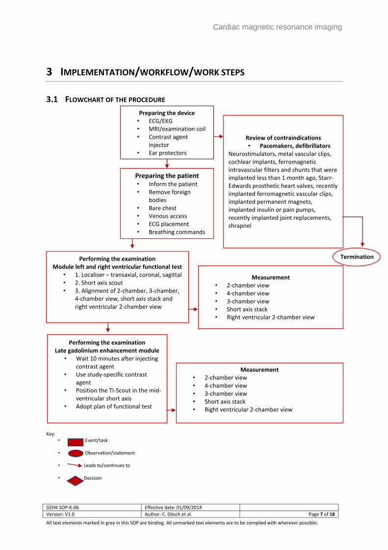
3 IMPLEMENTATION/WORKFLOW/WORK STEPS
3.1 FLOWCHART OF THE PROCEDURE
Key: • Event/task
• Observation/statement
• Leads to/continues to
• Decision
Preparing the device • ECG/EKG • MRI/examination coil • Contrast agent
injector • Ear protectors
Preparing the patient • Inform the patient • Remove foreign
bodies • Bare chest • Venous access • ECG placement • Breathing commands • Apply ear protectors
Performing the examination Module left and right ventricular functional test
• 1. Localiser – transaxial, coronal, sagittal • 2. Short axis scout • 3. Alignment of 2-chamber, 3-chamber,
4-chamber view, short axis stack and right ventricular 2-chamber view
Measurement • 2-chamber view • 4-chamber view • 3-chamber view • Short axis stack • Right ventricular 2-chamber view
Performing the examination Late gadolinium enhancement module
• Wait 10 minutes after injecting contrast agent
• Use study-specific contrast agent
• Position the TI-Scout in the mid-ventricular short axis
• Adopt plan of functional test
Measurement • 2-chamber view • 4-chamber view • 3-chamber view • Short axis stack • Right ventricular 2-chamber view
Review of contraindications • Pacemakers, defibrillators
Neurostimulators, metal vascular clips, cochlear implants, ferromagnetic intravascular filters and shunts that were implanted less than 1 month ago, Starr-Edwards prosthetic heart valves, recently implanted ferromagnetic vascular clips, implanted permanent magnets, implanted insulin or pain pumps, recently implanted joint replacements, shrapnel
• Key:
•
Termination
Cardiac magnetic resonance imaging
DZHK SOP-K-06 Effective date: 01/09/2014
Version: V1.0 Author: C. Dösch et al. Page 8 of 18
All text elements marked in grey in this SOP are binding. All unmarked text elements are to be complied with wherever possible.
3.2 PREPARING FOR THE EXAMINATION Check the Informed Consent Form
Check the laboratory values (creatinine)
Ask about height and weight
3.2.1 Preparing the workplace
None
3.2.2 Preparing the devices
Position and insert the coil, prepare the positioning aids
Prepare and connect the contrast agent injector
3.2.3 Principles of preparing the subject for the examination
Inform the patient about the course of the examination
Remove all foreign bodies (e.g. jewellery, ECG cables, etc.) and clothing (e.g. bra, zips, metal
buttons, metal threads, etc.), and any dental prosthesis that interfere with the examination
Ask the patient to strip down to the waist, surgical shirt (opening in front) or facility-internal
clothing
If necessary, prepare the venous access
Place the ECG, additional monitoring depending on the planned examination
Explain the breathing commands
Apply ear protectors
Positioning the patient
Supine position, head first
Apply the cardiac coil or the manufacturer-specific surface coil
Lay arms alongside the body
Ear protectors
Make the patient as comfortable as possible using positioning aids
If necessary, connect the patient to the contrast agent injector

Cardiac magnetic resonance imaging
DZHK SOP-K-06 Effective date: 01/09/2014
Version: V1.0 Author: C. Dösch et al. Page 9 of 18
All text elements marked in grey in this SOP are binding. All unmarked text elements are to be complied with wherever possible.
3.3 PERFORMING THE EXAMINATION Module left and right ventricular functional test *
*prior to commencing every study, details are adjusted according to current knowledge, during each study the
parameters are kept constant; furthermore, manufacturer-independent "generic" protocols are generated
that define the details of the sequences.
1. Localiser in all 3 patient axes (transaxial, coronal, sagittal)
2. Short axis scout
3. Alignment and measurement of 2-chamber, 3-chamber, 4-chamber view, short axis stack, right
ventricular 2-chamber view
Manual alignment is outlined below; however, automatic algorithms such as the three-point method
can also be applied.

Cardiac magnetic resonance imaging
DZHK SOP-K-06 Effective date: 01/09/2014
Version: V1.0 Author: C. Dösch et al. Page 10 of 18
All text elements marked in grey in this SOP are binding. All unmarked text elements are to be complied with wherever possible.
Figure 1: 2-chamber view
2-chamber view
The 2-chamber view is aligned on the
axial localiser through the centre of the
mitral valve and the tip of the left
ventricle.
Figure 2: 4-chamber view
4-chamber view
The 4-chamber view is aligned on the
short axis localiser and also using the 2-
chamber view, whereby it must be
ensured that the alignment runs
through the tip of the 2-chamber view
and verified on the short axis localiser
that the left ventricular outflow tract is
not included on the image.
Figure 3: 3-chamber view
3-chamber view
The 3-chamber view is aligned from the
4-chamber view and the short axis
localiser. A cross-section through the
left ventricular outflow tract is chosen
on the short axis stack, whereby for the
4-chamber view it must be ensured that
the cross-section runs through the apex
of the heart.
The 3-chamber view is needed for
assessing anteroseptal and inferolateral
wall motion disorders. Furthermore, it
can be used to determine the thickness
of the basal anterior septum (basal
septum) and of the inferior lateral wall
(basal lateral wall). In addition, the left
ventricular end-diastolic (LVEDD) and
end-systolic (LVESD) diameter, the
diameter of the left atrium and the
diameter of the left ventricular outflow

Cardiac magnetic resonance imaging
DZHK SOP-K-06 Effective date: 01/09/2014
Version: V1.0 Author: C. Dösch et al. Page 11 of 18
All text elements marked in grey in this SOP are binding. All unmarked text elements are to be complied with wherever possible.
tract (LVOT) can be measured in the 3-
chamber view.
Figure 4: Short axis stack
Short axis stack
The short axis stack is aligned in
diastole. The first layer should be
located inside the left atrium and the
last layer outside the left ventricle. The
short axis stack is aligned such that a
connection between the AV grooves
(white line) can be created at the height
of the mitral and tricuspid valve. The
remaining short axes cover the entire
left and right ventricle up to the apex of
the heart and are parallel to the initial
short axis.
Figure 5: Right ventricular 2-chamber view
Right ventricular 2-chamber view
In addition, a longitudinal section
through the right ventricle should be
made.

Cardiac magnetic resonance imaging
DZHK SOP-K-06 Effective date: 01/09/2014
Version: V1.0 Author: C. Dösch et al. Page 12 of 18
All text elements marked in grey in this SOP are binding. All unmarked text elements are to be complied with wherever possible.
Late gadolinium enhancement module *
*prior to commencing every study, details are adjusted according to current knowledge, during each study the
parameters are kept constant; furthermore, manufacturer-independent "generic" protocols are generated
that define the details of the sequences.
Figure 6: Late gadolinium enhancement module
Wait 10 minutes after injecting the contrast agent.
The contrast agent to be used (volume and type depending on relaxivity) are defined
specifically for each study. The injection rate (flow rate) is also adjusted accordingly.
TI-Scout to determine the zero-crossing point of the signal of the healthy myocardium.
Position the TI-Scout in the mid-ventricular short axis.
Recording of the late gadolinium enhancement images of all short axes, of the 2 left and right
ventricular 2-chamber views, of the 3-chamber view and of the 4-chamber view (in doing so,
the alignment should correspond to that of the functional test).
3.4 FOLLOW-UP AND DATA COLLECTION Evaluation of the MRI images using the respective evaluation software
3.5 PROCEDURE IN CASE OF DEVIATIONS None

Cardiac magnetic resonance imaging
DZHK SOP-K-06 Effective date: 01/09/2014
Version: V1.0 Author: C. Dösch et al. Page 13 of 18
All text elements marked in grey in this SOP are binding. All unmarked text elements are to be complied with wherever possible.
4 LITERATURE AND REFERENCES
Kramer CM et al. Journal of Cardiovascular Magnetic Resonance 2008, 10:35
Kramer CM et al. Journal of Cardiovascular Magnetic Resonance 2013, 15:91
5 CHANGE
Change compared to the previous version
Section Description of the change to the previous version
2.1
2.2
2.3
….
6 PERSONS INVOLVED
Name Role Involvement
PD Dr. Christina Dösch Initial author Creation of SOP
Dr. Christian Liebetrau Author Technical review
Prof. Dr. Joachim Lotz Author Technical review
PD Dr. Rolf Wachter Author Technical review
Prof. Dr. Jeanette Schulz-
Menger
Last author Technical review

Cardiac magnetic resonance imaging
DZHK-SOP-K-06 Effective date: 01/09/2014
Version: V1.0 Author: C. Dösch et al. Page 14 of 18
The text elements marked with the symbol ** in this SOP are binding. The text elements marked with the symbol * are to be complied with
wherever possible.
7 ANNEXES
7.1 ECRF MODULE
Cardiac magnetic resonance imaging
DZHK-SOP-K-06 Effective date: 01/09/2014
Version: V1.0 Author: C. Dösch et al. Page 15 of 18
The text elements marked with the symbol ** in this SOP are binding. The text elements marked with the symbol * are to be complied with
wherever possible.

Cardiac magnetic resonance imaging
DZHK-SOP-K-06 Effective date: 01/09/2014
Version: V1.0 Author: C. Dösch et al. Page 16 of 18
The text elements marked with the symbol ** in this SOP are binding. The text elements marked with the symbol * are to be complied with
wherever possible.
7.2 LIST OF FIGURES Figure 1: 2-chamber view ...................................................................................................................... 10 Figure 2: 4-chamber view ...................................................................................................................... 10 Figure 3: 3-chamber view ...................................................................................................................... 10 Figure 4: Short axis stack ....................................................................................................................... 11 Figure 5: Right ventricular 2-chamber view .......................................................................................... 11 Figure 6: Late gadolinium enhancement module ................................................................................. 12

Cardiac magnetic resonance imaging
DZHK-SOP-K-06 Effective date: 01/09/2014
Version: V1.0 Author: C. Dösch et al. Page 17 of 18
The text elements marked with the symbol ** in this SOP are binding. The text elements marked with the symbol * are to be complied with
wherever possible.
Cardiac magnetic resonance imaging
DZHK-SOP-K-06 Effective date: 01/09/2014
Version: V1.0 Author: C. Dösch et al. Page 18 of 18
The text elements marked with the symbol ** in this SOP are binding. The text elements marked with the symbol * are to be complied with
wherever possible.