e-cpr in germanywfsiccm2015.com/wfsiccm_ab/1100amthomas muller.pdf · 1. ecls in germany •...
TRANSCRIPT

E-CPR
in
Germany
PD Dr. Thomas Müller
Critical Care Medicine
Department of Internal Medicine II
University of Regensburg
conflict of interest: none
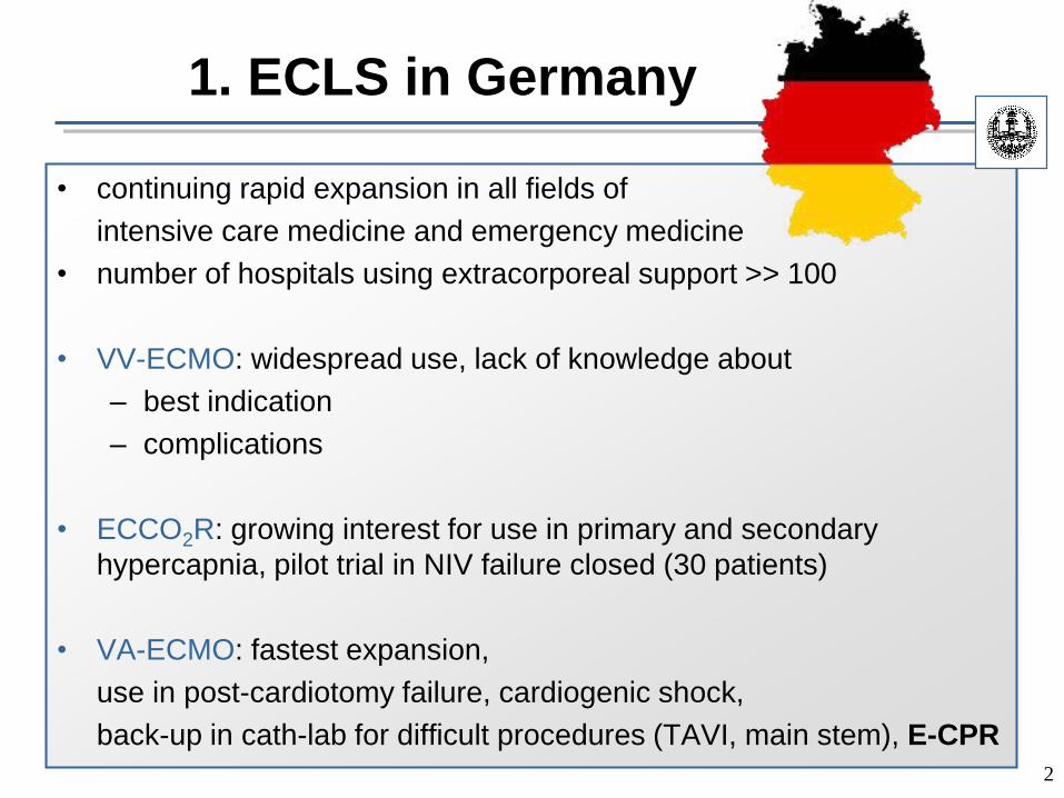
1. ECLS in Germany
• continuing rapid expansion in all fields of
intensive care medicine and emergency medicine
• number of hospitals using extracorporeal support >> 100
• VV-ECMO: widespread use, lack of knowledge about
– best indication
– complications
• ECCO2R: growing interest for use in primary and secondary
hypercapnia, pilot trial in NIV failure closed (30 patients)
• VA-ECMO: fastest expansion,
use in post-cardiotomy failure, cardiogenic shock,
back-up in cath-lab for difficult procedures (TAVI, main stem), E-CPR
2

3
ECMO Centers in Germany
•Essen •Göttingen•Leipzig
•Mainz•Frankfurt
•Aachen
•Halle•Dortmund
•München
•Regensburg
•Erlangen
•Ludwigsburg
•Freiburg
•Mannheim
•Würzburg
•Hamburg
•Berlin
•Gießen
•Bochum
•Dresden
•Hannover
•Kiel•Rostock
•Bonn •Marburg
•Münster•Osnabrück
2010 2015

2. In-Hospital Resuscitation
n = 64 339 , 2000 – 2008,
„Get with the Guidelines-Resuscitation Registry“
ROSC
< 25 % of patients who died, were resuscitated for > 30 min
31 198 (48.5 %) ROSC, 9 912 (15.4 %) discharged, of those 80.6 %
with good neurological outcome
4Goldberger et al, Lancet 2012;380:1473-81
duration of resuscitation
in non-survivors

E-CPR for IHCA - Overview
5Fagnoul D et al, Curr Opin Crit care 2014;20:259-65
25 – 55 min 20 – 40 %

6
Problems of peripheral VA-ECMO
1. Cannula associated
– damage to vessel
– peripheral ischemia
– thrombosis, pulmonary embolism
– poor access for intervention
2. „Harlekin Syndrome“
3. loading of left ventricle
4. bleeding
5. technical failure
– oxygenator thrombosis
– pump failure

7
Peripheral VA-ECMO: Less is more?
1. 15 Fr arterial cannula:
• less ischemia, less bleeding
2. Blood flow ≤ 3,5 L/min:
• reduced afterload
• allow LV to unload (RR amplitude > 10 mm Hg)
• avoid need of venting
3. Flow is more important than pressure:
• what matters, is oxygen transport
• vasopressors, inotropes
• MAP 50 – 60 mm Hg
4. Monitoring:
• NIRS for brain and leg
• venous saturation (pre oxy) > 65 %
• lactate
• urine output o.k.

Monitoring: Control of leg perfusion
Sensor NIRS
ECLS jugular vein > femoral artery
Monitoring:
• doppler US
• oxymetrie on toe
• capillary pulse
• NIRS
• documentation

Difficult Access for Intervention
• with VA ECMO, access for PCI or electrophysiologic treatment can be
difficult or prolonged
• Y-connector allows access through arterial cannula
Endemann D et al, Intensive Care Med 2011;37:2046-49
Ü cer E et al, Europace 2014;16:299-302

VA ECMO „less is more“: patients
10
n = 107
age 57.5 +/- 14 years
77 % male
indications:
• unsuccessful conventional resuscitation (n = 50)
• cardiogenic shock post resuscitation (n = 23)
• cardiogenic shock without previous resuscitation (n = 15)
• cardiogenic shock/resuscitation during intervent. cardiology (n = 19)
distal perfusion cannula: n = 20
difficult cannulation: n = 27
bleeding cannulation site: n = 12
time on ECMO 4.0 +/- 4.4 days
RBCs per day on ECMO: 0.6 +/- 1.0

VA ECMO „less is more“:
hemodynamics
11
** p< 0.001
**Blood Flow
MAP
**
Noradrenaline
**
**

VA ECMO „less is more“: results
12
** p< 0.001
** Laktate
ven. Saturation
**
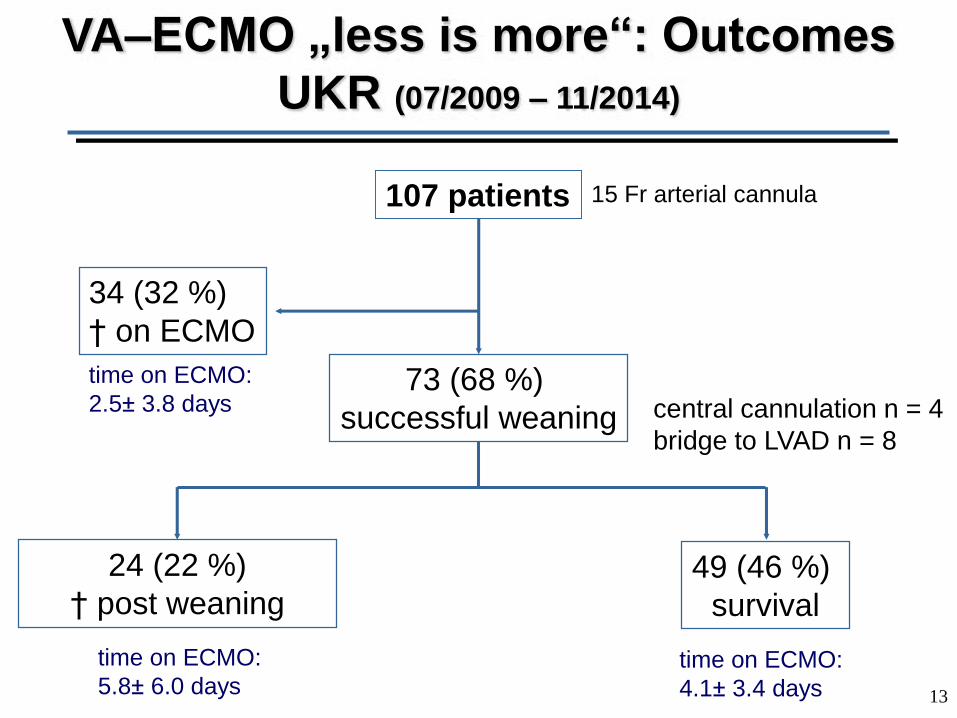
13
VA–ECMO „less is more“: Outcomes
UKR (07/2009 – 11/2014)
107 patients
73 (68 %)
successful weaning
49 (46 %)
survival
34 (32 %)
† on ECMO
24 (22 %)
† post weaning
time on ECMO:
2.5± 3.8 days
time on ECMO:
5.8± 6.0 daystime on ECMO:
4.1± 3.4 days
central cannulation n = 4
bridge to LVAD n = 8
15 Fr arterial cannula

3. E-CPR in OHCA?
• National Taiwan University Hospital Taipei
• 2007 – 2012; n = 230, 199 IHCA, 31 OHCA (selected patients)
• results:
• „Our results suggest that further investigation of the use of
ECMO in OHCA is warranted“
14Wang CH, Resuscitation 2014;85:1219-24
IHCA OHCA
time to ECLS (min) 44.4 ± 24.7 67.5 ± 30.6
survival to discharge 31.2 % 38.7 %
survival with good neurology 25.1 % 25.5 %

2010 – 6/2013; 91 ambulance stations in UK
n= 4471 patients randomized 2:1 manual : LUCAS-2
primary endpoint: 30 day survival
patient characteristics:
age 71 ys
80 % at home
60 % witnessed
43 % bystander resuscitation
time call to arrival 6.5 min
50 % asystole
Lancet 2014; online Nov.16

PARAMEDIC-Trial: Results
Lancet 2014; online Nov.16

E-CPR for OHCA - Overview
17Fagnoul D et al, Curr Opin Crit care 2014;20:259-65
44 – 120 min 4 – 39 %

18
all
(n = 79)
In-hospital
(n = 55)
Out-of-
hospital
(n = 24)
p Value
age, years 56 ± 15 64 ± 14 50 ± 15 0.0001
male, n (%) 57 (72) 40 (73) 17 (71) 0.99
CPR duration, min 52 ± 36 40 ± 29 82 ± 34 <0.0001
pH [venous, pre MO] 7.01 ± 0.22 7.08 ± 0.18 6.86 ± 0.24 <0.0001
lactate, mg/dL 99 ± 62 65 ± 51 132 ± 82 <0.0001
PvCO2, mmHg 71 ± 21 59 ± 17 86 ± 23 <0.0001
intervention possible, n (%) 52 (66) 36 (66) 16 (67) 0.99
complications, n (%) 23 (29) 15 (27) 8 (33) 0.60
outcome, n (%)
survival to discharge27 (34) 23 (42) 4 (17) 0.039
ECMO in ED
Resuscitation 2012,83:1331-37

RECA-Study
(Regensburg ECLS for Cardiac Arrest)
19
cardiac arrest with CPR > 10 min and
presumably cardiac etiology (VF/VT)
witnessed cardiac arrest
bystander-CPR or beginning of ACLS
within 5-10 min
age between 18 and 65 yrs

Out of Hospital E-CPR
• 61 yrs, observed collaps, immediate bystander resuscitation
• time response:
– alarm 11:58
– arrival ambulance + emergency team 12:08 (10 min)
– VF, continued CPR
– alarm mobile ECMO-Team 12:20 (12 min)
– arrival ECMO Team 12:32 (12 min)
– ECMO-start 12:47 (15 min)
• diagnosis: anterior myocardial infarction total: 49 min
20

Out of Hospital E-CPR
• 25 yrs, thoracic pain on inspiration for 5 days, progressive exertional
dyspnoe
• risk factors: oral contraception, Crohn s Disease + steroids
• 25/09/2014: observed collaps, immediate professional resuscitation
• time to ECMO 59 min
• Diagnosis: pulmonary embolism and right heart failure
21

4. Peri- and Post-Resuscitation Care:
Options for Improvement?
bystander CPR:
telephone guidance, compressions only
decrease time to ECMO: move ECMO to patient?
therapeutic hypothermia: early cold infusion
early diagnostic and therapeutic intervention
avoid hyperoxemia
avoid hypocapnia/alkalosis: „two circulations“
→ BGA right radial artery and post oxygenator
aspiration pneumonia: prophylactic antibiotics
post-resuscitation SIRS: hydrocortisone?
short-acting analgosedation
adapted, individualized anticoagulation
22Müller et al, Resuscitation 2013;84:1463

23
… a fool with a tool
is still a fool...
Summary: one step further
1. E-CPR can safe lifes
if done well
in the right patients
2. important:
reduce time to ECMO
reduce complications: “less is more”?
improve post-resuscitation care
find and treat cause of collaps
3. Cardiac Arrest Centres networking with
other hospitals may improve results

24

3. Loading of the Left Ventricle
• high afterload may hinder the LV to empty/ aortic valve to open
– high wall pressure
– no recovery
– pulmonary edema
– clotting in LV and pulmonary circulation
• management:
– preserve LV output: inotropes , vasopressors
– reduce ECMO flow, if possible (3.5 L/min)
– adapt ventilation
– increase anticoagulation
– venting (ultima ratio)

Decompression of the Left Ventricle
Rupprecht L et al, ASAIO Journal 2013;59:547-53