e d i t o r i a l b o a r d - uvlf.sk · subscription rate for 1 year is 200 sk, for foreigners 80...
TRANSCRIPT


FOLIA VETERINARIA is issued by the University of Veteri
nary Medicine in Košice (UVL); address: Komenského 73, 041
81 K o š i c e, The Slovak Republic (tel.: +421 55 633 51 03, fax:
+421 55 633 51 03, E−mail: [email protected]).
The journal is published quarterly in English (numbers 1—4)
and distributed worldwide.
Subscription rate for 1 year is 200 Sk, for foreigners
80 euros. Orders are accepted by The Department of The Scienti
fic Information — The Library of The University of Vete ri nary
Medicine, Košice (UVIK); the subscription is accepted by the
National bank of Slovakia in Košice (at the account number
mentioned below).
Bank contact: National bank of Slovakia, 040 01 Košice,
Strojárenská 1, our account number: 19−1924−512/0720.
FOLIA VETERINARIA, vydáva Univerzita veterinárskeho lekárstva v Košiciach (UVL), Komenského 73, 041 81 K o š i c e, Slovenská republika (tel.: 055/633 51 03, fax: 055/633 51 03, E−mail: [email protected]).
Časopis vychádza kvartálne (č. 1—4) a je distribuovaný celosvetove.
Ročné predplatné 200 Sk, pre zahraničných odberateľov 80 eur. Objednávky prijíma Ústav vedeckých infor mácií a knižnice Univerzity veterinárskeho lekárstva v Ko ši ciach (UVIK); predplatné Národná banka Slovenska v Košiciach (na nižšie uvedené číslo účtu).
Bankové spojenie: Národná banka Slovenska, Košice, Stro-járenská 1, číslo príjmového účtu: 19−1924−512/0720.
Tlač: EMILENA, Čermeľská 3, 040 01 KošiceSadzba: Aprilla, s.r.o., Hlavná 40, 040 01 Košice
Registr. zn. 787/93
E D I T O R I A L B O A R D
Editor in Chief : Rudolf C a b a d a jExecutive Editor : Emil P i l i p č i n e cMembers : Baumgartner, W. (Vienna), Bíreš, J. (Bratislava), Breza, M. (Košice), Buczek, J. (Lublin), Bugarský, A.
(Košice), Campo, M. S. (Glasgow), Cudlín, J. (Prague), Dianovský, J. (Košice), Huszenicza, Gy. (Buda-pest), Kottferová, J. (Košice), Legáth, J. (Košice), Levkut, M. (Košice), Lešník, F. (Košice), Mesároš, P. (Košice), Mikula, I. (Košice), Mojžišová, J. (Košice), Pistl, J. (Košice), Pogačnik, M. (Ljubljana), Rosival, I. (Košice), Šucman, E. (Brno), Totolian, A. A. (Saint Petersburg), Vilček, Š. (Košice), Zibrín, M. (Košice)
For basic information about the journal seeInternet home pages: www.uvm.sk
Indexed and abstractedin AGRIS, CAB Abstracts
KOŠICE

KORÉNEK, M., ŠUTIAK, V., KORÉNEKOVÁ, B., VAŠKO, L., ŠUTIAKOVÁ, I., NEUSCHL, J.:
Activity of α-chymotrypsin after addition of natural and synthetic inhibitors .......................................................................... 57
OLAYEMI, F. O., AKINSIKU, D. O., OJO, O. E., AZEEZ, O.: The haematology of the
Kuri breed of cattle .................................................................................................................................................................... 62
NOTTIDGE, H. O., OMOBOWALE, T. O., WASHIO, M., AJADI, R. A., TOIZUMI, Sh., TAKAHASHI, K.:
The prevalence of the dog erythrocyte antigen 1 (deA 1.1 and 1.2) in nigerian indigenous dogs ......................................... 66
ADEDAPO, A. A., AIYELOTAN, O.: The effects of graded doses of indomethacin on some
serum biochemical parameters of rats ........................................................................................................................................ 69
KALANIN, P., FLEŠÁROVÁ, S.: neuron damage elicited by cardiac arrest in a dog brain ................................................. 73
ADEHAN, R. K., AJUWAPE, A. T. P., ADETOSOYE, A. I.: The occurrence mycoplasma species
and other bacteria in preumonic lungs of sheep and goats from benin republic ...................................................................... 76
NOVOTNÝ, L., DVOŘÁK, P.: Manifestation of mycobacteriosis in cardinal tetras Paracheirodon axelrodi
(Schultz, 1956) during the Pleistophora hyphessobryconis (Schäperclaus, 1941) infection ...................................................... 80
REVAJOVÁ, V., LEVKUT, M., ALDAWEK, A., HERICH, R., DVOROŽŇÁKOVÁ, E., KRUPICER, I.:
Morphological, histological and immunohistochemical changes after multiple toxocara canis infection of lambs ................. 83
ONDREJKOVÁ, A., SÜLI, J., ONDREJKA, R., BENÍŠEK, Z.: detection of rabies antibodies
in dog sera by different serological methods ............................................................................................................................. 89
OKEWOLE, E. A.: A chemotherapeutic appraisal of canine hepatozoonosis in Ibadan, nigeria:
A retrospective study of 192 cases ............................................................................................................................................ 94
KOVALKOVIČOVÁ, N., PISTL, J., HOLOVSKÁ,s K., LEGÁTH, J.: Animal models
in immunotoxicity studies (A review) ........................................................................................................................................ 99
F O L I A V E T E R I N A R I A, 50, 2, 2006
C O N T E N T S
F O L I A V E T E R I N A R I A, 50, 2, 2006
C O N T E N T S

5757
www.uvm.sk
ACTIVITY OF α-CHYMOTRYPSIN AFTER ADDITION OF NATURAL AND SYNTHETIC INHIBITORS
Korének, M., Šutiak, V., Koréneková, B.Vaško, L., Šutiaková, I., Neuschl, J.
University of Veterinary Medicine, Komenského 73, 041 81 KošiceThe Slovak Republic
ABSTRACT
The effect of natural and synthetic inhibitors on α-chy-motrypsin activity has been studied under in vitro conditions. The chromogenic substrate Suc-(Gly)
2-Phe-NAn (20 mmol . 0.1
ml-1) and Tris/HCl buffer (0.7 ml) was incubated at 37 °C for 15 minutes together with other components. In the first experimental series, the natural inhibitors, hydrolysable tannin, Tanifarm, Farmatan and Pycnogenol at concentra-tions of 18.75, 37.5 and 75 µg.0.1 ml-1 were added. The same concentrations of synthetic inhibitors I
1 (Ala
2- Leu-NH-EtPh),
I2 (Suc-(Ala)
2-Pro-NH-EtPh), and I
3 (Glt-(Ala)
2-Pro-NH-EtPh)
were added in the second series of experiments. The reac-tion was initiated by α-chymotrypsin (200 µg . 0.1 ml-1). A significant decrease (P ≤ 0.001) activity of α-chymotrypsin was found after the addition of natural and synthetic in-hibitors in comparison to the control group. The activity of α-chymotrypsin was in order: Pycnogenol < Tanifarm < Tannin < Farmatan after the addition of natural inhibi-tors, and I
3 < I
1 < I
2 after addition of synthetic inhibitors.
The lowest activity of chymotrypsin was recorded after the addition of Pycnogenol (2.88 ηkat.l-1) and a synthetic inhibitor I
3 (3.57 ηkat.l-1). Our experiment has the potential
to contribute useful information regarding the activity of chymotrypsin after the addition of various natural and synthetic inhibitors.
Key words: α-chymotrypsin; natural inhibitors; synthetic inhibitors; tannins
FOLIA VETERINARIA, 50, 2: 57—61, 2006
INTRODUCTION
Serine proteases (chymotrypsin, trypsin, elastase) are a class of enzymes that cleave peptide bonds in proteins charac-terised by the presence of a serine in the active center of the enzyme. All three of these enzymes are similar in structure and are highly specific in the reactions they catalyze. Each of these digestive serine proteases targets different regions of the polypeptide chain, based upon the amino acid residues and side chains surrounding the site of cleavage (27).
Serine proteases were found in various mammal tissues and cells and are referred to as trypsin-like, chymotrypsin-like and elastase-like enzymes (20). Varying proportions of serine proteinases are related to their linkage with important physiological processes: digestion of proteins, coagulation and complement system and chemotaxis.
They are also involved in pathological states. Pulmonary diseases, pancreatitis, and digestive tract disorders are associated with changes in the activity of serine proteinases. The species differences in pathology resulting from neutrophil-mediated respiratory disease are determined by other factors such as differences in the abundance and function of intra- and extra-cellular protease inhibitors (7). Therefore, the development of specific inhibitors of proteases is a promising strategy in the treatment of these diseases.
Chymotrypsin as an important member of the family of serine proteases is a proteolytic enzyme acting in the digestive systems of mammals and other organisms. Chymotrypsin is synthesized in the pancreas by protein biosynthesis. Determi-nation of chymotrypsin activity in body fluids and faeces is relevant to pancreatitis, lung diseases and some other patho-logical changes (2, 26).

58
An enzyme inhibitor is a molecule that binds to an enzyme and decreases its rate of reaction. The natural inhibitors of serine proteinases are generally divided into progressive (inhibition lasts up to 120 minutes and is complete; this group includes α-macroglobulin, α-antitrypsin, antiplasmin), permanent (inhibition lasts up to eight to twelve days; to these belong Bowman-Birk’s and Kunitz’s inhibitors from soya), and temporary (inhibition lasts up to 24 hours with dissociation of the complex; this group includes Kazal’s porcine inhibitor) (1).
Serine proteases are inhibited by serine protease inhibitors (serpins), a group of enzymes that form a covalent bond with the serine protease, inhibiting its function , e.g. α-1-antitrypsin, antithrombin (23, 10).
Tannins are glycosides of plant origin, which cause inhibi-tion of peroxidation of lipids and lipooxygenase, xantioxidase and monoamino oxidase (5, 9, 15). They suppress production of proteolytic enzymes, such as colagenase and elastase. This non-competitive inhibition was observed also with β-glucuroni-dase, hyaluronidase, and β-glucosidase (18). These substances are anti-nutrients from the point of view of nutrition because they decrease the digestibility of proteins in the digestive tract by affecting the activity of serine proteinases (8, 6, 19).
The natural inhibitor – tannins and their derivatives (Farmatan and Tanifarm) and Pycnogenol are phenolic compounds with vari-ous antiviral, antioxidative, antitumorous and anti-inflammatory effects (3). Tanifarm and Farmatan, show an adstringent effect due to which they favourably affect inflammatory symptoms. They reduce the absorption of noxious substances and glandular secretion, act antiseptically and obstipatively, reduce intestinal peristalsis, prevent dehydration, reduce bleeding and support healing and epithelization of the mucosa and skin. The main indication for the use of these preparations is prevention and treatment of the diarrhoeic syndrome and respiratory diseases in animals (7, 17, 25, 19).
Various works have focused on an interesting biological tool for the evaluation of new synthetic inhibitors of serine proteases. These inhibitors are advantageous in several ways compared to natural inhibitors. They are not complete anti-gens and can be used orally but also as aerosols. They can be prepared according to need and are easily reabsorbed and transferred in an organism.
The synthetic compounds can protect against proteinases for even several days after development of organic disorders. Such an effect has been reported so far only with natural inhibitors (elastinal, α-antitrypsin), even ten days after the development of disorders (22). Synthetic inhibitors of serine proteinases have prophylactic effects in the course of pancreatic inflammatory processes (13, 12, 25). The aim of our study was to compare the activity of chymotrypsin after the addition of natural and synthetic inhibitors.
MATERIAL AND METHODS
Bovine α-chymotrypsin and Suc-(Gly)2-Phe-pNa (Sigma
Aldrich, Germany) was used in the experiment as an enzyme source and chromogenic substrate. The natural inhibitors tannin
and Tanifarm, investigated in the experiment, were supplied by the firm Pharmagal (the Slovak Republic) and Farmatan and Pycnogenol were obtained from the firm Sevnica (Slovenia). They contained the following pharmaceuticals: Tanin plv. (active ingredient tannin of PhBS IV pharmaceutical purity); Tanifarm plv. sol. a.u.v. (55 % tannin); Farmatan cps. a.u.v. (55 % tannin), and Pycnogenol tbl. (14 % pycnogenol®). Synthetic inhibitors I
1
Ala2-Leu-NH-EtPh, I
2 Suc-(Ala)
2-Pro-NH−EtPh, and I
3 Glt-(Ala)
2-
Pro-NH-EtPh, used throughout the study, were obtained from Res. Inst. of Bioch. and Pharm., Prague, the Czech Republic. Chymotrypsin activity was determined in the following buffer medium: 0.7 ml of 0.05 mol.l-1 Tris/HCl buffer (pH = 7.8) to which 100 µl substrate Suc-(Gly)
2-Phe-pNA (p-nitroanilid) at
a concentration of 20 mmol.l-1 was added. In the first experimental series, the natural inhibitors hydro-
lysable tannin, Tanifarm, Pycnogenol and Farmatan, all at the following concentrations: 18.75, 37.5, and 75 µg . 0.1 ml-1, were added to the samples. In the second series, synthetic inhibi-tors I
1, I
2, and
I
3 (18.75; 37.5; 75 µg . 0.1 ml-1) were added in the
same manner as in the first series. The reaction was initiated by α-chymotrypsin (200 µg . 0.1 ml-1). The sample contained only α-chymotrypsin without any inhibitors and served as a control. Each analysis was repeated six times.
Chymotrypsin activity was determined by the method de-scribed by R o s i v a l et al. (24). The rate of α-chymotrypsin catalysed hydrolysis Suc-(Gly)
2-Phe-pNA was established
kinetically at 405 nm using a Specol (Karl Zeiss Jena 200, Germany). The differences between experimental groups and the control (without inhibitors) were analysed statistically (Microsoft Excel 7.0) using Student’s t-test, at P ≤ 0.05, 0.01, and 0.001 levels of significance.
RESULTS
The exhibited highest activity of α-chymotrypsin was recorded in the control group without addition of inhibitors (6.03 nkat.l-1). According to Fig. 1, a sig-nificant decrease (P ≤ 0.001) in α-chymotrypsin activity was observed after addition of the natural inhibitors in comparison to the control group.
The mean activity of chymotrypsin declined consider-ably with increasing concentration of natural inhibitors in the first group of samples. The lowest activity of α-chymotrypsin was recorded at the concentration of 75 µg.ml-1.
The effect of natural inhibitors on the activity of α-chymotrypsin occurred in the following order: Tanifarm < Tannin < Pycnogenol < Farmatan at the concentration of 18.75 µg.ml-1.
However, after the addition of natural inhibitors at higher concentrations, the mean activity of α-chymot-rypsin was changed in the following order: Pycnogenol < Tanifarm < tannin < Farmatan. A similar sequence of inhibitory activity was observed at concentrations of 37 and 75 µg.ml-1.
The results of the present study showed that the lowest activity of α-chymotrypsin (2.88 nkat.l-1) was recorded

59
after the addition of Pycnogenol. On the other hand, Farmatan was the least efficient inhibitor of chymotrypsin activity (3.57 nkat.l-1).
The present study indicated that after the addition of synthetic inhibitors the activity of α-chymotrypsin was decreased. This decrease was indirectly related to the content of inhibitors in the samples (Fig. 2).
The variations in the activity of α-chymotrypsin induced by the synthetic inhibitors used were the following: I
2 <
I1
< I3 (at the concentration of 18.75 µg.ml-1). In spite of
previous results, higher concentrations (37; 75 µg.ml-1) of inhibitors resulted in changes in the mean activity of α-chymotrypsin in the following order: I
3 < I
1 < I
2.
The activity of α-chymotrypsin decreased significantly (P ≤ 0.001) after the addition of synthetic inhibitors in comparison to the control group. The results indicated that the lowest activity of α-chymotrypsin (3.57 nkat.l-1) was reached after the addition of synthetic inhibitor I
3
and the highest after the addition of synthetic inhibitor I
2 (4.19 nkat.l-1).
DISCUSSION
Protease inhibitors are widespread in plants, including cereals, but have been most studied in legumes, especially soybeans. Their common characteristic is an ability to bind to and inhibit proteolytic enzymes (16). The serine proteases with both tryptase and chymase-like proper-ties are inhibited by α-proteinase inhibitors (20). This inhibitor was also presented as a main elastase inhibitor (4) and was used for the immobilisation of chymotrypsin (21). However, the difference in the proteases is that they inhibit the nature of the binding site and in their primary amino acid sequences.
The protease inhibitors act by binding with the enzyme at its active site in the manner of a substrate peptide (11). According to D i e t z et al. (9), tannins
are also able to bind with proteins via the formation of protein-tannin complexes and to inhibit enzymes. Our results indicate clearly, that the tannins inhibited α-chymotrypsin and the increasing activity of natural inhibitors was in the following order: Pycnogenol < Tanifarm < tannin < Farmatan.
Hydrolysable tannin, used in our experiment, consists of polyphenolic acids esterified to a central monosac-charide unit. The non-competitive inhibitory effect does not depend on the structure and amount of glycoside, but on the number of free hydroxyl groups in the tannin structure (18). Some natural inhibitors, e.g. elastinal, act on α-chymotrypsin very promptly resembling tannins in our experiment.
The serine proteinases – trypsin, α-chymotrypsin and porcine pancreatic elastase – have a different specificity to various substrates. This specificity results from their amino acid substitution, primarily in the substrate-bind-ing site. Inhibitors have an affinity for the active site of an enzyme close to the affinity that the substrate of an enzyme has.
The inhibitor binds to the active site of the enzyme, so its substrate cannot bind there. In our experiment the peptidic synthetic inhibitors of α-chymotrypsin ( I
1, I
2,
I3
) used were bound to the active site of the enzyme chromogenic substrate Suc-Gly)
2-Phe-NAn without un-
dergoing a reaction. The substrate molecule was not able to enter the active site while the synthetic inhibitor was there. On the other hand, the synthetic inhibitor was not able to enter the site when the substrate was there.
Synthetic inhibitors can produce a tetrahedral con-figuration with the enzyme, which is either reversible or irreversible according to the strength of the bonds (2). The most intensive competitive inhibitory effect towards the activity of chymotrypsin was observed with the synthetic inhibitor N-aralkylamid tripeptid Glt-(Ala)
2-
Leu-NH-EtPh and the N−arylbenzoizotiazolines (14). The results of our experiment showed that the synthetic
*** — The differences in the means of compounds with and without
the addition of inhibitors were statistically significant (P ≤ 0.001)
Fig. 1. Changes in activity of α-chymotrypsin (µkat.l-1)
after the addition natural inhibitors
*** — The differences in the means of compounds with and without
the addition of inhibitors were statistically significant (P ≤ 0.001)
Fig. 2. Changes in activity of α-chymotrypsin (µkat.l-1)
after the addition syntetic inhibitors

60
inhibitor I3 was the most important competitive inhibitor
of chymotrypsin.On the other hand, after the addition of synthetic
inhibitors, the mean activity of α-chymotrypsin occurred in the following order: I
3 < I
1 < I
2 at concentrations of
18.75, 37.5, and 75 µg.0.1 ml-1. We observed a higher inhibitory effect of natural inhibitors on α-chymotrypsin activity compared to the synthetic inhibitors used in our experiment. A similar inhibitory effect was observed on the proteases activity of Aspergillus oryzae using in vitro techniques (12). It was found that tannins were better inhibitors than synthetic (I
1, I
2, I
3).
The present study showed that the best natural inhibitor, pycnogenol, caused a higher decrease in the activity of α-chymotrypsin than the best synthetic inhibitor I
3. The
in vitro technique used should provide accurate prediction of protein digestion in the gastrointestinal tract.
Our results show that tannins as well as synthetic inhibitors decrease the activity of α-chymotrypsin. It appears that synthetic inhibitors do not have so strong inhibitory properties against serine protease (chymotrypsin, trypsin, elastase) as the natural inhibitors – tannins.
The important action of tannins, their derivatives and synthetic inhibitors on proteolytic enzymes is based on the fact that through production of complexes they decrease the destructive activity of these enzymes result-ing in necrotic organ inflammations. Similar action was observed with animal inhibitors found in the blood and pancreas. Due to generation of a tetrahedral configura-tion through the action of Ser-OH enzyme and synthetic inhibitor and under the influence of phenolic OH groups of tannins, inactive enzyme complexes are produced. This inhibitory influence is important in practice in the prevention and therapy of various inflammatory diseases under in vivo conditions.
REFERENCES
1. Bartík, M., Šutiaková, I., Kasafírek, E., Šutiak, V., 1990: Development of method for analysis of pancreatic elastase a practical using of synthetic inhibitors of chymotrypsin and elastase (In Slovak). Final report: MPVž SR VII-4-8/04, UVL Košice, pp. 1—88.
2. Blahovec, J., Šlesárová, Ľ., 1991: Enzymes and clinical enzymology (In Slovak). Zenit Press, Košice, pp. 1—127.
3. Blazsó, G., Gábor, M., Rohdewald, P., 1997: An-tiinflammatory activities of procyanidin-containing extracts from Pinus pinaster Ait. after oral and cutaneous application. Pharmazie, 52, 380—382.
4. Brown, A., Farmerm, K., Mac Donald, L., Kalsheker, N., Pritchard, D., Haslett, C., Lamb, J., Sallenave, J. M., 2003: House dust mite Der p 1 downregulates defenses of the lung by inactivating elastase inhibitors. J. Respir. Cell Molec. Biology, 29, 381—389.
5. Butler, L. G., 1992: Anti-nutritional effect of condensed and hydrolysable tannins. In Hemingway. R. W. and Labs. P. E. (eds.): Plant Polyphenols. New York, pp. 693—698.
6. Clemente, A., Mac Kenzie, D., Jeenes, D., Gee, J., Johnson, I., Domoney, C., Vasil, I. K., 2002: Anticarcinogenic properties of plant protease inhibitors from the Bowman-Birk class. Plant Biotechnology and Beyond, 5, 429—431.
7. Dagleish, M. P., Pemberton, A. D., Brazil, T. J., Mac Aleese, S. M., Miller, H. R. O., Scudamore, C. L., 1999: Kinetics of equine neutrophil elastase release and superoxide anion generation following secretagoque activation: a potential mechanism for antiproteinase inactivation. Vet. Immunol. Im-munopath., 72, 257—275.
8. Deshimaru, M, Hanamoto, R., Kusano, C., Yoshimi, S., Terada, S., 2002: Purification and characterization of pro-teinase inhibitors from wild soya (Glycine soya) seeds. Biosci. Biotechnol. Biochem., 66, 1897—1903.
9. Dietz, B. A., Hagerman, A. E, Barret, G. N., 1994: Role of condensed tannin on salivary tannin-binding proteins, bioenergetics and nitrogen digestibility in Microtus pennsyl-vanicus, J. Mammol., 75, 880—889.
10. Gettins, P. G. W., 2002: Serpin structure, mechanism and function. Chem. Rev., 102, 751—803.
11. Korének, M., Neuschl, J., Šutiak, V., Koréneková, B., Čellárová, E., 1998: The use of adstringents in veterinary medicine (In Slovak). Infovet, No. 4, 36—37.
12. Korének, M., Lohajová, Ľ., Šutiaková, I., Šutiak, V., Vaško, L., Koréneková, B., Neuschl, J., Šlesárová, Ľ., Sobeková, A., 2000: Effect of tannins, their derivatives and some synthetic inhibitors on proteases activity of Aspergillus oryzae in vitro. Chem. Papers, 94, 571—591.
13. Kwon, D. Y., 1994: Effects of negative charges of a model for bovine pancreatic trypsin inhibitor folding inter-mediate on the peptide folding. Biosci. Biotechnol. Biochem., 58, 400—405.
14. Lange, F., Bignon, J., Dimicoli, J. L., Bieth, J., 1980: Comparative effects of reversible and irreversible specific elastase inhibitors on elastase induced emphysema. Bull. Europ. Physiopath. Resp., 16, 407—412.
15. Luck, G., Liao, H. L., Murray, J., Grimmer, H. R., Warminski, E. E., 1997: Polyphenols, astringency and proline-rich proteins. Phytochemistry, 37, 357—371.
16. Mc Bride, J. D., Freeman, H. N., Leatherbarrow, R. J., 2000: Identification of chymotrypsine inhibitors from a second generation template assisted combinatorial library. J. Pept. Sci., 6, 446—452.
17. Nguz, K., Gaver D. sr., Huyghebaert, A., Gaver, D. jr., 1998: Inhibition in vitro d'enzymes digestives par les tannins condenses du sorgho (Sorghum bicolor L.). Sciences des Aliments, 18, 507—514.
18. Pečiar, B., Košťálová, D., 1998: Plant tannins and their importance in prevention and therapy (In Slovak). Slovakofarma revue, 8, 54—60.
19. Pino, V. H. sr., Lajolo, F. M., Pino, V. H. jr., 2003: Efecto inhibitorio de los taninos del frijol Carioca (Phaseolus vulgaris) sobre la digestibilidad de la faseolina por dos siste-mas multienzimaticos. Ciencia e Tecnologia de Alimentos, 23, 49—53.
20. Pemberton, A. D., Belham, C. M., Hunley, J. F., Plevin, R., Miller, H. R., 1997: Sheep mast cell proteinase 1, a serine proteinase with both tryptase- and chymase-like properties,
61
is inhibited by plasma proteinase inhibitors and is mitogenic for bovine pulmonary artery fibroblasts. Biochem. J., 323, 719—725.
21. Polanowski, A., Wilimowska-Pelc, A., Kowalska, J., Grybel, J., Zelazko, M., Wilusz, T., 2003: Non-conventional affinity chromatography of serine proteinases and their inhibi-tors. Acta Bioch. Polon., 50, 765—773.
22. Powers, J. C., 1986: Serine proteases of leukocyte mast cell origin. Substrate specificity and inhibition of elastase, chymase and tryptase. Adv. Inflam. Res., 1, 145—157.
23. Silverman, G. A., Bird, P. I., Carrell, R. W., Church, F. C., Coughlin, P. B., Gettins, P. G., Irving, J. A., Lomas, D. A., Luke, C. J., Moyer, R. W., Pemberton, P. A., Remold-O'Donnell, E., Salvesen, G. S., Travis, J., Whisstock, J. C., 2001: The serpins are an expanding superfamily of structurally similar but functionally diverse proteins. Evolution, mechanism of inhibition, novel functions, and a revised nomenclature. J. Biol. Chem., 276, 293—296.
24. Rosival, I., Šulík, E., Šlesárová, Ľ., Korének, M., Blahovec, J., 1993: Determination of chymotrypsin activity in animal stools. Folia Vet., 37, 19—22.
25. Šutiak, V., Korének, M., Šutiaková, I., Koréneková, B., Sobeková, A., Vaško, L., Neuschl, J., 2002: The induction of inflammation in mouse tails with Aspergillus oryzae protease and the inhibitory effects of synthetic and natural substances in vitro. Acta Veter. Beograd, 52, 345—354.
26. Šutiaková, I., Šutiak,V., 1991: Synthetic inhibitors of serine proteinases. Bioch. Clin. Bohemoslov., 20, 193—202.
27. Yamasaki, Y., Satomi, S., Murai, N., Tsuzuki, S., Fushiki, T., 2003: Inhibition of membrane type serine protease 1-maptriptase by natural and synthetic protease inhibitors. J. Nutrit. Sci. Vitaminol., 49, 27—32.
Received June 1, 2006

6262
www.uvm.sk
ABSTRACT
To study the effects of sex and breed on the haemato-logical parameters of the Kuri breed of cattle in a warm humid tropical climate, twenty Kuri cattle and ten White Fulani cattle of both sexes were sampled.
The effects of sex and breed were determined on the haematological values: red blood cell counts (RBC), white blood cell counts (WBC), haemoglobin (Hb), packed cell value (PCV), mean corpuscular volume (MCV), mean corpuscular haemoglobin (MCH), mean corpuscular haemoglobin concen-tration (MCHC) and the differential leukocyte (neutrophil, lymphocyte, eosinophil, monocyte and basophil) counts. The value of Hb value was greater (P < 0.01) in the male than female Kuri cattle, however the other haematological values were similar in the male and female Kuri cattle. Furthermore, the Kuri cattle had higher PCV (P < 0.001) and Hb (P < 0.05) values than the White Fulani cattle but the other erythrocyte values and all the leukocyte values were similar in the two breeds of cattle. The differences in the erythrocyte values of the Kuri and White Fulani cattle suggests that though these cattle are both tropical breeds, the erythrocyte values of the White Fulani cattle
cannot be employed in the assessment of the health status of Kuri cattle.
Key words: breed; cattle; haematology; Kuri cattle; sex; White Fulani cattle
INTRODUCTION
The Kuri breed of cattle is also known as Kouri, Baharie, Buduma, Budduma, Budumu, Boudouma, Chad, Dongolé or White Lake Chad – R a h w a y (18) This breed of cattle is characterized by the gigantic bulbous horns, which is an un-mistakable trait of these cattle. The Kuri cattle are native to the shores of Lake Chad where Cameroon, Chad, Niger, and Nigeria join. They are believed to have descended from the Hamitic Longhorn cattle and have been herded by the Buduma and Kuri peoples for centuries.
The tribesmen were strict in the selection of animals for their horns, many of which grow in a lyre or crescent shape. The horns sometimes reach 130 cm in length and 55 cm in diameter. Most remarkable is the unique pear-shaped horns – R a h w a y (18).The animals are kept as dairy cattle in herds of approximately 30 females with one bull. The animals spend several hours each day in the water swimming in search of water plants for food. It is thought that the horns act as floats.
The cattle are acclimatised to water to such a degree that they survive with difficulty away from their indigenous area. They are easily affected by the sun if unable to bathe. Because of this the Kuri cattle are largely unsuitable as working ani-mals. The bulls which are docile and friendly in temperament
FOLIA VETERINARIA, 50, 2: 62—65, 2006
THE HAEMATOLOGY OF THE KURI BREED OF CATTLE
Olayemi, F. O*1, Akinsiku, D. O.1, Ojo, O. E.2, Azeez, O.
1Department of Veterinary Physiology and Pharmacology, University of IbadanIbadan
2Department of Veterinary Microbiology and Parasitology, University of Agriculture, AbeokutaOgun State
Nigeria
Corresponding author:
Dr F. O. Olayemi
Department of Veterinary Physiology and Pharmacology
University of Ibadan
Ibadan, Nigeria

63
are occasionally used as pack animals but they are slow and tire easily. The females yield four litres of milk a day after nursing their calves – M a s o n (5).
The Kuri are tall for an Africa breed, with a long back, shallow body and a large, bony rump. The legs are strong, long and bony with large, widely cleft hooves. Kuri cattle are usually white in colour. The females are 135 to 145 cm in height and average 400 kg in weight. The bulls range from 152 to 180 cm in height and average 475 kg – R a h w a y (18).
The normal haematological values have been reported for the White Fulani cattle – O l u s a n y a (13), O y e d i p e et al., (14), O l a y e m i et al. (9), O l a y e m i and O y e w a l e (11) and O l a y e m i (10), the N’dama cattle – O d u y e and O k u n a i- y a (10), O l a y e m i and O y e w a l e (11) and the Keteku breed of cattle – A w o l a j a et al. (1). These are all the breeds of cattle that found in Nigeria.
A thorough review of the literature shows that there are no available data at present on the haematological parameters of this unique and promising breed of cattle that are also popular in this country. Therefore, in this paper we present the effects of sex and breed on the haematology of the Kuri breed of cattle.
MATERIALS AND METHODS
A total of thirty cattle were used for this present study, twenty of these were of the Kuri breed of cattle (ten males and ten females), the remaining ten, were White Fulani breed of cattle (five males and five females). The animals used were among the animals brought for slaughter at the Bodija abattoir, Ibadan. All are apparently healthy with ages ranging between two and five years.
The Kuri cattle were transported into the country by road from the Lake Chad region, while the White Fulani cat-tle were reared locally within the country especially in the Northern part and transported to the abattoir by road. These animals are reared predominantly under the extensive system of management.
Both breeds were reared under a climate of high temper-ate that characterizes the sub-Saharan Africa, but the Kuri cattle had greater access to water in their native region. Both
groups of cattle had access to water and grass during transport to the abattoir. The blood samples were obtained from the jugular vein using sterile syringes and needles and into bijou bottles containing ethylene diamine tetraacetic acid (EDTA) (2 mg.ml-1 of blood) as anticoagulant. RBC and WBC were counted with haemocytometer. PCV was determined using the microhaematocrit method. Haemoglobin concentration was measured by the cyanmethaemoglobin method. From the above data the MCH, MCHC and MCV were calculated – J a i n (4). Blood smears were stained with Giemsa stain for differential WBC counts.
All data were analyzed statistically using Student’s t-test.
RESULTS
The erythrocyte values of the values of Kuri and White Fulani breeds of cattle are shown in Table 1. The PCV and Hb concentration were significantly higher (P < 0.001 and P < 0.05 respectively) in the Kuri than White Fulani cattle but the values of RBC, MCV, MCH and MCHC were similar in the two breeds.
Table 2 presents the erythrocyte values of Kuri cattle as influenced by sex. Except for the Hb concentration which was significantly higher (P < 0.01) in the male Kuri cattle than in the female, the values of all other erythrocytic parameters (PCV, RBC, MCV, MCH and MCHC) were not significantly different between sexes. The leukocyte values of the Kuri and White Fulani breeds of cattle are shown in Table 3. All the leukocytic parameters (total WBC counts and WBC differential counts of lymphocyte, neutrophil, eosinophil, monocyte, basophil) were not significantly different in the two breeds of cattle.
Table 4 presents the leukocyte values (total WBC counts and WBC differential counts of lymphocyte, neutrophil, eosinophil, monocyte, basophil) of the Kuri cattle as influenced by sex. In this breed of cattle, the total WBC counts and WBC differential counts were similar in the male and female.
The erythrocyte count of 9.34.106 ml-1 obtained in the Kuri cattle in this present study (Table 1) is higher
Table 1. Erythrocyte values (mean ± S.D.) of the
Kuri and White Fulani breeds of cattle
Parameters Kuri (20) White Fulani (10)
PCV (%) 41.8 ± 5.79 35.2 ± 2.30*
RBC (× 106 ml-1) 9.34 ± 2.40 8.63 ± 2.11
Hb (g.dl-1) 10.66 ± 1.90 9.31 ± 1.41**
MCV (fl) 47.24 ± 14.41 43.58 ± 11.46
MCH (pg) 12.15 ± 4.11 11.66 ± 4.34
MCHC (g.dl-1) 25.84 ± 5.02 26.28 ± 3.93
Number of animals in parenthesis
Value significantly lower than Kuri cattle at *P < 0.001 and **P < 0.05
Table 2. Erythrocyte values (mean ± S.D.) of the
Kuri Breed of Cattle as Influenced by Sex
Parameters Male (10) Female (10)
PCV (%) 43.00 ± 4.88 40.60 ± 6.62
RBC (× 10.ml-1) 9.69 ± 2.46 9.09 ± 2.37
Hb (g.dl-1) 11.74 ± 1.61 9.58 ± 1.56*
MCV (fl) 47.33 ± 14.56 47.14 ± 15.05
MCH (pg) 13.01 ± 4.18 11.30 ± 4.07
MCHC (g.dl-1) 27.54 ± 4.32 24.13 ± 5.31
Number of animals in parenthesis
Value significantly lower than Kuri cattle at *P < 0.01

64
than values obtained for other breeds of cattle breeds such as 5.46.106 ml-1 for N’dama cattle – O l a y e m i and O y e w a l e (11), 6.62.106 ml-1 for the Guernsey breed of cattle – P e n n y et al. (17), 5.95.106 ml-1 for the Ayrshire cattle – H o l m a n (3) and values of 6.28.106 ml-1 and 5.96.106 ml-1 for the Holstein breed of cattle – W i n g- f i e l d and T u m b l e s o n (21) and O l u s a n y a (13) respectively. However, the RBC value obtained for the Kuri cattle in the present study was similar to the value of 9.90.106 ml-1 obtained for the White Fulani cattle – O l b r i c h el al. (12). A higher PCV value of 41.8 % was obtained in the Kuri cattle in this present study (Table 1) when compared to the values of 37.13 % – O l a y e m i and O y e w a l e (11), 38.7 % – H i l l and E s u r u o s o (2) and 37.7 % – O d u y e and O k u n a i y a (8) obtained for the N’dama breed of cattle, and 29.92 % – A w o l a j a et al. (1) for the Keteku breed of cattle. However, this value is similar to those obtained for exo-tic breeds of cattle such as 43.3 % for Friesian, 40.2 % for Guernsey and 45.1 % for Ayrshire – P e n n y et al. (17).
The Hb concentration value of 10.66 g.dl-1 obtained in the Kuri cattle in this present study (Table 1) is similar to the values of 9.88 g.dl-1 obtained for N’dama – O l a- y e m i and O y e w a l e (11), 10.7 g.dl-1 for Friesian – R o w l a n d et al. (18) and 11.8 g.dl-1 for Holstein – W i n g f i e l d and T u m b l e s o n (21).
In the present study, the PCV and Hb values were significantly higher in the Kuri than the White Fulani cattle and the RBC count was also slightly higher (Table 1). O d u y e and O k u n a i y a (8) similarly observed that the Hb value was higher in the N’dama than the White Fulani cattle. It seems the higher erythrocyte values in the Kuri cattle, may be due to breed differences. The mean values of the RBC, MCV, MCH and MCHC were similar in the Kuri and the White Fulani cattle in this present study (Table 1). Similarly the RBC count of two different tropical breeds of cattle (N’dama and the
White Fulani cattle) was reported to be similar – H i l l and E s u r u o s o (2), O l a y e m i and O y e w a l e (11).
The male has greater Hb value than the female Kuri cattle (Table 2). Similarly the male donkey had a greater Hb concentration than the female – N a y e r i (6). However, there were also no sex differences in the Hb concentration of the African giant rat – O y e w a l e et al. (16), the mink – W e i s s et al. (20), the sheep – O d u y e (7) and the pangolin – O y e w a l e et al. (15). Nevertheless, the other erythrocyte values were similar in the male and female Kuri cattle. It seems the influence of testosterone on erythropoiesis in the Kuri breed of cattle is limited, this is because it has previously been suggested that male androgenic hormone, testosterone, stimulates the production of erythropoetin, which in turn stimulates the process of erythropoiesis and consequently the higher erythrocyte value in male animals – S w e n- s o n (19).
In the present study, there was also lack of sexual dimorphism in the total WBC counts (Table 4). Also the male and female White Fulani cattle were reported to have similar total WBC value – O l a y e m i (10). A higher total of WBC counts was however observed in the male than the female goat – J a i n (4), the donkey – N a y e r i (6) and the African giant rat – O y e w a l e et al. (16). Moreover, the female horse was reported to have a higher total WBC than the male – J a i n (4).
REFERENCES
1. Awolaja, A. O., Antia, R. E., Oyejide, A., 1997: Trace element levels in plasma and erythrocytes of Keteku and White Fulani cattle. Trop. Anim. Hlth. Prod., 29, 2—6.
2. Hill, D. S., Esuruoso, G. O., 1976: Trypanosomiasis in N’dama and White Fulani heifers exposed to natural infec-tion on a ranch in western Nigeria. Bull. Anim. Prod. Afr., 24, 117—124.
Table 3. Leukocyte values (mean ± S.D.) of the
Kuri and White Fulani Breeds of Cattle
Parameters Kuri (20) White Fulani (10)
WBC (× 103 ml-1) 10.46 ± 3.55 11.98 ± 4.60
Lymphocyte (× 103 ml-1) 4.58 ± 1.36 5.89 ± 3.83
(45.75 ± 13.08)a (46.50 ± 20.45)a
Neutrophil (× 103 ml-1) 5.51 ± 2.24 6.0 ±2.91
(53.40 ± 13.11)a (52.80 ± 20.03)a
Eosinophil (× 103 ml-1) 0.06 ± 0.13 0.051 ± 0.13
(0.60 ± 1.10)a (0.50 ± 1.27)a
Monocyte (× 103 ml-1) 0.028 ± 0.052 0.035 ± 0.11
(0.25 ± 0.44)a (0.20 ± 0.63)a
Basophil (× 103 ml-1) 0 0
Number of animals in parenthesisa — values expressed as percentage of total WBC count
Table 4. Leukocyte values (mean ± S.D.) of
the Kuri breed of cattle as influenced by sex
Parameters Male (10) Female (10)
WBC (× 103 ml-1) 11.62 ± 2.49 9.29 ± 4.18
Lymphocyte (× 103 ml-1) 5.34 ± 2.37 4.35 ± 2.34
(45.7 ± 15.41)a (45.80 ± 11.12)a
Neutrophil (× 103 ml-1) 6.18 ± 2.37 4.85 ± 2.0
(53.5 ± 15.38)a (53.30 ± 11.25)a
Eosinophil (× 103 ml-1) 0.035 ± 0.08 0.085 ± 0.17
(0.50 ± 0.85)a (0.60 ± 1.35)a
Monocyte (× 103 ml-1) 0.037 ± 0.061 0.019 ± 0.045
(0.30 ± 0.48)a (0.20 ± 0.48)a
Basophil (× 103 ml-1) 0 0
Number of animals in parenthesisa — values expressed as percentage of total WBC count

65
3. Holman, H. H., 1955: The blood picture of the cow. Brit. Vet. J., 111, 440—457.
4. Jain, N. C., 1986: Schalm’s Veterinary Haematology (4th edn.). Lea and Febiger, Philadelphia, USA.
5. Mason, I. L., 1996: A World Dictionary of Livestock Breeds, Types and Varieties (4th edn.). C. A. B. International, 273 pp.
6. Nayeri, D. D., 1978: Blood characteristics of the adult donkey. Zbl. Vet. Med. A, 25, 541—547.
7. Oduye, O. O., 1976: Haematological values of Nigerian goats and sheep. Trop. Anim. Hlth. Prod., 8, 131—136.
8. Oduye, O. O., Okunaiya, O. A., 1971: Haematological studies on the white Fulani and N’dama breeds of cattle. Bull. Epizoot. Dis. Afr., 19, 213—218.
9. Olayemi, F. O., Oyewale, J. O., Fajinmi, J. L., 2001: Plasma electrolyte, protein and metabolite levels in the Nigerian White Fulani cattle under two different management systems. Trop. Ani. Hlth. Prod., 33, 407—411.
10. Olayemi, F. O., 2004: Erythrocyte osmotic fragility, haematological and plasma biochemical parameters of the Nigerian White Fulani cattle. Bull. Anim. Hlth. Prod. Afr., 52, 208—211.
11. Olayemi, F. O., Oyewale, J. O., 2002: Comparative assessment of the erythrocyte osmotic fragility and of hae-matological and plasma biochemical values in the Nigerian white Fulani and N’dama breeds of cattle. Trop. Anim. Hlth. Prod., 34, 181—187.
12. Olbrich, S. E., Martz, F. A., Tumbleson, M. E., Johnson, H. D., 1971: Serum biochemical and haematological measure-ments of heat tolerant (Zebu) and cold treatment heifers. J. Anim. Sci., 33, 655—658.
13. Olusanya, S. K., 1979: Studies on some blood and body fluid characteristics in Zebu and European breads of cat tle in hot humid tropics of Nigeria. Bull. Anim. Hlth. Prod. Afr., 29, 231—236.
14. Oyedipe, E. O., Akerejola, O., Saror, O., Osri, D. I. K., 1984: Effect of dietary protein on some serum biochemical components of zebu heifer. Trop. Vet., 2, 231—236.
15. Oyewale, J. O., Ogunsanmi, A. O., Oziegbe, P., 1997: Haematology of the adult African White-Bellied Pangolin (Manis tricuspis). Vet. Arhiv, 67, 261—266.
16. Oyewale, J. O., Olayemi, F. O., Oke, O. A., 1998: Haematology of the wild adult African Giant Rat (Cricetomys gambianus). Vet. Arhiv, 68, 91—99.
17. Penny, R. H. C., Scofield, M. A., 1966: Haemato-logical values for the clinically normal bull. Br. Vet. J., 122, 239—247.
18. Rahway, N. J., 1985: Cattle Breeds of the World. MSO – AGRVET (Merck & Co., Inc.).
19. Swenson, M. J., 1984: Duke’s Physiology of Domestic Animals (10th edn.). Cornell University Press, Ithaca, USA.
20. Weiss, D. J., Wustenberg, W., Bucci, T. J., Perman, V., 1994: Haematologic and serum reference values for adult brown mink. J. Wildl Dis., 30, 599—602.
21. Wingfield, W. E., Tumbleson, M. E., 1973: Haemato-logical parameters as a function of age in dairy cattle. Cornell Vet., 63, 72—75.
Received May 23, 2006

6666
www.uvm.sk
ABSTRACT
The dog erythrocyte antigen (DEA) 1 is regarded as the most important canine blood group. This is because the group is highly antigenic and may cause a fatal transfusion reaction in a previously sensitized recipient. The prevalence of the DEA 1 blood group has been documented in several breeds of dog from different countries but there exists no information about Nigerian indigenous dogs. The blood of 178 Nigerian indigenous dogs were typed using the canine blood typing kit supplied by SHIGETA Animal Pharma-ceuticals Inc., Japan. The result showed a prevalence of 39.89 % for the DEA 1 blood group (32.02 % for DEA 1.1 and 7.87 % for DEA 1.2) while 60.11 % was negative for the DEA 1 blood group. The prevalence of the DEA 1 group was found to be lower than that reported for several breeds of dog.
Key words: blood typing; D.E.A 1; Nigerian indigenous dogs; prevalence
INTRODUCTION
Blood transfusion, which is defined as intravenous therapy with whole blood or blood products, has become commonplace in veterinary medicine (8). It is considered as a therapeutic option in conditions with haematocrit of 15 % or lower. It is always indicated when the haematocrit is 10 % or lower (10). However, as beneficial as blood transfusion can be to anaemic patients, it is also associated with some risks (16). An important rule
in transfusion medicine is never to administer to a patient, an antigen that she or he does not already have (5). Transfusion reactions can be classified as either immune or non-immune-mediated and they can be further categorized by the onset of their manifestation (8). Acute transfusion reactions may be seen within a few minutes after the transfusion has begun (12). Signs of transfusion reactions include acute haemolysis, depression, facial swelling, pulmonary oedema, pyrexia, shock, tremors or convulsions, urticaria and vomiting (8, 9).
Blood typing and cross matching are procedures, which eliminate most transfusion reactions when performed prior to transfusion. The best way to prevent DEA incompatibility is by determining the blood type using antisera. Crossmatching on the other hand determines the compatibility of donor and patient blood and assesses the effect of antibodies either in the donor or patient. It is an adjunct and not a substitute for blood typing (1, 5). Dog erythrocyte antigens (DEA) are defined by inherited antigens on the surface of the canine red blood cell (1). These antigens are cell membrane receptors responsible for initiating 70–80 % of immune mediated transfusion reactions in the dog. In health, they serve as participants in cell recognition (self versus non-self) and in disease, they serve as receptors for antibody or markers in disease occurrence (7).
In the dog, six blood group antigens have been described according to internationally standardized antisera. These are DEA 1 (1.1 and 1.2), 3, 4, 5 and 7 however more than twenty specificities have been described (4). A dog can have just one or a combination of all the blood groups but DEA 1.1 and 1.2 can not be phenotypically present in the same dog. This is because they are multiple alleles of the DEA 1 locus (7). There are no naturally occurring alloantibodies to DEA 1
THE PREVALENCE OF THE DOG ERYTHROCYTE ANTIGEN 1 (DEA 1.1 AND 1.2) IN NIGERIAN INDIGENOUS DOGS
Nottidge, H. O.1, Omobowale, T. O.*
Washio, M.2, Ajadi, R. A.1 , Toizumi, Sh.2, Takahashi, K.2
1Faculty of Veterinary Medicine, University of Ibadan, IbadanNigeria
2SHIGETA Animal Pharmaceuticals Inc, Oyabe city Japan
FOLIA VETERINARIA, 50, 2: 66—68, 2006

67
and so; a first transfusion of DEA 1 positive blood to a DEA 1 negative dog would not result in an acute reaction. This however, would cause the sensitization of the DEA 1 negative dog and subsequent transfusion of DEA 1 positive blood to such DEA 1 negative dog would result in an acute haemolytic transfusion reaction (7, 13).
A report of this kind of acute haemolytic reaction follow-ing DEA 1.1 incompatibility in a previously sensitized patient has been documented (6). It has been suggested that avoiding exposure to DEA 1.1 is sufficient in canine blood transfusions (7). This is because the DEA 1.1 antibody is known to be a haemolysin (14).
The breed prevalence of DEA 1.1 in several breeds of dog has been reported earlier (2, 11, 15, 16). This study was aimed at determining the prevalence of the DEA 1 blood group in the Nigerian indigenous dog population.
MATERIALS AND METHODS
Samples for DEA 1 typing were collected from one hundred and seventy eight (178) Nigerian indigenous dogs. Five ml of blood was collected from the cephalic or jugular veins into sample bottles containing ethylene di-amine tetra-acetic acid (EDTA). These samples were stored in the refrigerator at 4 °C and tested within 24 hours of collection. The DEA 1 status of the samples were determined using the canine blood typing materials supplied by SHIGETA Animal Pharmaceuticals Inc (Komorodani, Oyabe City, Toyama Pref., Japan). The blood typing procedure was carried out according to the manufac-turer’s instructions.
RESULTS
A total of seventy-one (71) out of the one hundred and seventy-eight (178) dogs were positive for the DEA 1 blood group (DEA 1.1 and 1.2). Out of these, 57 were positive for DEA 1.1 while fourteen were positive for DEA 1.2. This represented a prevalence of 32.02 % and 7.87 % for DEA 1.1 and 1.2 respectively. One hundred and seven (107) were negative for the DEA 1 blood group representing a prevalence of 60.11 % (Fig. 1).
DISCUSSION
The relatively low prevalence of DEA 1 blood group in Nigerian indigenous dogs shows their suitability as potential donor dogs. Such relatively low prevalence of the DEA 1 group has been reported in some other breeds of dog like the German shepherd, the Staffordshire terrier and the bull terrier (16). Many of these breeds of dog however are not usually found in Nigeria. Babesiosis, which usually results in life-threatening anaemia and thereby requiring blood transfusion, is known to be endemic in Nigeria (3). However because canine blood typing reagents are not commercially available, blood transfusion is not routinely done. Given the fact that these indigenous dogs are commonly found in several Nigerian households where they are usually kept for companion-ship, their careful recruitment and use as resident donor dogs in veterinary hospitals would be judicious.
ACKNOWLEDGEMENTS
The authors wish to thank the President and members of staff of SHIGETA Animal Pharmaceuticals Inc, Japan for providing the technical training and reagents that made this work possible.
REFERENCES
1. Abrams-Ogg, A., 2000: Practical blood transfusion. In Day, J. M., Mackin, A., Littlewood, J. D. (eds.): Manual of Canine and Feline Haematology and Transfusion Medicine. BSAVA, Gloucester, pp. 263—303.
2. Bell, K., 1987: The blood groups of domestic animals. In Agar, A. S., Board, P. G. (eds.): Red Blood Cells of Do-mestic Animals. Amsterdam: Elsevier Science Publications, pp. 133—164.
3. Bobade, P. A., Oduye, O. O., Aghomo, H. O., 1989: Preva-lence of antibodies against Babesia canis in dogs in an endemic area. Revue Elev. Med. Vet. Pays Trop., 42, 211—217.
4. Corato, A., Mazza, G., Hale, A. S., Barker, R. N., Day, M. J., 1997: Biochemical characterization of canine blood group antigens: immuno-precipitation of DEA 1, 2, 4 and 7 and iden-tification of a dog erythrocyte membrane antigen homologous to human rhesus. Vet. Immunol. Immunopathol., 59, 213—223.
5. Feldman, B. F., 1999: In-house canine and feline blood typing. J. Am. Anim. Hosp. Assoc., 35, 455—456.
6. Giger, U., Gelens, C. J., Callan, M. B., Oakley, D. A., 1995: An acute hemolytic transfusion reaction caused by dog erythrocyte antigen 1.1 incompatibility in a previously sensitized dog. J. Am. Vet. Med. Ass., 206, 1358—1362.
7. Hale, A. S., 1995: Canine blood groups and their im-portance in veterinary transfusion medicine. Vet. Clin. North Am. Small Anim. Pract., 25, 1323—1332.
8. Harell, K. A., Kristensen, A., 1995: Canine transfusion reactions and their management. Vet. Clin. North Am. Small Anim. Pract., 25, 1333—1364.
Fig 1. Percentage of Nigerian indigenous dogs
positive and negative for the DEA 1 blood group

68
9. Knottenbelt, C., Mackin, A., 1998: Blood transfusions in the dog and cat. Part 2. Indications and safe administration. In Pract., 20, 191–199.
10. Lobetti, R., 2000: Canine babesiosis. In Day, J. M., Mackin, A., Littlewood, J. D. (eds.): Manual of Canine and Feline Haematology and Transfusion Medicine. BSAVA, Gloucester, pp. 85—91.
11. Stone, M. S., Cotter, S. M., 1992: Practical guidelines for transfusion therapy. In Kirk, R. W., Bonagura, J. D., (eds.): Current Veterinary Therapy, (XI edn.), pp. 475—479. W. B. Saunders, Philadelphia.
12. Swisher, S. N., Young, L. E., 1961: The blood group system of dogs. Physiol. Rev., 1961, 495—520.
13. Swisher, S. N., Young, L. E., Trabold, N., 1962: In vitro and in vivo studies of the behaviour of canine erythrocyte iso-antibody systems. Annals New York Acad. Sci., 97, 15—25.
14. Symons, M., Bell, K., 1991: Expansion of the canine A blood group system. Anim. Genet., 22, 227—235.
15. Van der Merwe, L. L., Jacobson, L. S., Pretorius, G. J., 2002: The breed prevalence of dog erythrocyte antigen 1.1 in the Onderstepoort area of South Africa and its sig-nificance in selection of canine blood donors. J. S. Afr. Vet. Assoc., 73, 53—56.
Received May 10, 2006

6969
www.uvm.sk
ABSTRACT
The effect of graded doses of indomethacin (Indocid®) on the serum biochemical parameters of male rats was car-ried out in this study. The animals were divided into four groups of five animals per group. The drug was administered orally using canula to rats in three groups receiving doses of 25 mg, 50 mg and 75 mg but the fourth group served as the control and received distilled water only. Blood samples were collected for serum biochemical analysis. The serum biochemical parameters that were used in assessing this drug include: total protein, albumin, globulin; liver enzymes such as AST, ALT, and ALP; blood urea nitrogen (BUN). The following electrolytes Na+, K+, Cl-, HCO
3-, Ca2+, PO
42-,
were also assessed. The study showed that the drug caused a significant increase in the levels of total protein, albu-min, globulin, alanine amino transferase (ALT), Na+, K+, Cl-, HCO
3-, Ca2+, PO
42- and creatinine. The drug however,
caused a decrease in the level of aspartate amino transferase (AST), alkaline phosphatase but no changes in the level of HCO
3- in the groups administered with 50 and 75 mg. The
same was also noted for Na+ in the groups that received the drug in 25 and 75 mg doses. The study thus showed that indomethacin has a toxic effect on rats.
Key words: indomethacin; rats; serum biochemistry
*Corresponding author: Dr. Adeolu A. Adedapo, Department of Vet-
erinary Physiology, Biochemistry and Pharmacology, University of
Ibadan, Ibadan, Nigeria. Phone: 234 02 8102040, 2348023928512,
Fax: 234 02 810 1955. E-mail: [email protected]
INTRODUCTION
The widespread use of indomethacin as a rodenticide has been investigated in some studies – J o h n s o n et al. (15), O m o g b a i et al. (20), O m o g b a i et al. (21). The drug is usually easily obtained without any prescription from unlicensed sources and applied to bait: O m o g b a i et al. (21).
Indomethacin is a potent anti-inflammatory drug, com-parable to phenylbutazone. In addition, it is a potent and promptly acting antipyretic. Its analgesic action is better than phenylbutazone, but it relieves only inflammation or tissue related pain. It is a highly potent inhibitor of prostaglandin synthesis and suppresses neutrophil motility. In toxic doses it uncouples oxidative phosphorylation – T r i p a t h i (24). The gastrointestinal effects may include abdominal pain, diarrhoea, gastrointestinal haemorrhage, and pancreatitis – H a r d m a n et al. (14), K a t z u n g (16).
The consensus of opinion has been that its lethal effect on rats occurs due to gastrointestinal ulcerations and perforation – B r o d i e et al. (4), J o h n s o n et al. (15), O m o g b a i et al. (21). The incidence of analgesic-induced nephropathy and the well established inhibition of the synthesis of prostanoids has prompted further investigations. It seems likely that other organs and systems aside from the gastrointestinal tract could be affected by toxic doses of indomethacin – O m o g b a i et al. (21).
In this report, the effects of indomethacin on the serum chemistry of rats were carried out because biochemical changes are the earliest indicators of organic damage.
THE EFFECTS OF GRADED DOSES OF INDOMETHACIN ON SOME SERUM BIOCHEMICAL PARAMETERS OF RATS
*Adedapo, A., A., Aiyelotan, O.
Department of Veterinary Physiology, Biochemistry and PharmacologyUniversity of Ibadan, Ibadan
Nigeria
FOLIA VETERINARIA, 50, 2: 69—72, 2006

70
MATERIALS AND METHODS
Animals, Groupings and Experimental DesignTwenty adult male albino rats, bred and maintained at the
Experimental Animal Unit of the Faculty of Veterinary Medicine, University of Ibadan were used in this study. They were divided into four groups of five animals per group corresponding to the control (group A), 25 mg (group B), 50 mg (group C) and 75 mg (group D) dose groups. While group A did not receive any medicament, groups B, C and D were dosed with 25 mg, 50 mg and 75 mg of indomethacin respectively through the oral route using oral canula. The animals were then sacrificed after three hours of drug administration.
Technique for obtaining serum samplesBlood was collected by cardiac puncture from diethyl
anaesthetized rats into clean non-heparinised bottles and al-lowed to clot. The serum was separated from the clot and centrifuged according to groups into clean bottles for bio-chemical analysis.
Determination of Serum Biochemical ParametersThe serum total protein and albumin levels were determined
by the Biuret method – G o r n a l l et al. (12) while globulin was obtained from the difference between total protein and albumin – C o l e s (6). Serum urea and total bilirubin levels were determined using a photoelectric colorimeter (Gallenkamp and Sons Ltd. England) – C o l e s (6). Aspartate aminotrans-ferase (AST) and alanine aminotransferase (ALT) levels were determined using a photoelectric colorimeter (Gallenkamp and Sons Ltd.; England) – T o r o and A c k e r m a n n (23), D u n c a n et al. (7). The serum creatinine level was determined using a photoelectric colorimeter (Gallenkamp and Sons Ltd. England) as described by C o l e s (6), T o r o and A c k e r m a n n (23). The determination of sodium and potassium ions in the sera
was achieved using the flame photometer (Corning model 400, Corning Scientific Ltd; England) and the serum calcium level was measured by the cresolphthalein complexone technique – T o r o and A c k e r m a n n (23). The serum phosphate level was determined using a photoelectric colorimeter (Gallenkamp and Sons Ltd.; England) – G o m o r i (11).
Statistical Analysis The levels of significant differences between the mean
values of the treated and control were determined using the Student’s t-test- E s s e x - S o r l i e (8).
RESULTS
The effects of the graded doses of indomethacin in rats were determined by studying the changes in some of their serum biochemical parameters (Tab. 1). The study showed that the drug caused a significant increase in the levels of total protein, albumin, globulin, alanine amino transferase (ALT), Na+, K+, Cl-, HCO
3-, Ca2+, PO
42- and
creatinine. The drug however, caused a decrease in the level of aspartate amino transferase (AST), alkaline phosphatase but no changes in the level of HCO
3- in the
groups administered with 50 and 75 mg. The same was also noted for Na+ in the groups that received the drug in 25 and 75 mg doses. It should also be noted that no mortality was recorded during this period.
DISCUSSION
The graded doses of indomethacin caused significant increase in the levels of total protein, albumin and globulin. The changes observed may be as a result of the animals’
Table 1. Effects of the graded doses of indomethacin on the
serum biochemical parameters of rats (n = 5)
PARAMETERS Control (A) 25 mg (B) 50 mg (C) 75 mg (D)
Total Protein (g.l-1) 5.6 ± 0.1 6.9 ± 0.3a 7.2 ± 0.4a 7.9 ± 0.7a
Albumin (g.l-1) 2.6 ± 0.2 3.0 ± 0.1b 3.1 ± 0.2b 3.5 ± 0.5b
Globulin (g.l-1) 3.0 ± 0.3 3.9 ± 0.1c 4.1 ± 0.3c 4.4 ± 0.2c
ALT (IU) 28.3 ± 1.4 88.0 ± 1.5d 76.8 ± 2.5d 56.0 ± 1.5d
AST (IU) 94.1 ± 1.4 87.0 ± 1.3e 89.0 ± 3.2e 57.3 ± 2.1e
ALP (IU) 195.2 ± 1.4 107.0 ± 3.5f 137.8 ± 2.1f 73.8 ± 5.6f
BUN (mmol.l-1) 20.0 ± 0.1 22.3 ± 0.8g 37.5 ± 1.2g 26.8 ± 0.9g
Na+ (mmol.l-1) 145 ± 0.2 150 ± 0.5h 145 ± 0.8 149 ± 0.5h
K+ (mmol.l-1) 4.8 ± 0.2 6.6 ± 0.3i 8.3 ± 0.3i 6.7 ± 0.8i
Cl- (mmol.l-1) 99.7 ± 0.5 109.5 ± 0.3j 107.0 ± 0.9j 109.8 ± 0.6j
HCO3
- (mmol.l-1) 21.2 ± 0.4 22.5 ± 0.5k 21.0 ± 0.8 21.3 ± 0.7
Ca2+ (mmol.l-1) 8.5 ± 0.1 10.2 ± .2l 10.1 ± 0.4l 10.4 ± 0.8
PO4
2- (mmol.l-1) 5.1 ± 0.2 10.4 ± 0.3m 10.3 ± 0.2m l8.4 ± 0.5m
Creatinine (µmol.l-1) 0.7± 0.1 1.0 ± 0.1n 1.5 ± 0.1n 1.0 ± 0.1n
Superscripted items indicate significant values (P < 0.05). Note: Mean + S.D.

71
refusal to drink water and this may then lead to dehydra-tion – D u n c a n et al. (7). It is said that alteration in the plasma total protein is most often due to the decrease in the quantity of albumin which may be accompanied by a relative hyperglobulinaemia – C o l e s (6). In this study however, all the protein components experienced an increase. The major function of albumin is to provide colloid osmotic pressure in the plasma, which in turn prevents plasma loss from the capillaries. Globulins on the other hand, perform a number of enzymatic functions in the plasma and are principally responsible for both the natural and the acquired immunity that an individual has against invading organisms – G u y t o n and H a l l (13). It does show that the duration of exposure to this drug may go a long way in reflecting the toxic changes this drug may produce on the living system.
Serum sodium concentration is a function of the exchangeable cation content (i.e. the exchangeable so-dium in the extracellular fluid volume (ECF) plus the exchangeable potassium in the intracellular fluid volume (ICF) relative to total body water), as indicated in the following formula:
lysis. It should be noted that Na : K ratio has been used frequently as a diagnostic tool to identify adrenal insuf-ficiency – A j a d i et al. (1), but in this study, there was no significant difference from the control, suggesting that the adrenal gland may be intact.
This study also showed that indomethacin caused a significant increase in the level of chloride ions of the rats studied. However it is known that the plasma concentration level of this ion is maintained virtually constant even with severe decreases in the glomerular filtration rate (GFR), thus this increase in the level of chloride ion may be associated with severe destruction of nephrons – G u y t o n and H a l l (13).
The phosphate ions as well as bicarbonate ion are important in the buffering system. The amount of new bicarbonate contributed to the blood at any given time is equal to the amount of hydrogen ions secreted that end up in the tubular lumen with nonbicarbonate urinary buff-ers – G u y t o n and H a l l (13). For each ammonium ion excreted, a new bicarbonate ion is generated and added to the blood. It thus means that much of the glutamine is being metabolized and may eventually lead to a negative nitrogen balance. The effect of this drug on the level of calcium ion is that of increase. It has been suggested that the serum Ca2+ level must be estimated in conjunction with the serum albumin level as the former includes the sum of both ionized and albumin-bound Ca2+ fractions, though the ionized fraction is the physiologically active one – A m a n d (2), O l o w o o k o r u n and M a k i n d e (19). Hypercalcaemia (increased calcium concentration) depresses neuromuscular excitability and can lead to cardiac arrhythmias – G u y t o n and H a l l (13).
The study also showed an increase in the level of creatinine. Blood urea and serum creatinine are used as indices of renal function tests in mammal – O l o- w o o k o r u n and M a k i n d e (19). Serum creatinine is the most typical endogenous product in animals. If the creatinine level is high, it is suggestive of the fact that remarkable muscle wasting does occur – F i n c o (9), O l o r e d e and L o n g e (18) and it also suggests that kidney capacity may have been compromised. The in crease noted for blood urea may also be a pointer to the fact that the drug has a nephrotoxic effect – T i e z (22), O l a d e l e and A b a t a n (17).
Although the study showed that there was a signifi-cant reduction of the AST level, it may be safe to say that this drug does not produce a direct effect on the muscular system since extensive muscle necrosis tends to produce higher elevations of AST than severe liver necrosis – C a r l s o n (5). In the case of ALT and ALP, the drug caused a significant increase (P < 0.05). ALT is present in the liver and other cells and it is particularly useful in measuring hepatic necrosis, especially in small animals – B u s h (3). Since the effect of this drug on this enzyme is that of an increase, it means that this drug also has a hepatotoxic effect. Serum ALP is a sensitive indicator of cholestasis; however, there are many sources of ALP, including osteoblasts, chondroblasts, the hepa-
Serum Na mEq Exchangeable (Na + K) = L Total body water
Changes in sodium concentration reflect the net changes in this relationship and often do not represent accurately the changes in sodium balance – C a r l s o n (5), G u y t o n and H a l l (13). Changes in water bal-ance are responsible primarily for changes in serum sodium concentration. Hyponatraemia is an indication of a relative water excess, whereas hypernatraemia is an indication of a relative water deficit – C a r l s o n (5), G u y t o n and H a l l (13).
It may be safe to say that hypernatraemia occurred in this study. It needs be stressed that serum sodium concentration provides a means of categorizing dehydra-tion in a physiologically meaningful way. The prolonged administration of this drug may then lead to dehydra-tion. It should be noted that electrolyte and acid-base profiles are used primarily for assessment of the severity of body fluid disorders rather than the determination of a specific diagnosis. Sometimes they can be helpful in substantiating a diagnosis. Rarely, an electrolyte pattern is characteristic of a specific disease – G e o r g e (10).
Serum potassium concentration in this study also showed a significant increase which may suggest that hyperkalaemia exists in these results. Potassium plays a major role in the maintenance of cardiac and neuromus-cular excitability and changes in its concentration alter membrane potential – O l o w o o k o r u n and M a k i n d e (19). Factors such as insulin deficiency, aldosterone in-sufficiency, ß-adrenergic blockade, alkalosis, cell lysis and strenuous exercise are known to cause a shift of K+ out of cells – G u y t o n and H a l l (13). The hyperka-laemia noticed in this study may be attributable to cell

72
tobiliary system, gastrointestinal mucosa, renal tubules, and the placenta – D u n c a n et al. (7), O l a d e l e and A b a t a n (17). Since ALP is not a liver-specific enzyme it is difficult to pinpoint the source of its increase. The increase in the level of this enzyme is however an in-dication of on-going necrosis.
The effects of this drug on the serum biochemistry thus show that extreme caution must be exercised in its use and also show why it is often used as rodenticide in some communities.
REFERENCES
1. Ajadi, R. A., Ighalo, O., Sarumoh, B., 2002: Effect of organic contrast media on the haematology and serum electro-lytes in xylazine sedated dogs. Trop. Vet., 20, 209—217.
2. Amand, W. B., 1986: Avian clinical haematology and blood chemistry. In Fowler M. E. (ed.): Zoo and Wild Animal Medicine. Philadelphia (W. B. Saunders Company).
3. Bush, B. M., 1991: Interpretations of Laboratory Results for Small Animal Clinicians. Blackwell Scientific Publications, London.
4. Brodie, D. A., Cook, P. G., Bauner, B. J., Dagle, G. E., 1970: Indometacin-induced intestinal lesions in rats. Toxicol. Appl. Pharmacol., 17, 615—624.
5. Carlson, G. P., 1996: Clinical chemistry tests. In Smith, B. P. (ed.): Internal Animal Medicine (2nd edn.). Mosby Pub-lishers, U.S.A., pp. 441—469.
6. Coles, E. H., 1986: Veterinary Clinical Pathology (4th edn.). W. B. Saunders Co. Philadelphia.
7. Duncan, J. R., Praise, K. W., Mahaffey, E. A., 1994: Veterinary Laboratory Medicine (Clinical Pathology) (3rd edn.). Iowa State Univ. Press, U. S. A.
8. Essex-Sorlie, D., 1995: Medical Biostatistics and Epi-demiology. Appleton and Lange: Connecticut, U.S.A.
9. Finco, D. R. 1989: Kidney function. In Kaneko, J. J. (ed.): Clinical Biochemistry of Domestic Animals (4th edn.), Academic Press, Toronto.
10. George, J. W. 1994: Water, electrolytes and acid base. In Duncan et al. (eds.): Veterinary Laboratory Medicine (Clinical Pathology) (3rd edn.). Iowa State Univ. Press, Ames, pp. 94—111.
11. Gomori, G. ., 1942: Modification of the colorimetric phosphorus determination with the photoelectric colorimeter. J. Lab. Clin. Med., 27, 955—960.
12. Gornall, A. G., Bardwill, C. J., David, M. M., 1949: Determination of serum proteins by means of the Biuret reac-tion. J. Bio. Chem., 177, 751—756.
13. Guyton, A. C., Hall, J. E., 2000: Textbook of Medical Physiology (10th edn.). Saunders Publishers, Philadelphia.
14. Hardman, J. G., Limbird, L. E., Gilman, A. G., 2001: Goodman and Gilman’s The Pharmacological Basis of Thera-peutics (10th edn.). McGraw-Hill Publishers, New York.
15. Johnson, P. B., Aguiyi, J. C., Obi, C. I., Onwukaeme, K., Dafur, S. J., 1996: Studies on the rodenticidal activities of indomethacin. West Afr. J. Pharmacol. Drug Res., 12, 37—40.
16. Katzung, B. G., 2001: Basic and Clinical Pharmacology. (8th edn.). Lange Medical Books/McGraw-Hill, New York.
17. Oladele, G. M., Abatan, M. O., 2004: Histopathological and serum biochemical changes following oral administration of aqueous crude extracts of Hyptis suaveolens, Urena lobata and Cleome viscose in rats. Trop. Vet., 22, 9—15.
18. Olorede, B. R., Longe, O. G., 2001: Growth, nutrition retention, haematology and serum chemistry of broiler chicken fed a high sheabutter cake diet supplemented with molasses or palm oil. Trop. Vet., 19, 9—6.
19. Olowookorun, M. O., Makinde, M. O., 1998: Com-parative assessment of erythrocyte fragility, haematological and serum biochemical values in the domestic chicken and ostrich. Trop. Vet.,16 (1&2), 1—7.
20. Omogbai, E. K. I., Ozolua, R. I., Idaewor, P., Isah, A. O., 1999: Some studies on the rodenticidal action of in-domethacin. Drug Chem. Toxicol., 22, 629—642.
21. Omogbai, E. K. I., Ozolua, R. I., Enosolase, M. E., Idaewor, P., 2002: Some pathological studies on indometha-cin-induced lethality in rats. West Afr. J. Pharmacol. Drug Res. 18, (1&2), 21—25.
22. Tiez, N. W., 1986: Fundamentals of Clinical Chemistry. W. B. Saunders Company, Philadelphia.
23. Toro, G., Ackermann, P., 1975: Practical Clinical Chem-istry (1st edn.). Little Brown and Company, Boston, U.S.A.
24. Tripathi, K. D., 2003: Essentials of Medical Pharmacology (5th edn.). Jaypee Brothers Medical Publishers, New Delhi.
Received May 9, 2006

7373
www.uvm.sk
ABSTRACT
We evaluated the influence of a nine minute complete cerebral ischemia and eight hours survival on the devel-opment of postischemic neuronal argyrophilia and the subsequent fate of argyrophilic neurons in twelve dogs. Two groups of animals – ischaemic (n = 6) and control (n = 6) were evaluated. Histopathological examination of the vulnerable neocortical region was performed using the Nauta degeneration method. This silver degeneration method, originally developed for tracing axonal and ter-minal degenerations, has been found extremely useful for visualizing neuronal changes after transient CNS ischemia. Enhanced somato-dendritic impregnability was presumed to correspond to “dark” neurons which usually show other typical pathomorphological changes, such as shrunken outlines, piknotic nuclei and corkscrew like dendrites. To clarify this neuronal impregnability, the samples from animals surviving eight hours post arrest were processed for electron microscopy too. The results obtained demon-strate, that clear-cut neuronal argyrophilia was found, and the distribution of argyrophilic cells was confirmed to be identical with that of hyperchromatic or electron-dense cells. These findings indicate that enhanced silver impregnability observed in Golgi-like neurons surrounded the infarction site and in the vulnerable neocortical layers is related to detected cytochemical destructive processes, which were found in neurons with increased electron density.
Key words: cardiac arrest; dog; histopathology; reper-fusion; suprasplenial gyrus
INTRODUCTION
The brain is particularly vulnerable to ischemia. Complete interruption of blood flow to the brain for only five minutes triggers the death of vulnerable neurons in several brain re-gions. In part, the prominent vulnerability of brain tissue to ischemic damage reflects its high metabolic rate. Although the human brain represents only about 2.5 % of body weight, it accounts for 25 % of basal metabolism, a metabolic rate 3.5 times higher even that of the brains of other primate species. In addition, central neurons have a near-exclusive dependence on glucose as an energy substrate, and brain stores of glucose or glycogen are limited.
However, the considerations of energetics and energy substrate limitations are not solely responsible for the brain heightened vulnerability to ischemia. Rather, it appears that the brain intrinsic cell-cell and intracellular signaling mechanisms, normally responsible for information processing, become harm-ful under ischemic conditions, hastening energy failure and enhancing the final pathways underlying ischemic cell death in all tissues. It includes free radical production, activation of catabolic enzymes and membrane failure.
Cerebral ischemia may be either transient and followed by reperfusion, or essentially permanent. A region of the brain may be affected, as occurs during an arterial or venous stroke, or the entire brain may become globally ischemic, as occurs during a cardiac arrest. In addition to such settings where ischemia is the primary insult, ischemia may also contribute secondarily to brain damage in the setting of mass lesions, haemorrhage, or trauma. Within seconds of cerebral ischemia, local cortical activity as detected by electroencephalography ceases; if the
NEURON DAMAGE ELICITED BY CARDIAC ARREST IN A DOG BRAIN
Kalanin, P., Flešárová, S.*
Faculty of Medicine, P. J. Šafárik University, Tr. SNP 1, 040 66 Košice*University of Veterinary Medicine, Komenského 73, 041 81 Košice
The Slovak Republic
FOLIA VETERINARIA, 50, 2: 73—75, 2006

74
ischemia is global, unconsciousness rapidly ensues. This mas-sive shutdown of neural activity is induced by K+ efflux from neurons, mediated initially by the opening of voltage-depend K+ channels and later by ATP-dependent K+ channels, leading to transient plasma membrane hyperpolarization.
A few minutes later, despite this energy sparing response, an abrupt and dramatic redistribution of ions occurs across the plasma membrane, associated with membrane depolarization (efflux of K+ and influx of Na+, Cl-, and Ca2+). This “anoxic depolarization” results in the excessive release of neurotransmit-ters, in particular, glutamate, promoting further spatial spread of cellular depolarization, depletion of energy stores, and the advancement of the injury cascades (5, 8, 9).
Several aspects of pathophysiologic mechanisms involved in complete cerebral ischaemia are only poorly understood and their histologic manifestation still remains incompletely docu-mented. This inspired the authors to study light and electron-microscopic changes of vulnerable neocortical regions caused by a nine minute complete cardiac arrest cerebral ischemia and eight hours survival in a dog model using the Nauta method and electron microscopy.
MATERIAL AND METHODS
The experimental protocols were elaborated in compli-ance with the Animal Protection Act of the Slovak Republic No. 15/1995 and approved by the Ethical Commission of the Neurobiological Institute of the Slovak Academy of Sciences in Košice.
Adult mongrel male dogs (n = 12), free of heart worm disease, weighing between four and five kilograms and fasted 24 hours before intervention, were used in this study. Cardiac arrest was produced by a modified canine model (3) of nine minutes duration. Briefly, the animals were anaesthetized with pentobarbital (Pentobarbitalum natricum __ “Pentobarbital”, SPOFA, Prague) administered intravenously in a 40 mg.kg-1 dose, then intubated with an endotracheal cannula (“Portex”, BERCK, Paris) of a diameter 8—12 mm, and artificially ventilated with the volume ventilator (“Anemat N 8”, CHIRANA, Stará Turá, SR) using room air. The tidal volume and respiratory rate were adjusted to assure physiological levels. The femoral vein and artery were cannulated for fluid and drug administration and for the monitoring of the mean arterial blood pressure (MABP), respectively. A silicon catheter was introduced into the right cardiac atrium for KCl administration. Two-channel bilateral EEG was recorded. MABP, EEG and ECG were continuously monitored during intervention.
Cardiac arrest was induced by injection of 0.70 mEq KCl.kg-1 i.v., and ventilation was turned off. Cardiac arrest was confirmed by the absence of a pulse, a drop in MABP and loss of ECG activity. EEG activity became isoelectric within 40 seconds. Resuscitation was started with OCCM and venti-lation with 100 % oxygen, infusion of NaHCO
3 with heparin
and epinephrine. After stabilization of cardiac activity and arterial pressure the chest was closed. The animals survived eight hours (n = 6).
After their survival period, all the animals were deeply
anaesthetized with pentobarbital (50 mg.kg-1 i.v), then they were killed by transcardial perfusion with a fixative of 4 % paraformaldehyde in 0.1 mol-l phosphate buffer (pH = 7.4). After overnight immersion in the same fixative in situ, the brain was removed.
Blocks 5 mm long were dissected bilaterally from the suprasplenial gyrus and frozen sections cut were impregnated by the Nauta method (7). For comparison, blocks of the suprasplenial gyrus were processed for electron microscopy too. For electron microscopy the tissue was postfixed in 1 % buffered OsO
4. Ultrathin sections were stained with uranyl
acetate and lead citrate.
RESULTS
Nauta methodNo increased argyrophilia can be observed in the
control animals. Most neurons appear normal. Cell bod-ies as well as neuropil are lightly and uniformly stained. Neuronal perikarya are transparent with indistinet cell body outlines. In animals with eight hours post arrest survival, the infarction site was surrounded by many impregnated neurons. Most neurons in the vulnerable layers are heavily impregnated too. The Golgi-like ar-gyrophilia involves neurons completely, including their fine dendritic ramifications.
Electron microscopyIn the control animals, neuronal perikarya exhibit
normally distributed, well preserved organelles. The nuclei contain dispersed chromatin and well preserved nucleoli. In experimental dogs surviving eight hours post arrest, neurons with increased electron density, characterized by a great variation of changes were found. Ultrastructural changes show dispersed nuclear chromatin, cytoplasmic vacuolization, perikaryal swelling, dispersed free polyribosomes, swollen mitochondria and numerous small vacuoles in the cytoplasm.
DISCUSSION
Cardiac arrest refers to a sudden disturbance in the heart rhythm. The disturbance is so profound that the heart stops beating or does not beat enough to keep the body alive. Cardiac arrest is not the same as a heart attack. A heart attack, while life threatening, usually of-fers a short period of time in which treatment can save a person’s life. A cardiac arrest in humans and animals is the cessation of normal circulation of the blood due to failure of the ventricles of the heart to contract ef-fectively during systole (9). The resulting lack of blood supply results in cell death from oxygen starvation (5). Cardiac arrest is an emergency that, if left untreated, invariably leads to death within minutes (2). An ECG clarifies the exact diagnosis and guides treatment, but treatment should begin without waiting and an ECG.

75
The ECG may reveal asystole, ventricular fibrillation, ventricular tachycardia, severe bradycardia, complete heart block or even normal electrical activity (1, 6).
Cardiocirculatory arrest is the most common clinical cause of global cerebral ischemia. We studied neural cell damage and neuronal stress response after cardiocircula-tory arrest and subsequent cardiopulmonary resuscitation in dogs. The objective of the work was to create and analyse a model situation during which cardiac arrest create a histopathological changes in a dog’s brain. To characterize the contribution of central nervous system damage following cardiac arrest we measured mean arte-rial blood pressure, heart rate, urine output, arterial blood oxygen, PCO
2 values, arterial pH, temperature, plasma
glucose concentrations, haematocrit, malonaldehyde, erythrocyte-reduced glutathione, serum concentrations of alanine aminotransferase, aspartate aminotransferase, total bilirubin, alkaline phosphatase, gamma-glutamil transferase, creatinekinase, creatine, albumin, total pro-tein, plasma concentrations of inorganic phosphorus, blood urea nitrogen and electrolytes (sodium, chloride, calcium and potassium).
The Nauta method used in our study (7, 11) is a useful technique for visualizing neuronal damage. So-mato-dendritic argyrophilia, as a response to neuronal injury has been described under numerous pathological insults (10, 11). In general, increased cellular density was presumed to be in direct relation to cellular shrinkage. Comparison with observed electron microscopy analysis, it is therefore tempting to attribute the selective neuronal argyrophilia to some biochemical/biophysical alteration of the cytoplasm and cytoskeleton in response to cardiac arrest induced injury. In relation to pathophysiology, the onset of argyrophilia might reflect the stage when membrane functions fail and subsequent alterations of structural molecules come into play.
The results of the present study indicate that mortal-ity from cardio-respiratory arrest remains high. The high rate of neurologic death needs further improvements in the early phase of resuscitation maneuvers.
REFERENCES
1. Eckstein, M., Stratton, S. J., Chan, L. S., 2005: Ter-mination of resuscitative efforts for out-of-hospital cardiac arrest. Acad. Emerg. Med., 12, 65—70.
2. Hallstrom, A. P., Ornato, J. P., Weisfeldt, M., Trav-ers, A., Christenson, J., McBurnie, M. A., Zalenski, R., Becker, L. B., Schron, E. B., Proschan, M., 2004: Public access defibrillation trial investigators. N. Engl. J. Med., 351, 637—646.
3. Kumar K., Krause, G., Koestner, A., Hoehner, T., White, B., 1987: Effect of flunarizine on global brain isch-emia in the dog, a quantitative morphologic assessment. Exp. Neurol., 97, 115—127.
4. Lerner, E. B., Fairbanks, R. J., Shah, M. N., 2005: Identification of out-of-hospital cardiac arrest clusters using a geographic information system. Acad. Emerg. Med., 12, 81—84.
5. Lopez-Herce, J., Garcia, C., Dominguez, P., Car-rillo, A., Rodriguez-Nunez, A., Calvo, C., Delgado, M. A., 2004: Characteristics and outcome of cardiorespiratory arrest in children. Resuscitation, 63, 311—320.
6. Massetti, M., Tasle, M., O., Deredec, R., Babatasi, G., Buklas, D., Thuaudet, S., Charbonneau, P., Hamon, M., Grollier, G., Gerard, J. L., Khayat, A., 2005: Back from ir-reversibility: extracorporeal life support for prolonged cardiac arrest. Anm. Thorac. Surgl., 79, 178—183.
7. Nauta, W. G. H., 1957: Silver impregnation of degener-ating axons. In Windle, W. P. (ed.): New Research Techniques of Neuroanatomy. Charles C. Thomas Publisher, Springfield II, 17—26.
8. Neigh, G. N., Kofler, J., Meyers, J. L., Bergdall, V., LaPerle, K. M., Traystman, R. J., DeVries, A. C., 2004: Car-diac arrest/cardiopulmonary resuscitation increases anxie ty-like behavior and decreases social interaction. J. Cereb. Blood Flow Metab., 24, 372—382.
9. Swor, R., Fahoome, G., Compton, S., 2005: Potential impact of a targeted cardiopulmonary resuscitation program for older adults on survival from private-residence cardiac arrest. Acad. Emerg. Med., 12, 7—12.
10. Šula, I., Maršala, J., Lukáč, I., 2003: A light-micro-scopic study of an early phase of paraplegia induced by ischemia and reperfusion in dogs. Folia Veterinaria, 47, 200—203.
11. Šula, I., Maršala, J., Lukáč, I., 2004: A light-microscopic study of a delayed phase of paraplegia induced by ischemia and reperfusion in dogs. Folia Veterinaria, 48, 76—80.
Received September 25, 2005

7676
www.uvm.sk
ABSTRACT
In this cross-sectional study, a total of 100 pneumonic lungs from sheep and goats collected from the Republic of Benin, were examined bacteriologically. Thirty-eight (38 %) of the lungs had single Mycoplasma infections while 6 (6 %) pneumonic lungs had dual Mycoplasma infections (12 isolates). The overall prevalence of Mycoplasma spp. was 61 % (50/82). The frequency of isolation of the Mycoplasma spp., were as follows: M. mycoides subsp. mycoides LC 18 (36 %); M. capricolum subsp. capripneumoniae 10 (20 %); M. ovipneumoniae 8 (16 %); M. arginini 8 (16 %) and M. capricolum subsp. capricolum 6 (12 %). Other bacteria encountered were Staphylococcus aureus and Staphylococ-cus epidermidis with a prevalence of 12.2 % respectively. However, the Klebsiella species showed a prevalence of 4.9 %. While Mannheimia haemolytica, Proteus vulgaris and Nocardia species showed a prevalence of 1.2 % respectively. Whereas Arcanobacterium pyogenes showed a prevalence of 3.7 % and Escherichia coli showed 2.4 % prevalence. These microorganisms co-habited in the Mycoplasma spp. infected lungs. Mycoplasmal pneumonia is still ravaging the small ruminant population in the Benin Republic. A renewed annual international vaccination campaign and regular monitoring cannot be emphasized too strongly, so that animal protein will be available for our teeming population.
Key words: bacteria; goats; Mycoplasma spp.; lungs; pneumonia; respiratory infections; sheep
INTRODUCTION
Respiratory diseases are common in all livestock including goats and sheep with resultant economic losses in the small ruminant industry thereby constituting a major setback to the growth of this group of animals in many parts of the world. In Asia and Africa over 300 million goats are exposed to the risk of death due to contagious caprine pleuropneumonia (CCPP) (20). However, bacteria isolated from the normal respiratory tracts of goats, in many cases, are quantitatively similar to those isolated from tracts with respiratory diseases (17). This suggests that certain predisposing factors are important in the pathogenesis of the respiratory diseases of goats as is the case in other domestic animals (19).
Among bacterial respiratory diseases CCPP is one of the major health problems of small ruminants which may be caused by Mycoplasma spp. alone or in conjunction with other microbes (19). Mycoplasmal infections have caused indirect economic losses as a result of emaciation, delayed market weight and infertility, due to subacute or chronic pneumonia in sheep and goats. The aetiological agents of mycoplasmal pneumonia in goats include Mycoplasma mycoides subsp. mycoides LC, Mycoplasma mycoides subsp. capri, Mycoplasma capricolum subsp. capricolum, Mycoplasma capricolum subsp. capripneu-moniae, M. arginini, M. bovis, M. agalactiae and Mycoplasma ovipneumoniae (7, 15, 19, 21); while in sheep mycoplasmal pneumonia has been associated with Mycoplasma ovipneumo-niae, Mycoplasma capricolum subsp. capricolum, Mycoplasma mycoides subsp. mycoides LC and M. arginini (6).
However, isolation of Mycoplasma capricolum subsp. capripneumoniae (formerly Mycoplasma F-38) from sheep has
THE OCCURRENCE Mycoplasma species AND OTHER BACTERIA IN PNEUMONIC LUNGS OF SHEEP AND GOATS
Adehan, R. K1., Ajuwape, A. T. P2*., Adetosoye, A. I2.
1College Polytechnique Universitaire of National University of Republic of Benin, CotonouBenin Republic
2Department of Veterinary Microbiology and Parasitology, University of Ibadan, Ibadan Nigeria
FOLIA VETERINARIA, 50, 2: 76—79, 2006
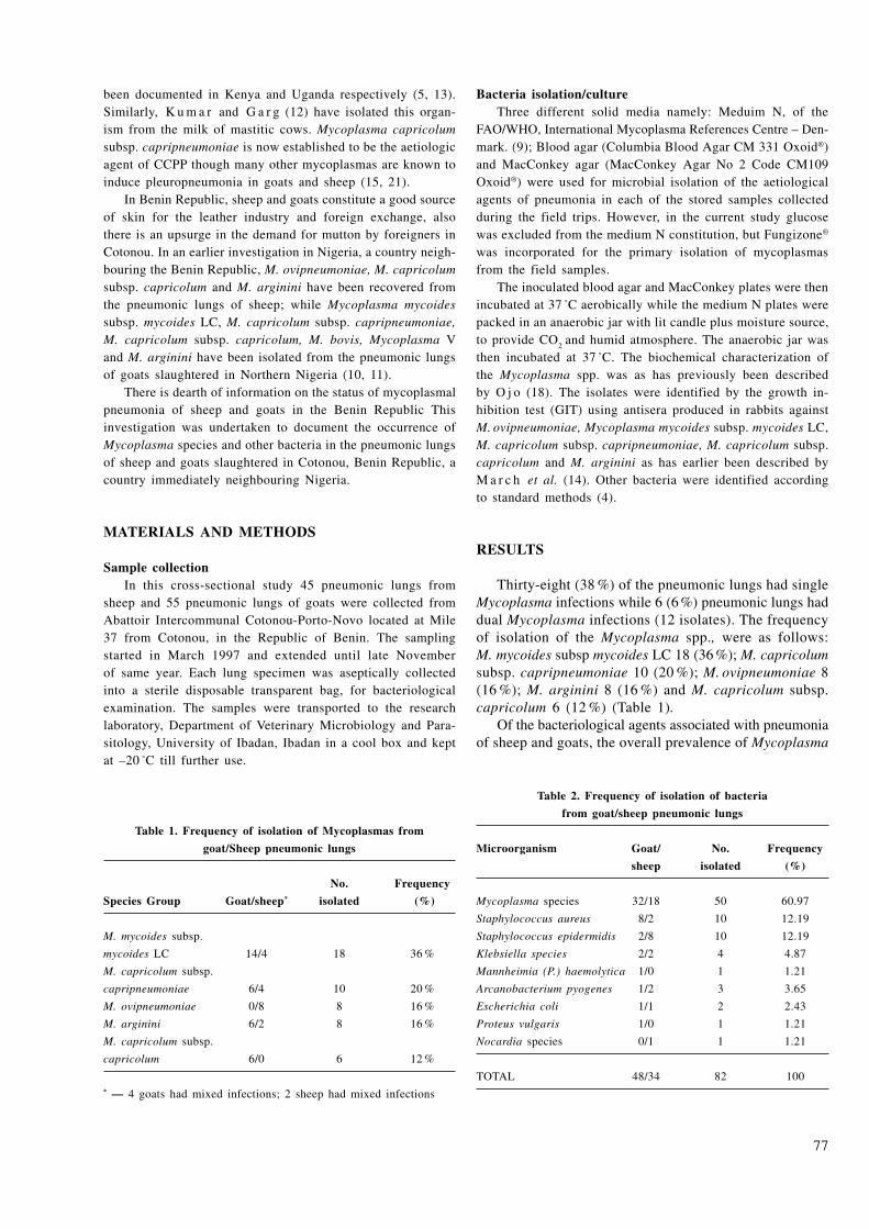
77
been documented in Kenya and Uganda respectively (5, 13). Similarly, K u m a r and G a r g (12) have isolated this organ-ism from the milk of mastitic cows. Mycoplasma capricolum subsp. capripneumoniae is now established to be the aetiologic agent of CCPP though many other mycoplasmas are known to induce pleuropneumonia in goats and sheep (15, 21).
In Benin Republic, sheep and goats constitute a good source of skin for the leather industry and foreign exchange, also there is an upsurge in the demand for mutton by foreigners in Cotonou. In an earlier investigation in Nigeria, a country neigh-bouring the Benin Republic, M. ovipneumoniae, M. capricolum subsp. capricolum and M. arginini have been recovered from the pneumonic lungs of sheep; while Mycoplasma mycoides subsp. mycoides LC, M. capricolum subsp. capripneumoniae, M. capricolum subsp. capricolum, M. bovis, Mycoplasma V and M. arginini have been isolated from the pneumonic lungs of goats slaughtered in Northern Nigeria (10, 11).
There is dearth of information on the status of mycoplasmal pneumonia of sheep and goats in the Benin Republic This investigation was undertaken to document the occurrence of Mycoplasma species and other bacteria in the pneumonic lungs of sheep and goats slaughtered in Cotonou, Benin Republic, a country immediately neighbouring Nigeria.
MATERIALS AND METHODS
Sample collectionIn this cross-sectional study 45 pneumonic lungs from
sheep and 55 pneumonic lungs of goats were collected from Abattoir Intercommunal Cotonou-Porto-Novo located at Mile 37 from Cotonou, in the Republic of Benin. The sampling started in March 1997 and extended until late November of same year. Each lung specimen was aseptically collected into a sterile disposable transparent bag, for bacteriological examination. The samples were transported to the research laboratory, Department of Veterinary Microbiology and Para-sitology, University of Ibadan, Ibadan in a cool box and kept at –20 °C till further use.
Bacteria isolation/culture Three different solid media namely: Meduim N, of the
FAO/WHO, International Mycoplasma References Centre – Den-mark. (9); Blood agar (Columbia Blood Agar CM 331 Oxoid®) and MacConkey agar (MacConkey Agar No 2 Code CM109 Oxoid®) were used for microbial isolation of the aetiological agents of pneumonia in each of the stored samples collected during the field trips. However, in the current study glucose was excluded from the medium N constitution, but Fungizone® was incorporated for the primary isolation of mycoplasmas from the field samples.
The inoculated blood agar and MacConkey plates were then incubated at 37 °C aerobically while the medium N plates were packed in an anaerobic jar with lit candle plus moisture source, to provide CO
2 and humid atmosphere. The anaerobic jar was
then incubated at 37 °C. The biochemical characterization of the Mycoplasma spp. was as has previously been described by O j o (18). The isolates were identified by the growth in-hibition test (GIT) using antisera produced in rabbits against M. ovipneumoniae, Mycoplasma mycoides subsp. mycoides LC, M. capricolum subsp. capripneumoniae, M. capricolum subsp. capricolum and M. arginini as has earlier been described by M a r c h et al. (14). Other bacteria were identified according to standard methods (4).
RESULTS
Thirty-eight (38 %) of the pneumonic lungs had single Mycoplasma infections while 6 (6 %) pneumonic lungs had dual Mycoplasma infections (12 isolates). The frequency of isolation of the Mycoplasma spp., were as follows: M. mycoides subsp mycoides LC 18 (36 %); M. capricolum subsp. capripneumoniae 10 (20 %); M. ovipneumoniae 8 (16 %); M. arginini 8 (16 %) and M. capricolum subsp. capricolum 6 (12 %) (Table 1).
Of the bacteriological agents associated with pneumonia of sheep and goats, the overall prevalence of Mycoplasma
Table 2. Frequency of isolation of bacteria
from goat/sheep pneumonic lungs
Microorganism Goat/ No. Frequency
sheep isolated (%)
Mycoplasma species 32/18 50 60.97
Staphylococcus aureus 8/2 10 12.19
Staphylococcus epidermidis 2/8 10 12.19
Klebsiella species 2/2 4 4.87
Mannheimia (P.) haemolytica 1/0 1 1.21
Arcanobacterium pyogenes 1/2 3 3.65
Escherichia coli 1/1 2 2.43
Proteus vulgaris 1/0 1 1.21
Nocardia species 0/1 1 1.21
TOTAL 48/34 82 100
Table 1. Frequency of isolation of Mycoplasmas from
goat/Sheep pneumonic lungs
No. Frequency
Species Group Goat/sheep* isolated (%)
M. mycoides subsp.
mycoides LC 14/4 18 36 %
M. capricolum subsp.
capripneumoniae 6/4 10 20 %
M. ovipneumoniae 0/8 8 16 %
M. arginini 6/2 8 16 %
M. capricolum subsp.
capricolum 6/0 6 12 %
* — 4 goats had mixed infections; 2 sheep had mixed infections

78
spp. was 60.97 % while other bacteria encountered were Staphylococcus aureus and Staphylococcus epidermidis with a prevalence of 12.19 % respectively. However, the Klebsiella species showed a prevalence of 4.87 %. While Mannheimia haemolytica, Proteus vulgaris and Nocardia spp. showed a prevalence of 1.21 % respec-tively. Whereas, Arcanobacterium pyogenes showed a prevalence of 3.65 % and Escherichia coli showed 2.43 % prevalence (Table 2).
DISCUSSION
The findings of this investigation have shown that mycoplasmal pneumonia constitutes a serious threat to the growth of the goat and sheep industry. Five Myco-plasma agents namely: M. ovipneumoniae, Mycoplasma mycoides subsp. mycoides LC, M. capricolum subsp. capripneumoniae, M. capricolum subsp. capricolum and M. arginini; have been isolated from pneumonic lungs of sheep and goats slaughtered in abattoir located in Cotonou, Republic of Benin.
Other bacteria encountered in order of decreasing prevalence were Staphylococcus aureus and Staphyloco-ccus epidermidis; Klebsiella species; Arcanobacterium pyogenes; Escherichia coli; Mannheimia haemolytica; Proteus vulgaris and Nocardia species respectively. The bacteriological findings of this study confirm the aetiologic-multi-factoral nature of pneumonia that respira-tory diseases tend to be of multiple causes (1, 3). It is therefore possible that they play some role in aggravating the disease or act as secondary invaders.
Interestingly, the overall prevalence (60.97 %) of Mycoplasma species recorded from the pneumonic lungs of sheep and goats slaughtered in Cotonou, Republic of Benin is lower than the prevalence (64.3 %) recorded in slaughtered goats alone in Northern Nigeria (11). The five Mycoplasma species encountered in this investiga-tion are similar to those earlier recorded in sheep and goats slaughtered in Nigeria by I k h e l o a et al. (10, 11). However, it is noteworthy that M. capricolum subsp. capripneumoniae was recovered from pneumonic lungs of sheep in Cotonou whereas this organism was not encountered in sheep lungs in Nigeria (10). This finding indicates that the affected sheep were exposed to goats with M. capricolum subsp. capripneumoniae infection before slaughter since cattle, sheep and goats are usually herded together by the pastoralists in the area under review.
However, this finding agrees with earlier reports of isolation of Mycoplasma F-38 from sheep in Kenya and Uganda respectively (5, 13). Similarly, K u m a r and G a r g (12) have isolated this organism from the milk of mastitic cows. In an experimental investigation M. capricolum subsp. capripneumoniae did not produce pneumonia in cattle, however when the organism was inoculated into cattle immunossupressed with T. congolense the cattle died with pleuropneumonic lesions (2).
The above findings confirm the pervasiveness of mycoplasmas in the mycoides cluster in small ruminants and the potential of interspecies transmission and disease when different animal taxa come in contact as has been reported by N i c h o l a s et al. (16). These investigators, reported an outbreak of systemic disease in Vaal rhebok (Pelea capreolus) caused by Mycoplasma capricolum subsp. capricolum, a caprine pathogen which is closely related to M. capricolum subsp. capripneumoniae.
Further more, no M. bovis and Mycoplasma V was encountered in sheep and goats slaughtered in Cotonou, Benin Republic whereas these organisms were found in sheep and goats slaughtered in Northern Nigeria (10, 11). This suggests that M. bovis pneumonia is common around northeastern Nigeria (1, 8, 10, 11); may have spread to Nigeria through neighbouring countries such as Niger, Chad or Cameroon, through uncontrolled cattle trade across the permeable borders.
From this investigation it was observed that myco-plasmal pneumonia is still ravaging the small ruminant population in Benin Republic and constitutes a threat to the livestock industry in the West African sub-region and Africa in general. With the imminent eradication of rinderpest from Africa mycoplasmal pneumonia may become the major source of economic loss in Africa. There is a need for a sustained annual international vaccination campaign coupled with abattoir surveillance by veterinarians and experienced meat inspectors, to be backed by rapid and sensitive diagnostic methods that can be used to confirm the initial diagnosis of ae-tiological agent based on clinical symptoms as well as pathological findings.
ACKNOWLEDGEMENTS
This investigation was financed by Nigeria National Ag-ricultural Research Project (NARP); RGS 024 and College Polytecnique Univeristaire, National University of Republic of Benin. The authors are also grateful to Dr Anne Vermelyen from Belgium for supplying reagents used in this study to Dr Adehan.
REFERENCES
1. Ajuwape, A. T. P., Adetosoye, A. I., Ikheloa, J. O., Ote-sile, E. B., Ojo, M. O., 2004a: Bacteriological examination of bovine pneumonic lungs with special reference to Mycoplasma species in Nigeria. Bull. Anim. Hlth. Prod. Afr., 57, 7—12.
2. Ajuwape, A. T. P., Adetosoye, A. I., Ikheloa, J. O., Alaka, O. O., Taiwo, V. O., Talabi, O. A., Otesile, E. B., Ojo, M. O., 2004b: Pathogenicity of Mycoplasma capricolum subspecies capripneumoniae for cattle immunosuppressed by Trypanosoma congolense. Trop. Vet., 22, 29—36.
3. Akpavie, S. O., Okoro, H. O., Salami, N. A. and Ikheloa, J. O., 1991: The pathology of adult bovine lung in Nigeria. Trop. Vet., 9, 151—161.

79
4. Barrow, G. H., Feltham, R. K. A., 1993: Cowan and Steel's Manual for Identification of Medical Bacteria (3rd edn.). Cambridge University Press, Cambridge 331.
5. Bölske, G., Johansson, K. E., Heinonen, R., Panvuga, P. A., Twinamasiko, E., 1995: Contagious caprine pleuropneu-monia in Uganda and isolation of M. capricolum subspecies capripneumoniae from goats and sheep. Vet. Record, 137, 594.
6. Carmichael, L. E., St. George, T. D., Sullivan, N. D., Horsfall, N., 1992: Isolation, propagation and characterization studies of an ovine Mycoplasma responsible for proliferative interstitial pneumonia. Cornell Vet., 62, 654—679.
7. DaMassa, A. J., Brooks, D. L., Alder, H. E., 1983: Caprine mycoplasmosis. Widespread infection in goats with Mycoplasma mycoides subsp. mycoides (LC type). Am. J. Vet. Res., 44, 322—325.
8. Egwu, G. O, Ameh, J. A., Aliyu, M. M., 1996: Detection of antibodies to Mycoplasma bovis in cattle by Enzyme Linked Immunosorbent Assay (ELISA). Trop. Vet., 14, 163—167.
9. Freundt, E. A., 1983: Culture media and classic myco-plasmas. In Razin, S. and Tully, J. G. (eds.): Methods in Mycoplasmology. Vol.1. Mycoplasma Characterization.Academic Press New York, U.S.A., pp.127—135.
10. Ikheloa, J. O., Ajuwape, A. T. P., Adetosoye, A., 2001: Ovine pneumonia: biochemical characterization and serological identification of mycoplasmas, isolated from pneumonic lungs of sheep slaughtered in northern states. Afr. J. Clin. Microb., 2, 13—15.
11. Ikheloa, J. O., Ajuwape, A. T. P., Adetosoye, A. I., 2004: Biochemical characterization and serological identifica-tion of mycoplasmas, isolated from pneumonic lungs of goats slaughtered in abattoirs in Northern Nigeria. Small Rum. Res. 52, 93—97.
12. Kumar, A., Garg, D. N., 1991: Isolation of mycoplasma F38 from the milk of mastitic cows. Vet. Record, 128, 429.
13. Litamoi, J. K., Wanyangu, S. W., Siman, P. K., 1990: Isolation of Mycoplasma biotype F-38 from sheep in Kenya. Trop. Anim. Health Prod., 22, 260—262.
14. March, J. B., Clark, J., Brodlie, M., 2000: Characte-rization of strains of Mycoplasma mycoides subsp. mycoides small colony type isolated from recent outbreaks of contagious bovine pleuropneumonia in Botswana and Tanzania: evidence of a new biotype. J. Clin. Microbiol., 38, 1419—1425.
15. Maré, C. J., 1996: Contagious caprine pleuropneumonia. In The Gray Book. Part IV: Foreign Animal Diseases. Available online at www.vet.uga.edu/VPP/gray-book/FAD/index.htm.5.
16. Nicolas, M. M., Stalis, H. H., Clippinger, T. L., Bush, M., Nordhausen, R., Maalouf, G., Screnzel, M. D., 2005: Systemic disease in Vaal rhebok (Pelea capreolus) caused by Mycoplasmas in the mycoides cluster. J. Clin. Microbiol., 43, 1330–1340.
17. Ojo, M. O., 1976a: Caprine pneumonia in Nigeria I: Epidemiology and bacterial flora of normal and diseased respiratory tracts. Trop. Anim. Hlth. Prod., 8, 85—89.
18. Ojo, M. O., 1976b: Caprine pneumonia II: Biochemical characterization and serological identification of Mycoplasmas. Trop. Anim. Hlth. Prod., 8, 137—146.
19. Ojo, M. O., Obi, T. U., 1996: Respiratory diseases of goat. Trop. Vet., 14, 85—99.
20. Rurangirwa, F. R., McGuire, T. C., 2003: Contagious caprine pleuropneumonia: Diagnosis and control. Available online at file://A:\.
21. Thiaucourt, F., Bölske, G., 1996: Contagious caprine pleuropneumonia and other pulmonary mycoplasmosis of sheep and goats. Revue Sci. et Tech. Office Intern. Epizoot. 15, 1397—1414.
Received July 12, 2005
8080
www.uvm.sk
ABSTRACT
In the described cases two illnesses of different etiol-ogy were diagnosed in three cardinal tetras (Paracheirodon axelrodi). The histopathological examination detected pans-poroblasts of Pleistophora hyphessobryconis in the muscle tissue and in the inner organs. Two types of granulomas of different morphology and localization were present. Ziehl-Neelsen staining identified acid-alcohol fast bacilli in one of the granulomas and staining with calcofluor detected spores of P. hyphessobryconis.
Key words: granulomatous; histopathology; inflam-mation; microsporidia; Mycobacterium
INTRODUCTION
Mycobacteriosis of fish is a chronic progressive disease spread all over the world. The disease occurs in sea, brack-ish and fresh water fishes kept both in aquacultures and free
FOLIA VETERINARIA, 50, 2: 80—82, 2006
nature which is characterized by the formation of granulomas enclosing AFR within the various tissues (12, 19).
Many mycobacterial species have been isolated from fish tissues (17). Most frequently detected mycobacterial species were the following: Mycobacterium marinum, M. gordonae and M. fortuitum (17). M. avium complex, M. chelonae, M. aurum, M. parafortuitum, M. poriferae, M. chelonae, and M. triplex have also been isolated from fish (1, 16, 2, 9, 14, 10).
Microsporidia are eukaryotic spore forming obligate intra-cellular protist parasites first recognized over 100 years ago. These organisms infect all of the major animal groups and are now recognized as opportunistic pathogens of humans (18).
Microsporidia Pleistophora hyphessobryconis (Shäper-claus, 1941) occures commonly in fresh water aquarium fish. Paracheirodon innesi and Hemigrammus erythrozonus are original hosts and in addition about 16 other fresh water fishes belonging to four families have been infected (3). Spores P. hyphessobryconis are ovoid 4 × 6 µm in size and one side is slightly more valued. Sporophorous vesicles with 20 to 130 spores may reach up to 35 µm in diameter. Muscle tissue is the most common affected and can be dispersed to the inner organs and body cavity in heavy infection (11).
MATERIAL AND METHODS
162 cardinal tetras were investigated from November 2000 till February 2006 from both amateur and professional stocks, were examined during the years. Fishes were euthanized by overdosing with MS 222 (tricaine methan sulphonate) prepara-tion. After gross examination, fresh mounts of skin scrapings
MANIFESTATION OF MYCOBACTERIOSIS IN CARDINAL TETRAS Paracheirodon axelrodi (Schultz, 1956) DURING THE
Pleistophora hyphessobryconis (Schäperclaus, 1941) INFECTION
Novotný, L.1, Dvořák, P.2
1Institute of Pathological Morphology, Faculty of Veterinary Medicine2Institute of Biochemistry and Biophysics, Faculty of Veterinary Hygiene and Ecology
University of Veterinary and Pharmaceutical Sciences Brno, Palackeho 1-3, 61242 Brno The Czech Republic
ADDRESS FOR CORRESPONDENCE
MVDr. Ladislav Novotný, PhD
Institute of Pathological Morphology, Faculty of Veterinary Medicine
University of Veterinary and Pharmaceutical Sciences Brno
Palackeho 1-3
612 42 Brno
Czech Republic
e-mail: [email protected]

81
and touch-print preparations of internal organs were examined by optic microscopy.
Samples for a histopathological examination were recovered and placed in 4 % buffered formaldehyde. The samples were treated by using a traditional paraffin technique and stained with haematoxylin-eosin. The Ziehl-Neelsen staining was used after the microscopic examination in the cases with granulomatous inflammation for visualization of the acid fast rods (AFR). The
Fig. 3. Pansporoblasts of Pleistophora hyphessobryconis stained with
calcofluor (asterisk) Calcofluor, fluorescent microscopy, × 400
Fig. 1. Paracheirodon axelrodi with the skin lesion (asterisk)
Fig. 2. Section through the skin and muscle tissue with granulo-
matous inflammation, scale is signed with asterisk. Haemotoxylin
and eosin, × 100
spores of P. hyphessobryconis were visualized with calcofluor staining and fluorescent microscopy (15).
RESULTS
Examination of the fresh mounts from the 162 in-dividuals revealed granulomatous inflammation in 48 tetras in the various organs and tissues. Only in 29 cases were diagnosed AFR after Ziehl-Neelsen staining method of the histopathological slides. In 15 tetras were diagnosed spores in the mussel tissue. Spores with AFR were diagnosed only in 3 individuals.
The histopathological examination of these three tetras revealed the skin ulceration, however, detected granu-lomas in the connective tissue and surrounding cutis. The size of the granulomas ranged from 150 × 80 µm to 250 × 100 µm. The granulomas had circular stratification from the peripheral layer of fibrocytes to the centre of granuloma. The necrotic centre with nuclear debris could be seen in the mature granulomas as well as epitheloid cells, which became more flat moving away from the center. Agglomerates of melanin were situated in and around the granulomas. In addition, short acid fast rods (AFR) were found freely in the granulomas and in the phagocytes.
Pansporoblasts of microsporidia and granulomas were discovered in the muscle tissue, spleen, mesentery and posterior kidney. Granulomas were found in the kidneys and were oval shape with two or three layers of fibro-cytes in the periphery. Large cells with unequal ellipsoid nucleus were under peripheral layer of fibrocytes. Spores of microsporidia were differed in the spaces among cells of the granulomas and visualized with the calcofluor staining. The size of these ovoid spores varied about 4 × 6 µm.

82
DISCUSSION
Mycobacteriosis is the opportune infection of weakened organisms. Weakening has a common factor – immune suppression. Immune suppression can arise for to nutrition deficiency, overcrowding of the tank, disease, etc. (8).
Pansporoblasts of P. hyphessobryconis were found in the inner organs too. The spread of pansporoblasts in the inner organs is usually observed in a heavy in-fection (11). It is possible to expect higher grade of immune suppression in heavy parasitic infection. Some pansporoblasts in the kidneys came under regression and defensive granulomatous process. These granulomas were negative in the Ziehl-Neelsen staining. The discovery of two types of granulomas could complicate the diagnosis and should lead to the specific staining method.
Mycobacteriosis is a severe and common illness of ornamental fishes. Miscellaneous factors apply in the etiopathogenesis, which put down immune response and which make possible penetration and multiplication of the pathogenic agent. The described cases of the my-cobacteriosis in Paracheirodon axelrodi with concurrent infection of the P. hyphessobryconis is evidence of sever immune suppression during these infections and both are dangerous for human health (18, 13).
.
REFERENCES
1. Bruno, D. W., Griffiths, J., Mitchell, C. G., Wood, B. P., Fletcher, Z. J., Drobniewski, F. A., Hastings, T. S., 1998: Pa-thology attributed to Mycobacterium chelonae infection among farmed and laboratory infected Atlantic salmon Salmo salar. Diseases of Aquatic Organisms, 33, 101—109.
2. Diamant, A., Banet, A., Ucko, M., Colorni, A., Knibb, W., Kvitt, H., 2000: Mycobacteriosis in wild rabbitfish Siganus rivulatus associated with cage farming in the Gulf of Eilat, Red Sea. Diseases of Aquatic Organisms, 39, 211—219.
3. Dyková, I., 2002: Phylum Microspora. In Woo, P. T. K.: Fish Diseases and Disorders (1st edn.). Vol. 1: Protozoan and Metazoan Infections. CABI Publishing, Oxon, United Kingdom, pp. 149—181.
4. Chinabut, S., 1998: Mycobacteriosis. In Woo, P. T. K., Bruno, D. W.: Fish Diseases and Disorders (1st edn.), Vol. 3: Viral, Bacterial and Fungal Infections. CABI Publishing, Oxon, United Kingdom, 319—331.
5. Fiedler, Z., Mazurová, J., Novotný, L., Dvořák, P., 2000: Fast diagnosis of mycobacteria in the Carassius auratus with using of DNA detection method. Veterinářství, 50, pp. 123—124.
6. Gomez, S., Bernabe, M., Gomez, A. M., Navarro, J. A., Sanchez, J., 1993: Fish mycobacteriosis: morphopathological and immunocytochemicals aspects. J. Fish Dis., 16, 137—141.
7. Gomez, S., Navarro, J. A., Gomez, A. M., Sanchez, J., Barnabe, A., 1996: Comparative study of immunohistochemical methods to diagnosis mycobacteriosis in swordtail Xiphophorus helleri. J. Fish Dis., 24, 117—120.
8. Hedrick, R. P., McDowell, T., Groff, J., 1987: Myco-bacteriosis in cultured striped bass from California. J. Wild. Dis., 23, 391—395.
9. Herbst, L. H., Costa, S. F., Weiss, L. M., Johnson, L. K., Bartell, J., Davis, R., Walsh, M., Levi, M., 2001: Granulomatous skin lesions in moray eels caused by a novel Mycobacterium species related to Mycobacterium triplex. In-fection and Immunity, 69, 4639—4646.
10. Leščenko, P., Mátlová, L., Dvorská, L., Bartoš, M., Vávra, O., Navrátil, S., Novotný, L., Pavlík, I., 2003: My-cobacterial infection in aquarium fish. Vet. Med.-Czech, 48, 71—78.
11. Lom, J., Dyková, I., 1992: Microsporidia (Phylum Microspora). In Lom, J., Dyková, I.: Protozoan Parasites of Fishes. Elsevier, Amsterdam. Developments in Aquaculture and Fisheries Science , 26, 134—149.
12. Mátlová, L., Fischer, O., Kazda, J., Kaustová, J., Bartl, J., Horváthová, A., Pavlík, I., 1998: Occurrence of mycobacteria in invertebrates and poikilothermic animals and their role in man and other animals (In Czech). Vet. Med.-Czech, 43, 115—132.
13. Novotný, L., Dvorská, L., Lorenzová, A., Beran, V., Pavlík, I., 2004: Fish: a potential source of bacterial pathogens for human beings. Vet. Med.-Czech, 49, 343—358.
14. Perez, A. T., Conroy, D. A., Quinones, L., 2001: Pres-ence of acid−fast bacteria in wild and cultured silver mullets (Mugil curema Val., 1836) from Margarita Island Venezuela. Interciencia, 26, 252—256.
15. Roberts, R. J., Smail, D. A., 2001: Laboratory methods. In Roberts, R.: Fish Pathology (3rd edn.). W. B. Saunders, Toronto, Canada, 380—412.
16. Talaat, A. M., Trucksis, M., Kane, A. S., Reimschues-sel, R., 1999: Pathogenicity of Mycobacterium fortuitum and Mycobacterium smegmatis to goldfish, Carassius auratus. Veterinary Microbiology, 66, 151—164.
17. Wayne, L. G., Kubica, G. P., 1986: Genus Myco-bacterium Lehmann and Neumann 1896, 363AL. In Sneath, P. H. A., Mair, N. S., Sharpe, M. E., Holt, J. G.: Bergey's Manual of Systematic Bacteriology. The Williams & Wilkins Co., Baltimore, pp. 1436—1457.
18. Weiss, L. M., 2001: Microsporidia: emerging pathogenic protists. Acta Trop., 78, 89—102.
19. Wolf, J. C., Smith, S. A., 1999: Comparative severity of experimentally induced mycobacteriosis in striped bass Morone saxatilis and hybrid tilapia Oreochromis spp. Diseases of Aquatic Organisms, 38, 191—200.
Received April 26, 2006

8383
www.uvm.sk
ABSTRACT
Morphological, histological and immunohistochemical changes of lambs following 23 days multiple oral infections with 1,000 embryonated Toxocara canis eggs were studied up to 49 days post infection (dpi). Five-month-old Valaška lambs were divided into an experimental group (n = 18) and controls (n = 5). The small intestine, liver, lungs, brain and mesenteric lymph nodes of both infected and control lambs were examined using the routine histological methods for paraffin sections after necropsy. Two experimental lambs were killed at days 1, 3, 7, 14, 21, 28, 35, 42, and 49 (n = 2) and controls at days 14 (n = 2) and 49 (n = 3). Morphologically, a few small grey-greenish nodules on the lungs and numerous disseminated yellow-whitish nodules on the liver were found from day 21 pi to the end of experiment. Eosinophil-rich hepatic granulomas, focal T. canis-induced pulmonary inflam-mation, massive mononuclear and eosinophilic infiltration of the small intestine lamina propria mucosae, as well as T. canis larvae were the most prominent histological changes. Immunohistochemical examination showed the parasite in the mesenteric lymph nodes. It was concluded that multiple larval toxocarosis followed the classical migratory path in the infected lambs.
Key words: immunohistochemistry; paratenic host; pathology; sheep-nematoda; Toxocara canis
INTRODUCTION
Larval toxocarosis is a serious disease transmissible from animals to humans occurring on a world-wide scale. The host response to T. canis infection is of an immunopathogenetical character, which results in clinical symptoms of the disease (7). The significant immune response of the host is not sufficient to fully eliminate T. canis larvae that may persist in the body for a relatively long period of time, which is associated with a relatively high level of specific antibodies (9, 4). Paratenic hosts reinfected with Toxocara spp. embryonated eggs cumulate viable larvae in their organs.
Excretion of parasite metabolic products leads to a permanent antigenic stimulation in hosts (5). Three-fold infections in mice with T. canis revealed the formation of typical eosinophilic granulomas in the liver and severe pulmonary inflammation in the lungs (2). Similar changes were observed in sheep af-ter oral infection with 10,000 embryonated T. canis eggs (3). Long-term experimental infection of sheep (23 days) with 1,000 Ascaris suum eggs showed that the number of larvae in the liver decreased more rapidly than in the lungs (6).
Morphological and histological examination was per-formed to find out the pathway of the parasite after multiple infection with a small dose, and immunocytochemistry was performed to observe the parasite and T. canis-positive cells in the organs.
MORPHOLOGICAL, HISTOLOGICAL AND IMMUNOHISTOCHEMICALCHANGES AFTER MULTIPLE Toxocara canis INFECTION OF LAMBS
Revajová, V., Levkut, M., Aldawek, A.*
Herich, R., Dvorožňáková, E.**, Krupicer, I.**
University of Veterinary Medicine, Komenského 73, 041 81 Košice, The Slovak Republic*Faculty of Veterinary Medicine, Al-Fateh University, Tripoli, Lybia
**Parasitological Institute, Slovak Academy of Science, Hlinkova 3, 040 01 Košice The Slovak Republic
FOLIA VETERINARIA, 50, 2: 83—88, 2006
84
MATERIAL AND METHODS
AnimalsTwenty-three five-month-old Valaška lambs were divided
into an experimental (n = 18) and a control group (n = 5). Be-fore starting the experiment the rectal faecal samples of lambs were monitored for the presence of parasites by helminthic examination of eggs (flotation and sedimentation methods), and larvae (Baermann method). The animals were treated with Sulfacox obtained from Pharmagal (Nitra, Slovakia) to remove detected coccidia. When the rectal faecal samples were proven to be negative for parasite eggs and larvae, the lambs were used in the experiment.
InfectionEach animal in the experimental group was daily infected
with 1,000 embryonated T. canis eggs before feeding during 23 days of the experiment. The eggs of T. canis was mixed in crumbs of bread. The control animals were not infected. The lambs of the experimental and control group were kept separately in two different places in pens, housed in a stable (Research Institute of Gnotobiology and Diseases of Young, UVM, Košice), and fed with a standard feed dose.
Parasite Toxocara canis eggs were isolated from the worm uteri
obtained from the intestine of two infected puppies. The eggs were cultured in 0.1 mol H
2SO
4 solution at room temperature
until infective larvae developed (four weeks). Larval develop-ment and viability were confirmed by light microscopy prior to inoculation.
Experimental design and samples The lambs were infected during 23 days, and the total
duration of experiment was 49 days. Experimental lambs were killed by decapitation at days 1, 3, 7, 14, 21, 28, 35, 42, and 49 (n = 2) and control at days 14 (n = 2) and 49 (n = 3).
Autopsy was performed after killing the lambs and samples from brain, liver, lungs, small intestine, and mesenteric lymph nodes were taken for histology and immunohistochemistry.
Histopathology Specimens were taken and immediately fixed in 10 % neutral
formalin. Samples were then submitted to routine histological processing. Prepared sections were stained with haematoxylin-eosin and Giemsa-Romanowski.
Immunohistochemistry Mouse anti-Toxocara canis hyperimmune serum was prepared
at first. Ten BALB/c mice were infected per os three times with 1,000 embryonated eggs of T. canis (in 0.5 ml phosphate buffered saline, PBS): the first dose on day 0, the second dose on day 14, and the third dose on day 21. Four weeks after the last dose the mice were bled. The obtained sera were tested with ELISA using an excretory/secretory antigen. A high posi-tive response (titre 1 : 3,200) was evident. The samples were used as the hyperimmune anti-T. canis serum.
Immunohistochemical procedure For an immunohistochemical reaction, samples from the small
intestine, liver, lungs, brain, and lymph nodes were collected in the PBS of both infected and control lambs. Analysis was conducted on frozen tissue sections using the biotin-streptavidin multilink staining kit (Biogenex, USA). In brief, frozen sec-tions of 6 µm thickness were mounted on glass slides, dried at room temperature, fixed in cold acetone for two minutes, and incubated with the previously prepared mouse hyperim-mune serum against T. canis (dilution 1 : 1,000) for one hour. After washing with PBS the sections reacted with biotinylated antiimmunoglobulins in the PBS for thirty minutes, were washed again with PBS, incubated with streptavidin-peroxidase conjugate for thirty minutes and subsequently stained with a substrate solution containing DAB (3,3-diaminobenzidine) for five minutes. The counterstaining of the sections was carried out with haematoxylin.
RESULTS
The morphological examination of the liver during the necropsy of the infected lambs showed disseminated pinheaded to millet size, slightly prominent yellowish-whit-ish nodules on seven dpi. Findings of multiplex nodules prevailed to the end of experiment with an in crease in their number and size on 21, 28, and 35 dpi. Typical milk spots together with nodules could be observed on 49 dpi.
The lungs were without any macroscopical changes on day 7. On days 14, sporadic red and greyish green nodules, 0.2—0.4 mm large were disseminated on the diaphragmatic side of the caudal lobes, with prominence on 21 dpi. Lobular alveolar emphysema, accentuation of interstitial tissue, and local pleural fibrosis predominated at the end of the experiment.
Only focal acute catarrhal enteritis was recorded on the small intestine.
In the infected lambs the histological changes that predominated most were observed in the small intes-tine, liver and lungs. The intestinal lamina propria mucosae showed submucosal congestion with a massive lymphocyte, plasmocyte, macrophage, eosinophil, and mastocyte infiltration. The hatching of larvae was seen in jejunal lamina propria on 24 hpi, and T. canis larvae were found at the same place on 14 dpi (Fig. 1).
The parasite was observed also on 21 dpi in the duo-denal and jejunal lamina propria. Granuloma consisted of central necrosis and infiltration by mononuclear cells was found on 47 dpi in ileal lamina propria. In the liver, early phase lesions were observed 24 hpi that consisted of focal hepatocellular degenerations with aggregates of lymphocytes, eosinophils, and neutrophils (Fig. 2).
Periportal lymphocytic and macrophage infiltration was found on 3 dpi. Some migratory tracts filled with erythrocytes, lymphocytes and eosinophils were observed 21 dpi. As the infection progressed typical eosinophilic granulomas were formed within the hepatic lobules

85
Fig. 1. Toxocara canis larva and extensive eosinophilic infiltration
in jejunal lamina propria mucosae on 14 dpi (HE; bar 5 µm) Fig. 2. Neutrophilic, eosinofilic, lymphocytic and macrophage
infiltration of liver parenchyma 24 hpi (Giemsa-Romanowsky;
bar 2 µm)
Fig. 3. Eosinophilic granuloma in the liver of infected lambs on
14 dpi (HE; bar 10 µm)
Fig. 4. Encapsulated eosinophilic granuloma in the liver on 28 dpi
(HE; bar 5 µm)
Fig. 5. Peribronchial infiltration by lymphocytes and eosinophils
on 21 dpi (HE; bar 10 µm)
from 14 dpi (Fig. 3), which became encapsulated on 28 dpi (Fig. 4).
The lung parenchyma exhibited congestion of the perialveolar and peribronchial blood vessels 24 hpi that was connected with massive peribronchial infiltration by lymphocytes, plasma cells, macrophages and eosinophils on 21 dpi (Fig. 5).
Plasma exsudation into the alveoli was intensive and led to oedema of the lungs. Perivascular oedema in the brain and nodular lymphocytic infiltration were observed on 21 dpi in one infected lamb (Fig. 6).
The spleen, mesenteric and hepatic lymph nodes reacted to the infection by follicular hyperplasia. All the examined sections of the control lambs showed no histological changes in the same organs.

86
Immunohistochemical examination using hyperim-mune anti-Toxocara serum showed a positive response by Lieberkühn cryptal epithelial cells and visualisation of T. canis larvae in the mesenteric lymph nodes on 42 dpi (Fig. 7). In the peripheral blood the larvae were micro-scopically found from 14 dpi (8 ± 2.8 larvae) to 35 dpi (2 ± 2.8 larvae) of the experiment, with a maximum on 28 dpi (13 ± 4.2 larvae).
DISCUSSION
A wide spectrum of paratenic hosts can be infected with larval toxocarosis either naturally or experimentally. These include: human (10), mice (2), cats (18), chicken (8), quail (15), and sheep (11, 3).
After administration of the infected eggs the preferen-tial site of parasite localization was the small intestine. There were no preferred sites for colonization; in all parts we were able to find larvae. Submucosal conges-tion and infiltration with eosinophils and lymphocytes was observed 12 hpi. Hatched larvae were found in Lieberkühn crypts of ileum 24 hpi. Our observation is in agreement with the findings of A b o - S h e h a d a et al. (1) who mentioned that the most T. canis larvae could be detected in the intestinal wall within the first 24 hours. In sheep, the elimination of larval and adult stages of some nematodes is thought to be dependent on the local intestinal immunity (16). The expansion of blood and mucosal eosinophils cell population is among the earliest events observed in nematode-infected sheep (20).
Migration of T. canis larvae through the liver and lungs in sheep and other paratenic hosts induces dif-ferent immune reactions in the destroyed tissue (11, 17), first of all acute focal hepatitis and the forming of granulomas (14).
In our experiment focal infiltrations in liver were observed already by 24 hpi. The liver parenchyma was disseminated by numerous minute aggregates consisted of neutrophils, eosinophils, lymphocytes and macroghages, sometimes localized very closely to the vena centralis with remnants of destroyed eggs. The changes in hepatic lobules were accompanied by periportal infiltration of lymphocytes and macrophages.
As the infection progressed a diffuse eosinophilic hepatitis was evident. In the advanced stages, typical encapsulated eosinophil-rich hepatic granulomas were observed. The structure of granulomas was the same as in mice (3) except that neutrophils were abundant in the lesions of murine tissues. Larvae were detected in the liver of infected sheep on 7 dpi. On twenty-one dpi they were trapped in yellowish-whitish abundant granulomas with different size from beginning to the end of experi-ment, when white spots composed of fibrous tissue were also observed on the surface of the liver.
The extensive lesions in the liver parenchyma and the larval trapping revealed a key role of liver in the host-parasite relationship. S u g a n e and O s h i m a (22), P a r- s o n s and G r i e v e (19) have claimed that the larvae of T. canis survive for extensive periods in the face of an ongoing and aggressive immune response.
The lung tissue of infected lambs showed extensive histological changes in spite of the absence of larvae. Congestion of perialveolar and peribronchial blood ves-sels was connected with massive infiltration of interstitial and alveolar tissue by eosinophils and lymphocytes. Peribronchial eosinophilic and lymphocytic infiltrates at 7 dpi was the reaction to T. canis antigen, localized on the surface of bronchial and bronchiolar epithelial cells,
Fig. 7. Larvae of T. canis and positive macrophages in mesenteric
lymph nodes on 42 dpi – frozen tissue section by using anti-T. canis
hyperimmune serum and biotin-streptavidin complex; bar 5 µm
Fig. 6. Perivascular oedematization and nodular lymphocytic in-
filtration in the brain on 21st dpi (10 µm)

87
as well as in infiltrated peribronchial macrophages. It is evidence of parasite migration.
At the advanced stages, the lesions became inter-spersed and the interstitial tissue was thickened due to lymphocyte and macrophage infiltration that finally gave a picture of diffuse interstitial pneumonia. K a y e s (12) has suggested that these findings might be the results of prolonged antigenic stimulation of the host immune system.
The brain tissue demonstrated perivascular oedema-tization and lymphocytic infiltration at 21 dpi in one animal, however no larvae were detected. A l d a w e k et al. (3) referred no histological changes in the brain after single infection with 10,000 embryonated T. canis eggs in sheep, but three-times infection with 1,000 in-fective T. canis eggs in mice led to a high incidence of larvae (2). Therefore, it can be assumed that multiple infections might indirectly aid in the establishment of the “neurotropic phase”, mainly in mice. This finding is of public health importance since infection in the environment follows the same pattern.
The migration of T. canis larvae in lambs may cause severe reactions in the host; however secondary implica-tions may be more important then primary effects. The inflammatory and haemorrhagic conditions that develop in the liver and lungs may serve as avenues for secondary invasion, and subadult parasitic organisms as possible disseminators of other diseases.
In conclusion, T. canis-infected sheep showed clas-sical histomorphological changes in the affected organs. Thus, it can be assumed that sheep might be used as a paratenic host model to study the pathology of larval toxocarosis and the migratory behaviour of larval toxo-carosis in the infected lambs is similar to that in other paratenic hosts.
ACKNOWLEDGEMENT
This work was supported by VEGA Scientific Grant 2/1150/21 from The Ministry of Education, The Slovak Republic.
REFERENCES
1. Abo-Shehada, M. N., AL-Zubaidy, B. A., Herbert, I. V., 1983/84: The migration of larval Toxocara canis in white mice I. The migration through the intestine in primary infections. Vet. Parasitol., 17, 65—73.
2. Aldawek, A. M., Levkut, M., Revajová, V., Dvorožňáková, E., Dubinský, P., 2001: Morphological changes in mice after multiple infection with Toxocara canis. Folia Veterinaria, 45, 31—34.
3. Aldawek, A. M., Levkut, M., Revajová, V., Kolodzieyski, L., Ševčíková, Z., Dubinský, P., 2002: Larval toxocarosis in sheep: the immunohistochemical characterization of lesions in some affected organs. Vet. Parasitol., 105, 207—214.
4. Chieffi, P. P., Peres, B. A., de Mello, E. O., Kanamura, H., Brandao, M. M., 1993: Persistence of specific antibody response in different experimental infections of mice with Toxocara canis larvae. Rev. Inst. Med. Trop. Sao Paulo, 37, 187—190.
5. Cuellar, C., Fenoy, S., Aguila, C., Guillen, J. L., 1990: Evaluation of chemotherapy in experimental toxocarosis by determination of specific immune complexes. J. Helminthol., 64, 279—289.
6. Dubinský, P., Švický, E., Kováč, G., Lenhardt, Ľ., Krupicer, I., Vasilková, Z., Dvorožňáková, E., Levkut, M., Papajová, I., Moncol, D. J., 2000: Pathogenesis of Ascaris suum in repeteated infection of lambs. Acta Vet. Brno, 69, 201—207.
7. Dvorožňáková, E., Borošková, Z., Dubinský, P., Tomašovičová, O., Machnicka, B., 2000: Toxocara canis in mice: immune response after infection and immunization. Helminthologia, 37, 199—204.
8. Glickman, L. T., Magnaval, J. F., Domanski, L. M., Ghofer, F. S., Lauria, S. S., Gottsstein, B., Brochier, B., 1987: Visceral larva migrans in French adults: a new disease syn-drome? Am. J. Epidemiol., 125, 1019—1034.
9. Grieve, R. B., Stewart, V. A., Parsons, J. C., 1993: Immunobiology of larval toxocarosis (Toxocara canis). A summary of recent research. In Lewis, J. W, Maizels, R. M. (eds.): Toxocara and Toxocarosis. Clinical, Epidemiological and Molecular Perspectives. Brit. Parasitol. Soc. Inst. Biol., London, 117—124.
10. Havasiová-Reiterová, K., Dubinský, P., Štefančíková, A., 1993: A seroepidemiological study of Toxocara infection in human in Slovak Republic. J. Helminthol., 67, 260—267.
11. Hayate, C. S., Rehmann, B., Ahmed, M. S., 1973: Migration of ascarids in sheep. Pakistan J. Zool., 5, 91—97.
12. Kayes, S. G., 1997: Human toxocariasis and the visceral larva migrans syndrome correlative immunopathol-ogy. In Fredman, D. O. (ed.): Immunopathogenetic Aspects of Disease Induced by Helminth Parasites. Chem. Immunol., Karger, Basel, 66, 92—124.
13. Levkut, M., Revajová, V., Dvorožňáková, E., Reiterová, K., Dubinský, P., Krupicer, I., Moncol, D. J., 1999: Effect of Ascaris suum on immunoreactivity in lambs. Helminthologia, 36, 69—74.
14. Levkut, M., Revajová, V., Aldawek A. M., Švický, E., Dubinský, P., Krupicer, I., Dvorožňáková, E., Levkutová, M., 2002: Paratenic host and Toxocara canis (In Slovak). Infovet, 11, 30—32.
15. Maruyama, S., Yamamoto, K, Katsube, Y., 1994: Infectivity of Toxocara canis larvae from Japanese quails in mice. J. Vet. Med. Sci., 56, 399—401.
16. McClure, S. J., Emery, D. L., Wagland., B. M., Jones, W. O., 1992: A serial study of rejection of Trichostrongylus colu-briformis by immune sheep. Int. J. Parasitol., 22, 227—234.
17. Medveďová, M., Pajerský, A., Tomašovičová, O., 1994: The macroscopic pictures of the reactive manifestation in relation to the distribution of Toxocara canis (Werner, 1782) larvae in paratenic hosts. Helminthologia, 31, 23—27.
18. Parsons, J. C., Bowman, D. D., Grieve, R. B., 1989: Pathological and hematological response of cats experimen-

88
tally infected with Toxocara canis larvae. Int. J. Parasitol., 19, 478—488.
19. Parsons, J. C., Grieve, R. B., 1990: Kinetics of liver trapping of infective larvae in murine toxocarosis. Int. J. Parasitol., 76, 529—536.
20. Pfeffer, A., Douch, P. G. C., Shaw, R. J., Gatehouse, T. K., Rabel, B., Green, R. S., Shirer, C. L., Jonas, W. E., Bisset, S., 1996: Sequential cellular and humoral responses in the abomasal mucosa and blood of Romney sheep dosed with Trichostrongylus axei. Int. J. Parasitol., 26, 765—773.
21. Revajová, V., Kolodzieyski, L., Aldawek, A. M., Ševčíková, Z., Leng, Ľ., Krupicer, I., 2001: Immunohistochemi-cal study of lesions after experimental invasion of sheep with Toxocara canis (In Slovak). Slov. Vet. Čas., 26, 298—301.
22. Sugane, K., Oshima, T., 1983: Trapping of large numbers of larvae in the liver of Toxocara canis-reinfected mice. J. Helminthol., 57, 95—99.
Received April 14, 2006

8989
www.uvm.sk
ABSTRACT
The authors compared the in vivo virusneutralisation test (VNT) of rabies antibodies determination with in vitro methods – rapid fluorescent focus inhibition test (RFFIT), fluorescent antibody test (FAVNT) and the immunoenzymatic assay ELISA. All serum samples of a low and sufficiently high titer of rabies antibodies have coincident results in determina-tion with all of these methods. The critical point seems to be the value close to the requested protective level (0.5 IU.ml-1). Moreover, in this range small differences can cause a change in the appraisal of the samples’ negative or positive response to sera. Therefore it is suitable to choose a method, which is strict enough in terms of the criteria and at the same time is more reproducible. The authors recommend FAVNT instead of RFFIT and VNT at the moment because of the greater chance to record insufficienly immunized animals, which have a level of rabies antibodies close to border value.
Key words: detection of rabies antibodies; dog; ELISA; FAVNT; RFFIT; VNT
INTRODUCTION
The detection and quantification of rabies antibodies is used mainly for control of the humoral immunity state after prophylactic vaccination and also for characterisation of the antigenic activity of rabies vaccines. The WHO Expert Commit-tee on Rabies has determined that the level of rabies antibodies minimal 0.5 IU.ml-1 indicates adequate protection of vaccinated individuals (9, 13, 18). Standardized and using multinational organizations’ recommended methodical procedures for the de-tection and quantification of rabies antibodies were developed,
DETECTION OF RABIES ANTIBODIES IN DOG SERA BY DIFFERENT SEROLOGICAL METHODS
Ondrejková, A., Süli, J.Ondrejka, R., Beníšek, Z.
University of Veterinary Medicine, Komenského 73, 041 81 KošiceThe Slovak Republic
first of all the virusneutralization test on mice (VNT) and the rapid fluorescent focus inhibition test (RFFIT) (10, 18). Other alternatives are the fluorescent antibody test (FAVNT) and the enzyme linked immunosorbent assay (ELISA) (3, 5). In fre-quent cases it is necessary to perform the determination of the rabies antibodies in the possible shortest time, for example on the arrival of animals in and their departure from some rabies free countries. A long-term quarantine has a great influence on travelling, so there is an effort to minimize it. Determination of the rabies antibodies levels by using in vitro techniques instead of a long in vivo test can be favourable.
MATERIAL AND METHODS
The sera of non-vaccinated dogs and sera obtained on day 30 after vaccination with commercial rabies vaccine were used in the experiment. 32 sera obtained from vaccinated dogs and eight from non-vaccinated dogs were used for the comparison of the listed serological methods. Before the examinations all sera were inactivated at 56 °C for thirty minutes.
The rabies antibodies were determined by four serological methods: RFFIT, FAVNT, ELISA and VNT.
Forty dog sera were examined by RFFIT, FAVNT and ELISA tests (eight sera from non-vaccinated dogs and 32 sera from vaccinated ones).
RFFIT was carried out according to S m i t h et al. (14) in modification according to Z á v a d o v á et al. (19). Each serum was examined twice in a three-fold dilution in a stable tissue culture BHK 21/C13, on 96 wells microtitration plates. A stand-ard reference strain of rabies virus CVS-11/Paris passaged on tissue culture BHK-21/C13 was used for virus-neutralisation. The rabies antibody level was evaluated by using the WHO reference serum (Statens Seruminstitute, Copenhagen, Denmark)
FOLIA VETERINARIA, 50, 2: 89—93, 2006

90
which contained 30 international units of rabies antibodies per milliliter of serum (30 IU.ml-1).
A FAVN test was carried out according to C l i q u e t et al. (5). Each serum was examined four times; the final triple dilu-tions were prepared directly in microtitration plate and the last
dilution was much higher because of recording the eventual high titres of rabies antibodies as well. WHO reference anti-rabies serum (Statens Seruminstitute, Copenhagen, Denmark, 30 IU.ml-1) and also reference positive serum as well as refer-ence negative serum OIE were used in the test.
Table 1. Results of rabies antibodies titration detected by RFFIT, FAVNT, ELISA in the sera of vaccinated
and non-vaccinated dogs (RFFIT, ELISA – mean values from two examinations; FAVN – mean values from four examinations)
T i t e r o f r a b i e s a n t i b o d i e s
RFFIT FAVNT ELISA
Serum No. (IU.ml-1) + / – (IU.ml-1) + / – (UE.ml-1) + / –
1 (NV) 0 – 0 – 0 –
2 (NV) 0 – 0 – 0.080 –
3 (NV) 0.22 – 0.17 – 0.356 –
4 (NV) 0 – 0 – 0.004 –
5 (NV) 0 – 0.06 – 0 –
6 (NV) 0 – 0 – 0 –
7 (NV) 0.22 – 0.11 – 0.254 –
8 (NV) 0 – 0 – 0.101 –
9 1.95 + 1.63 + 2.358 +
10 1.95 + 1.30 + 2.042 +
11 0.65 + 0.54 + 1.028 +
12 5.90 + 4.91 + 7.650 +
13 0.44 – 0.44 – 0.873 –
14 1.95 + 1.63 + 1.998 +
15 1.30 + 1.30 + 1.852 +
16 0.65 + 0.44 – 0.746 –
17 1.95 + 1.95 + 2.592 +
18 11.80 + 8.85 + 13.892 +
19 5.90 + 4.91 + 6.843 +
20 0.44 – 0.33 – 0.809 –
21 1.95 + 0.98 + 2.334 +
22 0.65 + 0.65 + 0.895 +
23 5.90 + 5.90 + 6.741 +
24 3.93 + 2.94 + 4.025 +
25 5.90 + 4.91 + 7.006 +
26 1.30 + 0.98 + 1.587 +
27 1.95 + 1.63 + 2.721 +
28 5.90 + 4.91 + 6.432 +
29 17.70 + 14.75 + 19.888 +
30 3.93 + 3.93 + 4.325 +
31 1.95 + 1.63 + 2.222 +
32 1.95 + 1.95 + 3.011 +
33 0.65 + 0.44 – 0.989 – (+)
34 1.95 + 1.63 + 2.536 +
35 1.95 + 1.63 + 2.345 +
36 1.95 + 1.95 + 2.964 +
37 5.90 + 4.91 + 6.873 +
38 5.90 + 3.93 + 6.002 +
39 1.30 + 1.30 + 2.369 +
40 0.65 + 0.54 + 0.986 – (+)
+ — titer of rabies antibodies exceeded the requested protective value 0.5 IU.ml-1 or 1.0 UE.ml-1
– — titer of rabies antibodies did not reach the requested protective value 0.5 IU.ml-1 or 1.0 UE.ml-1
NV — non-vaccinated; UE — unit equivalents

91
Each serum was examined twice by the ELISA test; con-trol positive antirabies dog serum (titer of rabies antibodies 2.5 IU.ml-1 – determined by VNT) and also negative dog serum were used in the test. The rabies antibodies were detected by a kit for detection of rabies antibodies, developed in the Joint Laboratory of Rabies Research of University of Veterinary Medicine and Research Institute of Veterinary Medicine in Košice, the Slovak Republic (2, 17).
Eight dog sera (two from non-vaccinated dogs and six from vaccinated dogs) were chosen for the determination of rabies antibodies by VNT. Sera were selected on the basis of ELISA and RFFIT results in order to represent sera with low, high and border titres. A virusneutralisation test was carried out on laboratory inbred mice weighing 8 grams according to A t a n a s i u (1). Research was conducted according to the principles presented in the “Guide for Care and Use of Labo-ratory Animals”, published by the Government of the Slovak Republic, No. 289/2003 (8).
RESULTS
The results of rabies antibodies titration in dog sera by RFFIT, FAVNT and ELISA test are presented in Table 1. Each serum from non-vaccinated dogs was seronegative; in two or three cases a low level of rabies antibodies was detected (0.22 IU.ml-1 by RFFIT; 0.11 and 0.17 IU.ml-1 by FAVN and 0.101, 0.254, 0.356 UE.ml-1 by ELISA test); they were probably a result of the survival of maternal antibodies, i.e. the mothers of these puppies were vaccinated shortly before mating.
The titer of rabies antibodies higher than the requested protective level – 0.5 IU.ml-1 by RFFIT and FAVNT or 1.0 UE.ml-1 by ELISA – was detected in 27 sera of vaccinated dogs (84.38 %). The titer of rabies antibod-ies lower than the requested level was detected by all three methods in two sera (serum No. 13 and 20), that
is 6.25 %. The level of rabies antibodies nearly below 0.5 IU.ml-1 or 1.0 UE.ml-1 was detected in the next two sera (No. 16 and 33) by the FAVNT and ELISA tests; the lower titre was detected by ELISA test in serum No. 40, as well.
The level of rabies antibodies in the selected sera was detected also by VNT on mice. The comparison of the results of in vivo and in vitro tests is presented in Table 2. The results of each detection methods are corre-late with the exception of serum No. 16, when the rabies antibodies level in the vaccinated dog did not reach the requested protective level. This result was confirmed by three methods – VNT, FAVNT and ELISA, the titer over the protective level was obtained only by RFFIT.
DISCUSSION
The WHO Expert Committee on Rabies has deter-mined that the rabies antibodies level 0.5 IU.ml-1 indicates an adequate protection of vaccinated individuals (18). The confirmation of this level evaluated by only one test determines, whether animals entering a rabies-free country must be placed in quarantine or not. Actually, only small differences between tests can cause a great economic loss for the owners of animals (4, 11).
The standard method of rabies antibodies detection is VNT on mice. Each new developed method for rabies antibodies detection is compared just with VNT. In our experiments the results of rabies antibodies obtained by VNT corresponded most strongly with the results of FAVNT and RFFIT.
Rabies antibodies determined by RFFIT are estab-lished on the basis of their ability to inhibit the virus replication in tissue cultures. Any non-specific factors in sample inhibiting the growth of virus may be measured by RFFIT as neutralizing antibodies. However, in all events
Table 2. Comparison of the results of rabies antibodies detected by VNT, RFFIT, FAVNT and ELISA in the sera of vaccinated and
non-vaccinated dogs (RFFIT, ELISA — mean values from two examinations; FAVN — mean values from four examinations)
T i t e r o f r a b i e s a n t i b o d i e s
Serum VNT RFFIT FAVNT ELISA
No. (IU.ml-1) + / – (IU.ml-1) + / – (IU.ml-1) + / – (UE.ml-1) + / –
1 (NV) 0.00 – 0.00 – 0.00 – 0.00 –
7 (NV) 0.11 – 0.22 – 0.11 – 0.254
11 0.54 + 0.65 + 0.54 + 1.028 +
16 0.33 – 0.65 + 0.44 – 0.746 –
17 1.95 + 1.95 + 1.95 + 2.592 +
23 5.90 + 5.90 + 5.90 + 6.741 +
24 2.94 + 3.93 + 2.94 + 4.025 +
29 14.75 + 17.70 + 14.75 + 19.888 +
+ — titer of rabies antibodies exceeded the requested protective value 0.5 IU.ml-1 or 1.0 UE.ml-1
– — titer of rabies antibodies did not reach the requested protective value 0.5 IU.ml-1 or 1.0 UE.ml-1
NV — non-vaccinated

92
the RFFIT is more advantageous than VNT; besides time and technical advantages also ethical principles are prominent (15). The great advantage of FAVNT in comparison with RFFIT is the error elimination by the four-fold examination of each sample and also the evalu-ation of results by the method “all or nothing” (5). In RFFIT the samples are examined twice and the result is deduced by percentage estimation of the fluorescence in wells. This needs great experience in a researcher and the result is largely subjective (5, 11). The RFFIT is better from the time aspect, it is carried out over 24 to 48 hours; it uses less volume of the examined serum (0.1 ml-1) (by FAVNT it is 0.2 ml-1) and also of the virus – 0.4 ml-1 (by FAVNT it is 0.8 ml-1). The final interpreta-tion is more advantageous with FAVNT, because with RFFIT it is more time-consuming and tiring (4).
Our results obtained by RFFIT and FAVNT were comparative with those obtained by B r i g g s et al. (4). In two sera of vaccinated dogs a lower level of rabies antibodies was detected by both methods. The required protective level 0.5 IU.ml-1 after rabies vaccination was achieved in 30 dog sera (93.75 %) when detected by both these methods.
In the recent years several ELISA techniques for the quantification of rabies antibodies have been described (6). These techniques are simple and quick, the results can be achieved in a few hours (16). However, the disadvantage of ELISA methods is that almost all IgG antibodies and not only the truly virus-neutralizing ones are bounded on the microtitration plate covered by antigen and are determined by them. When the used rabies antigen is not highly-purified enough, besides the targeted specific antibodies other ones may be detected and the results can be distorted and incorrect (7). This is one of the reasons why the antibodies level obtained by the ELISA method is expressed in unit equivalents (UE) and not in international units (IU). On the ground of detection of all and not only virus-neutralizing antibodies WHO recommends (18) increasing the protective level of rabies antibodies detected by ELISA to 1.0 UE.ml-1.
Titres of rabies antibodies obtained by ELISA are in correlation with those obtained by methods listed above. Rabies antibodies titres over 1.0 UE.ml-1 were detected in 84.38 % of 32 sera of the vaccinated dogs. In two cases the detected titre was only a few thousands less than the requested protective value 1.0 UE.ml-1. The rabies anti bodies titres detected by the ELISA method are in general higher in comparison with results from other tests (12). Each serum from the non-vaccinated dogs was determined by the ELISA method as seronegative.
The results of rabies antibodies detection reached in our experiment by the methods mentioned above are in direct correlation. Each serum from nonvaccinated dogs was seronegative, in two or three sera (ELISA) a low level of rabies antibodies was detected – probably because of the survival of maternal antibodies. The rabies antibodies level over the protective value (0.5 IU.ml-1, or 1.0 UE.ml-1) was detected in 84.38 % of vaccinated dogs; in 6.25 % of
sera this value was not achieved. The obtained results demonstrate that the in vitro detection methods are reliable and not time-consuming; the best seems to be FAVNT, which can register insufficiently immunized animals with a border level of rabies antibodies better. It can reliably replace the virus-neutralizing test, often critised from the economic, time and ethical aspect.
ACKNOWLEDGEMENTS
This work was financed by scientific project APVT (The Slovak Republic) No.20-043902.
REFERENCES
1. Atanasiu, P., 1973: Quantitative assay and potency test of antirabies serum and immunoglobulin. In Kaplan, M., Koprowski, H. (eds.): Laboratory Techniques in Rabies (3rd edn.). Geneva, WHO, 314—318.
2. Beníšek, Z., Süliová, J., Švrček, Š., Závadová, J., 1989: ELISA test for titration of the rabies antibodies (In Slovak). Veterinárske správy, III, 25—27.
3. Bourhy, H., Sureau, P., 1991: Laboratory methods for rabies diagnosis. Commission des Laboratoires de Référence et d'Expertise, Institut Pasteur, Paris, pp. 197.
4. Briggs, D. J., Smith, J. S., Mueller, F. L., Schwenke, J., Davis, R. D., Gordon, C. R., Schweitzer, K., Orciari, L. A., Yager, P. A., Rupprecht, C. E., 1998: A comparison of two serological methods for detecting the immune response after rabies vaccination in dogs and cats being exported to rabies-free areas. Biologicals, 26, 347—355.
5. Cliquet, F., Aubert, M. F. A., Sagné, L., 1998: Develop-ment of a fluorescent antibody virus neutralisation test (FAVN test) for the quantification of rabies neutralising antibody. J. Immunol. Methods, 212, 79—87.
6. Cliquet, F., Müller, T., Mutinelli, F., Geronutti, S., Brochier, B., Selhorst, T., Schereffer, J. L., Krafft, N., Burow, J., Schameitat, A., Schlüter, H., Aubert, M., 2003: Standardisation and establishment of a rabies ELISA test in European laboratories for assessing the efficacy of oral fox vaccination campaigns. Vaccine, 21, 2986—2993.
7. Elmgren, L. D., Wandeler, A. I., 1996: Competitive ELISA for the detection of rabies virus-neutralizing antibodies. In Meslin, F. X., Kaplan, M. M., Koprowski, H. (eds.): Laboratory Techniques in Rabies (4th edn.). Geneva, WHO, 200—208.
8. Guide for Care and Use of Laboratory Animals. Statute No. 289/2003, published by Government of Slovak Republic, 9.7.2003 (In Slovak).
9. Meslin, F. X., Kaplan, M. M., Koprowski, H. (eds.), 1996: Laboratory Techniques in Rabies. Geneva: WHO, 4th edn., pp. 476.
10. OIE – Office International des Epizooties. World Organisation for Animal Health, 2000: Lists A and B Dis-eases of Mammals, Birds and Bees. In Manual of Standards for Diagnostic Tests and Vaccines (MSDTV). Rabies (4th edn.), 276–291.

93
11. Ondrejková, A., Süli, J., Ondrejka, R., Beníšek, Z., Franka, R., Švrček, Š., Maďar, M., Bugarský, A., 2002: Comparison of the detection and quantification of rabies an-tibodies in canine sera. Vet. Med.-Czech, 47, 218—221.
12. Otachel-Hawranek, J., 2003: Comparison of RFFIT and ELISA in detecting post-vaccine anti-rabies antibodies in dog and fox sera (In Polish). Med. Wet., 59, 1099—1102.
13. Smith, J. S., 1991: Rabies serology. In Baer, G. M. (ed.): The Natural History of Rabies. 2nd edn. Boca Raton, CRC Press, 245—247.
14. Smith, J. S., Yager, P. A., Baer, G. M., 1973: A rapid reproducible test for determining rabies neutralizing antibody. Bull. WHO, 45, 535—541.
15. Smith, J. S., Yager, P. A., Baer, G. M., 1996: A rapid fluorescent focus inhibition test (RFFIT) for determining rabies virus-neutralizing antibody. In Meslin, F. X., Kaplan, M. M., Koprowski, H. (eds.): Laboratory Techniques in Rabies (4th edn.). Geneva, WHO, 181—192.
16. Stančeková, M., 1995: The laboratory diagnostics of virus diseases. In Žemla, J., Čiampor, F., Leššo, J. (eds.): General Virology. Slovak Academic Press, Bratislava, 189—204.
17. Süliová, J., Beníšek, Z., Švrček, Š., Ďurove, A., Zá-vadová, J., 1994: Quantification of rabies antibodies level in sera of vaccinated humans (In Slovak). Bratislavské lekárske listy, 95, 73—77.
18. WHO: WHO recommendations on rabies postexposure treatment and the correct technique on intradermal immuniza-tion against rabies. WHO, EMC, ZOO, 1996.
19. Závadová, J., Švrček, Š., Maďar, M., Ďurove, A., 1996: Titration of rabies antibodies by rapid fluorescent focus inhibition test (RFFIT). Vet. Med.-Czech, 41, 225—230.
Received April 26, 2006

9494
www.uvm.sk
ABSTRACT
A retrospective evaluation of chemotherapy of the 192 cases of canine hepatozoonosis treated at the four major city clinics in Ibadan, southwestern, Nigeria from Febru-ary 2000 to January 2002 was undertaken in order to identify possible case management flaws and advise for improvements. From a total of 1970 canine cases, recorded in the four city clinics for the two years, 192 (9.7 %) had hepatozoonosis in 132 (68.8 %) local and 60 (31.2 %) exotic breeds of pet dogs. The sex ratio was 99 (51.6 %) male to 93 (44.4 %) female, while 165 (85.9 %) were under one year in age and only 27 (14.1 %) were over one year in age. Treat-ments with diminazine, imidocarb, doxycycline, toltrazuril, clindamycin, flunixin and their various combinations that were reputed specific treatment agents for the respective diagnosed concomitant infections with hepatozoonosis, at their recommended protocols, effected mostly clinical remission without appreciable protozoacidal effect on the multiplication and pathogenic phase of the H. canis life cycle. Possible sequelae of the latter included high relapse rates of 6.7 % to 75.6 %. The complex multiplicative multi-organ life cycle of the H. canis parasite, immune-mediated involvements, non-inclusion of short-acting anti-inflammatory agents and a poor vector-control programme that permit re-infections that can be mistaken for relapses were thought to be contributing to the poor treatment responses. These factors are distinct but interrelated and their redress would optimistically improve hepatozoonosis control in dogs and other susceptible carnivores and prolong their lives.
Key words: canine; haemotherapeutic; hepatozoonosis; neutrophil
INTRODUCTION Canine hepatozoonosis is a tick-borne parasitic disease of
dogs and other carnivores, characterized by the hepatozoon parasitization of the leukocytes and other tissues to cause sub-clinical (15) or severe life-threatening clinical disease (5) usually in sick or immunosuppressed young animals. Its distribution is world-wide, being largely influenced by the geographical habitation of the common vector, the brown dog tick, Rhipicephalus sanguineus. H. canis is the species mostly identified in Nigeria (1, 9) and has also been reported from other African countries, Europe, Asia and the islands of Pacific and Indian oceans where the vector ticks thrive (6). Clinical or sub-clinical disease results commonly from the ingestion of the vector tick; R. sanguineus from the body of infected canine hosts.
Several predisposing conditions for the development of hepa-tozoonosis have been proposed (5) including, a genetic defect in neutrophil function, an immature immune system in puppies younger than four to six months, immunosuppressive conditions or therapy and infections with other infectious agents.
Clinical hepatozoonosis is characterized by chronic inter-mittent and antibiotic-unresponsive fever and emaciation (9, 5) but anaemia, diarrhoea, anorexia, depression, trunk rigidity and stiffness with reluctance to move have been additionally observed (22, 19).
Several antiprotozoal agents and their different combinations have been universally administered in an effort to control hepa-tozoonosis, but not one has been reported to consistently and permanently effect a cure even though clinical remissions and reduced parasitaemia were observed. Quite often, spontaneous recovery occurs in few days, only to be followed by relapses and death within a short period of diagnosis. C r a i g (6) has
A CHEMOTHERAPEUTIC APPRAISAL OF CANINE HEPATOZOONOSIS IN IBADAN, NIGERIA: A RETROSPECTIVE STUDY OF 192 CASES
Okewole, E. A.
Department of Veterinary Medicine, University of Ibadan, Ibadan Nigeria
FOLIA VETERINARIA, 50, 2: 94—98, 2006

95
speculated such treatment failure to be the chronic and complex nature of the disease, the intercurrent diseases, non-inclusion of a non-steroidal palliative agent in the treatment regimen and a poor tick control programme to prevent re-infection.
The aim of this study was to appraise the chemotherapeu-tic practice against canine hepatozoonosis in southwestern Nigeria with a view to identify possible clinical management factors that could be contributory to its mortality and advise for improvement.
MATERIALS AND METHODS
Time and place of studyThe 24-month retrospective case study was undertaken be-
tween February 2000 and January 2002 at the following major city small animal clinics in Ibadan, Nigeria, viz. the Vetech Clinic, Jesswool Clinic, Mokola Veterinary Hospital and the University of Ibadan, Veterinary Teaching Hospital Clinic that handles referrals from the other clinics.
Mode of study: Retrospective study of all cases treated in the preceding twenty four months, with special emphasis on the complicated and uncomplicated canine hepatozoonosis.
Study cases: Case records in the four city clinics were scrutinized for those diagnosed and treated for both compli-cated and uncomplicated canine hepatozoonosis in the two-year period. Treatment regimens, protocols and compliances, as well as the rates of relapses and mortality patterns were studied. Three criteria were adopted for inclusion in the study group, these included: (a) a clinical history and diagnosis of hepatozoonosis; (b) clinical pathological confirmation by serol-ogy or microscopic examination of Giemsa-stained blood or buffy-coat smears; (c) a full course of chemotherapy followed by three follow-up checks.
DIAGNOSTIC TECHNIQUES
Tick collection and identificationLive ticks were collected into a solution of chloroform and
10 % formalin from the inner pinnac, axillae and the interdigital spaces of the dogs presented to each of the city clinics on our visits. Identification was done by the microscopic examina-tion of the whole specimen in petri dishes under a binocular strereoscopic microscope. Body inornation, short hypostome and palpi, hexagonal dorsal basis capituli, presence of both adanal and accessory adanal shields in the males and a pair of comma-shaped spiracles morphologically identified R. sanguineus.
Diagnostic haemoprotozoologyRoutine diagnostic techniques for haemoparasitic infec-
tions in the clinics were based on the anamnesis and clinical examination findings, confirmed by the microscopic findings of the respective haemoparasite in the leukocytes or erythrocytes in the Giemsa-stained blood or buffy-coat smears. Because of the practical problems of detecting Ehrlichia organisms in routinely fixed tissue, the cell culture technique described by (21) was occasionally modified for confirmation.
Serological testToxoplasmosis was confirmed by the latex agglutination
test (LAT), using the commercial assay (Toxo-test-, Eicken chemical 60, Tokyo, Japan, sknkit Inc., Chasteworth, CA) as described (8).
Parasitological testDemodectic and sarcoptic mange were diagnosed and con-
firmed by the microscopic examination of the warm potassium hydroxide smears of the superficial and deep skin scrapings for the specific mite identification features.
RESULTS
Tick identification All the live ticks recovered were of the R. sangui-
neus species.
Case recordFrom a total of 1970 canine cases treated in the four
city clinics over the two-year period, 192 (9.7 %) were of complicated and uncomplicated hepatozoonosis of which 132 (68.8 %) were of local and 60 (31.2 %) of exotic breeds. Males were 99 (51.6 %), while females were 93 (48.4 %) with 165 (85.9 %) under one year in age and only 27 (14.1 %) of over one year in age (Table 1).
Treatment regimens consisted of antiprotozoal, anti-coccidial, antibiotics and acaricidal preparations while the protocols were as recommended by the manufactur-ers for each of the agent (Table 2). Only 16 (8.3 %) of the hepatozoonosis cases were uncomplicated while 60 (31.3 %), 45 (23.4 %), 25 (13.0 %), 20 (10.4 %), e. t. c., were complicated by babesiosis, ehrlichiosis, demodicosis, scabies, e.t.c. respectively. Relapse rates were 75.6 %, 61.1 %, 60.0 % and 43.8 % for hepatozoonosis/ehrlichio-sis, hepatozoonosis/babesiosis/ehrlichiosis, hepatozoo-nosis/demodicosis, and uncomplicated hepatozoonosis respectively (Table 3).
DISCUSSION The recovery of R. sanguineus ticks in the city clin-
ics, lend credence to the correctness of a diagnosis of hepatozoonosis in these dogs and also supported the claim of R. sanguineus as the possible vector ticks in this locality (5).
The results suggested a prevalence rate of 7.9 % for canine hepatozoonosis in southwestern Nigeria and also revealed that the pathogenic effects manifested principally through its intercurrence with ehrlichiosis, babesiosis, demodicosis and toxoplasmosis which probably provided the enabling immunosuppression usually in local puppies under one year of age. These are consistent with earlier reports (20, 9, 11, 12).
Some earlier reports have considered hepatozoonosis as incidental or non-pathogenic (16, 20) but the cur-
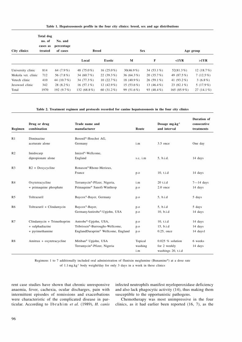
96
Table 1. Hepatozoonosis profile in the four city clinics: breed, sex and age distributions
Total dog
no. of No. and
cases as percentage
City clinics treated of cases Breed Sex Age group
Local Exotic M F <1YR >1YR
University clinic 814 64 (7.9 %) 48 (75.0 %) 16 (25.0 %) 30(46.9 %) 34 (53.1 %) 52(81.3 %) 12 (18.7 %)
Mokola vet. clinic 712 56 (7.8 %) 34 (60.7 %) 22 (39.3 %) 36 (64.3 %) 20 (35.7 %) 49 (87.5 %) 7 (12.5 %)
Vetech clinic 410 44 (10.7 %) 34 (77.3 %) 10 (22.7 %) 18 (40.9 %) 26 (59.1 %) 41 (93.2 %) 3 (6.8 %)
Jesswool clinic 342 28 (8.2 %) 16 (57.1 %) 12 (42.9 %) 15 (53.6 %) 13 (46.4 %) 23 (82.1 %) 5 (17.9 %)
Total 1970 192 (9.7 %) 132 (68.8 %) 60 (31.2 %) 99 (51.6 %) 93 (48.4 %) 165 (85.9 %) 27 (14.1 %)
Table 2. Treatment regimen and protocols recorded for canine hepatozoonosis in the four city clinics
Duration of
Drug or drug Trade name and Dosage mg.kg-1 consecutive
Regimen combination manufacturer Route and interval treatments
R1 Diminazine Berenil®-Hoechst AG,
aceturate alone Germany i.m 3.5 once One day
R2 Imidocarp Imizol®-Wellcome,
dipropionate alone England s.c, i.m 5, b.i.d. 14 days
R3 R2 + Doxycycline Ronaxon®-Rhone-Merieux,
France p.o 10, t.i.d 14 days
R4 Oxytetracycline Terramycin®-Pfizer, Nigeria, i.m 20 t.i.d 7—14 days
+ primaquine phosphate Primaquine® Sanofi-Winthrop p.o 2.0 once 14 days
R5 Toltrazuril Baycox®-Bayer, Germany p.o 5, b.i.d 5 days
R6 Toltrazuril + Clindamycin Baycox®-Bayer, p.o 5, b.i.d 5 days
GermanyAntirobe®-Upjohn, USA p.o 10, b.i.d 14 days
R7 Clindamycin + Trimethoprim Antrobe®-Upjohn, USA, p.o 10, t.i.d 14 days
+ sulphadiazine Tribrissen®-Burroughs-Wellcome, p.o 15, b.i.d 14 days
+ pyrimethamine EnglandDaraprim® Wellcome, England p.o 0.25, once 14 days1
R8 Amitrax + oxytetracycline Mitiban® Upjohn, USA Topical 0.025 % solution 6 weeks
Terramycin®-Pfizer, Nigeria washing for 2 weekly 14 days
i.m washings 20, t.i.d
Regimens 1 to 7 additionally included oral administration of flunixin meglumine (Bunamine®) at a dose rate
of 1.1 mg.kg-1 body weight/day for only 3 days in a week in these clinics
rent case studies have shown that chronic unresponsive anaemia, fever, cachexia, ocular discharges, pain with intermittent episodes of remissions and exacerbations were characteristic of the complicated disease in par-ticular. According to I b r a h i m et al. (1989), H. canis
infected neutrophils manifest myeloperoxidase deficiency and also lack phagocytic activity (14), thus making them susceptible to the opportunistic pathogens.
Chemotherapy was most unimpressive in the four clinics, as it had earlier been reported (16, 7), as the

97
Table 3. Treatment regimen and protocols for the complicated and uncomplicated hepatozoonosis and the rate of relapses
Hepatozoonosis complicated with
Uncomplicated Babesiosis Ehrlichiosis Babesiosis and Sarcoptic
Hepatozoonosis ehrlichiosis mange Demodicosis Toxoplasmosis
No. treated in the
four city clinics 16 60 45 18 20 25 8
Treatment regimen
and protocols adopted
as in Table 2 R 1 or R 2 R1 or R2 R2 or R3 R1, R2 or R3 R8 + R1 or R2 R8 + R1 +R2 R6 or R7
No. and percentage
of hepatozoonosis 7 4 34 11 4 15 —
in 24 months 43.8 % 6.7 % 75.6 % 61.1 % 20.0 % 60.0 % 0.00 %
rate of relapses was high at 43.8 % in the uncomplicated and was even higher in the complicated, ranging from 6.7 % to 75.6 % despite the specific treatment agents for the intercurrent infections. Four factors were identified (5) as contributing to treatment failure in hepatozoonosis viz the chronic and multiorgan nature of the disease, sometimes involving the immune-mediation, spontane-ous remissions and exacerbations (which make loss of parasitaemia, painlessness and drop in rectal temperatures invalid parameters for drug efficacy assessment), non-comprehensive nature of some treatment regimens that provide not for the chronic body inflammation and pain and the ineffective tick control programme that allows for re-infection which could clinically be mistaken for relapses.
Amyloid deposits in multiple organs, rheumatoid ar-thritis, pyelonephritis, glomerulonephritis, etc. that have been described in advanced hepatozoonosis (3) are evi-dences of immune-complex mediation which would have benefited from a medium course of corticosteroids like phenylbutazone, which was lacking in the listed regimen (Table 2). However, such corticosteroid phenylbutazone therapy should be of a very short course to avoid its enhancement effect on parasitaemia. (4).
Again, drugs like trimethoprim and sulphadiazine (R7) are only effective against the asexual stage but not the more pathogenic sexual multiplicative stage of the hepatozoon parasites (7); imidocarb (R2) clears only the blood forms (merozoites), sparing the highly multipli-cative and hence pathogenic gametocytes to thrive (5, 2,19), while diminazine aceturate (R1) had apparently little effect on the course of disease (5).
Treatment with oxytetracycline or doxycycline with or without primaquine also effected only clinical remission in cat (5), while oxytetracycline and imidocarb effected clinical remission and reduced parasitaemia in dogs with hepatozoonosis and ehrlichiosis (15), probably by targeting the concomitant ehrlichiosis. Toltrazutril (R5 and R6), a potent anticoccidial drug, was also reported to be unable
to completely eliminate hepatozoon parasites in dogs, though effected clinical remission (17, 19).
At present there seems to be no effective and con-sistent single protozoacidal agent or its combination for the successful clinical management of canine hepatozoo-nosis – a fact that is not unconnected with its complex multiplicative multiorgan life cycle. Furthermore, a possible defective functioning of the H. canis-parasitized neutrophils (13, 14) might have contributed more to the treatment failure than even the intercurrent immunosup-pressive infections that had their effective and specific treatment agents included in the routine treatment regi-men of these clinics.
A possible recipe for this important practice problem could be in the routine use of the recombinant canine granulocyte colony stimulating factor (G-CSF) (23), which have been commercially purified, molecularly cloned and expressed as recombinant protein for the stimulation of the production and function of neutrophils and monocytes. Their use as adjunct cytokine proteins in the management of hepatozoonosis can be worthwhile and fruitful as it has been with the parvoviral infection management (10).
Oral administration of flunixin meglumine (Banamine®) as practiced in these clinics, offers a palliative anti-inflammatory relief and should be encouraged in the treatment of all chronic inflammatory conditions like hepatozoonosis.
Because hepatozoonosis is transmissible also by mites, mosquitoes, lice, fleas and other biting flies (18) an effective vector control programme is as important as the treatment, if relapse or re-infection rate is to be reduced. This is particularly easy for a disease like hepatozoonosis that is of low parasitaemia and requires heavy ectoparasitism for successful transmission. Regular use of acaricides as dips, shampoos and on the floors and walls of the kennels would assist in the control of the vector ticks, Rhipicephalus sanguineus and the disease as well. The future development of a sensitive

98
serological assay for the early detection of inapparent carriers would also help control.
ACKNOWLEDGEMENTS
Doctors Y. A. Awoseyi, M. Lawal, S. Sansi and Y. Oladokun, the respective resident veterinarians at Vetech, Mokola Veteri-nary, Jesswool and Magma clinics are much acknowledged not just for their co-operation, but also for the anamneses provided and the access given to their case records.
REFERENCES
1. Aliu, Y. O., Sanusi, A., Ezeokoli, C. D., Lemu, I., 1976: Canine hepatozoonosis and its therapeutic management. Nig. Vet. J., 5, 9—11.
2. Baneth, G., Harmelin, A., Presentey, B. Z., 1995: Hepatozoon canis infection in two dogs. J. Am. Vet. Med. Assoc., 206, 1891—1894.
3. Barton, C. L., Russo, E. A., Craig, T. M. et al., 1985: Canine hepatozoonosis: a retrospective study of 15 naturally occurring cases. J. Am. Anim. Hosp. Assoc., 21, 125—134.
4. Clark, K. A., Rodson, R. M., Weishuhn, L. L. et al., 1973: Hepatozoon procyonis infections in Texas. J. Wildl. Dis., 9, 182—186.
5. Craig, T. M., 1990: Hepatozoonosis. In Greene, C. E., (ed.): Infectious Diseases of the Dog and Cat. Philadelphia, W. B. Saunders, pp. 778—785.
6. Craig, T. M., 1998: Hepatozoonosis. In Greene, C. E. (ed.): Infectious Diseases of the Dog and Cat. Philadelphia, W. B. Saunders, pp. 458—465.
7. Craig, T. M., Smallwood, J. E., Knauer, K. W. et al., 1978: Hepatozoon canis in dogs: clinical, radiographic and haemato-logical findings. J. Am. Vet. Med. Assoc., 173, 967—972.
8. Dubey, J. P., Beattie, C. P., 1988: Toxoplasmosis in Ani-mals and Man. C.R.C. Press, Boca Raton, Fl., pp. 1—220.
9. Ezeokoli, C. D., Ogunkoya, A. B, Abdullahi, R., Tak-dek, L. B., Sannusi, A., Ilemobade, A. A., 1983: Clinical and epidemiological studies on canine hepatozoonosis in Zaria, Nigeria. J. Sm. Anim. Pract., 24, 455—460.
10. Fox, L. M., Bruederle, J. B., 1996: Nearly foolproof parvovirus treatment. Vet. Forum, 13, 36—38.
11. Gosset, K. A., Gaunt, S. D., Aja, D. S., 1985: Hepato-zoonosis and ehrlichiosis in a dog. J. Am. Anim. Hosp. Assoc., 21, 265—267.
12. Harmelin, A., Dubey, J. P., Yakobson, B. et al., 1992: Concurrent Hepatozoon canis and Toxoplasma gondii infections in a dog. Vet. Parasitol., 43, 131—136.
13. Ibrahim, N. D. G., Rahamathulla, P. M., Njoku, C. O., 1989: Neutrophil myeloperoxidase deficiency associated with canine hepatozoonosis. Int. J. Parasitol., 19, 915—918.
14. Itoh, N., Itoh, S., 1991: A fundamental investigation of nitroblue tetrazolium (NBT) reduction test in canine neu-trophils. J. Jpn. Vet. Med. Assoc., 44, 1034—1036.
15. Jauregui, L. E., Lopez, G. M., 1995: Canine hepato-zoonosis. Vet. Internat., 1, 30—38.
16. Klopfer, U., Neuman, F., Nobel, T. A., 1973: Hepatozoon canis infection in dogs in Israel. Refu. Vet., 30, 116—120.
17. Krampitz, H. E., Habercorn, A., 1988: Experimental treatment of Hepatozoon infections with anticoccidial agent toltrazuril. J. Vet. Med. Sci., 35, 131—137.
18. Levine, N. D., 1973: Protozoan Parasite of Domestic Animals and of Man (2nd edn.). Minneapolis, Burgess Pub-lishing Co., 406 pp.
19. Macintire, D. K., Vincent-Johnson, N., Dillon, A. R., Blagburn, B., Lindsay, D., Whitley, E. M., Banfield, C., 1997: Hepatozoonosis in dogs: 22 cases (1989–1994). J. Am. Vet. Med., Assoc., 210, 916—922.
20. McCully, R. M., Basson, P. A., Bigalke, R. D. et al., 1975: Observations on naturally acquired hepatozoonosis of wild carnivores and dogs in the Republic of South Africa. Ondersteport J. Vet. Res., 42, 117—133.
21. Nyindo, M. B. A., Ristic, M., Huxsol, D. L., Smith, A. R., 1971: Tropical canine pancytopaenia: In vitro cultiva-tion of the causative agent – Ehrlichia canis. Am. J. Vet. Res., 32, 1651—1658.
22. Vincent-Johnson, N., Macintire, D. K., Bareth, G., 1997: Canine hepatozoonosis: pathophysiology, diagnosis and treatment. Compend. Cont. Educ. Pract. Vet., 19, 51—65.
23. Zinkl, J. G., Cain, G., Jain, N. C., Sousa, L. M., 1992: Haematological responses of dogs to canine recombinant granulocyte-colony stimulating factor (rcG-CSF). Comp. Hae-mat. Int., 2 , 151—156.
Received April 14, 2006

9999
www.uvm.sk
ABSTRACT
Xenobiotics present a risk due to their toxic effects and also due to their potential for causing damage to the im-mune system leading to immunosuppression or increased incidence of hypersensitivity disorders, auto-immune and infectious diseases or neoplasia. Experimental animal stud-ies have identified many immunotoxic agents and suggested potential risk for humans in terms of susceptibility to and prevalence of infectious diseases. The critical factors which affect the design and results of immunotoxicity studies are nutritional health, microbial disease status, the ordinary microflora, life stage of the animal, dose-response rela-tionships, redundancy, timing of immunotoxicity studies, genetic factors as well as marked differences in the immune systems that do exist cross species. The use of a rodent and non-rodent species for testing should be considered in this context. Dogs and non-human primates are widely used in toxicity studies, and attention should be given to their use in immunotoxicity investigations. Immunotoxic-ity of pesticides may be studied on free living, grazing animals which are among the animal groups immediately exposed to and most affected by the harmful influence of these chemical substances. Models need to be developed for closer extrapolation to humans by using severe combined immunodeficiency and transgenic and congenic mouse and human cell lines. In vitro immunologic assays that can be used for rapid detection of potential immunosuppressive agents need to be developed to determine cellular and subcellular sites of action.
Key words: animal models; immunotoxicity; risk asses-sment; xenobiotics
ANIMAL MODELS IN IMMUNOTOXICITY STUDIES (A Review)
Kovalkovičová, N.1, Pistl, J.1, Holovská, V.2, Legáth, J.1
1University of Veterinary Medicine, Komenského 73, 041 81 Košice2University of P. J. Šafárik, Medical Faculty, Tr. SNP 1, 040 11 Košice
The Slovak Republic
INTRODUCTION
The immune system is a sensitive target organ during exposure to xenobiotics. It is comprised of a number of organs, tissues and cells throughout the body. The cells are polymorphonuclears, monocytes/macrophages, lymphocytes of different populations and sub-populations and stromal cells in the lymphoid organs, for instance, epithelial cells in the thymus important for the differentiation of thymus-dependent cells (T-cells). A successful immune response is dependent on co-operation between different cell types. The reactions of lymphoid cells are associated with gene amplification, transcription, and translation; chemical compounds that affect these processes of cell proliferation and differentiation are especially immunotoxic (29).
Relevant data on the biological effects, dose-response rela-tionships, and exposure for a particular agent are analysed in an attempt to establish qualitative and quantitative estimates of adverse outcomes in the process of risk assessment. Typically, risk assessment comprises four major steps: hazard identification, hazard characterization (dose-response assessment), exposure assessment, and risk characterization. The utility of any overall risk assessment is critically dependent on the quality of the first step – hazard identification. Up until now, immunotoxicol-ogy has focused mainly on hazard identification, and to some extent on dose-response assessment, and very few studies have included exposure assessment or risk characterization.
The detection of immune changes after exposure to poten-tially immunotoxic compounds is more complicated in humans than in experimental animals. The testing possibilities are limited, level of exposure to the agent (i.e. dose) are difficult to establish, and the immune status of populations is extremely heterogeneous. Age, race, sex, pregnancy, acute stress and the
FOLIA VETERINARIA, 50, 2: 99—107, 2006

100
ability to cope with stress, coexistent disease and infections, nutritional status, tobacco smoke, and some medications con-tribute to this heterogeneity. Therefore adequate human data on immunotoxicity are seldom available and hazard identification is most often based on results obtained in animals.
Animal models are useful for identifying possible hazards that could attend human exposure to xenobiotics. Over the last decade, several important animal models have been developed to detect the immune potential of chemical agents and to elu-cidate immunologic mechanisms of injury. The following are two of the most productive areas of development:
1. animal models are essential for detecting immunosuppres-sive potential. They provide the means whereby dose-response and mechanistic studies can be performed to provide a basis for hazard evaluation as related to human risk;
2. when possible, the experimental design for animal studies should be applicable to humans with respect to the following: a) ability to measure the status of the immune sys-
tem; b) use of a route of administration that emulates human
exposure; c) matching of the toxicokinetics (i.e. absorption,
distribution, and metabolism of the xenobiotic in the experimental animal and humans).
The ultimate purpose of risk assessment is to protect hu-man health and the environment. Suitable model systems must therefore be chosen. Experimental animal provide an important and necessary means for detecting immunotoxic compounds and for determining their mechanisms of action in the induc-tion of disease. The results of animal studies are useful for determining chemical hazards, managing risk, and determining relatively safe conditions of exposure (15).
The potential for chemicals to cause injury to the immune system is of considerable public health significance. Due to many adverse effects reported in man, the immunotoxicity po-tential of every new molecular entity should be systematically and specifically evaluated in various animal models.
CHARACTERISTIC OF ANIMAL MODELS
An immunotoxicology program was first established within the National Toxicology Program of the National Institute of Environmental Health Sciences (USA) in 1978 with the goal of selecting, developing, and validating animal models – for use in assessing immunomodulation induced by xenobiotic substances.
The first choice of an experimental animal for toxicologic investigation is the one whose toxicokinetics of the test chemical are similar to those of the human. With some exceptions, the immune and metabolic systems of humans and experimental animals are similar enough for animal models to provide the conduit to detect immunotoxicants. Most immunosuppressive agents in humans produce similar results in animals, and the mechanisms of action are similar in experimental animals and humans (7).
Researchers have used various animal models to discover immunotoxic agents, to develop immune system profiles to identify mechanisms of action, and potential health risk asso-ciated with exposure to specific xenobiotics, either consumed as drugs or through environmental exposure.
ASSESSMENT OF IMMUNOTOXICITY IN RODENTS
The immune system is highly conserved among higher verte-brate species and the immune components and their interactions in mice, rats and humans are similar. Thus, if toxicokinetic properties of chemicals are similar, it is reasonable to test for potential adverse effects in humans using laboratory rodents (29). With regard to rodents being the best-known animal models, the review is aimed to describe non-rodent species that are now more frequently used to assess the immunotoxicity of chemical compounds.
MiceThe majority of the immunotoxicological tests were origi-
nally developed in mice because basic immunological reasearch has been conducted in this species. Although the mouse is not used routinely in initial toxicologic evaluations, except in the determination of carcinogenic potential, the mouse immune system is well characterized, and most of the necessary reagents for immunotoxicity testing are specific to them.
RatsWith the progress of immunotoxicology the rat has been
used increasingly. The rat is the standard animal in the early phase of toxicological testing. For this species most of the pharmacokinetic and toxicologic data are available. An effort has been made to use the rat to optimize and verify methods that assess immune function and immunotoxic effects and to develop the reagents necessary to assess immunotoxicity in the rat. Nowadays the rat is equally useful for most aspects of immunotoxicity assessment, however, transference of the tests is not always easy, partly because of the lack of suitable reagents (18).
Guinea pigsGuinea pigs are used to determine the sensitization poten-
tial of chemicals, cosmetics ingredients, consumer products, and drugs (4).
ASSESSMENT OF IMMUNOTOXICITY IN NON-RODENT SPECIES
While most immunotoxicological evaluations are conducted in mice or rats, use of other species is increasing, for example rabbits (28), Chinese hamsters (7), small breeds of pigs (17) and sheep (45), non-human primates (12), dogs (30), fish (59, 34) and chicken (6).

101
Non-human primatesIn immunotoxicological studies Macaca mulatta (rhesus
macaques), M. nemestrina (pig-tailed macaques), Cercocebus atys (sooty mangabeys), M. fascicularis (cynomolgus monkey), and marmosets have been used to determine how chemicals target the immune systems (35).
Many of the assays carried out in mice, rats or humans can be adapted for use with non-human primates (12). Many of the reagents available for human immune assessment can be used in monkeys. Monoclonal antibodies generated to human leukocyte subsets can be used in phenotyping the blood mononuclear cells of e.g. marmoset monkeys (Callithrix jacchus; 12), although the possibility of such use differs, depending on the evolutionary distance of the non-human primate from humans. More recently, the advent of humanized antibodies and vaccines has imposed a further demand on non-human primate models since many immunotherapeutics do not interact with rodent receptors, but frequently only cross-react with primate tissue.
The usefulness and suitability of nonhuman primates for assessing immunotoxicology has been the subject of thorough reviews (42, 24, 8, 27). H o u s e and T h o m a s (27) have recently provided a compilation of immunotoxicology tests available for nonhuman primates (Table 1). Use of the monkey as a test species is likely to increase as more and more biotechnology and recombinant products are produced.
Table 1. Available approaches for the assessment
of immmunotoxicity in the non-human primate (27)
Structural integrity
Haematology, clinical chemistry
Histopathology (thymus, spleen, lymph nodes, Peyer
patches, bone marrow)
Immunocytochemistry using specific CD antibodies
Flow cytometry
B-cell function
Antibody production
Mitogenesis
T-cell function
Delayed-type hypersensitivity
Cytokine analysis
Mitogenesis and mixed lymphocyte response/reaction
Natural imunity
Natural killer cell function
Macrophage/neutrophil function
DogsWhile dogs are not the species of choice for immunotoxi-
cological studies, they are used predominantly as a common non-rodent species for extrapolation to man for risk assess-ment, and virtually all of the assays used for assesssing im-munotoxic potential have been adapted for use in dogs (33, 10, 30, 19, 41).
The dog is an important immunological model for discovering the mechanism of diseases such as atopy, rheumatoid arthritis, autoimmune haemolytic anaemia, autoimmmune thrombocyto-penia, autoimmune thyroiditis, autoimmune dermal conditions
and systemic lupus erythematosus (30). Immunodeficient dwarf-ism is a relevant model for elucidating the endocrine role of the thymus in its relationships between the neuroendocrine and immune systems in pre-pubertal dogs (50). Moreover, the predictivity of pharmaceutical agent human toxicities by pre-clinical animal species is almost 50 % from dog or primate studies, but very few toxicities are identified from rat studies alone (43). Therefore, the scientific and medical rationale for using the dog as an alternative immunologic model to rodent species extends beyond being the non-rodent alternative in regulatory studies.
ASSESSMENT OF IMMUNOTOXICITY IN NON-MAMMALIAN SPECIES
For evaluating the potential adverse effects of compounds and agents on the immune system non-mammalian species are also used extensively, e.g. fish and chicken.
FishBecause of their environment, fish are an excellent model
for studying the effects of water- and sediment-borne pollut-ants. The choice of species depends on its biology (migratory or local, marine or freshwater, sediment-dwelling or pelagic) and on experience in the laboratory (20, 59, 34). Some species seem to be preferred, such as trout, salmon, and carp, which are practical, owing to their size, for sampling blood and tis-sues for laboratory studies.
For studies of saltwater species, bottom-dwelling flatfish are commonly used. In Europe, the flounder (Platichthys flesus) and dab (Limanda limanda) are popular target species since they are susceptible to certain recognizable diseases and are commonly available. Smaller species, such as guppies (Poecilia reticulata) and medaka (Oryzias latipes), have secured a niche in aquatic toxicology owing to the ease of husbandry and relatively low cost; moreover, because of their small size, whole animals can be used for histopathological examination, but their application in immunotoxicology may be limited because of the difficulty in obtaining adequate blood and tissue samples.
Recently, some of the same criteria used to determine immunotoxicity in mammalian systems have been used to assess chemical-induced immuntoxicity of selected metals (9, 56, 39, 57), pesticides (58, 3) and polycyclic aromatic hydro-carbons (13) in laboratory-reared Japanese medaka, feral fish populations or hatchery-raised rainbow trout. Further, studies in fish-eating birds (21) and harbor seals (49) add additional support to the utility of immune assays for use as immuno-toxicity biomarkers.
Studies such as these that employ fish to assess chemical-induced immunotoxicity fit well into the newly-emerging trends for immunotoxicity testing, which include more in vitro tests, greater use of computational methods, and the development and validation of non-mammalian alternative species (31). Moreover, the validity/accuracy of cross-species extrapolation studies is improved by the ability to assess chemical-induced immunoto-xicity in different animal models using the same assays.

102
ChickensAnother non-mammalian species that has been studied
extensively with regard to the structure and function of its immune response is the chicken. It is therefore not surprising that the chicken has emerged as the predominant avian model for assessing compounds for potential immunotoxicity (16, 1, 53, 6, 26).
FUTURE DIRECTIONS
Transgenic miceA transgenic animal is any animal into which cloned genetic
material has been transferred. Since the first description of the creation of transgenic mice in the early 1980s, further and rapid refinements of genetic engineering and animal modelling technologies have facilited remarkable advances in biological research, in particular toxicological evaluation (10, 40, 48). Transgenic technology can be used in immunology to gener-ate mice that lack virtually any genetic control mechanism or specific cell sub-populations. As a consequence, complex systemic responses can be dissected into individual components, and the mechanism by which immunosuppressive agents exert their effects can be better understood.
Two strategies are used to induce genetic aberrations in transgenic mice (5). One involves the induction of genes that produce toxins, such as diphtheria toxin or the A sub-unit of ricin, into targeted cell subpopulations. The second strategy involves the thymidine kinase (tk) gene from Herpes simplex virus: when certain nucleotide analogues are administered and metabolised exclusively by viral thymidine kinase, the metabolites are lethal only to cell subpopulations that express the tk gene. Both approaches are inducible systems for kill-ing cells in vivo and hold the promise for understanding the selective toxicity of drugs and environmental agents on the immune system.
Other promising avenues are the use of animals transgenic with respect to certain specificities of the T-cell receptor. If a gene that encodes for a certain antigen specificity is introduced into the genome, those effects of immunotoxicants affecting the (positive and/or negative) selection process which takes place in the thymus, could be studied elegantly with such models, when either undesired specificities (which should be negatively selected) or desired specificities (which should be positively selected) are introduced.
These in vivo models using genetically engineered animals allow the study of biological effects of expression of a range of genes under physiologically relevant conditions (55). From the point of view of the evaluation of xenobiotics, it is hoped that such models will provide results more rapidly, require fewer animals and may be more representative of human response (55). It is also possible that transgenic animal bio-assays can be used to estimate risk for specific human subpopulations that may be genetically more susceptible to certain diseases, for example some types of cancer (52).
Transgenic animals or adoptive transfer models can provide valuable insights into the immunotoxic mechanism of compounds in vivo. Transgenic and knockout mice can be used to address
the role of specific gene products in the response to toxicants, making these animals possibly more prone to develop certain conditions facilitating the identification of these toxicants. Transgenic mice can also be used to produce specific T-cells, which can be transferred to syngenic recipients to measure the response to activation. The OVA TCR transgenic mice are useful as an adoptive transfer model for mechanistic research. This model may be useful in screening, as the sensitivity for the endpoint is significantly increased.
Severe combined immunodeficient mice (SCID mice)Another approach that may warrant further exploration is
the use of severe combined immunodeficient CB-17 scid/scid (SCID) mice grafted with human immune cells. Xenogeneic lymphoid cells and/or tissues can be successfully transferred to SCID mice. SCID mice have been grafted with human foetal lymphoid tissue in order to study human haematopoiesis or with human peripheral blood lymphocytes to allow production of human immunoglobulins, including secondary antibody response. SCID mice have also been used to study auto-immunity and potential antiviral therapeutics. While these animal models still have limitations (46), they may ultimately provide predictive models for examining potential immunosuppressive agents.
In particular, SCID mice co-implanted with human foetal thymus and liver tissue fragments (SCID/hu mice) offer the possibility of studying the human thymus in vivo in an isolated xenogeneic environment and the effects of immunotoxicants on these grafts. This system is particularly interesting with regard to those immunotoxicants for which the thymus is one locus of action.
When data obtained in experimental animals are extrapolated to the human situation, a control model, between the SCID/hu mouse model and the intact laboratory animal (rat), is desirable in order to test for possible differences in thymic behaviour, because of its location under the kidney capsule: thymic blood flow and therefore the toxicokinetic behaviour of the thymus may differ. For this reason the SCID/ra model was developed, by implanting rat foetal thymus and liver tissue fragments under the SCID mouse renal capsule. The outcomes of exposure of rats and SCID/ra mice can be compared and the influence of thymus location and mouse metabolism on extrapolation from SCID/hu to humans can then be determined.
The outcome of experiments with SCID/hu and SCID/ra mice can be used to compare the sensitivity of the human and rat thymus and can thus yield important information for the process of human risk assessment (17, 46, 11).
Molecular approaches in immunotoxicology – cytokinesA promising avenue for early detection of immunotoxicity
may be measurement of the expression of various interleukins. Cytokines are involved both in regulation of the immune system and pathological phenomena, hence alterations in their pattern of expression may be early indicators of immunotoxicity (38). Such testing can be done at the level of mRNA expression, on mRNA extracted from lymphoid tissue taken from exposed animals, or in tissue sections, so that the alterations can be evaluated in the context of morphological indications of the toxic effects. The signal of the cytokine that is being tested
103
must therefore be strong enough to be picked up in material from exposed animals whose immune system has not received other stimuli, i.e. sensitization or infection (25).
Very sensitive analysis can be done with the semiquantitative polymerase chain reaction, which is a powerful technique for elucidating early kinetic changes of cytokine expression, before translation and secretion. In addition, since immunosuppressive agents can enhance or inhibit the ultimate production and secre-tion of cytokines at various stages such as transcription, the splicing of mRNA, translation of mRNA into polypeptides on ribosomes, post-translation processing, and secretion, potential molecular targets can be dissected by such techniques. Several other molecular approaches may be used, including northern blotting, dot-blot hybridization, and antisense oligonucleotides for inhibiting the translation of specific mRNAs.
Approaches to assessing immunosuppression in vitroA more recent development in risk assessment is use of
in vitro models as an adjunct to studies of experimental ani-mals. The advantages of this approach are that it improves the accuracy of extrapolation of data from animals to man and minimizes the use of animals; it also bridges the gap between those data, particularly when human experimentation is limited for ethical considerations.
The direct addition of compounds in various in vitro as-says, including those involving lymphocyte proliferation, mixed leukocyte reaction, and cytotoxic assays with NK-cells and Tc-lymphocytes, has been used to determine the mechanisms by which compounds alter the immune response at the cellular and subcellular level.
One of the limitations of in vitro systems is that ex-ogenous metabolic activation systems are often required. While lymphocytes can metabolize some compounds, such as benzo[a]pyrene, to active metabolites (36), other potent im-munosuppressive compounds such as cyclophosphamide and naphthalene require a metabolic activation system S9 (32) or a hepatocyte co-culture system (47).
Predictive in vitro systems based on immune cells of hu-man origin are particularly attractive, given the uncertainties of extrapolating the results of experimental studies to humans and the accessibility of immune cells in human peripheral blood. Although many of the immune cells obtained from hu-man blood are immature forms, the large numbers and diverse populations (i.e. polymorphonuclear leukocytes, monocytes, NK-cells, T-cells and B-cells) that can be obtained provide an attractive alternative or adjunct to conventional studies in experimental animals. As a consequence, a number of studies have been conducted to compare the functional response of human and rodent lymphocytes to putative immunosuppressive agents in vitro (47).
FACTORS INFLUENCING THE RESULTS OF THE IMMUNOTOXICOLOGICAL STUDIES IN ANIMALS
There are several factors that may have a profound influence on the final experimental outcome of a study of immunotoxicity
in animals. The most important of them are the genetic make-up of the experimental animal, interspecies differences, nutritional health, microbial disease status, ordinary microflora and life stage of the animal, dose-response relationships, redudancy and timing of immunotoxicity studies (2, 15).
The genetic make-up of the experimental animal The genetic make-up of experimental animals can be of
great importance in either masking or reducing toxicity or in facilitating the detection of toxic effects. The toxicodynamics features of chemicals are generally stable from one species to another, at least from a qualitative standpoint; however, the toxicokinetic behaviour of compounds, which is influenced by the genetics of the animal, often differs between species or even strains and therefore should be an important factor in the selection of an appropriate species to predict potential human immunotoxicity (2).
Genetic factors play a key role not only in the metabolic capacity of drug disposition, but also in the magnitude and nature of immune responses. Due to the intrinsic inter-individual variabitily of immune responses, the range of immunotoxicity results is typically wider than that of other conventional toxicity endpoints. As wide interindividual variability is unavoidable, sufficient numbers of animals, a cautious comparison with both study and historical controls, and the possible use of inbred or genetically modified animals are to be considered (15).
Interspecies differencesThe dramatic advancement in murine immunology over
the last 40 years has led to the incorrect tendency to assume that all laboratory animals are immunologically similar to mice, and that mice are immunologically identical to humans. Unfortunately, studies that specifically attempt to identify and characterize species differences in immune structure and func-tion, such as that by L a n g et al. (37) and S m i a l o w i c z et al. (51), are few, and the characterization of most species differences are based on the comparison of data obtained from published results from many different laboratories collected under highly variable conditions.
As has been nicely put by C u n n i g h a m (14): “a mouse is not a rat is not a human”. The investigation of immunotoxicity of chemical entities may require the use of different species, and while in many cases information concerning the structure and function of the immune system can be readily translated across species there are numerous and significant species differences that need to be considered. In some cases, the generation of meaningful immunotoxicology data can be adversely affected by the choise of a species that does not adequately share the immune function of concern with man. Likewise immunoto-xicology testing in one species may produce negative data in one species but positive data in another (22).
Marked interspecies differences in the immune system should lead to the use of more than one animal species and to confirm animal data during clinical trials (15).
H a l e y (22) has described numerous and significant dif-ferences in the structure and function of the immune system of various species of animals, e.g. in the structure of thymus, lymph nodes, spleen, mucosal associated lymphoid tissue,

104
cell-mediated immune responses, hypersensitivity reactions, circulating leukocyte populations and antibody production.
Circulating leukocyte populations display marked species differences (Table 2). The circulating leukocyte profile for humans is considered to be neutrophilic as it is for the dog whereas rodents have greater percentages of circulating lym-phocytes (44, 23, 54, 22).
Most species appear to produce similar subclasses of anti-bodies, but subclass function may vary (Table 3, 22).
The nutritional health of the animalThe animal’s diet and its composition, both prior to and
during the study, can have a major influence on the outcome of the study. National and international recommendations on optimal laboratory animal diets are available but ideas in this field are changing as the influence of “overnutrition” on longevity and toxicological outcomes in laboratory animals becomes better understood.
The composition of the diet and the nature of the individual ingredients are also major elements influencing the bioavail-ability of test chemicals or macro- and micro-nutrients from the dietary matrix, as well as the proper physiological functioning of the gut system (2).
The microbial disease status of the animal The absence of disease ensures the longevity of the animals
in chronic studies and allows the findings in a toxicity study to be properly interpreted. For example, when questions regarding
influences of food additives, contaminants, micro-nutrients, novel foods, etc., on the functioning of the immune system are to be explored in experimental animals it is important to know about past and present challenges to the immune system in order to identify the baseline of immune system function for the study (2).
The ordinary microflora of the animalThe aerobic and anaerobic microflora of the gut are of
utmost importance for the appropriate digestion of the diet and in determining the bioavailability of nutrients. Similarly, they may also play a role in the metabolism of test substances in the lower gut and may even generate toxic metabolites which can then be absorbed systemically. Programs have been intro-duced to control the presence of infectious diseases in animal populations (specific pathogen free, SPF), to make them totally germfree (gnotobiotic) or associated with a microbiologically-defined flora.
It is also probable that the gut flora in early life is of importance for the imprinting of the immature immune sys-tem (2).
The life stage of the animalTo make a scientifically sound assessment of the toxic
potential of any compound, it is important to identify the most sensitive life stage of the experimental animal for that compound (2).
Table 2. Approximate total white blood cells (WBC), percentage of neutrophils
and lymphocytes in the blood of different species (44, 23, 54, 22)
Species WBC Neutrophils Lymphocytes
(103 µl-1) % 103 µl-1 % 103 µl-1
Human 7.0–10.0 50–70 3.5–7.0 20–40 1.4–4.0
Cynomolgus monkey 5.0–18.0 10–42 0.5–7.5 40–67 2.0–12.0
Beagle dog 6.4–14.6 47–65 3.0–9.5 16–41 1.0–6.0
CD rat 3.0–14.5 10–21 0.3–3.0 83–100 3.0–12.0
Hamster 4.0–10.0 13–35 0.5–3.5 63–80 2.5–3.5
Guinea pig 2.0–12.0 25–30 0.5–3.5 75–83 1.5–10.0
CD-1 mouse 2.0–10.0 15–20 0.3–2.0 50–70 1.0–7.0
New Zealand white rabbit 4.0–13.0 25–46 1.0–6.0 50–70 2.0–9.0
Table 3. Immunoglobulins of different species (22)
Species IgG IgM IgE IgA IgD
Human IgG1, IgG2, IgG3, IgG4 + + IgA1, IgA2 +
Non-human primate + + + +
Dog IgG1, IgG2, + + +
Rabbit + + + +
Hamster IgG1, IgG2 + +
Rat IgG1, IgG2a, IgG2b, IgGc, IgG3, IgG4 + + +
Mouse IgG1, IgG2a, IgG2b + + +
Guinea pig IgG1, IgG2 + + +

105
Dose-response relationshipsIn immunotoxicology, dose-response relationships are often
atypical. As regards immunosuppression, a common finding is that high doses can induce a complete inhibition of im-mune responsiveness, whereas lower doses cause only a mild to moderate decrease in immune responses. Thus, a careful selection of doses is critical for the risk/benefit evaluation of new chemicals (47, 15).
Redundancy and timing of immunotoxicity studiesThe term redundancy refers to the capacity of the immune
system to trigger compensatory mechanisms when one effec-tor mechanism is inhibited. Thus, one single change in one immunolological endpoint does not necessarily mean that the tested chemical is immunotoxic. The logical consequences are that more than one endpoint should be used to predict the potential for immunotoxicity and that a global evaluation of all the results as well as general toxicity studies is essential to avoid misleading conclusions (15).
As the immune system is a significant target organ of tox-icity, it is essential that immunological endpoints should have been already investigated in early preclinical studies (15).
ACKNOWLEDGMENTS
This study was supported by VEGA Grant No. 1/1362/04 and the National Reference Laboratory for Pesticides, University of Veterinary Medicine in Košice.
REFERENCES
1. Baecher-Steppan, L., Nakaue, H. S., Matsumoto, M., Gainer, J. H., Kerkvliet, N. T., 1989: The broiler chicken as a model for immunotoxicity assessment. 1. Standardization of in vitro immunological assays. Fundam. Appl. Toxicol., 121, 773—786.
2. Barlow, S. M., Greig, J. B., Bridges, J. W., Carere, A., Carpy, A. J. M., Galli, C. L., Kleiner, J., Knudsen, I., Koëter, H. B. W. M., Levy, L. S., Madsen, C., Mayer, S., Narbonne, J. F., Pfannkuch, F., Prodanchuk, M. G., Smith, M. R., Steinberg, P., 2002: Hazard identification by methods of animal-based toxicology. Food Chem. Toxicol., 40, 145—191.
3. Beaman, J. R., Finch, R., Gardner, H., Hoffmann, F., Rosencrance, A., Zelikoff, J. T., 1999: Mammalian immu-noassays for predicting the toxicity of malathion in a laboratory fish model. J. Toxicol. Environ Health, 56, 523—542.
4. Berlin, A., Dean, J. H., Draper, M. H., Smith, M. B., Spreafico, F., 1987: Immunotoxicology. Martinus Nijhoff Publishers, Dordrecht, 125—134.
5. Bernstein, A., Breitman, M., 1989: Genetic ablation in transgenic mice. Mol. Biol. Med., 6, 523—530.
6. Bírešová, M., Naď, P., Makóová, Z., Skaliká, M., 1997: The effect of aflatoxin B1 on selected immune parameters of broilers (In Slovak). Slov. vet. čas., 22, 321—323.
7. Blair, P. C., Thompson, M. B., Morrissey, R. E., Moor-man, M. P., Sloane, R. A., Fowler, B. A., 1990: Comparative
toxicity of arsine gas in B6C3F1 mice, Fischer 344 rats and Syrian golden hamsters: System organ studies and comparison of clinical indices of exposure. Fundam. Appl. Toxicol., 14, 776—787.
8. Bleavins, M., de la Iglesia, F. A., 2000: Preclinical immune function testing. An approach using the cynomolgus monkey (Macaca fascicularis). In Korte, R., Weinbauer, G. F. (eds.): Towards New Horizons in Primate Toxicology. Waxmann, Münster.
9. Bowser, D. H., Frenkel, K., Zelikoff, J. T., 1994: Ef-fects of in vitro nickel exposure on the macrophage-mediated immune functions of rainbow trout (Oncorhynchus mykiss). Bull. Environ. Contam. Toxicol., 52, 367—373.
10. Burns, L. A., Meade, B. J., Munson, A. E., 1996: Toxic responses of the immune system. In Klaassen, C. D. (ed.): Casarett & Doulls' Toxicology – The Basic Science of Poisons. Mc Graw-Hill, New York, 355—402.
11. Burns C., Burns P., Holsapple M., 2000: ‘of Mice and men’ (John Steinbeck) – How do we determine the potential for immunotoxicity in humans? Ann. Epidemiol., 10, 471—472.
12. Buse, E., Habermann, G., Osterburg, I., Korte, R., Weinbauer, G. F., 2003: Reproductive/developmental toxic-ity and immunotoxicity assessment in the nonhuman primate model. Toxicology, 185, 221—227.
13. Carlson, E., Zelikoff, J. T., 1999: The teleost im-mune system: A sensitive biomarker of polycyclic aromatic hydrocarbon toxicity. Society of Environmental Toxicology and Chemistry, 20th Annual Meeting, p. 265.
14. Cunnigham, M. L., 2002: A mouse is not a rat is not a human: species differences exist. Toxicol. Sci., 70, 157—158.
15. Descotes, J., 2004: Importance of immunotoxicity in safety assessment: a medical toxicologist’s perspective. Toxicol. Lett., 149, 1—3, 103—108.
16. Dietert, R. R., Qureshi, M. A., Nanna, U. C., Bloom, S. E., 1985: Embryonic exposure to aflatoxin-B1: Mutagenicity and influence on development and immunity. Environ. Mutag., 7, 715—725.
17. Dietert, R. R., Golemboski, K. A., Kwak, H., Ha, R., Miller, T. L., Davison, T. F., 1996: Environment – immunity inter actions. In Davison, T. F., Morris, T. R., Payne, L. N. (eds.): Poultry Immunology. Carfax Publish. Co. Abingdon, UK, 343—356.
18. Germolec, D. R., 2004: Sensitivity and predictivity in immunotoxicity testing: immune end-points and disease resist-ance. Toxicol. Lett., 149, 1—3, 109—114.
19. Ginel, P. J., Margario, J. M., Lucena, R., Molleda, J. M., 1997: Concentrations of plasma immunoglobulins in the dog as determined by laser nephelometry. Comparison with radial immunodiffusion and enzyme-linked immunosorbent assay. Eur. J. Clin. Chem. Clin. Biochem., 35, 223—228.
20. Gogal, R. M., Jr., Smith, B. J., Kalnitsky, J., Holladay, S. D., 2000: Analysis of apoptosis of lymphoid cells in fish ex-posed to immunotoxic compounds. Cytometry, 39, 310—318.
21. Grasman, K. A., Fox, G. A., Scanlon, P. F., Ludwig, J. P., 1996: Organochlorine-associated immunosuppression in prefledgling Caspian terns and herring gulls from the Great Lakes: an ecoepidemiological study. Environ. Health Perspect., 104, 829—842.

106
22. Haley, P. J., 2003: Species differences in the structure and function of the immune system. Toxicology, 188, 49—71.
23. Hall, R., 1992: Clinical pathology of laboratory ani-mals. In Gad, S. C., Chengelis, C. P. (eds.): Animal Models in Toxicology. Marcel Dekker, Inc., New York, 765—811.
24. Hendrickx, A. G., Makori, N., Peterson, P., 2000: Nonhuman primates: their role in assessing developmental effects of immunomodulatory agents. Hum. Exp. Toxicol., 19, 219—225.
25. Herman, C., von Aulock, S., Graf, K., Hartung, T., 2003: A model of human whole blood lymphokine release for in vitro and ex vivo use. J. Immunol. Methods, 275, 69—79.
26. Holloway, J., Scheuhammer, A. M., Chan, H. M., 2003: Assessment of white blood cell phagocytosis as an immunological indicator of methylmercury exposure in birds. Arch. Environ. Contam. Toxicol., 44, 493—501.
27. House, R. V., Thomas, P. T., 2002: Immunotoxicology: fundamentals of preclinical assessment. In Derelanko, M. J., Hollinger, M. A. (eds.): Handbook of Toxicology. CRC Press, Boca Raton, 401—438.
28. Inns, R. H., Rice, P., 1993: Efficacy of dimercapto chelating agents for the treatment of poisoning by percutane-ously applied dichloro (2-chlorovinyl) arsine in rabbits. Hum. Exp. Toxicol., 12, 241—246.
29. IPCS (International Programme on Chemical Safety), 1996: Principles and Methods for Assessing Direct Immunoto-xicity Associated with Exposure to Chemicals. Environmental Health Criteria, Vol. 180. International Programme on Chemical Safety. World Health Organization, Geneva.
30. Jones, R. D., Offutt, D. M., Longmoor, B. A., 2000: Capture ELISA and flow cytometry methods for toxicologic assessment following immunization and cyclophosphamide challenges in beagles. Toxicol. Lett., 115, 33—44.
31. Karol, M. H., 1998: Target organs and systems: meth-odologies to assess immune system function. Environ. Health Perspect., 106, 533—540.
32. Kawabata, T. T., White, K. L., 1987: Suppression of the in vitro humoral immune response of mouse splenocytes by benzo(a)pyrene metabolites and inhibition of benzo (a)pyrene-induced immunosuppression by a-naphthoflavone. Cancer Res., 47, 2317—2322.
33. Keil, D., Luebke, R. W., Ansley, M., Gerard, P. D., Pruett, S. B., 1999: Evaluation of multivariate statistical methods for analysis and modeling of immunotoxicology data. Toxicol. Sci., 51, 245—258.
34. Kollner, B., Wasserrab, B., Kotterba, G., Fischer, U., 2002: Evaluation of immune functions of rainbow trout (Oncorhynchus mykiss) – how can environmental influences be detected? Toxicol. Lett., 131, 1—2, 83—95.
35. Korte, R., Weinbauer, G. F., 2000: Towards New Ho-rizons in Primate Toxicology. Waxmann, Münster.
36. Ladics, G. S., Kawabata, T. T., White, K. L., 1991: Suppression of the in vitro humoral immune response of mouse splenocytes by 7,12-dimethylbenz[a]anthracene metabolites and inhibition of immunosuppression by a-naphthflavone. Toxicol. Appl. Pharmacol., 110, 31—44.
37. Lang D., Meier K. L., Luster M. I., 1993: Compara-tive effects of immunotoxic chemicals on in vitro proliferative
responses of human and rodent lymphocytes. Fundam. Appl. Toxicol., 21, 535—545.
38. Langezaal, I., Hoffmann, S., Hartung, T., Coecke, S., 2002: Evaluation and prevalidation of an immunotoxicity test based on human whole-blood cytokine release. Altern. Lab. Anim., 30, 581—595.
39. Luebke, R. W., Hodson, P. V., Faisal, M., Ross, P. S., Grasman, K. A., Zelikoff, J. T., 1997: Aquatic pollution-induced immunotoxicity in wildlife species. Fundam. Appl. Toxicol., 37, 1—15.
40. Mahler, J. F., 2000: The use of genetically altered animals in toxicology. Toxicol. Pathol., 28, 447—449.
41. Mediano, J. F., Smith, R., Wojcieszyn, J., Thomas, J. S., Rosenbaum, B. A., Ball, C., Nicholds, E. A., Anthony, M. A., Barton, C. L., 1998: The use of cytochemistry, immuno-phenotyping, flow cytometry, and in vitro differentiation to determine the ontogeny of a canine monoblastic leukemia. Vet. Clin. Pathol., 27, 40—49.
42. Neubert, R., Helge, H., Neubert, D., 1996: Nonhuman primates as models for evaluating substance-induced changes in the immune system with relevance for man. In Smialowicz, R. J., Holsapple, M. P. (eds.): Experimental Immunotoxicology. CRC Press, Boca Raton, 63—98.
43. Olson, H., Betton, G., Stritar, J., Robinson, D., 1998: The predictivity of the toxicity of pharmaceuticals in humans from animal data – an anterim assessment. Toxicol. Lett., 102—103, 535—538.
44. Payne, B. J., Murchinson, T. E., Hart, E. A., 1976: Hematology of laboratory animals. In Melby, E. C. Jr., Altman, N. H. (eds.): CRC Handbook of Laboratory Animal Science. Vol. III. CRC Press, Cleveland, 383—461.
45. Pistl, J., Kovalkovičová, N., Holovská, V., Legáth, J., Mikula, I., 2003: Determination of the immunotoxic potential of pesticides on functional activity of sheep leukocytes in vitro. Toxicology, 188, 73—81.
46. Pollock, P. L., Germolec, D. R., Comment, C. E., Rosenthal, G. J., Luster, M. I., 1994: Development of human lymphocyte engrafted SCID mice as a model for immunotoxi-cology assessment. Fundam. Appl. Toxicol., 22, 130—138.
47. Putman, E., van der Laan, J. W., van Loveren, H., 2003: Assessing immunotoxicity: guidelines. Fundam. Clin. Pharmacol., 17, 615—526.
48. Rosenberg, M. P., 1997: Gene knockout and transgenic technologies in risk assessment: the next generation. Molecular Carcinogenesis, 20, 262—274.
49. Ross, P. S., deSwart, R. L., Addison, R. F., VanLoveren, H., Vos, J. G., Osterhaus, A. D. M. E., 1996: Contaminant-induced immunotoxicity in harbour seals: wildlife at risk. Toxicology, 112, 157—169.
50. Roth, J. A., Goff, L., Monroe, W. E., 1988: Immunode-ficient dwarfism in dogs: a model for neuroimmunomodulation. Int. J. Neurosci., 38, 443—454.
51. Smialowicz, R. J., Riddle, M. M., Williams, W. C., Diliberto, J. J., 1994: Effects of 2, 3, 7, 8-tetrachlorodibenzo-p-dioxin (TCDD) on humoral immunity and lymphocyte sub-populations: differences between mice and rats. Toxicol. Appl. Pharmacol., 124, 248—256.
52. Storer, R. D., 2000: Current status and use of short/me-

107
dium term models for carcinogenicity testing of pharmaceuticals-scientific perspective. Toxicol. Lett., 112—113, 557—566.
53. Sung, Y. J., Hotchkiss, J. H., Austic, R. E., Dietert, R. R., 1992: Direct measurement of nitric oxide in headspace gas produced by a chicken macrophage cell line in a closed culture system. Biochem. Biophys. Res. Commun., 184, 36—42.
54. Williams, W. J., Beutlar, E., Erslev, A. J., Lichtman, M. A., 1995: Williams Hematology (5th edn.). Mc Graw Hill Book Comp. N., New York.
55. Wolf, C. R., Henderson, C. J., 1998: Use of transgenic animals in understanding molecular mechanisms of toxicity. J. Pharmacy Pharmacol., 50, 567—574.
56. Zelikoff, J. T., Bowser, D., Squibb, K. S., Frenkel, K., 1995: Immunotoxicity of low level cadmium exposure in fish: an alternative animal model for immunotoxicological studies. J. Toxicol. Environ. Health, 45, 235—248.
57. Zelikoff, J. T., 1998: Biomarkers of immunotoxicity in fish and other non-mammalian sentinel species: Predictive value for mammals. Toxicology, 129, 63—71.
58. Zelikoff, J. T., Li, Y., Carlson, E., Raymond, A., 1998: The immune response of fish: a sensitive indicator of permethrin toxicity. Soc. Environ. Toxicol. Chem., 19, 249.
59. Zelikoff, J. T., Raymond, A., Carlson, E., Li, Y., Beaman, J. R., Anderson, M., 2000: Biomarkers of immuno-toxicity in fish: from the lab to the ocean. Toxicol. Lett., 15, 112—113, 325—331.
Received January 24, 2006