e-imrt user’s guide version 0.2 2009/02/23. contents welcome window & login commissioning...
TRANSCRIPT
e-IMRT User’s GUIDE
Version 0.22009/02/23

Contents
Welcome window & login Commissioning Tomograph management Creating a new treatment Launching a verification process Launching a optimization process
e-IMRT user’s GUIDE

Welcome window
Entrance window to e-IMRT can be found in the web direction: https://eimrt.cesga.es:8443/eIMRT2/client/.
To go to the login window, click “ENTER”.
e-IMRT user’s GUIDE Entrance window & login

Login
In this window you must enter your user name and password to access the platform.
You can send an e-mail to the administrator to request a new account.
e-IMRT user’s GUIDE Entrance window & login
e-IMRT main page
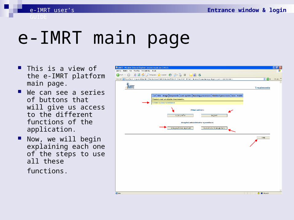
This is a view of the e-IMRT platform main page.
We can see a series of buttons that will give us access to the different functions of the application.
Now, we will begin explaining each one of the steps to use all
these functions.
e-IMRT user’s GUIDE Entrance window & login

Contents
Welcome window & login Commissioning Tomograph management Creating a new treatment Launching a verification process Launching a optimization process
e-IMRT user’s GUIDE

Commissioning of accelerators
e-IMRT user’s GUIDE Commissioning
The commissioning of an accelerator is the first step to perform before using the platform.
It consists in building a virtual accelerator that will reproduce its real behaviour (this is the physical properties and characteristics of the radiation beam which outputs the accelerator).
The aim is to reproduce in calculations certain measurements carried out in an accelerator.
This will assure that the result (in terms of dose) of any treatment calculated employing this virtual accelerator will be the same as the result of this treatment delivered to the patient with the real accelerator.

Accelerator management
To start the commissioning process, we must click in the button labelled as “Accelerator management”.
NOTE: The first time you launch an e-IMRT applet, a warning window will be shown. You have to accept to allow the applet storing temporary information on your hard disk.
e-IMRT user’s GUIDE Commissioning

Adding accelerators We will proceed to a new
window where we can select the accelerator which the hospital has and that we want to commission.
We will need to commission two virtual accelerators: one for optimization and another for verification. For each one of them we select the type of accelerator from among the available (in this case Siemens Primus with collimator 3D MLC).
Subsequently we have to give him an internal name (for example "Demo Siemens Verif" for verification and "Demo Siemens Optim" for optimization) and click in "Add accelerator to hospital".
e-IMRT user’s GUIDE Commissioning

Selecting algorithms In this applet we can see the
information of our accelerator.
Below, we can select the dose and commissioning algorithms.
For the verification accelerator, we will select the dose algorithm "BEAMnrc Monte Carlo" and the commissioning algorithm "BEAMnrc Monte Carlo Photon Beam Characterization".
For the optimization accelerator, we must select the dose algorithm "Monte Carlo C/S Optimization" and the commissioning algorithm with the same name.
e-IMRT user’s GUIDE Commissioning

Uploading data file Now we should upload the
data measured previously in the real accelerator.
For this purpose, first we must select the format of the file that contains the data (Mephisto, eIMRT or RFA300). In our case, we select Mephisto.
After that, we can select the measurements file ("Dummy Accelerator.exp") pressing “Browse...".
Clicking "Open", data is read and converted to internal format (eIMRT).
e-IMRT user’s GUIDE Commissioning

Data preview
When data is translated to internal format, we can visualize this data selecting a field size and clicking "View".
In a graphic we will see the PDD (Percentage Depth Dose) of that field.
In another graphic, we see the lateral profiles at different depths.
To execute the commissioning algorithm, we must click "Run".
e-IMRT user’s GUIDE Commissioning

Running
In the case of the commissioning of an accelerator for the verification tool, the process will last less than one minute.
Now, we should repeat the previous steps for the commissioning of the accelerator for the optimization tool.
e-IMRT user’s GUIDE Commissioning
Finished
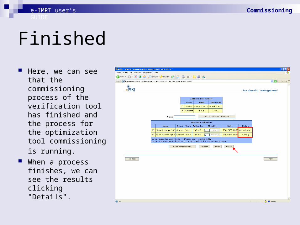
Here, we can see that the commissioning process of the verification tool has finished and the process for the optimization tool commissioning is
running. When a process
finishes, we can see the results clicking "Details".
e-IMRT user’s GUIDE Commissioning

Percent Depth Dose (PDD)
Here, we can see a new applet that will show us the measured and calculated PDD (one for each field size) normalized to a depth of 10 cm.
In the right side, we can see the percentage difference between measured and calculated points.
e-IMRT user’s GUIDE Commissioning

Lateral profiles Selecting the buttons "X
Profiles" or "Y Profiles", we can see the corresponding calculated and measured lateral profiles (for each field size) and its associated one-dimensional Gamma function.
If the value of this function in a point is <=1, this indicates that the measured data is within tolerance with the calculated data in that point.
We can change the tolerances. By defect, they are 2% in measured dose value and 2 mm in spatial separation.
e-IMRT GUIDE Commissioning

Validating accelerator (1)
If we agree with the obtained result, the following step is to validate the virtual accelerator.
For that we should click in "Validate".
e-IMRT user’s GUIDE Commissioning

Validating accelerator (2)
We can view again the obtained results. By clicking "Accept" we validate the accelerator.
If we do not agree with the results we must press in "Reject".
e-IMRT user’s GUIDE Commissioning

Ready!
Here, we can see the two validated accelerators.
Now, we can use them.
e-IMRT user’s GUIDE Commissioning

Contents
Welcome window & login Commissioning Tomograph management Creating a new treatment Launching a verification process Launching a optimization process
e-IMRT user’s GUIDE

Main window
The tomograph is the device used to imaging a patient by sections
The platform needs the values of the tomograph’s ramps to convert from Hounsfield Unities to densities before the calculations
To add a tomograph we must click in "Tomograph management" in the e-IMRT main window.
e-IMRT user’s GUIDE Tomograph management

Adding tomograph
Tomograph will translate CT (Computed Tomography) images of a patient in maps of electronic densities.
Clicking "Add tomograph" a new tomograph is created for which we will can define its parameters.
e-IMRT user’s GUIDE Tomograph management

Tomograph ramps
Now we should define the tomograph.
First, we have to give him a name.
Second, we have to indicate the equivalences HU (Hounsfield Units) vs density for each material.
These equivalences define the tomograph ramps.
This example shows a good definition, so we can click in “Add”.
e-IMRT user’s GUIDE Tomograph management
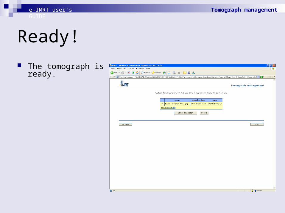
Ready!
The tomograph is ready.
e-IMRT user’s GUIDE Tomograph management

Contents
Welcome window & login Commissioning Tomograph management Creating a new treatment Launching a verification process Launching a optimization process
e-IMRT user’s GUIDE

Main window
We may click in “Create a new treatment" in the e-IMRT main window.
e-IMRT user’s GUIDE Creating a new treatment

Name & keywords
The first step to create a treatment is to give him a name.
Also we should assign him a series of keywords.
Additional information could be also added like patient Clinic history, detailed treatment explanation, etc
e-IMRT user’s GUIDE Creating a new treatment

Ready!
Finally, we may click in "Create" and the new treatment will be ready.
e-IMRT user’s GUIDE Creating a new treatment
Contents
Welcome window & login Commissioning Tomograph management Creating a new treatment Launching a verification process Launching a optimization process
e-IMRT user’s GUIDE

MC Treatment Verification Hospitals rely on commercial platforms, thoroughly tested and called
Treatment Planning Systems (TPS), to create treatments for their patients. As the amount of patients to treat is huge, dose calculation algorithms employed by TPS use limited accuracy to win calculation time.
In clinical routine, treatments created by commercial TPS must be verified experimentally (in a human-tissue-equivalent phantom) prior to its deliverance to the patient.
An alternative dose calculation method with higher accuracy would be extremely useful. Monte Carlo (MC) simulation fulfils the requirements in terms of precision but not in terms of speed. This fact may be bypassed by the use of vast computational resources (GRID)
Our verification tool may help radiophysicists at hospitals to replace or to complement experimental verifications, reducing accelerator use
e-IMRT user’s GUIDE Launching a verification process

Creating treatment
The first step is to create a new treatment.
We can see a list of treatments in the main window.
To launch a verification process we must select a treatment and click in "Treatment information".
e-IMRT user’s GUIDE Launching a verification process
Treatment information
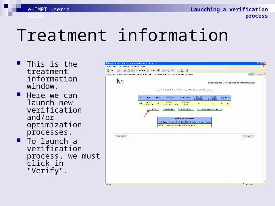
This is the treatment information window.
Here we can launch new verification and/or optimization processes.
To launch a verification process, we must click in "Verify".
e-IMRT user’s GUIDE Launching a verification process
Uploading Dicom files
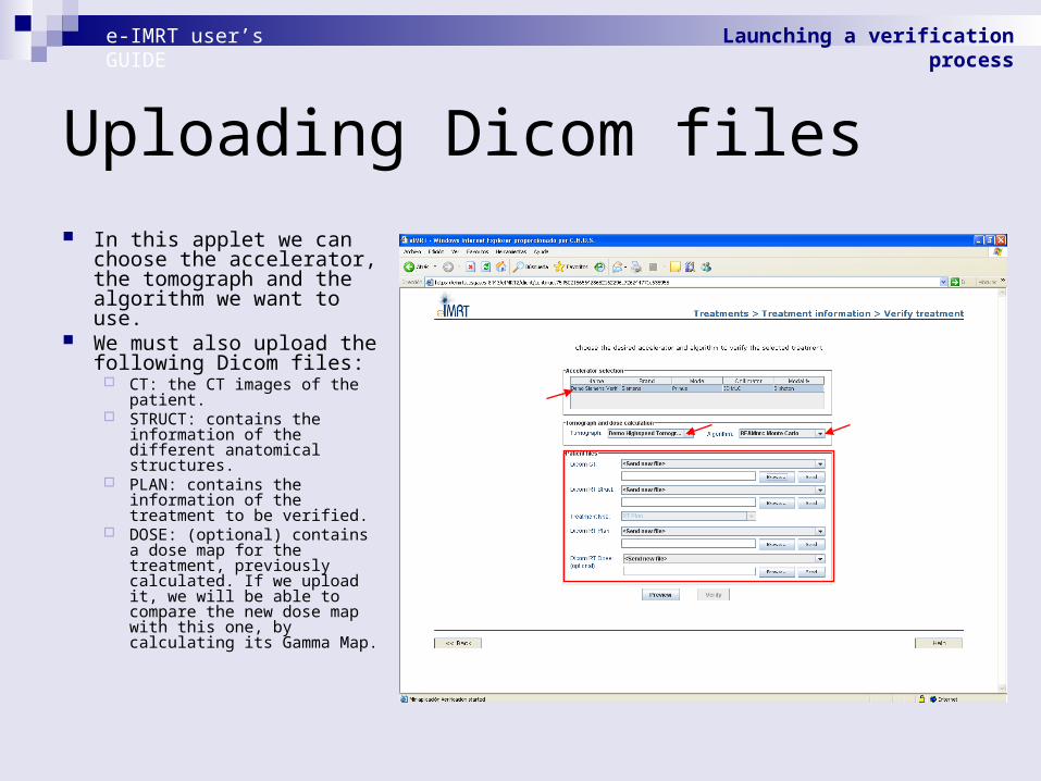
In this applet we can choose the accelerator, the tomograph and the algorithm we want to use.
We must also upload the following Dicom files:
CT: the CT images of the patient.
STRUCT: contains the information of the different anatomical structures.
PLAN: contains the information of the treatment to be verified.
DOSE: (optional) contains a dose map for the treatment, previously calculated. If we upload it, we will be able to compare the new dose map with this one, by calculating its Gamma Map.
e-IMRT user’s GUIDE Launching a verification process

Sending Dicom files
We select the corresponding Dicom files and we click in "Send".
The files suffer a process during which the private data of the patient are erased.
After this process, the files are saved in the server.
e-IMRT user’s GUIDE Launching a verification process
Preview
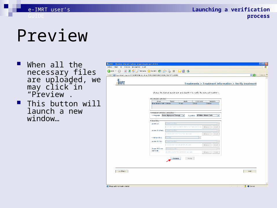
When all the necessary files are uploaded, we may click in “Preview”.
This button will launch a new window…
e-IMRT user’s GUIDE Launching a verification process
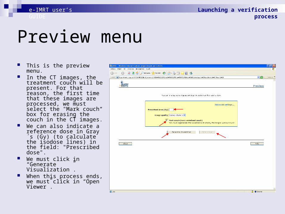
Preview menu This is the preview menu. In the CT images, the
treatment couch will be present. For that reason, the first time that these images are processed, we must select the "Mark couch“ box for erasing the couch in the CT images.
We can also indicate a reference dose in Gray´s (Gy) (to calculate the isodose lines) in the field: "Prescribed dose".
We must click in “Generate Visualization”.
When this process ends, we must click in “Open Viewer”.
e-IMRT user’s GUIDE Launching a verification process

Erasing couch (1)
This is the treatment viewer.
As we have selected the “Mark couch” box, we can see a red region over the CT image. We can move up or down the horizontal green bar until the red region is placed over the couch.
e-IMRT user’s GUIDE Launching a verification process

Erasing couch (2)
We have to be careful and do not cover any region within the body of the patient. It can be helpful to activate the box "Body“ in the “CONTOURS” list.
The selection shown in this example is valid. We will cut the images overlapped by the red film clicking "Mark couch".
e-IMRT user’s GUIDE Launching a verification process
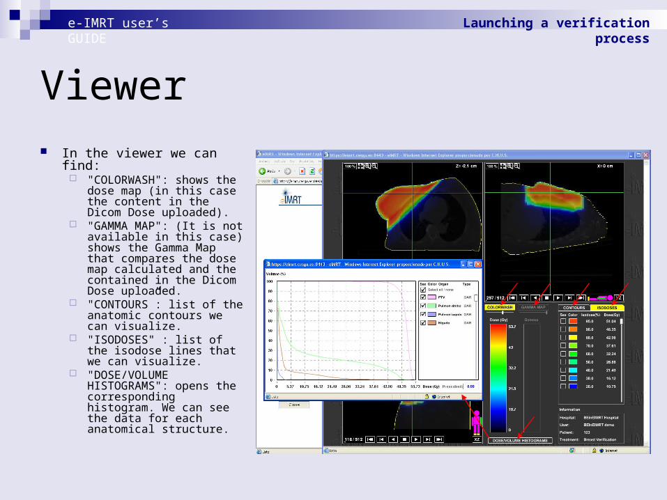
Viewer In the viewer we can find:
"COLORWASH": shows the dose map (in this case the content in the Dicom Dose uploaded).
"GAMMA MAP": (It is not available in this case) shows the Gamma Map that compares the dose map calculated and the contained in the Dicom Dose uploaded.
"CONTOURS”: list of the anatomic contours we can visualize.
"ISODOSES" : list of the isodose lines that we can visualize.
"DOSE/VOLUME HISTOGRAMS": opens the corresponding histogram. We can see the data for each anatomical structure.
e-IMRT user’s GUIDE Launching a verification process
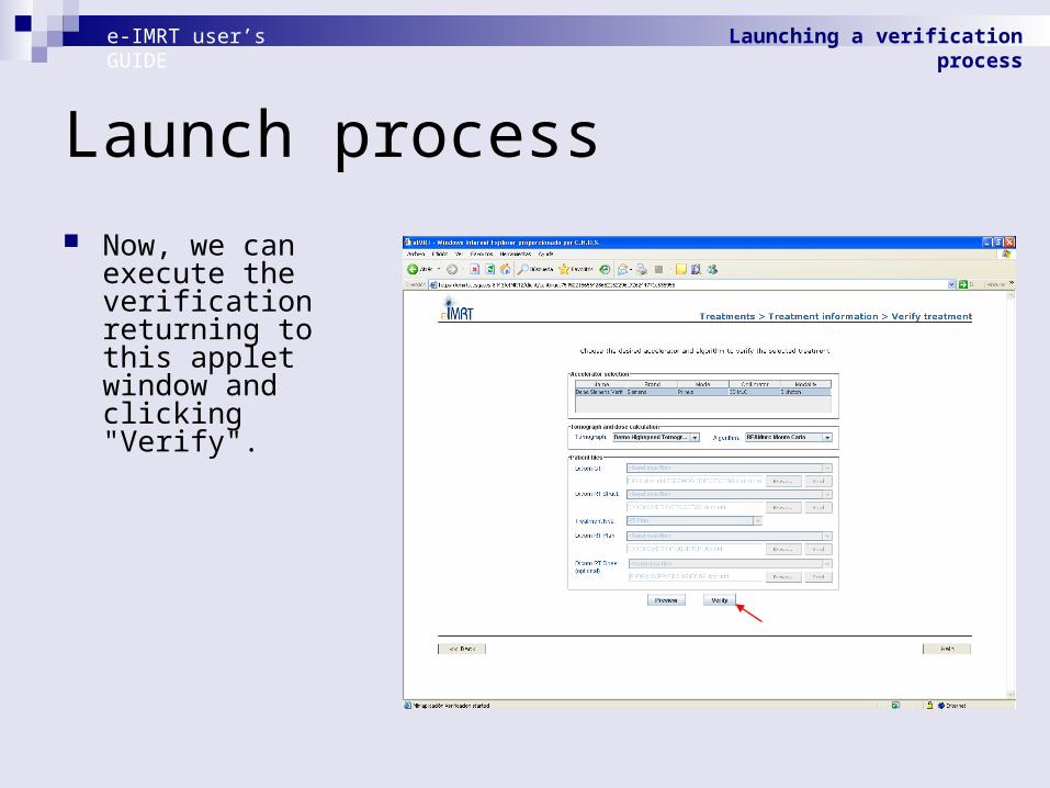
Launch process
Now, we can execute the verification returning to this applet window and clicking "Verify".
e-IMRT user’s GUIDE Launching a verification process
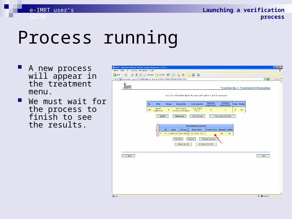
Process running
A new process will appear in the treatment menu.
We must wait for the process to finish to see the results.
e-IMRT user’s GUIDE Launching a verification process

Finished process
When the process has finished, we may click in "Show Results".
e-IMRT user’s GUIDE Launching a verification process
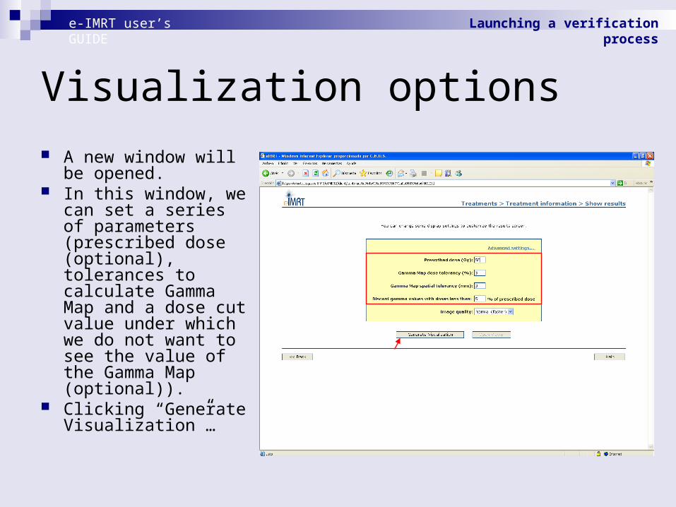
Visualization options
A new window will be opened.
In this window, we can set a series of parameters (prescribed dose (optional), tolerances to calculate Gamma Map and a dose cut value under which we do not want to see the value of the Gamma Map (optional)).
Clicking “Generate Visualization”…
e-IMRT user’s GUIDE Launching a verification process

Generating visualization
The visualization data will be generated (this process can take over 1 minute).
Now we may click in “Open viewer”…
e-IMRT user’s GUIDE Launching a verification process

Colorwash
In the viewer window, we will be able to activate all the options previously shown.
Here we can view the dose colorwash.
e-IMRT user’s GUIDE Launching a verification process
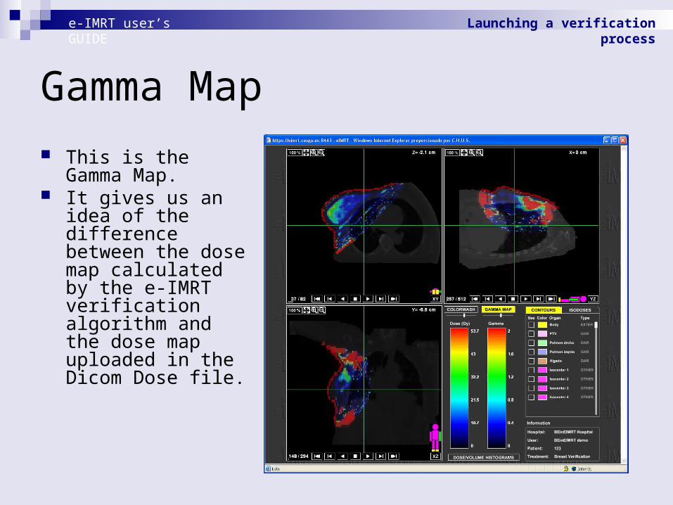
Gamma Map
This is the Gamma Map.
It gives us an idea of the difference between the dose map calculated by the e-IMRT verification algorithm and the dose map uploaded in the Dicom Dose file.
e-IMRT user’s GUIDE Launching a verification process
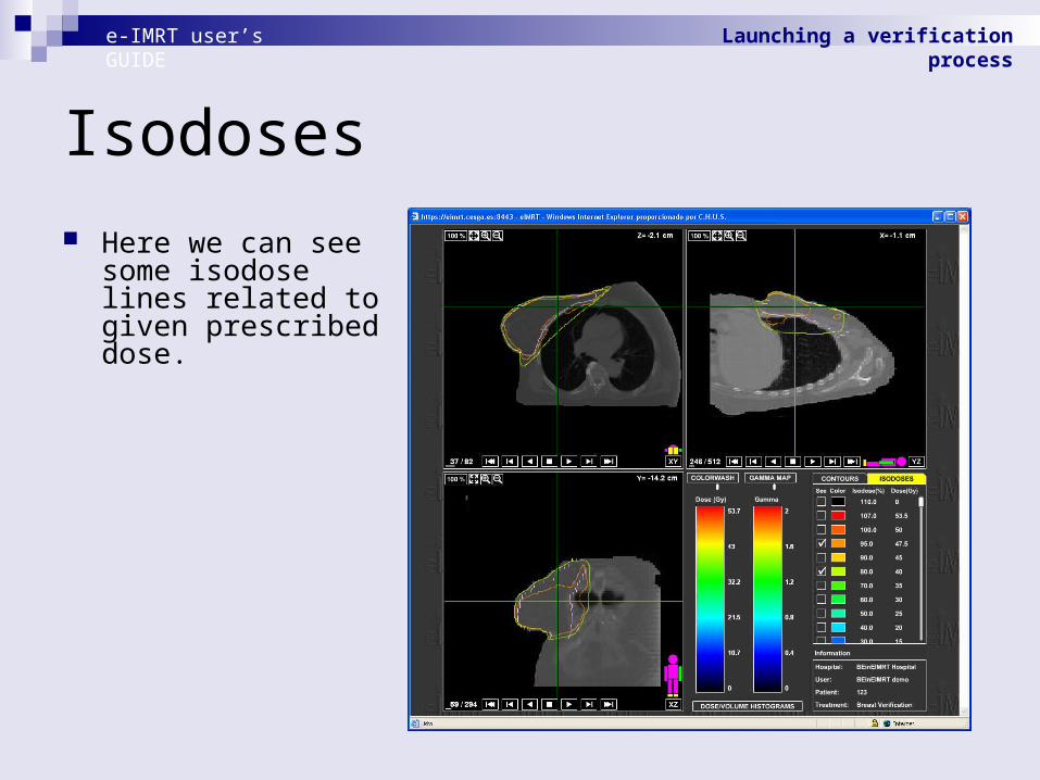
Isodoses
Here we can see some isodose lines related to given prescribed dose.
e-IMRT user’s GUIDE Launching a verification process

Dose-Volume Histogram
Here we can see a Dose-Volume Histogram.
e-IMRT user’s GUIDE Launching a verification process

Process viewed
If we close the viewer and return to the treatment information window, the process will be marked as viewed.
We can see again the results in the future clicking "Show Results"
e-IMRT user’s GUIDE Launching a verification process

Process viewed
If we close the viewer and return to the treatment information window, the process will be marked as viewed.
We can see again the results in the future clicking "Show Results"
e-IMRT user’s GUIDE Launching a verification process

Download DICOM RT Dose (1)
To download the resultant DICOM RT Dose press “Process DICOMs” to show the DICOMs associated with the Verification.
e-IMRT user’s GUIDE Launching a verification process

Download DICOM RT Dose (2)
The applet will show all the files of the process. You can download any of them pressing “Download” button.
To download the resultant DICOM RT Dose you must select it on Dicom RT Dose list and press Download. The name of the calculated DICOM starts with [OUT].
This file will be also available to use with a new Verification.
e-IMRT user’s GUIDE Launching a verification process

Contents
Welcome window & login Commissioning Tomograph management Creating a new treatment Launching a verification process Launching a optimization process
e-IMRT user’s GUIDE

Contents
Welcome window & login Commissioning Tomograph management Creating a new treatment Launching a verification process Launching a optimization process
e-IMRT user’s GUIDE
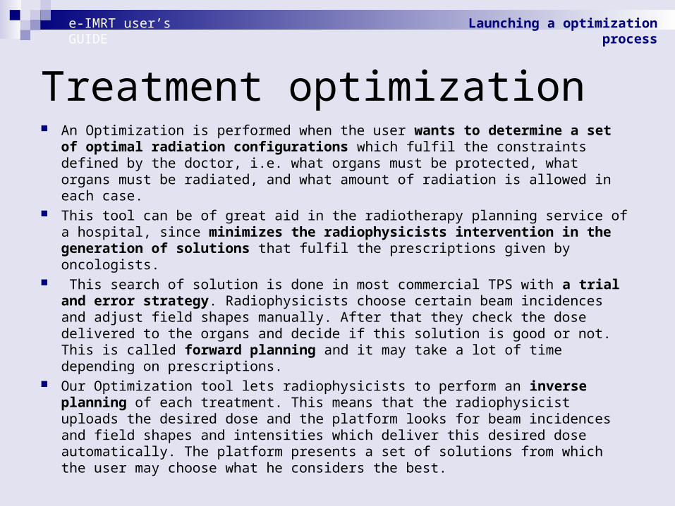
Treatment optimization An Optimization is performed when the user wants to determine a set of optimal
radiation configurations which fulfil the constraints defined by the doctor, i.e. what organs must be protected, what organs must be radiated, and what amount of radiation is allowed in each case.
This tool can be of great aid in the radiotherapy planning service of a hospital, since minimizes the radiophysicists intervention in the generation of solutions that fulfil the prescriptions given by oncologists.
This search of solution is done in most commercial TPS with a trial and error strategy. Radiophysicists choose certain beam incidences and adjust field shapes manually. After that they check the dose delivered to the organs and decide if this solution is good or not. This is called forward planning and it may take a lot of time depending on prescriptions.
Our Optimization tool lets radiophysicists to perform an inverse planning of each treatment. This means that the radiophysicist uploads the desired dose and the platform looks for beam incidences and field shapes and intensities which deliver this desired dose automatically. The platform presents a set of solutions from which the user may choose what he considers the best.
e-IMRT user’s GUIDE Launching a optimization process

Creating treatment
The first step is to create a new treatment or select one of the list and click in "Treatment information".
e-IMRT user’s GUIDE Launching a optimization process

Treatment information
This is the treatment information window.
To launch a optimization process, we must click in “Optimize".
e-IMRT user’s GUIDE Launching a optimization process

Uploading dicom files In this applet we must choose the
accelerator, the tomograph and the algorithms we want to use.
In this case, only the “CT” and “STRUCT” Dicom files are needed.
An optimization process will generate as output the information contained in the PLAN and DOSE Dicom files (we will be able to generate and download a PLAN Dicom file for each solution).
We can open the viewer (clicking "Preview") where we will be able to mark the couch (go to slides 35 and 36).
To continue with the process, we must click in "Prescript constraints".
e-IMRT user’s GUIDE Launching a optimization process

Prescriptions
In this applet, we can introduce the medical prescriptions.
Also, we can choose some parameters of the solutions that are going to be generated.
We can save our prescriptions in a xml file.
Also we can load the prescriptions from a xml file previously saved ...
e-IMRT user’s GUIDE Launching a optimization process
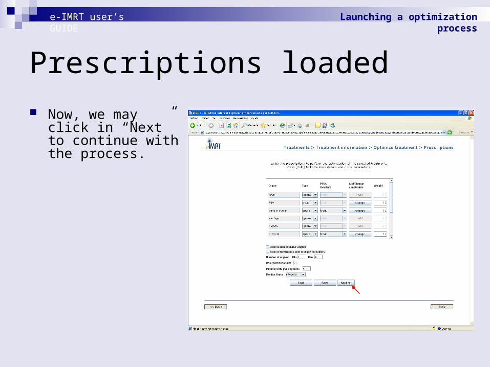
Prescriptions loaded
Now, we may click in “Next” to continue with the process.
e-IMRT user’s GUIDE Launching a optimization process

Summary
Here, we can see a summary of the prescriptions.
We can review them to find possible errors and, if it is necessary, return to the previous window to fix them.
To execute the optimization, we must click in "Optimize".
e-IMRT user’s GUIDE Launching a optimization process

Process running
A new process will appear in the treatment menu.
We must wait for the process to finish to see the results.
e-IMRT user’s GUIDE Launching a optimization process

Finished process
When the process finishes, we may click in "Show Results".
e-IMRT user’s GUIDE Launching a optimization process

Dose-Volume Histogram
A new window with a list of solutions will be opened (with information about its characteristics).
Selecting one of them we can view the DVH.
We can select the organs we want to see.
e-IMRT user’s GUIDE Launching a optimization process
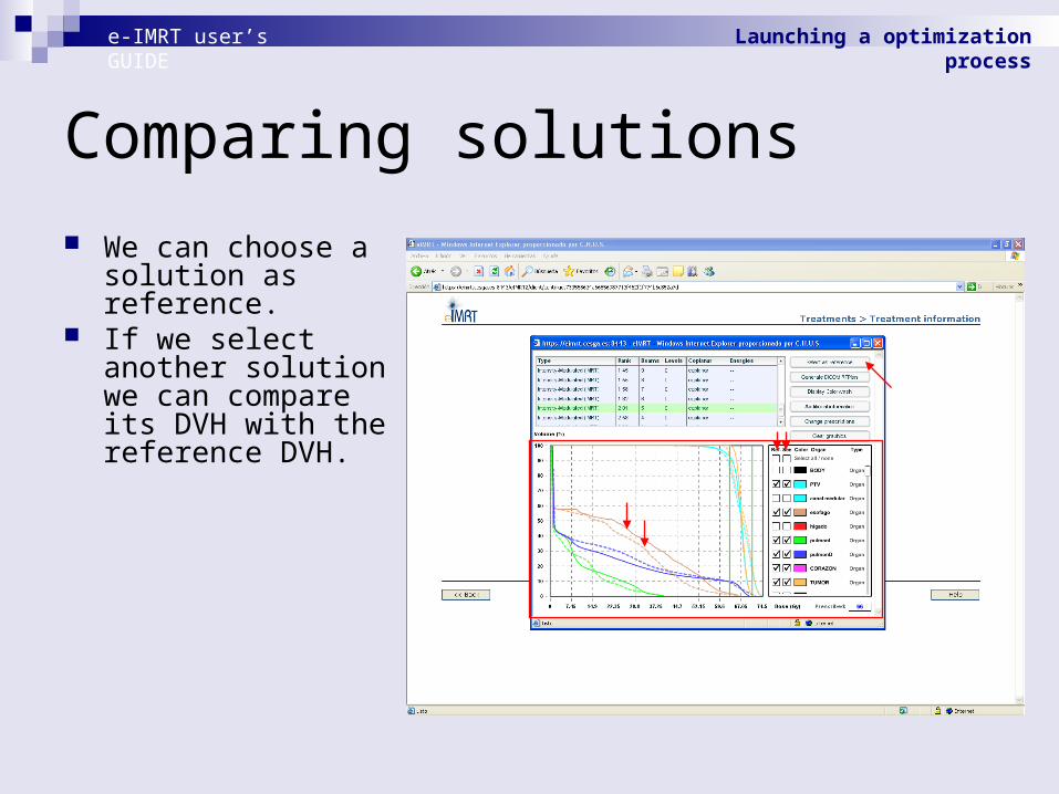
Comparing solutions
We can choose a solution as reference.
If we select another solution we can compare its DVH with the reference DVH.
e-IMRT user’s GUIDE Launching a optimization process

Additional information
We can open a new window with detailed information of each solution clicking “Additional information”.
e-IMRT user’s GUIDE Launching a optimization process

Display Colorwash (1)
Again, as with the verification results, we can open the viewer to display de dose map of each solution.
We must click in “Display Colorwash.
The visualization options window will be opened.
We can set the “Prescribed Dose” in Gy and click in “Generate Visualization”.
When the generation process finishes, we may click in “Open Viewer”…
e-IMRT user’s GUIDE Launching a optimization process

Display Colorwash
In the viewer, we can evaluate the solution.
e-IMRT user’s GUIDE Launching a optimization process

Download DICOM RT Plan (1)
If we find a good solution, we can generate and download the RTPlan in Dicom format clicking “Generate DICOM RTPlan”.
e-IMRT user’s GUIDE Launching a optimization process

Optimization DICOM files (1)
If we close the solutions window and return to the treatment information window, we can see that the optimization process is marked as viewed.
After generating the DICOM RT Plans, they keep stored on the platform. You can access easily to them by pressing “Process DICOMs” button.
e-IMRT user’s GUIDE Launching a optimization process

Optimization DICOM files (2)
e-IMRT user’s GUIDE Launching a optimization process
The applet will show all the files of the process. You can download any of them pressing “Download” button.
To download the resultant DICOM RT Plans you must select one on Dicom RT Plan list and press Download. The name of the generated DICOM starts with [OUT].
This files will be also available to use with a new Verification.

Finish visualization
If we close the solutions window and return to the treatment information window, we can see that the optimization process is marked as viewed.
e-IMRT user’s GUIDE Launching a optimization process

End
e-IMRT user’s GUIDE