ear, nose, and throat assessment in children · 8/2/2017 2 nasal anatomy the nose serves as an...
TRANSCRIPT

8/2/2017
1
Ear, Nose, and Throat Assessment in Children
Christina Landino
RN, BSN, CPN
Victoria Albano
MSN, CPNP
Contact Information:
Disclosure Agreement:We have no relevant financial or non-financial relationships
to disclose.
Overview
I. Nose and Sinus Assessment
a. Anatomy of the Nose and Sinus Cavities
b. Common Diagnoses of the Nose and Sinuses
II. Mouth and Throat Assessment
a. Anatomy of the Mouth and Throat
b. Common diagnoses of the Mouth and Throat
III. Ear Assessment
a. Anatomy of the Ear
b. Common Diagnoses of the Ear
IV. Hearing loss
a. Conductive vs. sensorineural
b. Music-induced hearing loss
c. Treatment

8/2/2017
2
Nasal Anatomy
The nose serves as an entrance to the respiratory system; acting to filter, warm, and moisten inhaled air.
Common Diagnoses of the Nose and Sinuses
•Rhinitis –allergic and non-allergic
•Epistaxis
•Nasal fracture
•Foreign body in the nasal cavity
•Sinusitis
Rhinitis
Irritation and inflammation of the nasal mucous membrane.
One or more of these symptoms present:
– Sneezing– Rhinorrhea (anterior and/or
posterior)– Nasal congestion – Nasal itching
Treatment is geared towardsthe underlying cause.
Persistent rhinitis may lead to nasal polyps, sinusitis, ear infections, and upper respiratory
infections.

8/2/2017
3
Allergic Rhinitis
• Present in 40 percent of children
• Symptoms:– “Allergic shiners”
– “Allergic salute”• nasal itching/rubbing helps
distinguish allergic rhinitis from most other forms
– Watery rhinorrhea
– Nasal congestion
– Sneezing
– Allergic conjunctivitis• 70 percent of cases
• red, itchy, watery eyes
Common allergens
• Seasonal rhinitis• tree, grass, weed pollens, outdoor
molds
• Perennial rhinitis• indoor allergens• house dust mites, cockroaches,
allergens from household fur bearing pets, rodents, and fungi
Allergic Rhinitis
“Allergic shiners” “Allergic salute”
Allergic Rhinitis
Allergic Rhinitis
Nasal mucosa classically appears edematous and pale
Normal Nasal Anatomy
Septum, turbinates

8/2/2017
4
Non-allergic Rhinitis
• Chronic presence of one or more of the following:
– nasal congestion
– rhinorrhea
– postnasal drainage
• Diagnosis of exclusion – rule out immune deficiency,
infectious diseases, structural abnormalities, etc.
• Nasal mucosa is usually normal in color
Clinically distinguished from allergic rhinitis by the following:
• Onset at a later age• Absence of nasal/ ocular itching and
prominent sneezing• Nasal congestion and postnasal
drainage are prominent symptoms• Symptoms are perennial
Typical triggers:• irritant odors and strong fragrances
(tobacco smoke, perfumes, diesel and car exhaust, cleaning products, newsprint)
• changes in temperature• alcoholic beverages
Rhinitis Algorithm
Epistaxis
Epistaxis (nosebleeds) occurs when the membranes and vessels of the nasal cavity are irritated.
CAUSES:
•Dry air, nose picking, colds, allergies, hard nose blowing, injury, high altitude, dry heat and various other irritants.
•Less common: Tumors (Juvenile nasopharyngeal angiofibroma)
Anterior vs. posterior bleed

8/2/2017
5
Epistaxis
MANAGEMENT:
1. Have the child sit up and lean forward
2. Pinch the nose firmly just below the nasal bones
• 5 minutes for children
• 10-15 minutes for teenagers/adults
3. Repeat step 2 if bleeding continues
4. Afrin administration
5. If all above measures fail Emergency room
Epistaxis
PREVENTION:• Warm or cool mist humidifier at night
• OTC nasal saline spray
• Nasal ointments
• Gentle nose blowing
• No nose picking, trim nails
• Avoid rough play and contact sports
• Avoid NSAIDS with severe bleeding
• Do not put tissues, gauze, cotton, tampons etc. in nose
Nasal Fracture
Nasal fractures affect both the bones and cartilage of the nose.
Approximately 40% of all facial injuries each year.
SYMPTOMS:-Nose pain-Swelling of the nose and face-Crooked or bent appearance of the nose -Possible nosebleed-Nasal obstruction-Bruised or blackened eyes

8/2/2017
6
MANAGEMENT PRIOR TO CONSULT: • Elevate nose, chin up
• Gently apply ice (cool gauze)
• Tylenol PRN pain (avoid motrin with bleeding)
• Call PCP or ORL for evaluation
– ORL exam needs to be within 5-7 days d/t swelling
– Closed reduction in clinic vs. in OR
Emergency room evaluation if:– severe trauma/facial lacerations
– clear drainage from nares; concern for skull fracture/CSF leak
– excessive bleeding
– concern for septal hematoma
Nasal Fracture
Foreign Body in Nasal Cavity
The most common objects: food material, paper, small toys, beads, and rocks.
SYMPTOMS:•Unilateral nasal drainage •Pain in, or trouble breathing from, the affected side of the nose•Anterior/posterior epistaxis•Foul odor from the nose•Infection
Emergency room evaluation if:1) The object moves to the back of the throat causing chocking or respiratory distress2) Object contains chemicals (i.e. batteries)
Foreign Body in Nasal Cavity

8/2/2017
7
Foreign Body in Nasal Cavity
Sinus Anatomy
Four sets of sinus cavities:• maxillary sinuses• ethmoid sinuses• sphenoid sinuses• frontal sinuses
Purpose:• humidify and heat entering air• cushion the skull in case of injury • to enhance our voices
Sinuses: a series of connected hollow cavities within the skull
Sinusitis
Definition:• The inflammation of the mucosal lining of
sinuses • Infections cause the normally air-filled sacs to
become filled with excess infected mucous– Bacterial, viral, fungal– Acute, chronic
Symptoms:• Nasal congestion and discharge• Cough• Headaches (pressure-like pain in the
general areas of the sinuses)• Fever – variable • Fatigue• Facial swelling
Symptoms:• Nasal congestion and discharge• Cough• Headaches (pressure-like pain in the
general areas of the sinuses)• Fever – variable • Fatigue• Facial swelling

8/2/2017
8
Sinusitis
Clinical presentation:
• Persistent symptoms • >10 days WITHOUT improvement
• Severe symptoms• fever >102.2• purulent nasal discharge x3
consecutive days• Worsening symptoms
• respiratory symptoms that worsen after initial improvement
• onset of new fever or severe headache
Treatment:
• Augmentin (first line) • Nasal saline spray/irrigations• Increase fluids• Rest• Decongestants/intranasal
corticosteroids • Recommended using with
underlying allergy component
Acute Bacterial Rhinosinusitis
Overview
I. Nose and Sinus Assessment
a. Anatomy of the Nose and Sinus Cavities
b. Common Diagnoses of the Nose and Sinuses
II. Mouth and Throat Assessment
a. Anatomy of the Mouth and Throat
b. Common diagnoses of the Mouth and Throat
III. Ear Assessment
a. Anatomy of the Ear
b. Common Diagnoses of the Ear
IV. Hearing loss
a. Conductive vs. sensorineural
b. Music-induced hearing loss
c. Treatment

8/2/2017
9
Mouth and Throat Anatomy
Common Diagnoses of the Mouth and Throat in Children
Pharyngitis
Tonsillitis Peritonsillar Abscess
Adenoiditis
Pharyngitis
Definition: Inflammation of the pharynx, “sore throat”
COMMON CAUSES:• Viral
• Adenoviruses, coxsackie A viruses, influenza
• Treatment: supportive measures
• Bacterial• Group A streptococcus (+
rapid strep)• Treatment: antibiotics
• Life threatening• Epiglottitis• Peritonsillar abscess Emergency Room
Symptoms • Painful swallowing• Redness in the throat• Fever• Headache• Enlarged tonsils

8/2/2017
10
Tonsillitis
Definition: inflammation of the tonsils
SYMPTOMS:• Sore throat • Tonsillar exudate• Difficulty swallowing• Fever and chills• Ear pain• Headache• Tenderness of jaw and throat• Cervical adenopathy
Treatment depends on culture results
Tonsillectomy considered for chronic infections
Adenoiditis
Definition: inflammation and swelling of the adenoids when they become overwhelmed by either bacterial or viral infections.
Symptoms include:• Sore throat• Swollen glands in the neck• Nasal congestion• Mouth breathing• Nasal speech • Difficulty sleeping, snoring, or sleep
apnea• Eustachian tube dysfunction • Otalgia (ear pain)
Treatment: antibiotics
Adenoidectomy considered for chronic infections
Obstructive Sleep Apnea (OSA)
Major risk factors• Tonsil/adenoid hypertrophy
– Influenced by genetic factors, infection, and inflammation• Moderate- severe obesity• Conditions that reduce upper airway size
– CP, Down Syndrome, Craniofacial anomalies, Neuromuscular disorders, etc.
Evaluation• Exam (oral- tonsils, nasal endoscopy- adenoids)• History• Sleep study
– Assesses breathing pauses, leg movement, oxygen, EEG
8/2/2017
11
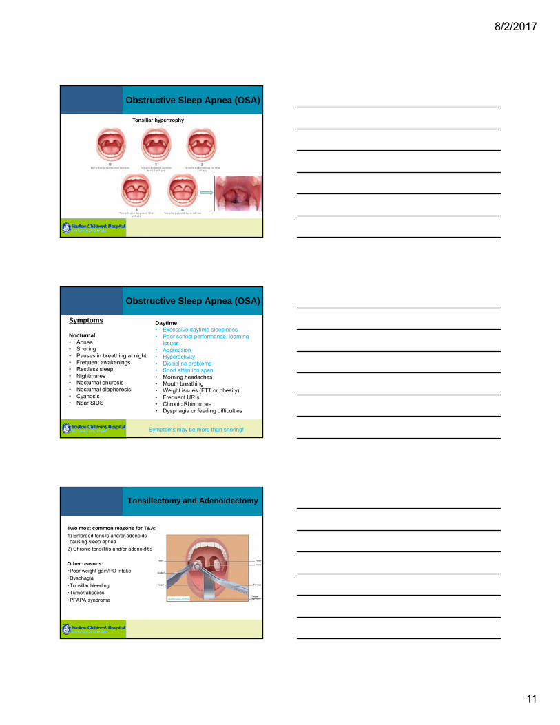
Obstructive Sleep Apnea (OSA)
Tonsillar hypertrophy
Obstructive Sleep Apnea (OSA)
Symptoms may be more than snoring!
Nocturnal• Apnea• Snoring• Pauses in breathing at night• Frequent awakenings• Restless sleep• Nightmares• Nocturnal enuresis• Nocturnal diaphoresis• Cyanosis• Near SIDS
Daytime• Excessive daytime sleepiness• Poor school performance, learning
issues• Aggression• Hyperactivity• Discipline problems• Short attention span• Morning headaches• Mouth breathing• Weight issues (FTT or obesity)• Frequent URIs• Chronic Rhinorrhea• Dysphagia or feeding difficulties
Symptoms
Tonsillectomy and Adenoidectomy
Two most common reasons for T&A:
1) Enlarged tonsils and/or adenoids causing sleep apnea
2) Chronic tonsillitis and/or adenoiditis
Other reasons:
• Poor weight gain/PO intake
• Dysphagia
• Tonsillar bleeding
• Tumor/abscess
• PFAPA syndrome

8/2/2017
12
Tonsillectomy and Adenoidectomy
Surgical Recovery
Tonsillectomy:•The recovery period is 14 days.•Post-operative symptoms include:
o Throat pain x2 weekso Painful swallowingo Bad breath due to scabbingo Ear paino Low grade fever (less than
101.5oF)o Cougho Nausea or vomiting (due to
anesthesia)
Adenoidectomy:•The recovery period is 3-5 days.•Post-operative symptoms include:
o Possible throat pain x3-5 dayso Bad breath due to scabbingo Low grade fever (less than
101.5oF)o Cougho Neck discomforto Mild changes to the tone of
child’s voice (hyponasal)o Rhinorrhea x2 weeks o Nausea or vomiting (due to
anesthesia)
Tonsillectomy and Adenoidectomy
Parents are instructed to seek medical attention if:
• Bleeding from mouth or nose
Emergency room
• Temperature over 101.5oF
• Dehydration– Poor PO intake
– Decreased urine output
– Persistent vomiting
• Decreased neck ROM
• Changes in child’s mental status or alertness
Tonsillectomy vs. Tonsillotomy
Tonsillectomy
• Removal of the entire palatine tonsil and the surrounding capsule
Tonsillotomy
• Typically done for diagnosis of sleep-disordered breathing, not recurrent infections.
• Devices are used to debulk the obstructing portions of the tonsil
• Some evidence suggests:– More rapid recovery compared with traditional tonsillectomy
– Reduces the risk of postoperative hemorrhage
– Disadvantages: there is a (small) risk of tonsillar regrowth

8/2/2017
13
Peritonsillar Abscess (PTA)
Definition: collection of pus located between the capsule of the palatine tonsil and the pharyngeal muscles.
SYMPTOMS:• Sore throat (may be severe and
one-sided)• Tender glands of jaw and throat• Difficulty opening mouth (trismus)• Difficulty swallowing (dysphagia)• Fever and chills• Headache• Drooling or facial swelling• Ear pain
TREATMENT:• Antibiotics (IV or PO)• Surgical drainage
If left untreated, PTA could lead to airway obstruction or cellulitis of jaw,
neck, or chest.
Overview
I. Nose and Sinus Assessment
a. Anatomy of the Nose and Sinus Cavities
b. Common Diagnoses of the Nose and Sinuses
II. Mouth and Throat Assessment
a. Anatomy of the Mouth and Throat
b. Common diagnoses of the Mouth and Throat
III. Ear Assessment
a. Anatomy of the Ear
b. Common Diagnoses of the Ear
IV. Hearing loss
a. Conductive vs. sensorineural
b. Music-induced hearing loss
c. Treatment
Ear Anatomy
Key Features of the Ear:
• External Auditory Canal• Tympanic Membrane• Middle Ear Space
• Malleus• Incus• Stapes Bones
• Inner Ear Space• Cochlea• Semicircular Canals
• Eustachian tube

8/2/2017
14
Common Diagnoses of the Ear in Children
•Otitis Media•Otitis Media with Effusion•Otitis Externa•Perforated Tympanic Membrane•Mastoiditis•Foreign Body in the Ear
Otitis Media (OM)
The most common ear-related illness affecting children.
COMMON CAUSES:• Primary eustachian tube
dysfunction (due to age)• Eustachian tube dysfunction
secondary to:• Craniofacial abnormalities• Allergies• Reflux
SYMPTOMS:• Signs of an acute infection
• Fever,• Otalgia (ear pain)• Irritability• Trouble sleeping
• Red, bulging tympanic membrane• Middle ear effusion and/or drainage
Otitis Media (OM)
• Otoscopy
Normal

8/2/2017
15
Otitis Media (OM)
Initial Management of Acute Otitis Media•Antibiotic Therapy vs. Observation
– The choice of strategy depends upon the age of the child and the laterality and severity of illness:
• Children <2 years with AOM are treated immediately with antibiotics
• Children >2 years who appear toxic; have persistent otalgia for more than 48 hours; have temperature ≥102.2°F (39°C) in the past 48 hours; have bilateral AOM or otorrhea; or have uncertain access to follow-up be immediately treated with an appropriate antibiotic.
• For children >2 years who are normal hosts (eg, immune competent, without craniofacial abnormalities) and have unilateral AOM with mild symptoms and no otorrhea, initial observation may be appropriate if the caretakers understand the risks and benefits of such an approach.
Long Term Management for Chronic Infections Tympanostomy Tubes
Otitis Media with Effusion (OME)
Otitis media with effusion occurs when the middle ear space is filled with fluid, but the fluid is not infected. It can follow an episode of OM or a URI.
• Symptoms do NOT usually include pain or fever, but symptoms DO include mild hearing loss and a feeling of fullness.
• Unlike OM, OME does not respond to antibiotic treatment! – Children typically observed x3 months, as OME will often resolve itself.
– If after 3-4 months fluid remains and there is evidence of hearing loss, a tympanostomy tube placement, adenoidectomy, or myringotomy may be considered.
Otitis Media with Effusion (OME)
• Otoscopy
Normal

8/2/2017
16
Tympanostomy Tubes
Tympanostomy tubes are small tubes that are surgically placed into the ear
drum to allow for drainage of fluid from, and the passage of air to, the
middle ear space.
Some complications include:•Infection with drainage•Persistent eardrum perforations after tubes fall out•Eardrum scarring•Cholesteatoma
INDICATIONS:• Eustachian tube dysfunction• Chronic OM • OME causing hearing loss or
speech delay
Tympanostomy Tube Insertion
Tube Insertion Video
Otorrhea with Tympanostomy Tubes
• The ear is draining now what?– Call PCP or ORL to
report
– Start ear drops (floxin, ciprodex, etc)
– Does not need to be on oral antibiotics unless the drainage is persistent despite drops
– Can be bloody!

8/2/2017
17
Otitis Externa (OE)
Otitis externa, more often referred to as “Swimmer’s Ear”, is the
inflammation of the external ear canal.
Symptoms include: Red, itchy, painful, or
swollen outer ears Drainage from the ear canal Swollen glands in the neck Conductive hearing loss
TREATMENT:• Ear drops • Debridement• Wick placement• Oral antibiotic
• Rare
CAUSES:• Bacterial or fungal infection
• Increased exposure to water• Warm humid areas• Harsh cleaning• Trauma • Foreign body in the ear canal
Perforated Tympanic Membrane
CAUSES:• Severe otitis media
• Acoustic trauma
• Barotrauma
• Foreign objects in the ear
• Injury
SYMPTOMS: Drainage from ear Hearing loss (mild or severe) Ear pain or discomfort Relief of significant ear pain during
acute ear infectionTREATMENT:• Ear drum will often heal itself.• If the eardrum does not heal itself,
myringoplasty or tympanoplasty may be necessary.
• Other treatment options for a perforated tympanic membrane are geared towards relieving pain and preventing infection.
• Perforations after ear tubes fall out is nonurgent, usually heals on its own within 2 months
Mastoiditis is an infection of the mastoid bone of the skull, often caused by the spread of
untreated otitis media infections.
SYMPTOMS:• Drainage from ear, accompanied by
ear pain or discomfort• Fever• Redness • Warmth• Swelling behind the ear• Hearing loss• Dizziness
Diagnostics: CT scan (rare), tympanocentesis for culture
Treatment: IV antibiotics (ceftriaxone), mastoidectomy for severe infections
Mastoiditis
*Send for ED evaluation as likely will need IV antibiotics

8/2/2017
18
Foreign Body in Ear
• May cause injury to ear canal or tympanic membrane.
• Some of the common objects found include: insects, food items, beads, toys, etc.
• Symptoms: pain otorrhea, infection
While most objects can be removed easily or by the child’s PCP or Otolaryngologist, some objects do require urgent removal or a trip to the emergency room, such as:• Small batteries• Food materials that may swell when
moistened• If an object is causing severe pain or
bleeding
Overview
I. Nose and Sinus Assessment
a. Anatomy of the Nose and Sinus Cavities
b. Common Diagnoses of the Nose and Sinuses
II. Mouth and Throat Assessment
a. Anatomy of the Mouth and Throat
b. Common diagnoses of the Mouth and Throat
III. Ear Assessment
a. Anatomy of the Ear
b. Common Diagnoses of the Ear
IV. Hearing loss
a. Conductive vs. sensorineural
b. Music-induced hearing loss
c. Treatment
Hearing Pathway

8/2/2017
19
Hearing Screens
• Newborn hearing screens
• MA screening regulation: 200.500:• (C) The school committee or board of health shall cause the hearing of each student in the public
schools to be screened in the year of school entry and annually through grade 3 (or by age nine in the case of ungraded classrooms), once in grades 6 through 8 (ages 12 through 14 in the case of ungraded classrooms), and once in grades 9 through 12 (ages 15 through 18 in the case of ungraded classrooms). The hearing of each student shall be tested by means-of some form of discrete frequency hearing test such as the Massachusetts Hearing Test or comparable method approved by the Department of Public Health.
• (D) Screenings of sight and hearing shall be performed by teachers, physicians, optometrists, nurses or others approved by the Department for this purpose, in accordance with guidelines of the Department.
• (E) For any student who does not pass a vision or hearing screening, a written plan shall be developed by the school nurse, in consultation to the extent possible with a student's parent or legal guardian, for appropriate follow up of the student. With the consent of the parent or legal guardian, the student's primary care provider shall be furnished with a copy of the record of screening tests performed in the school.
•
Hearing Loss
• CONDUCTIVE — Conductive losses imply a mechanical problem in the outer or middle ear: the pinna, external canal, tympanic membrane, or ossicles.
CAUSES:– Outer ear — Congenital anomalies, cerumen impaction, infections, and trauma.
– Middle ear — Infection, middle ear fluid, congenital anomalies, ossicular chain fixation, tympanic membrane perforation, or tumors.
• SENSORINEURAL — Disorders of the inner ear. Problems may occur at the level of the cochlea, eighth nerve, internal auditory canal, or brain.
CAUSES:– Congenital vs acquired
– Genetic
– Ototoxic drugs
– CMV
– Noise exposure
– Structural abnormalities
AC (AIR)
UNMASKED
MASKED
BC (BONE)
UNMASKED
MASKED
125 250 500 1000 2000 4000 8000750 1500 3000 6000
0
10
20
30
40
50
-10
60
70
80
90
100
110
120
FREQUENCY IN HERTZ (Hz)
HE
AR
ING
LE
VE
L (
HL
) IN
DE
CIB
EL
S (
dB
)
KEY
R L
SOUNDFIELD
S
SPEECH AUDIOMETRY
SDT
SRT
SPEECHDISCRIM.(WORDRECOG.)
R L
NORMAL HEARING ( -10-20 dBHL)
MILD LOSS (21-40 dBHL)
MODERATE LOSS (41-55 dBHL)
MODERATELY SEVERE LOSS (56-70 dBHL)
SEVERE LOSS (71-90 dBHL)
PROFOUND LOSS ( > 90 dBHL)
8/2/2017
20
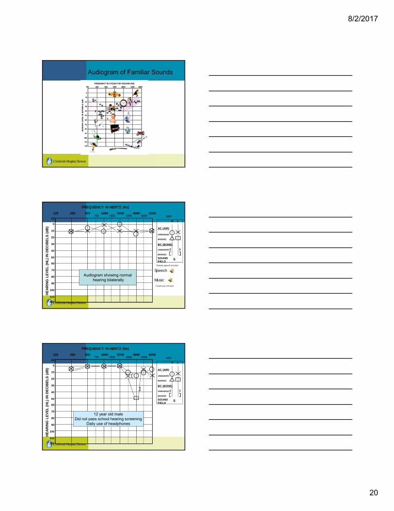
Audiogram of Familiar Sounds
AC (AIR)
UNMASKED
MASKED
BC (BONE)
UNMASKED
MASKED
125 250 500 1000 2000 4000 8000750 1500 3000 6000
0
10
20
30
40
50
60
70
80
90
100
110
120
FREQUENCY IN HERTZ (Hz)
HE
AR
ING
LE
VE
L (
HL
) IN
DE
CIB
EL
S (
dB
)
KEY
R L
SOUNDFIELD
S
-10
Audiogram showing normalhearing bilaterally
Speech
Music
Female speech nml.mp3
I found you nml.mp3
AC (AIR)
UNMASKED
MASKED
BC (BONE)
UNMASKED
MASKED
125 250 500 1000 2000 4000 8000750 1500 3000 6000
0
10
20
30
40
50
60
70
80
90
100
110
120
FREQUENCY IN HERTZ (Hz)
HE
AR
ING
LE
VE
L (
HL
) IN
DE
CIB
EL
S (
dB
)
KEY
R L
SOUNDFIELD
S
-10
12 year old maleDid not pass school hearing screening
Daily use of headphones

8/2/2017
21
Unilateral Hearing Loss
Bilateral Sudden SNHL
Risk for Music-Induced Hearing Loss
Hearing loss due to noise exposure can dislodge ossicles or permanently damage the hair cells that transmit sound in the cochlea.
Noise-induced hearing loss is cumulative - gradual and painless.
Signs a child may be developing hearing loss:
• Difficulty hearing or responding to commands
• Raising voice unnecessarily when talking
• Saying “what” frequently • Complaints of muffled sounds• Pain or tinnitus, especially after
exposure to loud noise

8/2/2017
22
Preventing Music-Induced Hearing Loss
Recommendations• Limit listening level to 60% of
maximum volume• Limit listening time to 1 hour a
day• Because in-ear earphones are
7-9 dB higher than over-the-ear at the same setting, shorter time use or lower level volume is necessary.
Hearing loss can be prevented by actively educating children
and families!
Avoiding loud noises, utilizing hearing protection such as earplugs or earmuffs, and
lowering the sound of appliances whenever possible.
Hearing loss in children: Treatment
Hearing aids•Provide sound amplification
•3 components: microphone, amplifier and speaker
• BTE – most appropriate for younger children
• CROS Aid
Hearing loss in children: Treatment
• FM Systems– Designed to improve hearing perception, especially in noisy
environments. They consist of a microphone for the speaker, a frequency modulated (FM) transmitter, and a receiver worn by the listener.
– Improves signal to noise ratio by eliminating background noise.
– Most FM assistive devices are used for educational purposes, but they can also help in other listening situations.
– Soundfield FM systems

8/2/2017
23
Hearing loss in children: Management
Bone conduction hearing devices• Device directly transmits sounds through the skull
• Indications: – Congenital ear canal atresia
– Allergic reaction to standard hearing aid
– Chronic infections of middle/outer ear
Hearing loss in children: Management
Cochlear implants • Surgically-implanted prosthetic devices that electrically stimulate the
cochlear nerve to provide hearing. – Battery-powered external processor (that looks like a hearing aid), a receiver coil implanted
below the scalp, and an electrode inserted directly into the cochlea through a surgical opening.
• Indications: – Severe to profound bilateral sensorineural hearing loss (SNHL)
– Little or no benefit from hearing aid use after six months
– Need to consider inner ear anatomy (imaging is needed)
ORL Urgencies
Refer to Emergency Room for:• Uncontrolled epistaxis
• Nasal septal hematoma after fracture
• Foreign body– Battery
– Airway occlusion
• Orbital cellulitis
• T&A post-operative bleeding
• Peritonsillar abscess
• Mastoiditis
• Sudden hearing loss

8/2/2017
24
Any questions?Thank you!
[email protected]@childrens.harvard.edu
Boston Children’s Hospital ORL nursing line:617-355-7147
http://www.childrenshospital.org/oto