early embryogenesis and...
TRANSCRIPT

HUMANHUMANEMBRYOLOGYEMBRYOLOGY
Department of Histology and EmbryologyDepartment of Histology and EmbryologyJilinJilin UniversityUniversity
Chapter 2 General Embryology
Second week:Implantation & bilaminar germ
Disc
Chapter 2 Chapter 2 General EmbryologyGeneral Embryology
Second week:Second week:Implantation & Implantation & bilaminarbilaminar germ germ
DiscDisc

human zygote → morula → blastocyst → moving towards uterine cavity →Zona pellucida disappears
Review: 1st-4th day of development

process in which blastocyst is embedded in endometrium of the uterustime: begin on 5th –6th day; finish on 11th –12th day place: fundus and body of uterus
5. Implantation/imbed
Seed
Soil
Root
Bud
plant
—— blastocyst
—— endometrium
—— placenta
—— embryo/fetus
—— infant
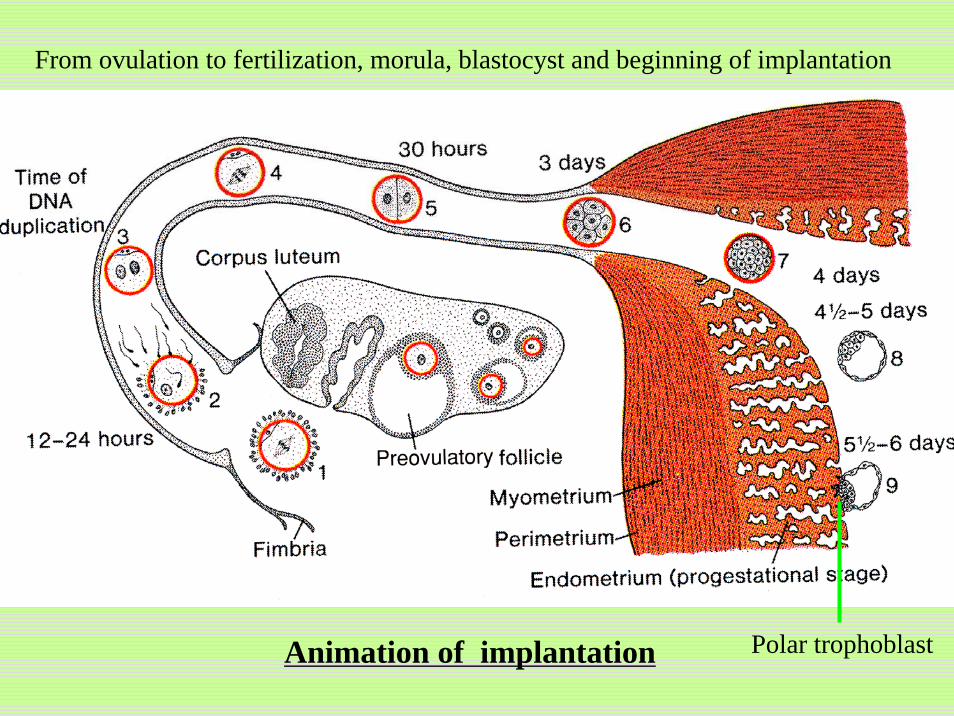
Animation of implantation
From ovulation to fertilization, morula, blastocyst and beginning of implantation
Polar trophoblast

5.1 Process of implantationDay 6
Polar trophoblastic cells penetrate endometrium. The endometrium is eroded by proteinase.Blastocyst begins to be immersed into the defect and be embedded into the endoemtrium.
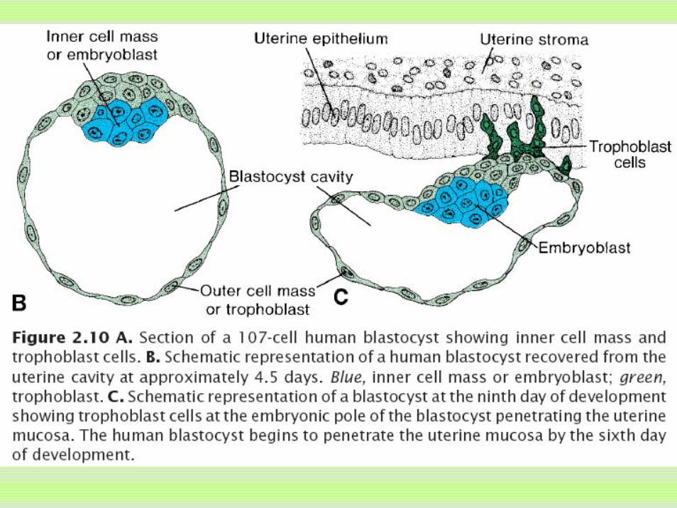

Day 8Blastocyst is partially embedded in endometrium. Trophoblast
outer syncytiotrophoblastinner cytotrophoblastCTs→STs
Embryoblasthigh columnar epiblastsmall cuboidal hypoblast
bilaminar germ disc primordium of human body.
amniotic cavity within the epiblast
endometrium
Blastocyst
endometriumamniotic cavity
blastocele
Superficial view
inside view
endometrium
blastocele
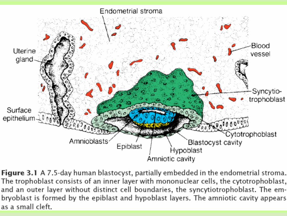

Day 9Blastocyst is deeply embedded in the endometrium.Defect in epithelium is closed by a fibrin coagulum.Vacuoles in trophoblast fuse to form large lacunae.Flattened cells from hypoblast form primitive yolk sac (exocoelomic cavity).
endometrium
amniotic cavity
fibrin coagulum
Primitive yolk sac
syncytiotrophoblast
cytotrophoblast
epiblast
hypoblast
Trophoblastic lacunea


Formation of bilaminar germ disc, amniotic cavity, primitive yolk sac,cytotrophoblast; syncytiotrophoblast from day 6 to day 9
blastocystendometrium
mucosa
hypoblast
epiblast
Polar trophoblast

Days 11 and 12Blastocyst is completely embedded in endometrium.surface epithelium almost entirely covers defect. Syncytiotrophoblast cells eroded maternal sinusoids, which become continuous with lacunae in syncytium, establishing uteroplacental circulation.
endometrium
amniotic cavity
fibrin coagulum
Primitive yolk sac
syncytiotrophoblast
cytotrophoblast
epiblast
hypoblast
Trophoblastic lacunea
Maternal sinusoid

endometrium
amniotic cavity
fibrin coagulum
Primitive yolk sac
syncytiotrophoblast
cytotrophoblast
epiblast
hypoblast
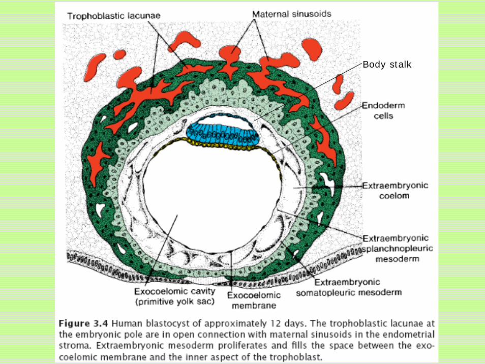
Extraembryonic mesoderm form and fills the space between cytotrophoblast, amnion and primitive yolk sac membrane. Extraembryonic coelom develops inside extraembryonic mesoderm.
Extraembryonic somatopleuric mesoderm lines cytotrophoblast and amnion Extraembryonic splanchnopleuric mesoderm lines yolk sac.
Germ disc is connected to trophoblast by body/connecting stalk.
Maternal sinusoid
Body stalk
SplanchnopleuricMesoderm
somatopleuricMesoderm
Extraembryoniccoelom
Body stalk

Day 13The defect in the endometrium has usually healed. Primary villi :cytotrophoblast penetrate into syncytiotrophoblastCells from hypoblast migrate along the inside of the primitive yolk sac membrane, forming secondary/definitive yolk sac.
amniotic cavity
Secondary yolk sac
syncytiotrophoblast
cytotrophoblast
epiblast
hypoblast
endometrium
Primary villous
Maternal sinusoid
trophoblast lacunea
Body stalk
Splanchnopleuricmesoderm
somatopleuricmesoderm
Extroembryoniccoelom
Degenerated primitive yolk sac


Implantation site at the end of the second week.
Body stalk
Splanchnopleuric mesoderm
somatopleuricmesoderm
Extraembryonic
coelom
syncytiotrophoblast
cytotrophoblast

uteroplacental circulation; Eextraembryonic mesodermExtraembryonic coelom ; secondary yolk sac
Formation of extraembryonic mesoderm and definitive yolk sac

5.2 Uterus at time of implantationMenstrual cycle
follicular/proliferative phaseSecretory/progestational phase
Coiled uterine glands and arteries, succulent tissuestroma cell → predecidual cell → decidual cell polyhedral and loaded with glycogen and lipids
menstrual phaseDecidua reaction: endometrium →decidua

Relationship between the embryo and the endometrium
Decidua capsularis
embryo
vagina
cervix
Uterine cavity
Decidua parietalis
Decidua basalis

Conditions of implantation:- endometrium is in secretory phase—— soil - morula reach uterine cavity on time—— spring- zona pellucide disappears in time—— covering coat
Almost all contraceptive measures are based on blocking these three conditions.
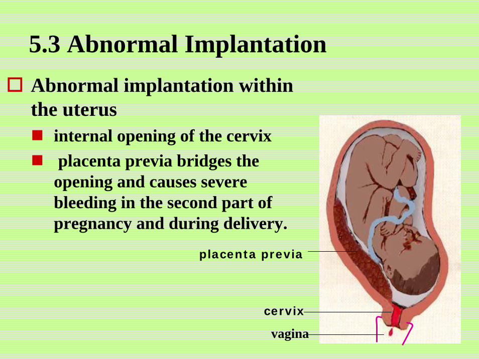
5.3 Abnormal ImplantationAbnormal implantation within the uterus
internal opening of the cervixplacenta previa bridges the opening and causes severe bleeding in the second part of pregnancy and during delivery.
placenta previa
cervix
vagina

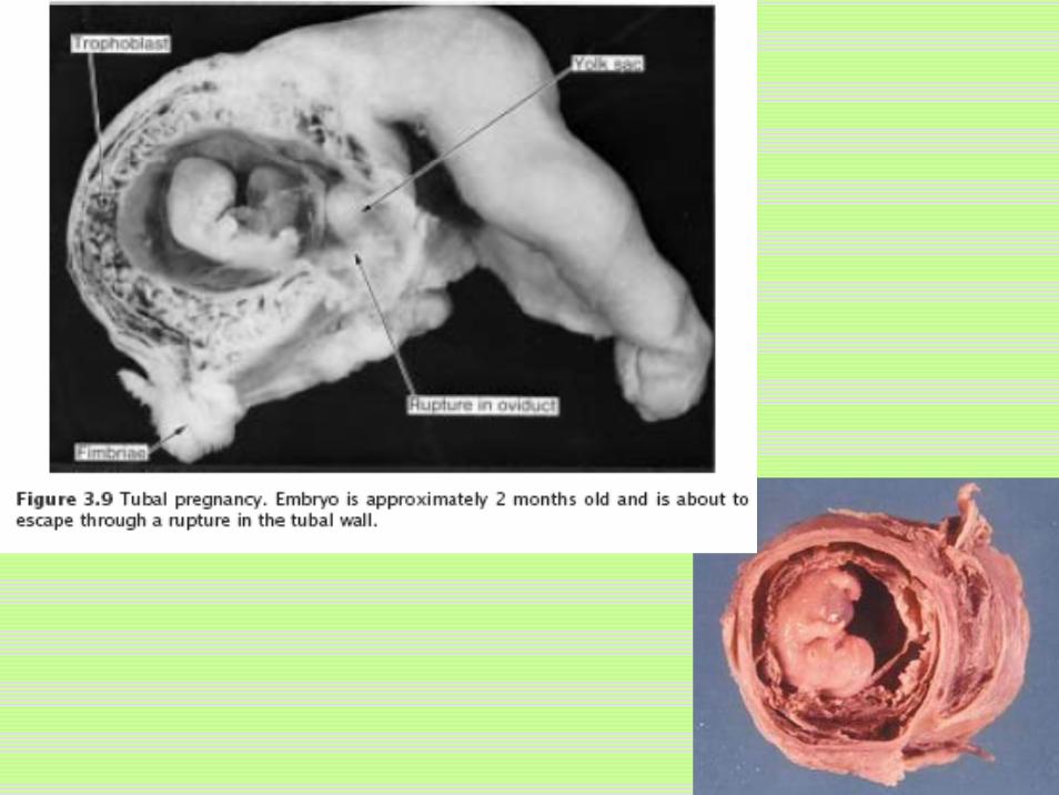
Extrauterine or ectopic pregnancyabdominal cavity, ovary, uterine tube, particularly ampullaEmbryo dies about the second month of gestation.

Chapter 2 General Embryology
Third week:Trilaminar germ disc
Chapter 2 Chapter 2 General EmbryologyGeneral Embryology
Third week:Third week:TrilaminarTrilaminar germ discgerm disc

The peacock is spreading its tail
sandwich

The third weekEpiblast form primitive streak at caudal midline.The cephalic end of the streak is primitive node. Primitive streak invaginate, forming primitive groove. Part of invaginated epiblast migrate into and displace hypoblast, creating embryonic endoderm. Others come to lie between epiblast and endoderm, forming mesoderm, which contacts with extraembryonicmesoderm.Cells remaining in the epiblast then form ectoderm.
6. Formation of trilaminar germ disc6.1 Formation of intraembryonic Mesoderm and Endoderm
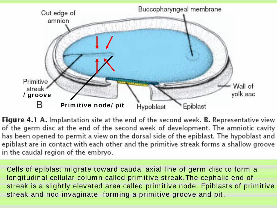
Cells of epiblast migrate toward caudal axial line of germ disc to form a longitudinal cellular column called primitive streak.The cephalic end of streak is a slightly elevated area called primitive node. Epiblasts of primitive streak and nod invaginate, forming a primitive groove and pit.
Primitive node/pit
/groove

Dorsal side of the germ disc from a 16-day embryo indicating the movement of surface epiblast cells (solid black lines) through the primitive streak and node and the subsequent migration of cells between the hypoblast and epiblast (broken lines).

Cross section through the cranial region of the streak at 15 days showing invagination of epiblast cells. The first cells to move inward displace the hypoblast to create the definitive endoderm. Once definitive endoderm is established, inwardly moving epiblast forms mesoderm.
extra-embryonic mesoderm

a sagittal section through the germ disc
Interembryonic mesoderm establishes contact with extraembryonicmesoderm covering the yolk sac and amnion. Cells remaining in the epiblast then form ectoderm.

epiblast
hypoblast
Primitive node
Primitive streak
cephalic
Caudal
Formation of intraembryonic mesoderm and notochord

Primitive streak disappears gradually.Sometimes, remnant of the primitive streak persists in sacrococcygeal region and form teratoma.

6.2 Formation of the NotochordPrenotochordal cells invaginating in the primitive pit move forward cephalad between epiblast and endoderm. The notochord extend cranially to the prechordal plate and caudally to the primitive pit. Notochord induces the formation of neural plate. Residual notochord degenerate to nucleus pulposus of intervertebral discs.

Drawing of a sagittal section through a 17-day embryo. The mostcranial portion of the definitive notochord has formed.
Schematic view showing the definitive notochord.
ectoderm
Primitive pit

Formation of notochord
The primitive streak shortens and disappears gradually following the formation of notochord.
hypoblastepiblast
Primitive node
Primitive streak

6.3 Formation of the allantoisThe cranial buccopharyngeal membrane
future opening of the oral cavity.The caudal cloacal membrane
future opening of reproductive tract and anus.
Primitive pitbuccopharyngeal membrane

allantois
body stalk
Posterior wall of the yolk sac forms allantoisextending into body stalk.

6.4 Growth of the Embryonic Disc
The flat and round embryonic disc gradually becomes elongated, with a broad cephalic and a narrow caudal end. Continuous migration of cells from primitive streak in cephalic direction. Thus formation of the germ layers continues in caudal segments while cranial structures are differentiating, causing the embryo to develop cephalocaudally.

6.5 Further Development of the Trophoblast
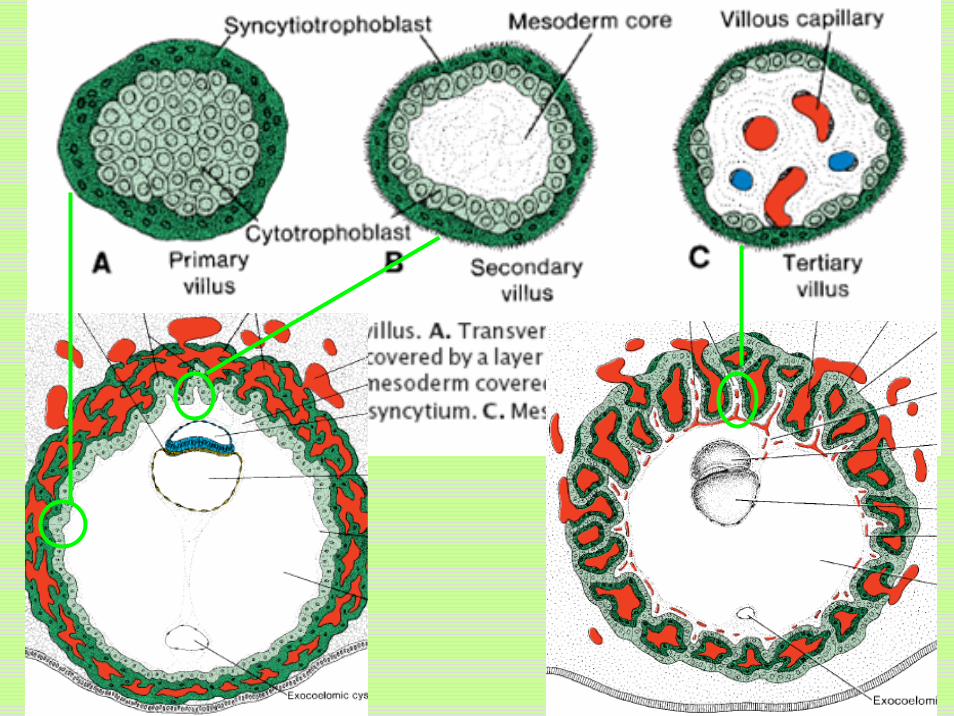
Third week:primary villus
cytotrophoblastic core covered syncytial layer. secondary villus
mesodermal cells penetrate primary villustertiary villus or definitive placental villus
mesodermal cells differentiate into blood cells and small blood vesselsCapillaries in tertiary villi contact with capillaries in mesoderm of the chorionic plate and in the connecting stalk.

Chorionic platetrophoblast and the extraembryonic mesoderm
Chorionchorionic plate and villi
Stem or anchoring villifrom chorionic plate to decidua basalis
Free (terminal) villibranch from the sides of stem villi
Cytotrophoblast shellsurrounding the trophoblast entirely and attaching the chorionic sac firmly to the decidua.
Intervillous lacuna filling with mother’s blood cells.The embryo is suspended in chorionic cavity by body stalk.

Stem/anchoring villus
Free/terminal villus
chorion


After 6 weeks, the villi penetrating the decidua basalis grow densely Villous chorion or chorion frondosum.Smooth chorion or chorion laeve.

Villous chorion Smooth chorion

Development of Chorion

Key pointsConcept, normal and abnormal site & phase of implantationStructure of deciduaStructure of two-layered embryo
epiblast and hypoblastStructure of three-layered embryo
ectoderm, mesoderm and endodermReason of sacrococcygeal teratomasFunction of notochordStructures of three types of villiConcept of chorionic plate, chorion, stem villus, free villus, cytotrophoblast shell, villous chorion, smooth chorion