early region 1 transforming functions are …jvi.asm.org/content/73/4/3071.full.pdf ·...
TRANSCRIPT

JOURNAL OF VIROLOGY,0022-538X/99/$04.0010
Apr. 1999, p. 3071–3079 Vol. 73, No. 4
Copyright © 1999, American Society for Microbiology. All Rights Reserved.
Early Region 1 Transforming Functions Are Dispensable forMammary Tumorigenesis by Human Adenovirus Type 9
DARBY L. THOMAS,1,2 SOOK SHIN,2† BERNARD H. JIANG,3‡ HANNES VOGEL,4
MARGERY A. ROSS,3§ MICHAEL KAPLITT,3\ THOMAS E. SHENK,3
AND RONALD T. JAVIER2*
Program in Cell and Molecular Biology,1 Division of Molecular Virology,2 and Department of Pathology,4
Baylor College of Medicine, Houston, Texas 77030, and Department of Molecular Biology,Howard Hughes Medical Institute, Princeton University, Princeton, New Jersey 085443
Received 3 December 1998/Accepted 4 January 1999
Some human adenoviruses are tumorigenic in rodents. Subgroup A and B human adenoviruses generallyinduce sarcomas in both male and female animals, and the gene products encoded within viral early region 1(E1 region) are both necessary and sufficient for this tumorigenicity. In contrast, subgroup D human adenovi-rus type 9 (Ad9) induces estrogen-dependent mammary tumors in female rats and requires the E4 region-en-coded ORF1 oncoprotein for its tumorigenicity. Considering the established importance of the viral E1 regionfor tumorigenesis by adenoviruses, we investigated whether this viral transcription unit is also necessary forAd9 to generate mammary tumors. The nucleotide sequence of the Ad9 E1 region indicated that the gene or-ganization and predicted E1A and E1B polypeptides of Ad9 are closely related to those of other human adeno-virus E1 regions. In addition, an Ad9 E1 region plasmid demonstrated focus-forming activity in both low-passage-number and established rat embryo fibroblasts, whereas a large deletion within either the E1A or E1Bgene of this plasmid diminished transforming activity. Surprisingly, we found that introducing the same trans-formation-inactivating E1A and E1B deletions into Ad9 results in mutant viruses that retain the ability to elicitmammary tumors in rats. These results are novel in showing that Ad9 represents a unique oncogenic adeno-virus in which the E4 region, rather than the E1 region, encodes the major oncogenic determinant in the rat.
Human adenoviruses cause primarily respiratory, gastroin-testinal, and eye infections in people and are divided into sixsubgroups (A to F) based upon several physical characteristics(25, 48). In rodents, however, the subgroup A and B adenovi-ruses are tumorigenic, eliciting undifferentiated sarcomas atthe site of viral inoculation in both male and female animals(22, 54). Although subgroup D adenoviruses are nononcogenicin hamsters (54), subgroup D human adenovirus type 9 (Ad9)elicits mammary tumors in rats (3, 4, 29). Three months aftersubcutaneous injection with Ad9, female rats develop exclu-sively estrogen-dependent mammary tumors, while male ratsfail to develop tumors of any kind. Tumors that form in thefemale rats are predominantly mammary fibroadenomas, themost common type of benign breast tumor found in youngwomen (29, 44).
For the subgroup A and B adenoviruses, the E1A and E1Bgene products encoded within the viral early region 1 (E1region) are both necessary and sufficient for oncogenic trans-formation of primary rodent cell cultures (22, 49, 51). Individ-ually, E1A is capable of immortalizing cells (26), whereas E1Bdisplays no transforming potential (55). Together, however,these viral genes cooperate to produce transformed cells (22).
The mechanism by which E1 region gene products transformcells can be attributed, in part, to their ability to inactivate thecellular tumor suppressor proteins pRB and p53 (48).
Unlike subgroup A and B adenoviruses, subgroup D Ad9requires the E4 region ORF1 oncoprotein to generate tumors(30, 32). Nevertheless, the facts that (i) E1A mRNA is ex-pressed in Ad9-induced mammary tumors (29) and (ii) theAd9 E1 and E4 regions together cooperate to induce focusformation in CREF cells (30) suggest that the viral E1 regionmay also be required for Ad9-induced mammary tumorigene-sis. To address this possibility, we constructed Ad9 mutantviruses containing transformation-defective E1A and E1Bgenes. Despite the critical role of the viral E1 region in onco-genesis by subgroup A and B adenoviruses, we present resultshere indicating that E1 region transforming functions are dis-pensable for Ad9 to induce mammary tumors in rats.
MATERIALS AND METHODS
Cell lines. Rat embryo fibroblasts (REFs) were cultured from 16-day Fisher ratembryos (Harlan Sprague-Dawley, Indianapolis, Ind.) by using standard methods(20). REF cultures, rat CREF (19) and 3Y1 cell lines (37), and human A549 and293 cell lines (2, 23) were maintained in culture medium (Dulbecco’s modifiedEagle medium supplemented with 20 mg of gentamicin per ml and 6 or 10% fetalbovine serum) under a 5% CO2 atmosphere at 37°C.
Nucleotide sequence analyses and plasmid construction. Plasmids pUC19-Ad9[0-7.5] and pSP72-Ad9[7.5-12.5] containing Ad9 DNA sequences from 0 to7.5 and 7.5 to 12.5 map units (m.u.), respectively, were used to determine thenucleotide sequence of the Ad9 E1 region.
A DNA fragment (0 to 12.5 m.u.) containing the Ad9 E1 region was insertedinto the KpnI and BglII sites of plasmid pSP72 (Promega) to make pAd9E1.Deletions within the Ad9 E1A and E1B genes were first introduced into pUC19-Ad9[0-7.5] by removing the Ad9 SacI-BspEI fragment (nucleotides [nt] 542 to1049) and the Ad9 NaeI-ClaI fragment (nt 1609 to 2495), respectively. These twodeletion mutations were subsequently transferred to pAd9E1 within the Ad9BamHI-EcoRI fragment (0 to 7.5 m.u.), resulting in pAd9E1(DE1A) andpAd9E1(DE1B), respectively. The presence of the correct deletion in each mu-tant plasmid was verified by restriction enzyme and limited sequence analyses.
* Corresponding author. Mailing address: Division of MolecularVirology, Baylor College of Medicine, One Baylor Plaza, Houston, TX77030. Phone: (713) 798-3898. Fax: (713) 798-3586. E-mail: [email protected].
† Present address: Department of Genetics, The Salk Institute, LaJolla, CA 92037.
‡ Present address: St. Joseph Mercy Hospital, Ann Arbor, MI 48106.§ Present address: Department of Biochemistry, University of Con-
necticut Health Center, Farmington, CT 06032.\ Present address: Department of Neurosurgery, New York Hospi-
tal-Cornell University Medical College, New York, NY 10021.
3071
on Septem
ber 8, 2018 by guesthttp://jvi.asm
.org/D
ownloaded from
3072 THOMAS ET AL. J. VIROL.
on Septem
ber 8, 2018 by guesthttp://jvi.asm
.org/D
ownloaded from
Construction of adenovirus mutants. Ad9 mutant viruses having the sameE1A and E1B gene deletions described above for plasmids pAd9E1(DE1A) andpAd9E1(DE1B) were generated. Briefly, the full-length Ad9 genome (0 to100 m.u.) consists of three EcoRI fragments: A (7.5 to 95 m.u.), B (0 to 7.5 m.u.),and C (95 to 100 m.u.). Deletions were first introduced into the Ad9 EcoRI Bfragment of a plasmid, pAd9-EcoRI(B1C), which contains properly orientedterminal Ad9 EcoRI B and C fragments but lacks the intervening Ad9 EcoRI Afragment. Full-length mutant Ad9 genomes were subsequently assembled byinserting a virion-derived Ad9 EcoRI A fragment in the correct orientation at theunique EcoRI site of mutant pAd9-EcoRI(B1C) plasmids. The resulting infec-tious pAd9-EcoRI(A1B1C) plasmids were digested with SpeI to release intactlinear viral genomes, which were transfected into 293 cells to complement ex-pected E1 region deficiencies of the mutant viruses (2, 23). Recovered viruseswere amplified and titrated in 293 cells (31, 48).
Isolation of RNA and Northern blot analyses. Total RNA was isolated frommock-infected or Ad9-infected A549 cells (multiplicity of infection of 10; 9 hpostinfection). Cells were washed with ice-cold phosphate-buffered saline(4.3 mM Na2HPO4, 1.4 mM KH2PO4, 137 mM NaCl, 2.7 mM KCl) and lysed inguanidinium solution (4 M guanidinium isothiocyanate, 20 mM sodium acetate[pH 5.2], 0.1 mM dithiothreitol, and 0.5% [wt/vol] Sarkosyl) (12). The resultinglysate was drawn through a 20-gauge needle to shear cellular DNA, layered ontoa 5.7 M CsCl cushion, and centrifuged at 150,000 3 g for 18 h. The RNA pelletwas dissolved in TES buffer (1 mM Tris-HCl [pH 7.5], 2.5 mM EDTA, 1%[wt/vol] sodium dodecyl sulfate [SDS]), precipitated with ethanol, and resus-pended in water.
For Northern blot analyses, total RNA was separated on a formaldehydeagarose gel and transferred to a nitrocellulose membrane (13). The membranewas preincubated in hybridization buffer (0.5 M Na2HPO4 [pH 7.2], 1 mMEDTA, 7% [wt/vol] SDS) at 65°C for 4 h and then incubated in hybridizationbuffer containing a radiolabeled DNA probe (4.3 3 106 cpm/ml) at 65°C for 16 h.E1A and E1B probes, derived from Ad9 E1 region DNA fragments SacI-SphI (nt542 to 1473) and NaeI-EcoRI (nt 1609 to 2563), respectively, were radiolabeledby the random priming method (17) and purified by gel filtration on NICKcolumns (Pharmacia). Probed membranes were washed in SSC wash buffer (45mM NaCl, 4.5 mM sodium citrate, 0.1% [wt/vol] SDS) at 65°C.
Isolation of virion and cellular DNA. For isolation of adenovirus virion DNA,293 cells were infected at a multiplicity of infection of 10 and, at 72 h postinfec-tion, were harvested and lysed in lysis buffer (55 mM Tris-HCl [pH 9.0], 0.5 mMEDTA, 0.2% [wt/vol] sodium deoxycholate, 10% [vol/vol] ethanol, 0.5 mMspermine-HCl). Cell lysates were cleared by centrifugation, treated with protein-ase K solution (0.75% [wt/vol] SDS, 12.5 mM EDTA, 2.5 mg of proteinase K perml) at 37°C for 1 h, and extracted with phenol and chloroform. Virion DNA wasprecipitated with ethanol and resuspended in water.
For isolation of cellular DNA, 400 mg of frozen tumor tissue was ground in aliquid nitrogen-chilled mortar and pestle. The resulting frozen tumor powder wassuspended in 4.8 ml of digestion buffer (10 mM Tris-HCl [pH 8.0], 100 mM NaCl,25 mM EDTA, 0.5% [wt/vol] SDS, 0.1 mg of proteinase K per ml), incubated at50°C for 16 h, and extracted with phenol (52). Cellular DNA was precipitatedwith ethanol and resuspended in TE buffer (10 mM Tris-HCl [pH 7.4], 1 mMEDTA).
PCR analyses. For PCR amplification of cDNAs (reverse transcription-PCRanalysis), 2 mg of total RNA was reverse transcribed with Moloney murineleukemia virus reverse transcriptase, using random hexamers, as suggested by themanufacturer (Gibco-BRL). Ad9 E1A cDNAs were PCR amplified with Taqpolymerase (Promega) by using E1A primers 1 (nt 551 to 570; 59 CTC CTG CAGTCC CAG AGA CCG AGA AAA AT 39) and 2 (nt 1430 to 1411; 59 CTC AAG
CTT AAG CGC ACG TGC GTC TAG TT 39). PstI and HindIII sites (under-lined) engineered within the E1A oligonucleotides allowed PCR products to beinserted at the same sites of plasmid ds56rII6HI (1) for sequencing. Portions ofthe Ad9 E1A and E1B genes and the entire Ad9 E4 ORF1 gene were PCRamplified from tumor DNAs, using the following oligonucleotide pairs: E1Aprimers a (nt 487 to 513; 59 CCA GTC GAG TCC GTC AAG AGG CCA CTC39) and b (nt 1487 to 1461; 59 CCA CAC CTT GCA TGC GTC ACA TAG AC39); E1B primers c (nt 1584 to 1609; 59 ATC CTT GCA GAC TTT AGC AAGACA CG 39) and d (nt 2651 to 2628; 59 CAT GCA GGG TCA TCT GGC TGTTGG 39); and Ad9 E4 ORF1 primers 1 (59 ATG GCT GAA TCT CTG TATGCT TTC 39) and 2 (59-CAT GGT TAG TAG AGA TGA GAG TCT GAA 39).For E1A and E1B nested PCRs, DNA products derived from each of the firstPCR amplifications described above were extracted with phenol, precipitatedwith ethanol, and resuspended in water. One-twentieth of each sample wassubjected to a second round of PCR amplification using the following oligonu-cleotide pairs: E1A primers e (nt 726 to 746; 59 CCC ATG ATG ACG ACC CTAACG 39) and b; and E1B primers c and f (nt 2116 to 2094; 59 CAA TCC AGCTCC TCT TCC GAC GG 39).
Immunoprecipitation and immunoblot analyses. Immunoprecipitations andimmunoblot analyses were performed as described previously (32). Briefly, fro-zen tumor powder, generated as described above for the isolation of cellularDNA, was suspended in ice-cold radioimmunoprecipitation assay buffer (50 mMTris-HCl [pH 8.0], 150 mM NaCl, 0.1% [wt/vol] SDS, 1% [vol/vol] Nonidet P-40,0.5% [wt/vol] deoxycholate) containing protease inhibitors (2 mg of aprotinin, 2mg of leupeptin, and 100 mg of phenylmethylsulfonyl fluoride per ml), sonicatedbriefly, and cleared by centrifugation (16,000 3 g, 10 min). The protein concen-tration of tumor lysates was determined by the method of Bradford (9). Threemilligrams of protein from tumor lysates was subjected to immunoprecipitationwith 15 ml of Ad9 E4 ORF1 antiserum prebound to 30 ml of protein A-Sepharosebeads (Pharmacia) (32). Beads were washed with ice-cold radioimmunoprecipi-tation assay buffer and boiled in 23 sample buffer (0.13 M Tris-HCl [pH 6.8], 4%[wt/vol] SDS, 20% [vol/vol] glycerol, 2% [vol/vol] b-mercaptoethanol, 0.003%[wt/vol] bromophenol blue). Proteins were separated by SDS-polyacrylamide gelelectrophoresis (40) and electrophoretically transferred to a polyvinylidene di-fluoride membrane, which was blocked in TBST (50 mM Tris-HCl [pH 7.5], 200mM NaCl, 0.1% [vol/vol] Tween 20) containing 5% (wt/vol) both nonfat dry milkand bovine serum albumin. In these assays, Ad9 E4 ORF1 antiserum (1:5,000 inTBST) (32) and horseradish peroxidase-conjugated goat anti-rabbit immuno-globulin G (1:5,000 in TBST; Southern Biotechnology Associates) were used asprimary and secondary antibodies, respectively. After extensive washing withTBST, the membrane was developed by enhanced chemiluminescence (Pierce).
Focus assays. Plasmid DNA purified by CsCl density gradient centrifugationwas transfected onto 50% confluent tertiary REF cultures or CREF cells on100-mm-diameter dishes, using the calcium phosphate precipitation method witha glycerol shock (38). At 72 h posttransfection, REF and CREF cells werepassaged 1:3 and maintained in culture medium containing 10 and 6% filteredfetal bovine serum, respectively. Four to six weeks posttransfection, cells werefixed in methanol and stained with Giemsa to quantify transformed foci (32).
Mammary tumorigenicity of viruses in rats. Female rats with 1- or 2-day-oldlitters were obtained from Harlan Sprague-Dawley; 12 to 24 h after arrival,newborn rats were injected subcutaneously with 0.4 ml of virus solution on theiranterior flanks, using a 26-gauge needle. Beginning 2 months postinfection,animals were examined weekly by palpation for the presence of tumors, until theexperiment was terminated at 8 months postinfection. At this time, animals wereeuthanized, and portions of tumors were removed and either fixed in 10%formalin for histological examination or frozen at 280°C for isolation of DNA or
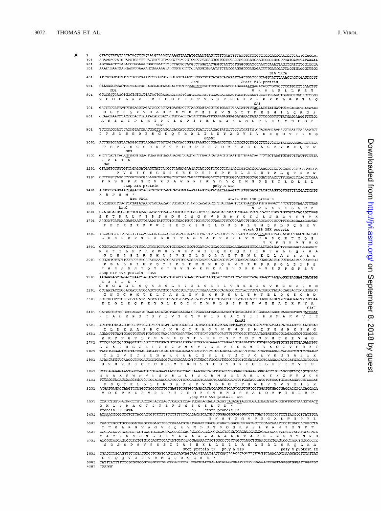
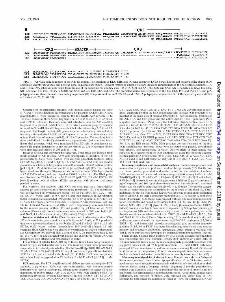
FIG. 1. (A) Nucleotide sequence of the Ad9 E1 region. The locations of E1A, E1B, and IX gene promoter TATA boxes, known and putative splice donor (SD)and splice acceptor (SA) sites, and poly(A) signal sequences are shown. Relevant restriction enzyme sites are indicated (underlined) on the nucleotide sequence. E1Aand E1B mRNA splice variants result from the use of the following SD and SA sites: 10S E1A, SD1 and SA1 plus SD2 and SA2; 12S E1A, SD2 and SA2; 13S E1A,SD3 and SA2; 13S E1B, SD4A or SD4B and SA3; and 22S E1B, SD5 and SA3. The predicted amino acid sequences of the 13S E1A, 19K and 55K E1B, and pIXpolypeptides are shown beneath their coding sequences. (B) Comparison of the Ad9 13S, 12S, and 10S E1A polypeptide sequences. CR1, CR2, spacer region, and CR3are indicated (22, 33, 48, 53).
VOL. 73, 1999 Ad9 TUMORIGENESIS DOES NOT REQUIRE THE E1 REGION 3073
on Septem
ber 8, 2018 by guesthttp://jvi.asm
.org/D
ownloaded from

protein. Animals were cared for and handled according to institutional guide-lines.
Protein sequence alignments. Sequences of Ad12, Ad7, Ad5, and Ad40 E1region polypeptides were obtained from GenBank. Alignments were made byusing the Pairwise Sequence Alignment program (ALIGN) of the BCM SearchLauncher (50).
Nucleotide sequence accession number. The nucleotide and polypeptide se-quences reported in this paper were submitted to GenBank (accession no.AF099665).
RESULTS
Gene organization and predicted polypeptides of the Ad9 E1region. To initiate our characterization of the subgroup D Ad9E1 region, we determined the sequence of the left 4006 nt ofthe Ad9 genome. From this analysis, we found that the geneorganization of the Ad9 E1 region closely resembles that ofother human adenovirus E1 regions (Fig. 1A) (48). In addition,the predicted Ad9 13S E1A, 19K and 55K E1B, and pIXproteins displayed significant sequence similarity with the cor-responding proteins from other human adenoviruses, althoughthey were most closely related to the E1 region polypeptides ofsubgroup B adenoviruses (Table 1).
Northern blot analyses of total cellular RNA isolated fromAd9-infected A549 cells were also performed to detect Ad9E1A and E1B mRNAs. In these assays, a diffuse E1A mRNAband migrating at approximately 1 kb and distinct 1.2- and2.2-kb E1B mRNA bands were observed (Fig. 2). The size ofthe E1A mRNA band was consistent with that predicted forthe 12S and 13S transcripts (see below), and the two E1BmRNAs corresponded well with the sizes predicted for 13S and22S transcripts (47).
Because E1A but not E1B mRNA is detected in Ad9-in-duced rat mammary tumors (29), we determined the structuresof Ad9 E1A transcripts by using reverse transcription-PCRtechniques on total RNA from Ad9-infected A549 cells. Se-quencing of PCR products obtained from these analyses re-vealed three Ad9 E1A splice-variant transcripts resembling13S, 12S, and 10S mRNAs from other human adenoviruses(Fig. 1A) (47). Ad9 E1A mRNA having a size consistent withthat expected for the 10S mRNA was not detected by Northernblot analyses (Fig. 2), presumably due to its low abundance atearly times after infection. The 13S, 12S, and 10S Ad9 E1AcDNAs are predicted to encode 251-, 189-, and 133-amino-acid-residue polypeptides, respectively (Fig. 1B). Between con-served regions 2 (CR2) and 3 (CR3), the 13S E1A protein ofsubgroup A virus Ad12 possesses an alanine-rich spacer regionwhich is, in part, responsible for the highly oncogenic pheno-type of this virus (33, 53). In contrast, the Ad9 13S E1A proteinwas found to contain a non-alanine-rich spacer region similarto the one present in 13S E1A proteins of weakly oncogenicsubgroup B adenoviruses (Fig. 1B).
A large deletion within the E1A or E1B gene abolishes focus-forming activity by the Ad9 E1 region. To investigate the trans-
forming potential of the Ad9 E1 region, we constructed an Ad9E1 region (0 to 12.5 m.u.) plasmid, pAd9E1, and examined itsability to induce transformed foci on low-passage-number REFcultures. Unlike other adenovirus E1 regions, the Ad9 E1region is unable to transform primary REF or baby rat kidneycell cultures (28). Consistent with these previous findings,pAd9E1 alone failed to generate transformed foci on REFs(Table 2). Nevertheless, whereas an activated ras plasmidalone also lacked detectable focus-forming activity on REFs,pAd9E1 and the activated ras plasmid together cooperated toproduce transformed foci on these cells (Table 2). To deter-mine whether Ad9 E1A and E1B gene functions were requiredfor this cooperation, we introduced a large deletion into eachof these genes within pAd9E1. A segment of the E1A gene cod-ing for the initiation codon, conserved region 1 (CR1), CR2,and half of CR3 (48) was removed in plasmid pAd9E1(DE1A),and E1B gene coding sequences downstream of E1B-19Kamino acid residue 14, as well as the first 208 amino acidresidues of the 495-residue E1B-55K protein, were removed inplasmid pAd9E1(DE1B) (Fig. 3). Each deletion would be an-ticipated to inactivate the transforming potential of the rele-vant gene (21, 22, 57). When cotransfected with the activatedras plasmid, pAd9E1(DE1A) failed to generate any foci onREFs, whereas pAd9E1(DE1B) retained significant focus-forming activity, albeit at a reduced efficiency compared towild-type pAd9E1 (Table 2). These results are concordant withprevious results showing that activated ras cooperates with theAd5 E1A but not the E1B gene (18). Therefore, our findingsprovided evidence that the transforming potential of the E1Agene is inactivated in pAd9E1(DE1A); however, it was un-
FIG. 2. Northern blot analyses of Ad9 E1A and E1B mRNAs. Total RNA(23 mg), isolated from Ad9-infected (9 h postinfection) or mock-infected A549cells, was separated on a formaldehyde agarose gel, transferred to a nitrocellu-lose membrane, and hybridized to either an E1A or E1B 32P-labeled DNA probe.RNA bands were visualized by autoradiography. Locations of 28S and 18S rRNAsare indicated. The indicated Ad9 mRNA species are predicted from their sizes.
TABLE 1. Amino acid sequence identities between subgroups A to D and F adenovirus E1A, E1B, and pIX proteinsa
VirusE1A 13S E1B 19K E1B 55K pIX
Ad12 Ad7 Ad5 Ad9 Ad40 Ad12 Ad7 Ad5 Ad9 Ad40 Ad12 Ad7 Ad5 Ad9 Ad40 Ad12 Ad7 Ad5 Ad9 Ad40
Ad12 100 100 100 100Ad7 41.9 100 41.6 100 47.9 100 53.7 100Ad5 39.9 37.5 100 43.2 48.1 100 48.6 53.8 100 49.3 49.6 100Ad9 39.3 43.1 38.1 100 43.2 52.4 47.6 100 45.3 56.2 52.2 100 50.0 58.2 46.5 100Ad40 38.4 38.5 33.7 40.1 100 48.5 45.5 43.6 41.8 100 55.9 48.3 48.6 44.5 100 62.5 51.7 49.3 49.6 100
a Sequence identities were determined with the full-length sequence of each polypeptide by using the ALIGN program (50). Ad12, subgroup A; Ad7, subgroup B;Ad5, subgroup C; Ad9, subgroup D; Ad40, subgroup F. Boldface values indicate amino acid sequence comparisons between Ad9 E1 region polypeptides and other E1region polypeptides; values in italics show that all Ad9 E1 region polypeptides are most closely related to those of subgroup B virus Ad7.
3074 THOMAS ET AL. J. VIROL.
on Septem
ber 8, 2018 by guesthttp://jvi.asm
.org/D
ownloaded from

clear from these REF assays whether the deletion in pAd9E1(DE1B) similarly affects the transforming potential of the E1Bgene.
In an attempt to reveal more striking transforming deficien-cies for pAd9E1(DE1B), we next performed focus assays in theestablished REF cell line CREF (19). Contrary to results ob-tained in REFs, transfection of pAd9E1 alone into CREF cellsled to the formation of numerous transformed foci (Table 2).The fact that a plasmid containing Ad9 sequences from 0 to17.5 m.u. exhibits weaker transforming activity in CREF cells(30) may indicate that Ad9 sequences from 12.5 to 17.5 m.u.interfere with focus formation in these cells. More important,when transfected individually into CREF cells, both pAd9E1(DE1A) and pAd9E1(DE1B) displayed significantly impairedfocus-forming activity compared to wild-type pAd9E1 (Ta-ble 2). Cotransfection of pAd9E1(DE1A) and pAd9E1(DE1B)into CREF cells, however, resulted in a moderate number oftransformed foci, revealing cooperation between the func-tional E1A and E1B genes retained collectively in the twoplasmids. Taken together, the results obtained for low-passage-number REFs and the cell line CREF showed that the dele-
tions within pAd9E1(DE1A) and pAd9E1(DE1B) greatly di-minish the transforming activity of the Ad9 E1A and E1Bgenes, respectively.
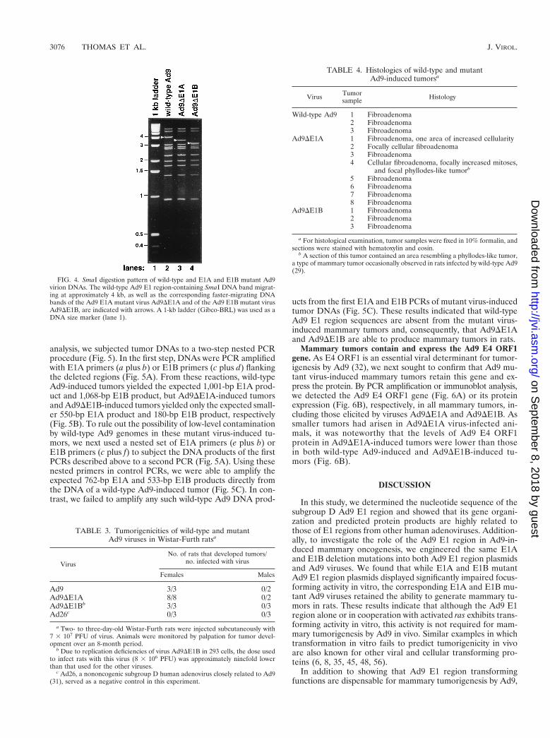
Isolation of Ad9 E1A or E1B deletion mutant viruses. Theimportance of the Ad9 E1 region in mammary oncogenesis wasassessed by introducing the same E1A and E1B deletion mu-tations of pAd9E1(DE1A) and pAd9E1(DE1B) into infectiousAd9 plasmids for recovery of mutant viruses. To complementtheir E1 region deficiencies, we transfected each of the mutantviral DNAs into human 293 cells, which stably express Ad5 E1region proteins (2, 23). In 293 cells, the E1A mutant virusAd9DE1A replicated to titers comparable to those of wild-typeAd9, whereas the E1B mutant virus Ad9DE1B replicated totiters approximately 10-fold lower. Because wild-type Ad9 failsto complement the replication defects of the Ad5 E1B-55Kmutant dl252 (28), the reduced replication of Ad9DE1B con-versely may be due to it being poorly complemented by Ad5E1B proteins expressed in 293 cells. Restriction enzyme anal-yses of virion DNA verified that Ad9DE1A and Ad9DE1Bcontained the expected deletions and further showed thatthese viruses had not acquired Ad5 E1 region sequences fromthe 293 cells (Fig. 4).
Ad9 E1A and E1B mutant viruses retain the ability to elicitmammary tumors in rats. We next tested the ability of mutantviruses Ad9DE1A and Ad9DE1B to generate mammary tu-mors in Wistar-Furth rats. In accordance with our previousresults (29, 30), wild-type Ad9 elicited mammary tumors in allof the female rats but none of the male rats, whereas subgroupD Ad26 failed to elicit tumors in any animals (Table 3). Sig-nificantly, we found that both Ad9DE1A and Ad9DE1B re-tained the ability to generate mammary tumors in female rats,despite the fact that Ad9DE1B-infected animals received aninefold-lower dose of virus than did animals infected witheither wild-type Ad9 or Ad9DE1A (Table 3). The tumorigenicphenotype of Ad9DE1B may not be surprising, consideringthat, unlike E1A mRNA, E1B mRNA is not detected in Ad9-induced mammary tumors (29). Furthermore, although mam-mary tumors elicited by all of the viruses were histologicallyidentical (Table 4), the tumors produced by Ad9DE1A weregenerally smaller than those induced by either wild-type Ad9or Ad9DE1B, both of which generated tumors of similar size(data not shown).
Mutant Ad9 virus-induced tumors do not contain wild-typeAd9 E1 region sequences. Because retention of tumorigenicityby both Ad9DE1A and Ad9DE1B was unanticipated, it wasimportant to demonstrate that the mammary tumors causedby these viruses do not contain wild-type Ad9 DNA. For this
TABLE 2. Focus formation by wild-type and mutant Ad9E1 region plasmids on low-passage-number REF
cultures and the CREF cell linea
Plasmid(s)
No. of transformed foci/2 100-mm-diam dishes
REF culturesb CREF cell linec
2ras 1rasExpt 1 Expt 2
Expt 1 Expt 2 Expt 1 Expt 2
pSP72 0 0 0 0 0 1pAd9E1 0 0 24 53 71 116pAd9E1(DE1A) 0 0 0 0 2 3pAd9E1(DE1B) 0 0 12 14 0 0pAd9E1(DE1A) 1
pAd9E1(DE1B)NDd ND ND ND 36 45
a 50% confluent tertiary REF or CREF cells on 100-mm-diameter dishes weretransfected with the indicated plasmid(s). At 72 h posttransfection, cells werepassaged 1:3 and then maintained in culture medium. REF and CREF cells werefixed in methanol and stained with Giemsa at 4 and 6 weeks posttransfection,respectively, to quantify the number of transformed foci.
b 15 mg of the indicated Ad9 E1 region plasmid plus 5 mg of empty pSP72(2ras) or 5 mg of pSP72-ras (1ras) plasmid (18) were transfected into REF cells.
c 10 mg of the indicated Ad9 E1 region plasmid plus 10 mg of empty pSP72plasmid were transfected into CREF cells. For the pAd9E1(DE1A)-plus-pAd9E1(DE1B) cotransfection, 10 mg of each plasmid was used.
d ND, not determined.
FIG. 3. Illustration of E1A and E1B deletion mutations introduced into the Ad9 E1 region of plasmid pAd9E1. Wild-type Ad9 sequences are represented by a blackline; deleted sequences are represented by a hatched line. The restriction enzyme sites used to generate the deletions are shown. The locations of E1A CR1, CR2, andCR3 (22, 48) are also indicated. ITR, inverted terminal repeat.
VOL. 73, 1999 Ad9 TUMORIGENESIS DOES NOT REQUIRE THE E1 REGION 3075
on Septem
ber 8, 2018 by guesthttp://jvi.asm
.org/D
ownloaded from
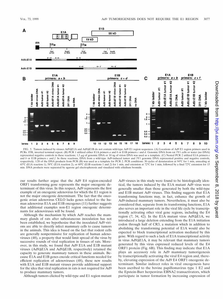
analysis, we subjected tumor DNAs to a two-step nested PCRprocedure (Fig. 5). In the first step, DNAs were PCR amplifiedwith E1A primers (a plus b) or E1B primers (c plus d) flankingthe deleted regions (Fig. 5A). From these reactions, wild-typeAd9-induced tumors yielded the expected 1,001-bp E1A prod-uct and 1,068-bp E1B product, but Ad9DE1A-induced tumorsand Ad9DE1B-induced tumors yielded only the expected small-er 550-bp E1A product and 180-bp E1B product, respectively(Fig. 5B). To rule out the possibility of low-level contaminationby wild-type Ad9 genomes in these mutant virus-induced tu-mors, we next used a nested set of E1A primers (e plus b) orE1B primers (c plus f) to subject the DNA products of the firstPCRs described above to a second PCR (Fig. 5A). Using thesenested primers in control PCRs, we were able to amplify theexpected 762-bp E1A and 533-bp E1B products directly fromthe DNA of a wild-type Ad9-induced tumor (Fig. 5C). In con-trast, we failed to amplify any such wild-type Ad9 DNA prod-
ucts from the first E1A and E1B PCRs of mutant virus-inducedtumor DNAs (Fig. 5C). These results indicated that wild-typeAd9 E1 region sequences are absent from the mutant virus-induced mammary tumors and, consequently, that Ad9DE1Aand Ad9DE1B are able to produce mammary tumors in rats.
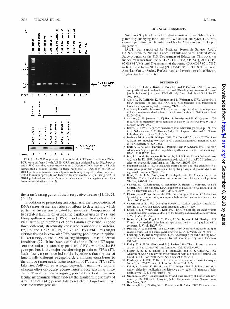
Mammary tumors contain and express the Ad9 E4 ORF1gene. As E4 ORF1 is an essential viral determinant for tumor-igenesis by Ad9 (32), we next sought to confirm that Ad9 mu-tant virus-induced mammary tumors retain this gene and ex-press the protein. By PCR amplification or immunoblot analysis,we detected the Ad9 E4 ORF1 gene (Fig. 6A) or its proteinexpression (Fig. 6B), respectively, in all mammary tumors, in-cluding those elicited by viruses Ad9DE1A and Ad9DE1B. Assmaller tumors had arisen in Ad9DE1A virus-infected ani-mals, it was noteworthy that the levels of Ad9 E4 ORF1protein in Ad9DE1A-induced tumors were lower than thosein both wild-type Ad9-induced and Ad9DE1B-induced tu-mors (Fig. 6B).
DISCUSSION
In this study, we determined the nucleotide sequence of thesubgroup D Ad9 E1 region and showed that its gene organi-zation and predicted protein products are highly related tothose of E1 regions from other human adenoviruses. Addition-ally, to investigate the role of the Ad9 E1 region in Ad9-in-duced mammary oncogenesis, we engineered the same E1Aand E1B deletion mutations into both Ad9 E1 region plasmidsand Ad9 viruses. We found that while E1A and E1B mutantAd9 E1 region plasmids displayed significantly impaired focus-forming activity in vitro, the corresponding E1A and E1B mu-tant Ad9 viruses retained the ability to generate mammary tu-mors in rats. These results indicate that although the Ad9 E1region alone or in cooperation with activated ras exhibits trans-forming activity in vitro, this activity is not required for mam-mary tumorigenesis by Ad9 in vivo. Similar examples in whichtransformation in vitro fails to predict tumorigenicity in vivoare also known for other viral and cellular transforming pro-teins (6, 8, 35, 45, 48, 56).
In addition to showing that Ad9 E1 region transformingfunctions are dispensable for mammary tumorigenesis by Ad9,
FIG. 4. SmaI digestion pattern of wild-type and E1A and E1B mutant Ad9virion DNAs. The wild-type Ad9 E1 region-containing SmaI DNA band migrat-ing at approximately 4 kb, as well as the corresponding faster-migrating DNAbands of the Ad9 E1A mutant virus Ad9DE1A and of the Ad9 E1B mutant virusAd9DE1B, are indicated with arrows. A 1-kb ladder (Gibco-BRL) was used as aDNA size marker (lane 1).
TABLE 3. Tumorigenicities of wild-type and mutantAd9 viruses in Wistar-Furth ratsa
Virus
No. of rats that developed tumors/no. infected with virus
Females Males
Ad9 3/3 0/2Ad9DE1A 8/8 0/2Ad9DE1Bb 3/3 0/3Ad26c 0/3 0/3
a Two- to three-day-old Wistar-Furth rats were injected subcutaneously with7 3 107 PFU of virus. Animals were monitored by palpation for tumor devel-opment over an 8-month period.
b Due to replication deficiencies of virus Ad9DE1B in 293 cells, the dose usedto infect rats with this virus (8 3 106 PFU) was approximately ninefold lowerthan that used for the other viruses.
c Ad26, a nononcogenic subgroup D human adenovirus closely related to Ad9(31), served as a negative control in this experiment.
TABLE 4. Histologies of wild-type and mutantAd9-induced tumorsa
Virus Tumorsample Histology
Wild-type Ad9 1 Fibroadenoma2 Fibroadenoma3 Fibroadenoma
Ad9DE1A 1 Fibroadenoma, one area of increased cellularity2 Focally cellular fibroadenoma3 Fibroadenoma4 Cellular fibroadenoma, focally increased mitoses,
and focal phyllodes-like tumorb
5 Fibroadenoma6 Fibroadenoma7 Fibroadenoma8 Fibroadenoma
Ad9DE1B 1 Fibroadenoma2 Fibroadenoma3 Fibroadenoma
a For histological examination, tumor samples were fixed in 10% formalin, andsections were stained with hematoxylin and eosin.
b A section of this tumor contained an area resembling a phyllodes-like tumor,a type of mammary tumor occasionally observed in rats infected by wild-type Ad9(29).
3076 THOMAS ET AL. J. VIROL.
on Septem
ber 8, 2018 by guesthttp://jvi.asm
.org/D
ownloaded from
our results further argue that the Ad9 E4 region-encodedORF1 transforming gene represents the major oncogenic de-terminant of this virus. In this respect, Ad9 represents the firstexample of an oncogenic adenovirus for which the E1 region isnot the major oncogenic determinant. The fact that the onco-genic avian adenovirus CELO lacks genes related to the hu-man adenovirus E1A and E1B oncogenes (11) further suggeststhat additional examples non-E1 region oncogenic determi-nants for adenoviruses will be found.
Although the mechanism by which Ad9 reaches the mam-mary glands of rats after subcutaneous inoculation has notbeen established, we hypothesize that the inoculated Ad9 viri-ons are able to directly infect mammary cells to cause tumorsin the animals. This idea is based on the fact that rodent cellsare generally nonpermissive for replication of human adeno-viruses (48), a property that would limit spread of the virus bysuccessive rounds of viral replication in tissues of rats. More-over, in this study, we found that Ad9 E1A and E1B mutantviruses (Ad9DE1A and Ad9DE1B, respectively) retained thecapacity to generate mammary tumors in these animals. Be-cause E1A and E1B genes encode critical functions needed forefficient replication of adenoviruses (48), these new resultswith E1A and E1B mutant viruses provide additional supportfor the idea that viral replication in rats is not required for Ad9to produce mammary tumors.
Although tumors elicited by wild-type and E1 region mutant
Ad9 viruses in this study were found to be histologically iden-tical, the tumors induced by the E1A mutant Ad9 virus weregenerally smaller than those generated by both the wild-typeand E1B mutant Ad9 viruses. This finding suggests that E1Atransforming functions may, in fact, enhance the growth ofAd9-induced mammary tumors. Nevertheless, it must also beconsidered that, separate from its transforming functions, E1Aalso serves an important role in the viral life cycle by transcrip-tionally activating other viral gene regions, including the E4region (7, 34, 42). In the E1A mutant virus Ad9DE1A, weintroduced a large deletion extending from the E1A initiationcodon through half of CR3, a mutation which in addition toabolishing the transforming potential of E1A would also beexpected to block transcriptional activation mediated by thisgene. With regard to such a lack of E1A transcriptional activityin virus Ad9DE1A, it may be relevant that mammary tumorsgenerated by this virus expressed reduced levels of the E4ORF1 protein (Fig. 6B). This finding may indicate that E1Aplays an accessory role in Ad9 mammary tumorigenesisby transcriptionally activating the viral E4 region and, there-by, elevating expression of the Ad9 E4 ORF1 oncogenic de-terminant. Similar indirect roles in viral oncogenesis havebeen ascribed to the bovine papillomavirus type 1 E2 andthe Epstein-Barr herpesvirus EBNA2 transactivators, whichparticipate in tumor formation by increasing expression of
FIG. 5. Tumors induced by viruses Ad9DE1A and Ad9DE1B do not contain wild-type Ad9 E1 region sequences. (A) Locations of Ad9 E1 region primers used inPCRs. ITR, inverted terminal repeat. (B) PCR 1 utilized either E1A primers a and b or E1B primers c and d. Genomic DNA from rat 3Y1 cells or water (no DNA)represented negative controls in these reactions; 1.5 mg of genomic DNA or 10 ng of virion DNA was used as a template. (C) Nested PCR 2 utilized E1A primers eand b or E1B primers c and f. In these reactions, DNA from a wild-type Ad9-induced tumor and 3Y1 genomic DNA represented positive and negative controls,respectively; 1/20 of the DNA products from PCR (B) was used as a template for PCR 2. PCR conditions: 30 cycles of denaturation at 94°C for 1 min, annealing at65°C (E1A reaction 1), 58°C (E1A reaction 2), or 60°C (E1B reactions 1 and 2) for 1 min, and extension at 72°C for 1 min, followed by a final 72°C extension for 15min. DNA products were separated by agarose gel electrophoresis and visualized with ethidium bromide.
VOL. 73, 1999 Ad9 TUMORIGENESIS DOES NOT REQUIRE THE E1 REGION 3077
on Septem
ber 8, 2018 by guesthttp://jvi.asm
.org/D
ownloaded from
the transforming genes of their respective viruses (14, 16, 24,36, 43).
In addition to promoting tumorigenesis, the oncoproteins ofDNA tumor viruses may also contribute to determining whichparticular tissues are targeted for neoplasia. Comparisons oftwo related families of viruses, the papillomaviruses (PVs) andfibropapillomaviruses (FPVs), can be used to illustrate thisidea. Although members of both families of viruses encodethree different, structurally conserved transforming proteins,E5, E6, and E7 (5, 10, 15, 27, 39, 46), PVs and FPVs targetdistinct tissues in vivo, with PVs causing papillomas in epithe-lial keratinocytes and FPVs causing fibropapillomas in dermalfibroblasts (27). It has been established that E6 and E7 repre-sent the major transforming proteins of PVs, whereas the E5gene product is the major transforming protein of FPVs (27).Such observations have led to the hypothesis that the use offunctionally different oncogenic determinants contributes tothe unique tumorigenic tissue tropisms of PVs and FPVs (27).Likewise, Ad9 causes estrogen-dependent mammary tumors,whereas other oncogenic adenoviruses induce sarcomas in ro-dents. Therefore, one intriguing possibility is that novel mo-lecular mechanisms which underly the transforming activity ofAd9 E4 ORF1 (41) permit Ad9 to selectively target mammarycells for tumorigenesis.
ACKNOWLEDGMENTS
We thank Stephen Hoang for technical assistance and Sylvia Lee forgenerously supplying REF cultures. We also thank Sylvia Lee, BrittGlaunsinger, Ezequiel Fuentes, and Nader Ghebranious for helpfulsuggestions.
D.L.T. was supported by National Research Service AwardCA09197 from the National Cancer Institute and by the Federal Work-Study program of the U.S. Department of Education. This work wasfunded by grants from the NIH (NCI R01 CA/AI58541), ACS (RP6-97-068-01-VM), and Department of the Army (DAMD17-97-1-7082)to R.T.J. and by an NIH grant (POI CA41086) to T.E.S. T.E.S. is anAmerican Cancer Society Professor and an Investigator of the HowardHughes Medical Institute.
REFERENCES
1. Abate, C., D. Luk, R. Gentz, F. Rauscher, and T. Curran. 1990. Expressionand purification of the leucine zipper and DNA-binding domains of fos andjun: both fos and jun contact DNA directly. Proc. Natl. Acad. Sci. USA 87:1032–1036.
2. Aiello, L., R. Guilfoyle, K. Huebner, and R. Weinmann. 1979. Adenovirus 5DNA sequences present and RNA sequences transcribed in transformedhuman embryo kidney cells. Virology 94:460–469.
3. Ankerst, J., and N. Jonsson. 1989. Adenovirus type 9-induced tumorigenesisin the rat mammary gland related to sex hormonal state. J. Natl. Cancer Inst.81:294–298.
4. Ankerst, J., N. Jonsson, L. Kjellen, E. Norrby, and H. O. Sjogren. 1974.Induction of mammary fibroadenomas in rats by adenovirus type 9. Int. J.Cancer. 13:286–290.
5. Baker, C. C. 1987. Sequence analysis of papillomavirus genomes, p. 321–385.In N. Salzman and P. M. Howley (ed.), The Papovaviridae, vol. 2. PlenumPublishing Corp., New York, N.Y.
6. Barbosa, M. S., and R. Schlegel. 1989. The E6 and E7 genes of HPV-18 aresufficient for inducing two-stage in vitro transformation of human keratino-cytes. Oncogene 4:1529–1532.
7. Berk, A. J., F. Lee, T. Harrison, J. Williams, and P. A. Sharp. 1979. Pre-earlyadenovirus 5 gene product regulates synthesis of early viral messengerRNAs. Cell 17:935–944.
8. Bos, J. L., A. G. Jochemsen, R. Bernards, P. I. Schrier, H. van Ormondt, andA. J. van der Eb. 1983. Deletion mutants of region E1a of AD12 E1 plasmids:effect on oncogenic transformation. Virology 129:393–400.
9. Bradford, M. M. 1976. A rapid and sensitive method for the quantitation ofmicrogram quantities of protein utilizing the principle of protein dye bind-ing. Anal. Biochem. 72:248–254.
10. Bubb, V., D. J. McCance, and R. Schlegel. 1988. DNA sequence of theHPV-16 E5 ORF and the structural conservation of its encoded protein.Virology 163:243–246.
11. Chiocca, S., R. Kurzbauer, G. Schaffner, A. Baker, V. Mautner, and M.Cotten. 1996. The complete DNA sequence and genomic organization of theavian adenovirus CELO. J. Virol. 70:2939–2949.
12. Chomczynski, P., and N. Sacchi. 1987. Single-step method of RNA isolationby acid guanidinium thiocyanate-phenol-chloroform extraction. Anal. Bio-chem. 162:156–159.
13. Chomczynski, R. 1992. One-hour downward alkaline capillary transfer forblotting of DNA and RNA. Anal. Biochem. 201:134–139.
14. Cohen, J. I., F. Wang, and E. Kieff. 1991. Epstein-Barr virus nuclear protein2 mutations define essential domains for transformation and transactivation.J. Virol. 65:2545–2554.
15. Danos, O., L. W. Engel, E. Y. Chen, M. Yaniv, and P. M. Howley. 1983.Comparative analysis of the human type 1a and bovine type 1 papillomavirusgenomes. J. Virol. 46:557–566.
16. DiMaio, D., J. Metherall, and K. Neary. 1986. Nonsense mutation in openreading frame E2 of bovine papillomavirus DNA. J. Virol. 57:475–480.
17. Feinberg, A. P., and B. Vogelstein. 1983. A technique for radiolabeling DNArestriction endonuclease fragments to high specific activity. Anal. Biochem.132:6–13.
18. Finlay, C. A., P. W. Hinds, and A. J. Levine. 1989. The p53 proto-oncogenecan act as a suppressor of transformation. Cell 57:1083–1093.
19. Fisher, P. B., L. E. Babiss, I. B. Weinstein, and H. S. Ginsberg. 1982.Analysis of type 5 adenovirus transformation with a cloned rat embryo cellline (CREF). Proc. Natl. Acad. Sci. USA 79:3527–3531.
20. Freshney, R. I. 1987. Culture of animal cells: a manual of basic technique,2nd ed., p. 107–126. Alan R. Liss, Inc., New York, N.Y.
21. Fukui, Y., I. Saito, K. Shiroki, and H. Shimojo. 1984. Isolation of transfor-mation-defective, replication-nondefective early region 1B mutants of ade-novirus type 12. J. Virol. 49:154–161.
22. Graham, F. 1984. Transformation by and oncogenicity of human adenovi-ruses, p. 339–398. In H. Ginsberg (ed.), The adenoviruses. Plenum Press,New York, N.Y.
23. Graham, F. L., J. Smiley, W. C. Russell, and R. Nairn. 1977. Characteristics
FIG. 6. (A) PCR amplification of the Ad9 E4 ORF1 gene from tumor DNAs.PCRs were performed with Ad9 E4 ORF1 primers as described for Fig. 5 exceptthat a 55°C annealing temperature was used. Genomic DNA from rat 3Y1 cellsrepresented a negative control in these reactions. (B) Detection of Ad9 E4ORF1 protein in tumors. Tumor lysates containing 3 mg of protein were sub-jected to immunoprecipitation followed by immunoblot analysis using Ad9 E4ORF1 polyclonal antiserum. Preimmune serum served as a negative control forimmunoprecipitations (lane 2).
3078 THOMAS ET AL. J. VIROL.
on Septem
ber 8, 2018 by guesthttp://jvi.asm
.org/D
ownloaded from
of a human cell line transformed by DNA from human adenovirus type 5.J. Gen. Virol. 36:59–72.
24. Groff, D. E., and W. D. Lancaster. 1986. Genetic analysis of the 39 earlyregion transformation and replication functions of bovine papillomavirustype 1. Virology 150:221–230.
25. Horwitz, M. S. 1996. Adenoviruses, p. 2149–2171. In B. N. Fields, D. M.Knipe, and P. M. Howley (ed.), Fields virology, vol. 2. Lippincott, Philadel-phia, Pa.
26. Houweling, A., P. van den Elsen, and A. van der Eb. 1980. Partial transfor-mation of primary rat cells by the leftmost 4.5% fragment of adenovirus 5DNA. Virology 105:537–550.
27. Howley, P. M. 1996. Papillomavirinae: the viruses and their replication, p.2045–2076. In B. N. Fields, D. M. Knipe, and P. M. Howley (ed.), Fieldsvirology, vol. 2. Lippincott, Philadelphia, Pa.
28. Jannun, R., and G. Chinnadurai. 1987. Functional relatedness between theE1a and E1b regions of group C and group D human adenoviruses. VirusRes. 7:33–48.
29. Javier, R., K. Raska, Jr., G. J. Macdonald, and T. Shenk. 1991. Humanadenovirus type 9-induced rat mammary tumors. J. Virol. 65:3192–3202.
30. Javier, R., K. Raska, Jr., and T. Shenk. 1992. Requirement for the adeno-virus type 9 E4 region in production of mammary tumors. Science 257:1267–1271.
31. Javier, R., and T. Shenk. 1996. Mammary tumors induced by human ade-novirus type 9: a role for the viral early region 4 gene. Breast Cancer Res.Treat. 39:57–67.
32. Javier, R. T. 1994. Adenovirus type 9 E4 open reading frame 1 encodes atransforming protein required for the production of mammary tumors inrats. J. Virol. 68:3917–3924.
33. Jelinek, T., D. S. Pereira, and F. L. Graham. 1994. Tumorigenicity of ade-novirus-transformed rodent cells is influenced by at least two regions ofadenovirus type 12 early region 1A. J. Virol. 68:888–896.
34. Jones, N., and T. Shenk. 1979. An adenovirus type 5 early gene functionregulates expression of other early viral genes. Proc. Natl. Acad. Sci. USA 76:3665–3669.
35. Kaur, P., and J. K. McDougall. 1988. Characterization of primary humankeratinocytes transformed by human papillomavirus type 18. J. Virol. 62:1917–1924.
36. Kieff, E. 1996. Epstein-Barr Virus and its replication, p. 2343–2396. In B. N.Fields, D. M. Knipe, and P. M. Howley (ed.), Fields virology, vol. 2. Lippin-cott, Philadelphia, Pa.
37. Kimura, G., A. Itagaki, and J. Summers. 1975. Rat cell line 3Y1 and itsvirogenic polyoma and SV40 transformed derivatives. Int. J. Cancer 15:694–706.
38. Kingston, R. E., C. A. Chen, and H. Okayama. 1990. Calcium phosphatetransfection, p. 9.1.1–9.1.9. In F. M. Ausubel, R. Brent, R. E. Kingston, D. D.Moore, J. G. Seidman, J. A. Smith, and K. Struhl (ed.), Current protocols inmolecular biology. Greene Publishing Associates and Wiley-Interscience,New York, N.Y.
39. Kulke, R., and D. DiMaio. 1991. Biological activities of the E5 protein of thedeer papillomavirus in mouse C127 cells: morphologic transformation, in-duction of cellular DNA synthesis, and activation of the platelet-derivedgrowth factor receptor. J. Virol. 65:4943–4949.
40. Laemmli, U. K. 1970. Cleavage of structural proteins during the assembly of
the head of bacteriophage T4. Nature 227:680–685.41. Lee, S. S., R. S. Weiss, and R. T. Javier. 1997. Binding of human virus
oncoproteins to hDlg/SAP97, a mammalian homolog of the Drosophila discslarge tumor suppressor protein. Proc. Natl. Acad. Sci. USA 94:6670–6675.
42. Nevins, J. 1981. Mechanism of activation of early viral transcription by theadenovirus E1A gene product. Cell 26:213–220.
43. Rabson, M. S., C. Yee, Y.-C. Yang, and P. M. Howley. 1986. Bovine papil-lomavirus type 1 39 early region transformation and plasmid maintenancefunctions. J. Virol. 60:626–634.
44. Robbins, S. L., M. Angell, and V. Kumar. 1981. Basic pathology, p. 564–595.The W. B. Saunders Co., Philadelphia, Pa.
45. Schlegel, R., W. C. Phelps, Y.-L. Zhang, and M. Barbosa. 1988. Quantitativekeratinocyte assay detects two biological activities of human papillomavirusDNA and identifies viral types associated with cervical carcinoma. EMBO J.7:3181–3187.
46. Schwarz, E., M. Durst, C. Demankowski, O. Lattermann, R. Zech, E. Wolf-sperger, S. Suhai, and H. zur Hausen. 1983. DNA sequence and genomeorganization of genital human papillomavirus type 6b. EMBO J. 2:2341–2348.
47. Sharp, P. A. 1984. Adenovirus transcription, p. 173–204. In H. Ginsberg(ed.), The adenoviruses. Plenum Press, New York, N.Y.
48. Shenk, T. 1996. Adenoviridae: the viruses and their replication, p. 2111–2148. In B. N. Fields, D. M. Knipe, and P. M. Howley (ed.), Fields virology,vol. 2. Lippincott, Philadelphia, Pa.
49. Shenk, T., and J. Flint. 1991. Transcriptional and transforming activities ofthe adenovirus E1A proteins. Adv. Cancer Res. 57:47–85.
50. Smith, R. F., B. A. Wiese, M. K. Wojzynski, D. B. Davison, and K. C. Worley.1996. BCM Search Launcher—an integrated interface to molecular biologydatabase search and analysis services available on the World Wide Web.Genome Res. 6:454–462.
51. Stillman, B. 1986. Functions of the adenovirus E1B tumor antigens. CancerSurv. 5:389–404.
52. Strauss, W. M. 1994. Preparation of genomic DNA from mammalian tissue,p. 2.2.1–2.2.3. In F. M. Ausubel, R. Brent, R. E. Kingston, D. D. Moore, J. G.Seidman, J. A. Smith, and K. Struhl (ed.), Current protocols in molecularbiology, vol. 1. Greene Publishing Associates and Wiley-Interscience, NewYork, N.Y.
53. Telling, G. C., and J. Williams. 1994. Constructing chimeric type 12/type 5adenovirus E1A genes and using them to identify an oncogenic determinantof adenovirus type 12. J. Virol. 68:877–887.
54. Trentin, J., Y. Yabe, and G. Taylor. 1962. The quest for human cancerviruses: a new approach to an old problem reveals cancer induction inhamster by human adenovirus. Science 137:835–841.
55. van den Elsen, P., A. Houweling, and A. van der Eb. 1983. Expression ofregion E1b of human adenoviruses in the absence of region E1a is notsufficient for complete transformation. Virology 128:377–390.
56. van Leeuwen, F. N., R. A. van der Kammen, G. G. M. Habets, and J. G.Collard. 1995. Oncogenic activity of Tiam 1 and Rac1 in NIH3T3 cells.Oncogene 11:2215–2221.
57. Yew, P. R., C. C. Kao, and A. J. Berk. 1990. Dissection of functional domainsin the adenovirus 2 early 1B 55K polypeptide by suppressor-linker insertionalmutagenesis. Virology 179:795–805.
VOL. 73, 1999 Ad9 TUMORIGENESIS DOES NOT REQUIRE THE E1 REGION 3079
on Septem
ber 8, 2018 by guesthttp://jvi.asm
.org/D
ownloaded from