early somatic mosaicism is a rare cause of long-qt syndrome · 13, 14). however, nearly 30% of...
TRANSCRIPT

Early somatic mosaicism is a rare cause oflong-QT syndromeJames Rush Priesta,b,c, Charles Gawadb,d,1, Kristopher M. Kahlige, Joseph K. Yuf,g, Thomas O’Haraf, Patrick M. Boylef,g,Sridharan Rajamanie,2, Michael J. Clarkh, Sarah T. K. Garciah,3, Scott Ceresnaka,b,c, Jason Harrish, Sean Boyleh,Frederick E. Deweya,i,4, Lindsey Malloy-Waltona,b,c,5, Kyla Dunna,j, Megan Grovea,i, Marco V. Pereza,i, Norma F. Neffk,Richard Chenh, Katsuhide Maedaa,l, Anne Dubina,b,c, Luiz Belardinellie, John Westh, Christian Antolikm,Daniela Macayam, Thomas Quertermousa,i, Natalia A. Trayanovaf,g, Stephen R. Quakek,n,o,6, and Euan A. Ashleya,b,i,6
aStanford Center for Inherited Cardiovascular Disease, Stanford University School of Medicine, Stanford, CA 94305; bChild Health Research Institute,Stanford University School of Medicine, Stanford, CA 94305; cDivision of Pediatric Cardiology, Stanford University School of Medicine, Stanford, CA 94305;dDivision of Pediatric Hematology-Oncology, Stanford University School of Medicine, Stanford, CA 94305; eDepartment of Biology, CardiovascularTherapeutic Area, Gilead Sciences, Fremont, CA 94555; fDepartment of Biomedical Engineering, Johns Hopkins University, Baltimore, MD 21218; gInstitutefor Computational Medicine, Johns Hopkins University, Baltimore, MD 21218; hPersonalis, Inc., Menlo Park, CA 94025; iDivision of Cardiovascular Medicine,Stanford University School of Medicine, Stanford, CA 94305; jLucile Packard Children’s Hospital Heart Center, Palo Alto, CA 94304; kDepartment ofBioengineering, Stanford University School of Medicine, Stanford, CA 94305; lDivision of Cardiothoracic Surgery, Stanford University School of Medicine,Stanford, CA 94305; mCardiogenetic Testing Services, GeneDx, Gaithersburg, MD 20877; nDepartment of Applied Physics, Stanford University, Stanford, CA94305; and oHoward Hughes Medical Research Institute, Stanford University School of Medicine, Stanford, CA 94305
Contributed by Stephen R. Quake, August 19, 2016 (sent for review May 5, 2016; reviewed by Leslie Biesecker and Christopher Semsarian)
Somatic mosaicism, the occurrence and propagation of genetic varia-tion in cell lineages after fertilization, is increasingly recognized to playa causal role in a variety of human diseases. We investigated the caseof life-threatening arrhythmia in a 10-day-old infant with long QTsyndrome (LQTS). Rapid genome sequencing suggested a variant in thesodium channel NaV1.5 encoded by SCN5A, NM_000335:c.5284G > Tpredicting p.(V1762L), but read depth was insufficient to be diagnostic.Exome sequencing of the trio confirmed read ratios inconsistent withMendelian inheritance only in the proband. Genotyping of single cir-culating leukocytes demonstrated the mutation in the genomes of 8%of patient cells, and RNA sequencing of cardiac tissue from the infantconfirmed the expression of the mutant allele at mosaic ratios. Heter-ologous expression of the mutant channel revealed significantlydelayed sodium current with a dominant negative effect. To investi-gate the mechanism by which mosaicism might cause arrhythmia, webuilt a finite element simulation model incorporating Purkinje fiberactivation. This model confirmed the pathogenic consequences of car-diac cellular mosaicism and, under the presenting conditions of thiscase, recapitulated 2:1 AV block and arrhythmia. To investigate theextent to which mosaicism might explain undiagnosed arrhyth-mia, we studied 7,500 affected probands undergoing commercialgene-panel testing. Four individuals with pathogenic variantsarising from early somatic mutation events were found. Herewe establish cardiac mosaicism as a causal mechanism for LQTSand present methods by which the general phenomenon, likely tobe relevant for all genetic diseases, can be detected through single-cell analysis and next-generation sequencing.
mosaicism | arrhythmia | genomics | computational modeling | single cell
There is growing recognition that somatic mosaicism, i.e., ge-netic variation within an individual that arises from errors in
DNA replication during early development, may play a role in avariety of human diseases other than cancer (1). However, theextent to which cellular heterogeneity contributes to disease isminimally understood. One report suggests that 6.5% of de novomutations presumed to be germline in origin may instead havearisen from postzygotic mosaic mutation events (2), and recentgenetic investigations directly interrogating diseased tissues inbrain malformations, breast cancer, and atrial fibrillation haverevealed postzygotic causal mutations absent from germlineDNA (3–6). Pathogenic mosaic structural variation is also de-tectable in children with neurodevelopmental disorders (7).However, a consequential category of genetic variation hasnot been surveyed systematically in clinical or research studies ofother human diseases.
The pathophysiological basis of long-QT syndrome (LQTS) isprolongation of cardiac ventricular repolarization by acquiredfactors such as drug exposure or genetic variation in the proteinscontrolling transmembrane ion-concentration gradients (8, 9);parental gonadal mosaicism is an infrequently described phe-nomenon in LQTS (10–12). Knowledge of the molecular sub-typing of disease in LQTS has provided a foundation forgenotype-specific risk stratification and treatment strategies (8, 9,
Significance
Most genetic studies and clinical genetic testing do not lookfor the possibility of mosaic variation. The genetic form oflong-QT syndrome (LQTS) can result in life-threatening ar-rhythmias, but 30% of patients do not have a genetic di-agnosis. We performed deep characterization of a mosaicvariant in an infant with perinatal LQTS and developed acomputational model showing how abnormal cellular re-polarization in only 8% of heart cells may cause arrhythmia.Finally we looked at the prevalence of mosaicism among pa-tients with LQTS; in a population of 7,500 individuals wefound evidence of pathogenic early somatic mosaicism inapproximately 0.17% of LQTS patients without a genetic di-agnosis. Together these data establish an unreported mech-anism for LQTS and other genetic arrhythmias.
Author contributions: J.R.P., K.M.K., T.O., S.R., S.C., R.C., K.M., A.D., L.B., J.W., D.M., T.Q.,and E.A.A. designed research; J.R.P., C.G., K.M.K., J.K.Y., P.M.B., L.M.-W., K.D., M.G.,M.V.P., and C.A. performed research; J.K.Y., T.O., P.M.B., F.E.D., N.F.N., D.M., N.A.T.,and S.R.Q. contributed new reagents/analytic tools; J.R.P., C.G., K.M.K., J.K.Y., P.M.B.,M.J.C., S.T.K.G., J.H., S.B., and J.W. analyzed data; and J.R.P., K.M.K., J.K.Y., T.Q., andE.A.A. wrote the paper.
Reviewers: L.B., NIH; and C.S., University of Sydney.
Conflict of interest statement: At the time of this work K.M.K., S.R., and L.B. were em-ployed by Gilead Sciences. M.J.C., S.T.K.G., S.B., J.W., and R.C. were employed by Person-alis, Inc. J.W. and E.A.A. are founders of Personalis, Inc. which offers clinical genetictesting but does not offer Clinical Laboratory Improvement Amendments (CLIA)-certifiedrapid-turnaround whole-genome sequencing. N.A.T. is a co-founder of CardioSolv LLC.
Freely available online through the PNAS open access option.1Present address: St. Jude Children’s Research Hospital, Memphis, TN 38105.2Present address: Amgen, Inc., South San Francisco, CA 94080.3Present address: 10X Genomics, Pleasanton, CA 94556.4Present address: Regeneron Pharmaceuticals, Tarrytown, NY 10591.5Mercy Children’s Hospital, Kansas City, MO 64108.6To whom correspondence may be addressed. Email: [email protected] or [email protected].
This article contains supporting information online at www.pnas.org/lookup/suppl/doi:10.1073/pnas.1607187113/-/DCSupplemental.
www.pnas.org/cgi/doi/10.1073/pnas.1607187113 PNAS | October 11, 2016 | vol. 113 | no. 41 | 11555–11560
MED
ICALSC
IENCE
S
Dow
nloa
ded
by g
uest
on
Dec
embe
r 25
, 201
9
13, 14). However, nearly 30% of probands remain undiagnosedusing standard commercial gene-panel testing, suggesting there isunrecognized genetic variation (genic, regulatory, or otherwise) yetto be associated with disease (15).In its most severe form, LQTS may occur in the neonatal
period with bradycardia and functional 2:1 AV block that occursecondary to a severely prolonged ventricular refractory periodgreater than the short R–R interval characteristic of a normalheart rate in infancy (16). Patients of all ages presenting withLQTS are predisposed to torsades de pointes (TdP), a life-threatening cardiac arrhythmia, with patients presenting in in-fancy showing particularly poor outcomes (13, 16–18). In thisstudy, we applied rapid-turnaround whole-genome sequencing(WGS) on day of life (DOL) 3 in a premature infant with peri-natal LQTS and life-threatening arrhythmia and investigated thecontribution of a discovered mosaic variant to abnormal cardiacelectrophysiology at the molecular and tissue levels. Additionallywe surveyed 7,500 individuals already tested for genetic ar-rhythmias, allowing us to estimate the prevalence of mosaicismin an unbiased population sample.
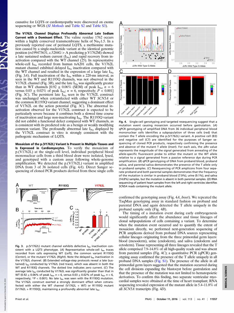
ResultsDeep Sequencing Identifies Mosaic Variation in SCN5A in an Infantwith a Prolonged QT Interval and Arrhythmia. A near-term 2.5-kgfemale infant of Asian ancestry with prenatally diagnosed 2:1 AVblock was delivered by caesarean section at 36-wk gestation. At 1 hof life the infant developed intermittent 2:1 AV block with multipleepisodes of TdP with a corrected QT interval (QTc) of 542 ms (Fig.1), which were relieved by placement of a dual-chamber epicardialimplantable cardioverter/defibrillator (ICD) accompanied by a bi-lateral stellate ganglionectomy. A standard commercially availablegenetic panel for LQTS was sent on DOL 1 (13, 17), and WGSwas performed on DOL 3. At 6 mo of age the patient developeddilated cardiomyopathy and subsequently received an orthotopicheart transplant.WGS yielded a 39.76 mean read depth, and coverage across
the coding regions of known LQTS and dilated cardiomyopathy(DCM) genes was 99.93% at 10× or greater (Table S1). Therapid RTG pipeline detected a mutation in the 28th exon ofthe voltage-gated sodium channel NaV1.5 encoded by SCN5ANM_000335: c.5284G > T, which predicts p.(V1762L) within thealpha subunit of NaV1.5, which was not present in the two addi-tional call sets originating from Burrows–Wheeler Aligner (BWA)/Genome Analysis Toolkit (GATK) or Issac (Issac Genome Align-ment Software and Variant Caller) pipelines. All three pipelinesdetected rs41261344, a polymorphism in SCN5A common in HanChinese NM_000335: c.3575G > A, which predicts p.R1193Q (22).Given the discrepant results between the WGS pipelines, we
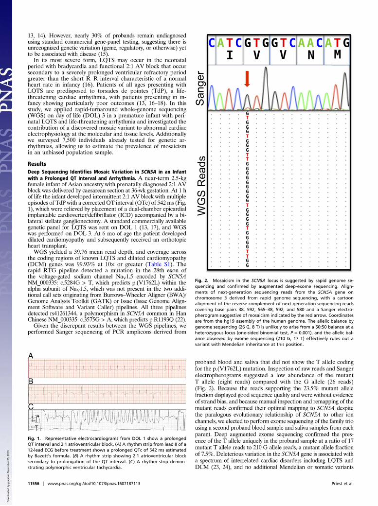
performed Sanger sequencing of PCR amplicons derived from
proband blood and saliva that did not show the T allele codingfor the p.(V1762L) mutation. Inspection of raw reads and Sangerelectropherograms suggested a low abundance of the mutantT allele (eight reads) compared with the G allele (26 reads)(Fig. 2). Because the reads supporting the 23.5% mutant allelefraction displayed good sequence quality and were without evidenceof strand bias, and because manual inspection and remapping of themutant reads confirmed their optimal mapping to SCN5A despitethe paralogous evolutionary relationship of SCN5A to other ionchannels, we elected to perform exome sequencing of the family triousing a second proband blood sample and saliva samples from eachparent. Deep augmented exome sequencing confirmed the pres-ence of the T allele uniquely in the proband sample at a ratio of 17mutant T allele reads to 210 G allele reads, a mutant allele fractionof 7.5%. Deleterious variation in the SCN5A gene is associated witha spectrum of interrelated cardiac disorders including LQTS andDCM (23, 24), and no additional Mendelian or somatic variants
B
C
A
Fig. 1. Representative electrocardiograms from DOL 1 show a prolongedQT interval and 2:1 atrioventricular block. (A) A rhythm strip from lead II of a12-lead ECG before treatment shows a prolonged QTc of 542 ms estimatedby Bazett’s formula. (B) A rhythm strip showing 2:1 atrioventricular blocksecondary to prolongation of the QT interval. (C) A rhythm strip demon-strating polymorphic ventricular tachycardia.
Fig. 2. Mosaicism in the SCN5A locus is suggested by rapid genome se-quencing and confirmed by augmented deep-exome sequencing. Align-ments of next-generation sequencing reads from the SCN5A gene onchromosome 3 derived from rapid genome sequencing, with a cartoonalignment of the reverse complement of next-generation sequencing readscovering base pairs 38, 592, 565–38, 592, and 580 and a Sanger electro-pherogram suggestive of mosaicism indicated by the red arrow. Coordinatesare from the hg19 assembly of the human genome. The allelic balance bygenome sequencing (26 G, 8 T) is unlikely to arise from a 50:50 balance at aheterozygous locus (one-sided binomial test, P = 0.001), and the allelic bal-ance observed by exome sequencing (210 G, 17 T) effectively rules out avariant with Mendelian inheritance at this position.
11556 | www.pnas.org/cgi/doi/10.1073/pnas.1607187113 Priest et al.
Dow
nloa
ded
by g
uest
on
Dec
embe
r 25
, 201
9
causative for LQTS or cardiomyopathy were discovered on exomesequencing or WGS (SI Methods and Table S2 and Table S3).
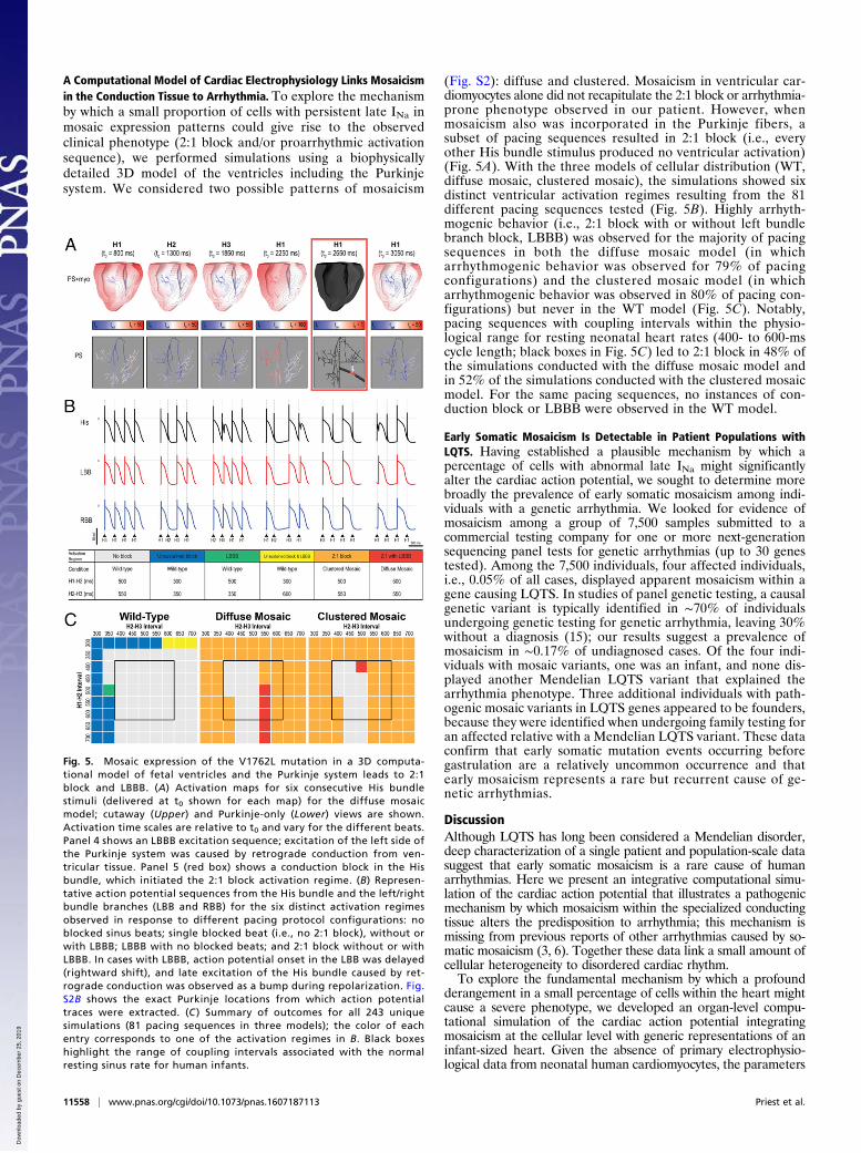
The V1762L Channel Displays Profoundly Abnormal Late SodiumCurrent with a Dominant Effect. The valine residue 1762 occurswithin a highly conserved transmembrane helix of NaV1.5. In apreviously reported case of perinatal LQTS, a methionine muta-tion caused by a single-nucleotide variant at the identical genomicposition (rs199473631, c.5284G > A predicting p.V1762M) showeda late sustained sodium current (INa) and rapid recovery from in-activation compared with the WT channel (25). In representativewhole-cell INa recorded from human tsA201 cells, the V1762Lmutant channel exhibited delayed INa inactivation compared withthe WT channel and resulted in the expression of a large late INa(Fig. 3A). Full inactivation of the INa within a 220-ms interval, asseen in the WT and R1193Q channels, was not observed in theV1762L channel (Fig. 3B), and the late INa was significantly greaterthan in WT channels [0.92 ± 0.06% (SEM) of peak INa, n = 6versus 0.03 ± 0.02% of peak INa; n = 6, respectively; P < 0.001](Fig. 3C). The persistent late INa seen in the V1762L constructwas unchanged when cotransfected with either WT SCN5A orthe common R1193Q variant channel, suggesting a dominant effectof V1762L on the action potential (Fig. 3C). The abnormal in-activation observed for the V1762L construct is expected to beparticularly severe because it combines both a slowed time courseof inactivation and large non-inactivating INa. The R1193Q variantdid not exhibit a functional defect compared with WT channels, asis consistent with its predicted role as a benign or weakly modifyingcommon variant. The profoundly abnormal late INa displayed bythe V1762L construct in vitro is strongly consistent with thepathogenic mechanism of LQTS.
Mosaicism of the p.(V1762L) Variant Is Present in Multiple Tissues andIs Expressed in Cardiomyocytes. To verify the mosaicism ofp.(V1762L) at the single-cell level, individual peripheral bloodmononuclear cells from a third blood draw were isolated, lysed,and genotyped with a custom assay following whole-genomeamplification. We detected the p.(V1762L) variant in amplifiedDNA from 3 of 36 isolated cells (Fig. 4A). Direct Sanger se-quencing of cloned PCR products derived from these single cells
confirmed the genotyping assay (Fig. 4A, Inset). We repeated theTaqMan genotyping assay in standard fashion on proband andparental DNA and again detected the T allele uniquely in theproband sample only (Fig. 4B).The timing of a mutation event during early embryogenesis
would significantly affect the abundance and tissue lineages ofclonal subpopulations of cells containing a variant. To determinewhen the mutation event occurred and to quantify the extent ofmosaicism directly, we performed next-generation sequencing ofPCR amplicons derived from proband DNA sources representingcellular lineages originating from the three primordial germ layers:blood (mesoderm), urine (endoderm), and saliva (endoderm andectoderm). Tissue representing all three lineages revealed that the Tallele comprised 7.9–14.8% of all high-quality reads and was absentfrom parental samples (Fig. 4C); a quantitative PCR (qPCR) gen-otyping assay confirmed the presence of the T allele uniquely in allproband DNA samples (Fig. S1). The presence of the allele in allprimordial germ layers suggested that the mutation occurred duringthe cell divisions expanding the blastocyst before gastrulation andthat the presence of the mutation was not limited to hematopoieticderivatives. To confirm this finding, two separate ventricular myo-cardial samples were obtained at the time of heart transplant; RNAsequencing revealed expression of the mutant allele in 5.4–11.8% ofall SCN5A transcripts (Fig. 4D).
Fig. 3. p.(V1762L) mutant channel exhibits defective INa inactivation con-sistent with a LQT3 phenotype. (A) Representative whole-cell INa tracesrecorded from cells expressing WT (Left), the common variant R1193Q(Center), or the mutant V1762L (Right). Note the delayed INa inactivation inthe V1762L channel. (B) Extended voltage-step protocols reveal a late (sus-tained) INa conducted by V1762L (red trace), which was absent in both theWT and R1193Q channels. The dotted line indicates zero current. (C) Theaverage late INa conducted by V1762L was significantly greater than that inWT (0.92 ± 0.06% of peak INa, n = 6, versus 0.03 ± 0.02% of peak INa, n = 6,respectively; *P < 0.001). No late INa was seen with the R1193Q mutation.The V1762L construct exerted a strongly dominant effect when cotrans-fected with either the WT channel (V1762L + WT) or R1193Q mutant(V1762L + R1193Q), maintaining a profoundly abnormal late INa.
75
Positive Cell (B3)
Rn Wild Type G Allele 2 3 4
0
1
2
3
4
Rn
Mut
ant T
Alle
le
0.7
0.8
0.9
1.0
Rn
Mut
ant T
Alle
le
2.8 3.0 3.2 3.4 3.6 Rn Wild Type G Allele
0
25
50
75
100
Per
cent
age
of R
eads
Base
T G C A
0
25
50
75
100
Per
cent
age
of R
eads
Base
T G C A
Negative Cell (C7)
A B
DC
Fig. 4. Single-cell genotyping and targeted resequencing suggest that amutation event causing mosaicism occurred before gastrulation. (A)qPCR genotyping of amplified DNA from 36 individual peripheral bloodmononuclear cells identifies a subpopulation of three cells (red) thatcontain the T allele encoding the p.(V1762L) variant. A positive cell (B3)and negative cell (C7) are identified for the purposes of Sanger se-quencing of cloned PCR products, respectively confirming the presenceand absence of the mutant T allele (Inset). For each axis, the ΔRn valuerepresents the magnitude of the signal generated from annealing of theallele-specific fluorescent probe to either the mutant or the WT allelerelative to a signal generated from a passive reference dye during PCRamplification. (B) qPCR genotyping of DNA from proband blood, probandsaliva, and parental saliva demonstrates the presence of the T allele onlyin proband samples. (C ) Resequencing of PCR amplicons from four sepa-rate proband and both parental samples demonstrates that the frequencyof the mutation is similar in proband blood (7.9%), urine (9.1%), and saliva(14.8%) samples, but the mutation is absent in both parental samples. (D) RNAsequencing of patient heart samples from the left and right ventricles identifiesSCN5A reads containing the mutant allele.
Priest et al. PNAS | October 11, 2016 | vol. 113 | no. 41 | 11557
MED
ICALSC
IENCE
S
Dow
nloa
ded
by g
uest
on
Dec
embe
r 25
, 201
9
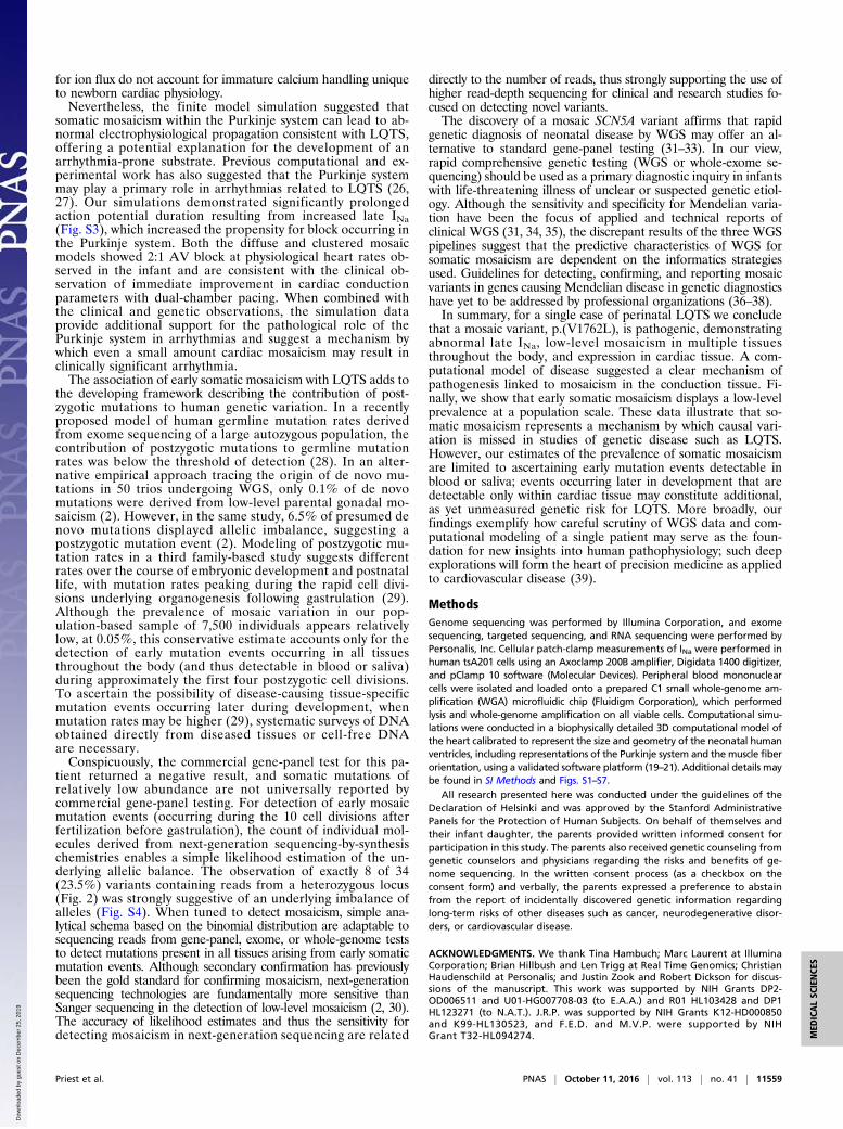
A Computational Model of Cardiac Electrophysiology Links Mosaicismin the Conduction Tissue to Arrhythmia. To explore the mechanismby which a small proportion of cells with persistent late INa inmosaic expression patterns could give rise to the observedclinical phenotype (2:1 block and/or proarrhythmic activationsequence), we performed simulations using a biophysicallydetailed 3D model of the ventricles including the Purkinjesystem. We considered two possible patterns of mosaicism
(Fig. S2): diffuse and clustered. Mosaicism in ventricular car-diomyocytes alone did not recapitulate the 2:1 block or arrhythmia-prone phenotype observed in our patient. However, whenmosaicism also was incorporated in the Purkinje fibers, asubset of pacing sequences resulted in 2:1 block (i.e., everyother His bundle stimulus produced no ventricular activation)(Fig. 5A). With the three models of cellular distribution (WT,diffuse mosaic, clustered mosaic), the simulations showed sixdistinct ventricular activation regimes resulting from the 81different pacing sequences tested (Fig. 5B). Highly arrhyth-mogenic behavior (i.e., 2:1 block with or without left bundlebranch block, LBBB) was observed for the majority of pacingsequences in both the diffuse mosaic model (in whicharrhythmogenic behavior was observed for 79% of pacingconfigurations) and the clustered mosaic model (in whicharrhythmogenic behavior was observed in 80% of pacing con-figurations) but never in the WT model (Fig. 5C). Notably,pacing sequences with coupling intervals within the physio-logical range for resting neonatal heart rates (400- to 600-mscycle length; black boxes in Fig. 5C) led to 2:1 block in 48% ofthe simulations conducted with the diffuse mosaic model andin 52% of the simulations conducted with the clustered mosaicmodel. For the same pacing sequences, no instances of con-duction block or LBBB were observed in the WT model.
Early Somatic Mosaicism Is Detectable in Patient Populations withLQTS. Having established a plausible mechanism by which apercentage of cells with abnormal late INa might significantlyalter the cardiac action potential, we sought to determine morebroadly the prevalence of early somatic mosaicism among indi-viduals with a genetic arrhythmia. We looked for evidence ofmosaicism among a group of 7,500 samples submitted to acommercial testing company for one or more next-generationsequencing panel tests for genetic arrhythmias (up to 30 genestested). Among the 7,500 individuals, four affected individuals,i.e., 0.05% of all cases, displayed apparent mosaicism within agene causing LQTS. In studies of panel genetic testing, a causalgenetic variant is typically identified in ∼70% of individualsundergoing genetic testing for genetic arrhythmia, leaving 30%without a diagnosis (15); our results suggest a prevalence ofmosaicism in ∼0.17% of undiagnosed cases. Of the four indi-viduals with mosaic variants, one was an infant, and none dis-played another Mendelian LQTS variant that explained thearrhythmia phenotype. Three additional individuals with path-ogenic mosaic variants in LQTS genes appeared to be founders,because they were identified when undergoing family testing foran affected relative with a Mendelian LQTS variant. These dataconfirm that early somatic mutation events occurring beforegastrulation are a relatively uncommon occurrence and thatearly mosaicism represents a rare but recurrent cause of ge-netic arrhythmias.
DiscussionAlthough LQTS has long been considered a Mendelian disorder,deep characterization of a single patient and population-scale datasuggest that early somatic mosaicism is a rare cause of humanarrhythmias. Here we present an integrative computational simu-lation of the cardiac action potential that illustrates a pathogenicmechanism by which mosaicism within the specialized conductingtissue alters the predisposition to arrhythmia; this mechanism ismissing from previous reports of other arrhythmias caused by so-matic mosaicism (3, 6). Together these data link a small amount ofcellular heterogeneity to disordered cardiac rhythm.To explore the fundamental mechanism by which a profound
derangement in a small percentage of cells within the heart mightcause a severe phenotype, we developed an organ-level compu-tational simulation of the cardiac action potential integratingmosaicism at the cellular level with generic representations of aninfant-sized heart. Given the absence of primary electrophysio-logical data from neonatal human cardiomyocytes, the parameters
Fig. 5. Mosaic expression of the V1762L mutation in a 3D computa-tional model of fetal ventricles and the Purkinje system leads to 2:1block and LBBB. (A) Activation maps for six consecutive His bundlestimuli (delivered at t0 shown for each map) for the diffuse mosaicmodel; cutaway (Upper) and Purkinje-only (Lower) views are shown.Activation time scales are relative to t0 and vary for the different beats.Panel 4 shows an LBBB excitation sequence; excitation of the left side ofthe Purkinje system was caused by retrograde conduction from ven-tricular tissue. Panel 5 (red box) shows a conduction block in the Hisbundle, which initiated the 2:1 block activation regime. (B) Represen-tative action potential sequences from the His bundle and the left/rightbundle branches (LBB and RBB) for the six distinct activation regimesobserved in response to different pacing protocol configurations: noblocked sinus beats; single blocked beat (i.e., no 2:1 block), without orwith LBBB; LBBB with no blocked beats; and 2:1 block without or withLBBB. In cases with LBBB, action potential onset in the LBB was delayed(rightward shift), and late excitation of the His bundle caused by ret-rograde conduction was observed as a bump during repolarization. Fig.S2B shows the exact Purkinje locations from which action potentialtraces were extracted. (C ) Summary of outcomes for all 243 uniquesimulations (81 pacing sequences in three models); the color of eachentry corresponds to one of the activation regimes in B. Black boxeshighlight the range of coupling intervals associated with the normalresting sinus rate for human infants.
11558 | www.pnas.org/cgi/doi/10.1073/pnas.1607187113 Priest et al.
Dow
nloa
ded
by g
uest
on
Dec
embe
r 25
, 201
9
for ion flux do not account for immature calcium handling uniqueto newborn cardiac physiology.Nevertheless, the finite model simulation suggested that
somatic mosaicism within the Purkinje system can lead to ab-normal electrophysiological propagation consistent with LQTS,offering a potential explanation for the development of anarrhythmia-prone substrate. Previous computational and ex-perimental work has also suggested that the Purkinje systemmay play a primary role in arrhythmias related to LQTS (26,27). Our simulations demonstrated significantly prolongedaction potential duration resulting from increased late INa(Fig. S3), which increased the propensity for block occurring inthe Purkinje system. Both the diffuse and clustered mosaicmodels showed 2:1 AV block at physiological heart rates ob-served in the infant and are consistent with the clinical ob-servation of immediate improvement in cardiac conductionparameters with dual-chamber pacing. When combined withthe clinical and genetic observations, the simulation dataprovide additional support for the pathological role of thePurkinje system in arrhythmias and suggest a mechanism bywhich even a small amount cardiac mosaicism may result inclinically significant arrhythmia.The association of early somatic mosaicism with LQTS adds to
the developing framework describing the contribution of post-zygotic mutations to human genetic variation. In a recentlyproposed model of human germline mutation rates derivedfrom exome sequencing of a large autozygous population, thecontribution of postzygotic mutations to germline mutationrates was below the threshold of detection (28). In an alter-native empirical approach tracing the origin of de novo mu-tations in 50 trios undergoing WGS, only 0.1% of de novomutations were derived from low-level parental gonadal mo-saicism (2). However, in the same study, 6.5% of presumed denovo mutations displayed allelic imbalance, suggesting apostzygotic mutation event (2). Modeling of postzygotic mu-tation rates in a third family-based study suggests differentrates over the course of embryonic development and postnatallife, with mutation rates peaking during the rapid cell divi-sions underlying organogenesis following gastrulation (29).Although the prevalence of mosaic variation in our pop-ulation-based sample of 7,500 individuals appears relativelylow, at 0.05%, this conservative estimate accounts only for thedetection of early mutation events occurring in all tissuesthroughout the body (and thus detectable in blood or saliva)during approximately the first four postzygotic cell divisions.To ascertain the possibility of disease-causing tissue-specificmutation events occurring later during development, whenmutation rates may be higher (29), systematic surveys of DNAobtained directly from diseased tissues or cell-free DNAare necessary.Conspicuously, the commercial gene-panel test for this pa-
tient returned a negative result, and somatic mutations ofrelatively low abundance are not universally reported bycommercial gene-panel testing. For detection of early mosaicmutation events (occurring during the 10 cell divisions afterfertilization before gastrulation), the count of individual mol-ecules derived from next-generation sequencing-by-synthesischemistries enables a simple likelihood estimation of the un-derlying allelic balance. The observation of exactly 8 of 34(23.5%) variants containing reads from a heterozygous locus(Fig. 2) was strongly suggestive of an underlying imbalance ofalleles (Fig. S4). When tuned to detect mosaicism, simple ana-lytical schema based on the binomial distribution are adaptable tosequencing reads from gene-panel, exome, or whole-genome teststo detect mutations present in all tissues arising from early somaticmutation events. Although secondary confirmation has previouslybeen the gold standard for confirming mosaicism, next-generationsequencing technologies are fundamentally more sensitive thanSanger sequencing in the detection of low-level mosaicism (2, 30).The accuracy of likelihood estimates and thus the sensitivity fordetecting mosaicism in next-generation sequencing are related
directly to the number of reads, thus strongly supporting the use ofhigher read-depth sequencing for clinical and research studies fo-cused on detecting novel variants.The discovery of a mosaic SCN5A variant affirms that rapid
genetic diagnosis of neonatal disease by WGS may offer an al-ternative to standard gene-panel testing (31–33). In our view,rapid comprehensive genetic testing (WGS or whole-exome se-quencing) should be used as a primary diagnostic inquiry in infantswith life-threatening illness of unclear or suspected genetic etiol-ogy. Although the sensitivity and specificity for Mendelian varia-tion have been the focus of applied and technical reports ofclinical WGS (31, 34, 35), the discrepant results of the three WGSpipelines suggest that the predictive characteristics of WGS forsomatic mosaicism are dependent on the informatics strategiesused. Guidelines for detecting, confirming, and reporting mosaicvariants in genes causing Mendelian disease in genetic diagnosticshave yet to be addressed by professional organizations (36–38).In summary, for a single case of perinatal LQTS we conclude
that a mosaic variant, p.(V1762L), is pathogenic, demonstratingabnormal late INa, low-level mosaicism in multiple tissuesthroughout the body, and expression in cardiac tissue. A com-putational model of disease suggested a clear mechanism ofpathogenesis linked to mosaicism in the conduction tissue. Fi-nally, we show that early somatic mosaicism displays a low-levelprevalence at a population scale. These data illustrate that so-matic mosaicism represents a mechanism by which causal vari-ation is missed in studies of genetic disease such as LQTS.However, our estimates of the prevalence of somatic mosaicismare limited to ascertaining early mutation events detectable inblood or saliva; events occurring later in development that aredetectable only within cardiac tissue may constitute additional,as yet unmeasured genetic risk for LQTS. More broadly, ourfindings exemplify how careful scrutiny of WGS data and com-putational modeling of a single patient may serve as the foun-dation for new insights into human pathophysiology; such deepexplorations will form the heart of precision medicine as appliedto cardiovascular disease (39).
MethodsGenome sequencing was performed by Illumina Corporation, and exomesequencing, targeted sequencing, and RNA sequencing were performed byPersonalis, Inc. Cellular patch-clamp measurements of INa were performed inhuman tsA201 cells using an Axoclamp 200B amplifier, Digidata 1400 digitizer,and pClamp 10 software (Molecular Devices). Peripheral blood mononuclearcells were isolated and loaded onto a prepared C1 small whole-genome am-plification (WGA) microfluidic chip (Fluidigm Corporation), which performedlysis and whole-genome amplification on all viable cells. Computational simu-lations were conducted in a biophysically detailed 3D computational model ofthe heart calibrated to represent the size and geometry of the neonatal humanventricles, including representations of the Purkinje system and the muscle fiberorientation, using a validated software platform (19–21). Additional details maybe found in SI Methods and Figs. S1–S7.
All research presented here was conducted under the guidelines of theDeclaration of Helsinki and was approved by the Stanford AdministrativePanels for the Protection of Human Subjects. On behalf of themselves andtheir infant daughter, the parents provided written informed consent forparticipation in this study. The parents also received genetic counseling fromgenetic counselors and physicians regarding the risks and benefits of ge-nome sequencing. In the written consent process (as a checkbox on theconsent form) and verbally, the parents expressed a preference to abstainfrom the report of incidentally discovered genetic information regardinglong-term risks of other diseases such as cancer, neurodegenerative disor-ders, or cardiovascular disease.
ACKNOWLEDGMENTS. We thank Tina Hambuch; Marc Laurent at IlluminaCorporation; Brian Hillbush and Len Trigg at Real Time Genomics; ChristianHaudenschild at Personalis; and Justin Zook and Robert Dickson for discus-sions of the manuscript. This work was supported by NIH Grants DP2-OD006511 and U01-HG007708-03 (to E.A.A.) and R01 HL103428 and DP1HL123271 (to N.A.T.). J.R.P. was supported by NIH Grants K12-HD000850and K99-HL130523, and F.E.D. and M.V.P. were supported by NIHGrant T32-HL094274.
Priest et al. PNAS | October 11, 2016 | vol. 113 | no. 41 | 11559
MED
ICALSC
IENCE
S
Dow
nloa
ded
by g
uest
on
Dec
embe
r 25
, 201
9
1. Biesecker LG, Spinner NB (2013) A genomic view of mosaicism and human disease. NatRev Genet 14(5):307–320.
2. Acuna-Hidalgo R, et al. (2015) Post-zygotic point mutations are an underrecognizedsource of de novo genomic variation. Am J Hum Genet 97(1):67–74.
3. Thibodeau IL, et al. (2010) Paradigm of genetic mosaicism and lone atrial fibrillation:Physiological characterization of a connexin 43-deletion mutant identified from atrialtissue. Circulation 122(3):236–244.
4. Ruark E, et al.; Breast and Ovarian Cancer Susceptibility Collaboration; WellcomeTrust Case Control Consortium (2013) Mosaic PPM1D mutations are associated withpredisposition to breast and ovarian cancer. Nature 493(7432):406–410.
5. Jamuar SS, et al. (2014) Somatic mutations in cerebral cortical malformations. N Engl JMed 371(8):733–743.
6. Gollob MH, et al. (2006) Somatic mutations in the connexin 40 gene (GJA5) in atrialfibrillation. N Engl J Med 354(25):2677–2688.
7. King DA, et al.; Deciphering Developmental Disorders Study (2015) Mosaic structuralvariation in children with developmental disorders. Hum Mol Genet 24(10):2733–2745.
8. Lu JT, Kass RS (2010) Recent progress in congenital long QT syndrome. Curr OpinCardiol 25(3):216–221.
9. Giudicessi JR, Ackerman MJ (2013) Genotype- and phenotype-guided management ofcongenital long QT syndrome. Curr Probl Cardiol 38(10):417–455.
10. Dufendach KA, Giudicessi JR, Boczek NJ, Ackerman MJ (2013) Maternal mosaicism con-founds the neonatal diagnosis of type 1 Timothy syndrome. Pediatrics 131(6):e1991–e1995.
11. Miller TE, et al. (2004) Recurrent third-trimester fetal loss and maternal mosaicism forlong-QT syndrome. Circulation 109(24):3029–3034.
12. Etheridge SP, et al. (2011) Somatic mosaicism contributes to phenotypic variation inTimothy syndrome. Am J Med Genet A 155A(10):2578–2583.
13. Cuneo BF, et al. (2013) Arrhythmia phenotype during fetal life suggests long-QTsyndrome genotype: Risk stratification of perinatal long-QT syndrome. Circ ArrhythmElectrophysiol 6(5):946–951.
14. Priori SG, et al. (2013) HRS/EHRA/APHRS expert consensus statement on the diagnosisand management of patients with inherited primary arrhythmia syndromes: Docu-ment endorsed by HRS, EHRA, and APHRS in May 2013 and by ACCF, AHA, PACES, andAEPC in June 2013. Heart Rhythm 10(12):1932–1963.
15. Lieve KV, et al. (2013) Results of genetic testing in 855 consecutive unrelated patientsreferred for long QT syndrome in a clinical laboratory. Genet Test Mol Biomarkers17(7):553–561.
16. Mitchell JL, et al. (2012) Fetal heart rate predictors of long QT syndrome. Circulation126(23):2688–2695.
17. Aziz PF, et al. (2010) Congenital long QT syndrome and 2:1 atrioventricular block: Anoptimistic outcome in the current era. Heart Rhythm 7(6):781–785.
18. Crotti L, et al. (2013) Long QT syndrome-associated mutations in intrauterine fetaldeath. JAMA 309(14):1473–1482.
19. Boyle PM, Veenhuyzen GD, Vigmond EJ (2013) Fusion during entrainment of ortho-dromic reciprocating tachycardia is enhanced for basal pacing sites but diminishedwhen pacing near Purkinje system end points. Heart Rhythm 10(3):444–451.
20. Rodríguez B, Li L, Eason JC, Efimov IR, Trayanova NA (2005) Differences between leftand right ventricular chamber geometry affect cardiac vulnerability to electric shocks.Circ Res 97(2):168–175.
21. Boyle PM, Massé S, Nanthakumar K, Vigmond EJ (2013) Transmural IK(ATP) hetero-geneity as a determinant of activation rate gradient during early ventricular fibril-lation: Mechanistic insights from rabbit ventricular models. Heart Rhythm 10(11):1710–1717.
22. Hwang HW, et al. (2005) R1193Q of SCN5A, a Brugada and long QT mutation, is acommon polymorphism in Han Chinese. J Med Genet 42(2):e7–author reply e8.
23. Shi R, et al. (2008) The cardiac sodium channel mutation delQKP 1507-1509 is associatedwith the expanding phenotypic spectrum of LQT3, conduction disorder, dilated cardio-myopathy, and high incidence of youth sudden death. Europace 10(11):1329–1335.
24. Chockalingam P, Wilde A (2012) The multifaceted cardiac sodium channel and itsclinical implications. Heart 98(17):1318–1324.
25. Chang C-C, et al. (2004) A novel SCN5Amutationmanifests as a malignant form of long QTsyndrome with perinatal onset of tachycardia/bradycardia. Cardiovasc Res 64(2):268–278.
26. Iyer V, Sampson KJ, Kass RS (2014) Modeling tissue- and mutation- specific electrophysi-ological effects in the long QT syndrome: Role of the Purkinje fiber. PLoS One 9(6):e97720.
27. Ben Caref E, Boutjdir M, Himel HD, El-Sherif N (2008) Role of subendocardial Purkinjenetwork in triggering torsade de pointes arrhythmia in experimental long QT syn-drome. Europace 10(10):1218–1223.
28. Narasimhan VM, et al. (2016) A direct multi-generational estimate of the humanmutation rate from autozygous segments seen in thousands of parentally relatedindividuals. bioRxiv:059436.
29. Rahbari R, et al.; UK10K Consortium (2016) Timing, rates and spectra of humangermline mutation. Nat Genet 48(2):126–133.
30. Beck TF, Mullikin JC, Biesecker LG; NISC Comparative Sequencing Program (2016)Systematic evaluation of Sanger validation of next-generation sequencing variants.Clin Chem 62(4):647–654.
31. Priest JR, et al. (2014) Molecular diagnosis of long QT syndrome at 10 days of life byrapid whole genome sequencing. Heart Rhythm 11(10):1707–1713.
32. Saunders CJ, et al. (2012) Rapid whole-genome sequencing for genetic disease di-agnosis in neonatal intensive care units. Sci Transl Med 4(154):154ra135.
33. Talkowski ME, et al. (2012) Clinical diagnosis by whole-genome sequencing of aprenatal sample. N Engl J Med 367(23):2226–2232.
34. Dewey FE, et al. (2014) Clinical interpretation and implications of whole-genomesequencing. JAMA 311(10):1035–1045.
35. Miller NA, et al. (2015) A 26-hour system of highly sensitive whole genome se-quencing for emergency management of genetic diseases. Genome Med 7(1):100.
36. Rehm HL, et al.; Working Group of the American College of Medical Genetics andGenomics Laboratory Quality Assurance Commitee (2013) ACMG clinical laboratorystandards for next-generation sequencing. Genet Med 15(9):733–747.
37. Directors ABO; ACMG Board of Directors (2015) ACMG policy statement: Updatedrecommendations regarding analysis and reporting of secondary findings in clinicalgenome-scale sequencing. Genet Med 17(1):68–69.
38. Hehir-Kwa JY, et al. (2015) Towards a European consensus for reporting incidentalfindings during clinical NGS testing. Eur J Hum Genet 23(12):1601–1606.
39. Shah SH, et al. (2016) Opportunities for the cardiovascular community in the PrecisionMedicine Initiative. Circulation 133(2):226–231.
40. Reumers J, et al. (2011) Optimized filtering reduces the error rate in detecting ge-nomic variants by short-read sequencing. Nat Biotechnol 30(1):61–68.
41. Raczy C, et al. (2013) Isaac: Ultra-fast whole-genome secondary analysis on Illuminasequencing platforms. Bioinformatics 29(16):2041–2043.
42. McKenna A, et al. (2010) The genome analysis toolkit: A MapReduce framework foranalyzing next-generation DNA sequencing data. Genome Res 20(9):1297–1303.
43. Bentley DR, et al. (2008) Accurate whole human genome sequencing using reversibleterminator chemistry. Nature 456(7218):53–59.
44. Wang K, Li M, Hakonarson H (2010) ANNOVAR: Functional annotation of geneticvariants from high-throughput sequencing data. Nucleic Acids Res 38(16):e164.
45. Reese MG, Eeckman FH, Kulp D, Haussler D (1997) Improved splice site detection inGenie. J Comput Biol 4(3):311–323.
46. Dogan RI, Getoor L, Wilbur WJ, Mount SM (2007) SplicePort–an interactive splice-siteanalysis tool. Nucleic Acids Res 35(Web Server issue):W285–91.
47. Boyle AP, et al. (2012) Annotation of functional variation in personal genomes usingRegulomeDB. Genome Res 22(9):1790–1797.
48. Wang H-Q, et al. (2012) Involvement of JNK and NF-κB pathways in lipopolysaccharide(LPS)-induced BAG3 expression in human monocytic cells. Exp Cell Res 318(1):16–24.
49. Whitfield TW, et al. (2012) Functional analysis of transcription factor binding sites inhuman promoters. Genome Biol 13(9):R50.
50. Raivich G, Behrens A (2006) Role of the AP-1 transcription factor c-Jun in developing,adult and injured brain. Prog Neurobiol 78(6):347–363.
51. Hess J, Angel P, Schorpp-Kistner M (2004) AP-1 subunits: Quarrel and harmony amongsiblings. J Cell Sci 117(Pt 25):5965–5973.
52. Kahlig KM, et al. (2014) Ranolazine reduces neuronal excitability by interacting withinactivated states of brain sodium channels. Mol Pharmacol 85(1):162–174.
53. Rajamani S, El-Bizri N, Shryock JC, Makielski JC, Belardinelli L (2009) Use-dependentblock of cardiac late Na(+) current by ranolazine. Heart Rhythm 6(11):1625–1631.
54. Ginestet C (2011) ggplot2: Elegant graphics for data analysis. J R Stat Soc Ser A StatSoc 174(1):245–246.
55. Mayo P, Hartshorne T, Li K (2010) CNV analysis using TaqMan copy number assays.Curr Protoc Hum Genet Chapter 2: Unit 2.13.
56. Vetter FJ, McCulloch AD (1998) Three-dimensional analysis of regional cardiac func-tion: A model of rabbit ventricular anatomy. Prog Biophys Mol Biol 69(2-3):157–183.
57. Boyle PM, Deo M, Plank G, Vigmond EJ (2010) Purkinje-mediated effects in the responseof quiescent ventricles to defibrillation shocks. Ann Biomed Eng 38(2):456–468.
58. O’Hara T, Virág L, Varró A, Rudy Y (2011) Simulation of the undiseased human cardiacventricular action potential: Model formulation and experimental validation. PLOSComput Biol 7(5):e1002061.
59. Ten Tusscher KH, Noble D, Noble PJ, Panfilov AV (2004) A model for human ven-tricular tissue. Am J Physiol Heart Circ Physiol 286(4):H1573–89.
60. Sampson KJ, Iyer V, Marks AR, Kass RS (2010) A computational model of Purkinje fibresingle cell electrophysiology: Implications for the long QT syndrome. J Physiol 588(Pt14):2643–2655.
61. Vigmond EJ, Aguel F, Trayanova NA (2002) Computational techniques for solvingthe bidomain equations in three dimensions. IEEE Trans Biomed Eng 49(11):1260–1269.
62. Vigmond EJ, Hughes M, Plank G, Leon LJ (2003) Computational tools for modelingelectrical activity in cardiac tissue. J Electrocardiol 36(Suppl):69–74.
63. Plank G, et al. (2008) From mitochondrial ion channels to arrhythmias in the heart:Computational techniques to bridge the spatio-temporal scales. Philos Trans A MathPhys Eng Sci 366(1879):3381–3409.
64. Pahlajani DB, Miller RA, Serratto M (1975) Patterns of atrioventricular conduction inchildren. Am Heart J 90(2):165–171.
65. Schwartz PJ, et al.; European Society of Cardiology (2002) Guidelines for the in-terpretation of the neonatal electrocardiogram. A task force of the European Societyof Cardiology. Eur Heart J 23(17):1329–1344.
66. Boyle PM, Williams JC, Ambrosi CM, Entcheva E, Trayanova NA (2013) A comprehensivemultiscale framework for simulating optogenetics in the heart. Nat Commun 4:2370.
67. Silvetti MS, Drago F, Ravà L (2010) Determinants of early dilated cardiomyopathy inneonates with congenital complete atrioventricular block. Europace 12(9):1316–1321.
68. Dewey FE, et al. (2015) Sequence to medical phenotypes: A framework for in-terpretation of human whole genome DNA sequence data. PLoS Genet 11(10):e1005496.
11560 | www.pnas.org/cgi/doi/10.1073/pnas.1607187113 Priest et al.
Dow
nloa
ded
by g
uest
on
Dec
embe
r 25
, 201
9