ebooksclub.org lymphedema diagnosis and treatment
TRANSCRIPT


Lymphedema

Lawrence L. Tretbar ⋅ Cheryl L. Morgan ⋅ B.B. Lee ⋅Simon J. Simonian ⋅ Benoit Blondeau
Lymphedema
Diagnosis and Treatment

Lawrence L. Tretbar, MD, ScD, FACS, Cheryl L. Morgan, PhD FRSM (Eng) Adjunct FacultyClinical Associate Professor of Surgery Rockhurst UniversityUniversity of Kansas School of Medicine Kansas City, MOKansas City, KS USAUSA
Byung-Boong Lee, MD, PhD, FACS Simon J. Simonian, MD, ScM, ScD,Professor of Surgery FACSGeorgetown University School of Clinical Professor of Surgery Medicine Georgetown University School ofWashington, DC MedicineUSA Washington, DC USA
Benoit Blondeau, MDAssistant Professor of SurgeryUniversity of Missouri-Kansas CitySchool of MedicineKansas City, MOUSA
British Library Cataloguing in Publication DataLymphedema : diagnosis and treatment 1. Lymphedema I. Tretbar, Lawrence L., 1933– 616.4′2 ISBN-13: 9781846285486
Library of Congress Control Number: 2007930548
ISBN: 978-1-84628-548-6 e-ISBN: 978-1-84628-739-0
© Springer-Verlag London Limited 2008
Apart from any fair dealing for the purposes of research or private study, or criticism or review, as permitted under the Copyright, Designs and Patents Act 1988, this publication may only be reproduced, stored or transmitted, in any form or by any means, with the prior permission inwriting of the publishers, or in the case of reprographic reproduction in accordance with the terms of licences issued by the Copyright Licensing Agency. Enquiries concerning reproduction outside those terms should be sent to the publishers.The use of registered names, trademarks, etc. in this publication does not imply, even in the absence of a specifi c statement, that such names are exempt from the relevant laws and regula-fitions and therefore free for general use.Product liability: The publisher can give no guarantee for information about drug dosage and application thereof contained in this book. In every individual case the respective user must check its accuracy by consulting other pharmaceutical literature.
9 8 7 6 5 4 3 2 1
springer.com

v
Foreword
Lower extremities do not only allow the upper part of the body containing heart, lungs, and alimentary tract to be transferred from one place to another,but they also carry our brain where it commands. We live longer, our brainswork longer, our legs become worn out. Mentally fully capable older indi-viduals become home-, chair-, bed-confi ned. Sick legs eliminate them from fiprofessional and social life. Any new information on diagnosis and treatment of diseases of lower extremities is desperately needed—not only for educa-tion of medical professionals but also for patients, who are often the earliest“diagnosers” of this illness. One of the pathological conditions affectinghuman legs is edema. Of course, edema is only a symptom of an ongoing process in soft and hard tissues of the limb. Hundreds of millions of people around the world either already suffer from pathological events in their extremities or will suffer in the future. Each pathological process in the limbs involves the lymphatic system. The lymph system is a regulatory and defence organization regulating water and chemical environment of cells, partici-pates in healing and defends against penetrating microorganisms. There are pathological factors specifically damaging the limb lymphatic system, but fithis system may also be adversely affected by diseases specific for other fitissues (e.g. vein, tendons, ligaments, bones and nerves). The book Lymphe-dema by Lawrence L. Tretbar, Cheryl L. Morgan, B.B. Lee, Simon J. Simonian, and Benoit Blondeau gives a comprehensible insight into all aspects of the lymphatic system under physiological and pathological conditions. The authors are authorities in the fi eld of lymphology and phlebology andfimanaged to present their knowledge in a most condensed fashion. The book has been written in simple language and can be useful even for those who are far away from clinical medicine. Multiple color fi gures do not only per-fifectly illustrate what happens to the extremity in case of damage of the lymphatics and lymph nodes, they are so expressive that having seen them no professional or patient would neglect early limb swelling and postpone referring to or seeing a specialist.
Waldemar L. OlszewskiProfessor of Surgery
Former President, International Society of Lymphology

vii
Preface
Lymphology is fi nally recognized by American medicine as a distinct medical fispecialty. Its acceptance has no doubt been hastened by a more completeunderstanding of the embryologic and microscopic changes found in lym-phatic diseases. Technologic advances (e.g., scanning electron microscopy or lymphangioscintigraphy) have also contributed to our understanding of the lymphatic system.
As an increasing amount of knowledge emerges from the study of the lymphatics, it is clearly apparent that the venous system is intimately associ-ated with it. This fact is readily observed by the authors.
Members of the public, as well as health professionals, are looking for new and reliable information on lymphedema. As cancer survivors age, their risk of developing lymphedema increases. Unfortunately, one common source of information, the Internet, may present information that is incomplete, mis-leading, inadequate, and often inappropriate.
Using evidence-based sources and their own extensive clinical exper -ience, our authors have assembled an impressive amount of clinical and research material. They have created a pool of information that should help us form a more unified concept of the lymphatic system, and its many fiaberrations.
One goal of this book is to provide some simple guidelines for the physi-cian who must direct and follow the patient’s progress during treatment of lymphatic or venous disorders. This requires a basic understanding of the vascular disorders, available interventions and the need for life-longfollow-up.
Another goal is to inform a knowledgeable public that lymphedemais a problem that can be treated—successfully. No longer should a person accept the concept that lymphedema is something they “must learn to live with.”
It is our collective experience that patients who face this complex, po -tentially disfiguring and disabling medical problem achieve superior out-ficomes when managed by a knowledgeable physician and an experienced therapist. They also demonstrate greater adherence to the recovery program.
Lastly, only through the concerted efforts of the “team,” physicians, thera-pists, and patients, can the insurance carriers be convinced that people with

lymphatic diseases deserve the same economic support as those with other vascular diseases.
My gratitude to the authors, especially Dr. Cheryl L. Morgan, for theircontributions, and to Emily Iker, MD, for the use of her many photographs.
Lawrence L. Tretbar
viii Preface

ix
Contents
Foreword by Waldemar L. Olszewski . . . . . . . . . . . . . . . . . . . . . . . . . . . vPreface . . . . . . . . . . . . . . . . . . . . . . . . . . . . . . . . . . . . . . . . . . . . . . . . . . . . . vii
1 Structure and Function of the Lymphatic System . . . . . . . . . . . . . 1Lawrence L. Tretbar
2 Differential Diagnosis of Lymphedema . . . . . . . . . . . . . . . . . . . . . . 12Simon J. Simonian, Cheryl L. Morgan, Lawrence L. Tretbar,and Benoit Blondeau
3 Classifi cation and Staging of Lymphedema . . . . . . . . . . . . . . . . . . . 21fiCheryl L. Morgan and B.B. Lee
4 Lymphatic Malformation . . . . . . . . . . . . . . . . . . . . . . . . . . . . . . . . . . 31B.B. Lee
5 Medical Management of Lymphedema . . . . . . . . . . . . . . . . . . . . . . 43Cheryl L. Morgan
6 Surgical Management of Lymphedema . . . . . . . . . . . . . . . . . . . . . . 55B.B. Lee
7 Chronic Lymphovenous Disease . . . . . . . . . . . . . . . . . . . . . . . . . . . . 64Lawrence L. Tretbar
Index . . . . . . . . . . . . . . . . . . . . . . . . . . . . . . . . . . . . . . . . . . . . . . . . . . . . . . . 71

1
1Structure and Function of the Lymphatic System
Lawrence L. Tretbar
Early Investigations
Like many anatomical discoveries, the early anatomists described the lymphatic system inmorphologic terms. Little was recognized aboutthe function of the system until some centurieslater.
The visibility of the blood circulatory systemmade it an easy system to study and encouraged early investigators to examine it thoroughly. Nev-ertheless, many curious anatomists recognized the differences between the blood circulatory system and the lymphatic system.
Hippocrates described “chyle” in the intestinaltract. Of interest, too, is his equation of lymphaticstates with emotional states. He described 3 lym-phatic temperaments: phlegm (lymph and chyle),yellow bile, and black bile (1,2).
Similarly, during the Middle Ages “physiks,” i.e.academic physicians, promulgated the concept of 4 “humors” within the body: blood, black bile,yellow bile, and phlegm. According to this belief,if you had too much phlegm you became phleg-matic, too much black bile caused melancholy, and an excess of yellow bile made you to feel bilious.An overfl ow of blood allowed you to become san-flguine, clever, and thoughtful. We still talk about humors, for example one may be in a good or bad humor. We still describe pulmonary mucous asphlegm. While the descriptive terms phlegmatic,sanguine, bilious, and melancholy are perhapsarchaic, they nevertheless remain a part of our language.
During the early 17th century, Aselli pointed out the differences between lymph vessels andveins and was the fi rst to describe the lacteals, fi“venae albae et lacteae,” or white and milklike veins (3,4). He died before publishing his findings, fibut fortunately 2 colleagues proceeded to publishthem in 1627, a year after his death (5) (Figure 1-1).
A young Swedish anatomist, Rudbeck, furtheridentifi ed the nature of the lymphatic system. Hefirecognized it as a distinct system, separate from the blood circulatory system, that ultimately drains its contents into the upper veins (6,7).
During this period of discovery, Harvey, aformer student of Fabricius in Padua, defined the fiblood circulatory system. However, his major pub-lication in 1628, a year after Aselli’s, made nomention of the lymphatics and only described the circulation of blood through its various compart-ments (8,9). Later, he did make many commentson the fi ndings of other anatomists regarding fitheir contributions to lymphatic research.
The English school of investigation providedother contributions to the understanding of thelymphatics. The Hunter brothers in London estab-lished the Anatomy School on Windmill Street (10) (Figure 1-2).
Mascagni, a professor of anatomy in Siena, created and published a magnificent compendium fiof work in 1787. It contributed enormously to the understanding of the lymphatic system, especially its anatomy. Its illustrations are as useful today as they were then (11) (Figure 1-3).

2 L.L. Tretbar
FIGURE 1-1. (A) Gaspar Aselli (1581–1626), age 42 years. (B) The fron-tispiece of Aselli’s posthumously published dissertation shows theexpected array of cherubs and armless angels. Note the image belowthe inscription; it is the same as T IIII shown in (C), but rotated coun-terclockwise and flopped left to right. This beautifully illustratedmanuscript reveals in vivid detail the many contributions of the lym-phatics to the body’s circulation—the many intestinal lacteals and their joining lymphatic vessels. They were drawn from canine dissec-tions, as attested by the multiple lobes of the liver. Peyer patches had not yet been described. (C) The first 3 plates show the intestine andits mesenteries. The final illustration shows the relationship of theymph channels with the liver; its lobes and gall bladder are well
depicted. These are probably the first anatomic illustrations to bepublished in color. These reproductions are from the original poly-chrome woodcuts, the portrait from a copperplate etching. Fold lines are evident from hundreds of years of use. (Image courtesy the Clen-dening History of Medicine Library, University of Kansas Medical Center.)
A B
C

1. Structure and Function of the Lymphatic System 3
FIGURE 1-2. (A) William Hunter wrote that the lymphaticsystem created a “grand system for absorption, in men and quadrupeds.” He combined the lacteals and lym-phatics under the term “absorbent vessels,” a common belief at the time. (B) John Hunter was probably more interested in “osteology” than circulation, but he hadmany thoughts about his and other’s investigations. This classic etching reveals him as a thoughtful author. Of interest is the image of the skeleton of the famed Irish giant James Byrne, in the upper right. The skeleton is still on display in the Royal College of Surgeons’ museum inLondon. (Photos courtesy the Royal Society of Medicine,London.)
A
B

4 L.L. Tretbar
A
B
FIGURE 1-3. Paulo Mascagni (1755–1815) published this magnifi-cent group of illustrations, a result of his meticulous dissections. (A) The frontispiece is of a classical nature and less ornate than earlier forms. (B) This illustration demonstrates the spectrum of lymphatic structures—peripheral lymphatics joining lymph nodes,intestinal lymphatics, and multiple valves within the vessels. The cylindrical structure is a pipette used to intubate and opacify lymphchannels. (Image courtesy the Clendening History of Medicine Library, University of Kansas Medical Center.)
Lymphatic Embryology
The embryonic development of lymphatics was studied extensively during the beginning of the last century. Since then, however, this field of fiexploration has advanced slowly because of the lack of specifi c lymphatic markers, andfibecause the issue of histogenetic origin remainscontroversial.
In the early 1900s, Sabin introduced a now widely accepted theory of lymphatic development. Her experimental evidence suggested that lym-
phangiogenesis parallels that of the venous system.She proposed that isolated primitive lymph sacsbud from endothelial cells during the sixth toseventh week of gestation and appear in the neckas extensions of the anterior cardinal veins (12).These primordial lymph sacs coalesce to form a large jugulo-axillary sac that eventually becomes the thoracic duct. Continued proliferation and segregation of distal lymphatic structures begins about the 10th week of gestation. Functionallymph nodes first appear in the axillae and in thefiprimitive thymus and spleen at about the sixth

1. Structure and Function of the Lymphatic System 5
week. Valves in superfi cial and deep lymphatics fiare seen during the fifth month.fi
Configuration of the
Lymphatic System
The term microcirculation generally refers to the distal-most portion of the circulatory system,arterioles, capillaries, and venules. Capillaries arecomposed of a single layer of endothelial cells,many of which are separated by a cleft or pore. Precapillary sphincters, formed by a thin layer of muscle, help regulate the flow of blood into the flcapillary bed and the egress of fluids from them fl(11,13). Capillaries are the site of fluid, nutrient, fland waste exchange.
Unlike blood vessels, the peripheral lymphaticsare dead-ended. They originate in the distal-mosttissues of the skin, muscles, visceral organs, lung, and intestine. Most lie within the neurovascular bundle that contains nerve elements, arteries,veins, and lymphatics. Therefore, lymph flow is flcentripetal, i.e. from distal to proximal (11,13–15)(Figure 1-4).
The lymphatic transport system is generally divided into 3 categories:
• the superfi cial system that drains the skin and fisubcutaneous tissues
• the deeper subfascial system that drains muscles,joints, synovial sheaths, and bones
• the visceral system that drains the small intes-tine, spleen, liver, thymus, and lungs
Lymphatic vessels are not normally present in avascular structures such as the epidermis, hair, nails, cartilage, and cornea, or in some vascular-ized organs like the brain and retina (13,15–19).
From their distal origins, the lymphatics con-tinue as 2 distinct structures: transport vessels and solid organs. Collecting vessels transportlymphatic fluid to lymph nodes and ultimately toflthe neck veins (15,16,19,20).
Prelymphatic Tissue Channels
Prelymphatic channels are the original part of the lymph-collecting system. Although these tiny vas-cular structures contain no endothelium and are not usually considered lymphatic channels, they direct interstitial fluid to the capillaries (16,17).fl
Lymph Capillaries
Capillaries are valveless and lined with a single-cell layer of continuously overlapping endothelialcells. Reticulated, fi lamentous, fifi brous strands fianchor the capillary to the surrounding tissue fibrils. They play an important role in regulating fifluid flfl ow into and out of the capillary by alterna-fltively stretching and relaxing (13,14,18). Thisstructure makes the capillary quite permeable; it permits the absorption and drainage of enormous quantities of protein-rich lymph from the extra-cellular spaces. Lymphatic capillaries have beenimproperly called terminal lymphatics; they origi-nate in the periphery, rather than terminatingthere (18,19,21).
Lymph Precollectors
Sometimes considered the initial lymph vessel,the precollectors contain 1 or more layers of muscle cells. They are singly or doubly valved (bicuspid), may contain collagenous fibers, and fiare dispersed from 6 to 20 cm apart. Their mainfunction is to initiate fl ow through the chain of fl
FIGURE 1-4. Three levels of lymphatic collectors are represented inthis drawing. The upper layer is the skin with the originating, dead-ended collectors. In a normal situation, they are almost empty asthey transport lymph. Subcutaneous tissue lies beneath the skin,and its own collecting systems carry the fluid to the fascia andmuscles below. (Illustration by LL Tretbar.)

6 L.L. Tretbar
lymphatic structures, whereas valves maintaincentripetal flow (14,17,19).fl
Lymph Collectors
At this level, collectors represent the main trans-port mechanism for lymph and begin to resembleother vascular structures. Their walls are com-posed of an endothelial-lined intima, a mediacomposed of muscle cells and collagen fibers and fian adventitia of collagen fibers that extend intofithe perivascular tissues. Vasa vasora, the tiny blood vessels that nourish the lymphatic tissues, begin to emerge (15,19,20).
The lymph vessel lying between valves is called a lymphangion. Liberally supplied with sympa-thetic and parasympathetic nerves, lymphangionsform a unique muscular unit that initiates spon-taneous contractions (20–26) (Figure 1-5).
Peyer Patches
Another unique situation occurs within the small intestine. In the submucosa of the distal ileum, the lining is a velvety surface of villi—millions of fingerlike projections that increase the totalfisurface area. Peyer patches form the lymphaticinterface of the mucosa with the products of diges-tion. After the digestion of fat, these substancesin the form of chylomicrons, e.g. free fatty acids,
cholesterol, phospholipids, and lipoproteins, areabsorbed into the lymphatics, where the fluid isflcalled chyle (20,21,26). The lymphatic vessels con-ducting lymph from the intestinal mucosa to themesenteric nodes are known as lacteals, a termwhich suggests milk; in an early observation in awell-fed dog, the vessels appeared milky, thus the term lacteal (20). Any surgeon who has had tooperate on an accident victim in the middle of the night recognizes the milky lacteals, opacifi ed by filate-evening snacks of fast foods (Figure 1-6).
Lymph Ducts
The ducts are the largest of the transport struc-tures and mimic other vascular structures in their
FIGURE 1-5. A lymphangion is seen between 2 valves. Lymphaticvalves are similar to venous valves in their construction and func-tion. Unlike venous structures, the lymphangion creates spontane-ous contractions that propel the lymph toward the next valve orlymph node. (Modified and redrawn from Casteviholz A., Lymphol-ogy 31(3):101–118, computer enhanced to improve visual clarity.)y
FIGURE 1-6. Peyer patches are easily seen within the small intes-tine in this illustration. A unique component of the lymphaticsystem, they provide another method for the body to absorb fats.(Netter medical illustrations, used with permission of Elsevier. All rights reserved.)

1. Structure and Function of the Lymphatic System 7
complexity. As these structures progress proxi-mally, the space between valves increases, the tunica media thickens, and nerve endings increase(19,20,26).
As described above, lymph flow begins in tiny flperipheral vessels and then proceeds to the nearest lymph node. From the medial foot and leg, the superfi cial lymphatics travel up the long saphe-finous vein to nodes in the popliteal space and thelarger superficial nodal groups in the inguinalfiarea. Lateral foot and leg lymph proceeds to thepopliteal nodes (19,20,25,26).
Deeper lymphatics follow the blood vessels to end in the deep popliteal nodes. Of clinical interest are the lymph vessels that bypass the inguinalnodes and follow the sciatic nerve to the deep iliac nodes. These alternate routes permit lymph to be redirected when one system is nonoperative(15,20,22).
From the pelvic and lumbar nodal system, lymph is carried to the cisterna chyli, a large col-lecting basin situated in front of the first or second filumbar vertebra. It is the confl uence of the many fllymph channels from the lower extremities andcontinues cephalad as the large thoracic duct, the final pathway for transport of lymph into the neck fiveins. The intercostals and many other interven-ing lymph channels join the thoracic duct alongthe way (20,22,26) (Figure 1-7).
Lymph Nodes
Most people are familiar with lymph nodes because they become palpable when inflamed. flStructurally, the node is kidney shaped and encompassed by a fi brous capsule containing col-filagen and single smooth muscle fibers. Many fiafferent lymph vessels enter the convex surface. Internally, trabeculae surround lymph sinuseswhere afferent collectors direct the infl ux of lymph flto different parts of the sinuses, i.e. marginal, intermediate, and terminal. The concave surface is the hilum, where 1 and occasionally 2 efferentlymph channels direct lymph to the next set of nodes. Arterioles and venules enter and exit the node only at the hilum, as do tiny nerve fibers fi(19,21,23,26).
Lymph nodes are arranged in chains or groups and number as many as 600 to 700, most found in the abdominal and neck areas (13,26).
Wastes filtered by the nodes may consist of fiunwanted substances like high molecular proteins,fats, cellular debris, foreign organisms, viruses,and bacteria.
Large concentrations of macrophages, plasma cells, and lymphocytes within the nodal systeminitiate an immune response that kills livemicrobes and destroys other noxious substances.Lymph nodes produce lymphocytes and encour-age their maturation along with reticuloendothe-lial cells, for example monocytes. Some lympho -cytes remain in the node, whereas those passing through the node increase with the addition of those formed in the node and those contributedby the arterial and venous flow through the node fl(19,21,23,26).
Lymph is concentrated in the node, where almost half of its volume is removed by venous
FIGURE 1-7. Mascagni provides another view of the lymphatics’pathways. This beautiful and anatomically correct illustrationincludes the pelvic distribution of multiple channels, nodes, thecisterna chyli, and the thoracic duct with its intercostal channelsemptying into the subclavian veins. (Courtesy the ClendeningHistory of Medicine Library, University of Kansas Medical Center.)

8 L.L. Tretbar
flow. Slight pressure on the lymphatics, such as flmanual lymph drainage, further stimulates flow fland therefore the production of lymphocytes (23) (Figure 1-8).
Functions of the Lymphatic System
Two or three major functions are attributed to thelymphatic system: the transport of lymph fromthe periphery of the body to the large veins of the neck; the maintenance of homeostasis, i.e. thebalance of fl uid volumes, pH, and electrolytes; andflthe regulation of immunity (22,26).
Interstitial Fluid/Edema
Lymph formation begins in the interstitial(intercellular) space, the space between somatic
cells. Fluid leaks from blood capillaries, fi llingfithe interstitial space. Most of the fluid is reab-flsorbed into the venules or the initial lymphatics(16,24).
Interstitial fluid is usually colorless and oftenflconsidered serum, that is, blood without red blood cells and platelets. Its contents vary with differentconditions, but in general it contains about 96% water plus these other items, proteins, lipids, car-bohydrates, enzymes, glucose, urea, hormones,dissolved gases (carbon dioxide, oxygen), cells (lymphocytes, macrophages), unwanted toxins,bacteria and viruses, cellular debris, and otherbodily wastes. Colloids are present as well—sodium, potassium, chloride, calcium, phospho-rous, magnesium, and zinc or copper—in aboutthe same concentrations as in plasma. When inter-stitial fl uid fifl nally enters an initial lymph capillary,fiit becomes lymph (16,21,23,24).
Edema is a condition in which the amount of interstitial fluid increases and the area becomesflswollen with excess fl uid. Factors that increaseflfluid discharge from the arteriovenous capillaries, flsuch as trauma or infection, or that decreases itsreabsorption into the lymphatics can cause edema (23,24).
Fluid Exchanges
According to Starling hypothesis, transport of fluids or particles through capillary fifl lters dependsfion 4 variables: capillary blood pressure, intersti-tial tissue pressure, intravascular colloidal osmotic pressure (capillary), and extravascular colloidalosmotic pressure (tissue). It is important to under-stand the basic concepts of fluid and soluteflexchange between the arteriovenous circulationand the lymphatic system (18,25).
Diffusion
Many gases and solutes (dissolved solids) crossthe arterial-capillary wall by diffusion, either through the endothelial cell wall or through struc-tural clefts and pores. Diffusion is a passive process in which molecules move from an area of greater concentration to an area of lesser concentration.The type of diffusion usually depends on whether the gas or solute is lipid-soluble. Gases like O2 or CO2 are highly fat soluble and diffuse easily
FIGURE 1-8. This familiar view of a lymph node shows the many afferent lymph vessels entering the node. Generally, there is1efferent channel leaving the node, but there may be more. Arte-rioles and venules enter and exit only at the hilum. (Illustrationmodified from Schaeffer JP, ed, Morris’ Human Anatomy, 11th ed.yyNew York: Blakiston Company; 1953.)
1. Structure and Function of the Lymphatic System 9
through the endothelial cell wall (18,23,24). Water-soluble substances, such as water, glucose, amino acids, and ionized substances, are not fat-soluble and must therefore pass through the intercellularclefts.
The process of diffusion is effective and effi-ficient over short distances but less so at greater distances. When edema is present, the dis -tance between cells increases and the transfer of nutrients and gases becomes more difficult fi(18,23).
Osmosis
Although diffusion accounts for much of the body’s fl uid exchange, osmosis remains the most flimportant method of fluid transfer. Osmosis is the flmovement of fluid across a semipermeable mem-flbrane (the cell wall). A semipermeable membraneallows the passage of some molecules through it but not others. The movement of water is due to a difference in solute concentration, from an area of higher water concentration (less solute) to one of lower water concentration. The process is similarto diffusion, except that it is water and a few small molecules which pass through the membrane,rather than the opposite (19).
Two principal forces, hydrostatic pressure and osmotic pressure, regulate osmosis. Hydro-static pressure is an external force transmitted to the blood vascular capillaries and the lymphaticcapillaries. Arterial blood pressure, intrinsiclymphatic pressure, and gravity are the major hydrostatic forces applied to the 2 systems. Hydro-static pressure in the initial lymphatics is negative, usually −5 to −6 cm water, and +6 cm in the kidney (23,24,26).
Osmotic pressure is determined by the type and amount of solute, especially protein, present in thefluid. The term osmotic pressure is misleading, flbecause the “pressure” is actually a force attractingfluid through the membrane to the area of greater flconcentration; the greater the solute concentra-tion, the greater the osmotic pressure. Osmotic pressure is also known as oncotic pressure (15,21,23).
In the physiologic state, it is estimated thatbetween 2 to 4 L of interstitial fl uid are fifl ltered fieach day and returned via the lymphatics to the neck veins (23).
Lymph transport away from the interstitialspace depends on a number of factors: local lym-phatic contractions, contractions of adjacentmuscles, arterial pulsations, changes in intraab-dominal and thoracic pressures, and mechanicalstimulation (16,23,25).
Spontaneous Contractions
Unlike the blood vascular system, in which theheart provides most of the propulsive force for circulation, the lymph system has its own distinct mode of propulsion. Lymph fl uid is propelled by flspontaneous segmental contractions within the lymphangion, the lymph channel between valves.The number of contractions within the lymphan-gion average about 6 to 15 per minute. Spontane-ous contractions of the lymphangion can be augmented by external pressure, arterial pulsa-tions, exercise, muscular contractions, and inspi-ration, among multiple other factors. As lymphenters the subclavian vein, where flow is rapid, a flsiphoning effect may also contribute to lymph movement (25,26).
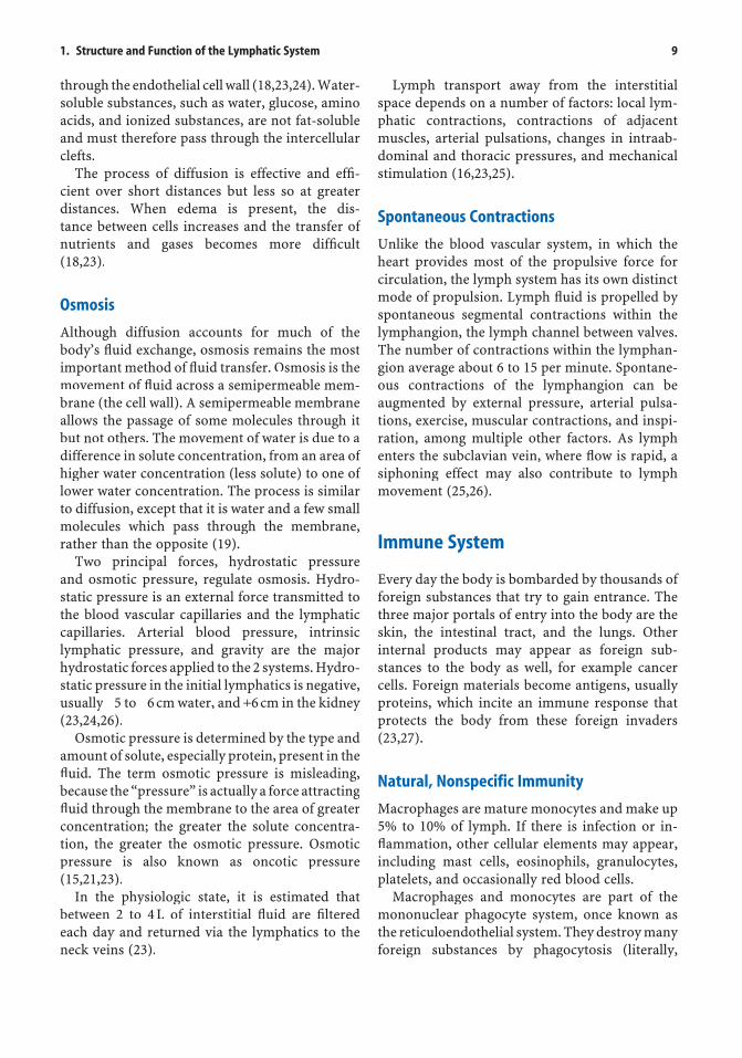
Immune System
Every day the body is bombarded by thousands of foreign substances that try to gain entrance. The three major portals of entry into the body are the skin, the intestinal tract, and the lungs. Other internal products may appear as foreign sub-stances to the body as well, for example cancer cells. Foreign materials become antigens, usually proteins, which incite an immune response thatprotects the body from these foreign invaders (23,27).
Natural, Nonspecific Immunity
Macrophages are mature monocytes and make up5% to 10% of lymph. If there is infection or in -flammation, other cellular elements may appear,flincluding mast cells, eosinophils, granulocytes,platelets, and occasionally red blood cells.
Macrophages and monocytes are part of the mononuclear phagocyte system, once known asthe reticuloendothelial system. They destroy many foreign substances by phagocytosis (literally,

10 L.L. Tretbar
eating cells) and by secreting enzymes destructiveto the antigen. They maintain a portion of the nonspecific immune system, the system that doesfinot adapt to different antigens (17,27,29).
Specific Immunity
Many other cellular products make up lymph. Lymphocytes are the most abundant cell, repre-senting about 80% to 85%, whereas they encom-pass only 20% to 40% of the blood’s white cells.Found in both blood and lymph, they contribute to immune and phagocytic activities. Two types of lymphocytes develop: B cells that mature in the bone marrow, and T cells that mature in thethymus.
B cells are present in the germinal centers of lymph nodes and are found frequently in bonemarrow and the spleen. When an antigen entersthe body, B cells produce noncellular antibodies,or immunoglobulins, which contribute to the immune defense. Both cellular and noncellular substances complement the primary naturalimmune system (17,23,28).
When activated by a foreign substance, B cellsmay divide into 2 other types of cells: plasma cells and memory cells. Plasma cells are those that produce antibodies (immunoglobulins). Memory cells are what their name implies—they act as a memory of previous antigen invasions and protect the body for many years from the antigen, e.g.tetanus or small pox (17,18,23).
T cells are also found in blood (80% to 85% of lymphocytes), lymph nodes, the thymus, and the spleen. They, too, produce subsets of cells. Generally, they assist B and other T cells by aligning antigens in proper order for destruction.There are memory cells in this group as well (18,19,23).
Antibodies
Numerous stimuli cause lymphocytes to produce antibodies. A number of antibodies, or immuno-globulins, circulate within the vascular systems and may deactivate antigens or tag them for later destruction. Immunoglobulin G (IgG) is the main immunoglobulin of the body; others include IgM, IgA, IgD, and IgE. All have rather specific duties fiwithin the immune system (17–19,23,29).
Summary
Anatomically, the lymphatic system consists of multiple channels that begin in the periphery of the body, run through a series of lymph nodes,and return lymph to the venous system. In the normal state, the lymphatic system equilibrates the fl ow of interstitial flfl uid and mediates the many flimmune elements for the protection of the inter-nal systems.
References
1. Bartholinus, Th. De lacteis thoracis in homine brutisque nuperrime observatis. Copenhagen.Martzan M, 1652.
2. Hippocrates. The Genuine Books of Hippocrates. Adams F, trans. London: Sydenham Society;1849.
3. De Lactibus Siue lacteis venis Quarto Vasorum Mes-araicorum genere Nouo Inuento. Gasparis AselliCremon Is, Anatomici Ticinensis, Dissertatio. Milan: Baptum Bidellium; 1627.
4. Foster M. Lectures on the History of Physiology.Cambridge: Cambridge University Press; 1901.
5. Choulant JL. History and Bibliography of Anatomic Illustration. Frank M, trans. and annot. New York:Hafner Publishing Company; 1934.
6. Rudbeck O. Nova exercitation anatomica, exhibens ductus hepaticos aquosos et vasa glanularum serosa,nunc primum in venta, aeneisque figures delineate.fifiVesteras: E Lauringer; 1653.
7. Fulton JF. The early history of the lymphaticswith particular reference to Bartholin, Rudbeck,and Joliffe. Bull Hennepin County Med Society.1938;9:5–10.
8. Harvey W. Exercitatio anatomica de motu cordis et sanguini in animalibus. Frankfurt: W Fitzer;1628.
9. Wyall H. William Harvey. London: Parsons; 1924.10. Hunter W. Medical Commentaries. No. 26. London:
1762.11. Mascagni P. Vasorum lymphaticorum corporis
humani description et iconographi. Siena: P Carli; 1787.
12. Picard J-D. Lymphatic Circulation. Lavaur, France:Editions Médicales Pierre Fabre; 1995.
13. Rouviere H. Anatomy of the Human Lymphatic System. trans. Tobias MJ, Ann Arbor, MI: MJEdwards; 1938.
14. Sappey P. Anatomie, physiologie, pathologie des vaisseaux lymphatiques consideres chez l’homme et les vertibres. Paris: A Delahaye; 1874.

1. Structure and Function of the Lymphatic System 11
15. Kubik S. Zur Klinischen Anatomie des Lympsys-tesm, Vehr Anat Ges. 1975.
16. Bollinger A, Partsch H, Wolfe J, eds. Initial Lymphat-ics, New Methods and Findings. International Sym-posium; Zurich 1984. Stuttgart and New York: Georg Thieme Verlag; 1985.
17. Olszewski WL. Peripheral Lymph: Formation and Immune Function. Boca Raton, FL: CRC Press;1985.
18. Weissleder H, Schuchhardt C, eds. Lymphedema: Diagnosis and Therapy. 3rd ed. Bonn: Kagerer Kommunikation; 2001.
19. Földi M, Kubik S, eds. Lehrbuch der Lymphologie für Mediziner. 5th ed Munich-Jena: Urban & Fisher; 2002.
20. Zhdanov DA. General Anatomy and Physiology of the Lymphatic System. Leningrad: Medgiz; 1952.
21. Kappert A, ed. Lehrbuch und Atlas der Angiologie:Erkrankungen der Arterien, Venen, Kapillaren und Lymphgefässe. 12th ed. Bern: Verlag Hans Huber;1989.
22. Casley-Smith JR. Lymph and lymphatics. In: Micro-circulation. New York: University Park Press; 1977;1:421–430.
23. Kasseroller RG. Compendium of Dr. Vodder’sManual Lymph Drainage. Heidelberg, Germany:Haug; 1998.
24. Guyton AC, Granger HJ, Taylor AE. Interstitial fluidflpressures. Physiol Rev. 1971.
25. Yoffey JM, Courtice FC. Lymphatics, Lymph and Lymphoid Tissue. London: Arnold; 1956.
26. Kubik S. The Lymphatic System. New York: Springer;1985.
27. Olszewski WL, Engeset A. Immune proteins, enzymes, and electrolytes in human peripherallymph. Lymphology. 1978;11:156–164.
28. Ryan TJ, Mallon EC. Lymphatics and the processingof antigen. Clin Dermatol. 1995;13(5):485–492.
29. Olszewski WL, Grzelak I, Ziolkowska A, Engeset A. Immune cell traffic from blood through the finormal human skin to lymphatics. Clin Dermatol.1995;13:473–483.

12
2Differential Diagnosis of Lymphedema
Simon J. Simonian, Cheryl L. Morgan, Lawrence L. Tretbar, and Benoit Blondeau
Pathophysiology of Edema
As described in chapter 1, there is always fluidflwithin the interstitial space, the space between tissue cells. The amount of fluid depends on 2flfactors: the amount introduced into the interstitialspace, and the amount removed from it. Fluid enters the space from arterioles and venules; some returns to the venules, and the remainder is takenup by the lymphatics. In the normal physiologicstate, entrance and exit are approximately equal,so that tissues retain their usual morphologic appearance and function (1,2).
Edema (swelling) develops when the volume of interstitial fluid increases, either from increasedflinfl ow or decreased outflfl ow, or both (3).fl
Lymphatic Causes of Edema
Primary Lymphedema
Primary lymphedema results when the lymphat-ics do not or cannot propel the lymph in adequateamounts, and the fl uid sequesters within the inter-flstitial or lymphatic spaces. It develops from analteration or defi ciency within the lymphatic col-filecting or transport systems. Primary lymph-edema often occurs in the lower extremities, andaffects women more than men (4–6). Many lym-phatic malformations are described in chapter 4;a few are briefly reviewed below:fl
• Milroy disease (hereditary lymphedema type I) is a familial congenital disease and appears at orsoon after birth (7,8) (Figure 2-1).
• Meige disease (hereditary lymphedema type II) develops later, e.g. at puberty, often after a minorinjury, and causes foot and ankle swelling. Girls are affected more often than boys (9,10).
• Lymphedema praecox, another term for the2 syndromes described above, has an early onset up to age 35 years (10,11).
• Lymphedema tardum is similar to praecox, buthas an onset after age 35 years (10,11).
• Lymphangiomas are congenital, benign, often cystic malformations of the lymphatics and may be associated with other vascular malforma-tions (4,12).
Secondary Lymphedema
Secondary lymphedema is swelling that follows some other incident or event, such as infection orinjury (12,13).
Infection
• Filariasis, the most common cause of secondary lymphedema in the world, affects patients who have lived in or traveled in areas endemic with the disease; the worm’s larvae migrate to thelymphatics, causing obstruction and damage(13,14) (Figure 2-2).
• Recurrent cellulitis, e.g. erysipelas, is acute and unilateral in the affected limb, often enteringskin from a fungal skin infection (15,16).
• Lymphogranuloma venereum, a sexually trans-mitted disease, caused by chlamydia, often with enlarged inguinal lymph nodes.
• Scrofula, old term for tuberculous lymph nodesof the neck.

2. Differential Diagnosis of Lymphedema 13
FIGURE 2-1. (A) An example of Milroy disease, the right leg of this 2-year-old baby has been swollen since birth. (B) This woman had mild swelling of the legs noted at age 10 years. The swelling wasprogressive until her lymphedema was finally diagnosed andtreated at age 36. Dermal backflow is more prominent in the right leg, but is present in both (see Figure 2.8). The feet show thetypical changes of lymphedema—swelling of the toes and feet,with deep flexion creases. (Photos courtesy Emily Iker.)
FIGURE 2-2. Filariasis, as seen in this man’s legs, is endemic inHaiti and many other countries. (Photo courtesy CL Morgan.)
crystal absorption into the foot with lymphaticdamage (Figure 2-3)
Cancer Treatment and Other Types
of Trauma
• Any treatments, whether surgical or irradiation,can interrupt the normal fl ow of lymph, that flresults in the accumulation of lymph, i.e., lymphedema.
• Lymphadenectomy, or surgical excision of the inguinal, iliac, or auxiliary lymph nodes, themost common non-infectious cause worldwide, especially following breast cancer treatment, of chronic unilateral swelling (17).
• Nonsurgical trauma, e.g. radiation therapy tolymph node groups, may cause chronic unilat-eral swelling.
• Surgery of the prostate, uterus, or cervix may cause bilateral swelling.
• Recurrent and metastatic malignancy.• Hodgkin and non-Hodgkin lymphoma.• Reconstructive arterial surgery, e.g. saphenous
vein harvest for coronary artery bypass
A
B
Inflammation
• Any of a group of non-infectious diseases causing edema, pain, or erythema.
• Systemic lupus erythematosus• Rheumatoid arthritis, which may have com-
bined muscle pump failure due to fixation and fistiffening of the joints.
• Psoriatic arthritis• Chronic dermatitis• Retroperitoneal fibrosisfi• Panniculitis• Systemic diseases, e.g. Grave’s disease, myxedema• podoconiosis, elephantiasis caused by silica

14 S.J. Simonian et al.
A
B
FIGURE 2-3. (A) Although the swelling in this patient’s hands ismild, it nevertheless represents myxedema. (B) Her feet begin toshow typical changes of lymphedema, with deepening of theflexion creases and swelling of the toes. (Photos courtesy CL Morgan.)
FIGURE 2-4. (A) This 46-year-old woman had surgery and radia-tion for uterine cancer. Lymphedema in her left leg is confirmedby the lymphoscintigram, which shows marked dermal backflow.(B) In another patient, the right greater saphenous vein was har-
vested for a coronary artery bypass; lymphatic damage caused later lymphedema. Here the left leg shows the changes of chronicvenous insufficiency with hyperpigmentation, slight edema, andhyperkeratosis. (Photos courtesy Emily Iker.)
A B

2. Differential Diagnosis of Lymphedema 15
• Factitious, self-infl icted trauma, e.g. constrict-fling a limb with a rubber band to induce swelling (18).
• External injury to the body, e.g. an auto accident (Figure 2-4)
Other Causes of Edema
Chronic Venous Insufficiency
As explained in chapter 1, properly functioning venous and lymphatic systems help maintain a balance between interstitial and lymphatic fluid. A flchange in one system affects the other (12,19–22).
With venous disease in the legs, there is usually chronic damage to the veins and their valves. The result of valve failure is continuing reflux fl(backward flow of blood), increasing pressure on flnormal veins and damage to the surroundingtissues and lymphatic structures—chronic venousinsufficiency (4,20,22). (See chapter 7 for morefiinformation.)
Hypoalbuminemia
Reduced blood concentration of proteins, espe-cially albumin, reduces osmotic (oncotic) pressureand the reabsorption of interstitial fluid into the flvenous capillaries. Swelling is usually chronic,often bilateral (12,17).
• Excessive loss of albumin, glomerulonephritis,nephrotic syndrome, extensive burns.
• Malabsorption syndromes, inadequate ab -sorption of protein, steatorrhea syndromes,protein calorie malnutrition, starvation, e.g.Kwashiorkor.
• Inadequate synthesis of albumin, cirrhosis, liverfailure.
Drug-induced Edema
Several medications can cause or contribute to edema, complicating diagnosis (12,17,18). They include:
• hormones, estrogens, testosterone, corticoste-roids, progesterone, and androgens
• antihypertensives, guanethidine, β-adrenergic blockers, calcium channel blockers, clonidine, hydralazine, methyldopa, minoxidil, reserpine,and labetalol
• nonsteroidal anti-in ammatory drugs, (NSAIDs)fl• antidepressants, e.g. trazodone• hypoglycemics, e.g. pioglitazone and
rosiglitazone• cytokines: GM-CSF, G-CSF, Il-4, Il-2,
Interferon-a• chemotherapeutics, e.g. cyclophosphamide,
cyclosporine, mitramycin, and cytosinearabinoside
• antiviral, e.g. acyclovir
Differential Diagnosis of Edema
The differential diagnosis of edema requires a thorough historical review, physical examination,evaluation of treatment response, and occasion-ally special laboratory investigations. The exami-nation report should include a diagram of thelimb or affected area and standard measurements, e.g. limb circumference. Staging using these crite-ria is discussed in chapter 3.
Duration and Distribution
If the duration of the swelling is short, perhaps hoursor days, and it is unilateral and painful, an acuteprocess is suggested: deep vein thrombosis, cellulitis,abscess, ruptured Baker cyst, trauma, muscle com-partment syndrome, ruptured gastrocnemius muscle, or re ex sympathetic dystrophy (12,18).fl
If the onset is gradual and progressive, over aperiod of weeks or months, and it is unilateral and painless, a chronic process is suggested: chronic venous insufficiency, post-thrombotic syndrome, filymphedema, (primary or secondary type), exter-nal venous compression, arteriovenous fistula, fiKlippel-Trenaunay syndrome, and soft-tissue orvascular tumors (12,17).
If the onset is gradual and progressive and swelling is bilateral, possible causes include:congestive heart failure, glomerulonephritis, thenephrotic syndrome, hypoproteinemia, drug reac-tions, cirrhosis of the liver, pretibial myxedema, constrictive pericarditis, lower limb dependency syndrome, lipedema, bilateral chronic venous insufficiency, bilateral lymphedema, or a malig-financy in the pelvis, abdomen, or the retroperito-neal space. Advanced prostate carcinoma cancause lymphatic obstruction and bilateral legedema, as can ovarian carcinoma or other pelvic tumors (12,17,18).

16 S.J. Simonian et al.
Dermatologic Changes
Skin changes are common with many types of swelling (22,23). They include:
• venous hypertensive pigmentation due to hemo-siderin deposition, eczema, atrophie blanche(white atrophy), lipodermatosclerosis, or ulcer-ation (Figure 2-5)
• taut skin, the inability to pinch a fold of skinat the base of the second toe (Kaposi-Stemmersign), or prominent skin creases in the feet
• hyperkeratosis or papillomatosis (a warty skin texture)
• cellulitis, especially when recurrent (Figure2-6)
Diagnostic Laboratory Tests for
Systemic Disease
• The cardiopulmonary system is evaluatedwith chest x-rays, electrocardiograms, andechocardiograms.
• Kidney and liver functions are assessed with standard blood testing, which should include the blood albumin concentration.
• Thyroid function is also tested with standardblood tests, e.g. T3, T4.
FIGURE 2-5. Superficial venous insufficiency caused these advanced cutaneous complications—edema, pigmentation, eczema, andulceration. (Photos courtesy LL Tretbar.)
Testing for Venous Disease
• different non-invasive and invasive tests areused to evaluate the veins for blood clots and function
• D-dimer, blood testing for deep veinthrombosis
• ultrasonographic imaging, to assess the deep, perforator, and superficial venous systems of thefilegs for evidence of old deep vein thrombosisand for the degree of valvular incompetence andreflux (Figure 2-7)fl
• contrast venography, to check for the presenceof pelvic or abdominal thrombus
• computed tomography (CT) to check for pelvicpathology, especially malignancies and retro-peritoneal fibrosisfi
• assessment of the ankle-brachial pressure index (ABPI), which is useful in determining arterialinsuffi ciency in the legs of older patients andfidiabetics; compression therapy may worsenperipheral arterial disease.
Testing for Lymphedema
Lymphangioscintigraphy is the best laboratory test for lymphedema. A radioisotope-labeledcolloid is injected into the web space between the

2. Differential Diagnosis of Lymphedema 17
FIGURE 2-6. This man suffered a right deep vein thrombosis 12years before this photograph was taken. There is marked hyper-pigmentation, eczematoid changes in the calf with white atrophy,and small ulcerations on the ankle. Little edema had developed. (Photo courtesy LL Tretbar.)
FIGURE 2-7. Duplex ultrasonography is noninvasive, safe, andreproducible; it is relatively inexpensive and exhibits a high degree of accuracy. (Photo courtesy LL Tretbar.)

18 S.J. Simonian et al.
first and second toes. Using a gamma camera, theficolloid movement is measured as it travels toward the proximal lymph nodes (24–26) (Figure 2-8). Slow progress of the radioisotope, compared withthe normal lower limb, suggests hypoplasia of theperipheral lymphatics, as in primary lymphedema. If the radioisotope escapes from the main lymphchannels, especially into the skin, it is called dermal backflow. This fifl nding suggests lymphfireflux, often seen in secondary lymphedema with flproximal lymph obstruction.
Lymphangiography is another option that is rarely used today. This test uses radio-opaque lipi-odol injected directly into a peripheral lymphvessel; x-rays monitor its proximal progress. This test is used for planning surgical lymphovenousanastomoses and for research purposes (27,28).
Both computed tomography (CT) and magnetic resonance imaging (MRI) show a typical sub-cutaneous honeycomb pattern in lymphedema, but
B
A
FIGURE 2-9. In the (A) sagittal view and (B) axial view of the lower extremities, an MRI shows an extensive honeycombed pattern inthe soft tissue, a hallmark of chronic lymphedema not found inother edemas. Here the muscle compartment is unchanged,although it may be enlarged in the postthrombotic syndrome. MRIs can also provide quantitative assessment of lymphedemavolume. (Photos courtesy BB Lee.)
not in other edemas. Of the 2, MRI is the superior test, as it also detects excess fluid (Figure 2-9).fl
Lipedema
In determining lipedema, an MRI will show that the soft-tissue swelling consists only of fat. The peripheral lymphatics are normal; there is nohoneycomb appearance, and subcutaneous edemais absent. However, late lymphatic changes may develop (27) (Figure 2-10).
BA
FIGURE 2-8. Lymphangioscintigraphy remains the basic test forassessing lymphatic function within the extremities. (A) Thisnormal scan at 20 minutes demonstrates bilateral proximal flow of colloid that opacifies the inguinal, iliac, and axillary nodes and liver. (B) At 40 minutes, most of the tracer has dissipated. When the test is abnormal (see Figure 2-4), dermal backflow can be seenas the tracer leaks from the lymphatics. (Photos courtesy BB Lee.)

2. Differential Diagnosis of Lymphedema 19
A
B C
FIGURE 2-10. (A) Lipedema is characterized by a swelling of the hips and legs, but usually with none in the feet. In this case, alymphoscintigram was normal. The fatty changes developed over a number of years; the feet are beginning to show lymphedema-tous changes, e.g. deep flexion creases and swelling of the dorsumand toes. (Photo courtesy Emily Iker.) (B) This woman’s legs werenormal until she had a car accident that traumatized them. Thelipedema developed only in her legs; the torso and feet werespared. An ulcer that formed after a recent infection is seen on her left leg. (Photo courtesy CL Morgan.) (C) Diagnosis is difficult inthis case: the feet are reasonably normal, but there are changesin the lower calves that suggest venous insufficiency. Extremeobesity is apparent as well, with a panniculus hanging below thepatient’s clothing. This patient deserves a complete evaluation that should include lymphangioscintigraphy. (Photo courtesy LLTretbar.)

20 S.J. Simonian et al.
Summary
Information obtained from clinical and labora-tory testing can help determine the probable causeof edema. Clinical evaluation with a thoroughmedical history is usually sufficient, as laboratory fitesting (e.g. lymphangioscintigraphy, MRI) is not always available. As accurate diagnosis is impor-tant, so that an appropriate treatment programcan be planned and expectations of the treatmentcan be reviewed with the patient.
References
1. Drinker CK, Field E, Ward HK, Leigh OC. The com-position of edema fluid and lymph in edema and flelephantiasis resulting from lymphatic obstruction.Am J Physiol. 1934;109:572–586.
2. Ryan TJ, DeBerker D. The interstitium, the connec-tive tissue environment of the lymphatic, andangiogenesis in human skin. Clin Dermatol.1995;13(5):451–458.
3. Daroczy J. Pathology of chronic lymphedema.Lymphology. 1994;6:91–106.
4. Browse NL, Stewart G. Lymphoedema: patho-physiology and classification.fi J Cardiovasc Surg.1985.
5. Olszewski WL. Lymph Stasis: Pathophysiology, Diag-nosis, and Treatment. Boston: CRC Press; 1991.
6. Cluzan RV. Lymphatics and edema. In: Cluzan RV, Pecking AP, Lokiec FM, eds. Progress in LymphologyXIII.Amsterdam: Excerpta Medica, Elsevier Science;International Congress Series; 1992:716–717.
7. Nonne M. Vier Falle von Elephantiasis Congenita Hereditaria. Virchow’s Arch Pathol Anat. 1891;125(1):189–196.
8. Milroy WF. Chronic hereditary edema: Milroy’s disease. JAMA. 1928;91:1172–1175.
9. Dale RF. The inheritance of primary lymphoedema. J Med Genet. 1985;22:274–278.
10. Kinmonth JB, Taylor GW, Tracy GD, Marsh JD.Primary lymphoedema. Br J Surg. 1957;45(189):1–9.
11. Wright NB. The swollen leg and primary lymph-edema. Arch Dis Child. 1994;71:44–49.
12. Weissleder H, Schuchhardt C, eds. Lymphedema:Diagnosis and Therapy. 3rd ed. Bonn: KagererKommunikation; 2001.
13. Dreyer G, Addiss D, Dreyer P, et al. Basic Lymphoe-dema Management, Treatment and Prevention of Problems Associated with Lymphatic Filariasis.Hollis, NH: Hollis Publishing; 2002.
14. Olszewski WL, Jamal S. Recurrent dermatolymphan-gioadenitis (DLA) is responsible for progression of lymphedema. Progress in Lymphology XV.Lymphology. 1996;(Suppl.)29:331–334.
15. Herpertz U. Lymphödem und Erysipel. Lymph-Forsch. 1998.
16. Blumberg H, Janig W. Clinical manifestation of reflex sympathetic dystrophy and sympathetically flmaintained pain. In: Wall P, Melzack R, eds. Text-book of Pain. Edinburgh: Churchill Livingstone;1993.
17. Földi M, Kubik S, eds. Lehrbuch der Lymphologie für Mediziner. 5th ed. Munich-Jena: Urban & Fisher;2002.
18. Browse N, Burnand K, Mortimer P, eds. Diseasesof the Lymphatics. London: Arnold; 2003.
19. Blondeau B, Helling TS, Morgan, CL. Insuffisancefiveineuse et obesité. Phlebologie. 2003;56.
20. Klippel M, Trenaunay P. Du naevus variqueux osteo-hypertrophique. Arch Gen Med. 1900;185:641–672.
21. Tretbar LL. Venous Disorders of the Legs. London:Springer-Verlag; 1999.
22. Partsch H, Urbanek B, Wenzel-Hora B. Dermal Lymphangiopathie ei chronisch venoser Insuffi-fizienz. In: Bollinger A, Partsch H, eds. Initiale Lym-phstrombahn-Internationales Symposium. Zurich:Thieme; 1984:205–209.
23. Ramelet AA, Monti M. Phlebologie. 2nd ed. Bonn:Kagerer Kommunikation; 1993;22:238–239.
24. Stemmer R. Ein klinisches Zeichen zur Früh-und Differentialdiagnose des Lymphodems. Vasa. 1976;5(3):261–262.
25. Pecking A, Cluzan R, Desprez-Cureley A. Indirect lymphangioscintigraphy in patient with limb edema.In: Heim L, ed. Immunology and Hematology Research Foundation; 1984;3(4):327–328.
26. Weissleder H, Weissleder R. Lymphedema: evaluation of qualitative and quantitative lymphangioscintigraphy in 238 patients. Radiol.1988;167(3):729–735.
27. Hummel E, Weissleder H. Lymphgefasse bie Lipodem. In: Clodius L, Baumeister RGH, Foldi E,eds. Lymphologica. Munich: Medikon; 1989;16:89–98.
28. Picard J-D. Lymphatic Circulation. Lavaur, France:Editions Médicales Pierre Fabre; 1995.

21
3Classification and Staging of Lymphedema
Cheryl L. Morgan and B.B. Lee
Introduction
Although there have been numerous internationalattempts to classify and stage lymphedema, thereis no single method that provides a comprehen-sive view of the disease that defines its etiopatho-fiphysiology or describes the significant overlap fibetween the recorded stages. Not only is the process of classifi cation and staging defifi cient, it is ficonfusing to many physicians because the defini-fitions and terminology differ widely.
Therefore, the authors propose a method of staging that includes the spectrum of identifiable fidisease processes, defined not only by physical fichanges but also by their etiopathophysiology.Further, an improved method of staging shouldcoalesce research, and help advance educationalprograms for the health professionals.
This chapter presents some of the controversialaspects of current staging methods and a new pro-posed staging system for lymphedema. However,until a consensus is reached for the adoption of amore standardized approach, the current stagingsystems may be used.
Condition
When evaluating a swollen limb or tissue, first its ficondition, the general type of change in the tissue,is described. This may be a cursory look at the swelling, or a more specifi c and thorough descrip-fition of the overall changes that cause it to differ from the normal limb or tissue. One traditional
idea is that lymphedema is a simple accumulationof fluid or lymph in the tissues; we now know, flhowever, that the development of lymphedemarepresents a dynamic, actively changing process.Chronic lymphedema can no longer be considered a benign, static condition, but is a progressive anddegenerative disease (1,2). This demands that all changes in the tissues be acknowledged, not justthe swelling.
The importance of accurate staging is obvious—without proper intervention at the appropriatetime, the condition is destined to develop seriouscomplications (3). Improved classification and fistaging standards can better identify these com-plications and determine the most useful inter-ventions, all of which depend on the generalhealth, age, and medical history of the patient.
Classification
Currently, the classification of lymphedema isfidefined by the origin of the condition, i.e. primary fior secondary (4). It must be remembered that clas-sifi cation of lymphedema is not a staging tech-finique. This system (of classification) is used fiinternationally and meets the authors’ presentexpectations.
Primary Lymphedema
Simply defi ned primary lymphedema results fromfiany defi ciency of proper lymphatic developmentfithat cannot be attributed to injury, trauma, illness, treatment, or disease, e.g. medication, radiation, or phlebolymphedema (4–6).

22 C.L. Morgan and B.B. Lee
Depending on the patient’s age when clinicalsymptoms manifest, primary lymphedema hastraditionally been classifi ed into 2 groups:filymphedema praecox becomes evident early,and up until age 35 years; lymphedema tardum develops at or after age 35. There is some suspi-cion that lymphedema tardum may be a second-ary lymphedema; often there is little evidence of congenital defects to ascertain its etiology (4,6)(see chapter 4).
Primary lymphedema can also be categorized as congenital or hereditary forms. Hereditary forms are familial and may be traced. The con-genital form represents a birth defect in the lym-phatic system caused by a genetic abnormality during embryogenesis. Using this classification,fithe true congenital type is limited to only a frac-tion of primary lymphedemas that are apparentat birth. Not all lymphedemas presenting at birthare a consequence of a genetic abnormality (7); lymphedematous defects can be secondary to traumatic events during pregnancy.
To further confound the issue, not all congenital lymphedemas are clinically detectable at birth but
may manifest later. These variations in presenta-tion make it diffi cult, if not sometimes impossible,fito classify the involvement of the affected body part (6,8,9).
Secondary Lymphedema
Secondary (acquired) lymphedema can resultfrom many external factors that alter, both ana-tomically and functionally, an otherwise normal lymph-conducting system (6,10). Identifying theexact cause of the swelling is the most important part of the diagnostic investigation. Eight caus-ative factors are usually considered (11–15): trauma, malignancy, venous disease, infection,infl ammation, immobility, parasites, and facti-fltious conditions.
Because several causes of secondary lymph-edema can be treated, with a reasonable chance of recovery, no effort should be spared in this criti-cally important part of chronic lymphedema management. Proper etiologic identification andfitreatment may also halt the progression of the
BA
FIGURE 3-1. (A) This represents a secondary stage I lymphedema.The edema recedes with elevation and there are no other physicalproblems. Bilateral stripping of the saphenous veins for varicosities in this 36-year-old woman caused enough lymphatic damage to the stripping sites that lymphedema developed. Note the loss of the Achilles space and the hyperemia of the legs. Four weeks of
complete decongestive therapy reduced the distal symptoms, and the patient continued home self-care. (B) A case of primary lymph-edema, stage 1. [Both images (A and B), are photographs taken atinitial evaluation.] Primary lymphedema is present in the right leg of this 15-year-old girl, whose symptoms appeared at age 12 withthe onset of menses. (Photographs courtesy of CL Morgan.)
3. Classification and Staging of Lymphedema 23
disease; there is no cure for an already damagedlymphatic system. All lymphedemas should beconsidered secondary until proven otherwise (13,14).
Obviously, signifi cant ambiguities exist in the fidiagnosis of primary and secondary lymphede-mas. Unfortunately, the present classification and fistaging systems fail to clarify them. Therefore, an advanced staging system can assist in proper diagnosis.
Staging
Once an etiologic diagnosis of lymphedema isestablished, staging based on clinical manifesta-tions is mandated. Staging provides a reasonably objective system recording abnormalities andplays an essential role in the planning of appropri-ate medical intervention and in determining the effectiveness of the intervention. New stagingstrategies should provide more defi nitive catego-firization of the disease, based on the degree andseverity of anatomic and physiologic changes(Figures 3-1 to 3-3) (15,16).
Staging of any disease process should accom-plish at least 5 primary goals:
1. Evaluate and identify the distinctive character-istics of the condition, e.g. possible etiology and symptoms.
2. Determine the duration, extent, and severity of the disease.
3. Outline appropriate medical intervention andexpected outcomes.
4. Help patients understand their disease, possi-ble management options, and anticipated results, and promote adherence to recom-mended treatment.
5. Help insurance companies ascertain the possi-ble expenses necessary to obtain the anti -cipated outcome.
Current Staging Methods
The Consensus Document on the Diagnosis andTreatment of Peripheral Lymphedema was first fipublished in 1995 by the International Society of Lymphology (ISL) and updated in 1998 following
the ISL Congress held in Madrid the prior year (17). However, neither document mentioned staging. The fi rst description of staging was in the firevised ISL Consensus Document of 2001, pub-lished in Lymphology that same year (17). The staging was enumerated as follows:
Stage I represents an early accumulation of fluid flrelatively high in protein content (when com-pared with venous edema), and which subsides with limb elevation.
Stage II occurs when limb elevation rarely reducestissue swelling and pitting is manifest. As tissuefibrosis supervenes late in stage II, the limb may fior may not pit.
Stage III encompasses lymphostatic elephantiasis, in which the skin does not pit, and trophic skinchanges such as acanthosis, fat deposits, and warty overgrowths develop.
The severity of each stage is based on volumedifferences: minimal, <20% increase in limb volume; moderate, 20% to 40% increase in limb volume; severe, >40% increase in limb volume.
The most recent revision of the ISL ConsensusDocument (2003) describes 4 stages of lymph-edema, based on risk, local skin changes, andthe severity and degree of edema (17). This revi-sion includes stage 0, described as a latent or sub-clinical condition where swelling is not evident despite impaired lymph transport. It may exist months or years before overt edema occurs. Many lymphologists contend that stage 0 representsonly the risk of developing lymphedema and may create diffi culty with insurance coverage and inap-fipropriate expectations for future treatment plan-ning (18).
The 2003 Consensus Document is the docu-ment most referred to in publications. It is incor-rect to identify it as the staging system endorsedby the ISL; the document itself states, “. . . a moredetailed and inclusive classification needs to be fiformulated in accordance with understanding of the pathogenic mechanism of lymphedema” (17).
A few examples of staging systems used across the globe are presented here to illustrate the com-plexities involved and the need for standardiza-tion to adequately address various issues, from diagnosis to intervention.

24 C.L. Morgan and B.B. Lee
BA
FIGURE 3-3. (A) This is an example of stage III lymphedema sec-ondary to surgery and radiation for melanoma. This 38-year-old male presents with significant volume increase of the left leg, mul-tiple hospitalizations for cellulitis, and recent disability from thesize and weight of the limb. Symptoms presented immediatelyafter groin dissection. Postradiation, the swelling involved thegenitalia, and 6 months later the right leg began to swell. (B) This
67-year-old woman presented with massive lymphedema of both lower extremities, which she reports she has had since childhood.Unable to ambulate, dress, bathe, or transfer without assistance,she has recently experienced 3 episodes of cellulitis and recurrentskin breakdown. Distally, the edematous tissues are nonpitting,with significant trophic changes, including acanthosis and multiple skin folds. (Photos courtesy of CL Morgan.)
BA
FIGURE 3-2. (A) This 52-year-old woman presented with a history of swelling of approximately 7 years, 9 pregnancies, and diabetes. Venous studies reveal moderate reflux in both saphenous veins. Edema is not reduced with elevation, significant fibrosis is evident,and 3 to 4 episodes of cellulitis per year have been reported for thepast 3 years. This lymphedema is considered stage II and secondary to venous insufficiency under the current ISL system. (B) Complica-
tions of diabetes and obesity experienced by this 61-year-old woman have led to 8 hospitalizations for cellulitis in the past 22 months. She has reported symptoms since she was 9 years old that suggest primary lymphedema, and that her mother and sister have “heavy legs.” Extensive fibrosclerosis is present, and distally theskin will no longer pit with pressure. (Photographs courtesy of CL Morgan.)

3. Classification and Staging of Lymphedema 25
Földi Method of Staging
For years the Földi staging system has been thepredominant method of classification among fiEuropean and American clinicians. In recentpublications, it too has included a stage 0 (14). Thestages are detailed as follows:
Stage 0, latency: asymptomatic, focal fibro -fisclerotic tissue changes, diagnosis by lymphangioscintigraphy.
Stage I, spontaneously reversible: high-proteinedema; focal fibrosclerotic tissue alterations; fipitting edema reduced by elevation; possible“pain of congestion”; diagnosis by basic procedures.
Stage II, spontaneously irreversible: extensivefibrosclerosis; proliferation of adipose tissue;fibrawny hard swelling not reduced with eleva-tion; diagnosis by basic procedures.
Stage III, elephantiasis: extensive fibrosclerosis (as fiwith stage II); proliferation of adipose tissue, invalidism; diagnosis by basic procedures.
Pitting Edema Scale
Pitting edema refers to an indentation of the skinthat occurs when simple fi nger pressure is applied. fiIf the skin can pit, the inference is that the onset of swelling is recent and therefore less serious. Edema is not detectable clinically until the inter-stitial fluid volume reaches 30% above normalfl(19). The pitting edema scale is detailed as follows:
1+ edema is barely detectable2+ a slight indentation occurs with pressure and
remains momentarily3+ a deeper impression occurs, but skin returns to
normal within 5 to 30 seconds4+ the impression remains more than 30 seconds
Nonpitting edema is present when pressure is applied but no indentation occurs. This suggeststhat more advanced subcutaneous tissue changeshave developed and fl uid cannot be displaced with flmere pressure. Although this manual measuringtechnique has been replaced by more objective and reliable measuring devices, like the tonometer,it is still widely used by physicians and health care professionals.
Staging by Limb Size
As simple as it is to measure the diameter of the limbs, there is signifi cant concern by health fiprofessionals that this system does not adequately represent the patient’s true condition. It doesnot, for example, provide for the evaluation of lymphedema of the trunk or other parts of thebody, nor does it indicate the duration of thedisease process, the development of skin changes,or other important considerations required forplanning management and determining outcomeresults (20). Staging by limb size is determined as follows:
Mild: less than 3 cm differential between affected and unaffected limbs
Moderate: 3 to 5 cm differential between affected and unaffected limbs
Severe: more than 5 cm differential between limbs
All of these examples of staging are internation-ally adapted, as indicated in the chapter’s intro-duction. These attempts by health care professionals to modify the current methods described above are indicative of a need for change. This further complicates communications with patients orinsurance companies in describing the severity, treatment options, outcomes, and length of treat-ment usually required to achieve the goals.
In many of the staging models, the values assigned to each stage primarily describe compli-cations of changes in tissues of affected limbs (20,21). These changes range from lymphoceles,fistulas, vesicles, chylous reflfi ux, skin folds, and fleczemas, to sepsis, infections, and chronic wounds.Described as knobs, bumps, protuberances, blis-ters, sclerosing panniculitis, lipodermatosclerosis, or mossy foot, all are manifestations of impaired lymphatics that present differently but are not well described in literature or medical reference materials (21).
Staging by Clinical Symptoms
Clinical evaluation is the most common method used to diagnose and stage lymphedema, since laboratory tests are too expensive or simply not available. This particular staging system addressesspecifi c changes in the lower extremities of fipatients with lymphedema secondary to parasitic

26 C.L. Morgan and B.B. Lee
infections. It was developed by physicians and theresearch team for the Lymphatic Filariasis Preven-tion and Treatment Program in Recife, Brazil (12).(Another, completely different scale identifyingonly 4 stages was devised by the Brazilian Society of Lymphology [22]).
Staging by clinical symptoms is detailed asfollows:
Stage I: swelling increases during the day, and dis-appears overnight
Stage II: swelling does not go away overnight;occasional infections; may have entry lesions
Stage III: persistent edema; beginning of shallow skin folds; occasional infections
Stage IV: persistent edema; presence of skin knobs, protrusions, and bumps; occasionalinfections
Stage V: persistent edema; the presence of 1 or more deep skin folds; occasional infections
Stage VI: mossy lesions develop on the skinsurface; numerous deep skin folds; frequent infections
Stage VII: unable to perform daily activities; numerous deep skin folds and skin lesionspresent; frequent infections
The method developed by the Italian Society of Lymphangiology (Campisi-Michelini) providesfurther example of the diverse clinical symptomstaging systems (22). The stages are detailed as follows:
Stage I: no edema in an at-risk patient(preclinical)
Stage II: edema that reduces spontaneously withelevation and during the night
Stage III: edema that does not reduce spontane-ously and only partially with treatment; recur-rent episodes of lymphangitis
Stage IV: fibrotic edema; disappearance of tendonfiand bone shapes
Stage V: elephantiasis complicated with relapsing skin infections, with involvement of deep layers(muscles, joints)
Location, Volume, Fibrosis Scale
To clarify the degree to which lymphedemais manifested beyond standard volumetric mea-surements, Austrian physicians Kasseroller and
Brenner developed the location, volume, fibrosis fi(LVF) scale (23). This approach can help to identify and collect data that can be numerically repre-sented and allows progress to be tracked more effectively. This tool is not a method of staging lymphedema, but is one that quantifies 3 elementsfiof the condition most often focused on in treatment plans. This scale could be used in con-junction with staging lymphedema to provideclear data that can be followed to measure improve-ment (Table 3-1).
Similar to the Clinical Etiologic AnatomicPathologic (CEAP) classification for the sympto-fimology of chronic venous disease, it is not as complex to employ, and its objectives are clear (24). The objectives are to more easily identify the method of treatment most effective, present patients with expected outcomes, and provide insurance carriers with measurable data and cri-teria to determine reasonable reimbursements foreach case.
Proposed Staging of Lymphedema
To compensate for deficits of the numerous stagingfimethods, the authors propose modifying the ISL stages into a more specifi c 4-stage system.fiAcknowledging that lymphangioscintigraphy isnot consistently available or used for diagnosis, the 4-stage system would permit both clinicalassessment and laboratory data to defi ne the stagefi(2). However, establishing a fourth stage with quantification of symptoms and correlated treat-fiment options necessitates an aggressive educa-tional program for physicians, to help themprescribe and monitor the patients’ treatmentsand outcomes. This expanded staging method canbe helpful in selecting treatment options, deter-mining clinical results, evaluating the progress of the disease for the patient and health professional,and predicting risk, especially as insurance com-panies determine policy coverage. The proposed stages are defined as stages I to IV.fi
Clinical Staging
Clinical factors are based primarily on the condi-tion of the skin and subcutaneous soft tissues found with subjective and objective evaluation;

3. Classification and Staging of Lymphedema 27
the severity of skin changes and the degree of dermatofibrosclerosis are especially important fi(14,16).
Other conditions are scored independently:local, and/or systemic infection, limitation of daily activities (ADL) by subjective symptoms that include pain, sensory complaints (heaviness, tight-ness, numbness), and diffi culties in sleeping, per-fiformance of work, domestic duties, or recreationalactivities (Table 3-2).
Functional limitations comprising measurable physical factors such as the strength of an affectedlimb, the volume of edema, limitation of range of motion (e.g. brachioplexus), and restricted mobil-ity (e.g. wheelchair bound) are objective reports considered in this method (14,21).
A separate laboratory staging, based on lym-phangioscintigraphy fi ndings, complements theficlinical findings. The lymphoscintigraphic fifi nd-fiings are also divided into 4 stages, I to IV.
Laboratory Staging:
Lymphangioscintigraphy
Laboratory staging is based on the lymphan-gioscintigraphy findings observed initially and fithroughout the course of treatment. These repeat-able tests help determine the effectiveness of treatment or possible progression of the disease(25) (Table 3-2).
Combining careful clinical staging with labora-tory evaluation has greatly improved the diagnos-tic capabilities, especially in difficult situations. fiThese 2 systems appear to provide better predict-ability of treatment outcomes and support more
rational decision making for potential surgicalintervention (2,5,11).
When lymphangioscintigraphy is not available, values can be established for other available tests, such as magnetic resonance imaging (MRI) orcomputed tomography (CT) scanning (26–28).The results do not necessarily alter the treatment approach. The 4 stages are based on the number of fi ndings in each of the clinical and laboratory fifactors. This system can meet many requirements of health professionals and significantly aid in the fiamalgamation of research.
A fourth stage helps identify cases that may prove unresponsive to therapies, e.g. patients with multiple comorbidities, limited functional capac-ity, frequent infections, and could indicate when surgical intervention might provide better resultsfor the patient’s quality of life (14,22).
Quality of Life
Quality of life measurements are a vital inclusion in the diagnosis and treatment of lymphedema (29). They are equally useful during the course of treatment, but because they are too subjective to standardize, a simplified scale can be used. An fiexample is provided below (see Table 3-3). As anadjunct measurement, it helps determine theimpact of the disease on the patient, family members, work or social settings, and ultimately the patient’s ability to cope with the condition (29,30). A quality of life assessment can also help determine the effectiveness of contemporary management methods (31). It is the authors’ expe-rience that utilizing a quality of life tool assists in
TABLE 3-1. Location, volume, fibrosis scale (LVF).
Location: VolumetricAxilla Inguinal Increase Fibrosis
S Left S Left V0 1.00–1.05 F0 <1.25D Right D Right V1 1.05–1.10 F1 1.25–2.00L1 Trunk L1 Trunk V2 1.10–1.25 F2 2.00–3.50L2 Lower arm L2 Calf V3 1.25–1.50 F3 >3.50L3 Lower arm & hand L3 Calf & foot V4 >1.50 F4 + color changesL4 Upper & Lower arm L4 Calf & thigh — — F5 + secondary complicationsL5 Entire arm L5 Entire leg — — — —L6 Hand L6 Foot VX Not defined more precisely
LX + Genitals
From Kasseroller RG, reference 23.

28 C.L. Morgan and B.B. Lee
TABLE 3-3. Quality of life scale.
Excellent No limitation of daily activity or difficulty on extra5 points activity (e.g. hobbies) physically, psychologically,
and/or socioeconomicallyGood Some limitation on extra activity occasionally3 points physically, psychologically, and/or socioeconomically,
but with no limitation to daily activityFair Significant limitation on extra activity but no limitation1 point on daily activity physically, psychologically, and/or
socioeconomically, or occasionally some limitationon both daily and extra activity.
Poor Significant limitation on both daily activity and extra−1 point activity, frequently physically, psychologically,
and/or socioeconomicallyBad Profound limitation on daily activity as well as extra−3 points activity or no feasible daily activity without
assistance physically, psychologically, and/orsocioeconomically
BB Lee, J Bergan. Lymphology 2005:38(2).
TABLE 3-2. Clinical staging and laboratory staging (lymphoscintigraphic staging).
Clinical Laboratory
Stage I Stage I
Edema (swelling): mild and/or easily reversible (+) Lymph node uptake (LN): decreased (±)Skin changes: none without dermatofibrosclerosis (DFS) (−) Dermal backflow (DBF): none (−)Sepsis (systemic and/or local): none (−) Collateral lymphatics (CL): good visualization (+)Function limitations (FL): none (−) Main lymphatics (ML): decreased visualization (±)Daily activity limitation (ADL): no limitation (−) Clearance of radioisotope from injection site (CR): decreased
lymphatic transport (±)
Stage II Stage II
Edema: moderate and/or reversible with effort (+) Lymph node uptake (LN): decreased to none (−)Skin changes: none to minimal without DFS (±) Dermal backflow (DBF): visualization (+)Sepsis: none to occasional (±) IIA: extent of DBF does not exceed 1/2 of each limbFunction limitations (FL): mild to moderate (+) IIB: exceeds 1/2 of each limbADL: occasional and/or moderate limitations (±) CL: decreased visualization (±)
ML: poor to no visualization (±)CR: greater decrease (±)
Stage III Stage III
Edema: moderate to severe and/or minimally reversible to irreversible (±) to (−) Lymph node uptake (LN): decreased to none (−)Skin changes: moderate with significant DFS (+) Dermal backflow (DBF): visualization (+)Sepsis: common (+) less than 4 times a year CL: poor visualization ( )Function limitations (FL): moderate to significant (+) ML: no visualization (−)ADL: frequent and significant limitations (+) CR: no clearance (−)
Stage IV Stage IV
Edema: severe and/or irreversible (−) Lymph node uptake (LN): none (−)Skin changes: severe with advanced DFS (+) Dermal backflow (DBF): poor to no visualization (−)Sepsis: very frequent (+) more than 4 times a year CL: no visualization ( )Function limitations (FL): moderate to severe (+) ML: no visualization (−)ADL: constant and severe limitations (+) CR: no clearance (−)
Clinical stage is established with a composite of 3 or more findings. Laboratory stage is established with a minimum of 2 or more lymphoscintigraphic findings.
BB Lee, CL Morgan, J Bergan. Findings: (+) present, (−) Absent, (±) Either : both
promoting adherence with the treatment plan,thus improving the outcome of treatment.
Outcome Measurements
The patient’s response to treatment must bemonitored both during and after the treatmentprogram. Outcome measurements are based on the improvement of clinical and laboratory evalu-ation (31–33).
Assessment of the clinical response to treat-ment relies on subjective improvement of symp-toms, e.g. pain, discomfort, and fatigue, and onevidence of objective improvement, e.g. reductionof swelling, improved range of motion of joints,and elimination of sepsis. These combined find-fiings are used to assign the appropriate stage andthereby determine the effectiveness of treatment

3. Classification and Staging of Lymphedema 29
(32). Improvement of the quality of life can beadded separately to the final evaluation.fi
When progression of the disease is arrested by the treatment program, as determined by the new clinical and laboratory staging, the result is cate-gorized as “excellent.” If there is no progression of the disease, but moderate clinical improvement, a“good” result is recorded. When there is minimal progression of the disease, with or without clinical improvement and an unchanged clinical staging,the result is “fair.” If there is steady progression of the disease and simultaneous clinical deteriora-tion, the response is “poor.”
When more conservative methods fail to provide desired results, the new staging criteriashould identify this situation and provide the opportunity to evaluate the possible use of surgi-cal therapies. This new clinical staging, based onnew clinical criteria, appears to be more precise than the current ISL staging or its modifications. fiIt can be used by physicians for laboratory diag-nosis, or by therapists who can use clinical find-fiings to establish a tentative diagnosis (33). Eitherassessment can determine the proper stage for documentation and insurance purposes.
Summary
Although fully functional, the proposed stagingsystem can be improved. As it attempts to include the entire spectrum of related medical and non-medical factors in the staging process, it itself requires continuous evaluation. For example, establishing other criteria for testing methods,e.g. MRI, CT, or bioimpedence, would make it more universal and more compatible with other staging methods. A prospective and periodicreview of the testing techniques and the informa-tion obtained by them is essential for long-term improvement (34).
The goal is to improve patient care by improv-ing the evaluation of the patient and his or herdisease. These collective data can help establish more accurate staging that in turn will allow moreaccurate intervention at the appropriate time.
This is only possible when the medical com-munity is willing to form a consensus on:
• defining a common diagnostic and treatmentfilanguage
• creating a more universal vocabulary of terms• improving the investigation, collection, and
reporting of patient information• maintaining consistency in research, documen-
tation, and publication• developing new educational opportunities for
the health professional
With all concerned health providers workingtogether to achieve these, this goal is not only pos-sible, but also obtainable.
References
1. Nieto S. Stages of lymphedema according to corre-lation among pathophysiology, clinical features,imaging, and morphology of the affected limbs.Lymphology.
2. Lee BB, Bergan JJ. New clinical and laboratory staging systems to improve management of chroniclymphedema. Lymphology. 2005;38:122–129.
3. Olszewski WL. Lymph Stasis: Pathophysiology,Diagnosis and Treatment. Boston: CRC Press; 1991.
4. Kinmonth JB. The Lymphatics: Diseases, Lymphog-raphy and Surgery. London: Arnold; 1972.
5. Bruna J, Miller AJ, Beninson J. The clinical grading and simple classifi cation of lymphedema. fi Lymphol-ogy. 2002;35.
6. Weissleder H, Schuchhardt C. Lymphedema: Diag-nosis and Therapy, 2nd ed. Bonn: Kagerer Kommu-nikation, 1997.
7. Kinmonth JB, Taylor GW, Tracy GD, Marsh JD.Primary lymphoedema. Br J Surg. 1957;45(189):1–10.
8. Casley-Smith JR. The pathophysiology of lymphoe-dema. In: Heim LR, ed. IXth International Society of Lymphology. Tel Aviv, Israel Immunology Research Foundation, Newburgh, USA, 125–130.
9. Browse NL, Stewart G. Lymphoedema: patho-physiology and classificationfi . J Cardiovasc Surg. 1985;26:91–106.
10. Daroczy J. Pathology of chronic lymphoedema.Lymphology. 1995;28(3):151–158.
11. Weissleder H, Schuchhardt C, eds. Lymphedema:Diagnosis and Therapy. 3rd ed. Bonn: Kagerer Kom-munikation; 2001.
12. Dreyer G, Addiss D, Dreyer P, et al. Basic Lymphoe-dema Management, Treatment and Prevention ofProblems Associated with Lymphatic Filariasis.Hollis, NH: Hollis Publishing; 2002.
13. Olszewski WL, Jamal S. Recurrent dermatolymphan-gioadenitis (DLA) is responsible for progression of lymphedema. Progress in Lymphology XV. Lym-phology. 1996;(Suppl.)29:331–334.

30 C.L. Morgan and B.B. Lee
14. Földi M, Kubik S, eds. Lehrbuch der Lymphologie fürMediziner. 5th ed. Munich-Jena: Urban & Fisher;2002.
15. Browse N, Burnand K, Mortimer P, eds. Diseases ofthe Lymphatics. London: Arnold; 2003.
16. Stemmer R. Ein klinisches Zeichen zur Früh-und Differentialdiagnose des Lymphodems. Vasa. 1976;5(3):261–262.
17. International Society of Lymphology. The diagnosisand treatment of peripheral lymphedema. Consen-sus Document of the International Society of Lym-phology. Lymphology. 2003;36:84–91.
18. International Congress of Lymphology. ConsensusMeeting on Staging Lymphedema, Salvador, Brazil,2005.
19. Mortimer PS. Managing lymphedema. Clin ExpDermatol. 1995;13(5):499–505.
20. Földi E, Földi M, Weissleder H. Conservative treatment of lymphedema of the limbs. Angiology. 1985;36(3):171–180.
21. Lee BB, Kim DI, Hwang JH, et al. Contemporary management of lymphedema—personal experi-ences. Lymphology. 2002;38(1):28–31.
22. International Congress of Lymphology. Salvador,Brazil, 2005.
23. Kasseroller RG. LVF—Lymphödemklassifikationfides inguinal und axillären Tributargebeites. Journalfür Lymphologie 2. Jhg. 2/2002:2–7.
24. Labropoulus N. CEAP in clinical practice. Vasc Surg.1997;31.
25. Pecking A, Cluzan R, Desprez-Cureley A. Indirect lymphangioscintigraphy in patient with limb edema. Progress in lymphology. Proceedings of 9th internation congress; Tel Ariv. 1985;201–206.
26. Weissleder H, Weissleder R. Lymphedema: evalua-tion of qualitative and quantitative lymphangios-cintigraphy in 238 patients. Radiol. 1988;167(3):729–735.
27. Yeo YC, Lee ES, Lee BB. Ultrasonographic evaluation of lymphedema. Ann Dermatol. 1997(9):126.
28. Picard J-D. Lymphatic Circulation. Lavaur, France:Editions Médicales Pierre Fabre; 1995.
29. Azevedo WF, Boccardo F, Zilli A, et al. A reliableand valid quality of life in patients with lymph-edema—version of the SF-36. Lymphology. 2002;35(1):177–180.
30. McWayne J, Heiney SP. Psychologic and socialsequelae of secondary lymphedema: a review. Cancer. 2005;104:457–466.
32. De Godoy JMP, Braile DM, et al. Quality of life and peripheral lymphedema. Lymphology. 2002;35(2):44–45.
33. Choi JY, Lee KH, Kim SE. Quantitative lymphos-cintigraphy in post-mastectomy lymphedema:correlation with circumferential measurements(abstract). Korean J Nucl Med. 1997;31.
34. Hwang JH, Kwon JY, Lee BB, et al. Changes inlymphatic function after complex physical therapy for lymphedema. Lymphology. 1999;32(1):15–21.

31
4Lymphatic Malformation
B.B. Lee
Lymphatic malformation is the most commonform of congenital vascular malformation andmay exist alone or with other vascular malforma-tions, e.g. lymphovenous malformations (1–4).
To clarify these differences, at the Hamburg consensus meeting of 1988 the InternationalSociety for the Study of Vascular Anomaly divided lymphatic malformations into 2 groups: the trun-cular form, and the extratruncular form (5,6).
Although both forms are the consequence of genetic arrest, the arrest occurs at different stages of lymphangiogenesis. This difference is impor-tant when planning appropriate interventions.
When developmental arrest occurs at an early stage of embryogenesis, it remains in the extra-truncular form; when arrest occurs at a later stageof angiogenesis, it becomes the truncular form(7,8).
Because the lymphatic arrest of the extratrun-cular form occurs at an earlier stage of embryo-genesis than the truncular form, it retains many unique characteristics of its evolutionary poten-tial. Accordingly, given the appropriate stimuli, e.g. trauma, surgery, hormones, or pregnancy, itslesions can continue to grow (9).
The primitive evolutionary potential of theextratruncular form may cause difficulty in man-fiagement—for example, an ill-planned treatmentstrategy for lesions like cystic or cavernous lymph-angioma may provoke recurrences (2).
Truncular Forms
Primary Lymphedema
Primary lymphedema represents the truncular form of lymphatic malformation and is frequently
considered a separate disease, although its close relationship with the extratruncular form is notalways appreciated. Nor is its close tie to other forms of congenital vascular malformations fully recognized. Because of its clinical presentation, it is often grouped with secondary lymphedema as chronic lymphedema (10,11).
In contrast, the truncular form does not possess an evolutionary potential, because its defect occurs at a later, more mature stage of deve-lopment. Other alterations in structure and func-tion develop: ectasia, aplasia, hypoplasia, andhyperplasia of lymph nodes and the lymph-col-lecting system. Depending on their severity, the defects create lymphodynamic changes that aremanifested clinically as chronic lymphedema,(2,12–14) (Figure 4-1).
Diagnosis
Diagnostic investigation of the swollen limb is ini-tiated with a carefully taken history and completephysical examination. These clinical examinationsmay be adequate to make an appropriate diagno-sis. If not, they should be followed by noninvasive diagnostic tests designed specifi cally to assess the fitruncular form, from a simple tape measurementof the limb to infrared optometric determination of limb volume.
When available, lymphoscintigraphy (see chapter 2) should be used to assess lymphatic function within the extremities (15–17). It also evaluates the degree and extent of anatomictruncular form involvement throughout the col-lecting and conducting systems and the lymph nodes (18).

32 B.B. Lee
Magnetic resonance imaging (MRI) andcomputed tomography (CT) scanning are occa-sionally added, e.g. for quantitative assessment of lymphedema volume. Newly developed ultrasono-graphic and MR lymphangiography may be considered before treatment plans are fully devel-oped (1,2).
An investigation of the extratruncular form may be warranted during clinical evaluation of thetruncular form, to rule out simultaneous involve-ment. The remainder of the differential diagnosisis outlined in chapter 2. In addition to evaluationof the lymphatic system, the superficial and deepfivenous systems must be assessed for patency and function with duplex ultrasonography.
Management
When developing management strategies, a mul-tidisciplinary team is essential; this can minimizemorbidity and prevent an improper approach to this lifelong medical challenge (19).
A program of complete decongestive therapy (CDT) that includes manual lymphatic drainage, wrapping, exercise, and skin care should initiate the clinical management of truncular-formlymphatic malformations (20,21). Sequential,intermittent, pneumatic compression may beadded in selected cases.
Surgical intervention should be considered only if medical treatments have failed during the earli-est possible stages of lymphedema (4). Fortunately,a number of reconstructive surgical procedures are available (4,10,22–25). Multiple lympho-venous microsurgical anastomoses (24,25) can be performed if the primary lymphedema is caused by lymph node dysplasia alone. Free lymph node transplant (4,26) can also be performed forlymph node dysplasia, especially when dysplasia is combined with inadequate lymph-collectordevelopment.
The free-graft technic transplants the harvestedlymph node groups to the recipient site; it requires an end-to-end anastomosis of donor artery andvein to recipient vessel. It is therefore unavailableto those patients receiving lympho-venous anas-tomotic reconstruction.
Outcome Assessment
Follow-up assessment by the multidisciplinary management team is recommended on an averageof every 6 months (4,10). This includes a clinicaland basic laboratory evaluation that looks forsubjective improvement of symptoms, as well as objective evidence of physical improvement(see chapter 5). Where available, lymphoscinti-graphic evaluation should be included on anannual basis for patients with truncular forms of lymphatic malformations, regardless of treatmenttype (18).
The author included MR and/or ultrasono-graphic lymphangiography in the postoperativeevaluation. Both tests are still in the investigativestage for assessing the clinical results of recon-structive, venolymphatic anastomotic, or freelymph node transplantation.
FIGURE 4-1. This image coveys the essence of advanced primarylymphedema. The swelling developed during this woman’s ado-lescent years and continued until it was treated in her early 20s.The calves are enormous and show cutaneous changes of hyper-pigmentation and hyperkeratosis. The feet show the typical signsof lymphedema, with swollen toes with deepening flexion creases. CDT-based treatment was successful, and the patient was able tolive a relatively normal life. (Photo courtesy LL Tretbar.)

4. Lymphatic Malformation 33
Candidates for ablative (excisional) surgery arethose who, in the later stages of lymphedema,develop grossly disfigured limbs that cannot be fieffectively treated by conventional conservativemethods (27–29) (see chapter 6). The addition of surgical therapy might improve the response of conservative management for the truncular form;however, experience thus far has been too limited to draw any reliable conclusions for long-termsynergistic effects.
Extratruncular Forms
Depending on the severity of the clinical presenta-tion, the extratruncular forms of lymphatic mal-formations are grouped as lymphangioma, andlymphangiomatosis. They are different from lymphangiosarcoma (30–33).
Lymphangiomas (34–36) are hamartomatousand neoplastic tumors of different sizes. They areembryonic tissue remnants that usually behaveindependently from other malformations. They may occasionally communicate with a normally developed lymph-conducting system but seldominterfere with normal functions.
Depending upon the size and the ability of the embryonic tissue remnant to hold lymph fluid, fllymphangiomas are further divided into 3 groups:lymphangioma simplex, lymphangioma caver-nosa, and cystic lymphangioma (hygroma). Herethe term “hygroma” is identified more specififi cally fias cystic hygroma, which has become synonymouswith lymphangioma (37).
These forms are entirely different from lym-phangiectasia of the lymph-conducting system, which is a simple ectatic change within the lymph-collecting vessels; it usually belongs to the trun-cular form, which includes aplasia, hypoplasia,and hyperplasia.
Diagnosis
The diagnosis of this extratruncular form of lym-phatic malformations is made with noninvasiveand invasive tests (1,2). The diagnostic testing is used to differentiate it from other extratruncularforms, especially from combined vascular defectsand hemolymphatic malformations.
Usually the diagnosis can be made without invasive tests. Ultrasonography (38), lymphoscin-
tigraphy, MRI, and CT are the basics for the initialdiagnostic workup (39). More esoteric tests—such as Tc-99m RBC whole-body blood pool scin-tigraphy (WBBPS) (40) and transarterial lungperfusion scintigraphy (TLPS), using Tc-99m macro aggregated albumin (41)—are occasionally indicated.
If the relationship of the lymphangioma withthe lymph-conducting system is unclear, ascend-ing or percutaneous direct puncture lymphography or bipedal lymphography, using lipiodol-basedcontrast material, should be considered.
Management
In general, cavernous lesions are more likely to develop postoperative lymph fistulae. An fiattempt to control the skin vesicles with super-ficial therapies, e.g. laser, electrocautery, orfidiathermy, is destined to fail. It provides only temporary relief, because the vesicular lesionmaintains an elevated contractile pressure andcan rupture.
Once the extent and degree of tissue involve-ment is established, the multidisciplinary teamshould select the lesions that require therapy first. fiTreatment priority should be given to lesions located near vital organs or structures that mightthreaten critical functions, including respiration, vision, hearing, or eating. Early treatment shouldalso be considered for lesions with accompanying complications, lymph leakage, hemorrhage, or recurrent infections. Other candidates for early treatment include those with symptomatic lesions,with or without cosmetically severe deformities orfunctional disability, e.g. hand, foot, wrist, and ankle (1,2,34–37) (Figure 4-2).
Fortunately, the extratruncular form of lym-phatic malformation generally is less serious thanother forms of congenital vascular malformation,e.g. venous or arteriovenous malformations. They rarely become a life- or limb-threatening condi-tion. In contrast to limbs with arteriovenous mal-formations, a conservative approach has beenaccepted for managing the extratruncular form of lymphatic malformation (1,2).
De nitive treatment of children’s problems fishould be deferred until the child attains a level of development that diminishes the risk of therapy.Should complications develop, such as acuterespiratory or alimentary embarrassment,

34 B.B. Lee
emergency life-saving measures may be required,especially in neonatal and young pediatric patients.
Sclerotherapy
Sclerotherapy is usually preferred as the initialtreatment and entails the injection of a solutionthat irritates the tissues and causes them to scle-rose (collapse). It is often adequate treatment, butshould it fail, excision of deep-seated subcutaneous
FIGURE 4-2. (A) Like this child, candidates for early treatment are those with severe functional disability. (B) Early aggressive management is warranted when normal function is seriously compromised. Here skin lesions are already apparent on this child’s right leg.
FIGURE 4-3. (A) From the surface, a mild swelling appears along the left upper anterior chest wall and subclavicular region. Itrepresents the extratruncular form of lymphatic malformation.
(B) Deeper macrocystic lesions, often known as cystic lymphangio-mas, were treated successfully with direct percutaneous-puncture OK-432 sclerotherapy.
or submucous extratruncular lesions may be indicated (1,2).
As recurrence following sclerotherapy iscommon, we recommend a less vigorous agent,OK-432, as the first choice for sclerotherapy fi(42,43). OK-432 is a lyophilized exotoxin of the low-virulence Su strain of type III group A Strep-tococcus pyogenes. It may be less effective thanmore powerful agents, like ethanol, but diminishesthe risk of complications.
A B
A B
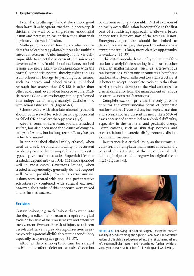
4. Lymphatic Malformation 35
Even if sclerotherapy fails, it does more good than harm if subsequent excision is necessary; itthickens the wall of a single-layer endothelial lesion and permits an easier dissection than witha primary thin-walled lesion.
Multicystic, lobulated lesions are ideal candi-dates for sclerotherapy alone, but require multipleinjection sessions. Unfortunately, it is virtually impossible to inject the sclerosant into microsize cavernous lesions. In addition, these honeycombedlesions are more likely to communicate with the normal lymphatic system, thereby risking injury from sclerosant leakage to perilymphatic tissues,such as nerves and blood vessels. Published research has shown that OK-432 is safer than other sclerosant, even when leakage occurs. Mul-tisession OK-432 sclerotherapy can be performedas an independent therapy, mainly to cystic lesions,with remarkable results (Figure 4-3).
Sclerotherapy with absolute alcohol (ethanol)should be reserved for select cases, e.g. recurrentor failed OK-432 sclerotherapy cases (1,2).
Another common sclerosant, sodium tetradecylsulfate, has also been used for closure of congeni-tal cystic lesions, but its long-term efficacy has yet fito be determined.
In our published clinical trials, ethanol, whenused as a sole treatment modality to recurrent or deeply seated lesions—preferably the cystictypes—gave excellent results. Superficial lesions fitreated independently with OK-432 also respondedwell in most cases. Cavernous lesions, when treated independently, generally do not respondwell. When possible, cavernous extratruncularlesions were treated with pre- and perioperative sclerotherapy combined with surgical excision; however, the results of this approach were mixed and of limited success.
Excision
Certain lesions, e.g. neck lesions that extend intothe deep mediastinal structures, require surgical excision because of their massive size and extensive involvement. Even so, the risk of injury to adjacentvessels and nerves is great during dissection; injury may result in potentially life-threatening conditions,especially in a young age group (34–37).
Although there is no optimal time for surgical excision, it is safer to defer an extensive dissection
or excision as long as possible. Partial excision of an easily accessible lesion is acceptable as the first fipart of a multistage approach; it allows a better chance for a later excision of the residual lesion.Emergency operations should be limited todecompressive surgery designed to relieve acute symptoms until a later, more elective opportunity is available (34–37).
This extratruncular lesion of lymphatic malfor-mation is rarely life threatening, in contrast to other vascular malformations, such as arteriovenous malformations. When one encounters a lymphaticmalformation lesion adherent to a vital structure, it is better to accept incomplete excision rather than to risk possible damage to the vital structure—a crucial difference from the management of venous or arteriovenous malformations.
Complete excision provides the only possiblecure for the extratruncular form of lymphaticmalformations. Nevertheless, incomplete excision and recurrence are present in more than 50% of cases because of anatomical or technical difficulty, fiespecially in the neonatal and pediatric group.Complications, such as skin fl ap necrosis and flpost-excisional cosmetic disfi gurement, disillu-fision many surgeons.
Recurrence is a critical issue, as the extratrun-cular form of lymphatic malformation retains theoriginal characteristic of the mesenchymal cell,i.e. the pluripotential to regrow its original tissue(1,2) (Figure 4-4).
FIGURE 4-4. Following ill-planned surgery, recurrent massiveswelling is pervasive along the right incisional scar. The soft tissuelesions of this child’s neck extended into the retropharyngeal and left submandibular region, and necessitated further excisionalsurgery to relieve vital functions for breathing and swallowing.

36 B.B. Lee
Outcome Assessment
Treatment results are based on findings of subjec-fitive and objective clinical improvement: pain, fluid leakage, infection, and reduction of lesion flsize or swelling. Both clinical and laboratory assessment may be necessary. The results aregraded as “excellent” when the treated lesion hasdisappeared completely, “good” when there is sub-stantial reduction or nearly complete disappear-ance of the lesion, and “fair” for moderateimprovement.
Evaluation of the extratruncular form should be made every 6 months by the team; in additionto clinical assessment, duplex ultrasonography and MRI may be needed as well.
Lymphangiosarcoma
Primary lymphedema has an inherent risk, however slight, of developing a malignancy—lym-phangiosarcoma derived from the chronically infl amed lymphedematous tissue (30,31). It is theflonly malignant disease that develops in the lymphatic system and is often known as Stewart-Treves sarcoma. Although lymphangiosarcoma isan extremely rare complication of primary lymphedema, the possibility of its development cannot be ignored.
It starts as painless, reddish-purple skin nodulesthat steadily increase in size and number. Because of its rarity, it is invariably neglected or misdiag-
nosed as a common skin disorder. Ulceration,bleeding, and infection complicate the process that eventually spreads throughout the body; atthat time, it is usually too late for a cure.
When discovered early, however, a combination of radiation therapy, intravenous, and intra-arterial chemotherapies can effect temporary relief or remission—a better alternative thanpalliative amputation (32,33). Once the diagnosis is confi rmed, an extensive investigation should befiperformed to determine the extent of metastasis, and whether an aggressive treatment program is justified (Figure 4-5).fi
Lymphangioma Circumscriptum
Lymphangioma circumscriptum is a rare extra-truncular lesion that requires special attentionto determine the extent of connective tissue involvement with deeper tissues. Masquerading as benign lesions in different sizes and colors, mul-tiple vesicular lesions develop on the skin and mucous membranes. Their appearance may be misleading, because the process involves transder-mal penetration of the cavernous extratruncularlesions into subdermal, submucosal, or subcuta-neous tissues. High contractile pressure vesicula-tion of the surface tissues may cause rupture(Figure 4-6).
Transdermal or transmucosal communications,causing visible skin or mucosal lesions, mustbe eradicated before other treatment is begun.
FIGURE 4-5. (A) Although infrequent, patients with lymphangio-sarcoma may present themselves to treatment centers. This personwas treated with chemotherapy and radiation that improved the
condition of her arm and chest. (B) Local swelling in the arm was treated with manual lymph drainage and compression. (Photocourtesy CL Morgan.)
A B

4. Lymphatic Malformation 37
It must be shown that a communication does not exist between the lesion and adjacent lymph-conducting systems. The lesions should beassessed by appropriate methods, such as ultraso-nography or MRI.
Lymphangiomatosis
Diagnosis
Lymphangiomatosis is defined as a “diffuse, fiinfi ltrating type of extratruncular form of lym-fiphatic malformation” by the Hamburg classifica-fition system. Subcutaneous and skeletal lesionsdominate this vascular malformation. Althoughlymphangiomatosis is rare, it presents a challengeto the clinician who has managed the simplerlesions of cystic or cavernous lymphangiomas and now must manage these extensive subcutaneousand skeletal lesions.
Part or the entire limb may be involved; thesubcutaneous lesions present as a diffuse swelling,exhibit an extensive involvement throughout thelimb, and create a striking physical appearance.
Spongiform lymphangiomatosis shows fluid flmovement that shifts according to elevation orposition of the limb, a phenomenon found in
other vascular malformations of the lowerextremity.
Skeletal lymphangiomatosis, an extratruncularlesion involving the bone, may manifest itself asan independent disease without evidence of con-comitant soft tissue involvement. It too deserves afull investigation. X-ray findings of demineralized fibone may mimic other demineralizing diseases:distant malignant metastatic lesions, multiple myeloma, hyperparathyroidism, polyostoticfibrous dysplasia, or metabolic lipid disease.fi
Lymphangiomatosis deserves a complete investigation to determine the possibility of other organ involvement. An evaluation should includethe visceral and skeletal systems using CT/MRI of the abdomen and mediastinum, whole-body lymphoscintigraphy with delayed and interval evaluation, and percutaneous direct-puncturelymphangiography.
Management
The management of lymphangiomatosis, depend-ing on the extent of involvement, is relatively straightforward and similar to that of other extra-truncular lesions—sclerotherapy and surgical excision. Unfortunately, pathological long bonefractures are frequent during the growing age and
A
B
FIGURE 4-6. (A) Here lymphangioma circumscriptum is visible with its seemingly benign lesions appearing in different sizes and colors on the skin and mucous membranes. (B) Penetration into
pelvic structures was confirmed by MRI; lymphoscintigraphy showed extensive dermal backflow.

38 B.B. Lee
FIGURE 4-7. Lymphangiomatosis extends the length of this child’sforearm and fingers and showed both macro- and microcysticlesions (left). Complete resection of the microcystic lesions fol-lowed preoperative OK-432 and ethanol sclerotherapy to the mac-rocystic lesions (right).
may cause devastating outcomes with severe disability. New multidisciplinary approaches mustbe found to improve the treatment results of these complex lesions (Figure 4-7).
Hemangioma
Congenital vascular malformations are frequently called capillary or cavernous hemangiomas by cli-nicians and histopathologists, even though a con-genital vascular malformation is entirely different from a genuine infantile-neonatal hemangioma(44,45).
Originating from proliferative endothelium,hemangiomas are the most common vascular tumor to arise during infancy. They appear duringfetal or early neonatal life and are characterized by rapid initial growth followed by spontaneousregression during later childhood (46).
A true peripheral congenital vascular malfor-mation, however, comprises a group of birthdefects that develop in various peripheral vascular systems, following developmental arrest in various stages of embryonic life. In contrast to hemangio-mas, in which increased mitotic activity is often observed, a congenital vascular malformation is
not hypercellular but is composed of normalmature endothelium.
The congenital vascular malformation is usually recognizable at birth, grows commensurate with the child, has a tendency to expand throughpuberty, and unlike a hemangioma does notregress.
Proper differentiation between the 2 vascularproblems is mandatory. Each requires a differenttreatment strategy, even though they belong to the same category of vascular anomalies (47).
Lymphovenous Malformation
Klippel-Trenaunay Syndrome
Klippel-Trenaunay syndrome is the most common combined form of congenital vascularmalformations—lymphovenous malformationsembodied within a larger group of hemolymphatic malformations (2,48–51). Klippel-Trenaunay syn-drome is not an inherited disorder, although it is congenital. Earlier reports, including the originalpaper by Klippel and Trenaunay, failed to recog-nize the lymphatic component, which produces many of the edema symptoms.
In contrast to the older eponym-based terminology, the Hamburg method classifies fiKlippel-Trenaunay syndrome as a venolymphaticmalformation. This terminology provides a clue to its etiology, anatomy, and pathophysiology. The new classifi cations are based on the “predominant ficomponent” of the chronic vascular malforma-tions and refer to the specific vascular systemfiaffected by the developmental arrest (5,6).
A triad of symptoms characterizes Klippel-Trenaunay syndrome (50,51): cavernous heman-gioma (port-wine stain); bone and limb hypertrophy (52,53), which develop in most cases;and varicose veins that appear in about three-quarters of the patients. Approximately 65% of patients have the 3 tissue changes. It is sometimescalled the angio-osteohypertrophy syndrome, andfemales are affected approximately twice as often as males (52,53).
Port-wine stains, often appearing on the lateral leg, are caused by dysplastic vasculature. Varicoseveins are common and extensive. Although thegreater saphenous vein is usually spared congeni-

4. Lymphatic Malformation 39
tal alteration, the deep veins may be valveless,aneurysmal, or absent. A large lateral marginal thigh vein often serves the limb as its major venous outfl ow conduit. Lymphedema develops fromflthese vascular changes, notably in the legs, and is treated in a fashion similar to that for otherprimary lymphedemas (Figure 4-8).
Osteoarticular changes may present in only 1 leg and evolve during the first 5 years of life. fiThey include soft tissue and bony hypertrophy, which may cause postural deformities and spinal scoliosis (53).
Lymphangiomas are less common, but they may accentuate other lymphatic changes. Visceral
involvement, e.g. liver, lungs, kidneys, or large intestine, may produce complications, such as spontaneous rupture of hemangiomas andinternal bleeding. Other complications of Klippel-Trenaunay syndrome include skin ulceration, cellulitis, deep vein thrombosis, and pulmonary embolism.
Parkes Weber Syndrome
Diagnosis
Arteriovenous fi stulae are rarely found in the fiextremities of Klippel-Trenaunay syndrome patients. They are, however, the primary feature distinguishing Klippel-Trenaunay syndrome fromthe Parkes Weber syndrome (54). The absence of fistulae and the presence of low-velocity venousfimalformations suggest a diagnosis of Klippel-Trenaunay syndrome, rather than Parkes Webersyndrome.
With an improved understanding of theses syn-dromes, we must recognize the different truncularand extratruncular forms and consider theircontributions to the pathophysiologic process.For example, the extratruncular forms presentin venolymphatic malformations will exert theirpluripotential characteristics originating from the embryologic mesenchymal cells. The truncu-lar forms, however, will only exert hemodynamicchanges on the venous and lymphatic vascular systems and on the bony and soft tissues as well.
Treatment
Treatment should be directed at specifi c physio-filogical problems. In the truncular form—forexample Klippel-Trenaunay syndrome—theembryonic lateral marginal vein is frequently excised or sclerosed to arrest severe reflux and flprogressive venous insufficiency.fi
In contrast, lymphangioma, an extratruncularform of lymphatic malformation, is seldomsited for treatment except to control complica-tions, lymph leakage, intralesional bleeding, orsepsis. As with other primary lymphedemas, thetruncular form of lymphatic malformations—such as aplasia, hypoplasia, hyperplasia of lymph
FIGURE 4-8. This patient made her own diagnosis of Klippel-Trenaunay syndrome, having reviewed her symptoms on the Inter-net. During this examination, a flat Doppler probe determines thedirection and velocity of blood flow in the lateral thigh vein. Duplex ultrasonography was also performed to further delineate thestatus of the venous system. The discrepancy in her leg lengthsuggests vascular bone involvement. A port-wine nevus completesthe triad of symptoms. (Photo courtesy LL Tretbar.)

40 B.B. Lee
collecting vessels and lymph nodes—is treated with CDT-based conservative treatment.
The interrelationships among the multiple vas-cular malformations are complex. Their interac-tions often create unpredictable, adverse responsesto each other, either by their natural presence orin the treatment.
Genetic Prospects
Although presently handled as separate entities, the truncular and extratruncular forms of lym-phatic malformations may need to be combinedespecially for the future treatment strategy; they are the result of the same developmental geneticdefi cit, although occurring at different stages of fidifferentiation.
Recently, new genetic research has suggestedthat gene therapy holds promise for the treatmentof lymphatic malformations within the fields of fiangiogenesis and vasculogenesis (55). Althoughthe feasibility for therapeutic implementation of gene therapy to lymphatic malformation is far from reality, there is vigorous pursuit of the geneticpossibilities.
The correction of defective lymphangiogenesiswould be benefi cial for contemporary manage-fiment of lymphangioma, as well as for primary lymphedema.
Exogenous growth factor administration wouldopen a new chapter for effective gene and gene-product therapy. Recently, it was confirmed thatfi“Anti-VEGFR-3 neutralizing antibody” seems tocompletely halt lymphatic vascular regeneration.Therefore, a “molecular antidote” to the pathologi-cal overgrowth of lymphangiomatosis couldbecome a major force that creates a chance for acure in the near future (56).
Recognition of this enormous potential for con-temporary management has caused some lym-phologists to alter their approach to the treatmentof lymphatic malformations.
Summary
During a period when lymphologists are increas-ingly aware of the complexity of vascular malfor-mations, they are using newer methods of classification and improved testing technology to fi
plan treatments more specific to the lesions’ fipathophysiology. Many treatment modalities areunder re-evaluation for inclusion in the therapist’sarmamentarium; they can provide an increasingly individualized and effective approach to these difficult problems.fi
References
1. Lee BB. Lymphedema-angiodysplasia syndrome: aprodigal form of lymphatic malformation (LM). Phlebolymphology. 2005;47:324–332.
2. Lee BB, Seo JM, Hwang JH, et al. Current conceptsin lymphatic malformation (LM). Vasc Endovascu-lar Surg. 2005;39:67–81.
3. Jacob AG, Driscoll DJ, Shaughnessy WJ, et al. Klippel-Trenaunay syndrome: spectrum and management. Mayo Clin Proc. 1998.
4. Lee BB. Current issue in management of chroniclymphedema: personal reflection on an experienceflwith 1065 patients. (Commentary). Lymphology.2005;38.
5. Belov ST. Anatomopathological classification of ficongenital vascular defects. Semin Vasc Surg. 1993.
6. Belov S. Classifi cation, terminology, and nosology fiof congenital vascular defects. In: Belov ST, LooseDA, Weber J, eds. Vascular Malformations. Reinbek,Germany: Einhorn-Presse; 1989.
7. Bastide G, Lefebvre D. Anatomy and organogenesisand vascular malformations. In: Belov ST, LooseDA, Weber J, eds. Vascular Malformations. Reinbek,Germany: Einhorn-Presse; 1989.
8. Woolard HH. The development of the principalarterial stems in the forelimb of the pig. ContribEmbryol. 1922.
9. Lee BB. Critical issues on the management of congenital vascular malformation. Ann Vasc Surg.2004;18:380–392.
10. Lee BB, Kim DI, Hwang JH, et al. Contemporary management of chronic lymphedema—personal experiences. Lymphology. 2002;35.
11. Lee BB. Chronic lymphedema, no more stepchild to modern medicine. Eur J Lymphology. 2004;14:6–12.
12. Mortimer PS. The pathophysiology of lymphedema. Cancer. 1998.
13. Papendieck CM. Lymphangiomatosis and dermo-epidermal disturbances of lymphangioadenodys-plasias. Lymphology. 2002;35.
14. Wolfe JH, Kinmonth JB. The prognosis of primary lymphedema of the lower limbs. Arch Surg. 1981.
15. Williams WH, McNeill GC, Witte MH, et al. Evalua-tion of peripheral lymphedema by longitudinallymphangioscintigraphy. Lymphology. 2002;35.

4. Lymphatic Malformation 41
16. Olszewski WL. Lymphoscintigraphy helps to differ-entiate edema of various etiologies (inflammatory, flobstructive, posttraumatic, venous). Lymphology.2002;35.
17. Bernas MJ, Witte CL, Witte MH. The diagnosis andtreatment of peripheral lymphedema. Lymphology. 2001.
18. Lee BB, Bergan JJ. New clinical and laboratory staging systems to improve management of chroniclymphedema. Lymphology. 2005;38:122–129.
19. Lee BB, Bergan JJ. Advanced management of congenital vascular malformations: a multidisci-plinary approach. Cardiovasc Surg. 2002;10:523–533.
20. Casley-Smith JR, Mason MR, Morgan RG. Complex physical therapy for the lymphedematous leg. Int JAngiol. 1995;4:134–142.
21. Hwang JH, Kwon JY, Lee BB, et al. Changes in lym-phatic function after complex physical therapy forlymphedema. Lymphology. 1999;32.
22. Baumeister RGH, Siuda S. Treatment of lymphede-mas by microsurgical lymphatic grafting: what is proved? Plas Reconstr Surg. 1990.
23. Campisi C, Boccardo F, Zilli A, et al. Long-termresults after lymphatic-venous anastomoses for the treatment of obstructive lymphedema. Microsur-gery. 2001.
24. Krylov V, Milanov N, Abalmasov K. Microlymphatic surgery of secondary lymphoedema of the upperlimb. Ann Chir Gynaecol. 1982.
25. Olszewski WL. The treatment of lymphedema of theextremities with microsurgical lympho-venous anastomoses. Int Angiol. 1988.
26. Becker C, Hidden G, Godart S, et al. Free lymphatic transplant. Eur J Lymphol. 1991;6:75–80.
27. Homans J. The treatment of elephantiasis of thelegs. N Engl J Med. 1936.
28. Kim DI, Huh S, Lee BB, et al. Excision of subcutane-ous tissue and deep muscle fascia for advanced lymphedema. Lymphology. 1998;31.
29. Auchincloss H. New operation for elephantiasis. P RJ Public Health Trop Med. 1930.
30. Wysocki WM, Komorowski A. Stewart-Treves syn-drome. J Am Coll Surg. 2007;205(1):194–195.
31. Laguerre B, Lefeuvre C, Kerbat P, et al. Stewart-Treves syndrome arising in post-traumatic lymph-edema. Bull Cancer. 1999.
32. Goetze S, Schmook T, Audring H, et al. Success -ful treatment of Stewart-Treves syndrome with liposomal doxorubicin. J Dtsch Dermatol Ges. 2004.
33. Lans TE, de Wilt JH, van Geel AN, et al. Isolated limb perfusion with tumor necrosis factor and melpha-lan for nonresectable Stewart-Treves lymphangio-sarcoma. Ann Surg Oncol. 2002.
34. Alqahtani A, Nguyen LT, Flageole H, et al. Twenty-fi ve years’ experience with lymphangiomas in fichildren. J Pediatr Surg. 1999.
35. Al-Salem AH. Lymphangiomas in infancy and child-hood. Saudi Med J. 2004.
36. Orvidas LJ, Kasperbauer JL. Pediatric lymphangio-mas of the head and neck. Ann Otol RhinolLaryngol. 2000.
37. Stromberg BV, Weeks PM, Wray RC Jr. Treatment of cystic hygroma. South Med J. 1976.
38. Lee BB, Mattassi R, Choe YH, et al. Critical role of duplex ultrasonography for the advanced manage-ment of a venous malformation (VM). Phlebology.2005.
39. Lee BB, Choe YH, Ahn JM, et al. The new role of MRI(magnetic resonance imaging) in the contemporary diagnosis of venous malformation: can it replace angiography? J Am Coll Surg. 1984.
40. Lee BB, Mattassi R, Kim BT, et al. Contem -porary diagnosis and management of venousand AV shunting malformation by whole body blood pool scintigraphy (WBBPS). Int Angiol.2004.
41. Lee BB, Mattassi R, Kim BT, et al. Advanced manage-ment of arteriovenous shunting malformation with transarterial lung perfusion scintigraphy (TLPS) for follow-up assessment. Int Angiol. 2005;24:173–184.
42. Ogita S, Tsuto T, Deguchi E, et al. OK-432 therapy for unresectable lymphangiomas in children. J PedSurg. 1991;26.
43. Kim KH, Kim HH, Lee BB, et al. OK-432 intrale-sional injection therapy for lymphangioma in chil-dren. J Korea Asso Ped Surg. 2001.
44. Mulliken JB. Cutaneous vascular anomalies. SeminVasc Surg. 1993.
45. Mulliken JB, Glowiczki J. Hemangiomas and vascu-lar malformations in infants and children: a classi-fi cation based on endothelial characteristics.fi PlastReconstr Surg. 1982.
46. Mulliken JB, Zetter BR, Folkman J. In vivo charac-teristics of endothelium from hemangiomas and vascular malformations. Surg. 1982.
47. Lee BB. Statutes of new approaches to the treatment of congenital vascular malformations (CVMs)—single center experiences [editorial review]. Eur JVasc Endovasc Surg. 2005;30:184–197.
48. Klippel M, Trenaunay J. Du noevus variqueux etosteohypertrophique. Arch Gén Méd. 1900.
49. Servelle M. Klippel and Trenaunay’s syndrome. AnnSurg. 1985.
50. Lee BB, Kim DI, Huh S, et al. New experiences with absolute ethanol sclerotherapy in the managementof a complex form of congenital venous malforma-tion. J Vasc Surg. 2001.

42 B.B. Lee
51. Lee BB, Do YS, Byun HS, et al. Advanced management of venous malformation with ethanol sclerotherapy:mid-term results. J Vasc Surg. 2003;37:533–538.
52. Mattassi R. Differential diagnosis in congenital vascular-bone syndromes. Semin Vasc Surg. 1993.
53. Belov ST. Correction of lower limbs length discrepancy in congenital vascular-bone disease by vascular surgery performed during childhood.Semin Vasc Surg. 1993.
54. Ziyeh S, Spreer J, Rossler J, et al. Parkes Weber orKlippel-Trenaunay syndrome? Non-invasive diag-nosis with MR projection angiography. Eur Radiol. 2004.
55. Szuba A, Skobe M, Karkkainen MJ, et al. Therapeuticlymphangiogenesis with human recombinant VEGF-C. FASEB J. 2002;16:1985–1987.
56. Bridenbaugh E. Literature watch. Lymphat Res Biol.2005.
43
5Medical Management of Lymphedema
Cheryl L. Morgan
Historical Review
While chapter 1 documents the early lymphaticdiscoveries, many of today’s interventions devel-oped from investigations into the causes andmanagement of lymphedema over the past 2 centuries.
In the 1870s, Still, the founder of the AmericanSchool of Osteopathy, created an approach tomanual therapies acknowledging the lymphatics as a vital system and designed to correct anatomi-cal deviations that interfered with the fl ow of fllymph and blood. Still proposed a relationship in which cerebrospinal fluid is reabsorbed by flthe lymph, a relationship supported by recentresearch (1).
In the 1890s, Winiwarter, an Austrian physicianfrom Vienna, described a treatment for swollen limbs that seems surprisingly similar to that rec-ommended today; the conservative treatment included elevation, compression, massage, and exercise. Unfortunately, Winiwarter’s suggestions were not advanced further for some time (2).
A few decades later, in the 1920s, Miller devel-oped the Miller thoracic pump technique, whichhe described as effective in creating intrathoracic pressure changes on lymphatic flow. Many of the flconditions that he treated successfully with this technique include forms of edema (3,4).
In 1922, Millard published the first osteopathic fimedical textbook examining only the lymphaticsystem, Applied Anatomy of the Lymphatics, and was the founder and president of the InternationalLymphatic Research Society in Kirksville, MO (5).
In the 1930s, Vodder and his wife began workingwith manual techniques to affect lymph flow. fl
Although the Vodders were Danish, the majority of their work was performed and published in France, where they lived and worked from 1928 to 1939. Here they developed their technique, then called lymph drainage massage. They later coined the term manual lymph drainage. The Vodder technique of lymph drainage is the most widely taught component of conservative lymphedematreatment (6,7).
In the 1960s, Asdonk, a German physician, scientifically tested the Vodder technique of fimanual lymph drainage in his clinic on 20 000 patients. From this research, he established theindications, contraindications, and effects of thetechniques (8).
In the following decade, the Földis combined the Vodders’ manual lymph drainage with ban-daging, exercise, and specific skin care into the fitreatment program termed complete decongestivephysiotherapy (CDP), later renamed completedecongestive therapy (CDT) (9). The Földis’ many contributions to lymphology include their exten-sive research as well as directing the Földi Clinicin Germany.
The first North American lymphedema treat-fiment centers opened in the 1980s, where specially trained therapists initiate the use of CDT to treat lymphedema patients.
In the 1990s, an increase of scientifi c literature, fipatient awareness, and interested therapists andphysicians stimulates an increase of treatment centers in the United States. In 1992, Medicare(KS) approves CDT treatment for lymphedema. In 1999, a new CPT code describing manual thera-pies, including manual lymph drainage (97140), isapproved by the American Medical Association.

44 C.L. Morgan
In recent years, continued use of CDT has encouraged controlled clinical studies and research to focus on quality of life, treatment outcomes, and cost effectiveness. The American Society of Lymphology (ASL) has initiated efforts to stan-dardize educational requirements of health care professionals treating disorders of the lymphatics. Legislation has been introduced by the NationalLymphedema Network (NLN) to improve reim-bursement for medical supplies for lymphedema. Research has moved to the forefront thanks to the Lymphatic Research Foundation (LRF).
Among individuals, Australian physiciansCasley-Smith devoted more than 40 years to research, treatment, and the education of thera-pists (10). Their abundant and diverse researchusing the electron microscope has received world-wide recognition.
The significant contributions through thefiCenters for Disease Control and Prevention led by Dreyer and Addiss in the treatment of filarialfilymphedema and vector control (11) have pro-duced extraordinary developments that willbenefi t millions suffering with disfifi guring conse-fiquences of parasitic infection.
Witte, a professor and general surgeon, mademajor contributions as a researcher and educator (12). He and his wife, also a professor of surgery, established the first lymphological laboratory fiin the United States devoted to both basic andclinical investigation of lymphatic circulatory disorders.
Many of these accomplishments above could not have been realized without the contributions of medical professionals, such as the prominent physicians Guyton and Stemmer (13,14). The pro-lifi c research of Olzsewski and Partsch continue toficontribute to the develop of treatment options and new diagnostic methods to improve equally thequality of research, medical intervention, and thequality of life of lymphedema patients.
Successful Treatment for
Lymphedema
Optimal treatment results require patient adher-ence. This begins with an accurate diagnosis, as outlined in chapter 2. To date, complete deconges-
tive therapy is recognized internationally as the most successful treatment option for lymphedema (15,16). Other names used to describe the programinclude complete (or combined, or complex) decongestive physiotherapy (17).
The author has designated the term Compre-hensive Decongestive Therapy©, to indicate thattherapeutic interventions may not have completeresults, and need not be considered complex. This program, introduced in 1989, also includescomprehensive instruction in the disease, its treatment, and the expected results. Nutritionalcounseling and psychosocial intervention, e.g.counseling, support groups, and resource man-agement, are essential components of the programas well. These additional components are describedon pages 51 and 52. The results of this program are demonstrated by one patient’s progress through treatment (see Figures 5-1 to 5-8).
A fundamental CDT-based program describedby the International Society of Lymphology includes manual lymph drainage, specialized
FIGURE 5-1. A 51-year-old female presenting with bilateral, chronic, stage III lymphedema (elephantiasis). Vena cava clipping25 years prior following orthopedic surgery and deep vein throm-bosis was performed to prevent reoccurring pulmonary embolism. Massive lymphedema developed despite the patient’s efforts toobtain medical intervention. (Photo courtesy of CL Morgan.)
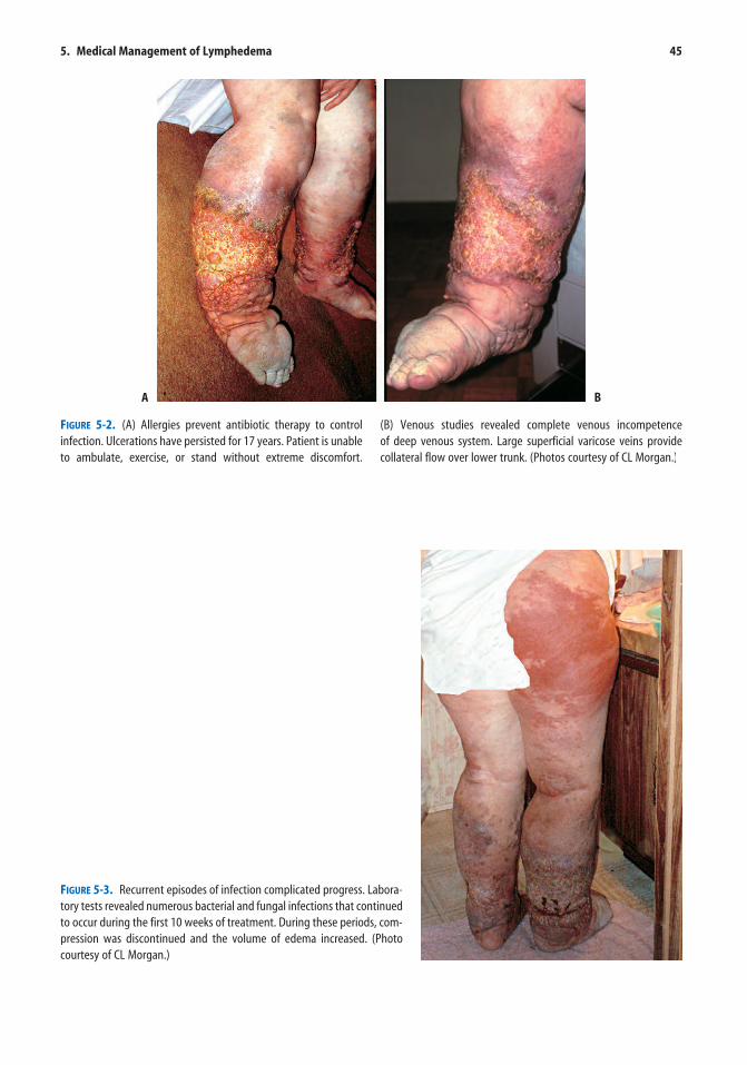
5. Medical Management of Lymphedema 45
FIGURE 5-2. (A) Allergies prevent antibiotic therapy to control infection. Ulcerations have persisted for 17 years. Patient is unableto ambulate, exercise, or stand without extreme discomfort.
(B) Venous studies revealed complete venous incompetenceof deep venous system. Large superficial varicose veins provide collateral flow over lower trunk. (Photos courtesy of CL Morgan.)
FIGURE 5-3. Recurrent episodes of infection complicated progress. Labora-tory tests revealed numerous bacterial and fungal infections that continued to occur during the first 10 weeks of treatment. During these periods, com-pression was discontinued and the volume of edema increased. (Photo courtesy of CL Morgan.)
A B

FIGURE 5-4. (A) A Stage IV ulceration measured 28.4 cm by 14.3 cm.Treatment was initiated with bedrest, elevation of the lowerextremity, and compression-aided wound care. Dressing changes were required hourly. Surface infections were treated with a 2%benzethonium chloride solution, and the ulcer base debrided withtrypsin gel. Manual lymph drainage and passive range of motionwere initiated at the second week, once wound drainage was
reduced and episodes of infection were under control. (B) Here fibrosclerotic tissues have begun to soften, hyperpigmentation hasimproved, episodes of infection have been reduced, and ulcershave decreased in diameter. Wounds are treated daily with non-ionic isotonic surfactant cleanser and polymeric membrane dress-ing once infections have resolved. Dressing changes are required every 2 to 3 hours. (Photos courtesy of CL Morgan.)
FIGURE 5-5. At 33 weeks, the wounds have closed without sur -gical intervention. Papillomas have been excised, and are benign. There is no episode of infection for 8 weeks. Patient is able to ambulate, stand, increase exercise, donn and doff bandages fol-lowing instruction in self-care program. (Photograph courtesy of CL Morgan.)
FIGURE 5-6. Results of comprehensive treatment after 47 weeks.No recurrence of wounds. Bilateral bandaging is applied by the patient each evening. The patient has returned to work. Circumfer-ence of the lower limbs has been reduced and continued improve-ment is expected with demonstrated adherence to a self-careprogram. (Photo courtesy of CL Morgan and LL Tretbar.)
A B

5. Medical Management of Lymphedema 47
FIGURE 5-7. (A) Review of bandaging techniques performed withpatient improves patient adherence and self care skills. (B) Anexample of bandaging used for lymphedema management taughtto the patient for intensive program and subsequent self-care. Thepatient is now able to wear shoes and increase activity.
FIGURE 5-8. The patient has returned to all normal activities andhas continued with intermittent week-long treatment sessions twice per year to improve the integrity of tissue and improve bandaging techniques as progress continues. Results have been sustained, without recurrent ulceration or a decrease in func-tional abilities achieved during the initial course of treatment 14years ago. (Photo courtesy of CL Morgan and LL Tretbar.)
bandaging, exercise, skin care, and a self-manage-ment program (16).
The goals of intervention are to improve theintegrity of the affected skin and connectivetissues, to reduce or eliminate infections, to reduce edema, and to facilitate patients’ ability to manage their condition once the intensive treatment phaseis completed.
Two-Phase Intervention
The management of lymphedema using CDTis divided into 2 phases. The first addresses the fiprimary need of the patient, such as treatmentof the swollen limb, increased functional capacity, and prevent infections. Trained health care
B
A

48 C.L. Morgan
professionals initiate the fi rst phase with CDT. Thefisecond phase addresses the continuing needs of the patient through self-management.
Many European models of CDT recommend twice daily treatment visits for an average of 4 to6 weeks. However, constraints of insurance carri-ers limit this successful model and, subsequently,the results. Patients with upper extremity lymph-edema might receive daily treatments for 3 to 4weeks, whereas those with lower extremity lymph-edema might receive an average of 4 to 6 weeks of daily visits. Some clinics report that patients may receive CDT only 1 to 3 times per week, due to insurance limitations, work limitations, or physi-cal distances traveled.
Health professionals must be aware that symptom reversal is a slow process. Even in the best circumstances where appropriate interven-tion occurs and patient adherence is optimal,symptom reversal may require 8 months or moreof treatment. When improper treatment or rapidly paced shortcuts are employed, patients rarely havelasting outcomes and often experience exacerba-tion of their symptoms. Frequently, individuals with comorbidities do not respond as quickly to treatment.
The components of the 2-phase treatmentmethod are:
Phase I: Intensive Treatment
• manual lymph drainage• skin care (including wound care if indicated)• bandaging (specialized)• exercise (in bandages)• compression garment (if appropriate)
Phase II: Self-management
• skin care• manual lymphatic drainage (as needed)• daytime compression garment, nighttime
bandaging• exercise (in bandages or garment)• support groups
Manual Lymph Drainage
Manual lymph drainage is based on physiologicprinciples that regulate the flow of lymph, and itflinfl uences the general lymph circulation primar-fl
ily by affecting the initial and superfi cial lymphat-fiics. The techniques are designed to stimulatelymph fl ow from distal lymphatics to proximalfllymphatics (7,18). This objective requires that manual lymph drainage move excess lymph fromthe swollen areas or sites of obstruction, to adja-cent areas that can efficiently transport the addi-fitional volume.
To facilitate lymph fl ow into adjacentflbody areas, manual techniques include treat -ment of the contralateral or ipsilateral lymphnode regions. Lymphography and lymphoscintig-raphy, used for research and diagnostic purposes, confirm that superfifi cial lymph flfi uid moves aroundflblocked areas toward more central vessels whenthese specialized manual techniques are used (15,16).
Manual lymph drainage has many effects on the lymph system by:
• increasing lymph transport capacity, thereby increasing the volume of lymph fluid trans-flported proximally
• increasing the frequency of lymph vessel contractions
• increasing pressure in the lymph collector vessels
• redirecting natural flow patterns toward collat-fleral vessels, anastomoses, and uninvolved lymph node regions
• increasing arteriolar blood flowfl
A concept, now under investigation, suggests thatmanual lymph drainage enhances angiogenesis; if true, the number of functioning lymphatics should increase. Another theory under investigation isthat the compression bandages are responsible forthe promotion of angiogenesis.
Starting Manual Lymph Drainage
Before initiating treatment, the program should be reviewed with the patient by the referring phy-sician and therapist. Physician support greatly increases patient adherence to an intensive therapy program. The physician should know that mobi-lizing the lymphatic fl uid will cause a temporary flincrease in circulating blood volume. Confirmfiwith the physician if there are any comorbiditiesthat would contraindicate CDT (see Summary at

5. Medical Management of Lymphedema 49
end of Chapter, page 53). Once these issues are settled, treatment may begin (19).
Manual lymph drainage recruits functioninglymph vessels and nodes closest to the regions that are not adequately performing. Therefore,regardless of the site of insufficient flfi ow, treatment flcan include manual work on the neck, back,abdominal region, and uninvolved inguinal andaxillary lymph nodes (6,16,18). Initially, manualtherapy begins from the contralateral trunk area toward the congested area. Treatment then contin-ues to move from the involved area to uninvolvedareas.
The manual techniques of manual lymph drain-age are light, slow, and precise. Manual lymph drainage does not use the deep strokes and pres-sures of effl euragefl , a standard therapeutic massage technique. Rather, a specifi ed number of light fipressure strokes without friction that direction-ally stretch tissue are used (6,18,20,21). Themanual lymph drainage therapy session usually requires a minimum of 45 to 60 minutes, depend-ing on the size of the limb(s) or body parts involved, the severity of the symptoms, and theamount of fi brosis (20).fi
Skin Care
Meticulous skin care is an essential component of both treatment phases. The goal of careful and thorough skin care is to avoid or eliminate fungaland bacterial infections such as cutaneous celluli-tis, erysipelas, or lymphangitis (22). Patients with lymphedema experience skin changes and arehighly susceptible to infections. Dermatologicalconditions often associated with lymphedema caninclude:
• dry skin• hyperkeratosis• contact dermatitis• lipodermatosclerosis• fungal infections• folliculitis• lymph fistulasfi• papillomatosis• lymphangiectasia• lymphorrhea• eczema
• dermatolymphangioadenitis (DLA) (cellulitis,lymphangitis, erysipelas)
• lymphangiosarcoma• chronic ulceration
The protein-rich fluid that accumulates in the fllymphedematous tissues serves as a culture medium for pathogens which circulate within thebody or enter through skin lesions (23,24).
Wound Care
When the integrity of the skin is compromised by chronic edema or lymphedema, regardless of theetiology, basic skin care regimens can reduce therisk of complications from small lesions to chroniculcers (23,24). Should a wound develop withlymphedema, compression-aided wound caremethods are critical for the patient’s continued progress (25).
Bandaging
Because skin elasticity is partially lost in lymph-edema, tissue hydrostatic pressures must be main-tained by the use of external support. This support must be continuous until the volume reductionstabilizes and tissues remodel with improved functional lymphatic capacity (25).
During the treatment phase, bandaging per-formed by a health care professional with special-ized training will achieve the best results. A specific ficombination of padding, foam, protective gauze,and short-stretch bandages is applied in precise layers following each session of manual lymphdrainage. Before lymphedema bandaging, the involved skin is cleansed and protected with an appropriate moisturizer; in the case of ulcerations, protective dressing that suffi ciently captures fiexudate is applied. Experienced lymphologistsrecognize that most of the success observed during the treatment phase is due to compressionbandaging (2,23,25,26).
Bandaging affects the entire lymphatic systemby:
• providing support for tissues that have lostelasticity
• improving muscle pump effi ciency during fiactivity

50 C.L. Morgan
• reducing the rate of ultrafiltrationfi• facilitating colloidal protein reabsorption• softening fibrotic tissue with localized pressurefi• providing a mild increase in tissue pressure,
assisting lymph vessels to empty• reducing the hydrostatic pressure gradient
between blood and lymphatic tissues, thus pre-venting refi lling of the interstitium with flfi uidfl
Types of Bandages
A distinction must be made between short-stretch (low elastic) bandages and long-stretch (high elastic) bandages and the type of compressionthat each offers.
As their name implies, short-stretch bandageshave little or no stretch in them. When applied to the limb, they form an envelope or cocoon around the limb. In the resting state, compression is minimal but constant. Compression develops only during exercise as the muscles expand and pressagainst the wrap. Therefore, exercise is importantfor the effect of short-stretch bandages to be opti-mized. Because of the low resting pressure, they can be worn at night (25–27).
Long-stretch bandages are highly elastic and allow extension to perhaps 3 times their restinglength. Thus they create a continuous compres-sion on the limb with a high resting pressure. However, during exercise they stretch as the muscles expand and the compressive force dec-reases. This diminution of force follows the Laplace law (P = T/r) that states that external pressure (P)is directionally proportional to the tension (T) of the elastic fabric, and inversely proportional to theradius (r) (25). Because of high resting pressure, these bandages are not recommended for wear at night; the pressure can compromise arterial circu-lation and already compromised tissues, especially over bony prominences (25,27).
Although bandages can be cumbersome patientsshould continue, if not increase, their activity levelto facilitate muscle pumps and stimulate lymphatic function. After a period of adjustment, most ban-dages are well tolerated by informed and moti-vated patients. For optimum patient adherence, patients should thoroughly understand the impor-tance of the bandages during the treatment phase.During this period the bandages are worn continu-ously except for the time required to cleanse, treat, or rewrap the limb. Patients and their families
often begin to learn to apply the bandage toward the middle of the treatment phase (16,20,23).
The bandaging portion of a treatment sessionmay require 20 to 90 minutes; the amount of time depends on the number of custom foam pieces tobe produced, how much patient education andassistance is required, how many limbs are affected,the size of the involved limb(s) or body parts, andif any lesions require care.
By the third or fourth week of the treatmentphase, the clinician will spend less time actively bandaging, concentrating on instructing thepatient in basic application and technique. Thepatient’s progress may yet require alterations incustom pieces, education, and modifications infiapplication to continue improvement. Patient edu-cation is necessary to safely and accurately apply lymphedema bandages. Regular monitoring by a qualifi ed health care professional is essential and fihighly recommended.
Exercise
During the intensive treatment phase, exercises are performed while wearing lymphedema ban-dages. The exercise program, customized to a patient’s functional ability, includes activities that promote the emptying of the affected lymphregions. The activities should also assist the func-tioning lymphatics to work more efficiently. During fithe self-management phase, when the patient has achieved a reasonable volume reduction, a com-pression garment may be worn (23,27).
The amount of time spent on exercise during a session in the treatment phase will vary, based on the number of measured limitations and theintervention goals. Strategies for conservingenergy and achieving balance between exerciseand rest should be developed for each person. If there are no other existing functional limitations,a patient may be able to accept responsibility forexercise even during the initial treatment phase(28,29).
Many patients with lymphedema experiencesome level of limitation in joint range of motion, muscle strength, and posture or gait deficits. Any fisuch limitations should be considered when devel-oping an exercise program for the patient.
Exercise improves cardiovascular function,muscular strength, functional capacity, and endur-
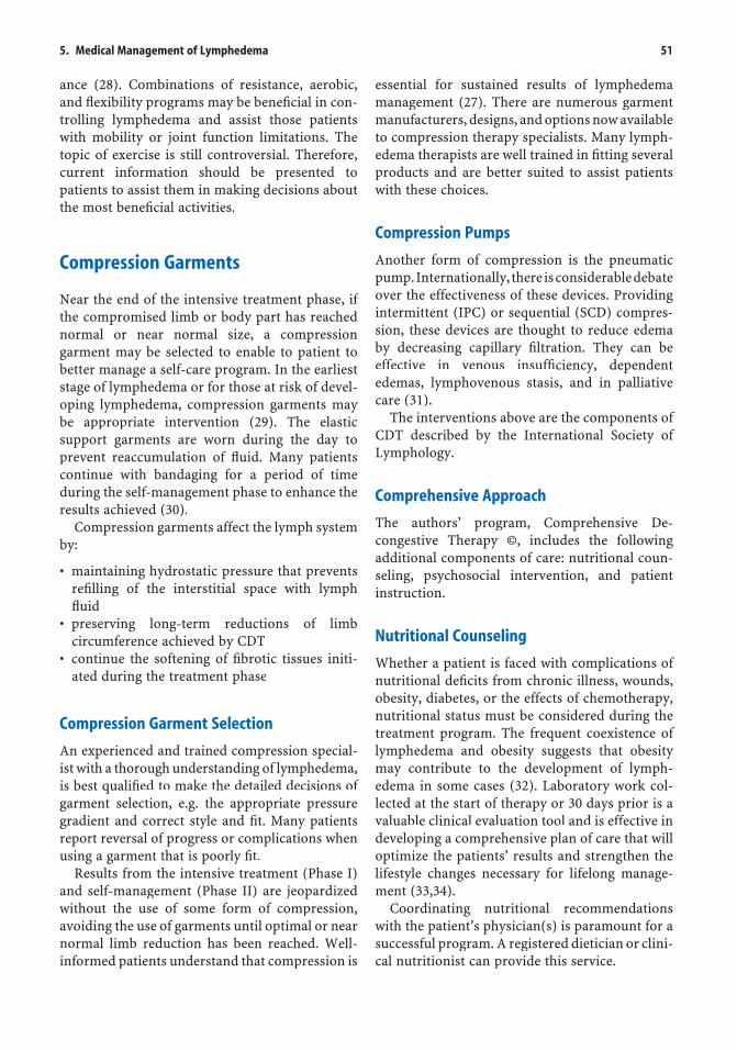
5. Medical Management of Lymphedema 51
ance (28). Combinations of resistance, aerobic,and fl exibility programs may be benefifl cial in con-fitrolling lymphedema and assist those patientswith mobility or joint function limitations. The topic of exercise is still controversial. Therefore, current information should be presented topatients to assist them in making decisions aboutthe most benefi cial activities.fi
Compression Garments
Near the end of the intensive treatment phase, if the compromised limb or body part has reached normal or near normal size, a compression garment may be selected to enable to patient to better manage a self-care program. In the earlieststage of lymphedema or for those at risk of devel-oping lymphedema, compression garments may be appropriate intervention (29). The elasticsupport garments are worn during the day toprevent reaccumulation of fluid. Many patients flcontinue with bandaging for a period of time during the self-management phase to enhance theresults achieved (30).
Compression garments affect the lymph systemby:
• maintaining hydrostatic pressure that preventsrefilling of the interstitial space with lymphfifluidfl
• preserving long-term reductions of limbcircumference achieved by CDT
• continue the softening of fibrotic tissues initi-fiated during the treatment phase
Compression Garment Selection
An experienced and trained compression special-ist with a thorough understanding of lymphedema, is best qualified to make the detailed decisions of figarment selection, e.g. the appropriate pressuregradient and correct style and fi t. Many patients fireport reversal of progress or complications whenusing a garment that is poorly fit.fi
Results from the intensive treatment (Phase I)and self-management (Phase II) are jeopardizedwithout the use of some form of compression,avoiding the use of garments until optimal or near normal limb reduction has been reached. Well-informed patients understand that compression is
essential for sustained results of lymphedemamanagement (27). There are numerous garmentmanufacturers, designs, and options now availableto compression therapy specialists. Many lymph-edema therapists are well trained in fi tting several fiproducts and are better suited to assist patientswith these choices.
Compression Pumps
Another form of compression is the pneumatic pump. Internationally, there is considerable debateover the effectiveness of these devices. Providing intermittent (IPC) or sequential (SCD) compres-sion, these devices are thought to reduce edema by decreasing capillary fi ltration. They can be fieffective in venous insuffi ciency, dependent fiedemas, lymphovenous stasis, and in palliativecare (31).
The interventions above are the components of CDT described by the International Society of Lymphology.
Comprehensive Approach
The authors’ program, Comprehensive De -con gestive Therapy ©, includes the followingadditional components of care: nutritional coun -seling, psychosocial intervention, and patientinstruction.
Nutritional Counseling
Whether a patient is faced with complications of nutritional defi cits from chronic illness, wounds,fiobesity, diabetes, or the effects of chemotherapy,nutritional status must be considered during the treatment program. The frequent coexistence of lymphedema and obesity suggests that obesity may contribute to the development of lymph-edema in some cases (32). Laboratory work col-lected at the start of therapy or 30 days prior is a valuable clinical evaluation tool and is effective indeveloping a comprehensive plan of care that will optimize the patients’ results and strengthen thelifestyle changes necessary for lifelong manage-ment (33,34).
Coordinating nutritional recommendations with the patient’s physician(s) is paramount for asuccessful program. A registered dietician or clini-cal nutritionist can provide this service.

52 C.L. Morgan
Psychosocial Intervention
An integral part of this approach is the provisionof support for patients and their family or caregiv-ers to take a positive role in daily management of their condition(s). Encouraging participation inthe process and results (adherence) is preferentialto compliance (35). Some patients with advanceddisease require palliative care and a well-orga-nized clinical team to address the physical andpsychological needs presented. Many patientswith lymphedema have concomitant health con-cerns, which contribute to psychosocial problemssuch as depression, isolation, loneliness, anxiety, and poor coping skills (36,37).
Initial assessment of a patient’s quality of life, the need for community resources (e.g. trans-portation, financial assistance, supplies), andfiindividual counseling or support groups promotes increased adherence to the intensive phase of therapy (32,38). Another successful therapeutictool for recovery and coping with chronic illnessis art therapy (39,40). These services can be pro-vided by a professional therapist or counselor, a licensed clinical social worker, or a psychologist.
Patient Instruction
The establishment of a comprehensive programrequires a structured and thorough patient educa-tion component that can be individualized. Educa-tion about the condition, effective treatment and results, instruction in problem solving, nutritionalguidelines, programs for exercise, and information on proper hygiene, skin care, and care of bandages and garments all provide a foundation for improved adherence and outcomes of therapy (41). Allmembers of the clinical team provide this service.
Phase II, Self-management
Some patients or their caregivers are candidatesfor training in some of the manual techniques toassist in self-management. They must be carefully instructed and educated in the appropriate pres-sures and directions of the strokes to avoid exac-erbation of the symptoms. If a patient experiencesan increase in swelling or a plateau in reduction following the treatment phase, follow-up with additional sessions of manual lymph drainage inthe clinic is appropriate. A patient who reports increased swelling should be examined and
cleared of possible complications, such as recur-rent malignancy, before continuing treatment. Atthis point, manual therapy may enhance the gainsachieved during the initial treatment phase. Oncethe intensive treatment phase is complete, most patients have learned how to manage their condi-tion with bandaging. The progress achieved andseverity or persistence of the symptoms deter-mine whether a patient continues to wear ban-dages at night. Most patients apply the bandagesat night to optimize improvements in tissue integ-rity and volume reduction during the self-management phase.
Patients often report that their symptoms fluc-fltuate with climate, body weight, and changes in lifestyle. Most concur that lymphedema is bestmanaged with consistent use of all components of the self-management phase. During the self-management phase, patients wear the compres-sion garments during daytime hours and continue with bandaging at night. As a result of the educa-tion provided during the intensive treatment(Phase 1), the patient should understand and beable to perform donning, doffi ng, and care of bothfibandages and compression garments.
Many patients will need to wear a compression garment for the remainder of their lives to effec-tively control symptoms and retain reduction of volume achieved. Nevertheless, tissue swellingand fi brosis can occur in nearby areas if garments fifit poorly. Patients must remain attentive to any fitissue changes in involved or adjacent areas.Garment fit and condition should be checked atfileast every 6 months, particularly if the patientexperiences difficulties with fifi t, comfort, or fisymptom control.
Patients are encouraged to continue their exer-cise program while wearing compression during the self-management phase. Periodic clinic visitsmay be needed to review the self-management home program or to modify for improvements or decline in function (42).
Summary
Complex decongestive therapy has proven to be an effective treatment protocol for lymph-edema management for more than 70 years(23,27,42). It cannot be overly emphasized that itis essential for health professionals to obtain in-

5. Medical Management of Lymphedema 53
depth education about lymphedema and relateddisorders and specialized training in each of thetherapeutic interventions. Of equal importance isthat they acquire extensive experience to ade-quately prepare for designation as a lymphedemaspecialist.
Additional indications for CDT include thefollowing:
• chronic venous insufficiencyfi• post-thrombotic (deep venous thrombosis)
syndrome• chronic wounds (associated with edema)• traumatic edema (iatrogenic, postsurgical, post-
musculoskeletal injury)• complex regional pain syndrome (CRPS), for-
merly known as refl ex sympathetic dystrophyfl• rheumatoid arthritis• possibly lipedema
There are some contraindications or precau-tions to CDT/manual lymph drainage to be noted.Most patients will tolerate the treatment, since itis a gentle, smooth manual approach rather than a deep powerful massage. However, monitoring of cardiac, pulmonary, and renal functions is stillnecessary because of the temporary increase of blood volume. The following are relative contrain-dications and precautions to CDT:
• acute infections, local or systemic, viral or bacte-rial: cellulitis, erysipelas
• secondary acute inflammationfl• acute bronchitis or bronchial asthma
(uncontrolled)• diabetes• cardiac failure, hypertension• malignancy• renal insuffi ciencyfi• sclerotic arteries or venous insufficiency (in fi
patients above age 60 years)• hyperthyroidism or hypothyroidism• pregnancy• recent surgery• Crohn disease, diverticulitis• deep venous thrombosis, acute superficial fi
thrombophlebitis
References
1. Still AT. The Philosophy and Mechanical Principlesof Osteopathy. Kansas City, MO: Hudson-Kimberly; 1902;65–66.
2. Földi M, Kubik S, eds. Lehrbuch der Lymphologie für Mediziner. 5th ed. Munich-Jena: Urban & Fisher; 2002.
3. Miller CE. The mechanics of lymphatic circulation.J Am Osteopath Assoc. 1923;22:397–398.
4. Miller CE. Osteopathic principles and thoracicpump therapeutics proved by scientific research. fiJ Am Osteopath Assoc. 1927;26:910–914.
5. Millard FP. Applied Anatomy of the Lymphatics. Kirksville, MO: AG Walmstey ed. InternationalLymphatic Research Society; 1922.
6. Vodder E. Le drainage lymphatique, une nouvelle méthode thérapeutique. Santé Pour Tous: Paris, 1936.
7. Wittlinger H, Wittlinger G. Introduction to Dr. Vodder’s Manual Lymph Drainage. Heidelberg: Haug; 1986.
8. Asdonk J. Lymphdrainage und Molekularmassage. Phys Medizin und Rehabilitation, Heft. 1967;10:62–65.
9. Földi E, Földi M, Weissleder H. Conservative treat-ment of lymphedema of the limbs. Angiology. 1985;36:171–180.
10. Casley-Smith JR. The Pathophysiology of Lymphoe-dema. In: Heim LR, ed. IXth International Society of Lymphology. Tel Ariv, Israel. Immunology ResearchFoundation, Newburgh, USA, 1983:125–130.
11. Dreyer G, Addiss D, Dreyer P, et al. Basic Lymphoe-dema Management, Treatment and Prevention of Problems Associated with Lymphatic Filariasis.Hollis, NH: Hollis Publishing; 2002.
12. Bernas MJ, Witte CL, Witte MH. The diagnosis and treatment of peripheral lymphedema.Lymphology. 2001;34:84–91.
13. Guyton AC, Granger HJ, Taylor AE. Interstitial fluid flpressures. Physiol Rev. 1971;51:527–563.
14. Stemmer R. Ein klinisches Zeichen zur Früh-und Differentialdiagnose des Lymphodems. Vasa. 1976;5(3):261–262.
15. Browse N, Burnand K, Mortimer P, eds. Diseases of the Lymphatics. London: Arnold; 2003.
16. Weissleder H, Schuchhardt C, eds. Lymphedema: Diagnosis and Therapy. Bonn: Kagerer Kommuni-kation; 1997.
17. International Society of Lymphology. The diagnosisand treatment of peripheral lymphedema. Consen-sus Document of the International Society of Lymphology. Lymphology. 2003;36:84–91.
18. Kasseroller RG. Compendium of Dr. Vodder’s Manual Lymph Drainage. Heidelberg: Haug; 1998.
19. Browse NL, Stewart G. Lymphoedema: patho-physiology and classificationfi . J Cardiovasc Surg. Torino, 1985;26:156–168.
20. Mortimer PS. Managing lymphedema. Clin Exp Dermatol. 1995;20:98–106.

54 C.L. Morgan
21. Eliska O, Eliskova M. Are peripheral lymphatics damaged by high-pressure manual massage?Lymphology. 1995;28:21–30.
22. Olszewski WL. Lymph Stasis: Pathophysiology,Diagnosis and Treatment. Boston: CRC Press; 1991.
23. Weissleder H, Schuchhardt C, eds. Lymphedema:Diagnosis and Therapy. 3rd ed. Bonn: Kagerer Kom-munikation; 2001.
24. Olszewski WL, Jamal S. Recurrent dermatolymphan-gioadenitis (DLA) is responsible for progressionof lymphedema. Progress in Lymphology XV. Lym-phology. 1996;(Suppl.)29:331–334.
25. Partsch H, Rabe E, Stemmer R. Compression Therapyof the Extremities. Paris: Editions Phlébologiques;2000.
26. Partsch H. Understanding the pathophysiologicaleffects of compression. In: European Wound Man-agement Association. Position Document. Under-standing compression therapy. London: MEP Ltd.;2003;2–4.
27. Földi E, Földi E, Weissleder H. Conservative treatment of lymphedema of the limbs. Anbiology 1985;36:171–180.
28. Casley-Smith, JR, Mason MR, et al. Complex physi-cal therapy for the lymphedematous leg. Int J Angio. 1995;4:134–142.
29. Pappas CJ, O’Donnell TF, Kalisher L, et al. Long-term results of compression treatment of lymph-edema. J Vasc Surg. 1992;16(4):555–564.
30. Badger CMA, Peacock JL, Mortimer PS. A random-ized controlled, parallel-group clinical trial com-paring multi-layer bandaging with hosiery versus
hosiery alone in the treatment of lymphedema of the limb. Cancer. 2000;88(12):2832–2837.
31. Vowden K. The use of intermittent pneumatic com-pression in venous ulceration. Br J Nurs. 2001;10(8):491–509.
32. Mortimer PS. The pathophysiology of lymphedema.Cancer. 1998;(Am Suppl.)12(83):2798–2802.
33. Olszewski WL, Engeset A. Immune proteins, enzymes, and electrolytes in human peripherallymph. Lymphology. 1978;11:156.
34. Ryan TJ, Mallon EC. Lymphatics and the processingof antigen, Clin Dermatol. 1995;13(5):485–492.
35. McWayne J, Heiney SP. Psychologic and social sequelae of secondary lymphedema: a review.Cancer. 2005;104:457–466.
36. Azevedo WF, Boccardo F, Zilli A, et al. A reliable andvalid quality of life in patients with lymphedema—version of the SF-36. Lymphology. 2002;35(1):177–180.
37. De Godoy JMP, Braile DM, et al. Quality of life andperipheral lymphedema. Lymphology. 2002;35(2):44–45.
38. Falvo D. Medical and psychosocial aspects of chronic illness and disability. Jones and Bartlettt,Sudbury, MA, 2005.
39. Rubin JA. Art as Therapy. Psychology Press: 1999.40. Malchiodi CA. Handbook of Art Therapy. Guilford
Press. 2003.41. Hwang JH, Kwon JY, Lee BB, et al. Changes in lym-
phatic function after complex physical therapy forlymphedema. Lymphology. 1999;32(1):15–21.
42. Lee BB. Chronic lymphedema, no more stepchild tomodern medicine. Eur J Lymphology. 2004;14:6–12.

55
6Surgical Management of Lymphedema
B.B. LeeWith contributions from V. Krylov and C. Becker
Introduction
An ideal treatment for the lymphedematouslimb should restore both function and cosmeticappearance. Unfortunately, it is impossible toachieve these goals using current treatmentmodalities.
Although manual lymph drainage-based com-plete decongestive therapy (CDT) effectively con-trols the progression of the disease in most casesand remains the primary management for chroniclymphedema, surgical therapy provides an adjunctto nonsurgical treatments (1–8).
Surgical treatments of lymphedema are usually categorized as excisional (ablative) or reconstruc-tive (9–28). As with all forms of lymphedema((treatment, surgical management necessitates a lifetime commitment to treatment. It too has limi-tations similar to those of conservative treatment,e.g. failure to improve the lymphedematous limband the inability to prevent progression of the disease (29,30). Surgical treatment, whether abla-tive or reconstructive, has many advocates and isknown to provide control of chronic lymphedema. Reconstructive surgery, in particular, has anattractive theoretical basis that in the future mightprovide an opportunity for cure. The use of recon-structive surgery, however, remains controversialand unavailable to most clinicians.
A dedicated and experienced microsurgicalteam for lymphovenous and lympholymphaticanastomoses is a prerequisite for successful long-term results (15,17,19,21,23). Unfortunately, only a limited number of institutions have surgicalteams that can meet these conditions.
As stated previously, CDT must accompany any type of surgical treatment, the combination of which may achieve complementary benefits not fiobtained by CDT alone (31,32).
Excisional/Ablative Surgery
Excisional procedures, often known as reductionor debulking operations, remove scarred and dis-figured lymphedematous tissue from the limb.fiThese procedures were condemned for many years because of general morbidity and significant com-fiplications; their application had been nonselectiveand used for any type of lymphedema. Some of these once-abandoned operations, however, havebeen reassessed and provide alternative palliativeoperations for chronic end-stage lymphedema.Large numbers of long-term results have yet to beassessed.
Early Debulking Operations
Excisional procedures have been described by Charles in 1912, Sistrunk in 1918, Homans in 1936,and Thompson in 1962 (9,10,12,13,33).
Charles recommended a radical operation forreducing the size of a massively swollen calf or foot (33). The entire skin and subcutaneous fat of the lower leg are removed circumferentially to the mus-cular fascia. Split-thickness skin grafts are applied over the denuded areas. The excision includes major veins and nerves of the saphenous system in addition to the lymphedematous tissues. The foot can be selectively treated in a similar manner.

56 B.B. Lee
Sistrunk described a noncircumferential deb-ulking procedure. It removes a wedge of skin andsubcutaneous fat to the muscular fascia (12). Skingrafting is not required inasmuch as the adjacentflaps, created by the excision, are approximated fland sutured.
Homans extended Sistrunk’s operation (10). He first excised a medial wedge of lymphedematous ficalf tissue then undermined the flaps to extendflthe area of tissue removal. Today, these 2 surgical approaches may be combined to reduce the circumference of the thigh.
Thompson’s operation is seldom performedtoday because it failed to provide relief of symp-toms. Its thesis suggested that the lymphatics of transected skin (dermis), if buried beneath the deep muscular fascia, would form a bridge between the 2 lymph systems. The anticipated drainage from superficial to deep lymphatics failed tofidevelop (12,13).
Selection of Candidates for
Excisional Surgery
Failure to obtain satisfactory control of the lymph-edematous process or to prevent disease progres-sion during a year of vigorous nonsurgicaltreatment is a major criterion for selection for surgical treatment (29,32). Another important cri-terion, for any type of lymphatic surgery, is a serious commitment to a lifetime of maintenance. Because postoperative maintenance care withCDT relies heavily on a self-initiated, home-basedmaintenance program, it requires an initial hospi-tal-based educational program for both patient and family (32).
A multidisciplinary team should evaluate all candidates for surgery. The final selection shouldfibe made by consensus after a vigorous and criticalassessment is performed. Patients with lymph-edema in clinical stage IV or those progressing to stage III with profound soft tissue changes, hard-ened fibrosclerotic tissues with distortion, disfifi g-fiurement, and/or elephantiasis are ideal candidates for excisional surgery. Sepsis should have recurred more than 3 times during the year, even under adequate antibiotic protection (31,32).
Surgery should be considered only when the epifascial (superficial) lymphatic system has beenfi
irreversibly damaged. As with all debulking opera-tions, the surgical goal is to reduce the amount of fibrosclerotic lymphedematous tissue, to reducefithe incidence of sepsis, and to improve the effec-tiveness of CDT.
Lymphoscintigraphy should demonstrate aprogressive deterioration of the lymphatic sys-tem’s residual function, e.g. increased dermal backflow, decreased lymph node uptake, decreased flradiotracer clearance, and the disappearance of collaterals (34–36). Exclusion criteria includefactors that interfere with conservative treatmentand negate positive long-term results, such aspoor compliance, lack of family support, age over 70 years, and infrequent infections.
Excisional Surgery
Our institution* uses a modified Auchincloss-fiHomans operation (9,10). It is usually performedon the lower limb and excises damaged skin and subcutaneous tissue to the muscular fascia. This procedure helps prevent tissue necrosis observedwith procedures requiring more extensive under-mining. Tissue edges are approximated and sutured immediately. As with many surgical procedures,morbidity consists of infection, hematoma, poor healing, skin loss, and recurrent swelling from lymphatic damage (11) (Figure 6-1).
Postoperatively, clinical evidence suggestsincreased lymph absorption through the deep subfascial lymphatic system, although lymphos-cintigraphy has yet to confirm this clinical obser-fivation (29,32).
Liposuction
The swelling in a limb is often a combination of lymph fl uid and edematous fat globules. It seemsfllogical that removing at least 1 portion of the problem—the edematous fat—should improvethe limb’s condition.
Liposuction is a surgical technique that insertsa tiny cannula into the subcutaneous tissuesand aspirates the surrounding fat (27,28). Theimagined ease of performing liposuction (actually ablation by aspiration) created an intense inter -
*SamSung Medical Center & SungKyunKwan Univer-sity, Seoul, Korea.
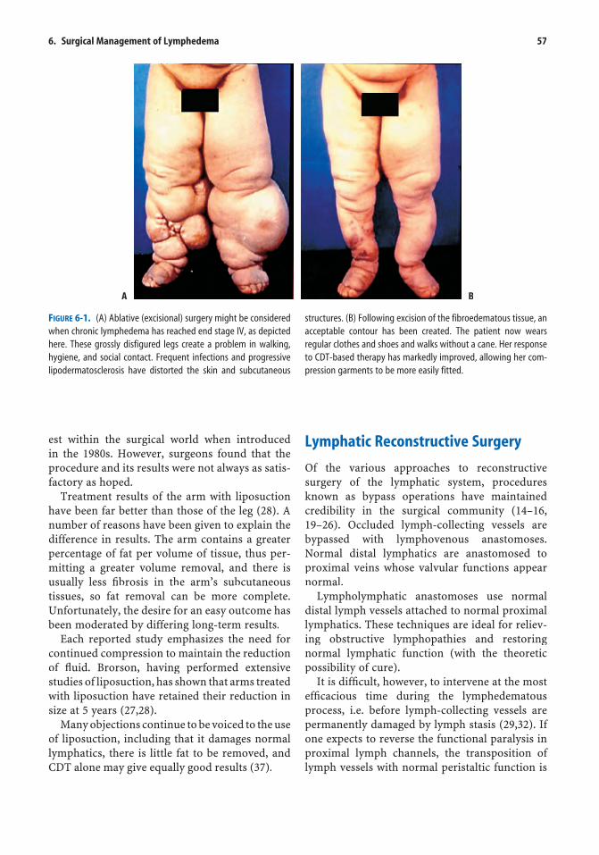
6. Surgical Management of Lymphedema 57
est within the surgical world when introduced in the 1980s. However, surgeons found that the procedure and its results were not always as satis-factory as hoped.
Treatment results of the arm with liposuction have been far better than those of the leg (28). A number of reasons have been given to explain thedifference in results. The arm contains a greater percentage of fat per volume of tissue, thus per-mitting a greater volume removal, and there isusually less fibrosis in the arm’s subcutaneous fitissues, so fat removal can be more complete. Unfortunately, the desire for an easy outcome has been moderated by differing long-term results.
Each reported study emphasizes the need forcontinued compression to maintain the reductionof fluid. Brorson, having performed extensiveflstudies of liposuction, has shown that arms treated with liposuction have retained their reduction insize at 5 years (27,28).
Many objections continue to be voiced to the useof liposuction, including that it damages normallymphatics, there is little fat to be removed, and CDT alone may give equally good results (37).
Lymphatic Reconstructive Surgery
Of the various approaches to reconstructive surgery of the lymphatic system, procedures known as bypass operations have maintained credibility in the surgical community (14–16,19–26). Occluded lymph-collecting vessels are bypassed with lymphovenous anastomoses. Normal distal lymphatics are anastomosed toproximal veins whose valvular functions appearnormal.
Lympholymphatic anastomoses use normaldistal lymph vessels attached to normal proximallymphatics. These techniques are ideal for reliev-ing obstructive lymphopathies and restoringnormal lymphatic function (with the theoreticpossibility of cure).
It is difficult, however, to intervene at the mostfieffi cacious time during the lymphedematous fiprocess, i.e. before lymph-collecting vessels arepermanently damaged by lymph stasis (29,32). If one expects to reverse the functional paralysis in proximal lymph channels, the transposition of lymph vessels with normal peristaltic function is
A B
FIGURE 6-1. (A) Ablative (excisional) surgery might be consideredwhen chronic lymphedema has reached end stage IV, as depictedhere. These grossly disfigured legs create a problem in walking,hygiene, and social contact. Frequent infections and progressive lipodermatosclerosis have distorted the skin and subcutaneous
structures. (B) Following excision of the fibroedematous tissue, an acceptable contour has been created. The patient now wears regular clothes and shoes and walks without a cane. Her responseto CDT-based therapy has markedly improved, allowing her com-pression garments to be more easily fitted.
58 B.B. Lee
essential. Before reconstructive surgery can bewidely accepted, either as an independent or anadjunctive therapy, positive long-term resultsmust be demonstrated.
Selection of Candidates for Anastomotic/
Reconstructive Surgery
In view of the massive financial, medical, and sur-figical commitments for reconstructive surgery, thescreening of candidates for lymphovenous (19,20) or lympholymphatic (15,16) anastomotic surgery must be stringent (32).
To be considered for surgery, candidates mustmeet at least 3 criteria:
• show substantial clinical progression of the disease, from stage I to stage II or from stage II to stage III, in spite of an adequate 12-month CDP treatment program
• demonstrate progressive lymph fluid accumula-fltion, especially below the knee, by lymphoscin-tigrams that show dermal backflowfl
• show decreasing effectiveness of manuallymph drainage-based CDT, especially below the knee
Treatment failure should be documented at least twice within a 6-month interval during a2-year period.
Preoperative Evaluation
Before reconstructive lymphovenous or lympho-lymphatic anastomotic surgery is contemplated,the anatomic and functional status of the proxi-mal lymph nodes and lymph-collecting vessels must be adequately assessed. Determination of function is necessary to identify normal, unpara-lyzed vessels (29,32).
Response to manual lymph drainage can be anindirect indication of the vessels functional status.Lymphoscintigraphy should be included in the preoperative evaluation to confirm these evalua-fitions; additional information may be required from duplex ultrasonography. The newly available ultrasonographic lymphangiography and mag-netic resonance lymphangiography (both in the investigative stage) are sometimes required to more accurately identify malfunctioning lymph-
collecting systems; their fi ndings help confifi rm or firule out paralyzed lymph vessels (32).
Lymphovenous Anastomotic Surgery
Lymphovenous reconstruction is ideal for treating secondary lymphedema, which develops aftercancer surgery or radiation, by restoring a more normal lymphatic function (21,30). Here there isselective damage to proximal lymph nodes, whiledistal lymph-collecting vessels remain intact.However, primary congenital lymphedema, withdysplasia of lymph nodes, is best treated by free lymph node transplantation (17,18).
The technique of lymphovenous anastomosisrequires a microscopic end-to-end or end-to-side anastomosis between healthy, well-functioninglymphatic vessels and healthy veins (23,24). At minimum, 3 to 4 lymph-collecting vessels areanastomosed to defunctionalized branches of the saphenous or adjacent veins in the lower limb(19,20,25,26).
The operation can be performed at the inguinal (19,20) or popliteal level (23,24). The classic ingui-nal approach is preferred during the early stages of lymphedema, when good long-term results canbe expected, e.g. in stage I before the collectingvessels are irreversibly paralyzed. A popliteal anastomosis may be necessary if the disease has progressed to a clinical stage II or early stage III. Evidence of progressive damage and paralysis of lymph-collecting vessels at the inguinal level further recommends a popliteal anastomosis(Figure 6-2).
Lympholymphatic Anastomotic Surgery
Lympholymphatic anastomotic reconstructionuses similar surgical principles and procedures (15,16). It is theoretically superior to a lympho-venous anastomosis, because it avoids the possi-bility of blood regurgitating into the lymph vessel and clotting.
Free Lymph Node Transplantation
The free lymph node transplantation technique isbased on the principle of the free-fl ap autotrans-fl

6. Surgical Management of Lymphedema 59
FIGURE 6-2. (A) Lymphovenous reconstructive surgery was per-formed on the left leg for progressive lymphedema, still in an earlyclinical stage. A preoperative lymphoscintigram shows markeddermal backflow (arrows). (B) This operative view of the poplitealarea reveals functioning lymph-collecting vessels (arrows) that are to be anastomosed end-to-end to the defunctionalized vein above.(Russian method, modified by Krylov.) (C) One month after surgery,a good clinical response is noted, with less edema and improvedskin turgor in the leg. The arrow points to the healing incision.A year later, dermal backflow is markedly diminished, as thescintigram demonstrates.
A
C
B
plantation technique (17,18). A vascularized pedicle fl ap is taken from the donor site, and aflmicroscopic anastomosis is created between theflap and 1 or 2 sets of arteries and veins at theflrecipient site.
Lymphoscintigraphic evaluation is necessary toselect the appropriate lymph node group fortransplantation, e.g. inguinal, cervical, or axillary group (32). Adequate numbers of nodes must be left at the donor site, so that normal function canbe maintained after harvesting. Selection of thebest donor site is as important as selecting thebest recipient site for transplantation. Otherwise,iatrogenic lymphedema may be induced by an unnecessarily aggressive nodal harvest. The patency and function of the patient’s arterial and venous systems should be confirmed with ultraso-finography as well. Local disease status at ankle,popliteal, or inguinal regions helps determine the recipient site.
The indications our institution adopted forfree lymph node transplantation are more liberalthan those for lymphovenous reconstructivesurgery. The authors recommend free lymphnode transplantation when lymphedema controlis only minimal, or when there is suspicion of disease progression and increased sepsis (29,32)(Figure 6-3).
Vein Grafting
Another microsurgical technique uses free vein grafts to bridge lymphatic obstructions, eithercongenital or acquired. As with free nodal trans-plantation, long-term results are improved if the surgery is performed in the earlier stages of lymphedema. Campisi reported an 81% improve-ment, although the exact outcome criteria were not expressed (20).
Postoperative Management
After excisional or reconstructive surgery, contin-ued CDT and compression are mandatory (32). When a successful free lymph node transplantis performed in stage II, CDT may be needed for 12 months or less (18). However, lymphoven -ous anastomotic surgery, performed for the

60 B.B. Lee
FIGURE 6-3. (A) Lymphedema in the left arm (arrows) followeda mastectomy and irradiation. A preoperative lymphoscintigramfailed to demonstrate a viable lymph node in the left axilla.Reconstructive surgery with free lymph node transplantation was recommended. (B) Donor lymph node-bearing tissue har-vested from the right axilla has intact feeding arteries and drainingveins making it suitable for free-flap transplantation. It wastransplanted from the right to the left axilla using microscopicend-to-end anastomoses (arrows) between donor and recipientarteries and veins. (French method, modified by Becker.) (C)Clinical improvement is evident after surgery (arrows). The patient reported less edema and demonstrated improved range of motion. Lymphoscintigraphy highlighted the newly transplantedlymph nodes in the left axilla (arrows). (Photos courtesyC Becker.)
A
C
B
pro gression of the disease from stage II to III, often requires a semipermanent commitment toCDT (19).
This need is accentuated when advanced diseasedictates a lymphovenous anastomosis at the pop-
liteal level, rather than a standard anastomosisat the inguinal level, which is used in earlier disease stages (24,29). Proximal inguinal lym-phatic paralysis would require supplemental CDT to assist in evacuating accumulated proximal edema (29,32).
Evaluation of Bypass Surgery Results
The multidisciplinary care team should eval -uate the patient’s clinical and laboratory treatment response at least twice annually. Postoper -ative assessment of treatment response includesinfrared optic limb volumetry of the extremity on each visit; lymphoscintigraphy is obtained at 6 months and at each anniversary thereafter.If indicated, an ultrasonographic scan of thelimb’s veins is added at the end of the follow-up period. When episodes of cellulitis or othersepsis intervene, additional tests may be necessary (32).
Ideally, the results of treatment should be recorded at 1, 2, 3, 6, 12, 18, and 24 months aftersurgery. It should then be extended from 24 to 48 months, and long-term results should be based onan 8- to 10-year experience.
Clinical Experience
From January 1995 to December 2002, 1065patients with chronic lymphedema were treated atthe Samsung Medical Center in Seoul, SouthKorea.
Of these, 65 limbs of 54 patients were acceptedfor surgical treatment because of documented medical treatment failure—disease progressionin spite of adequate manual lymph drainageand compression therapy performed for at least a year (11,29,31,32).
Their diagnoses were made with a variety of testing methods—ultrasonography, radionuclidelymphoscintigraphy, magnetic resonance imaging(MRI), and laboratory studies.
Treated limbs were divided into 3 groups:
Excisional surgery group = 22 patientsmean age, 46 years

6. Surgical Management of Lymphedema 61
F = 19, M = 3primary disease = 5, secondary disease = 17unilateral disease = 11, bilateral disease = 22
• Lymphovenous anastomotic surgery group = 19 patients� mean age, 49 years� F = 18, M = 1� primary disease = 4, secondary disease = 15
Free lymph node transplantation group = 13 patients
� mean age, 34 years� F= 10, M = 3� primary disease = 6, secondary disease = 7
Excisional Surgery Group
Using a modified Homans operation, 6 patients in ficlinical stage III and the remaining 16 patients instage IV underwent excisional surgery in a total of 33 lower limbs.
Twenty-eight of the 33 limbs showed an over -all improvement at 12 months; of these 28 limbs, 18 were able to retain satisfactory improvementat 24 months only when the patients were compliant with compression therapy. Others inpatients with poor compliance failed to maintainimprovement. Eight limbs among patients withgood compliance to compression therapy main-tained good results at 48 months; as with the other groups, poorly compliant patients showeddeterioration of their legs, both locally andsystemically.
Lymphovenous Anastomotic Surgery
Group
All 19 patients in clinical stages I to II selected as candidates for lymphovenous anastomotic surgery met the criteria for progression of disease under adequate treatment. Follow-up assessment was conducted at 24 and 48 months.
Sixteen of the patients, who complied with postoperative CDT, demonstrated clinically satis-factory improvement. The remaining 3 noncom-pliant patients showed continued deterioration of the limb.
Postoperatively, vigorous CDT was required forall patients as part of the total care management program.
Free Lymph Node Transplantation Group
According to the evaluation criteria, 13 patients, mostly in clinical stages I to II, were selected for surgery. Lymph node-bearing tissue, mainly fromthe posterior axilla, was transplanted to the ingui-nal region.
Ten patients compliant with postoperative CDTprograms showed clinical evidence of a successful graft at 12 months. The remaining 3 poorly com-pliant patients failed to prosper.
Summary
When combined with CDT, surgical therapy remains an adjunctive intervention that can help improve the treatment results of selectedlymphedema patients. This management approach,however, has lacked popular support becausethe microsurgical technique demands unerring surgical expertise and a tremendous amount of time and effort. The results remain equivocal as well.
Both excisional and reconstructive surgeriesappear to control the progression of lymphedema,at least in early postoperative periods. During follow-up periods ranging between 24 and 48 months, the authors’ findings have always shown fithat long-term results improve when patient com-pliance with self-initiated CDT is optimal. Free lymph node transplantation-treated limbs also show an improved outcome when accompaniedwith postoperative CDT, although it is necessary only until transplanted lymph node survival isassured.
To better understand the role of surgical opera-tions in the management of chronic lymphedema,well-constructed trials are warranted. They mustconsist of well-organized multicenter studies thatuse similar protocols and incorporate peer-reviewed outcome results. For the future, surgery remains the only possible treatment methodcapable of providing a cure.

62 B.B. Lee
References
1. Theys S, Deltombe TH, Scavée V, et al. Safety of long-term usage of retrograde-intermittent pneumaticcompression in lower limb lymphedema. Lymphol-ogy. 2002;35.
2. Leduc O, Bourgeois P, Leduc A. Manual of lymphatic drainage: scintigraphic demonstration of its effi-ficacy on colloidal protein reabsorption. In: Partsched. Progress in Lymphology IX. Excerpta Medica.Amsterdam: Elsevier; 1988.
3. Pappas CJ, O’Donnell TF. Long-term results of com-pression treatment for lymphedema. J Vasc Surg. 1992.
4. Hwang JH, Kwon JY, Lee KW, et al. Changes in lym-phatic function after complex physical therapy forlymphedema. Lymphology. 1999.
5. Foldi E, Foldi M, Weissleder H. Conservativetreatment of lymphedema of the limbs. Angiology. 1985.
6. Casley-Smith JR, Casley-Smith JR. Modern Treat-ment for Lymphoedema. Adelaide: LymphoedemaAssociation of Australia; 1994.
7. Hwang JH, Lee KW, Chang DY, et al. Complex physi-cal therapy for lymphedema. J Korean Acad RehabMed. 1998.
8. Hwang JH, Kim TU, Lee KW, et al. Sequential inter-mittent pneumatic compression therapy in lymph-edema. J Korean Acad Rehab Med. 1997.
9. Auchincloss H. New operation for elephantiasis. P RJ Publ Health Trop Med. 1930.
10. Homans J. The treatment of elephantiasis of thelegs. N Engl J Med. 1936.
11. Kim DI, Huh S, Lee SJ, et al. Excision of subcutane-ous tissue and deep muscle fascia for advancedlymphedema. Lymphology. 1998.
12. Sistrunk WE. Further experiences with the Kondo-leon operation for elephantiasis. JAMA. 1918;1:800–805.
13. Kinmonth JB, Patrick J II, Chilvers AS. Commentson operations for lower limb lymphedema.Lymphology. 1975.
14. Degni M. New technique of lymphatic-venous anas-tomosis for the treatment of lymphedema. J Cardio-vasc Surg (Turin). 1978.g
15. Baumeister RGH, Seifert J, Hahn D. Autotransplan-tation of lymphatic vessels. Lancet. 1981.
16. Baumeister RGH, Siuda S. Treatment of lymphede-mas by microsurgical lymphatic grafting: what isproved? Plast Reconst Surg. 1990.
17. Becker C, Hidden G, Godart S, et al. Freelymphatic transplant. Eur J Lymphol. 1991;6:75–80.
18. Becker C, Hidden G, Pecking A. Transplantation of lymph nodes: an alternative method for treatmentof lymphedema. Progr Lymphol. 1990;6:487–493.
29. Campisi C, Boccardo F, Zilli A, et al. Long-termresults after lymphatic-venous anastomoses for thetreatment of obstructive lymphedema. Microsur-gery. 2001.
20. Campisi C, Boccardo F, Zilli A, et al. The use of veingrafts in the treatment of peripheral lymphedemas:long-term results. Microsurgery. 2001.
21. Gloviczki P. Review. Principles of surgical treatmentof chronic lymphoedema. Int Angiol. 1999.
22. Huang GK. Results of microsurgical lymphovenous anastomoses in lymphedema—report of 110 cases. Langenbecks Arch Surg. 1989.
23. Krylov VS, Milanov NO, Abalmasov KG, et al. Recon-structive microsurgery in treatment of lymphoe-dema in extremities. Int Angiol. 1985.
24. Krylov VS, Milanov NO, Abalmassov KG. Efficacy fiof lympho-venous anastomosis. J Mal Vasc. 1988.
25. Olszewski WL. The treatment of lymphedema of the extremities with microsurgical lympho-venousanastomoses. Int Angiol. 1988.
26. Gloviczki P, Fisher J, Hollier LH, et al. Microsurgical lymphovenous anastomosis for treatment of lymph-edema: a critical review. J Vasc Surg. 1988.
27. Brorson H. Liposuction gives complete reduction of chronic large arm lymphedema after breast cancer.Acta Oncol. 2000.
28. Brorson H. Liposuction in arm lymphedema treat-ment. Scand J Surg. 2003.
29. Lee BB, Kim DI, Hwang JH, et al. Contemporary management of chronic lymphedema—personalexperiences. Lymphology. 2002.
30. Lee BB. Lymphoedeme post-chirurgical: un terme frappe d’interdiction pour les chirurgiens. Angeiol-ogie. 2004.
31. Lee BB. Chronic lymphedema, no more step -child to modern medicine. Eur J Lymphology. 2004.
32. Lee BB. Current issues in management of chronic lymphedema: personal reflection on an experienceflwith 1065 patients. [Commentary.] Lymphology. 2005;38.
33. Dellon Al, Hoopes JE. The Charles procedure for primary lymphedema. Plas Reconstr Surg. 1977.
34. Lee BB, Bergan JJ. New clinical and laboratory staging to improve management of chronic lymph-edema. Lymphology. 2005;38.
35. Choi JY, Hwang JH, Park JM, et al. Risk assessment of dermatolymphangioadenitis by lymphoscintig-

6. Surgical Management of Lymphedema 63
raphy in patients with lower extremity lymph-edema. Korean J Nucl Med. 1999.
36. Choi JY, Lee KH, Kim SE, et al. Quantitative lympho-scintigraphy in post-mastectomy lymphedema:
correlation with circumferential measurements. Korean J Nucl Med. 1997.
37. Slavin S. Liposuction in the consensus document.Lymphology. 2004.

64
7Chronic Lymphovenous Disease
Lawrence L. Tretbar
Introduction
Although the surgical treatment of lymphedema warrants a thorough discussion, it requires ahighly trained and experienced team to obtain successful results, and eligible patients are few.
Lymphovenous disease remains a more preva-lent problem than pure lymphedema. However, many physicians experienced in the treatment of early venous diseases are frustrated by the com-plications of advanced lymphovenous diseases—lipodermatosclerosis, ulceration, cellulitis, lymph -ovenous edema (phlebolymphedema)—whichprevent traditional treatment methods of thevenous syndrome.
Healthy Leg Veins
Under normal conditions, venous blood is pumped back to the heart by contractions of the leg muscles,against the pull of gravity. Valves within the veinsmaintain unidirectional fl ow and prevent bloodflfrom falling backward. But because of our uprightstance, there is constant hydrostatic pressurewithin the leg veins. Fortunately, as blood is pumped away, there is a concomitant fall in pres-sure. Hydrostatic pressure, altered by exercise, is calculated as ambulatory venous pressure and measured in mmHg. For example, the venouspressure in the foot when an individual stands at rest is about 100 mmHg. When the leg is exercised, the pressure (ambulatory venous pressure) candrop by 20% to 80%. Leg vein health depends on the ability of the valve/muscle system to decrease
ambulatory venous pressure during exercise(1,3).
Chronic Venous Insufficiency
As long as venous structures remain healthy and their muscular pumps function properly, fluidflentering the interstitial spaces is easily removed by the venules and lymphatics. Should the leakage of fl uid from the arteriovenous capillary network flincrease, lymphatics normally respond by increas-ing their outfl ow. If the lymphatics can continue to flevacuate the increased fluid load, further damagefland edema may be avoided. But if pressure is notattenuated by exercise, the states of chronic venoushypertension and chronic venous insuffi ciency are ficreated; continued damage to the venous and lym-phatic structures is almost inevitable (1–3).
Numerous causes can create valvular incompe-tence and venous insufficiency, including thefiinfl ammatory damage of venous thrombosis,fltrauma, hereditary factors, or the hormonal effectsof pregnancy. Both deep and superficial venousfisystems may be affected.
Chronic venous insufficiency can develop dev-fiastating consequences; high-protein fluid escapesflinto the interstitial space, and the lymphatics are at risk of damage. In most cases the superficialfiveins dilate and the valves are unable to prevent reflux—the backward flfl ow of blood. Starling equa-fltion is upset by the continued elevation of ambu-latory venous pressure and hydrostatic pressure, thereby perpetuating the state of chronic venoushypertension (3–6).

7. Chronic Lymphovenous Disease 65
Other causes of venous damage, reflux and flswelling include:
Endoluminal Venous Obstruction
• acute deep vein thrombosis—unilateral edema• acute superficial vein thrombosis—unilateral fi
edema• combined acute deep and superficial vein throm-fi
bosis—marked unilateral edema• inferior vena caval obliteration, filter, local fi
thrombosis—chronic, marked bilateral edema• late post-thrombotic syndrome—chronic, may
be bilateral edema (see Figure 7-1)
Extraluminal Venous Obstruction
• ruptured medial gastrocnemius muscle head incalf—acute, unilateral edema
• ruptured popliteal synovial (Baker) cyst atknee—acute, unilateral edema
• iliac artery or vein compression—chronic, unilateral edema
• pregnancy—chronic, may be bilateral edema• chronic distended urinary bladder, stricture,
enlarged prostate—chronic, may be bilateraledema
• aortic, iliac, popliteal arterial aneurysm—usually chronic, may be bilateral edema
• malignancy, iliac, intra-abdominal—chronic,usually unilateral
• retroperitoneal fi brosis—chronic, may be fibilateral
Muscle Pump Failure/Dependent
Leg Syndrome
• spinal cord paralysis or stroke with mobility loss—chronic, bilateral
• confi nement to chair—chronic, bilateralfi• arthritis with ankle joint fi xation—chronic, may fi
be bilateral
A B
FIGURE 7-1. (A) This man developed bilateral deep vein thrombo-ses following prolonged bedrest. His legs show all of the complica-tions related to the post-thrombotic syndrome: nonpitting lymphedema of the entire leg, hyperpigmentation, lipodermato-sclerosis, and tiny ulcerations in the calves. The feet begin to showthe signs of lymphedema—deep flexion creases, puffy feet and toes—but are spared much of the pigmentation because of the
compressive effect of tight shoes. (B) Viewed from behind, manyof the same features are evident. Note the creases left by his cloth-ing in the upper legs. Swelling obscures Achilles tendons, andpigmentation is circumferential. This patient was not considered acandidate for surgery; his legs were improved with CDT-basedtreatment and continued compression. (Photos courtesy of LL Tretbar.)
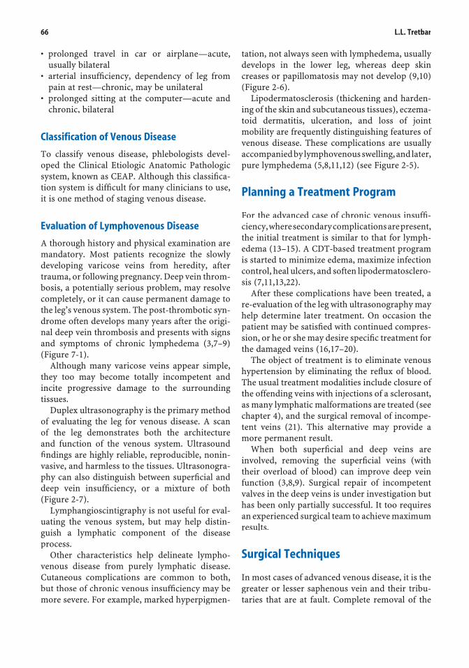
66 L.L. Tretbar
• prolonged travel in car or airplane—acute, usually bilateral
• arterial insuffi ciency, dependency of leg fromfipain at rest—chronic, may be unilateral
• prolonged sitting at the computer—acute and chronic, bilateral
Classification of Venous Disease
To classify venous disease, phlebologists devel-oped the Clinical Etiologic Anatomic Pathologic system, known as CEAP. Although this classifica-fition system is diffi cult for many clinicians to use,fiit is one method of staging venous disease.
Evaluation of Lymphovenous Disease
A thorough history and physical examination are mandatory. Most patients recognize the slowly developing varicose veins from heredity, aftertrauma, or following pregnancy. Deep vein throm-bosis, a potentially serious problem, may resolvecompletely, or it can cause permanent damage tothe leg’s venous system. The post-thrombotic syn-drome often develops many years after the origi-nal deep vein thrombosis and presents with signsand symptoms of chronic lymphedema (3,7–9) (Figure 7-1).
Although many varicose veins appear simple, they too may become totally incompetent andincite progressive damage to the surrounding tissues.
Duplex ultrasonography is the primary method of evaluating the leg for venous disease. A scanof the leg demonstrates both the architectureand function of the venous system. Ultrasound findings are highly reliable, reproducible, nonin-fivasive, and harmless to the tissues. Ultrasonogra-phy can also distinguish between superficial andfideep vein insufficiency, or a mixture of bothfi(Figure 2-7).
Lymphangioscintigraphy is not useful for eval-uating the venous system, but may help distin-guish a lymphatic component of the disease process.
Other characteristics help delineate lympho-venous disease from purely lymphatic disease. Cutaneous complications are common to both, but those of chronic venous insuffi ciency may befimore severe. For example, marked hyperpigmen-
tation, not always seen with lymphedema, usually develops in the lower leg, whereas deep skin creases or papillomatosis may not develop (9,10)(Figure 2-6).
Lipodermatosclerosis (thickening and harden-ing of the skin and subcutaneous tissues), eczema-toid dermatitis, ulceration, and loss of jointmobility are frequently distinguishing features of venous disease. These complications are usually accompanied by lymphovenous swelling, and later, pure lymphedema (5,8,11,12) (see Figure 2-5).
Planning a Treatment Program
For the advanced case of chronic venous insuffi-ficiency, where secondary complications are present,the initial treatment is similar to that for lymph-edema (13–15). A CDT-based treatment program is started to minimize edema, maximize infectioncontrol, heal ulcers, and soften lipodermatosclero-sis (7,11,13,22).
After these complications have been treated, are-evaluation of the leg with ultrasonography may help determine later treatment. On occasion thepatient may be satisfied with continued compres-fision, or he or she may desire specific treatment forfithe damaged veins (16,17–20).
The object of treatment is to eliminate venoushypertension by eliminating the reflux of blood.flThe usual treatment modalities include closure of the offending veins with injections of a sclerosant, as many lymphatic malformations are treated (seechapter 4), and the surgical removal of incompe-tent veins (21). This alternative may provide amore permanent result.
When both superficial and deep veins are fiinvolved, removing the superficial veins (withfitheir overload of blood) can improve deep vein function (3,8,9). Surgical repair of incompetent valves in the deep veins is under investigation but has been only partially successful. It too requiresan experienced surgical team to achieve maximum results.
Surgical Techniques
In most cases of advanced venous disease, it is thegreater or lesser saphenous vein and their tribu-taries that are at fault. Complete removal of the

7. Chronic Lymphovenous Disease 67
FIGURE 7-2. (A) A flexible stripper is introduced andadvanced through the vein that is to be removed. Tiny inci-sions allow the instrument to remove the veins frombeneath the skin with minimal trauma and scarring. (B) Aphlebectomy hook draws the side branch varicosities from the subcutaneous tissues. (Photos courtesy of LL Tretbar.)
damaged veins, or stripping, has been a traditionalmethod of eliminating saphenous veins whoserefl ux causes venous hypertension. Side branchflvaricosities are removed with tiny hooks, a tech-nique termed hook phlebectomy (3,9) (Figure7-2).
Experienced surgeons perform the surgery as an outpatient procedure using local anesth -esia, often supplemented with conscious sedation.Continued compression is essential, at leastduring the healing phase; occasionally, use of compressive garments is discontinued at a later date.
Endoluminal Ablation
Rather than removing the veins, endoluminalablation closes the incompetent venous conduitthat maintains the reflux. This technique obliter-flates the lumen of the saphenous vein with heat or
with injections of a sclerosant (3,17,19) (seechapter 4).
Results of Surgical Treatment
In a 10-month retrospective study of 105patients who received surgical treatment for lym-phovenous disease in 113 legs, one-third hadswelling or advanced lymphedema of the limb. Other cutaneous complications were found in 76 limbs.
At the completion of the study, all limbs hadimproved: hyperpigmentation decreased, lipoder-matosclerosis softened, “intractable” ulcers healed,edema and episodes of inflammation and infec-fltion were reduced. Only half of the limbs requiredcontinued compression, usually in the form of compression garments. The illustrations (Figures 7-3 to 7-5) demonstrate the results of surgicaltreatment.
A
B
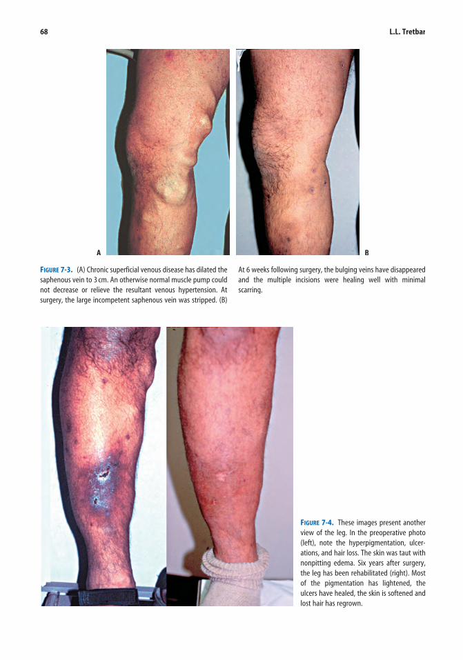
68 L.L. Tretbar
FIGURE 7-4. These images present anotherview of the leg. In the preoperative photo(left), note the hyperpigmentation, ulcer-ations, and hair loss. The skin was taut withnonpitting edema. Six years after surgery,the leg has been rehabilitated (right). Mostof the pigmentation has lightened, theulcers have healed, the skin is softened andlost hair has regrown.
A B
FIGURE 7-3. (A) Chronic superficial venous disease has dilated thesaphenous vein to 3 cm. An otherwise normal muscle pump couldnot decrease or relieve the resultant venous hypertension. At surgery, the large incompetent saphenous vein was stripped. (B)
At 6 weeks following surgery, the bulging veins have disappearedand the multiple incisions were healing well with minimalscarring.
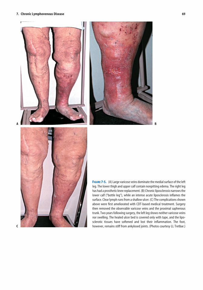
7. Chronic Lymphovenous Disease 69
FIGURE 7-5. (A) Large varicose veins dominate the medial surface of the leftleg. The lower thigh and upper calf contain nonpitting edema. The right leg has had a prosthetic knee replacement. (B) Chronic liposclerosis narrows the lower calf (“bottle leg”), while an intense acute liposclerosis inflames the surface. Clear lymph runs from a shallow ulcer. (C) The complications shownabove were first ameliorated with CDT-based medical treatment. Surgery then removed the observable varicose veins and the proximal saphenous trunk. Two years following surgery, the left leg shows neither varicose veinsnor swelling. The healed ulcer bed is covered only with tape, and the lipo-sclerotic tissues have softened and lost their inflammation. The foot, however, remains stiff from ankylosed joints. (Photos courtesy LL Tretbar.)
A
C
B
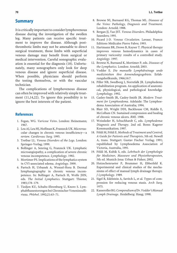
70 L.L. Tretbar
Summary
It is critically important to consider lymphovenousdisease during the investigation of the swollen leg. Many patients can receive specific treat -fiment to improve the disease. Although post-thrombotic limbs may not be amenable to directsurgical treatment, those limbs with superficialfivenous damage may benefi t from surgical or fimedical intervention. Careful sonographic evalu-ation is essential for the diagnosis (18). Unfortu-nately, many sonographers only look for deepvenous disease and ignore superficial disease.fiWhen possible, physicians should performthe testing themselves, or with the vasculartechnician.
The complications of lymphovenous diseasecan often be improved with relatively simple treat-ment (11,14,22). To ignore this possibility is toignore the best interests of the patient.
References
1. Fegan, WG. Varicose Veins. London: Heinemann;1967.
2. Leu AJ, Leu HJ, Hoffman K, Franzeck UK. Microvas-cular changes in chronic venous insuffi ciency—a fireview. Cardiovasc Surg. 1995.
3. Tretbar LL. Venous Disorders of the Legs. London:Springer-Verlag; 1999.
4. Bollinger A, Senring G, Franzeck UK. Lymphatic microangiopathy, a complication of severe chronicvenous incompetence. Lymphology. 1982.
5. Mortimer PS. Implications of the lymphatics system in CVI-associated edema. Angiology. 2000.
6. Partsch H, Urbanek A, Wenzel-Hora B. Dermallymphangiography in chronic venous incom-petence. In: Bollinger A, Partsch H, Wolfe JHN,eds. The Initial Lymphatics. Stuttgart: Thieme;1985;178–179.
7. Tiedjen KU, Schultz-Ehrenburg U, Knorz S. Lym-phabflussestorengen bei Chronischer Veneninsuffifl -fizienz. Phlebol. 1992;21:63–71.
8. Browse NI, Burnand KG, Thomas ML. Diseases of the Veins: Pathology, Diagnosis and Treatment.London: Arnold; 1988.
9. Bergan JJ, Yao JST. Venous Disorders. Philadelphia:Saunders; 1991.
10. Picard J-D. Venous Circulation. Lavaur, France:Editions Médicales Pierre Fabre; 1993.
11. Hartmann BR, Drews B, Kayser T. Physical therapy improves venous hemodynamics in cases of primary varicosity: results of a controlled study. Angiology. 1997.
12. Browse N, Burnand K, Mortimer P, eds. Diseases of the Lymphatics. London: Arnold; 2003.
13. Vodder E. Die manuelle Lymphdrainage undemedizinishen ihre Anwendungssgebiete. Erfah-rungsheilkunde, 1966;16:7.
14. Piller NB, Swedborg I, Norrefalk JR. Lymphedemarehabilitation program. An application of anatomi-cal, physiological, and pathological knowledge.Lymphology. 1992.
15. Casley-Smith JR, Casley-Smith JR. Modern Treat-ment for Lymphoedema. Adelaide: The Lymphoe-dema Association of Australia; 1994.
16. Blair SD, Wright DDI, Backhouse CM, Riddle E, McCollum CN. Sustained compression and healingof chronic venous ulcers. BMJ. 1988.
17. Weissleder H, Schuchhardt C, eds. Lymphedema:Diagnosis and Therapy. 2nd ed. Bonn: Kagerer Kommunikation; 1997.
18. Földi M, Földi E. Methods of Treatment and Control, A Guide for Patients and Therapists, 5th ed. NewellA, trans. Stuttgart: Gustav Fischer Verlag; 1991;republished by Lymphoedema Association of Victoria, Australia, 1991.
19. Földi M, Kubik S, eds. Lehrbuch der Lymphologie für Mediziner, Masseure und Physiotherapeuten,5th ed. Munich-Jena: Urban & Fisher; 2002.
20. Hutzschenrueter P, Brummer H, Ebberfeld K.Experimental and clinical studies of the mecha-nisms of effect of manual lymph drainage therapy. J Lymphology. 1989.
21. Sigel B, Edelstein A, Savitch L, et al. Types of com-pression for reducing venous stasis. Arch Surg.1975.
22. Kasseroller RG.Compendium of Dr. Vodder’s Manual Lymph Drainage. Heidelberg: Haug; 1998.

71
AAblation treatment. See Excisional
(ablative) treatmentAmerican Society of Lymphology,
44Angiogenesis, 40, 48Angio-osteohypertrophy
syndrome. See Klippel-Trenaunay syndrome
Ankle-brachial pressure index (ABPI), 16
Antibodies, 10Aplasia, primary lymphedema-
related, 31Arteriovenous malformations, 33Arthritis, as lymphedema cause,
13Aselli, Gaspar, 1, 2, 3
BBandaging, for lymphedema
management, 46, 47, 49–50, 52
Blood circulation, 1“Bottle leg,” 69
CCancer, as lymphedema cause,
15–16Cancer treatment, as lymphedema
cause, 13, 14, 24Cellulitis
as lymphedema cause, 12, 15, 16,24
as lymphovenous diseasecomplication, 64
Children, lymphatic malformationtreatment in, 33–34
Classifi cation and staging, of filymphedema, 21–30
staging methods in, 23–29Clinical Etiologic Anatomic
Pathologic System (CEAP),26, 66
Complete decongestive therapy (CDT), 55
combined with surgicalprocedures, 55, 56, 57, 58, 59–60, 61
contraindications to, 48–49, 53development of, 43for elephantiasis, 44–47indications for, 53for truncular lymphedema, 32as two-phase intervention,
47–52Comprehensive decongestive
physiotherapy (CDP), 43, 44Comprehensive Decongestive
Therapy©, 51–52Compression garments, 51, 52Computed tomography
of extratruncular lymphaticmalformations, 33
of lymphedema, 18, 33
DDiabetes mellitus, 24Differential diagnosis, of
lymphedema, 12–20Diffusion, 8–9Drugs, as edema cause, 15
EEctasia, primary lymphedema-
related, 31
Edema. See also Lymphedemadefi nition of, 8fidifferential diagnosis of,
15–20lymphatic causes of,
12–14nonpitting, 25pathophysiology of, 12pitting, 25venous causes of, 15
Effl eurage, 49flElephantiasis
complete decongestion therapy for, 44–47
lymphostatic, 23silica crystal absorption-related,
13, 14Embolism, pulmonary, 39,
44Ethanol, as sclerotherapy agent,
33–34Excisional (ablative) treatment
endoluminal, for lymphovenousdisease, 67
of extratruncular lymphaticmalformations, 33
of truncular lymphaticmalformations, 32
Exercise programs, forlymphedema patients,50–51
FFilariasis, 12, 13, 25–26Fistulae, arteriovenous, 15, 39Fluid exchange, 8Free lymph node transplantation,
58–59, 60, 61
Index

72 Index
GGene therapy, for lymphatic
malformations, 40
HHarvey, William, 1Hemangioma, 38
cavernous, 38, 39spontaneous rupture of, 39
Hippocrates, 1Hunter, John, 1, 3Hunter, William, 1, 3Hyperkeratosis, 14, 32Hyperpigmentation, 46
deep venous thrombosis-related, 17
edema-related, 16, 17lymphedema-related, 32lymphovenous disease-related,
67, 68post-thrombotic syndrome-
related, 65, 66Hyperplasia, primary
lymphedema-related, 31Hypertension
chronic venous, 64venous, 66–67, 68
Hypertrophy, of bone or limbs, 38,39
Hypoalbuminemia, 15Hypoplasia, primary lymphedema-
related, 31
IImmune system/immunity,
9–10Infection
cutaneous, lymphedema-associated, 49
as lymphedema cause, 12, 13during lymphedema treatment,
45, 46Infl ammation, as lymphedema fl
cause, 13International Lymphatic Research
Society, 43International Society of
Lymphology, 44, 47Consensus Document on the
Diagnosis and Treatment of Peripheral Lymphedema, 23
Interstitial space, fluid within, 8, fl12
KKaposi-Stemmer sign, 16Klippel-Trenaunay syndrome, 15
LLeg veins, healthy, 64Lipedema, 18–19Lipodermatosclerosis, 25, 64, 65, 66,
67Liposclerosis, 69Liposuction, 56–57Lymphangiography, 18Lymphangioma, 12, 33, 40
Klippel-Trenaunay syndrome-related, 39
Lymphangioma cavernosa, 31, 33Lymphangioma circumscriptum,
36, 37Lymphangioma cystic (hygroma),
31, 33–35Lymphangioma simplex, 33Lymphangiomatosis, 33, 37, 38Lymphangiosarcoma, 36Lymphangioscintigraphy, for
lymphovenous disease evaluation, 66
Lymphatic Research Foundation, 44
Lymphatic systemembryology of, 4–5structure and functions of,
1–11early investigations of, 1–4, 43lymph capillaries, 5lymph collectors, 5, 6lymph ducts, 6–7lymph nodes, 6–7lymph precollectors, 5–6Peyer patches, 6prelymphatic tissue channels,
5Lymph ducts, structure and
functions of, 6–7Lymphedema
bilateral, chronic stage III, 44–47chronic, 31congenital, 22–23diagnostic tests for, 16, 18fi liarial, 44fihereditary
type II (Meige disease), 12type I (Milroy disease), 12, 13
primary, 12, 31–32classifi cation of, 21–22fi
as lymphangiosarcoma cause, 36
secondary, 12–14, 31classifi cation of, 22–23fi
Lymphedema praecox, 12, 22Lymphedema tarda, 12, 22Lymph nodes, structure and
functions of, 6–7Lymphogranuloma venereum, 12Lymphography, of manual lymph
drainage, 48Lymphology, 23Lymphoscintigraphy
of cancer treatment-related lymphedema, 14
of extratruncular lymphatic malformations, 33
for lymphedema diagnosis, 16, 18
for lymphedema staging, 26of manual lymph drainage, 48preoperative, 56, 58, 59, 60, 61of primary lymphedema, 31,
32Lymphovenous disease. See also
Venous insuffi ciency,fichronic
causes of, 64, 65–66chronic, 64–70
classifi cation of, 66ficomplications of, 64evaluation of, 16, 17, 66
surgical treatment of, 66–69
MMagnetic resonance imaging
of extratruncular lymphatic malformations, 33
of lipedema, 18of lymphedema, 18, 32–33
Malformations, lymphatic, 12, 31–42
congenital vascular malformations associated with, 38–39
extratruncular forms of, 31, 33–35
gene therapy for, 40truncular forms of, 31–32, 39
Manual lymph drainage, 46, 48–49
Mascagni, Paulo, 1, 4Medical management, of
lymphedema, 45–54

Index 73
historical review of, 45–46successful, 46–47as two-phase intervention,
47–52Meige disease, 12Microcirculation, 5Milroy disease, 12, 13Muscle pump failure/dependent
leg syndrome, 65–66
NNational Lymphedema Network,
44Nutritional counseling, 51
OObesity
as lipedema cause, 19as lymphedema cause, 24, 51
OK-432, as sclerotherapy agent, 33–34
Osmosis, 9
PPalliative care, 52Panniculitis, sclerosing, 25Papilloma, 46Peyer patches, 6Phlebectomy, hook, 67Phlebolymphedema, 64Pneumatic pumps, 51Podoconiosis, 13, 14Port-wine stain, 38, 39Post-thrombotic syndrome, 65, 66Psychosocial interventions, with
lymphedema patients, 52
QQuality of life, assessment of,
27–28
RReconstructive surgery, lymphatic,
55, 57–58, 59
SSclerotherapy, 34–35, 38, 67Scrofula, 12Self-management, of lymphedema,
44, 47–48, 52Skin care, in lymphedema patients,
49Starling equation, 8Stewart-Trevers sarcoma. See
LymphangiomaSurgery, as lymphedema cause, 13Surgical management
of lymphedema, 55–63clinical outcomes in, 60–61combined with complete
decongestive therapy (CDT), 55, 56, 57, 58, 60, 61
excisional/ablation surgery,55–57, 61, 67
free lymph nodetransplantation, 58–59, 60,61
liposuction, 56–57lymphatic reconstructive
surgery, 55, 57–58, 59lymphovenous anastomotic
surgery, 55, 57–58, 59, 61postoperative management in,
59–60vein grafting, 59
of lymphovenous disease, 66–69
TThrombosis
deep venous, 17, 65as venous insuffi ciency cause, 64fi
Traumaas lipedema cause, 19as lymphedema cause, 13, 14,
15as venous insuffi ciency cause, fi
64Tuberculosis, of the lymph nodes,
12
UUlcers
deep venous thrombosis-related,17
dressings for, 46, 49elephantiasis-related, 45,
46infection-related, 17lymphovenous disease-related,
16, 64, 65, 66, 67, 68,69
Ultrasonography, duplex,17
for lymphovenous diseaseevaluation, 66, 70
VVaricose veins, 22, 38, 45, 66, 67,
69Vascular malformations,
congenital, 38–39Vasculogenesis therapy, 40Vein grafting, as lymphedema
treatment, 59Venous insuffi ciency, chronicfi
as lipedema cause, 19as lymphedema cause, 14, 15, 16,
24, 64–70
WWound care, 46, 49