ebv essential antigen ebna3c attenuates h2ax...
TRANSCRIPT

1
EBV essential antigen EBNA3C attenuates H2AX expression 1 2 3 Hem C Jha±, Mahadesh Prasad AJ±, Abhik Saha, Shuvomoy Banerjee, Jie Lu, and Erle S 4
Robertson* 5 6
7 Department of Microbiology and Tumor Virology Program, 8
Abramson Cancer Center, Perelman School of Medicine at the University of 9 Pennsylvania, Philadelphia, PA-19104, USA 10
11 ± Equal Contribution 12 13 * Corresponding Author: 14 E. mail: [email protected] 15 Phone: 1-215-746-0114; Fax: 1-215-898-9557 16 17 Running Title: EBNA3C deregulates H2AX 18 19 Keywords: EBNA3C; H2AX; Ubiquitination; Ser139 H2AX mutation; EBV infection; 20 Cell proliferation 21 22 23
JVI Accepts, published online ahead of print on 15 January 2014J. Virol. doi:10.1128/JVI.03568-13Copyright © 2014, American Society for Microbiology. All Rights Reserved.
on May 23, 2018 by guest
http://jvi.asm.org/
Dow
nloaded from

2
ABSTRACT 24 EBV latent antigen EBNA3C is implicated in B-cell immortalization and linked 25
to several B-cell malignancies. Deregulation of H2AX can induce genomic instability 26 with increased chromosomal aberrations which ultimately leads to tumorigenesis. Here 27 we demonstrated that EBNA3C can attenuate H2AX expression at the transcript and 28 protein levels. A reduction of total H2AX levels were clearly observed on infection of 29 primary B-cells with wild type EBV, but not with EBNA3C knockout EBV recombinant 30 virus. H2AX also interacted with EBNA3C through its N-terminal domain (residues 1-31 100). Furthermore, H2AX mutated at Ser139 failed to interact with EBNA3C. Luciferase 32 based reporter assays also revealed that the binding domain of EBNA3C is sufficient for 33 transcriptional inhibition of the H2AX promoter. EBNA3C also facilitated H2AX 34 degradation through recruitment of components of the ubiquitin proteasome pathway. We 35 further demonstrated that knockdown of H2AX in LCLs led to up-regulation of the Bub1 36 oncoprotein, and down-regulated expression of the p53. Overall, our study provides 37 additional insights into EBV-associated B-cell lymphomas which is linked to regulation 38 of the DNA damage response system in the infected cells. 39 40 Importance: 41 I. EBNA3C down-regulates H2AX expression at the protein and transcript levels in 42 epithelial cells, B-cells and EBV transformed LCLs . 43 II. EBNA3C binds with wild type H2AX but not with the ser139 mutant of H2AX. 44 III. The N-terminal (residue 1-100 amino acid) of EBNA3C is critical for binding to 45 H2AX. 46
on May 23, 2018 by guest
http://jvi.asm.org/
Dow
nloaded from

3
IV. Localization of H2AX is predominantly nuclear in the presence of EBNA3C. 47 V. H2AX knocked down in LCLs led to enhanced expression of Bub1 and 48 downregulation of the tumor suppressor p53 both important for driving the oncogenic 49 process. 50 51 52 53 54 55 56 57 58 59 60 61 62 63 64 65 66 67 68 69
on May 23, 2018 by guest
http://jvi.asm.org/
Dow
nloaded from

4
INTRODUCTION 70 Epstein Barr Virus (EBV) is a human gammaherpesvirus associated with 71
infectious mononucleosis, and it is estimated that more than 95% of adults are carriers of 72 EBV throughout their lifetime (1, 2). The contributory role of EBV in driving the 73 oncogenic process is continually being explored. EBV transforms latently infected 74 primary B-cells into constantly proliferating lymphoblastoid cell lines (LCLs) (3). EBV is 75 also commonly involved in numerous malignancies, including Burkitt’s Lymphoma 76 (BL), Post transplant lympho-proliferative disorders (PTLDs), Nasopharyngeal 77 carcinoma (NPC), HIV associated lymphomas, some types of T cell lymphomas, and 78 Gastric Cancer (4, 5). 79
Transformation of human B-cells into LCLs by EBV establishes a latent type of 80 infection typically known as type III latency (6). Three major viral latency programs have 81 been described with deferential expression profiles of specific viral gene transcription (7). 82 EBV latency patterns are characterized by the expression of different EBV nuclear 83 antigens (EBNAs) including, EBNA 1, 2, 3A, 3B, 3C, LP/5, and the latent membrane 84 proteins (LMP) 1, 2A and 2B (8). Importantly, these latent proteins are significantly 85 expressed during the latency III program (9) (10). Previous studies showed that EBNA2, 86 EBNA3A, EBNA3C and LMP1 play critical roles in B-cell transformation (11, 12). 87 Earlier studies showed that one of the essential EBV latent antigens, EBNA3C is 88 important for modulating B-cell activation. For example, the B-cell activation marker 89 CD21 was up-regulated in the presence of EBNA3C in Burkitt’s lymphoma cell lines (13, 90 14). EBNA3C binds to RBP-Jk, an important regulator of the Notch signaling pathway, 91 through an amino terminal motif, and the acidic domains are responsible for nuclear 92
on May 23, 2018 by guest
http://jvi.asm.org/
Dow
nloaded from

5
translocation due to the presence of the nuclear localization signals (15). Recently, we 93 reported that the p53 tumor suppressor is negatively regulated by EBNA3C both at the 94 transcriptional and post-transcriptional level (16). Critically, EBNA3C has also been 95 shown to regulate the major cell cycle checkpoints (17). Recently it was suggested that 96 EBV has a potential role in inducing genomic instability and that viral proteins associated 97 with the latency III program can regulate the DNA damage response (DDR) (18). In 98 addition, earlier studies from our lab demonstrated that EBNA3C binds to Chk2, a major 99 effecter of the DDR, which also deregulates the cell cycle of EBV infected cells at the 100 G2/M phase (19, 20). 101 EBV infection of primary B-cells was shown to activate the DDR by inducing 102 phosphorylation of H2AX at Ser139 (�-H2AX) (20). H2AX is a histone variant, which 103 has a key regulatory function during induction of the DDR. Induction of �-H2AX is a 104 hallmark of the DDR and recruits various DNA damage proteins, repair proteins as well 105 as cell cycle checkpoints (21). Recently, we found that H2AX phosphorylation is 106 important for KSHV-induced oncogenesis, which is mediated through one of its major 107 latent protein LANA (22). However, upon EBV infection, the mechanism by which cells 108 trigger the DDR and proceed towards oncogenesis is still not clearly understood. Further, 109 it is still not determined how the DDR progresses without repairing the damaged DNA to 110 bypass cell cycle arrest or apoptosis (16, 23, 24). In this study, we now demonstrate that 111 the EBV latent antigen EBNA3C deregulates total H2AX levels transcriptionally and 112 post-translationally through involvement of the ubiquitin-mediated proteasome 113 degradation pathway. Additionally, our study also showed dramatic changes in 114 expression patterns of the tumor suppressor p53 and oncoprotein Bub1 in H2AX 115
on May 23, 2018 by guest
http://jvi.asm.org/
Dow
nloaded from

6
knockdown LCLs. These results provide further clues as to the biological relevance of 116 H2AX deregulation in EBV-induced oncogenesis. Overall, our study suggests that 117 EBNA3C can play an important role in EBV-mediated oncogenesis through down-118 modulation of H2AX. 119 120 121 122 123 124 125 126 127 128 129 130 131 132 133 134 135 136 137 138
on May 23, 2018 by guest
http://jvi.asm.org/
Dow
nloaded from

7
MATERIALS AND METHODS 139 Ethics Statement - The University of Pennsylvania School of Medicine CFAR 140
(centre for AIDS research) Immunology core provided us human peripheral blood 141 mononuclear cells (PBMC) from unidentified donors, with written consent, which is 142 approved by IRB based on Helsinki recommendations. 143
Constructs and Transfection - Full length EBNA3C plasmid or its various 144 truncated domains were previously mentioned (25, 26). Plasmid expressing wild type 145 H2AX was previously described (27). Constructs expressing Flag-tagged wild type 146 H2AX and its mutated version H2AX Ser139 (H2AX S-A) were kindly provided by Dr 147 Alvaro N.A. Monteiro (H. Lee Moffitt cancer center, Tampa, FL) (28). pGL3-H2AX wild 148 type reporter construct was generously provided by Dr. Toru Ouchi (Roswell Park cancer 149 Institute, Buffalo, NY) (29). All construct sequences were further confirmed by DNA 150 sequencing at the core facility in University of Pennsylvania. All transfection procedures 151 were as described earlier (30). 152
Cell cultures and Antibodies EBV positive LCL1 and LCL2, BJAB stably 153 expressing EBNA3C clones (BJAB#7 and BJAB#10) and EBV negative cell lines BJAB, 154 DG75, and Ramos were cultured in RPMI 1640 medium and as described earlier (22). 155
Rabbit anti-H2AX antibody was obtained from Bethyl laboratories Inc. 156 (Montgomery, TX). GAPDH (anti-mouse) was purchased from US biological corp. 157 (Swampscott, MA). Mouse antibodies reactive to the Myc epitope (9E10), Flag epitope 158 (M2) and EBNA3C (A10) were described earlier (27). 159
Induction and Infection of recombinant EBV - BAC GFP-EBV was generated 160 as previously described (13). EBNA3C null mutant (ΔEBNA3C BAC GFP-EBV) was 161
on May 23, 2018 by guest
http://jvi.asm.org/
Dow
nloaded from

8
generated by deleting EBNA3C region between 98370-102891 bp from WT BAC GFP 162 EBV Plasmid (JL, HCJ and ESR, Personal communications). Southern Blot analysis, 163 PCR, and sequencing confirmed that the mutation was correct. Cells harboring BAC 164 GFP-EBV and ΔEBNA3C GFP- EBV were induced with TPA (12-O-165 tetradecanoylphorbol) and butyric acid (BA, 3mM: Sigma-Aldrich Corp., St. Louis, MO) 166 for 4-5 days in DMEM. Cell suspension was centrifuged at 1,800 rpm for 15 min and 167 filter through 0.45μ cellulose acetone filters. The viral particles were concentrated by 168 ultracentrifugation at 23,500 rpm at 4oC and viral particles were stored at -80oC. 169
For infection studies, we followed similar procedures as described earlier (31). 170 Ten million PBMCs were mixed with either wild type or ΔEBNA3C virus supplemented 171 with RPMI 1640 containing 10% FBS and incubated overnight at 37oC. Cells were 172 centrifuged at 2,000 rpm for 10 min. Pelleted cells were resuspended in 2 ml of cell 173 culture medium supplemented with 10% FBS. GFP expression was visualized to monitor 174 infection and RT-PCR of EBNA1 confirmed infection. 175
RNA interference - Short-hairpin oligonucleotides directed against EBNA3C 176 were designed (Dharmacon Research, Chicago, IL). All procedure and sequence for sh-177 EBNA3C- clone were followed as described earlier (28). To clone sh-H2AX, we used 178 similar strategy and sequences described earlier (32). Cell cultures were selected with 179 Puromycin. 180
Immunoprecipitation and Western blotting - Transiently transfected cells were 181 harvested and washed with ice-cold PBS followed by lysing cells using RIPA buffer 182 (10mM Tris, pH 7.5, 150mM Sodium chloride, 2mM EDTA, 1% NP40) and the protease 183 inhibitor cocktail was added before lysing the cells. The cell debris was removed by 184
on May 23, 2018 by guest
http://jvi.asm.org/
Dow
nloaded from

9
centrifugation at 21,000g for 12 min (4oC), supernatant was transferred to autoclaved 185 micro-centrifuge tubes. The lysate were then pre-cleared by spinning with normal mouse 186 serum and 1:1 mixture of Protein A and Protein G conjugated Sepharose beads for 1.5 187 hours at 4oC. The beads were spun at low speed and the supernatant transferred to a fresh 188 micro-centrifuge tube. The specific protein of interest was incubated with 1 μg of suitable 189 interacting antibody for overnight at 4oC in a rotating chamber. Immune complexes were 190 collected by binding with Protein A and G beads, pelleted and washed with ice cold 191 RIPA buffer 3 times. WB was performed as described previously (33). 192
193 RNA isolation and Quantitative real time PCR - Trizol reagent (Invitrogen Inc, 194
Carlsbad, CA) was used to isolate total RNA as described by manufacturer's instruction. 195 RNA to cDNA was prepared by superscript II reverse transcription kit (Invitrogen Inc, 196 Carlsbad, CA). The primers used for RT-PCR were as follows: H2AX 5’-197 GTTCCCAGTGGGCCGTGTA-3’ and 5’-CGGTGAGGTACTCCAGCACT-3’ for 198 EBNA3C 5’-AGAAGGGGAGCGTGTTTGT-3’ and 5’-GGCTCGTTTTGACGTCGGC-199 3 for EBNA1 5’-CATTGAGTCGTCTCCCCTTTGGAAT-3’ and 5’-200 TCATAACAAGGTCCTTAATCGCATC-3’ and for GAPDH 5’-201 TGCACCACCAACTGCTTAG-3’ and 5’-CATGCAGGGATGATGTTC-3’. 202 Quantitative RT-PCR was carried out using Step One Plus Real-time PCR system 203 (Applied Bio Systems, Foster city, CA), and all experiments were performed in 204 triplicates. 205
on May 23, 2018 by guest
http://jvi.asm.org/
Dow
nloaded from

10
Reporter assay - 10 million HEK-293 cells were co-transfected with 10μg of 206 pGL3H2AX reporter plasmids and increased concentration of EBNA3C, respectively. All 207 procedures were followed as described earlier (22). 208
Ubiquitination assay - HEK293 cells were transfected with vector, Flag- 209 EBNA3C, HA-Ub and Myc-H2AX. Cells were treated with 20 μM MG132 drug (Enzo 210 Life Sciences Inc, Plymouth meeting, PA). All procedures were essentially performed as 211 described earlier (53). Briefly, after 36 hours post-transfection cell lysates were taken and 212 immunoprecipitated with specific antibodies. Immunoprecipitated samples were resolved 213 by SDS-PAGE electrophoresis and transferred onto nitrocellulose membranes. Levels of 214 ubiquitination were evaluated by HA-specific antibody (12CA5). H2AX antibody was 215 used for immunoprecipitation, and Western blots were performed using the anti-ubiquitin 216 specific antibody. 217
Immunofluorescence (IF) analysis - IF was carried out essentially as described 218 previously (34). Briefly, U2OS cells layered on coverslips were transiently transfected 219 with the indicated plasmids by using Lipofectamine 2000 (Invitrogen, Carlsbad, CA). B- 220 cells BJAB, BJAB#7, BJAB#10, LCL1 and LCL2 were air dried and fixed. All 221 procedures were followed as described earlier (22). 222
GST pull down assay - Escherichia Coli BL21-DE3 was transformed (by heat 223 shock method) with Glutathione-S-Transferase (GST) and GST H2AX plasmid 224 constructs. All procedures were followed as described earlier (22). 225
Colony formation assay - 10 million HEK-293 cells were transfected with 226 EBNA3C and H2AX mutant and wild type plasmids by electroporation. 36 hours post-227 transfection cells were selected with G418 in DMEM supplemented with 5μg/ml 228
on May 23, 2018 by guest
http://jvi.asm.org/
Dow
nloaded from

11
Puromycin. The selection media was changed on alternate days. After two weeks cells 229 were fixed on the plates using 3% paraformaldehyde (PFA) for 30 min followed by 230 staining with 0.1% crystal violet. The total intensity of colonies was calculated by 231 scanning the dish using the Li-Cor Odyssey (LiCor Biosciences, Salt Lake city, Utah) 232 system and quantified. 233
234 235 236
237 238 239 240 241 242 243 244 245 246 247 248 249 250 251
on May 23, 2018 by guest
http://jvi.asm.org/
Dow
nloaded from

12
RESULTS 252 H2AX expression is down-regulated in the presence of EBNA3C during primary 253 infection and latency- It was recently demonstrated that the EBV essential antigen 254 EBNA3C contributes to attenuation of the DNA damage response (DDR) during latency, 255 and is particularly active during early infection of naive B-cells in vitro by EBV (31). 256 However, the mechanism of attenuation has not been elucidated. H2AX is ubiquitously 257 expressed in mammalian cells and the phosphorylation of H2AX at Ser139 is a well 258 known marker for the DDR (35). This is supported by earlier studies which demonstrated 259 that reduction in H2AX levels is associated with higher genomic instability and tumor 260 predisposition (18, 23, 26). To investigate the effects of EBNA3C on H2AX regulation, 261 we evaluated H2AX levels in the presence of EBNA3C expression as well as in EBV 262 infected B-cells. We first compared H2AX expression in EBV-transformed 263 lymphoblastoid cell lines (LCL1 and LCL2) with EBV-negative Burkitt’s lymphoma 264 (BL) cells, Ramos and DG75 (Fig. 1A). The results showed that cellular H2AX levels 265 were down-regulated by approximately 3-4 fold in LCL1 and LCL2, compared to the 266 EBV negative cell lines DG75 and Ramos by Western blot analysis (Fig. 1A). To 267 determine if EBNA3C alone can affect H2AX expression, we next investigated the levels 268 of H2AX in BJAB cells stably expressing EBNA3C cell lines (BJAB#7 and BJAB#10) 269 compared to a BJAB vector control cell line (Fig. 1A). Our results from Western blot 270 analysis showed changes in H2AX levels with a drop of at least 2-fold in BJAB#7, and 271 BJAB#10 compared to BJAB cells (Fig. 1A). In addition, when EBNA3C expression was 272 stably knocked down using specific lentiviral constructs in LCLs, a dramatic 273 enhancement of approximately 5-fold in H2AX levels was observed (Fig. 1A). These 274
on May 23, 2018 by guest
http://jvi.asm.org/
Dow
nloaded from

13
results strongly suggest that EBNA3C contributes directly to down-regulation of H2AX 275 in EBNA3C-expressing cells. 276
EBNA3C is a strong transcription factor, capable of regulating transcription of 277 both viral and cellular genes (16, 30, 32, 34). To determine if H2AX was directly 278 modulated by EBNA3C at the transcript level, we evaluated H2AX transcripts in EBV 279 transformed, and EBNA3C stable cell lines compared to EBV negative B-cells. The cells 280 include two lymphoblastoid cell lines (LCL1 and LCL2), two EBNA3C stably expressing 281 BL cell lines (BJAB#7 and BJAB#10), and LCL1 stably knocked down for EBNA3C 282 using a specific lentiviral construct along with its isogenic negative cell line (Fig. 1B). 283 The results from real-time PCR analysis indicated that the H2AX levels were 284 significantly reduced in both EBV positive as well as EBNA3C positive cell lines; 285 whereas, this was mostly reversed when EBNA3C was knocked down in an LCL 286 background (Fig. 1B). These results indicated that EBNA3C can block H2AX expression 287 under normal physiological conditions. 288 To provide another link to the physiological relevance of H2AX expression 289 during primary infection, we performed an in vitro infection study using peripheral blood 290 mononuclear cells (PBMCs) with recombinant viruses, including both wild type as well 291 as an EBNA3C deleted EBV (ΔEBNA3C) (Fig. 1C). 10 million resting PBMCs were 292 infected and grew over a period of seven days post EBV infection (Fig. 1C). H2AX 293 expression level was attenuated within day 2 post-infection, and this decrease was 294 recovered at day 7 to just over 50% based on quantitation using a LiCor Odyssey in the 295 linear range (Fig. 1C). However, PBMC’s infected with the ΔEBNA3C virus showed 296 enhanced H2AX levels in post-infected cells to almost 2-fold which was similar up to 7 297
on May 23, 2018 by guest
http://jvi.asm.org/
Dow
nloaded from
14
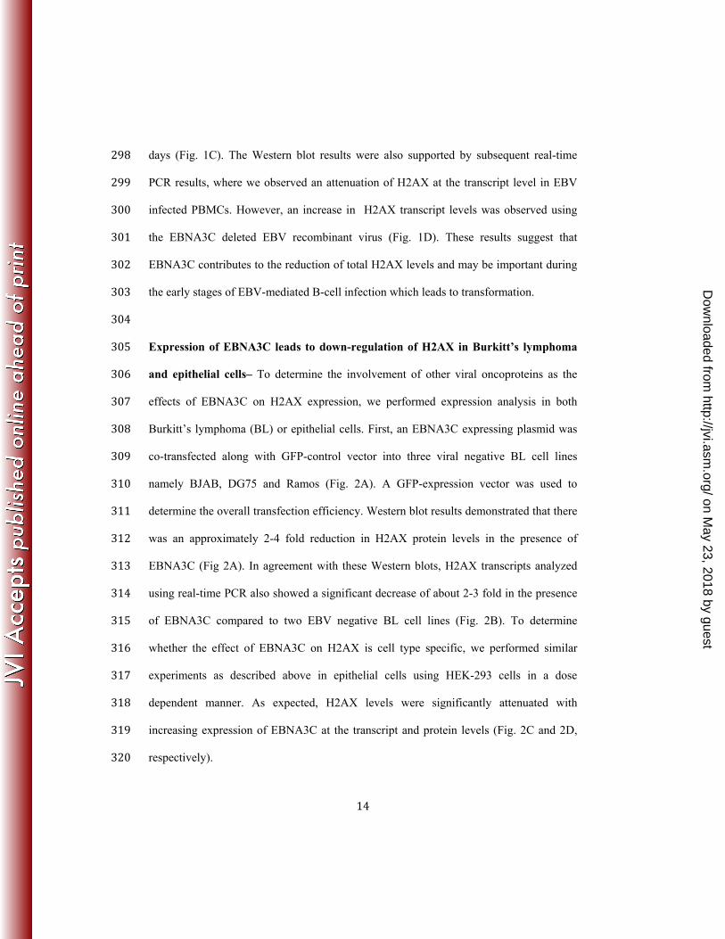
days (Fig. 1C). The Western blot results were also supported by subsequent real-time 298 PCR results, where we observed an attenuation of H2AX at the transcript level in EBV 299 infected PBMCs. However, an increase in H2AX transcript levels was observed using 300 the EBNA3C deleted EBV recombinant virus (Fig. 1D). These results suggest that 301 EBNA3C contributes to the reduction of total H2AX levels and may be important during 302 the early stages of EBV-mediated B-cell infection which leads to transformation. 303 304 Expression of EBNA3C leads to down-regulation of H2AX in Burkitt’s lymphoma 305 and epithelial cells– To determine the involvement of other viral oncoproteins as the 306 effects of EBNA3C on H2AX expression, we performed expression analysis in both 307 Burkitt’s lymphoma (BL) or epithelial cells. First, an EBNA3C expressing plasmid was 308 co-transfected along with GFP-control vector into three viral negative BL cell lines 309 namely BJAB, DG75 and Ramos (Fig. 2A). A GFP-expression vector was used to 310 determine the overall transfection efficiency. Western blot results demonstrated that there 311 was an approximately 2-4 fold reduction in H2AX protein levels in the presence of 312 EBNA3C (Fig 2A). In agreement with these Western blots, H2AX transcripts analyzed 313 using real-time PCR also showed a significant decrease of about 2-3 fold in the presence 314 of EBNA3C compared to two EBV negative BL cell lines (Fig. 2B). To determine 315 whether the effect of EBNA3C on H2AX is cell type specific, we performed similar 316 experiments as described above in epithelial cells using HEK-293 cells in a dose 317 dependent manner. As expected, H2AX levels were significantly attenuated with 318 increasing expression of EBNA3C at the transcript and protein levels (Fig. 2C and 2D, 319 respectively). 320
on May 23, 2018 by guest
http://jvi.asm.org/
Dow
nloaded from

15
Importantly, co-expression of H2AX tagged with the Myc epitope and EBNA3C 321 tagged with the Flag epitope from heterologous promoters also showed attenuation of 322 H2AX at the protein level as using the Myc specific mouse monoclonal antibody (Fig. 323 2E). These results indicated that EBNA3C can also deregulate H2AX at the 324 transcriptional level. 325 326 EBNA3C forms a complex with H2AX - In an attempt to understand whether this 327 attenuation of H2AX levels in the presence of EBNA3C expression is regulated through 328 an interaction between these two molecules, we further investigated their binding 329 activities in B-cells. To support this, we performed immunoprecipitation assays with anti-330 H2AX antibody in BJAB, BJAB#7, BJAB#10 and LCL1, LCL2 cells (Fig. 3A). The 331 results demonstrated that H2AX was stably associated with EBNA3C in both EBNA3C 332 stably expressing cells (BJAB#7 and BJAB#10) as well as in LCL1 and LCL2 (Fig. 3A). 333 Further to support our interaction results, we performed a reverse immunoprecipitation 334 assay, where we used the A10 mouse monoclonal antibody specific for EBNA3C using 335 similar cell lines as described above (Fig. 3B). These association studies provided strong 336 evidence that EBNA3C interacted with H2AX in both EBV-transformed cells as well as 337 in EBNA3C positive Burkitt's lymphoma cells (Fig. 3A). 338 339 H2AX autophosphorylation plays a critical role in interaction between H2AX with 340 EBNA3C - H2AX phosphorylation at Ser139 has previously been shown to be critical 341 for maintenance of latency during multiple gammaherpesviruses infection including 342 mouse gammaherpesvirus 68 (MHV-68) and KSHV (22, 36). Interaction between 343
on May 23, 2018 by guest
http://jvi.asm.org/
Dow
nloaded from

16
EBNA3C and H2AX prompted us to further evaluate whether mutation at the Ser139 344 residue of H2AX was critical for complex formation with EBNA3C (Fig. 3C). To address 345 this question, we performed an immunoprecipitation assay using HEK-293 cells where 346 the Flag-tagged Ser139 H2AX (where the Serine residue was replaced by an Alanine 347 residue) was co-expressed with Myc-tagged EBNA3C (Fig. 3D). Subsequently, 348 immunoprecipitation was performed by using anti-Myc antibody to target the Myc-349 tagged EBNA3C (Fig. 3D). The immunoprecipitated bands were visualized with anti-350 Flag antibody. Interestingly, no signal was detected for EBNA3C when H2AX was 351 mutated at the Ser139 residue (Fig. 3D). This indicated that the Ser139 residue of H2AX 352 was important for complex formation of H2AX with EBNA3C. Moreover, we found 353 reduced expression levels of gamma-H2AX in EBV positive LCL1 and LCL2 compared 354 to EBV negative DG75 and Ramos cells (data not shown). 355
356 The first 100 residues of EBNA3C are responsible for the interaction of EBNA3C 357 with H2AX - To identify the binding residues of EBNA3C important for interaction with 358 H2AX, we further performed similar immunoprecipitation experiments in a heterologous 359 expression system using HEK-293 cells. Cells were transfected with plasmids expressing 360 Flag tagged full-length EBNA3C (residues 1-992), N-terminal region (residues 1-365), 361 middle region (residues 366-620), and the C-terminal region (residues 621-992) along 362 with a Myc-tagged H2AX expression vector (Fig. 4A). Immunoprecipitation assays were 363 performed using a Flag specific mouse monoclonal antibody (M2). The results revealed 364 that H2AX forms a complex with residues within the N- terminal region of EBNA3C, but 365 not with the middle or carboxyl-terminal domains (Fig. 4A). 366
on May 23, 2018 by guest
http://jvi.asm.org/
Dow
nloaded from

17
To further confirm the above association studies, we performed GST pull-down 367 experiments using bacterially purified GST-fused H2AX with the HEK-293 cell extracts 368 transiently transfected with plasmids expressing full length EBNA3C and the three major 369 truncated domains of EBNA3C (N-terminal, M-domain and C-terminal) (Fig. 4B). The 370 results from GST pull-down assays also confirmed a strong interaction between full-371 length EBNA3C and the N- terminal (1-365aa) residues with H2AX (Fig. 4B, top 372 panels). The amount of control GST and GST-H2AX fusion protein used in the GST pull 373 down experiment was similar as shown by corresponding Coomassie brilliant blue 374 stained SDS-PAGE (Fig. 4B, bottom panel). Additionally, to narrow down the binding 375 residues with the N-terminal domain of EBNA3C, we further performed GST pull-down 376 assays using various truncated regions of the EBNA3C N-terminal region (Fig. 4C). A 377 strong association was clearly detected between GST-fused H2AX with EBNA3C 378 residues 1-300, 1-200 and 1-100, while none were found beyond the first 100 amino acid 379 residues of EBNA3C (Fig. 4C). These results demonstrate that the EBNA3C 1-100 380 residues are responsible for formation of a stable complex with H2AX in cells. The 381 schematic represents the mapping of the H2AX interaction domain within EBNA3C and 382 further outlines the studies to identify the interacting residues (Fig. 4D). 383
384 EBNA3C colocalizes with H2AX in EBV-transformed LCLs - Our results from both 385 immunoprecipitation as well as GST pull-down assays strongly supported the formation 386 of a complex between EBNA3C and H2AX under physiological conditions using EBV-387 transformed B cells. This prompted us to investigate whether EBNA3C and H2AX can 388 colocalize within similar cellular compartments. Immunofluorescence (IF) studies were 389
on May 23, 2018 by guest
http://jvi.asm.org/
Dow
nloaded from

18
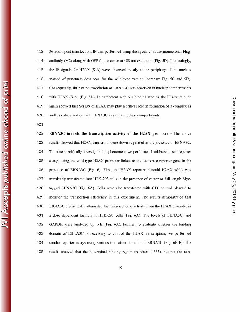
performed using EBNA3C stably expressing B-cells as well as EBV transformed LCLs 390 and EBV-negative controls (Fig. 5). First, IF was carried out in BJAB control cells along 391 with the isogenic EBNA3C expressing cells (BJAB#7 and BJAB#10) (data not shown). 392 The assay was carried out using specific antibodies against EBNA3C (A10, mouse 393 monoclonal), and H2AX (rabbit polyclonal) along with Texas-Red mouse and FITC-anti 394 rabbit secondary antibodies for visualization of the complexes. The results showed a 395 strong colocalization between H2AX and EBNA3C in similar nuclear compartments 396 (data not shown). Furthermore, an analysis of the fluorescence intensity for H2AX 397 signals in EBNA3C expressing BJAB cells demonstrated a significant reduction 398 compared to control BJAB cells (data not shown). This further corroborated our 399 biochemical studies as discussed above, which showed reduced H2AX levels in the 400 presence of either EBV or EBNA3C. 401 Importantly, IF analysis using LCL1 and LCL2 showed a similar co-localization 402 pattern of H2AX and EBNA3C (Fig. 5A-B). To further validate this observation, we 403 performed similar IF experiments using GFP-tagged EBNA3C and Myc-tagged H2AX in 404 U2OS epithelial cells at 36 hours post-transfection (Fig. 5C). Monitoring the GFP signal 405 of EBNA3C and a specific mouse monoclonal antibody against H2AX tagged with the 406 Myc epitope clearly showed a punctuate pattern of co-localization between H2AX 407 molecules and EBNA3C in similar nuclear compartments (Fig. 5C). 408 Our binding results previously demonstrated that phosphorylation of H2AX at 409 Ser139 is crucial for binding with EBNA3C. To further determine whether this residue 410 was also important for colocalization, we next performed an IF study with GFP-tagged 411 EBNA3C and a Flag tagged mutated version of H2AX (S-A) in U2OS cells (Fig. 5D). At 412
on May 23, 2018 by guest
http://jvi.asm.org/
Dow
nloaded from
19
36 hours post transfection, IF was performed using the specific mouse monoclonal Flag-413 antibody (M2) along with GFP fluorescence at 488 nm excitation (Fig. 5D). Interestingly, 414 the IF-signals for H2AX (S-A) were observed mostly at the periphery of the nucleus 415 instead of punctuate dots seen for the wild type version (compare Fig. 5C and 5D). 416 Consequently, little or no association of EBNA3C was observed in nuclear compartments 417 with H2AX (S-A) (Fig. 5D). In agreement with our binding studies, the IF results once 418 again showed that Ser139 of H2AX may play a critical role in formation of a complex as 419 well as colocalization with EBNA3C in similar nuclear compartments. 420
421 EBNA3C inhibits the transcription activity of the H2AX promoter - The above 422 results showed that H2AX transcripts were down-regulated in the presence of EBNA3C. 423 To more specifically investigate this phenomena we performed Luciferase based reporter 424 assays using the wild type H2AX promoter linked to the luciferase reporter gene in the 425 presence of EBNA3C (Fig. 6). First, the H2AX reporter plasmid H2AX-pGL3 was 426 transiently transfected into HEK-293 cells in the presence of vector or full length Myc-427 tagged EBNA3C (Fig. 6A). Cells were also transfected with GFP control plasmid to 428 monitor the transfection efficiency in this experiment. The results demonstrated that 429 EBNA3C dramatically attenuated the transcriptional activity from the H2AX promoter in 430 a dose dependent fashion in HEK-293 cells (Fig. 6A). The levels of EBNA3C, and 431 GAPDH were analyzed by WB (Fig. 6A). Further, to evaluate whether the binding 432 domain of EBNA3C is necessary to control the H2AX transcription, we performed 433 similar reporter assays using various truncation domains of EBNA3C (Fig. 6B-F). The 434 results showed that the N-terminal binding region (residues 1-365), but not the non-435
on May 23, 2018 by guest
http://jvi.asm.org/
Dow
nloaded from

20
binding domains (residues 366-992) of EBNA3C substantially attenuated transcription 436 from the H2AX promoter (Fig. 6B-D). In agreement, the smaller N-terminal binding 437 residues 1-100 amino acid binding region of EBNA3C also showed a similar inhibitory 438 trend of transcriptional regulation at the H2AX promoter (Fig. 6E). This indicated that 439 the amino-terminal binding residues of EBNA3C is required, as well as sufficient for 440 transcriptional inhibition of H2AX promoter. To further validate our results above we 441 transfected HEK-293 cells with the N-terminal deleted region of EBNA3C (ΔN 442 EBNA3C) along with the reporter and GFP expression plasmids (Fig. 6F). This result 443 supported our above data that the N-terminal binding residues of EBNA3C are important 444 for regulating H2AX transcription. All reporter experiments were carefully examined by 445 WB analysis to monitor the expression levels of EBNA3C and GAPDH as an internal 446 loading control (Fig. 6). 447 EBNA3C can induce degradation of H2AX by recruiting the ubiquitin-proteasome 448 system - To establish latency EBV has evolved strategies for targeted inactivation of 449 cellular factors to escape from the DDR induced during early infection (37-40). However, 450 the underlying mechanism is still not clear. Overall, our results described above, strongly 451 suggested that in addition to transcriptional regulation, EBNA3C can also attenuate 452 H2AX at the protein level. Moreover, it has also been reported that the H2AX protein 453 level can be regulated through poly-ubiquitination (23). Interestingly, EBNA3C has been 454 previously shown to utilize a ubiquitin-mediated proteosome degradation strategy for 455 targeting and degrading multiple tumor suppressor proteins to establish successful latent 456 infection in infected B-cells (31, 32). Along these lines, we wanted to determine whether 457 down-regulation of H2AX protein levels were associated with EBNA3C-mediated 458
on May 23, 2018 by guest
http://jvi.asm.org/
Dow
nloaded from
21
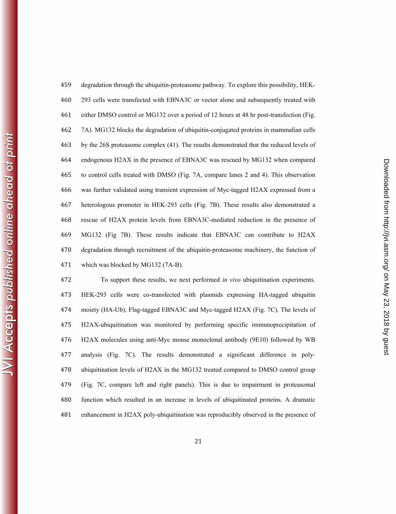
degradation through the ubiquitin-proteasome pathway. To explore this possibility, HEK-459 293 cells were transfected with EBNA3C or vector alone and subsequently treated with 460 either DMSO control or MG132 over a period of 12 hours at 48 hr post-transfection (Fig. 461 7A). MG132 blocks the degradation of ubiquitin-conjugated proteins in mammalian cells 462 by the 26S proteasome complex (41). The results demonstrated that the reduced levels of 463 endogenous H2AX in the presence of EBNA3C was rescued by MG132 when compared 464 to control cells treated with DMSO (Fig. 7A, compare lanes 2 and 4). This observation 465 was further validated using transient expression of Myc-tagged H2AX expressed from a 466 heterologous promoter in HEK-293 cells (Fig. 7B). These results also demonstrated a 467 rescue of H2AX protein levels from EBNA3C-mediated reduction in the presence of 468 MG132 (Fig 7B). These results indicate that EBNA3C can contribute to H2AX 469 degradation through recruitment of the ubiquitin-proteasome machinery, the function of 470 which was blocked by MG132 (7A-B). 471 To support these results, we next performed in vivo ubiquitination experiments. 472 HEK-293 cells were co-transfected with plasmids expressing HA-tagged ubiquitin 473 moiety (HA-Ub), Flag-tagged EBNA3C and Myc-tagged H2AX (Fig. 7C). The levels of 474 H2AX-ubiquitination was monitored by performing specific immunoprecipitation of 475 H2AX molecules using anti-Myc mouse monoclonal antibody (9E10) followed by WB 476 analysis (Fig. 7C). The results demonstrated a significant difference in poly-477 ubiquitination levels of H2AX in the MG132 treated compared to DMSO control group 478 (Fig. 7C, compare left and right panels). This is due to impairment in proteasomal 479 function which resulted in an increase in levels of ubiquitinated proteins. A dramatic 480 enhancement in H2AX poly-ubiquitination was reproducibly observed in the presence of 481
on May 23, 2018 by guest
http://jvi.asm.org/
Dow
nloaded from

22
EBNA3C with MG132 treatment, but not with DMSO control (Fig. 7C, compare left and 482 right panels, lane 3). To support the above findings, ubiquitination experiments were also 483 carried out in BJAB, BJAB#7 and BJAB#10 cells by performing H2AX 484 immunoprecipitation using an anti-H2AX specific antibody (Fig. 7D). The results further 485 demonstrated a relatively higher level of H2AX poly-ubiquitination in EBNA3C 486 expressing BJAB#7 and BJAB#10 cells compared to the BJAB control cells (Fig. 7D, 487 compare lane 1, with lanes 2 and 3). This provided additional evidence to suggest that 488 EBNA3C can facilitate H2AX destabilization through ubiquitin-mediated proteasomal 489 degradation. 490
491 EBNA3C inhibits the growth suppressive activity of H2AX – The spindle 492
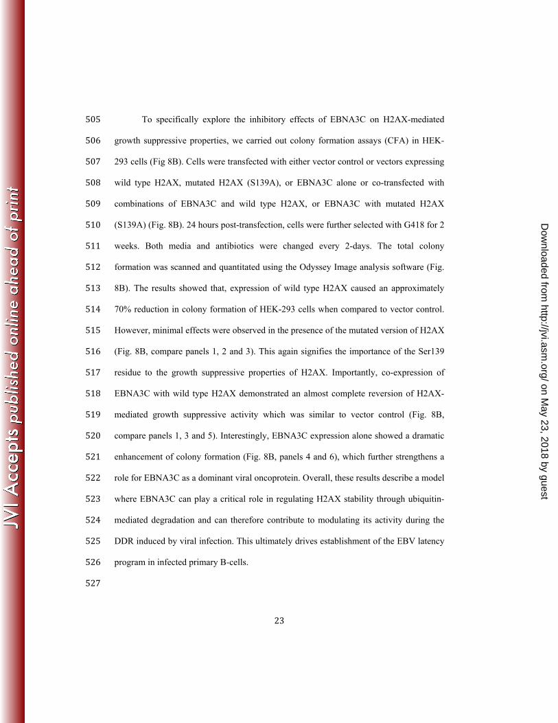
assembly checkpoint (SAC) and DDR are two critical mechanisms by which mammalian 493 cells can maintain genomic stability (42). Moreover, H2AX was shown to induce cell 494 cycle arrest via the p53/p21 pathway (43). In addition, knockdown of H2AX was shown 495 to strongly suppress apoptosis in lung cancer cells (44). To elucidate the biological 496 significance of H2AX degradation through EBNA3C in LCLs as well as to explore the 497 underlying molecular mechanism of this DNA damage protein in EBV-induced B-cell 498 lymphomagenesis, we used H2AX knockdown LCL1 cells compared to control vector 499 LCL1. Interestingly, H2AX knockdown resulted in a reduction in levels of the p53 tumor 500 suppressor protein of about 2-3 fold, while the level of Bub1, a critical kinetochore 501 protein essential for SAC was upregulated about 3-5 fold (Fig. 8A). These results now 502 provide a potential molecular strategy for H2AX downregulation in LCLs, and highlights 503 a critical role for EBNA3C in regulating the DDR during B-cell lymphomagenesis. 504
on May 23, 2018 by guest
http://jvi.asm.org/
Dow
nloaded from
23
To specifically explore the inhibitory effects of EBNA3C on H2AX-mediated 505 growth suppressive properties, we carried out colony formation assays (CFA) in HEK-506 293 cells (Fig 8B). Cells were transfected with either vector control or vectors expressing 507 wild type H2AX, mutated H2AX (S139A), or EBNA3C alone or co-transfected with 508 combinations of EBNA3C and wild type H2AX, or EBNA3C with mutated H2AX 509 (S139A) (Fig. 8B). 24 hours post-transfection, cells were further selected with G418 for 2 510 weeks. Both media and antibiotics were changed every 2-days. The total colony 511 formation was scanned and quantitated using the Odyssey Image analysis software (Fig. 512 8B). The results showed that, expression of wild type H2AX caused an approximately 513 70% reduction in colony formation of HEK-293 cells when compared to vector control. 514 However, minimal effects were observed in the presence of the mutated version of H2AX 515 (Fig. 8B, compare panels 1, 2 and 3). This again signifies the importance of the Ser139 516 residue to the growth suppressive properties of H2AX. Importantly, co-expression of 517 EBNA3C with wild type H2AX demonstrated an almost complete reversion of H2AX-518 mediated growth suppressive activity which was similar to vector control (Fig. 8B, 519 compare panels 1, 3 and 5). Interestingly, EBNA3C expression alone showed a dramatic 520 enhancement of colony formation (Fig. 8B, panels 4 and 6), which further strengthens a 521 role for EBNA3C as a dominant viral oncoprotein. Overall, these results describe a model 522 where EBNA3C can play a critical role in regulating H2AX stability through ubiquitin-523 mediated degradation and can therefore contribute to modulating its activity during the 524 DDR induced by viral infection. This ultimately drives establishment of the EBV latency 525 program in infected primary B-cells. 526 527
on May 23, 2018 by guest
http://jvi.asm.org/
Dow
nloaded from

24
DISCUSSION 528 Viral infection and the cellular DNA damage response (DDR) have been widely 529
debated and explored in recent years (24, 45). Viruses have evolved complex regulatory 530 mechanisms to persist in infected cells by manipulating several DNA repair mechanisms 531 (46). H2AX plays a key role in the DDR (47). H2AX is well known for its positive 532 regulation of DNA repair and activation of the DNA damage checkpoints (48). H2AX 533 also varies from H2A with a unique C-terminal region (23), which contains a highly 534 conserved SQE motif (49). However, it has been suggested that H2AX is not utterly 535 important for recognition of DNA damage but facilitates the damage response through 536 interaction with proteins that recognizes the phosphorylated form of H2AX (γ-H2AX) 537 (48). However, previous studies by Bassing et al reported that cells from H2AX deficient 538 mice are more prone to chromosomal instability and less efficient in DNA double 539 stranded break repair (37). 540
Earlier it was reported that the EBV latent proteins EBNAs and LMPs can 541 contribute towards chromosomal instability and cellular DNA damage by inhibiting as 542 well as facilitating degradation of DNA repair elements, or through mis-localization (50, 543 51). Reports from our lab and others also revealed that EBNA3C can contribute to the 544 oncogenic process through degradation of p53 or interaction with Chk2 (16, 20). 545 However, limited information is available for demonstrating a role for EBNA3C in 546 modulating H2AX. 547
Here, we identified H2AX as an interacting partner with EBNA3C during EBV 548 infection. We showed that in both EBV as well as EBNA3C positive B cell lines, cellular 549 H2AX levels were significantly down-regulated. This initial observation was further 550
on May 23, 2018 by guest
http://jvi.asm.org/
Dow
nloaded from

25
corroborated using RNA interference strategy by knocking down EBNA3C expression in 551 EBV transformed LCLs. Using an in vitro model system for resting B cell infection with 552 an EBNA3C deleted recombinant virus in comparison to wild type EBV infection, we 553 confirmed that EBNA3C was directly involved in regulating H2AX levels. The 554 attenuation of H2AX levels was clearly more significant in the wild type virus infection, 555 whereas, the EBNA3C deleted EBV did not show any significant change in H2AX 556 expression. These results supported our hypothesis that through modulation of the total 557 H2AX levels, EBNA3C can contribute to EBV latency and cell proliferation. 558
These initial findings were further corroborated in HEK-293 cells, further 559 strengthening our initial hypothesis that EBNA3C is directly involved in deregulating 560 H2AX expression. The reduction of H2AX levels were found to be inversely proportional 561 to the increase in EBNA3C levels at the protein and transcript levels. While unable to 562 bind DNA directly, EBNA3C was previously shown to be a strong transcriptional factor 563 that can regulate transcription of multiple cellular and viral genes. EBNA3C formed a 564 stable complex with H2AX through interaction with its amino-terminal first 100 amino 565 acid residues. Interestingly, we know that the N-terminal region of EBNA3C interacts 566 with various cell cycle regulatory molecules such as Cyclin D1, Cyclin A, p53, Mdm2, 567 E2F1, IRF4 and Aurora Kinase B (16, 30, 32, 34, 52, 53). 568
Phosphorylation of Ser139 of H2AX (referred to as gamma-H2AX) is 569 indispensible for recruitment of DNA repair elements (54). Furthermore, H2AX is 570 phosphorylated by ATM in response to double stranded breaks (54). More specifically, 571 mutation at Ser139 completely abolished the interaction between H2AX and EBNA3C. 572 This suggests, that H2AX phosphorylation may play an important role in EBNA3C 573
on May 23, 2018 by guest
http://jvi.asm.org/
Dow
nloaded from

26
regulated cell proliferation. This also suggests the involvement of EBNA3C in regulating 574 the DDR and consequently also opens a new avenue for further exploration. Our reporter 575 analysis of the H2AX promoter demonstrated that the N-terminal binding residues were 576 sufficient for blocking H2AX transcription. Furthermore, H2AX localization is strictly 577 confined to the nucleosomes (55). However, some reports demonstrated that H2AX also 578 functions outside the nucleosomes (56, 57). Our results demonstrated that EBNA3C can 579 localize with H2AX within similar nuclear compartments in EBV transformed LCLs as 580 well as EBNA3C expressing stable cell lines. The staining intensity of H2AX in 581 EBNA3C negative and EBNA3C positive cell lines showed a reduction in the levels of 582 H2AX in the presence of EBNA3C further supporting our hypothesis that EBNA3C can 583 attenuate H2AX expression. 584
In addition to transcriptional regulation, our results also indicated that EBNA3C 585 can regulate H2AX protein levels. Previous reports showed that H2AX is degraded by 586 poly-ubiquitination through the proteosome degradation pathway, and that inhibition of 587 the proteosome can stabilize H2AX (40, 58, 59). We hypothesized that EBNA3C 588 mediated H2AX deregulation may be through poly-ubiquitination and degradation since 589 our previous studies showed that EBNA3C recruits the ubiquitin-proteasome machinery 590 for targeted proteolysis of multiple tumor suppressor proteins through its N-terminal 591 residues (31, 60). Recently Feng et al hypothesized that Ubc13 and RNF8 are involved in 592 DDR-induced H2AX ubiquitination (61). How these proteins are involved in the 593 functional regulation of H2AX levels in EBV positive cells remains undetermined. In 594 addition to this, mono-ubiquitination of H2AX was shown to be an essential epigenetic 595 marker during the DNA damage response (62, 63). This would be interesting to 596
on May 23, 2018 by guest
http://jvi.asm.org/
Dow
nloaded from

27
determine whether EBNA3C could also initiate H2AX mono-ubiquitination to regulate 597 nucleosomal activity and thereby gene transcription. For now, our results demonstrated 598 that EBNA3C efficiently induces H2AX degradation most likely through poly-599 ubiquitination. 600
To determine the biological implications of H2AX deregulation in EBV-driven 601 oncogenesis, we generated LCLs knocked down for H2AX and evaluated the expression 602 levels of the p53 tumor suppressor and the oncoprotein Bub1. Earlier Fragkos et al 603 suggested that H2AX was required for cell cycle arrest through p53 (43). There is a 604 growing body of evidence for crosstalk between the DDR elements and SAC components 605 (42). Moreover, Bub1 was shown to be an essential mitotic protein in gammaherpesvirus 606 induced oncogenesis (64). Depletion of H2AX expression in LCLs led to a significant 607 down-regulation of p53 and up-regulation of Bub1. This supports an anti-proliferative 608 role for H2AX in the context of EBV-induced transformation of B- cells. Subsequent 609 colony formation assays further supported the anti-proliferative properties of H2AX. Our 610 results also clearly demonstrated that EBNA3C can effectively neutralize the growth 611 suppressive effects of H2AX. Therefore this essential latent antigen EBNA3C may have 612 evolved to modulate the DDR and so contribute to viral latency in addition to all its other 613 functions. Overall this study provides strong evidence to support a role for EBNA3C-614 mediated attenuation of total H2AX levels upon EBV infection (Fig. 8C) and may be 615 important for targeted therapeutic strategies against EBV associated B-cell lymphomas 616 617 618 619
on May 23, 2018 by guest
http://jvi.asm.org/
Dow
nloaded from

28
FIGURE LEGENDS 620 Figure 1: EBNA3C down-regulates total H2AX levels in B cells - (A) 621
Endogenous expression of H2AX in EBV positive cell lines (LCL1 and LCL2) and EBV 622 negative Burkitt’s lymphoma cell lines (Ramos and DG75) were analyzed at the protein 623 level (Fig 1A-left panel). Endogenous expression of H2AX in BJAB (EBV negative) 624 BJAB#7, BJAB#10 (EBNA3C stably expressing) cells were analyzed by WB (Fig. 1A, 625 mid panel). LCL1 cells stably transduced for sh-control and sh-EBNA3C were harvested 626 to determine the protein expression levels of H2AX. The relative density of H2AX was 627 analyzed by normalizing with GAPDH. (B) All groups of cells were extracted using 628 Trizol reagent and RNA concentration was estimated by using Nanodrop (Eppendorf 629 Inc., Hamburg, Germany). Transcript levels for the H2AX gene is represented by fold 630 change and the experiments were performed in triplicates. Results of LCL1 and LCL2 631 compared to Ramos and DG75 are shown in Fig. 1B left panel. Results of BJAB 632 compared to BJAB#7 and BJAB#10 are shown in Fig. 1B mid panel and LCL1 stably 633 knocked down for EBNA3C and control vector results were shown in Fig. 1B right panel. 634 (C, D) PBMC’s were infected with wild type (WT) EBV virus and ΔEBNA3C virus up to 635 7 days. Cells were harvested and expression of H2AX was monitored in a time-dependent 636 manner for protein and transcript levels by performing WB analysis and real-time PCR. 637 All panels show a representative picture of the repeated experiments. 638
Figure 2: EBNA3C attenuates H2AX levels in Burkitt’s lymphoma and 639 epithelial cells - (A) Approximately 30 million BJAB, DG75 and Ramos cells were 640 harvested for WB analysis of H2AX, EBNA3C, GAPDH and GFP levels after transient 641 transfection of EBNA3C and GFP constructs (B) Endogenous expression of H2AX levels 642
on May 23, 2018 by guest
http://jvi.asm.org/
Dow
nloaded from

29
in Burkitt’s lymphoma cell lines transfected with EBNA3C was analyzed for transcript 643 levels by real-time PCR. Approximately 15 million cells of BJAB, DG75 and Ramos 644 were co-transfected with the GFP expressing vector either with EBNA3C or control 645 vector. Transcript levels of the H2AX gene are represented as fold change. The 646 experiments were performed in triplicate. (C) H2AX levels in HEK-293 cells were 647 transfected with EBNA3C in a dose-dependent manner and analyzed for transcript levels 648 by RT-PCR. Transcript levels of the H2AX gene was represented as fold change 649 performed in triplicate. (D) H2AX expression levels in HEK-293 cells transfected with 650 EBNA3C in a dose-dependent manner was analyzed for protein level. (E) H2AX and 651 EBNA3C constructs were transfected in HEK-293 cells and analyzed for the detection of 652 H2AX protein levels. HEK-293 cells were transfected with GFP, EBNA3C Flag or vector 653 with Myc-H2AX. All panels are representative pictures from repeat experiments. 654
Figure 3: H2AX associates with EBNA3C in cells - (A) Complex formation 655 between EBNA3C and H2AX was evaluated using anti-H2AX antibody in Burkitt's 656 lymphoma (Fig. 3A, left panel) and LCLs (Fig. 3A, right panel). (B) Association between 657 EBNA3C and H2AX was evaluated using the anti-EBNA3C (A10) antibody in Burkitt's 658 lymphoma (Fig. 3B, left panel) and LCLs (Fig. 3B, right panel). (C) A schematic 659 showing H2AX wild type and specific serine139 mutation with alanine in full length 660 protein is shown. (D) Mutant Flag-H2AX Ser139 (H2AX-S-A) was co-transfected either 661 with Myc-EBNA3C or control vector. WB was performed with anti-Myc and anti-Flag 662 antibodies. 663
Figure 4: The first 100 amino acid residues of EBNA3C protein are critical 664 for complex formation with H2AX - (A) Complex formation between EBNA3C and 665
on May 23, 2018 by guest
http://jvi.asm.org/
Dow
nloaded from

30
H2AX was investigated in HEK-293 cells by immunoprecipitation. Approximately 10 666 million HEK-293 cells were transfected either with control vector, full length EBNA3C 667 or different truncations of EBNA3C (Amino, Middle and carboxy-terminal) region 668 tagged with Flag vector. (B) GST pull down assay was performed by using EBNA3C 669 truncations (Full-length EBNA3C 1-992 aa, N-terminal 1-365 aa, Middle domain 366-670 620 aa and C- terminal 621-992 aa) fused to GST. Smaller truncations of the N-terminal 671 region of EBNA3C (1-300, 1-200, 100-200, 1-100, 1-50 and 130-190 aa) were cloned in 672 pA3M vector and monitored for binding activity with H2AX in the right panel. 673 Coomassie stained gel is shown for GST-control and GST-H2AX fusion protein. (C) A 674 schematic shows different EBNA3C domains associated with other reported proteins as 675 well as its binding domain for H2AX. 676
Figure 5: EBNA3C colocalizes with H2AX in nuclear compartments - (A) In 677 vitro EBV transformed B-cell lines LCL1 and LCL2 were plated on slides and 678 subsequently air-dried at room temperature. Cells were fixed using 3% PFA and blocked 679 with 5% skimmed milk. Protein expression were detected by using anti-mouse EBNA3C 680 A10 monoclonal antibody (1:150), and anti-rabbit H2AX antibody (1:500) followed by 681 anti-rabbit Alexa flour 594 (red) for H2AX and anti-mouse Alexa flour 488 (green) for 682 EBNA3C. DAPI was used for nuclear staining. (B) 0.3 million epithelial U2OS cells 683 were transfected with GFP-EBNA3C and Myc-H2AX plasmids using Lipofectamine. 684 H2AX was detected using Anti-Myc (9E10) antibody (1:200) followed by Anti-rabbit 685 Alexa flour 594 (red) secondary antibody. Co-localization of H2AX with GFP-EBNA3C 686 was monitored using an Olympus confocal microscope. (C) 3x10⁵ U2OS cells were 687 seeded on coverslip and transfected with GFP-EBNA3C and mutant H2AX S-A plasmid 688
on May 23, 2018 by guest
http://jvi.asm.org/
Dow
nloaded from

31
using Lipofectamine. (D) HEK-293 cells were co-transfected with plasmids expressing 689 Flag-EBNA3C and Myc-H2AX. 690
Figure 6: The N-terminal binding residues of EBNA3C are sufficient for 691 suppressing H2AX transcription - HEK-293 cells were transfected with 10 μg of 692 pGL3-H2AX and 1, 2 and 4 μg of EBNA3C truncations (EBNA3C 1-992 aa, 1-365 aa, 693 366-620 aa and 621-992 aa). The N-terminal deleted region of EBNA3C (ΔEBNA3C-N) 694 was also included. Data from the reporter assays are represented as relative Luciferase 695 unit (RLU) with mean and standard deviation represented by error bars. The fold 696 activation was calculated by comparison of the promoter activity in the presence of 697 promoter pGL3-H2AX with the value of the PA3M vector alone. 698
Figure 7: EBNA3C facilitates H2AX degradation through the ubiquitin-699 mediated proteasomal pathway - (A) HEK-293 cells were transfected with plasmids 700 and treated with either MG132 (20μg/ml) or DMSO as control for 30 hours post-701 transfection to inhibit the proteasome activity. (B) HEK-293 cells were transfected with 702 plasmids expressing EBNA3C-Flag or Myc-H2AX. (C) HEK-293 cells were transfected 703 with various combinations of expression vectors as showed. (D) In vivo ubiquitination 704 assays were carried out using 50 million BJAB (EBNA3C negative) BJAB#7 and 705 BJAB#10 (EBNA3C positive stable cell lines). H2AX antibody (2μg) was used for pull 706 down experiments and subsequently detected using ubiquitin and H2AX specific 707 antibodies. The input was also analyzed for loading control using the anti-GAPDH 708 antibody. 709
Figure 8: EBNA3C blocks H2AX-mediated growth suppressive activity - (A) 710 Knockdown for H2AX cells were evaluated to determine H2AX, EBNA3C, Bub1 and 711
on May 23, 2018 by guest
http://jvi.asm.org/
Dow
nloaded from

32
p53 expression levels using their respective antibodies. Experiments were independently 712 performed three times with similar results. (B) Colony formation assays was performed 713 in HEK-293 cells. Cells were transfected with control vector, wild type H2AX, mutant 714 H2AX (S-A), EBNA3C and EBNA3C combined with wild type H2AX or H2AX (S-A). 715 24 hours post-transfection the cells were selected with G418 for 2 weeks. On alternative 716 days the media and antibiotics were replaced. Cells were fixed by 3% PFA and stained 717 with 0.1% crystal violet. The total number of colonies was measured using the Odyssey 718 Image analysis software. (C) A schematic showing the role of EBNA3C in degradation of 719 H2AX which results in modulation of p53 and Bub1 levels. These changes can contribute 720 to EBV-induced oncogenesis. 721 722 723 724 725 726 727 728 729 730 731 732 733 734 735 736 737 738 739 740 741 742 743 744 745 746 747
on May 23, 2018 by guest
http://jvi.asm.org/
Dow
nloaded from

33
ACKNOWLEDGMENTS 748 749 We are grateful to Dr Alvaro N.A. Monteiro (H. Lee Moffitt cancer center, Tampa, FL), 750 Dr. Toru Ouchi (Roswell Park cancer Institute, Buffalo, NY), and Elliott Kieff (Harvard 751 Medical School, Boston, MA) for kindly providing reagents. The work was supported by 752 NCI grant CA137894-05, NIDDK grant DK050306-17. ESR is a scholar of the Leukemia 753 and Lymphoma Society of America. 754 755 756 757 758 759 760 761 762 763 764 765 766 767 768 769 770 771 772 773 774 775 776 777 778 779 780 781 782 783 784 785 786 787 788
on May 23, 2018 by guest
http://jvi.asm.org/
Dow
nloaded from

34
REFERENCES 789 790 791 1. Epstein MA, Achong BG, Barr YM. 1964. Virus Particles in Cultured 792 Lymphoblasts from Burkitt's Lymphoma. Lancet 1:702-703. 793 2. Young LS, Murray PG. 2003. Epstein-Barr virus and oncogenesis: from 794 latent genes to tumours. Oncogene 22:5108-5121. 795 3. Pegtel DM, Middeldorp J, Thorley-Lawson DA. 2004. Epstein-Barr virus 796 infection in ex vivo tonsil epithelial cell cultures of asymptomatic carriers. J 797 Virol 78:12613-12624. 798 4. Chan AS, To KF, Lo KW, Ding M, Li X, Johnson P, Huang DP. 2002. 799 Frequent chromosome 9p losses in histologically normal nasopharyngeal 800 epithelia from southern Chinese. Int J Cancer 102:300-303. 801 5. Cho WC. 2007. Nasopharyngeal carcinoma: molecular biomarker discovery 802 and progress. Mol Cancer 6:1. 803 6. Thompson MP, Kurzrock R. 2004. Epstein-Barr virus and cancer. Clin 804 Cancer Res 10:803-821. 805 7. Cohen JI. 2000. Epstein-Barr virus infection. N Engl J Med 343:481-492. 806 8. Amon W, Farrell PJ. 2005. Reactivation of Epstein-Barr virus from latency. 807 Rev Med Virol 15:149-156. 808 9. Rezk SA, Weiss LM. 2007. Epstein-Barr virus-associated 809 lymphoproliferative disorders. Hum Pathol 38:1293-1304. 810 10. Gandhi MK, Tellam JT, Khanna R. 2004. Epstein-Barr virus-associated 811 Hodgkin's lymphoma. Br J Haematol 125:267-281. 812 11. Marchini A, Kieff E, Longnecker R. 1993. Marker rescue of a 813 transformation-negative Epstein-Barr virus recombinant from an infected 814 Burkitt lymphoma cell line: a method useful for analysis of genes essential for 815 transformation. J Virol 67:606-609. 816 12. Maruo S, Wu Y, Ishikawa S, Kanda T, Iwakiri D, Takada K. 2006. Epstein-817 Barr virus nuclear protein EBNA3C is required for cell cycle progression and 818 growth maintenance of lymphoblastoid cells. Proc Natl Acad Sci U S A 819
103:19500-19505. 820 13. Halder S, Murakami M, Verma SC, Kumar P, Yi F, Robertson ES. 2009. 821 Early events associated with infection of Epstein-Barr virus infection of 822 primary B-cells. PLoS One 4:e7214. 823 14. Wang F, Tsang SF, Kurilla MG, Cohen JI, Kieff E. 1990. Epstein-Barr virus 824 nuclear antigen 2 transactivates latent membrane protein LMP1. J Virol 825 64:3407-3416. 826 15. Maruo S, Wu Y, Ito T, Kanda T, Kieff ED, Takada K. 2009. Epstein-Barr 827 virus nuclear protein EBNA3C residues critical for maintaining 828 lymphoblastoid cell growth. Proc Natl Acad Sci U S A 106:4419-4424. 829 16. Saha A, Bamidele A, Murakami M, Robertson ES. 2011. EBNA3C 830 attenuates the function of p53 through interaction with inhibitor of growth 831 family proteins 4 and 5. J Virol 85:2079-2088. 832 17. McClellan MJ, Khasnis S, Wood CD, Palermo RD, Schlick SN, Kanhere AS, 833 Jenner RG, West MJ. 2012. Downregulation of integrin receptor-signaling 834
on May 23, 2018 by guest
http://jvi.asm.org/
Dow
nloaded from

35
genes by Epstein-Barr virus EBNA 3C via promoter-proximal and -distal 835 binding elements. J Virol 86:5165-5178. 836 18. Nikitin PA, Luftig MA. 2011. At a crossroads: human DNA tumor viruses and 837 the host DNA damage response. Future Virol 6:813-830. 838 19. Choudhuri T, Verma SC, Lan K, Murakami M, Robertson ES. 2007. The 839 ATM/ATR signaling effector Chk2 is targeted by Epstein-Barr virus nuclear 840 antigen 3C to release the G2/M cell cycle block. J Virol 81:6718-6730. 841 20. Nikitin PA, Yan CM, Forte E, Bocedi A, Tourigny JP, White RE, Allday MJ, 842 Patel A, Dave SS, Kim W, Hu K, Guo J, Tainter D, Rusyn E, Luftig MA. 2010. 843 An ATM/Chk2-mediated DNA damage-responsive signaling pathway 844 suppresses Epstein-Barr virus transformation of primary human B cells. Cell 845 Host Microbe 8:510-522. 846 21. Summers KC, Shen F, Sierra Potchanant EA, Phipps EA, Hickey RJ, 847 Malkas LH. 2011. Phosphorylation: the molecular switch of double-strand 848 break repair. Int J Proteomics 2011:373816. 849 22. Jha HC, Upadhyay SK, M AJP, Lu J, Cai Q, Saha A, Robertson ES. 2013. 850 H2AX phosphorylation is important for LANA-mediated Kaposi's sarcoma-851 associated herpesvirus episome persistence. J Virol 87:5255-5269. 852 23. Ikura T, Tashiro S, Kakino A, Shima H, Jacob N, Amunugama R, Yoder K, 853 Izumi S, Kuraoka I, Tanaka K, Kimura H, Ikura M, Nishikubo S, Ito T, 854 Muto A, Miyagawa K, Takeda S, Fishel R, Igarashi K, Kamiya K. 2007. DNA 855 damage-dependent acetylation and ubiquitination of H2AX enhances 856 chromatin dynamics. Mol Cell Biol 27:7028-7040. 857 24. Turnell AS, Grand RJ. 2012. DNA viruses and the cellular DNA-damage 858 response. J Gen Virol 93:2076-2097. 859 25. Bajaj BG, Murakami M, Cai Q, Verma SC, Lan K, Robertson ES. 2008. 860 Epstein-Barr virus nuclear antigen 3C interacts with and enhances the 861 stability of the c-Myc oncoprotein. J Virol 82:4082-4090. 862 26. Knight JS, Sharma N, Robertson ES. 2005. SCFSkp2 complex targeted by 863 Epstein-Barr virus essential nuclear antigen. Mol Cell Biol 25:1749-1763. 864 27. Lu J, Verma SC, Murakami M, Cai Q, Kumar P, Xiao B, Robertson ES. 2009. 865 Latency-associated nuclear antigen of Kaposi's sarcoma-associated 866 herpesvirus (KSHV) upregulates survivin expression in KSHV-Associated B-867 lymphoma cells and contributes to their proliferation. J Virol 83:7129-7141. 868 28. Rios-Doria J, Fay A, Velkova A, Monteiro AN. 2006. DNA damage response: 869 determining the fate of phosphorylated histone H2AX. Cancer Biol Ther 870 5:142-144. 871 29. Kang MA, So EY, Ouchi T. 2012. Deregulation of DNA damage response 872 pathway by intercellular contact. J Biol Chem 287:16246-16255. 873 30. Jha HC, Lu J, Saha A, Cai Q, Banerjee S, Prasad MA, Robertson ES. 2013. 874 EBNA3C-mediated regulation of aurora kinase B contributes to Epstein-Barr 875 virus-induced B-cell proliferation through modulation of the activities of the 876 retinoblastoma protein and apoptotic caspases. J Virol 87:12121-12138. 877 31. Saha A, Lu J, Morizur L, Upadhyay SK, Aj MP, Robertson ES. 2012. E2F1 878 mediated apoptosis induced by the DNA damage response is blocked by EBV 879 nuclear antigen 3C in lymphoblastoid cells. PLoS Pathog 8:e1002573. 880
on May 23, 2018 by guest
http://jvi.asm.org/
Dow
nloaded from

36
32. Yi F, Saha A, Murakami M, Kumar P, Knight JS, Cai Q, Choudhuri T, 881 Robertson ES. 2009. Epstein-Barr virus nuclear antigen 3C targets p53 and 882 modulates its transcriptional and apoptotic activities. Virology 388:236-247. 883 33. Gao J, Cai Q, Lu J, Jha HC, Robertson ES. 2011. Upregulation of cellular Bcl-2 884 by the KSHV encoded RTA promotes virion production. PLoS One 6:e23892. 885 34. Saha A, Halder S, Upadhyay SK, Lu J, Kumar P, Murakami M, Cai Q, 886 Robertson ES. 2011. Epstein-Barr virus nuclear antigen 3C facilitates G1-S 887 transition by stabilizing and enhancing the function of cyclin D1. PLoS Pathog 888 7:e1001275. 889 35. Paull TT, Rogakou EP, Yamazaki V, Kirchgessner CU, Gellert M, Bonner 890 WM. 2000. A critical role for histone H2AX in recruitment of repair factors to 891 nuclear foci after DNA damage. Curr Biol 10:886-895. 892 36. Tarakanova VL, Stanitsa E, Leonardo SM, Bigley TM, Gauld SB. 2010. 893 Conserved gammaherpesvirus kinase and histone variant H2AX facilitate 894 gammaherpesvirus latency in vivo. Virology 405:50-61. 895 37. Bassing CH, Suh H, Ferguson DO, Chua KF, Manis J, Eckersdorff M, 896 Gleason M, Bronson R, Lee C, Alt FW. 2003. Histone H2AX: a dosage-897 dependent suppressor of oncogenic translocations and tumors. Cell 114:359-898 370. 899 38. Celeste A, Fernandez-Capetillo O, Kruhlak MJ, Pilch DR, Staudt DW, Lee 900 A, Bonner RF, Bonner WM, Nussenzweig A. 2003. Histone H2AX 901 phosphorylation is dispensable for the initial recognition of DNA breaks. Nat 902 Cell Biol 5:675-679. 903 39. Fernandez-Capetillo O, Lee A, Nussenzweig M, Nussenzweig A. 2004. 904 H2AX: the histone guardian of the genome. DNA Repair (Amst) 3:959-967. 905 40. Ikura T, Ogryzko VV, Grigoriev M, Groisman R, Wang J, Horikoshi M, 906 Scully R, Qin J, Nakatani Y. 2000. Involvement of the TIP60 histone 907 acetylase complex in DNA repair and apoptosis. Cell 102:463-473. 908 41. Nawaz Z, Lonard DM, Dennis AP, Smith CL, O'Malley BW. 1999. 909 Proteasome-dependent degradation of the human estrogen receptor. Proc 910 Natl Acad Sci U S A 96:1858-1862. 911 42. Yang C, Wang H, Xu Y, Brinkman KL, Ishiyama H, Wong ST, Xu B. 2012. 912 The kinetochore protein Bub1 participates in the DNA damage response. 913 DNA Repair (Amst) 11:185-191. 914 43. Fragkos M, Jurvansuu J, Beard P. 2009. H2AX is required for cell cycle 915 arrest via the p53/p21 pathway. Mol Cell Biol 29:2828-2840. 916 44. Lu C, Xiong M, Luo Y, Li J, Zhang Y, Dong Y, Zhu Y, Niu T, Wang Z, Duan L. 917 2013. Genome-wide transcriptional analysis of apoptosis-related genes and 918 pathways regulated by H2AX in lung cancer A549 cells. Apoptosis 18:1039-919 1047. 920 45. Nikitin PA, Luftig MA. 2012. The DNA damage response in viral-induced 921 cellular transformation. Br J Cancer 106:429-435. 922 46. Chaurushiya MS, Weitzman MD. 2009. Viral manipulation of DNA repair 923 and cell cycle checkpoints. DNA Repair (Amst) 8:1166-1176. 924
on May 23, 2018 by guest
http://jvi.asm.org/
Dow
nloaded from

37
47. Chadwick BP, Willard HF. 2001. Histone H2A variants and the inactive X 925 chromosome: identification of a second macroH2A variant. Hum Mol Genet 926 10:1101-1113. 927 48. Kinner A, Wu W, Staudt C, Iliakis G. 2008. Gamma-H2AX in recognition and 928 signaling of DNA double-strand breaks in the context of chromatin. Nucleic 929 Acids Res 36:5678-5694. 930 49. Li A, Eirin-Lopez JM, Ausio J. 2005. H2AX: tailoring histone H2A for 931 chromatin-dependent genomic integrity. Biochem Cell Biol 83:505-515. 932 50. Kamranvar SA, Masucci MG. 2011. The Epstein-Barr virus nuclear antigen-933 1 promotes telomere dysfunction via induction of oxidative stress. Leukemia 934 25:1017-1025. 935 51. Sancar A, Lindsey-Boltz LA, Unsal-Kacmaz K, Linn S. 2004. Molecular 936 mechanisms of mammalian DNA repair and the DNA damage checkpoints. 937 Annu Rev Biochem 73:39-85. 938 52. Jha HC, Lu J, Saha A, Cai Q, Banerjee S, Prasad MA, Robertson ES. 2013. 939 EBNA3C-mediated regulation of Aurora Kinase B contributes to EBV-induced 940 B-cell proliferation through modulation of the activities of Rb and apoptotic 941 Caspases. J Virol 87:12121-12138. 942 53. Banerjee S, Lu J, Cai Q, Saha A, Jha HC, Dzeng RK, Robertson ES. 2013. The 943 EBV Latent Antigen 3C Inhibits Apoptosis through Targeted Regulation of 944 Interferon Regulatory Factors 4 and 8. PLoS Pathog 9:e1003314. 945 54. Burma S, Chen BP, Murphy M, Kurimasa A, Chen DJ. 2001. ATM 946 phosphorylates histone H2AX in response to DNA double-strand breaks. J 947 Biol Chem 276:42462-42467. 948 55. Downs JA, Nussenzweig MC, Nussenzweig A. 2007. Chromatin dynamics 949 and the preservation of genetic information. Nature 447:951-958. 950 56. Bewersdorf J, Bennett BT, Knight KL. 2006. H2AX chromatin structures 951 and their response to DNA damage revealed by 4Pi microscopy. Proc Natl 952 Acad Sci U S A 103:18137-18142. 953 57. Liu Y, Tseng M, Perdreau SA, Rossi F, Antonescu C, Besmer P, Fletcher 954 JA, Duensing S, Duensing A. 2007. Histone H2AX is a mediator of 955 gastrointestinal stromal tumor cell apoptosis following treatment with 956 imatinib mesylate. Cancer Res 67:2685-2692. 957 58. Bassing CH, Alt FW. 2004. H2AX may function as an anchor to hold broken 958 chromosomal DNA ends in close proximity. Cell Cycle 3:149-153. 959 59. Bassing CH, Chua KF, Sekiguchi J, Suh H, Whitlow SR, Fleming JC, Monroe 960 BC, Ciccone DN, Yan C, Vlasakova K, Livingston DM, Ferguson DO, Scully 961 R, Alt FW. 2002. Increased ionizing radiation sensitivity and genomic 962 instability in the absence of histone H2AX. Proc Natl Acad Sci U S A 99:8173-963 8178. 964 60. Saha A, Murakami M, Kumar P, Bajaj B, Sims K, Robertson ES. 2009. 965 Epstein-Barr virus nuclear antigen 3C augments Mdm2-mediated p53 966 ubiquitination and degradation by deubiquitinating Mdm2. J Virol 83:4652-967 4669. 968 61. Feng L, Chen J. 2012. The E3 ligase RNF8 regulates KU80 removal and NHEJ 969 repair. Nat Struct Mol Biol 19:201-206. 970
on May 23, 2018 by guest
http://jvi.asm.org/
Dow
nloaded from

38
62. Bergink S, Jentsch S. 2009. Principles of ubiquitin and SUMO modifications 971 in DNA repair. Nature 458:461-467. 972 63. Lukas J, Bartek J. 2009. DNA repair: New tales of an old tail. Nature 973 458:581-583. 974 64. Xiao B, Verma SC, Cai Q, Kaul R, Lu J, Saha A, Robertson ES. 2010. Bub1 975 and CENP-F can contribute to Kaposi's sarcoma-associated herpesvirus 976 genome persistence by targeting LANA to kinetochores. J Virol 84:9718-977 9732. 978 979 980
on May 23, 2018 by guest
http://jvi.asm.org/
Dow
nloaded from