ecg workshop - ctafp.org · ecg workshop a review of commonly (and not-so-commonly…) encountered...
TRANSCRIPT
ECG Workshop
A Review of Commonly
(and not-so-commonly…)
encountered ECG Tracings for Family Physicians
Eugene Orientale, Jr. MD
Professor and Program Director
UCONN/St. Francis Family Medicine
October 25, 2012

Financial Disclosures
• I am conflicted about many things, but they have nothing to do with money or this presentation
Workshop: 90 minutes
• ECG Intro [5 mins]
• Clinical Cases (30)
• Use Web-enabled Smartphone
• Use an Audience Response Card (36)

Audience Participation: Use your smartphone
• Use your smartphone’s internet browser
– www.rwpoll.com
– Session ID: uconn
• Then press [join]
– Next screen:
• Don’t bother entering a First Name, Last Name, or User Data
– just press the [continue] button
– Should say “Welcome to Session UCONN”

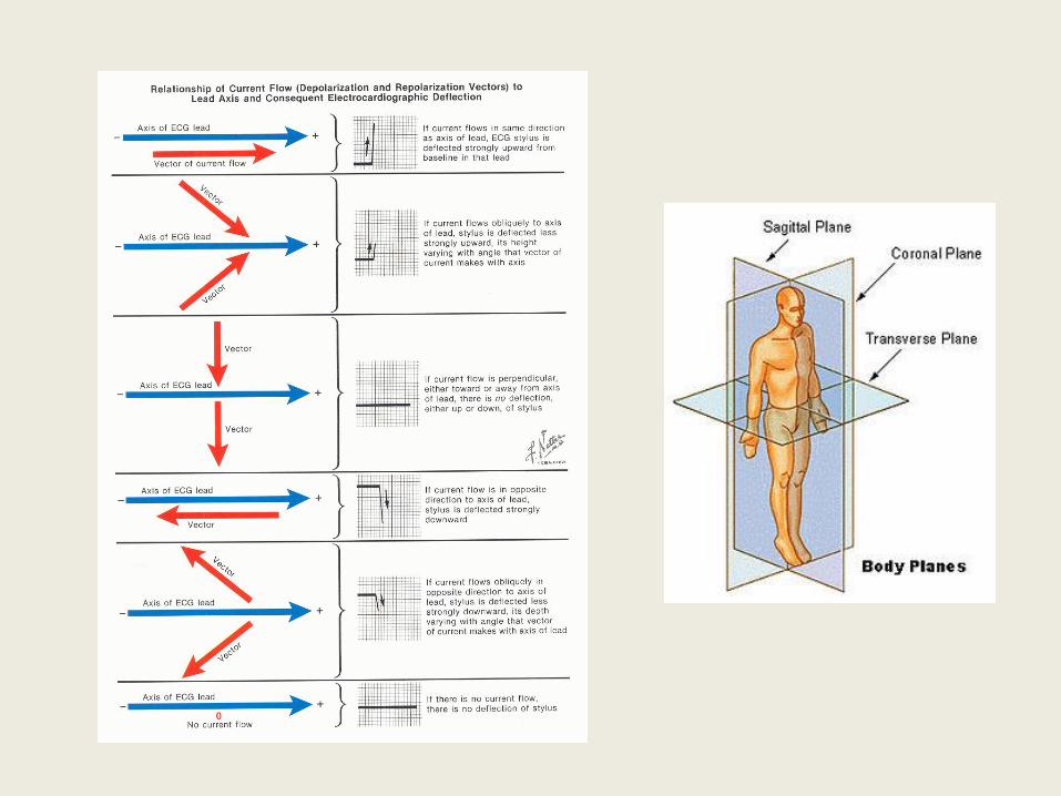
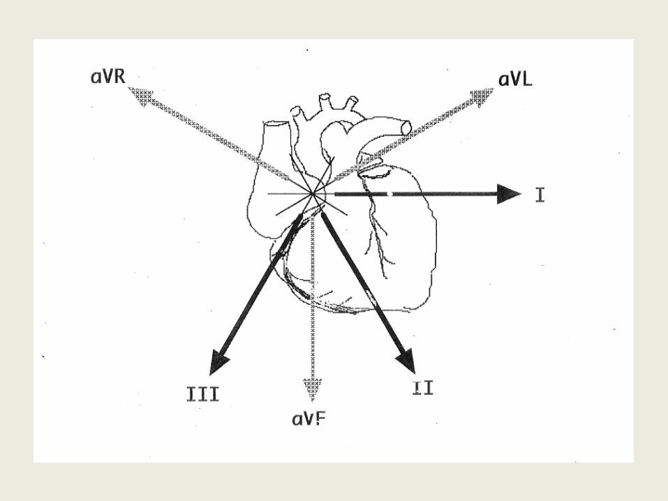
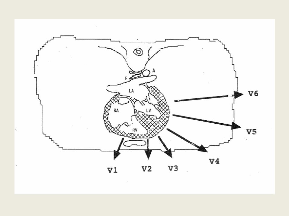
ECG Algorithm • Clinical Context
• Validity
• Rate
• Rhythm
• Axis
• 4 I’s – Intervals
– I(e)nlargements
– Ischemia / Injury / Infarction
– Interpretation
• Action

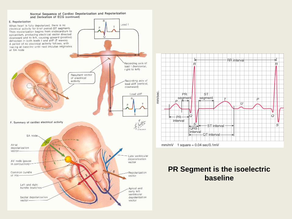
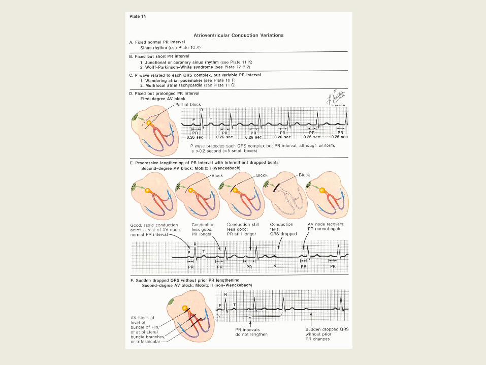
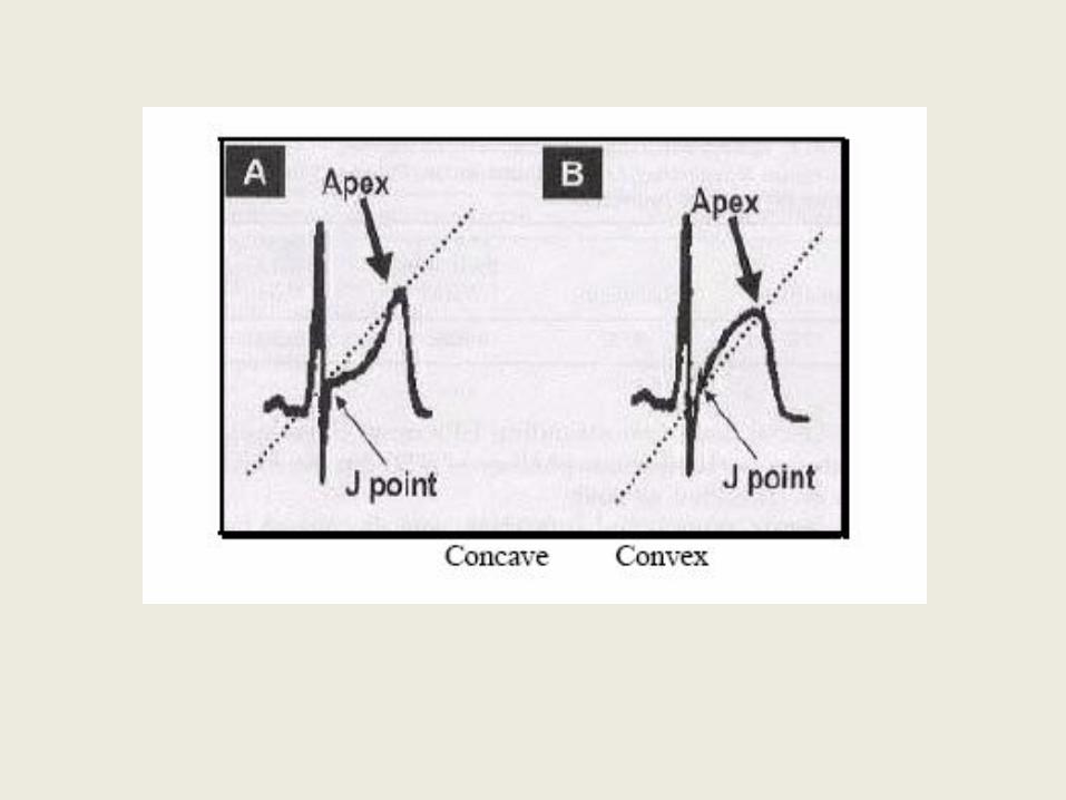
PR Segment is the isoelectric
baseline
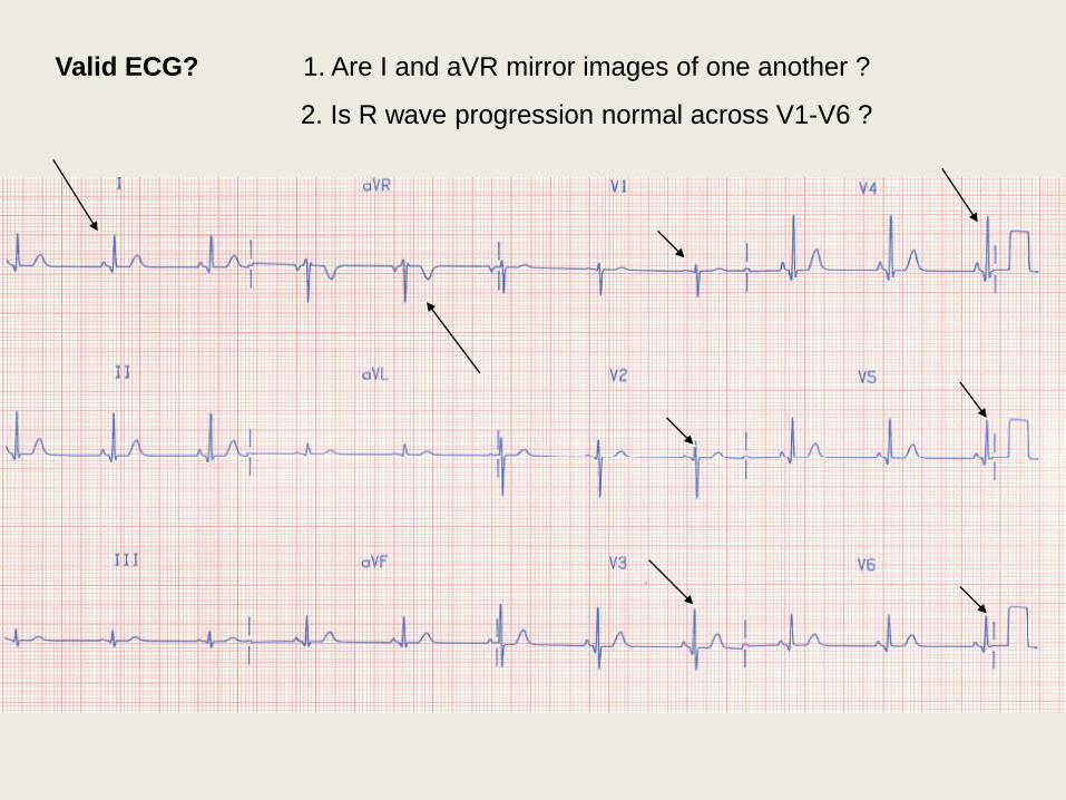
Valid ECG? 1. Are I and aVR mirror images of one another ?
2. Is R wave progression normal across V1-V6 ?

Let’s Begin !
Audience Participation Use your web-enabled Smartphone
OR Use an Audience Responsecard
[Simply click on correct numerical answer]

This ECG was taken from an otherwise healthy 13 yo female. The ECG is suggestive of:
1. Normal Sinus Rhythm
2. Sinus Bradycardia
3. Sinus Arrhythmia
4. First Degree Heart Block
5. None of the above
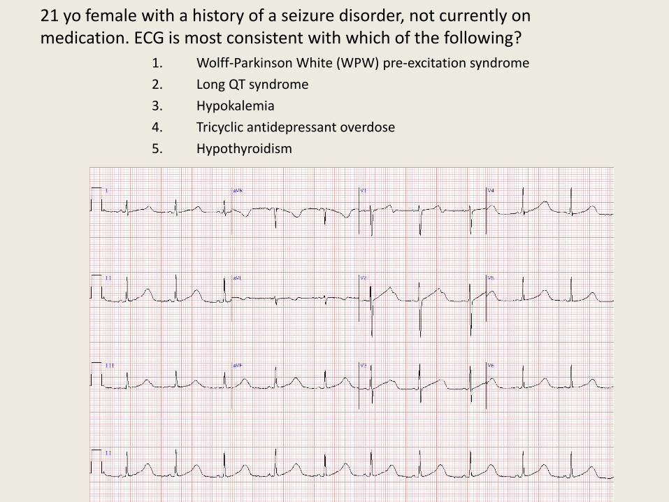
21 yo female with a history of a seizure disorder, not currently on medication. ECG is most consistent with which of the following?
1. Wolff-Parkinson White (WPW) pre-excitation syndrome
2. Long QT syndrome
3. Hypokalemia
4. Tricyclic antidepressant overdose
5. Hypothyroidism
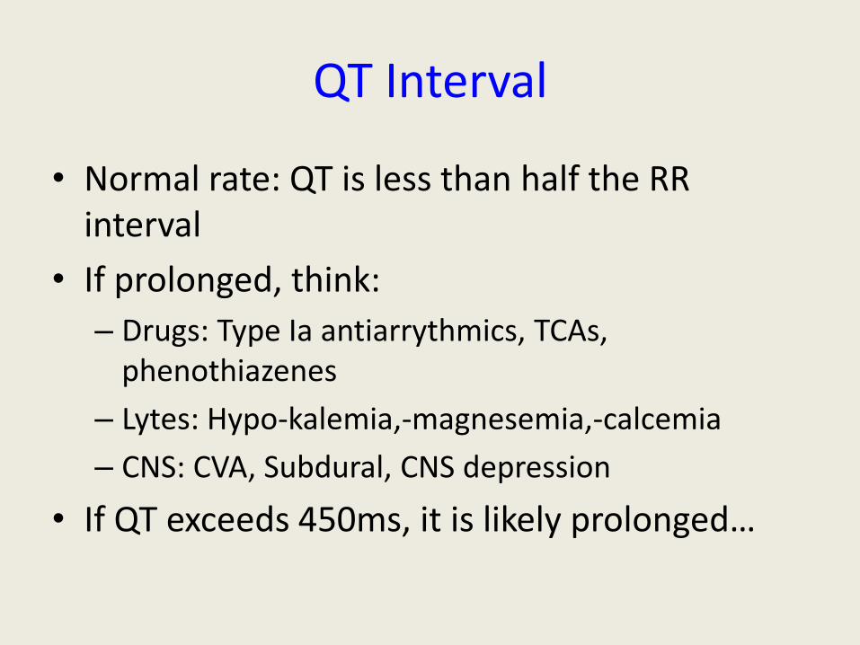
QT Interval
• Normal rate: QT is less than half the RR interval
• If prolonged, think:
– Drugs: Type Ia antiarrythmics, TCAs, phenothiazenes
– Lytes: Hypo-kalemia,-magnesemia,-calcemia
– CNS: CVA, Subdural, CNS depression
• If QT exceeds 450ms, it is likely prolonged…
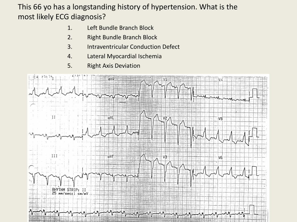
This 66 yo has a longstanding history of hypertension. What is the most likely ECG diagnosis?
1. Left Bundle Branch Block
2. Right Bundle Branch Block
3. Intraventricular Conduction Defect
4. Lateral Myocardial Ischemia
5. Right Axis Deviation

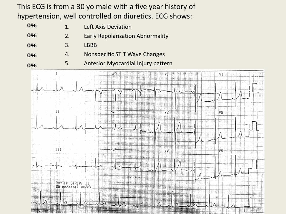
This ECG is from a 30 yo male with a five year history of hypertension, well controlled on diuretics. ECG shows:
0%
0%
0%
0%
0% 1. Left Axis Deviation
2. Early Repolarization Abnormality
3. LBBB
4. Nonspecific ST T Wave Changes
5. Anterior Myocardial Injury pattern

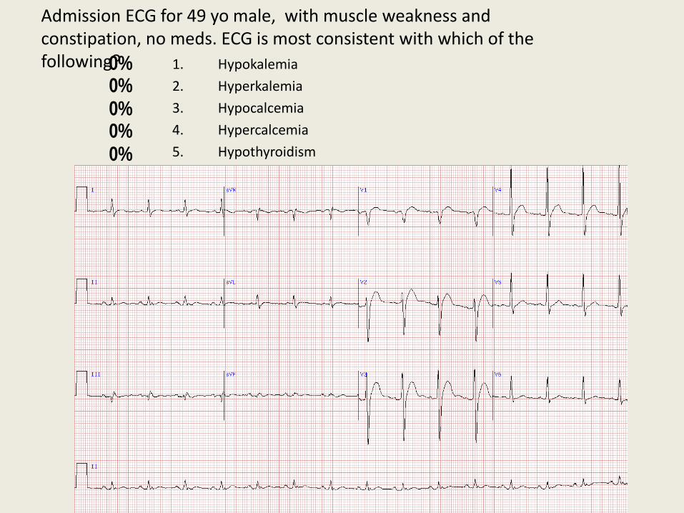
Admission ECG for 49 yo male, with muscle weakness and constipation, no meds. ECG is most consistent with which of the following?
0%0%0%0%0% 1. Hypokalemia
2. Hyperkalemia
3. Hypocalcemia
4. Hypercalcemia
5. Hypothyroidism
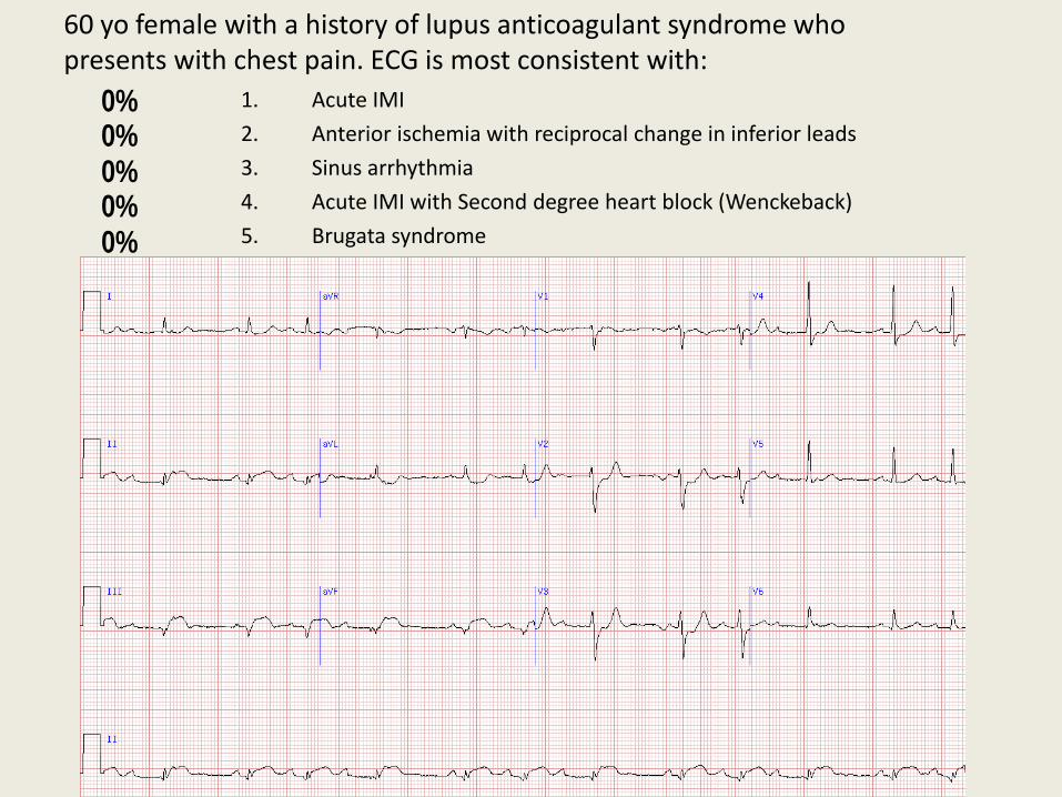
60 yo female with a history of lupus anticoagulant syndrome who presents with chest pain. ECG is most consistent with:
0%0%0%0%0% 1. Acute IMI
2. Anterior ischemia with reciprocal change in inferior leads
3. Sinus arrhythmia
4. Acute IMI with Second degree heart block (Wenckeback)
5. Brugata syndrome

This is an ECG from a 58 yo male with which of the following ECG diagnoses?
0%0%0%0%0% 1. Brugada Pattern
2. Left Bundle Branch Block
3. Right Bundle Branch Block with acute Anteroseptal MI
4. Hyperkalemia
5. Right Ventricular Hypertrophy
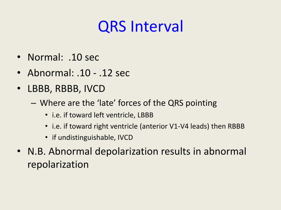
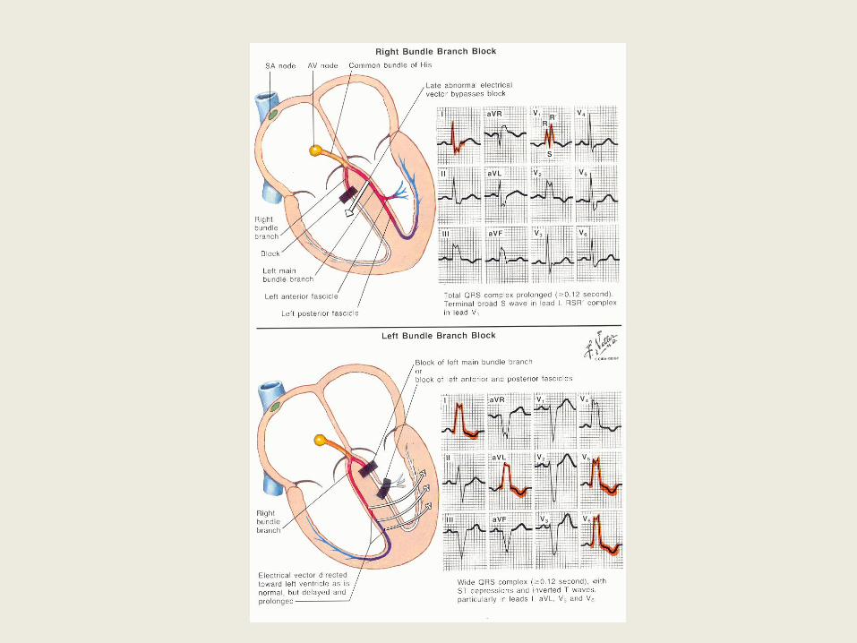
QRS Interval
• Normal: .10 sec
• Abnormal: .10 - .12 sec
• LBBB, RBBB, IVCD
– Where are the ‘late’ forces of the QRS pointing • i.e. if toward left ventricle, LBBB
• i.e. if toward right ventricle (anterior V1-V4 leads) then RBBB
• if undistinguishable, IVCD
• N.B. Abnormal depolarization results in abnormal repolarization
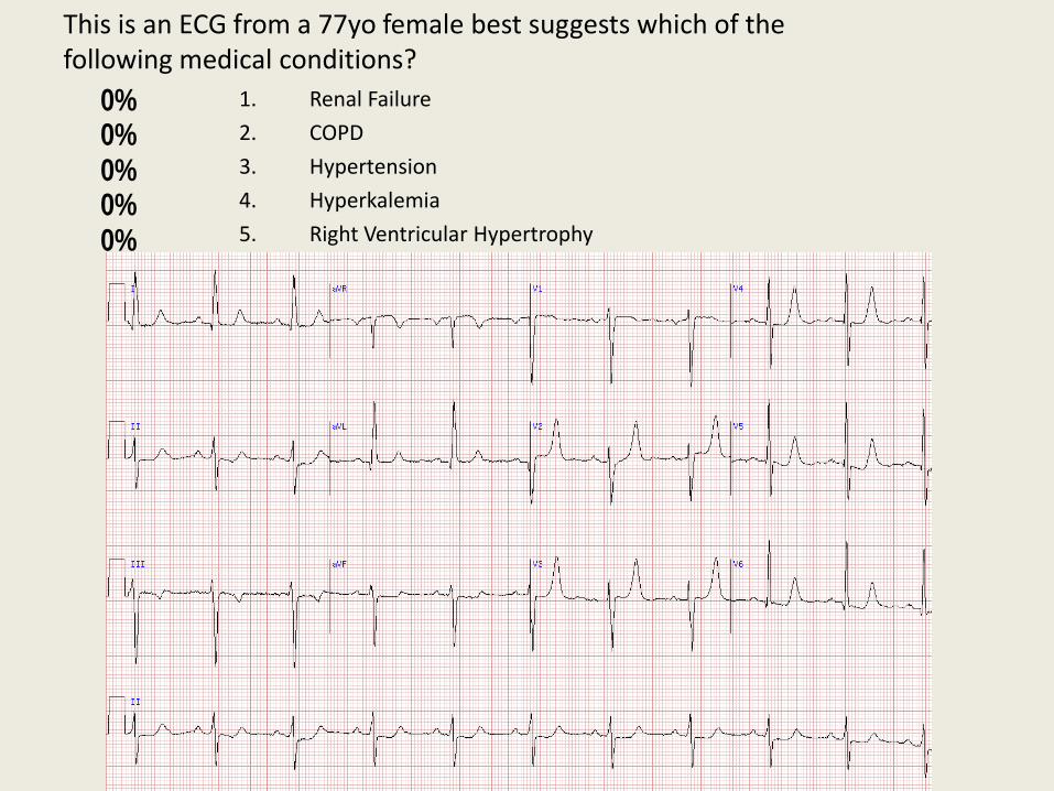
This is an ECG from a 77yo female best suggests which of the following medical conditions?
0%0%0%0%0% 1. Renal Failure
2. COPD
3. Hypertension
4. Hyperkalemia
5. Right Ventricular Hypertrophy
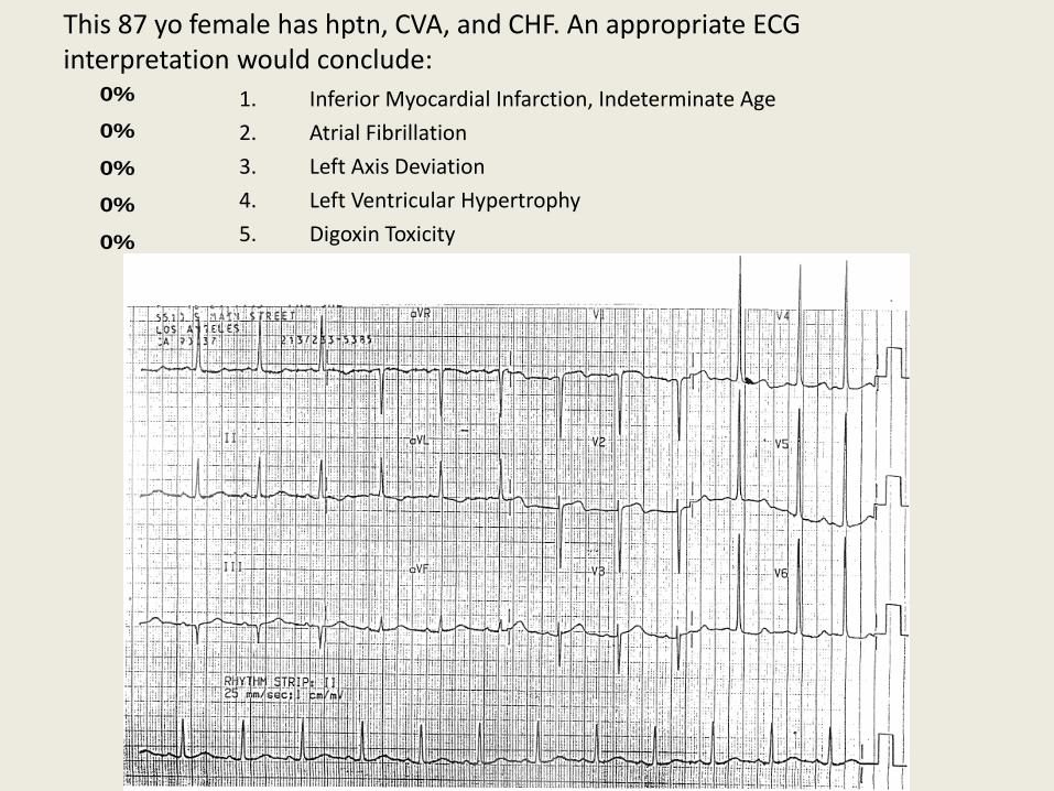
This 87 yo female has hptn, CVA, and CHF. An appropriate ECG interpretation would conclude:
0%
0%
0%
0%
0% 1. Inferior Myocardial Infarction, Indeterminate Age
2. Atrial Fibrillation
3. Left Axis Deviation
4. Left Ventricular Hypertrophy
5. Digoxin Toxicity
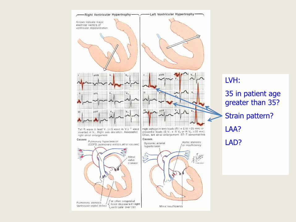
LVH:
35 in patient age greater than 35?
Strain pattern?
LAA?
LAD?
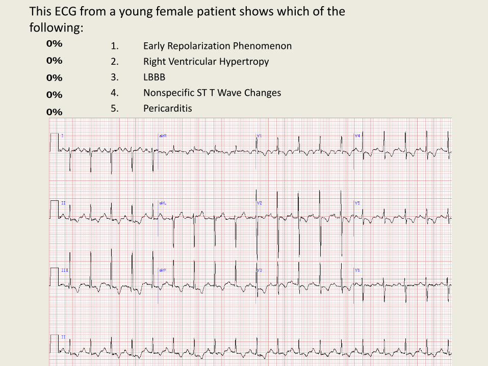
This ECG from a young female patient shows which of the following:
0%
0%
0%
0%
0% 1. Early Repolarization Phenomenon
2. Right Ventricular Hypertropy
3. LBBB
4. Nonspecific ST T Wave Changes
5. Pericarditis

• Leftward Axis
• Normal Axis • Right Axis
Deviation
• Left Axis Deviation
Axis
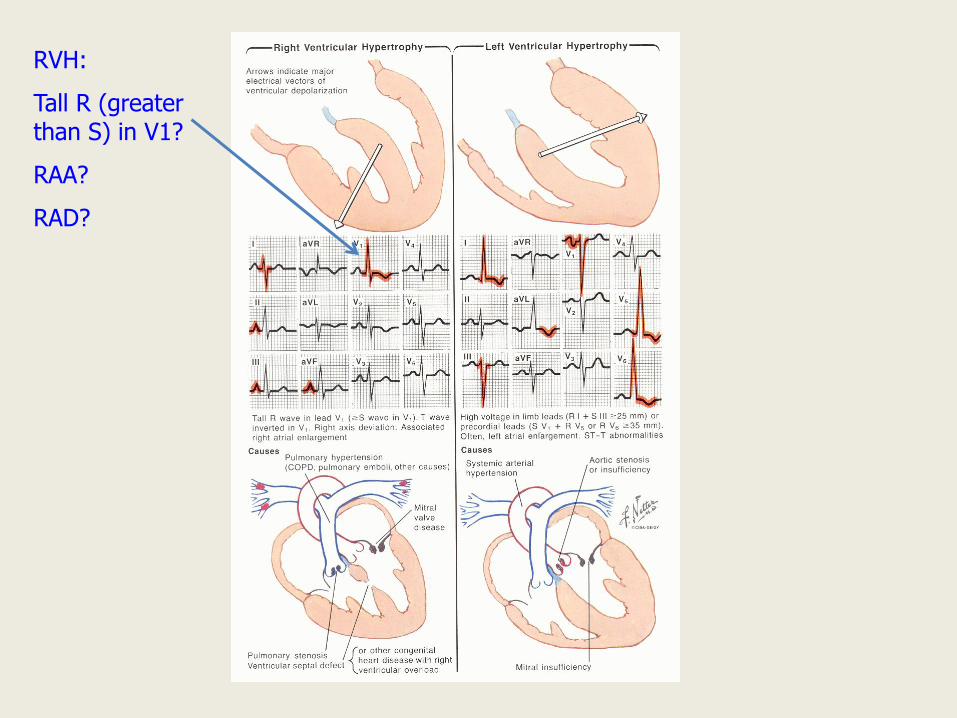
RVH:
Tall R (greater than S) in V1?
RAA?
RAD?
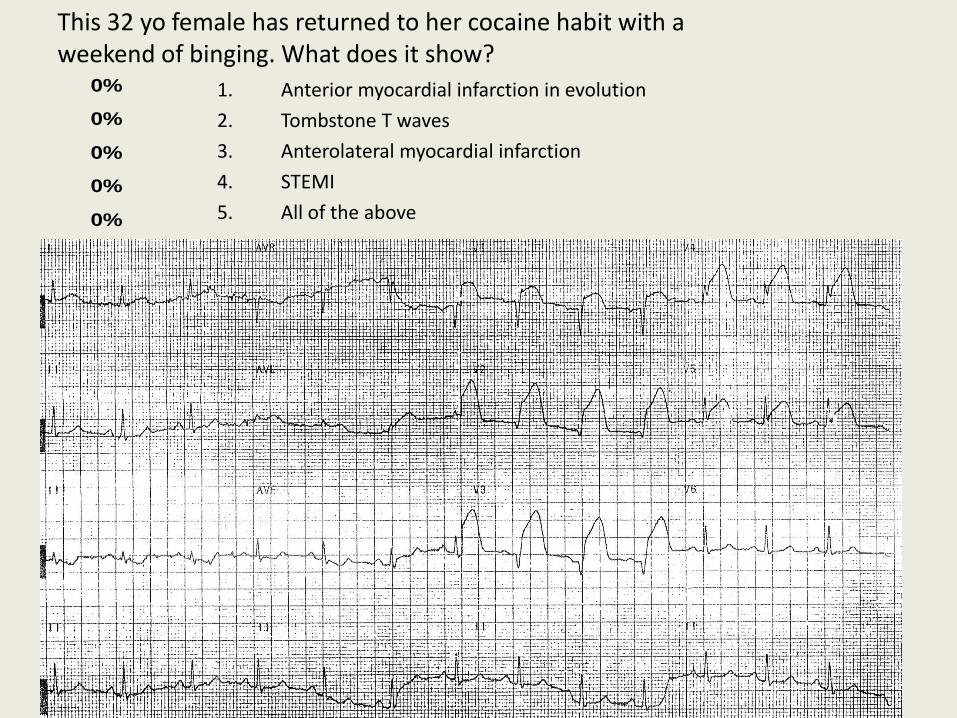
This 32 yo female has returned to her cocaine habit with a weekend of binging. What does it show?
0%
0%
0%
0%
0% 1. Anterior myocardial infarction in evolution
2. Tombstone T waves
3. Anterolateral myocardial infarction
4. STEMI
5. All of the above
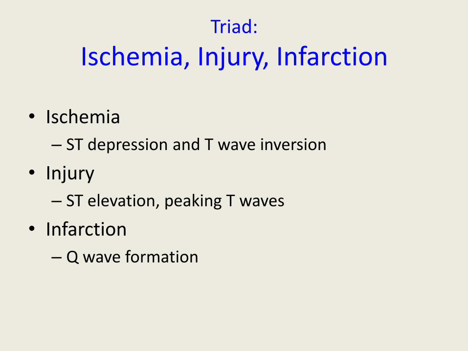
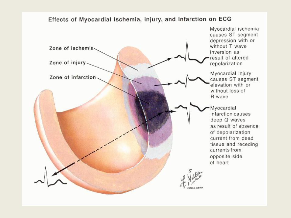
Triad:
Ischemia, Injury, Infarction
• Ischemia
– ST depression and T wave inversion
• Injury
– ST elevation, peaking T waves
• Infarction
– Q wave formation
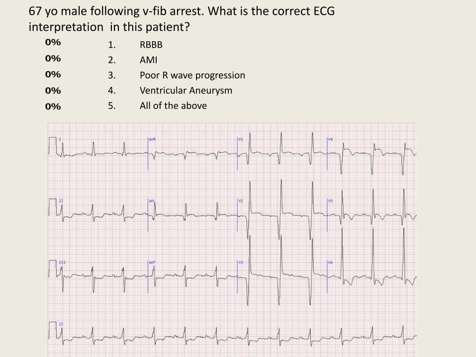
67 yo male following v-fib arrest. What is the correct ECG interpretation in this patient?
0%
0%
0%
0%
0% 1. RBBB
2. AMI
3. Poor R wave progression
4. Ventricular Aneurysm
5. All of the above
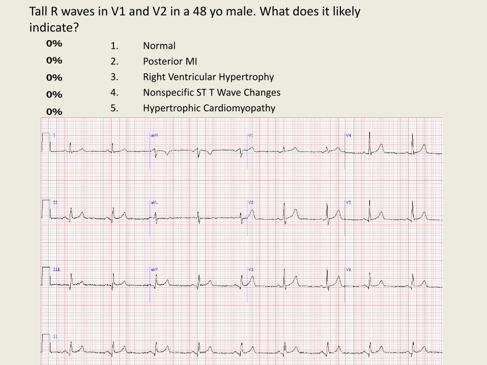
Tall R waves in V1 and V2 in a 48 yo male. What does it likely indicate?
0%
0%
0%
0%
0% 1. Normal
2. Posterior MI
3. Right Ventricular Hypertrophy
4. Nonspecific ST T Wave Changes
5. Hypertrophic Cardiomyopathy
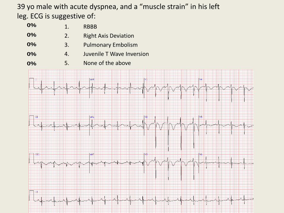
39 yo male with acute dyspnea, and a “muscle strain” in his left leg. ECG is suggestive of:
0%
0%
0%
0%
0% 1. RBBB
2. Right Axis Deviation
3. Pulmonary Embolism
4. Juvenile T Wave Inversion
5. None of the above
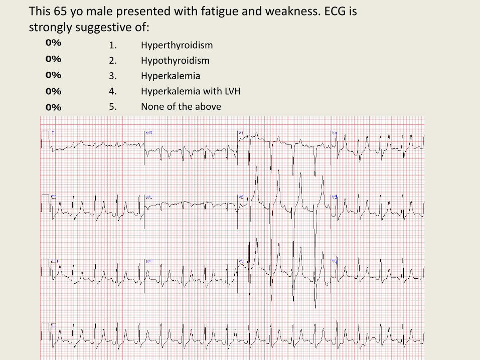
This 65 yo male presented with fatigue and weakness. ECG is strongly suggestive of:
0%
0%
0%
0%
0% 1. Hyperthyroidism
2. Hypothyroidism
3. Hyperkalemia
4. Hyperkalemia with LVH
5. None of the above

Can you predict what might happen to this patient (on haldol and erythromycin) in the near future?
0%
0%
0%
0%
0% 1. Third Degree Heart Block
2. Sinus Arrhythmia
3. Torsades de Pointes
4. Renal Failure
5. None of the above
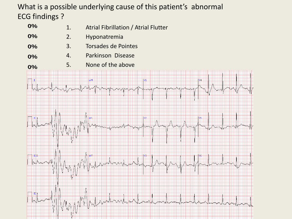
What is a possible underlying cause of this patient’s abnormal ECG findings ?
0%
0%
0%
0%
0% 1. Atrial Fibrillation / Atrial Flutter
2. Hyponatremia
3. Torsades de Pointes
4. Parkinson Disease
5. None of the above
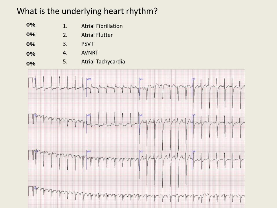
What is the underlying heart rhythm?
0%
0%
0%
0%
0% 1. Atrial Fibrillation
2. Atrial Flutter
3. PSVT
4. AVNRT
5. Atrial Tachycardia

71 yo female who has been ordered a consult. To which service is the consult likely requested?
0%
0%
0%
0%
0% 1. Cardiology
2. Pulmonary
3. Renal
4. Endocrinology
5. Hematology/Oncology
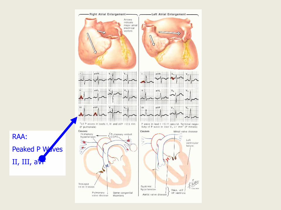
RAA:
Peaked P Waves
II, III, aVF
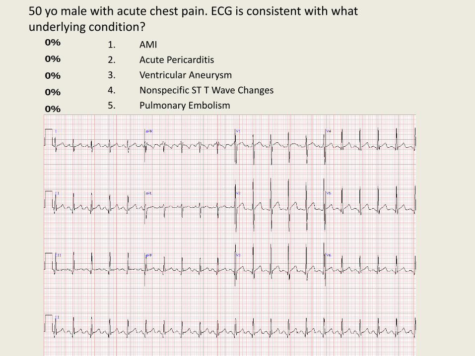
50 yo male with acute chest pain. ECG is consistent with what underlying condition?
0%
0%
0%
0%
0% 1. AMI
2. Acute Pericarditis
3. Ventricular Aneurysm
4. Nonspecific ST T Wave Changes
5. Pulmonary Embolism
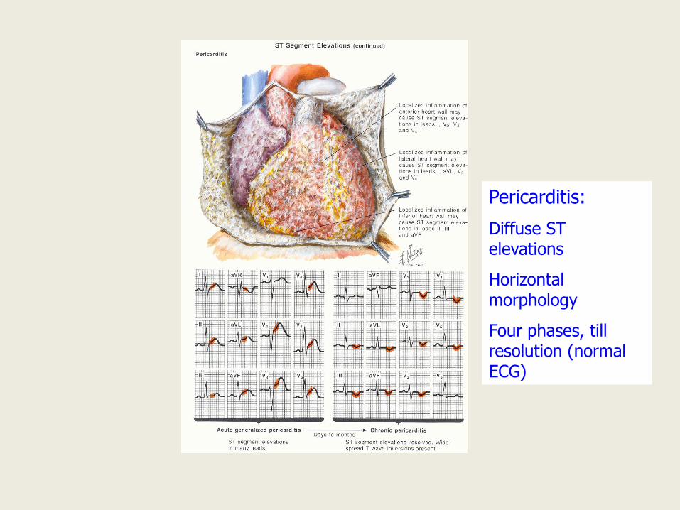
Pericarditis:
Diffuse ST elevations
Horizontal morphology
Four phases, till resolution (normal ECG)
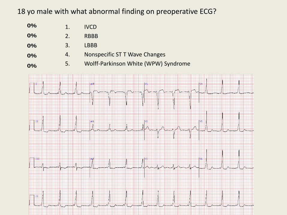
18 yo male with what abnormal finding on preoperative ECG?
0%
0%
0%
0%
0% 1. IVCD
2. RBBB
3. LBBB
4. Nonspecific ST T Wave Changes
5. Wolff-Parkinson White (WPW) Syndrome
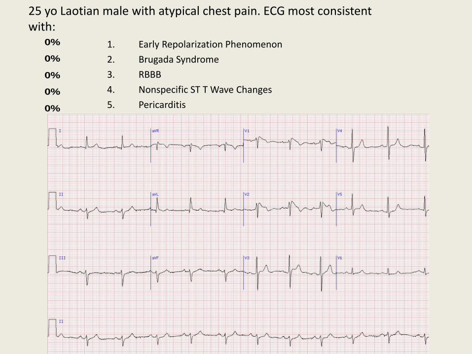
25 yo Laotian male with atypical chest pain. ECG most consistent with:
0%
0%
0%
0%
0% 1. Early Repolarization Phenomenon
2. Brugada Syndrome
3. RBBB
4. Nonspecific ST T Wave Changes
5. Pericarditis
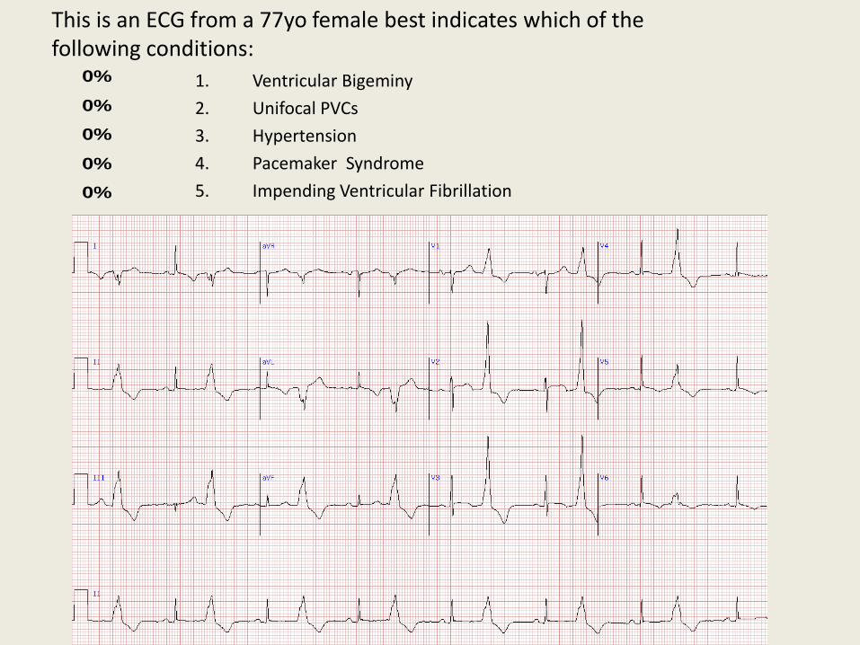
This is an ECG from a 77yo female best indicates which of the following conditions:
0%
0%
0%
0%
0% 1. Ventricular Bigeminy
2. Unifocal PVCs
3. Hypertension
4. Pacemaker Syndrome
5. Impending Ventricular Fibrillation
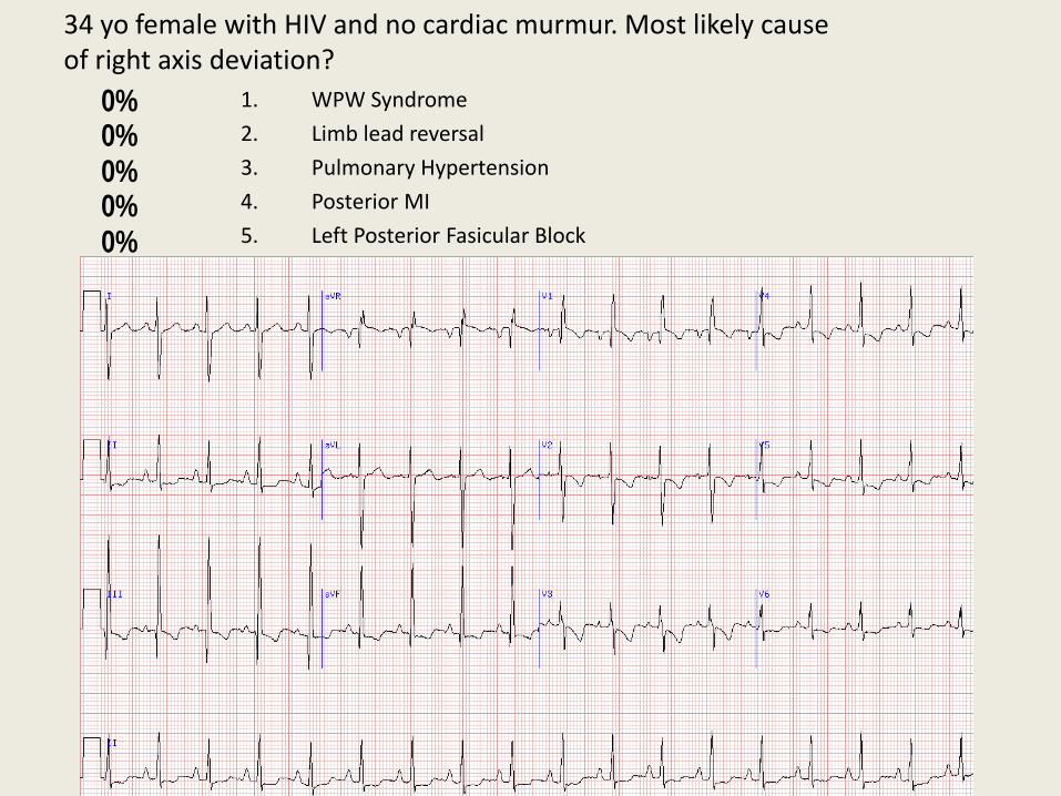
34 yo female with HIV and no cardiac murmur. Most likely cause of right axis deviation?
0%0%0%0%0% 1. WPW Syndrome
2. Limb lead reversal
3. Pulmonary Hypertension
4. Posterior MI
5. Left Posterior Fasicular Block

RAA:
Peaked P Waves
II, III, aVF
LAA:
Biphasic P wave
Lead V1
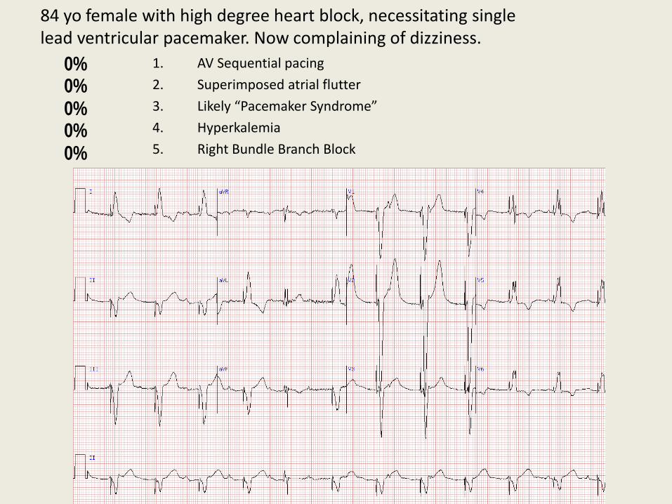
84 yo female with high degree heart block, necessitating single lead ventricular pacemaker. Now complaining of dizziness.
0%0%0%0%0% 1. AV Sequential pacing
2. Superimposed atrial flutter
3. Likely “Pacemaker Syndrome”
4. Hyperkalemia
5. Right Bundle Branch Block

What is the nature of this rhythm disturbance?
0%
0%
0%
0%
0% 1. AV sequential pacing
2. Ventricular pacing
3. Sinus Arrhythmia
4. Wandering Atrial Pacemaker
5. Atrial Pacing
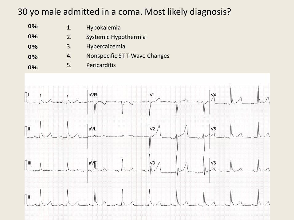
30 yo male admitted in a coma. Most likely diagnosis?
0%
0%
0%
0%
0% 1. Hypokalemia
2. Systemic Hypothermia
3. Hypercalcemia
4. Nonspecific ST T Wave Changes
5. Pericarditis
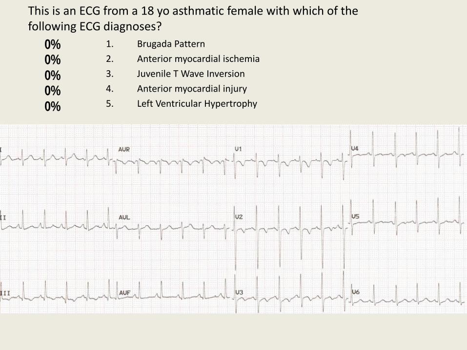
This is an ECG from a 18 yo asthmatic female with which of the following ECG diagnoses?
0%0%0%0%0% 1. Brugada Pattern
2. Anterior myocardial ischemia
3. Juvenile T Wave Inversion
4. Anterior myocardial injury
5. Left Ventricular Hypertrophy

Thank You
• Turning Technologies – www.turningtechnologies.com
– Free use of internet enabled polling system for the CT Academy of Family Physicians Annual Meeting
• Scheidt, Steven: Clinical Electrocardiography
• Nathanson L A, McClennen S, Safran C, Goldberger AL. ECG Wave-Maven: Self-Assessment Program for Students and Clinicians. http://ecg.bidmc.harvard.edu
• Aneesh Tolat, MD