echo-planar imaging ofintravoxel incoherent motion’ · this attenuation s/sohas...
TRANSCRIPT

Robert Turner, PhD #{149}Denis Le Bihan, MD, PhD #{149}Joseph Maler, MS (EEE) #{149}
Robert Vavrek, MS (EEE) #{149}L Kyle Hedges, PhD #{149}James Pekar, PhD
Echo-Planar Imaging of IntravoxelIncoherent Motion’
Abbreviations CS! chemical-shift imaging, EPI echo-planar imaging, IVCM intravoxel co-herent motion, WIM intravoxe! incoherent motion, MBEST modulus blipped echo-planar sin-g!e-pulse technique, RF radio frequency, ROl region of interest, S/N signal-to-noise ratio,
2DFT � two-dimensional Fourier transform.
407
The recently established single-shottechnique of echo-planar imaging ofintravoxel incoherent motion(IVIM) for determining and imag-ing the variations of microscopicmotions of water has been appliedto studies of water perfusion inphantoms and to in vivo studies ofdiffusion and perfusion in cat andhuman brains. The phantom resultsdemonstrate that perfusion levelscomparable with those found invivo have easily observable and re-producible effects on signal ampli-tude that are consistent with previ-ous IVIM theory. Reliable measure-ments of the diffusion coefficient invarious types of brain tissue havebeen obtained. The results for whitematter are consistent with the exis-tence of anisotropic diffusion In on-ented bundles of myelinated nervefibers. The results for gray mattercan be fitted to the IVIM theory andsuggest a value of up to 14% for thefraction of the signal contributed byrandomly perfusing fluid in normalcerebral cortex.
Index terms: Blood, flow dynamics e Blood
vessels, MR studies, 17.1214 #{149}Brain, MR stud-
ies, 10.1214 e Brain, perfusion #{149}Cerebral blood
vessels, flow dynamics e Cerebral blood yes-
sels, MR studies, 17.1214 #{149}Cerebrospinal fluid,
flow dynamics #{149}Cerebrospinal fluid, MR stud-
ies e Magnetic resonance (MR), echo planar
Radiology 1990; 177:407-414
t From the Biomedical Engineering and In-
strumenta! Program, National Center for Re-search Resources (R.T., L.K.H., J.P.), and theDepartment of Diagnostic Radiology, WarrenG. Magnuson Clinical Center (D.L.B.), NationalInstitutes of Health, Bethesda, Md, and from
Genera! Electric Medical Systems, Waukesha,Wis (J.M., R.V.). From the 1989 RSNA scientificassembly. Received March 29, 1990; revision re-
quested May 25; revision received July 18; ac-cepted July 20. Address reprint requests toR.T., Bldg 10, Rm B1D125, National Institutesof Health, Bethesda, MD 20892.
C RSNA, 1990See also the editorial by Le Bihan (pp 328-
329) and the article by Chenevert et a! (pp 401-405) in this issue.
S INCE the pioneering work of LeBihan et al (1-3) interest in the
physiologic and pathologic irtforma-tion based on knowledge of the mo-tion of body fluids at the cellular 1ev-el has grown rapidly. Such motionshave been classified as intravoxel in-coherent motion (WIM) (2) and intra-voxel coherent motion (IVCM) (4).IVIM can be further analyzed as dif-fusion, which arises from the brown-ian motion of individual moleculesmoving with large random thermalvelocities, and perfusion, which de-scribes the pseudorandom flow atlow velocities of blood moving alongthe finely divided structures of thecapillaries. To the extent that perfu-sive flow is anisotropic, as in thehighly oriented capillary beds char-acteristic of skeletal muscle, it is cate-gorized as WCM.
Diffusion and perfusion variationsboth represent bulk manifestations ofhistologic and physiologic differ-ences in tissue at a microscopic level.If the size of a cell, for instance, issmaller than the distance that a watermolecule travels by diffusion during
the time of observation in bulk Wa-ter, the diffusion coefficient may befound to be reduced; we speak of “re-stricted diffusion.” Variations in thetransverse relaxation time T2 in veg-
etable tissue, where the cell wall isalmost impermeable to water trans-port, have been successfully associat-ed with restricted diffusion in differ-ing cell sizes (5,6), and there aresome reports of similar effects in ani-mal tissue, although such cell mem-
branes are quite permeable to water(7). The relative viscosity and proper-tions of intra- and extracellular watercan also cause variations in the diffu-sion coefficient. Thus, it is likely that
studies of diffusion alone, if it can beaccurately measured, could play amajor role in diagnosis of tissue ab-normalities such as those associatedwith stroke and cancer.
The clinical and research possibili-ties offered by a study of perfusionare even wider. Since regional cere-bral blood flow is closely correlatedwith brain functional activity (8), tru-
ly noninvasive dynamic studies ofbrain neural organization may bepossible, and the origins of function-al deficit in trauma or cerebral infarc-
tion might be rapidly assessed.Despite these significant opportu-
nities, progress in the imaging ofIVIM has not been entirely satisfac-tory. The most serious problem hasbeen that the technique for sensitiz-ing a multipuise magnetic resonance(MR) imaging sequence to micro-scopic flows also gives it great sensi-tivity to bulk motions, such as cardi-
ac-cycle-related pulsations, involun-
tary movements, and flow ofcerebrospinal fluid. The method also
makes extreme demands on imaging-hardware stability, and the largemagnetic field gradients that it is ad-
visable to use can cause eddy cur-rents in surrounding conductingstructures (9), with severe conse-quences for image interpretability.Of course, large eddy currents with ashort time constant rule out any kindof rapid imaging technique.
Presented here is an attempt toavoid this problem in the study ofthe diffusion of water and perfusionin the human brain. This is achievedby using a low-eddy-current gradientconfiguration to acquire single-shotimages via echo-planar imaging (EPI)(10), which, because the entire imagedataset is captured in less than 0.1

Motion Artifact
IVIM Imaging
with
408 #{149}Radiology
second, show no visible trace of mo-tion artifact.
IMAGING THEORY
Diffusion Imaging
The effects of molecular diffusionon spin echoes have been studied
since the early days of MR (1 1-13). In
the presence of a magnetic field gra-dient, random spin displacementsproduce random dephasings that de-structively interfere with each other,resulting in an attenuation of the
spin-echo amplitude. Due to thegaussian profile of brownian motion,
this attenuation S/SO has an exponen-
tial dependence, such that
S/SO = exp(-bD),
where D is the diffusion coefficient
(considered here as isotropic) and b is
a factor that depends only on themagnetic field gradients applied be-fore data acquisition.
The concept of “diffusion ima-ging” is much more recent (1,2,14).Due to the multiple gradient pulsesused in an imaging sequence, the ex-pression for b must be generalized
and now takes the form
eTE
b = I Ik(t)I2dtJo
k(t) = -y I G(t’)dt’,Jo
where in a spin-echo sequence thesign of G(t’) is reversed for t’ > TE/2(TE = echo time). Due to the vectornature of this relation, the contribu-
tions b1, b�, and b� for each coordinate
axis can be considered separately. Ifdiffusion is anisotropic, the differentcomponents of the diffusion tensormust be taken into account:
S/S0 = exp(_ � bD,
\ (I =
For a typical two-dimensional Fou-rier transform (2DFT) imaging se-quence, the b factor remains low,typically less than 1 sec #{149}mm2, so
the diffusion effect is negligible (forD = 2.10� mm2 sec’, the diffusion
coefficient of water at room tempera-ture, S/S0 = 0.998). To increase thesensitivity of an imaging sequence to
diffusion, it is necessary to incorpo-rate additional gradient pulses intothe sequence (1,2,14). It then be-
comes possible to compute diffusion
images (ie, maps where the diffusioncoefficient is displayed in each pixel)by using two or more of such se-quences differently sensitized to dif-fusion (1,2).
Different variants of diffusion im-aging have been proposed. With thespin-echo scheme, it is possible tovary the strength or the duration ofthe diffusion-sensitizing gradients(i5,i6), or their direction, to enhance
anisotropic diffusion effects (i7).Other schemes have been used, suchas the stimulated-echo sequence (i8)or the steady-state free-precessiontechnique (19-21).
The concept of diffusion imaging(1) can be extended to encompass IVIM
imaging if we consider the signal at-
tenuation produced by any addition-a! incoherent motion of spins insideeach voxel. The attenuation is in-creased, compared with that causedby diffusion alone, and the depen-
dence on diffusion gradient may bedifferent. However, anatomic datasuggest that the microcirculation of
blood in capillaries can be seen as apseudodiffusion process, at least insuch tissues as the brain (3). We then
have to combine the effects of diffu-
(2) sion and microcirculation, which oc-cur simultaneously in each voxel. Us-ing a simple approach, one obtainsfor the signal attenuation (3)
(3) S/SO = (1 - I) exp(-bD) + f exp(��bD*),
where D and D* are, respectively, thetrue diffusion coefficient and thepseudo-diffusion coefficient and f is
the fractional volume occupied in thevoxel by flowing spins, or perfusionfactor. This simple two-compartmentmodel neglects exchanges betweenthe capillaries and the tissue and dif-ferences in relaxation between thesetwo compartments. Nevertheless, a
(4) biexponential variation of the signalattenuation versus the b factor mani-fests the presence of incoherent mo-tion other than diffusion. Further-more, estimates of D, D*, and f maybe made by fitting the signal intensi-ties obtained in images acquired withdifferent b factors to the above equa-tion.
Signal-to-noise ratio (S/N) consid-erations require the use of many datapoints in order to estimate these pa-rameters with reasonable accuracy. Itis likely that only single-shot tech-niques, such as EPI, will allow the ac-
quisition of enough data within the
short time compatible with clinicaluse.
In conventional multipulse imag-ing, even without diffusion gradi-ents, motion artifact can arise whenthe object, or part of the object, is dis-placed by a distance �x, say, between
successive acquisition cycles. Thesubsequent echo is phase modulatedby the function exp(ik�x), where k is
the phase-space coordinate, so thereis a discontinuity between this and
the previous echo. Once the com-plete dataset has been collected, andthe 2DFT performed to produce animage, such a discontinuity manifestsitself as the familiar ghost artifactdistributed in the phase-encode di-
rection seen in many images. Sincethe magnitude of the discontinuitydepends on k (a first-order effect),the power in the ghost images is notusually large.
However, the situation is muchworse when the large gradients usedin diffusion imaging are applied, orwhen the object to be imaged is mov-ing rapidly, as in conventional ab-dominal imaging. The signal now de-pends on the velocity of coherentand incoherent flows within the ob-ject, as well as its displacement. Forno artifact to appear, all of these van-ables must be the same at each echoacquisition. Cardiac gating is of somebenefit, especially in brain imaging,but noncyclic changes in blood flow
(5) and perfusion, cerebrospinal fluidflow, and involuntary patient motionhave often caused unreliable results(2i,2.2). The phase factor introducedby a variation of velocity of t�v be-tween successive echoes is now ofthe form exp(i’yG �vt2), where t is thetime during which the gradients areapplied. There is a discontinuity atall values of k (a zeroth-order effect),and the ghost images may have alarge enough amplitude to make cal-culation of the diffusion imagemeaningless.
With EPI, on the other hand, theentire set of echoes that is Fourier-transformed to form an image is col-lected in a single acquisition periodof 25-100 msec. No discontinuity canpossibly arise between successivedata points, and hence there can beno motion-derived ghosting. Even ifthere were bulk motion as large asseveral voxel widths during thisshort acquisition, only blurring andbanding of the image would be likelyto result. Such velocities are not nor-mally encountered in the brain. Rap-
November 1990

rt4�
Acquire
11 1 1 1 1 A A A A AIi 64 echoes
Gk�
a.
U slice selection gradients
TEJ2�
diffusion gradients
,�*
�J echo-planar imaging gradients
TE/2�
G�
90
rf4
iso.
�
Acquire
1ftRflfifia�G1 ill hAil AA IU 64 echoes
Gk �-
U slice selection gradients diffusion gradients �J echo-planar imaging gradients
b.
Figure 1. (a) Gradient-echo diffusion-weighted EPI sequence. (b) Spin-echo diffusion-weighted EPI sequence. Typical duration of diffusion gradients was 20 msec per lobe.
TE/2 TE2
Volume 177 #{149}Number 2 Radiology #{149}409
idly flowing blood in larger vessels
and cerebrospinal fluid in the thirdventricle and the aqueducts have acharacteristic variation in appearance
over the cardiac cycle during EPI, butno distributed motion artifact is everobserved, as has been repeatedlydemonstrated in cardiac EPI (23). Thepulsatile component of capillary flowin the brain is, of course, negligible
(24).
The IVIM-EPI Sequence and ItsGradient-Echo Counterpart
It is quite straightforward to sensi-
tize EPI (iO) to diffusion and perfu-sion. The simplest EPI technique toimplement on commercial MR equip-
ment is the MBEST (modulus blippedecho-planar single-pulse technique)sequence (25,26), either in its spin-echo (27) or gradient-echo (28) form.The arrangement of the diffusiongradient pulses is slightly different
in each case, as shown in Figure 1.
Since EPI describes only a method
for acquiring image data, it is com-
patible with any technique for pre-paring the spin magnetization thatcreates diffusion contrast (29).
Special Hardware Requirementsfor IVIM-EPI
For EPI capability, an MR imagermust have certain unusual features.The most stringent of these are con-cerned with maximum gradientstrength and gradient switchingspeed. It is also necessary to use ra-dio-frequency (RF) transmitter andreceiver coils that cannot supporteddy currents induced by the rapidlyswitched gradients. In practice thismeans that RF coils with extensiveareas of copper, such as the Alder-man-Grant design (30), are unsuit-able, and designs such as the bird-cage or saddle-coil type are favored.
To obtain echo-planar images, ei-ther actively shielded, low-induc-tance gradient coils (10,31,32) or agradient-coil insert that is very muchsmaller than the magnet bore of animaging system (33) must be used. A
rise time of less than 200 �isec is de-sirable; the rise time of 1 msec ormore typically found with commer-cial unshielded gradient coils makesEPI impossible.
MATERIALS AND METHODS
Sequence Implementation
Both of the IVIM-EPI sequences de-scribed above have been successfully im-plemented on a i.5-T whole-body imager
(Signa; GE Medical Systems, Milwaukee)and on 2-T and 4.7-T medium-bore chem-ical-shift imaging (CS!) systems (GE Mcd-ical Systems). On all of these imaging ma-chines, 128 X 128 images of phantomswith long T2s have been obtained, butthe data acquisition rate currently avail-able makes the acquisition window toolong for in vivo work at this resolution,giving rise to unacceptable image distor-tion. The studies reported here were per-formed at 64 X 64 resolution.
In the course of image reconstruction,special care must be taken to ensure thatthe data are correctly aligned in k-spaceafter alternate echoes are time reversed. Ifthis is not done properly, an aliasingghost appears halfway across the field ofview from the desired image. For the CS!systems the onset of sampling was care-fully adjusted, and the dataset was shiftedleft until the ghost disappeared (29). Forthe Signa system a different strategy wasadopted. A reference image, with phase-encode gradients switched off, was ob-tamed at the start of each set of image ac-quisitions. Suitably transformed datafrom the reference image were used toperform a phase correction on the subse-quent image data. This has the same ef-fect as properly aligning the echoes forFourier transformation and also correctsto some extent the effects of field inho-mogeneity.
The CS! results shown in this articlewere obtained by means of a set of shield-ed gradients (Acustar 260; GE MedicalSystems) that gave a maximum gradientof 40 mT m’, with a rise time of 170 �sec.A 64 X 64 resolution enabled a minimum
field of view of 40 mm to be achieved,giving a pixel size of 0.625 X 0.625 X 2.0
mm.The standard Signa gradient coils have
a good ratio of current efficiency to in-ductance. However, the current amplifi-ers that drive them cannot deliver suffi-dent current to allow EPI of a reasonablysized field of view. For this reason, novellocal gradient coils, which give a muchlarger z gradient, were designed andbuilt, initially as inserts for the standardquadrature head RF coil. Having a diame-ter of only 27 cm, such coils are muchmore efficient than the whole-body gra-dient set and have a much lower induc-tance of only about 100 MH. Their onlyimportant limitation is that transaxial
EPIs cannot be obtained.The first such coil had the standard
Maxwell configuration, resulting in a us-
Syringe
Filter
Water out
C
a
0�1
100 200Gradlant Factor b
Water in
410 #{149}Radiology November 1990
G-50-150 gel
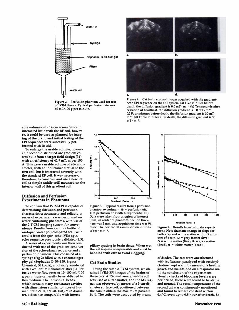
Figure 2. Perfusion phantom used for test
of IVIM theory. Typical perfusion rate was
60 mL/100 g per minute.
Figure 4. Cat brain coronal images acquired with the gradient-
echo EPI sequence on the CSI system. (a) Five minutes before
death, the diffusion gradient is 0.0 mT - m’. (b) Ten seconds after
cessation of heartbeat, the diffusion gradient is 0.0 mT - m1.(c) Four minutes before death, the diffusion gradient is 30 mT-
m1. (d) Three minutes after death, the diffusion gradient is 30
mT - m1.
able volume only 14 cm across. Since it
interacted little with the RF coil, howev-er, it could be used as planned for imag-
ing of the brain, and initial testing of the
EPI sequences were successfully per-
formed with its aid.To enlarge the usable volume, howev-
er, a second distributed-arc gradient coilwas built from a target field design (34),with an efficiency of 42.9 mT/m per 100
A. This gave a usable volume of 20-cm di-ameter, with an inductance similar to thefirst coil, but it interacted severely with
the standard RF coil. It was necessary,
therefore, to construct and use a new RFcoil (a simple saddle coil) mounted on the
interior wall of this gradient coil.
Diffusion and PerfusionExperiments in Phantoms
To confirm that IVIM-EPI is capable ofdetermining diffusion and perfusion
characteristics accurately and reliably, aseries of experiments was performed onwater-containing phantoms, with use ofthe 2-T CS! imaging system for conve-
nience. Results from a simple bottle ofundoped water (29) compared well withresults from the spin-echo IVIM spin-echo sequence previously validated (2,3).
A series of experiments was then con-ducted with use of the gradient-echo ver-
sion of the echo-planar sequence and a
perfusion phantom. This consisted of a
syringe (Fig 2) filled with a chromatogra-phy gel (Sephadex G-50-150, SigmaChemical, St Louis), a polyacrylamide gel
with excellent MR characteristics (3). Per-
fusive water flow rates of 10-100 mL/ 100
g per minute can easily be established inthis medium. The individual beads,which contain many reentrance cavitieswith dimensions similar to those of hu-
man brain cells, are 50-150 �im in diame-ter, a distance comparable with interca-
Figure 3. Typical results from a perfusion
phantom experiment. B = perfusion off,
. = perfusion on (with biexponential fit).
Data were taken from a region of interest
(ROl) in center of phantom. Section thick-
ness was 2 mm, and acquisition time was 94
msec. The horizontal axis is shown in units
of sec - mm2.
pillary spacing in brain tissue. When wet,
the gel is quite compressible and must be
handled with care to avoid clogging.
Cat Brain Studies
Using the same 2-T CS! system, we ob-
tamed IVIM-EPI images of the brains of
three cats. A 15-cm-diameter saddle coilwas used as a transmitter, and the MR sig-nal was observed by means of a 5-cm-di-
ameter surface coil, positioned betweenthe ears to obtain the maximum possible
S/N. The coils were decoupled by means
OrdI.nt teeter b
Figure 5. Results from cat brain experi-
ment. Note dramatic change of slope for
both gray and white matter within 5 mm-
utes of death. 0 gray matter (live),
0 = white matter (live), U gray matter(dead), #{149}= white matter (dead).
of diodes. The cats were anesthetizedwith isoflurane, paralyzed with succinyl-
choline, kept warm by means of a heatingjacket, and maintained on a respirator un-
til the conclusion of the experiment.Hourly checks of blood gas levels were
performed; these were found to be stable
and normal. The rectal temperature of the
second cat was continuously monitoredand found to remain constant within
0.4#{176}C,even up to 0.5 hour after death. Be-
1�41 1?P .‘,
#{248}��A
� � � �
Volume 177 #{149}Number 2 Radiology #{149}411
fore the experiment was concluded, each
cat was killed with 3 mL of T-61 euthana-
sia solution (Hoechst-Roussel [Agrivet],Somerville, NJ) administered into the
femoral vein, in accordance with Nation-a! Institutes of Health animal care guide-lines. The uniquely high speed of EPI
made it possible to obtain brain imageswithin seconds of death, which was mdi-cated by the cessation of heartbeat and
loss of blood pressure.
Studies in Human Brain
By use of the small head gradient coil
described earlier, coronal diffusion-
weighted echo-planar images were ob-
tamed of the heads of volunteers and pa-
tients. Results from patients are not
shown here. To obtain good-quality im-
ages, it was necessary to use the spin-echo
version of the MBEST sequence. The S/Nfor a 64 X 64 image with a 16-cm field ofview and 10-mm section thickness was
measured to be about 50:1, allowingquantitative measurements to be made of
the effects of diffusion on the MR signal.
The spatial resolution in plane is 2.5 mm.
Cardiac gating was sometimes used toavoid possible effects due to changes inthe contrast of moving blood.
Phantoms
RESULTS
Typical results are shown in Figure
3. The effect on the MR signal of aflow rate of as little as 60 mL/100 g
per minute is quite dramatic. Use ofthe Marquardt nonlinear least-
squares fit algorithm shows that the
points are well fitted by a biexponen-
tial variation with b, and the resul-tant perfusion parameters are entire-ly reasonable, confirming previously
published results obtained with a
2DFT spin-echo sequence (3).
Cat Brain
Typical EN images, again acquiredby means of the gradient-echo EPI se-
quence, are shown in Figure 4. These
show 64 X 64-pixel coronal sections
of 3-mm section thickness and 40-
mm field of view, interpolated byzero filling to 128 X 128 pixels. The
decrease in signal from top to bottom
arises from the nonuniform sensitiv-
ity of the surface coil. The total acqui-
sition time for each image was 140msec, the largest echo of the train of
64 echoes occurring at about 100
msec after the initial RF pulse. This
gives considerable T2 weighting tothe images, and some distortion ow-ing to imperfect shimming is also ev-
ident.
Figure 4a was acquired when the
cat was alive, and Figure 4b was ac-
quired 40 seconds after injection of T-
61 euthanasia solution, that is, 10 sec-
onds after cessation of heartbeat. All
imaging parameters were kept iden-
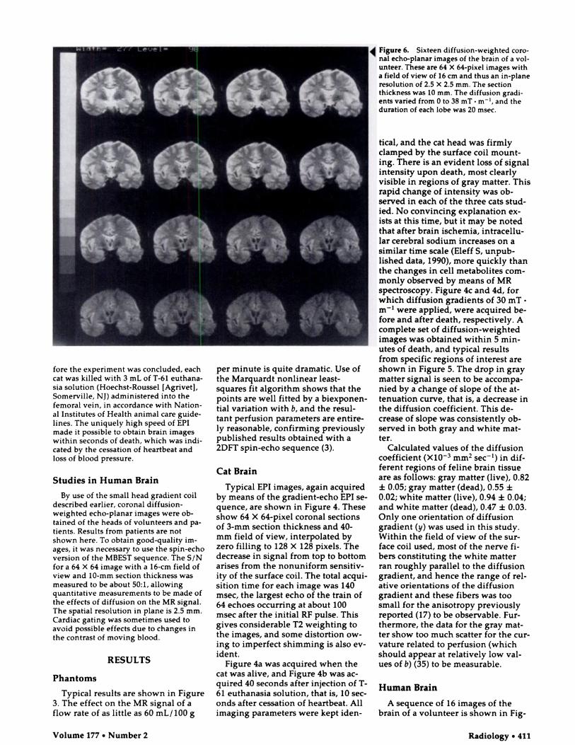
Figure 6. Sixteen diffusion-weighted coro-nal echo-planar images of the brain of a vol-
unteer. These are 64 X 64-pixel images witha field of view of 16 cm and thus an in-plane
resolution of 2.5 X 2.5 mm. The sectionthickness was 10 mm. The diffusion gradi-ents varied from 0 to 38 mT . mt, and the
duration of each lobe was 20 msec.
tical, and the cat head was firmly
clamped by the surface coil mount-ing. There is an evident loss of signalintensity upon death, most clearly
visible in regions of gray matter. This
rapid change of intensity was ob-
served in each of the three cats stud-ied. No convincing explanation ex-ists at this time, but it may be noted
that after brain ischemia, intracellu-lar cerebral sodium increases on a
similar time scale (Eleff 5, unpub-
lished data, 1990), more quickly than
the changes in cell metabolites com-monly observed by means of MR
spectroscopy. Figure 4c and 4d, for
which diffusion gradients of 30 mT.m’ were applied, were acquired be-
fore and after death, respectively. Acomplete set of diffusion-weightedimages was obtained within 5 mm-
utes of death, and typical results
from specific regions of interest are
shown in Figure 5. The drop in gray
matter signal is seen to be accompa-nied by a change of slope of the at-
tenuation curve, that is, a decrease in
the diffusion coefficient. This de-
crease of slope was consistently ob-
served in both gray and white mat-ter.
Calculated values of the diffusion
coefficient (X103 mm2 sec’) in dif-
ferent regions of feline brain tissue
are as follows: gray matter (live), 0.82
± 0.05; gray matter (dead), 0.55 ±0.02; white matter (live), 0.94 ± 0.04;
and white matter (dead), 0.47 ± 0.03.Only one orientation of diffusion
gradient (y) was used in this study.
Within the field of view of the sur-face coil used, most of the nerve fi-
bers constituting the white matter
ran roughly parallel to the diffusiongradient, and hence the range of rel-
ative orientations of the diffusion
gradient and these fibers was too
small for the anisotropy previously
reported (17) to be observable. Fur-
thermore, the data for the gray mat-ter show too much scatter for the cur-vature related to perfusion (whichshould appear at relatively low val-
ues of b) (35) to be measurable.
Human Brain
A sequence of 16 images of the
brain of a volunteer is shown in Fig-
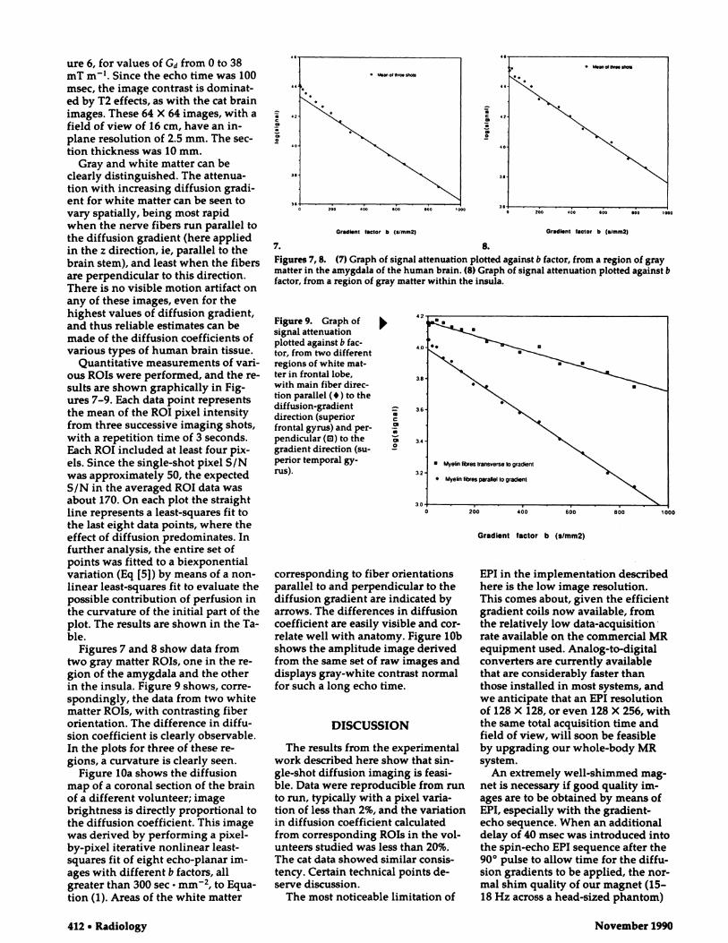
. Mea�otIh�eshoI�
7.
Gradient factor b (sJmm2)
8.
Gr#{149}d�.nt factor b (slmm2)
Figures 7, 8. (7) Graph of signal attenuation plotted against b factor, from a region of graymatter in the amygdala of the human brain. (8) Graph of signal attenuation plotted against bfactor, from a region of gray matter within the insula.
a
C
a
0
Gradient factor b (s/mm2)
412 #{149}Radiology November 1990
ure 6, for values of Gd from 0 to 38mT m�. Since the echo time was 100msec, the image contrast is dominat-ed by T2 effects, as with the cat brainimages. These 64 X 64 images, with afield of view of 16 cm, have an in-plane resolution of 2.5 mm. The sec-tion thickness was 10 mm.
Gray and white matter can beclearly distinguished. The attenua-tion with increasing diffusion gradi-
ent for white matter can be seen tovary spatially, being most rapidwhen the nerve fibers run parallel tothe diffusion gradient (here appliedin the z direction, ie, parallel to thebrain stem), and least when the fibersare perpendicular to this direction.There is no visible motion artifact onany of these images, even for thehighest values of diffusion gradient,
and thus reliable estimates can bemade of the diffusion coefficients ofvarious types of human brain tissue.
Quantitative measurements of vari-ous ROIs were performed, and the re-sults are shown graphically in Fig-
ures 7-9. Each data point representsthe mean of the ROl pixel intensityfrom three successive imaging shots,with a repetition time of 3 seconds.Each ROI included at least four pix-els. Since the single-shot pixel S/N
was approximately 50, the expectedS/N in the averaged ROl data was
about 170. On each plot the straightline represents a least-squares fit tothe last eight data points, where theeffect of diffusion predominates. Infurther analysis, the entire set ofpoints was fitted to a biexponentialvariation (Eq [5]) by means of a non-linear least-squares fit to evaluate thepossible contribution of perfusion inthe curvature of the initial part of theplot. The results are shown in the Ta-ble.
Figures 7 and 8 show data fromtwo gray matter ROIs, one in the re-gion of the amygdala and the otherin the insula. Figure 9 shows, corre-
spondingly, the data from two whitematter ROIs, with contrasting fiberorientation. The difference in diffu-sion coefficient is clearly observable.In the plots for three of these re-gions, a curvature is clearly seen.
Figure lOa shows the diffusionmap of a coronal section of the brainof a different volunteer; imagebrightness is directly proportional tothe diffusion coefficient. This imagewas derived by performing a pixel-by-pixel iterative nonlinear least-squares fit of eight echo-planar im-ages with different b factors, allgreater than 300 sec . mm2, to Equa-tion (1). Areas of the white matter
Figure 9. Graph of
signal attenuationplotted against b fac-tor, from two differentregions of white mat-ter in frontal lobe,with main fiber direc-
lion parallel (#{149})to thediffusion-gradientdirection (superior
frontal gyros) and per-
pendicular (D) to the
gradient direction (su-perior temporal gy-rus).
corresponding to fiber orientationsparallel to and perpendicular to thediffusion gradient are indicated byarrows. The differences in diffusioncoefficient are easily visible and cor-relate well with anatomy. Figure lObshows the amplitude image derivedfrom the same set of raw images anddisplays gray-white contrast normalfor such a long echo time.
DISCUSSION
The results from the experimentalwork described here show that sin-gle-shot diffusion imaging is feasi-ble. Data were reproducible from runto run, typically with a pixel varia-tion of less than 2%, and the variationin diffusion coefficient calculated
from corresponding ROIs in the vol-unteers studied was less than 20%.The cat data showed similar consis-tency. Certain technical points de-serve discussion.
The most noticeable limitation of
EPI in the implementation describedhere is the low image resolution.This comes about, given the efficient
gradient coils now available, fromthe relatively low data-acquisition#{149}rate available on the commercial MRequipment used. Analog-to-digitalconverters are currently availablethat are considerably faster thanthose installed in most systems, andwe anticipate that an EPI resolutionof 128 X 128, or even 128 X 256, withthe same total acquisition time andfield of view, will soon be feasibleby upgrading our whole-body MR
system.An extremely well-shimmed mag-
net is necessary if good quality im-ages are to be obtained by means ofEPI, especially with the gradient-echo sequence. When an additionaldelay of 40 msec was introduced intothe spin-echo EPI sequence after the90#{176}pulse to allow time for the diffu-sion gradients to be applied, the nor-
mal shim quality of our magnet (15-18 Hz across a head-sized phantom)
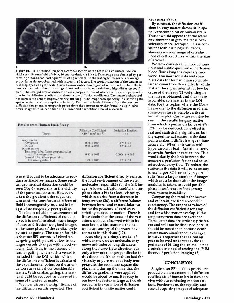
Figure 10. (a) Diffusion image of a coronal section of the brain of a volunteer. Section
thickness, 10 mm; field of view, 16 cm; resolution, 64 X 64. This image was obtained by per-
forming a nonlinear least-squares fit of Equation (1) to the last eight images of a 16-image
echo-planar dataset obtained with increasing b factor. The spatial variation of the parameter
D is displayed as a gray scale. Curved arrow indicates a region of white matter where the fi-
bers are parallel to the diffusion gradient and thus shows a relatively high diffusion coeffi-
cient. The straight arrows indicate an area (corpus callosum) where the fibers are perpendic-
ular to the diffusion gradient and shows a low diffusion coefficient. The image background
has been set to zero to improve clarity. (b) Amplitude image corresponding to a showing the
spatial variation of the amplitude factor S0. Contrast is clearly different from that seen on
diffusion image and corresponds precisely to the contrast normally found in a spin-echo
brain image with an echo time of 130 msec and a repetition time of 4 seconds.
Results from Human Brain Study
Diffusion Coefficient Perfusion FractionTissue (X103 mm2 sec�) (%)
Gray matterAmygdalaInsula
0.66 ± 0.060.68 ± 0.08
10.9 ± 4.06.9 ± 5.3
White matterTemporal lobe, fibers perpendicular
to diffusion gradientFrontal lobe, fibers parallel to
diffusion gradient
0.43 ± 0.03
1.00 ± 0.05
0.0006 ± 0.002
7.9 ± 2.3
Volume 177 #{149}Number 2 Radiology #{149}413
was still found to be adequate to pro-
duce artifact-free images. Some resid-
ual geometrical distortion could be
seen (Fig 6), especially in the vicinity
of the paranasal sinuses. However,
when the gradient-echo sequence
was used, the unrefocussed effects offield inhomogeneity resulted in im-ages of unacceptably poor quality.
To obtain reliable measurements ofthe diffusion coefficients of tissue in
vivo, it is useful to obtain each image
of a set of diffusion-weighted images
at the same phase of the cardiac cycle
by cardiac gating. The reason for thisis that the EPI contrast of blood un-
dergoing rapid, pulsatile flow in thelarger vessels changes with blood ye-
locity (24). Thus, in the absence ofcardiac gating, when such a vessel is
included in the RO! within whichthe diffusion coefficient is calculated,
the experimental points on the atten-
uation curve can show considerable
scatter. With cardiac gating, the scat-
ter should be reduced, as observed in
some of the studies made.We now discuss the significance of
the diffusion results reported. The
diffusion coefficient directly reflectsthe local environment of the water
molecules responsible for the MR im-
age. A lower diffusion coefficient im-
plies either a higher local viscosity,which can arise from a decrease intemperature (36), a different balance
between intra- and extracellular wa-
ter, or the presence of barriers re-
stricting molecular motion. There islittle doubt that the cause of the van-ations we have observed within hu-man brain white matter is the ex-
tneme anisotnopy of the water envi-
ronment in this tissue (17).According to a simple model of
white matter, water molecules maymove unhindered long distances
along the nerve-fiber direction but
only a few microns perpendicular tothis direction. If this medium had theviscosity of pure water at body tem-perature, the root mean square dis-
placement during the time that the
diffusion gradients were applied
would be about 16 tim. It is easy to
see how the factor as large as two ob-
served in the variation of diffusioncoefficient in white matter could
have come about.
By contrast, the diffusion coeffi-
cient in gray matter shows little spa-
tial variation in cat or human brain.
Thus it would appear that the water
environment in gray matter is con-siderably more isotropic. This is con-
sistent with histologic evidence,
showing a wider range of onienta-
tions of cell structures within the size
of a voxel.
We now consider the more conten-
tious and subtle question of perfusive
blood flow along the capillary net-
work. The most accurate and corn-
plete data for human brain so far ob-
tamed come from this study. In white
matter, the signal intensity is low be-
cause of the heavy T2 weighting in
the images obtained, and thus there
is considerable scatter in the ROl
data. For the region where the fibers
lie parallel to the diffusion gradient,some curvature is visible on the at-
tenuation plot. Curvature can also be
seen in the results for gray matter,
from which a perfusion factor of 6%-
12% may be deduced. This effect isreal and statistically significant, but
the experimental scatter in the data
points makes it difficult to quantitate
accurately. Whether it varies with
hypercarbia or brain functional activ-
ity awaits further investigation. Thiswould clarify the link between the
measured perfusion factor and actual
microcirculatory flow. To reduce thescatter in the data it will be necessary
to use larger ROIs or to average re-
sults from a larger number of images,which must be done after the image
modulus is taken, to avoid possible
phase interference effects arising
from system instability.
Comparing the results for human
and cat brain, we find reasonable
consistency. The ranges of values ofthe diffusion coefficients for gray
and for white matter overlap, if the
cat postmortem data are excluded.
These latter data are strikingly differ-
ent and will not be discussed here. It
should be noted that, because deathcauses many simultaneous changesin tissue properties that do not ap-pear to be well understood, the ex-
periment of killing the animal is not
a suitable method of testing the IVIM
theory of perfusion imaging (3).
CONCLUSION
Single-shot EPI enables precise, re-
producible measurement of diffusion
coefficients of human brain tissue in
vivo without confusing motion anti-
facts. Furthermore, the rapidity and
ease of acquiring images of adequate

414 #{149}Radiology November 1990
quality facilitate far more detailedanalyses of the attenuation curvethan possible previously. Effectshave been observed on the MR signalthat are consistent with those pre-dicted to be caused, according to theLe Bihan theory, by the superim-posed perfusive motion of blood incapillaries. Further improvements indata analysis should lead to observa-tions of changes in the IVIM parame-ters associated with brain function.
The excellent time resolution ofEPI has enabled the observation ofrapid changes in diffusion character-istics, which have been noted afterdeath in the brain of a cat. If thesechanges are associated primarily withischemia, it is likely that IVIM-EPIwill be of great clinical benefit in di-agnosis and treatment of cerebrovas-cular disease and other abnormalitiesinvolving the microcirculation. #{149}
Acknowledgments: We acknowledge use ofthe facilities of the NIH In Vivo NMR Center
and thank Chrit Moonen, PhD, for helpful ad-vice and Daryl Despres, Steve Merritt, and A.
Scott Chesnick for technical assistance. We alsothank Scott Eleff, PhD, for useful discussionsand for making available unpublished observa-tions regarding sodium MR during ischemia ina dog model.
References1. Le Bihan D, Breton E. Imagerie de diffu-
sion in-vivo par resonance magnetique. CR Acad Sci [III 1985; 15:1109-1112.
2. Le Bihan D, Breton E, La!lemand D, Gren-icr P, Cabanis E, Laval-Jeantet M. MRimaging of intravoxel incoherent motions:
application to diffusion and perfusion inneurologic disorders. Radiology 1986;
161:401-407.3. Le Bihan D, Breton E, Lallemand D, Aubin
M-L, Vignaud J, Laval-Jeantet M. Separa-tion of diffusion and perfusion in intra-voxel incoherent motion imaging. Radiol-
ogy 1988; 168:497-505.4. Young IR, Hall AS, Bryant DJ, et a!. As-
sessment of brain perfusion with MR im-aging. J Comput Assist Tomogr 1988;12:721-727.
5. Brownstein KR, Tarr CE. Importance ofclassical diffusion in NMR studies in bio-logical cells. Phys Rev A 1979; 19:2446-2453.
6. Whittall KP, MacKay AL. Quantitativeinterpretation of NMR relaxation data.
Magn Reson 1989; 84:134-152.
7. Hansen JR. Pulsed NMR study of watermobility in muscle and brain tissues. Bio-chem Biophys Acta 1971; 230:482-486.
8. Roy CS, Sherrington CS. On the regula-tion of the blood supply of the brain.
Physiol 1890; 11:85-108.9. Turner R, Bowley RM. Passive screening
of switched magnetic field gradients.Phys E: Sci Instrum 1986; 19:876-879.
10. Mansfield P. Multi-planar image forma-tion using NMR spin echoes. J Phys C: So!State Phys 1977; 10:L55-L58.
11. Hahn EL. Spin echoes. Phys Rev 1950;
80:580-594.12. Carr HY, Purcell EM. Effects of diffusion
on free precession in nuclear magneticresonance experiments. Phys Rev 1954;94:630-635.
13. Stejskal EO, TannerJE. Spin diffusionmeasurements: spin echoes in the pres-ence of a time-dependent field gradient.Chem Phys 1964; 42:288-292.
14. Taylor DC, Bushel! MC. The spatial map-ping of translational diffusion coefficientsby the magnetic resonance imaging tech-nique (abstr). In: Book of abstracts: Societyof Magnetic Resonance in Medicine 1985.Vol 1. Berkeley, Calif: Society of MagneticResonance in Medicine, 1985; 612-613.
15. Ahn CB, Lee SY, Nalcioglu 0, Cho ZH.An improved nuclear magnetic resonancediffusion imaging method using an opti-mised pulse sequence. Med Phys 1986;
13:789-793.16. Thomsen C, Ring P. Henrikson 0. In
vivo measurement of water self-diffusionby magnetic resonance imaging (abstr).
In: Book of abstracts: Society of MagneticResonance in Medicine 1988. Vol 2. Berke-Icy, Ca!if: Society of Magnetic Resonancein Medicine, 1988; 890.
17. Moseley ME, Cohen Y, Mintorovitch J, eta!. Early detection of regional cerebra!ischemia in cats: comparison of diffusion-and T2-weighted MRI and spectroscopy.Magn Reson Med 1990; 14:330-346.
18. Merbo!dt KD, Haenicke W, Frahm J. Self-diffusion NMR imaging using simulatedechoes. J Magn Reson 1985; 64:479-486.
19. Le Bihan D. Intravoxe! incoherent mo-tion imaging using steady-state free pre-cession. Magn Reson Med 1988; 7:346-351.
20. Le Bihan D, Turner R, McFal! JR. Effectsof intra-voxel incoherent motions (IVIM)in steady state free precession (SSFP) im-
aging: application to molecular diffusionimaging. Magn Reson Med 1989; 10:324-337.
21. Merbo!dt KD, Bruhn H, Frahm J, GyngellML, HSnicke W, Diemling M. MRI of‘diffusion’ in the human brain: new re-suits using a modified CE-FAST sequence.Magn Reson Med 1989; 9:423-429.
22. Chenevert TL, Brunberg JA, Schielke GP.Quantitative improvement of in vivo tis-sue perfusion and diffusion imaging(abstr). In: Book of abstracts: Society ofMagnetic Resonance in Medicine 1989.Vol 1. Berkeley, Calif: Society of MagneticResonance in Medicine, 1989; 62.
23. Doyle M, Turner R, Cawley M, et a!.Real-time cardiac imaging of adults at vid-eo frame rates by magnetic resonance im-aging. Lancet 1986; 2:682.
24. Pawlik G, Rack! A, Bing RJ. Quantitative
capillary topography and blood flow inthe cerebra! cortex of cats: an in vivo mi-croscopic study. Brain Res 1981; 208:35-58.
25. Johnson C, Hutchinson JMS. The limita-tions of NMR recalled-echo imaging tech-niques. J Magn Reson 1985; 63:14-30.
26. Howseman AM, Steh!ing MK, ChapmanB, et a!. Improvements in snapshot nu-
clear magnetic resonance imaging. BrRadio! 1988; 61:822-828.
27. Avram HE, Crooks LE. Effect of se!f-dif-
fusion on echo-planar imaging (abstr). In:Book of abstracts: Society of Magnetic Res-
onance in Medicine 1988; Vol 2. Berkeley,Calif: Society of Magnetic Resonance inMedicine, 1988; 980.
28. Turner R. Perfusion studies and fast im-
aging. In: Rescigno A, Boicelli A, eds. Cer-ebral blood flow. New York: Plenum,1988; 245-258.
29. Turner R, Le Bihan D. Single-shot diffu-sion imaging at 2.0 Tesla. J Magn Reson1990; 86:445-452.
30. Alderman DW, Grant DM. An efficientdecoupler coil design which reduces heat-ing in conductive samples in supercon-ducting spectrometers. J Magn Reson1979; 36:447-451.
31. Mansfield P. Chapman B. Active magnet-ic screening of gradient coils in NMR im-
aging. J Magn Reson 1986; 66:573-576.32. Roemer PB, Edelstein WA, Hickey JS.
Self-shielded gradient coils. In: Book ofabstracts: Society of Magnetic Resonancein Medicine 1986. Vol 3. Berkeley, Calif:
Society of Magnetic Resonance in Mcdi-cine, 1986; 1067.
33. Blackband 5, Chatham JC, O’Dell W, Day
S. Echo-planar imaging of isolated per-fused rat hearts at 4.7 T: a comparison ofLangendorff and working heart prepara-tions. Magn Reson Med 1990; 15:240-245.
34. Turner R. A target field approach for op-
timal coil design. J Phys D: App! Phys1986; 19:L147-L151.
35. Le Bihan D, Moonen CTW, Van Zij! PCM,Pekar J, DesPres D. Evaluation of water
molecu!ar diffusion and blood microcircu-lation in the cat brain at 4.7 tesla (abstr).
In: Book of abstracts: Society of MagneticResonance in Medicine 1989. Vol 2. Berke-ley, Calif: Society of Magnetic Resonancein Medicine, 1989; 1051.
36. Le Bihan D, De!annoy J, Levin RL. Tem-perature mapping with MR imaging ofmolecular diffusion: application to hyper-thermia. Radiology 1989; 171:853-857.