echocardiography role in assessing cardiovascular changes
TRANSCRIPT

47
Review Article
Echocardiography Role in Assessing Cardiovascular Changes in Very Low Birth Weight Babies, With Emphasis on the Presence of the Ductus ArteriosusAdriana Mello Rodrigues dos Santos1,2, Zilda Maria Alves Meira1, Maria do Carmo Nunes Pereira1
Universidade Federal de Minas Gerais1; Hospital Sofia Feldman2, Belo Horizonte, MG – Brazil
AbstractThis manuscript consisted in a literature review on
the cardiovascular alterations in premature neonates. Such alterations have a high prevalence in neonatal intensive-care units, with frequent need for specific treatment and prognostic implications. Review was conducted in a nonsystematic manner, based on the following sources of research: PubMed, BVS and Medline.
The causes of such hemodynamic alterations, the methods often used to detect them and more objective and efficient alternative proposals in this assessment were defined, with emphasis on different echocardiographic parameters and limitations of each method.
IntroductionChanges in the cardiovascular system are common in
premature neonates (gestational age less than 37 weeks), especially in very low birth weight infants (VLBW), i.e., with birth weight lower than 1,500g, usually with gestational age less than 30 weeks as well. These changes take place in the first hours of life1-4, for various reasons, as listed below:
a) Because the myocardium of premature infants contains less contractile tissue in relation to older children, it may be unable to adapt to increased systemic vascular resistance, resulting from the withdrawn of the placenta after birth and peripheral vasoconstriction triggered by the release of hormones during labor;5-7
b) Very low birth weight infants (VLBW), whose mothers have chorioamnionitis may have, in contrast, myocardial dysfunction associated with hypotension with normal or even increased cardiac output (low systemic vascular resistance);
c) premature neonates with a history of perinatal suffering may show myocardial dysfunction and/or abnormal vasomotor response;8
d) VLBW infants can manifest hypotension and/or low systemic blood flow secondary to a hemodynamically significant ductus arteriosus, associated or not to the left-to-right flow through the interatrial septum, although clinically silent.5,7,9 In this case, if inotropes are used to increase the systemic vascular resistance in relation to pulmonary vascular resistance, the systemic blood flow may be paradoxically reduced, despite hypotension is corrected, or systemic hypotension may be even aggravated with an increased pulmonary flow through the ductus arteriosus;4,7,10
e) hypotension and/or low systemic blood flow may occur due to relative adrenal insufficiency7 and resistance to inotropes / vasopressors or systemic inflammatory response syndrome, as observed in sepsis or necrotizing enterocolitis;10,11
f) neonatal hemodynamic instability can be caused by pericardial effusion, sometimes associated with the use of intracardiac catheters,5
g) low cardiac output may also be a consequence of pulmonary hypertension by reducing the left ventricular filling, secondary to reduced pulmonary venous return or mechanical effects caused by pressure overload of the right ventricle (RV).5
Hemodynamic adequacy of the premature should always be assessed globally, also taking into consideration any possible harmful effects of therapeutic treatments commonly used in the neonatal period. In this context, we emphasize the use of mechanical ventilation, often essential to the treatment of these newborns (NB), but which can cause reduced systemic venous return, increased pulmonary vascular resistance and even myocardial dysfunction, when higher mean pressures are used in airways12 In such cases, mechanical ventilation could improve arterial oxygenation, but the resulting reduced systemic perfusion would compromise tissue oxygenation.4
Proper understanding of systemic hemodynamics in neonates, however, is quite limited. The impact of prematurity in this context is even more unknown and the acquisition of this knowledge comes up against technical7,13 and ethical principles.14 As a result, the treatment of cardiovascular disorders in VLBW infants is rarely based on its pathophysiology.11 What can be seen in daily practice is the treatment of these newborns with intravenous infusions of physiological saline, sometimes at a high volume. However, it has been shown that there is no volume depletion in most hypotensive preterm infants in the first few hours of life.6
Vasopressors and/or inotropic agents are also administered, with little focus on the etiology, phase or pathophysiology of the hemodynamic instability. It is possible to notice different patterns in the way of treating these premature infants, as to
KeywordsEchocardiography; Very Low Birth Weight Infants;
Premature; Ductus Arteriosus.Mailing Address: Adriana Mello Rodrigues dos Santos •Av. Prof. Alfredo Balena, 190, sala 533. Postal Code 30130100, Belo Horizonte, MG - BrazilE-mail: [email protected] received November 16, 2015; revised December 13, 2015 and approved January 18, 2016.
DOI: 10.5935/2318-8219.20160014

48
Review Article
Santos et al.Cardiovascular changes in very low birth weight babies
Arq Bras Cardiol: Imagem cardiovasc. 2016;29(2):47-57
neonatal units at which they will be treated and not to clinical parameters proper.1,7,15,16 It is worthy of emphasis the limited ability of premature myocardium to increase contractility and ejection volume in response to volumetric overload.5 Moreover, in most neotatal intensive care units (NICU), the hemodynamic parameters used are mostly clinical, including capillary refill time, diuresis, amplitude in pulse rate and non-invasive blood pressure (BP). It is known, however, that these parameters were not correlated with more objective measures of systemic blood flow, especially in the first 24 hours of life.2,5,11,12,17-19
The focus in the treatment of hypotension is particularly common in neonatal intensive care units,1,7 which should be done with some important caveats. Hypotension is defined as the BP level at which there is loss of autoregulation of blood flow to the target organs. BP decreasing levels beyond this “autoregulation threshold” reach function loss levels and, finally reaching the “ischemic threshold”, they result in tissue ischemia and permanent injury.7,20 BP levels that would cause loss of self-regulation or permanent tissue ischemia in VLBW infants, is not known.1,11,15,21,22 However, it is known that lower blood pressure levels are more common in lower gestational ages and less frequent for those with higher postnatal ages.1,6
In general clinical practice, mean BP is used, as this is deemed to be more representative of the perfusion pressure than systolic and diastolic pressures.6,15 In addition, hypotension is defined as the mean BP below the fifth or 10th percentile for gestational and postnatal age. On the first day of life, these values are equivalent to gestational age in weeks.2,3,7,11,12,20,23 The choice for this parameter can be challenged, as the clinical objective should be to maintain blood pressure levels above a safe threshold, not a normal reference value based on statistical criteria21
Some studies state that the self-regulatory threshold must be around 30 mmHg, even in extreme low weight preterm infants (birth weight less than 1,000g) on their first day of life. Using the mean BP parameter equivalent to gestational age in weeks, the most premature infants, who are the most vulnerable, may be exposed to the risks of loss of autoregulation.15,20 In addition, the BP is determined by the interaction between cardiac output and systemic vascular resistance. Ideally, therefore, both cardiac output and/or systemic vascular resistance should be monitored beyond the BP value, in order to more accurately assess cardiovascular condition.12,15 Individually, pressure levels are poorly predictive of tissue perfusion and tissue oxygen supply1,2,5,6,8,19,21 Some authors speculate that the very premature newborn’s brain, immediately after labor, would be considered a non-vital organ and therefore possibly poorly perfused in a compensated shock phase, when BP levels are still normal.7,20
Ductus arteriosus in premature infantsThe prevalence of the ductus arteriosus in the group of
extreme preterm infants (gestational age less than 30 weeks) is high, ranging from 50% to 100% in the first days of life in different series, depending on the diagnostic criterion and gestational/postnatal ages.5,13,19,24-30 The prevalence is inverse to the gestational age and multiple morbidities
are associated with its presence, especially in extremely underweight NBs: peri-intraventricular hemorrhage (PIVH), necrotizing enterocolitis (NEC), pulmonary hemorrhage, bronchopulmonary dysplasia (BPD), increased hospital stay and death.2,3,29,31-42
The direct causal relationship between these complications and pulmonary hyperflow/systemic hypoflow, both present in the premature newborn with hemodynamically significant ductus arteriosus,43 was considered true until recent years. However, some studies showed no reduction of most of these comorbidities or its consequences, such as survival free of neurological sequelae,31,44 with prophylaxis or (pharmacological or surgical) treatment for closure of ductus arteriosus. It is noteworthy that these studies were not designed to define the role of the ductus arteriosus in the prediction of adverse clinical outcomes.30-32,39
Some authors have questioned whether the ductus arteriosus was not but a worse prognosis “marker” associated with prematurity and not a causal factor, i.e., the ductus arteriosus would be patent in severe preterm infants and, therefore, so would the complications such as PIVH, NEC, BPD and death.24,45,46 Other authors have questioned even if such morbidities would not be caused by pharmacological or surgical therapy, established for treatment of infants with ductus arteriosus.39,45,47-52
As a result, it has been observed different behaviors in relation to the treatment of premature infants with patent ductus arteriosus, not only among centers considered to be of excellence, but even in a same center, depending on the attending neonatologist. 30,47,50,53 This lack of consensus reflects the limited knowledge that literature has provided in recent years. Studies are conflicting as small groups and, therefore, with reduced statistical power, are studied54 including different populations in relation to gestational age, 55 hemodynamic compromise level, size of the ductus arteriosus,30,56-58 postnatal ages and, Consequently, also different exposure times are .46 The studies were not designed to evaluate the role of a patent ductus arteriosus in neonatal morbidities, but rather to detect the relationship between treatment and closure of the ductus.
Finally, all studies have rescue therapies if the patent ductus arteriosus persists.46,55,59 When the premature previously randomized for no treatment of the ductus arteriosus remained symptomatic, they were then treated. Thus, the results of the studies show the ductus arteriosus closure capacity with a given drug and not the possible benefits from the closure compared to no treatment of the ductus arteriosus.24,32,43,46,60-62
It is worthy of emphasis that, given the scientific evidence and the relatively high incidence of spontaneous closure of the ductus arteriosus in premature infants with greater gestational ages and higher birth weights,34,63 during quite varied periods of time, the approach to the ductus arteriosus changed53,56,64 tending to reduce indication or delay the clinical treatment for 65 closure of the ductus arteriosus. As a result, there was an increase of the need for surgical treatment of the patent ductus arteriosus,43especially at the lower gestational ages.64
Surgical closure of the ductus arteriosusUntil recently, the procedure for surgical correction of the
ductus arteriosus has been considered safe. Currently, however,

49
Review Article
Santos et al.Cardiovascular changes in very low birth weight babies
Arq Bras Cardiol: Imagem cardiovasc. 2016;29(2):47-57
perioperative complications such as preoperative low coronary flow, systemic hypotension, myocardial dysfunction, respiratory deterioration and loss of capacity for cerebral autoregulation were reported43,46,47,56,61,66 and may be implicated in the genesis of possible long-term adverse events. On the other hand, some studies have not identified the surgical ligation as a predictor of unfavorable neurological development, 45,54 but the comorbidities associated with it, such as prematurity and prolonged mechanical ventilation.54
Other authors have associated a worse neurological development to the surgical bandages prematurely applied to the ductus arteriosus (less than 10 days of life).67 BPD and retinopathy of prematurity may also be associated with surgical ligation of the ductus in premature infants,45,50,68 and there is also a debate about the safety of all anesthetic-surgical procedures in neonates and specifically in VLBW infants.54,69 The cause of worse neurological outcome and the association with retinopathy of prematurity in patients undergoing surgical ligation of the ductus arteriosus are not evident yet. Lesions may have preceded the surgery in some patients. Patients undergoing surgery may be sicker than those clinically treated or the lesion may have resulted from perioperative changes such as hypothermia, excessive handling during transport or direct exposure to anesthetic drugs.69
Despite some controversy, the surgical correction of the ductus arteriosus should be considered when there is contraindication to drug treatment or when the ductus arteriosus remains with hemodynamic repercussions after the drug treatment. There was a four to eight times increased mortality in the group of neonates who remained with a significant ductus arteriosus after drug treatment compared to those with no significant ductus arteriosus or closed ductus after drug treatment, even after adjusting for confounding factors such as gestational age and severity score.35,41
Pharmacological treatment of patent ductus arteriosus in preterm infants
Contrary to the current trend to question the real benefits of closing the patent ductus arteriosus in preterm infants, some studies have shown quite undesirable hemodynamic effects of the ductus arteriosus as early as the first hours of life, detected by echocardiography,2,4,13,25,26,33,60,61,70-74 with early pharmacologic treatment being therefore postulated. Early pre-symptomatic treatment of patent ductus arteriosus in selected preterm infants differs from what has been described in the literature as a prophylactic treatment. Prophylaxis is defined as the treatment of all preterm infants, whether or not they have a pervious patent ductus arteriosus. In this case, about 60% of the infants received the medication unnecessarily.42,60 Early pre-symptomatic treatment contradicts previous paradigms, whereby the existence of a ductus arteriosus in the first days of life of preterm infants was considered “normal”.60
As there are no randomized clinical trials comparing the use of cyclooxygenase inhibitors in a prophylactic, early pre-symptomatic, early symptomatic or late symptomatic manner, with surgical treatment or even with no treatment, no specific recommendations can be made.61,62,75
Although the side effects of cyclooxygenase inhibitors are considered reversible in most cases, the increased risk of BPD when indomethacin is used in preterm infants without ductus arteriosus has been emphasized,52 in addition to the increased risk of bowel perforation when the cyclooxygenase inhibitor is used in combination with corticosteroids. 24,51,76
As there are signs of deleterious effects caused by the patent ductus arteriosus in preterm infants, as well as increased mortality,35,41 and potential complications of prophylactic treatments, especially in NBs receiving the drug unnecessarily, as they have no ductus arteriosus, and possible consequences after surgical closure of the ductus, pre-symptomatic drug treatment becomes an interesting alternative. The distinction between preterm infants with hemodynamically significant patent ductus arteriosus and preterm infants whose ductus arteriosus is a physiological adaptation in the transitional period,25,27,56,60,73 based on the echocardiographic findings, allows to select the subgroup that is at a greater risk to exposure to patent ductus arteriosus and would mostly benefit from treatment.3
Functional Echocardiography in premature infants
Functional echocardiography could therefore become a fundamental tool for decision making on either drug or surgical treatment of the ductus arteriosus, avoiding exposure to nonsteroidal anti-inflammatory drugs (NSAIDs), thus restricting the use of these drugs to preterm infants who actually present hemodynamically significant ductus arteriosus and limiting the time of exposure to these drugs.62,73 In addition, early pharmacologic treatment before the emergence of clinical signs as murmurs, hyperdynamic precordium and large pulses,72,77 which is possible with the use of functional echocardiography, is associated with reduced need for surgery.46
Therefore, evaluations and validations of echocardiography protocols are necessary for a better assessment of hemodynamic status of preterm infants and, for this, the literature suggests including the following parameters:
1) estimate of the magnitude and direction of blood flow through the foramen ovale. The left-to-right interatrial flow can increase the pulmonary blood flow and reduce the systemic flow, both already caused by the ductus arteriosus.4,13,25,78-80 A diameter of the foramen ovale larger than 3 mm is usually considered significant;79
2) estimate of the magnitude of the blood flow from the aorta to the pulmonary artery through the ductus. Some authors consider significant a ductus arteriosus diameter greater than 1.5mm, correlating this cutoff point with potential complications.3,13,56,81,56,81 However, this estimate is not only related to ductal diameter13,26 (Figure 1), but also to the systemic and pulmonary vascular resistance, as well as to the contractility of the immature myocardium.
To that end, the measures of both mean and end-diastolic velocities in the left pulmonary artery (LPA)13,26,43,82 (Figure 2), and that of the pressure gradient between the aorta and pulmonary artery through the ductus arteriosus could be used as auxiliary measures instead of just calculating the ductus arteriosus diameter.

50
Review Article
Santos et al.Cardiovascular changes in very low birth weight babies
Arq Bras Cardiol: Imagem cardiovasc. 2016;29(2):47-57
Figure 1 – High left parasternal view. Ao = aorta; PA = pulmonary artery. A: Ductus arteriosus closed; B: Narrow ductus arteriosus (1mm) with left-to-right blood flow; C: Wide ductus arteriosus (3.1mm) with left-to-right blood flow. Source: personal archive of author AMRS.
A
PAAo
M
B
PADuctusAo
M
C
PA
Ductus arteriosus
Ao
M
Figure 2 – The white arrows point to the end diastolic velocities in the left pulmonary artery (LPA). A: End diastolic flow velocity of LPA at 0 cm/s, of a 29-week preterm infant with 13 hours of life and 1-mm ductus arteriosus; B: End diastolic flow velocity of LPA at 28.21 cm/s, in a 29-week-and-3-day preterm infant, with 9 hours of life and 2.1-mm ductus arteriosus. Source: Personal archive of author AMRS.
A
Flow velocity of left pulmonary artery
M
B
Flow velocity of left pulmonary artery
M
2.1) The mean diastolic flow velocity of LPA is obtained at the high left parasternal sagittal view (section of the ductus arteriosus) with the pulsed Doppler sample volume positioned in the proximal third of that artery, performing the planimetry of the area under the velocity-time curve TVI. The echocardiography apparatus program, which divides TVI by the duration of the cardiac cycle in seconds, provides the mean
flow velocity. The end diastolic flow velocity is measured at this same location. The mean diastolic flow velocity of LPA is considered high when ≥ 0,42m/s26 and end diastolic flow velocity is considered increased when ≥ 0.20m/s26,74 (Figure 2)
2.2) Flow direction and aorta-pulmonary artery pressure gradient through the ductus arteriosus can be evaluated in high left parasternal sagittal view, by trying to minimize

51
Review Article
Santos et al.Cardiovascular changes in very low birth weight babies
Arq Bras Cardiol: Imagem cardiovasc. 2016;29(2):47-57
the angle between the ultrasound beam and the blood flow through the ductus arteriosus. The flow in the ductus arteriosus generally travels from left to right, even in the first hours of life, but bidirectional flow is also frequent;60,74 In such cases, when the right-to-left flow is greater than 60% of the length of systole, the pulmonary pressure is generally suprasystemic.83 Using aorta-pulmonary artery gradient and obtaining, in an invasive manner (usually via umbilical artery catheter) or not, the systolic BP, it is possible to estimate systolic pressure in the pulmonary artery. e.g.: in a NB whose direction of blood flow through the ductus arteriosus is from left to right, and maximum flow velocity in the ductus is 3m/s (aorta-pulmonary artery gradient of 36 mmHg, calculated by Bernoulli’s equation) and systolic BP is 65 mmHg, the estimated systolic pressure in the pulmonary artery is of 29 mmHg (65 mmHg – 36 mmHg). Narrow ductus arteriosus with restriction signs generally have continuous flow, with maximum velocities higher than 2m/s,56 while large channels have pulsatile flow of velocities below 1.5m/s;27
3) evaluation of myocardial performance, which shall not be carried out by means of ejection fraction or fractional shortening testing, as such measures are overestimated in the presence of a ductus arteriosus. In such situation, the diastolic diameters of the left ventricle (LV) may be increased by an increased pulmonary venous return, while systolic diameters may be reduced by reducing afterload, which is now defined not only by systemic vascular resistance, but the pulmonary vascular resistance13,43,65,66 An interesting option would be therefore the relation between the velocity of circumferential shortening of myocardial fibers with heart-rate corrected (VCSHR) and systolic wall stress (SWS), resulting in a value less influenced by heart rate, pre- and afterload.84-88
VCSHR = (circumferential shortening/LV ejection time)
R – Rwhere the circumferential shortening = [(end diastolic
circumference – end systolic circumference) / end diastolic circumference] obtained in parasternal short-axis view at
the level of the mitral valve leaflets; the LV ejection time is obtained by means of a M-mode recording of the aortic valve, at a velocity of 100 mm/s, by measuring the opening and closure interval of this valve; and the square root of the R – R ( R – R) interval is used to correct the ejection time for a HR of 60 bpm. Normal values for preterm babies in the first hours of life are 0.8 circ/s ± 0.15.
SWS (g/cm²)=0,34 x SBP x LVSD
LVST x (1+ LVST /LVSD)
where SBP is the systolic blood pressure, LVSD is the LV systolic diameter, calculated by dividing the final systolic circumference by π instead of measuring the diameter in two-dimensional mode, in order to reduce errors due to distortion in the LV geometry (which are frequent, mainly due to the flattening of the interventricular septum); and LVST is the systolic thickness of the LV posterior wall.
The SWS corresponds to the calculation of afterload; the VCSHR measures how quickly the myocardium contracts (and not the distance of its contraction, as measured in the fractional shortening). However, the measurement of VCSHR is, just like the fractional shortening, also influenced by pre- and afterload . Thus, we should ideally calculate the VCSHR-SWS ratio, which is used as an index of myocardial contractility. There is an inverse ratio between VCSHR and SWS in normal preterm infants. Compared to older children, preterm infants’ myocardium has been shown to exhibit a higher basal contractile state, but a lower heart rate reserve, which means that, with an increase in afterload, the VCSHR in premature infants declines faster than that of older children86 (Figure 3).
Moreover, babies who develop low systemic flow also have a steeper VCSHR-SWS ratio curve, thus showing a worse ability of the myocardium of these children to deal with afterload increases89 (Figure 4).
4) estimate of the adequacy of the systemic circulation, which cannot be analyzed only by measuring LV or RV cardiac output because of the high frequency of flows through the
Figure 3 – Confidence interval of 95% for the VCSHR-SWS ratio in normal preterm infants (black line) and older children (gray line). Adapted from Barlow et al.
21.81.61.41.2
10.80.60.40.2
00 20 40 60
SWS (g/cm2)
VCS HR
(circ
/s)
80 100
1.5
1.0
0.5
0.00 0
SWS (g/cm2)
Normal flow Low flow
VCS HR
(circ
/s)
SWS (g/cm2)25 2550 5075 75
Figure 4 – Circumferential shortening velocity to systolic wall stress ratio (VCSHR-SWS ratio) in infants with adequate systemic flow (left) and low systemic blood flow (right). Babies who developed low systemic flow have a steeper curve, suggesting limited myocardial response to increased afterload (EPS). Adapted from Osborn et al.
52
Review Article
Santos et al.Cardiovascular changes in very low birth weight babies
Arq Bras Cardiol: Imagem cardiovasc. 2016;29(2):47-57
ductus arteriosus and atrial septum.4,18,19,25,71 In this context, measurements of flow in the superior vena cava (SVC), diastolic flow pattern in the descending aorta and superior mesenteric artery appears as interesting alternatives,18,28,70,71 but still require validation in daily practice. Moreover, some of these measures are technically more complex.18
4.1) Flow in the SVC can be used as an estimate of the systemic blood flow, as it represents the venous return of the upper part of the body and brain.2,5,70
SVC flow = VTI SVC x 3.14 x mean diameter SVC2 x HR
4
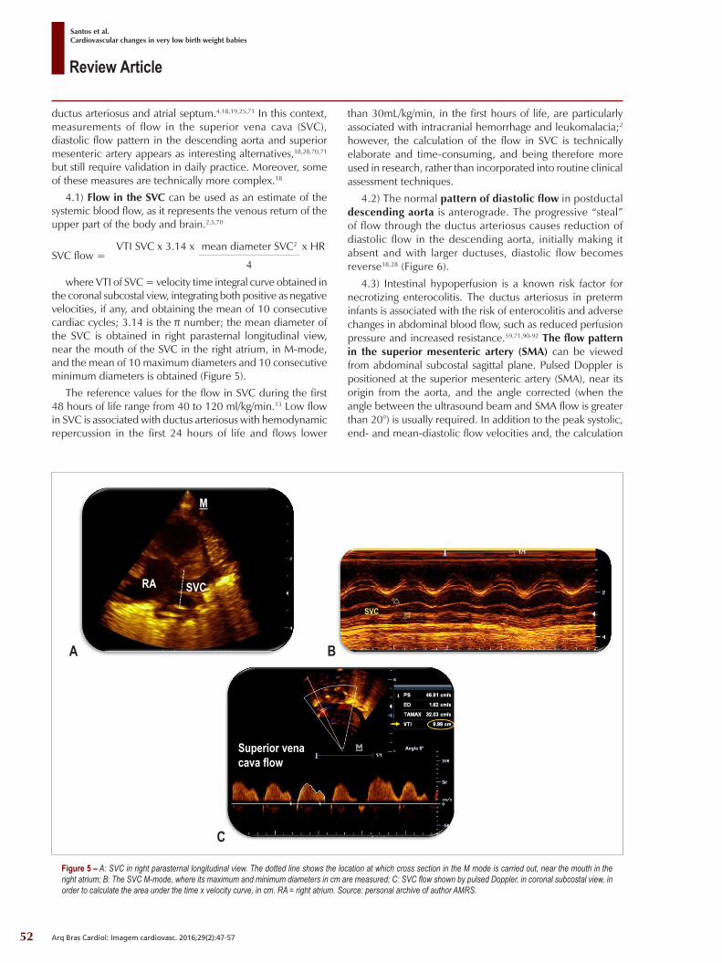
where VTI of SVC = velocity time integral curve obtained in the coronal subcostal view, integrating both positive as negative velocities, if any, and obtaining the mean of 10 consecutive cardiac cycles; 3.14 is the π number; the mean diameter of the SVC is obtained in right parasternal longitudinal view, near the mouth of the SVC in the right atrium, in M-mode, and the mean of 10 maximum diameters and 10 consecutive minimum diameters is obtained (Figure 5).
The reference values for the flow in SVC during the first 48 hours of life range from 40 to 120 ml/kg/min.13 Low flow in SVC is associated with ductus arteriosus with hemodynamic repercussion in the first 24 hours of life and flows lower
than 30mL/kg/min, in the first hours of life, are particularly associated with intracranial hemorrhage and leukomalacia;2 however, the calculation of the flow in SVC is technically elaborate and time-consuming, and being therefore more used in research, rather than incorporated into routine clinical assessment techniques.
4.2) The normal pattern of diastolic flow in postductal descending aorta is anterograde. The progressive “steal” of flow through the ductus arteriosus causes reduction of diastolic flow in the descending aorta, initially making it absent and with larger ductuses, diastolic flow becomes reverse18,28 (Figure 6).
4.3) Intestinal hypoperfusion is a known risk factor for necrotizing enterocolitis. The ductus arteriosus in preterm infants is associated with the risk of enterocolitis and adverse changes in abdominal blood flow, such as reduced perfusion pressure and increased resistance.59,71,90-92 The flow pattern in the superior mesenteric artery (SMA) can be viewed from abdominal subcostal sagittal plane. Pulsed Doppler is positioned at the superior mesenteric artery (SMA), near its origin from the aorta, and the angle corrected (when the angle between the ultrasound beam and SMA flow is greater than 20°) is usually required. In addition to the peak systolic, end- and mean-diastolic flow velocities and, the calculation
Figure 5 – A: SVC in right parasternal longitudinal view. The dotted line shows the location at which cross section in the M mode is carried out, near the mouth in the right atrium; B: The SVC M-mode, where its maximum and minimum diameters in cm are measured; C: SVC flow shown by pulsed Doppler, in coronal subcostal view, in order to calculate the area under the time x velocity curve, in cm. RA = right atrium. Source: personal archive of author AMRS.
A
M
RA SVC
B
SVC
C
Superior vena cava flow

53
Review Article
Santos et al.Cardiovascular changes in very low birth weight babies
Arq Bras Cardiol: Imagem cardiovasc. 2016;29(2):47-57
Figure 6 – High parasternal sagittal view, flow in the descending aorta detected on pulsed Doppler. Presence of diastolic reverse flow. Source: personal archive of author AMRS.
Reverse flow
Descending aorta flow
Figure 7 – A: Blood flow in abdominal, superior mesenteric aorta and celiac trunk detected on Doppler color flow mapping; B: Blood flow in superior mesenteric artery detected on pulsed Doppler, obtained after correction of the angle between the flow of said artery and the ultrasound beam. peak systolic flow velocity of 115,5 cm/s, end diastolic flow velocity of 0 cm/s and mid-velocity of 55,45 cm/s, resulting in a pulsatility index of 2.08. Newborn with gestational age of 28 weeks and 5 days, with a 2.2 mm ductus arteriosus; examined with 14 hours of life. Source: Personal archive of author AMRS.
A
superior mesenteric artery
aorta
celiac trunk
B
superior mesenteric artery
of the pulsatility index (PI), which is a measure of blood flow velocity variability in a given vessel (Figure 7), is possible using the following formula:93
PI= (systolic flow velocity in SMA – end-diastolic flow
velocity in SMA)
mean-velocity in SMAWith the progressive increase in the diastolic “steal”
by ductus arteriosus, a reduced or absent diastolic flow is observed, and then, reversal of diastolic flow in SMA.56,92 Recently, author AMRS noted, in her Masters dissertation, an increased PI (≥1.38), as early as in the first 24 hours of life of preterm infants aged from 26 to less than 30 weeks, who, later, needed treatment for closure of the ductus arteriosus.74
5) evaluation of LV volume overload signs, as in the study of the ratio between left atrial diameters (LA) and aorta. This ratio is traditionally measured when a ductus arteriosus is present, but a few caveats regarding to its measurement should be made. The increase in the LA and pre-load of the LV, due to increased pulmonary venous return, may not occur when there is a great left-to-right flow through the interatrial septum.25,80 Therefore, a reduced pre-load could even involve further reduction in systemic blood flow.43,79
Thus, a large ductus arteriosus associated with an exuberant
left-to-right flow through the foramen ovale, with consequent normal or slightly increased LA diameters could indicate the possibility of an inadequate systemic circulation flow and its possible consequences such as NEC, PIVH and kidney failure.65 Moreover, increases in ascending aorta diameters may accompany larger ductuses, reflecting in an increased pre-load and the classic LA-to-aorta diameter ratio result could be normal.36 Other measures that could support the left volume overload is the peak velocity of the mitral regurgitation jet more exceeding 2m/s, as well as evidence of high filling pressures in left chambers: E/A mitral ratio > 1 in moderate ductuses and > 1.5 in large ductuses.56,60,66 All, however, are difficult to interpret when left-to-right communications are concomitant through the atrial septum;
6) A full anatomical study using the segmental-sequential analysis prior to functional evaluation is paramount, as some congenital heart defects, such as total anomalous pulmonary venous drainage and aortic coarctation, cause neonatal hemodynamic instability and are difficult to detect.5 It is necessary to recognize structures that are considered normal, such as the persistent left superior vena cava. This would change the interpretation of echocardiographic data, such as the flow in superior vena cava (right), losing importance in the diagnosis of low systemic cardiac output.

54
Review Article
Santos et al.Cardiovascular changes in very low birth weight babies
Arq Bras Cardiol: Imagem cardiovasc. 2016;29(2):47-57
There is controversy in the literature regarding the clinical and echocardiographic parameters that should be taken into account when the hemodynamic repercussion is assessed, and hence, when decision has to be made as to whether the ductus arteriosus needs treatment. Some authors advocate that the diameter of a ductus arteriosus with predominant left-to-right flow would be the primary determinant of hemodynamic repercussion and that the direction of diastolic flow in the descending aorta and diastolic flow velocity in the left pulmonary artery would be the confirmatory additional measures,83 while other authors advocate the need for clinical and several others echocardiographic parameters in order to confirm the hemodynamic significance of the ductus arteriosus.56
Added to these challenges is the possibility that even with the spontaneous closure of the ductus arteriosus in a given premature infant24 the finding of hemodynamic overload or reduction in systemic blood flow in the first hours of life may result in future sequels, despite the spontaneous or not, later closure of the ductus.28,60,62 The most vulnerable neurological period in premature infants is the first 12 to 24 postnatal hours, during which the correlation between significant ductus arteriosus, low flow in superior vena cava and PIVH has already been proven.2
Other hemodynamic monitoring techniques in preterm infants
Future challenges include the use of continuous hemodynamic monitoring techniques,94 as even the functional echocardiography performed at the bedside by neonatologists, rather than as follow-ups by pediatric cardiologists, still is an intermittent evaluation method. There is the need for repeated evaluations of these premature infants, because pulmonary hypertension is frequently found with bidirectional flow pattern in ductus arteriosus, and even predominant pulmonary artery toward the aorta in the first hours of life, when pulmonary resistance is high, followed by a decrease in pulmonary resistance with an increase in aortic flow toward the pulmonary artery, leading to pulmonary blood hyperflow and systemic hypoflow with all related complications (pulmonary hemorrhage, ICH, NEC, kidney failure, among others).
Non-invasive measures for cardiac output and cerebral blood flow, such as electrical velocimetry by thoracic bioimpedance, the near-infrared spectroscopy (NIRS) and the integrated amplitude electroencephalogram look promising, precisely because these technologies are applicable at the bedside, on a continuous basis. These measures still require validation, particularly in the population of preterm infants.20,95,96 The use of biomarkers, such as natriuretic peptide (BNP), the amino-terminal portion of the B-type natriuretic peptide (NT-proBNP) and troponin T associated with clinical and echocardiographic parameters, have also been proposed in the evaluation of these patients and predicting the high risk of hemodynamic subgroups. 97-99
It is expected that the combined use of some of these techniques may in the future add more robust information on the hemodynamic status of premature infants in the NICU. It seems unlikely that only one hemodynamic parameter is sufficient to portray the systemic and cerebral blood flow continuously and in real time in this population. The most appropriate hemodynamic monitoring these infants may be useful in choosing and in the titration of vasoactive drugs as well as the decision as to treatment, contributing to the reduction of morbidity and mortality associated with cardiovascular disorders in preterm infants.
Authors’ contributionsManuscript drafting: Santos AMR; Critical revision of the
manuscript as for important intellectual content: Santos AMR, Meira ZMA, Pereira MCN.
Potential Conflicts of InterestThere are no relevant conflicts of interest.
Sources of FundingThis study had no external funding sources.
Academic AssociationThis study is part of the thesis of master submitted in
Universidade Federal de Minas Gerais.
1. Laughon M, Bose C, Allred E, O’Shea TM, Van Marter LJ, Bednarek F, et al. Factors associated with treatment for hypotension in extremely low gestational age newborns during the first postnatal week. Pediatrics. 2007;119(2):273-80.
2. Kluckow M, Evans N. Low superior vena cava flow and intraventricular haemorrhage in preterm infants. Arch Dis Child Fetal Neonatal Ed. 2000;82(3):F188-94.
3. Evans N, Kluckow M. Early ductal shunting and intraventricular haemorrhage in ventilated preterm infants. Arch Dis Child Fetal Neonatal Ed. 1996;75(3):F183-6.
4. Evans N, Kluckow M. Early determinants of right and left ventricular output in ventilated preterm infants. Arch Dis Child Fetal Neonatal Ed. 1996;74(2):F88-94.
5. Mertens L, Seri I, Marek J, Arlettaz R, Barker P, McNamara P, et al. Targeted Neonatal Echocardiography in the Neonatal Intensive Care Unit: practice guidelines and recommendations for training. Writing Group of the American Society of Echocardiography (ASE) in collaboration with the European Association of Echocardiography (EAE) and the Association for European Pediatric Cardiologists (AEPC). J Am Soc Echocardiogr. 2011;24(10):1057-78.
References

55
Review Article
Santos et al.Cardiovascular changes in very low birth weight babies
Arq Bras Cardiol: Imagem cardiovasc. 2016;29(2):47-57
6. Engle WD. Blood pressure in the very low birth weight neonate. Early Hum Dev. 2001;62(2):97-130.
7. Kluckow M, Seri I. Clinical presentation of neonatal shock. The very low birth weight neonate during the first postnatal day. In: Kleeinman C, Seri I (editors). Neonatology questions and controversies:hemodynamics and cardiology: Philadelphia:Saunders/Elsevier;2012.p.237-67.
8. K luckow M. Funct ional echocardiography in assessment of the cardiovascular system in asphyxiated neonates. J Pediatr. 2011;158(2Suppl):e13-8.
9. Urquhart DS, Nicholl RM. How good is clinical examination at detecting a significant patent ductus arteriosus in the preterm neonate? Arch Dis Child. 2003;88(1):85-6.
10. Noori S, Seri I. Neonatal blood pressure support: the use of inotropes, lusitropes, and other vasopressor agents. Clin Perinatol. 2012;39(1):221-38.
11. Seri I. Management of hypotension and low systemic blood flow in the very low birth weight neonate during the first postnatal week. J Perinatol. 2006;26 (Suppl 1):S8-13.
12. Kluckow M, Evans N. Relationship between blood pressure and cardiac output in preterm infants requiring mechanical ventilation. J Pediatr. 1996;129(4):506-12.
13. Evans N. Functional echocardiography in the neonatal intensive care unit. In: Kleinman C, Seri I (editors). Neonatology questions and con t rove r s ie s :hemodynamics and ca rd io logy. 2 nd ed . Philadelphia:Saunders/Elsevier;2012.Chap.5.
14. Batton BJ, Li L, Newman NS, Das A, Watterberg KL, Yoder BA, et al. Feasibility study of early blood pressure management in extremely preterm infants. J Pediatr. 2012;161(1):65-9.e1.
15. Engle WD. Definition of normal blood pressure range: the elusive target. In: Kleinman C, Seri I (editors). Neonatology questions and cont rover s ie s :hemodynamics and ca rd io logy : 2 nd ed . philadelphia:Saunders/Elsevier;2012.p.49-77.
16. Vain NE, Barrington KJ. Feasibi l i ty of evaluating treatment of early hypotension in extremely low birth weight infants. J Pediatr. 2012;161(1):4-7.
17. Tibby SM, Hatherill M, Marsh MJ, Murdoch IA. Clinicians’ abilities to estimate cardiac index in ventilated children and infants. Arch Dis Child. 1997;77(6):516-8.
18. Groves AM, Kuschel CA, Knight DB, Skinner JR. Echocardiographic assessment of blood flow volume in the superior vena cava and descending aorta in the newborn infant. Arch Dis Child Fetal Neonatal Ed. 2008;93(1):F24-8.
19. Osborn DA, Evans N, Kluckow M. Clinical detection of low upper body blood flow in very premature infants using blood pressure, capillary refill time, and central-peripheral temperature difference. Arch Dis Child Fetal Neonatal Ed. 2004;89(2):F168-73.
20. McLean CW, Noori S, Cayabyab RG, Seri I. Cerebral circulation and hypotension in the premature infant: diagnosis and treatment. In: Kleinman C, Seri I (editors). Neonatology questions and controversies- neurology. 2nd ed. Philadelphia:Saunders/Elsevier;2012. p.3-25.
21. Dempsey EM, Barrington KJ. Treating hypotension in the preterm infant: when and with what: a critical and systematic review. J Perinatol. 2007;27(8):469-78.
22. Dempsey EM, Al Hazzani F, Barrington KJ. Permissive hypotension in the extremely low birthweight infant with signs of good perfusion. Arch Dis Child Fetal Neonatal Ed. 2009;94(4):F241-4.
23. Development of audit measures and guidelines for good practice in the management of neonatal respiratory distress syndrome. Report of a Joint Working Group of the British Association of Perinatal Medicine and the Research Unit of the Royal College of Physicians. Arch Dis Child. 1992;67(10 Spec):1221-7.
24. Bose CL, Laughon MM. Patent ductus arteriosus: lack of evidence for common t reatments . Arch Dis Chi ld Feta l Neonata l Ed. 2007;92(6):F498-502.
25. Evans N, Iyer P. Assessment of ductus arteriosus shunt in preterm infants supported by mechanical ventilation: effect of interatrial shunting. J Pediatr. 1994;125(5 Pt 1):778-85.
26. El Hajjar M, Vaksmann G, Rakza T, Kongolo G, Storme L. Severity of the ductal shunt: a comparison of different markers. Arch Dis Child Fetal Neonatal Ed. 2005;90(5):F419-22.
27. Visconti LF, Morhy SS, Deutsch ADA, Tavares GMP, Wilberg TJM, Rossi FdS. Características clínicas e ecocardiográficas associadas à evolução do canal arterial em recém nascidos com peso de nascimento inferior a 1500g. Einstein. 2013;11(3):317-23.
28. Groves AM, Kuschel CA, Knight DB, Skinner JR. Does retrograde diastolic flow in the descending aorta signify impaired systemic perfusion in preterm infants? Pediatr Res. 2008;63(1):89-94.
29. Cassady G, Crouse DT, Kirklin JW, Strange MJ, Joiner CH, Godoy G, et al. A randomized, controlled trial of very early prophylactic ligation of the ductus arteriosus in babies who weighed 1000 g or less at birth. N Engl J Med. 1989;320(23):1511-6.
30. Sosenko IR, Fajardo MF, Claure N, Bancalari E. Timing of patent ductus arteriosus treatment and respiratory outcome in premature infants: a double-blind randomized controlled trial. J Pediatr. 2012;160(6):929-35.
31. Schmidt B, Davis P, Moddemann D, Ohlsson A, Roberts RS, Saigal S, et al. Long-term effects of indomethacin prophylaxis in extremely-low-birth-weight infants. N Engl J Med. 2001;344(26):1966-72.
32. Laughon MM, Simmons MA, Bose CL. Patency of the ductus arteriosus in the premature infant: is it pathologic? Should it be treated? Curr Opin Pediatr. 2004;16(2):146-51.
33. Kluckow M, Evans N. Ductal shunting, high pulmonary blood flow, and pulmonary hemorrhage. J Pediatr. 2000;137(1):68-72.
34. Koch J, Hensley G, Roy L, Brown S, Ramaciotti C, Rosenfeld CR. Prevalence of spontaneous closure of the ductus arteriosus in neonates at a birth weight of 1000 grams or less. Pediatrics. 2006;117(4):1113-21.
35. Brooks JM, Travadi JN, Patole SK, Doherty DA, Simmer K. Is surgical ligation of patent ductus arteriosus necessary? The Western Australian experience of conservative management. Arch Dis Child Fetal Neonatal Ed. 2005;90(3):F235-9.
36. Afiune JY, Singer JM, Leone CR. Evolução ecocardiográfica de recém-nascidos com persistência do canal arterial. Jornal de Pediatria. 2005;81(6):454-60.
37. Sadeck L. Persistência do canal arterial no recém-nascido pré-termo: quando e como tratar. in: Procanoy RS, Leone CR. PRORN - Programa de atualização em neonatologia. Porto Alegre: Sociedade Brasileira de Pediatria/Artmed Panamericana; 2008.p.133-53.
38. Cotton RB, Stahlman MT, Bender HW, Graham TP, Catterton WZ, Kovar I. Randomized trial of early closure of symptomatic patent ductus arteriosus in small preterm infants. J Pediatr. 1978;93(4):647-51.
39. Fowlie PW, Davis PG, McGuire W. Prophylact ic intravenous indomethacin for preventing mortality and morbidity in preterm infants. Cochrane Database Syst Rev. 2010; 7:CD000174.
40. Garland J, Buck R, Weinberg M. Pulmonary hemorrhage risk in infants with a clinically diagnosed patent ductus arteriosus: a retrospective cohort study. Pediatrics. 1994;94(5):719-23.
41. Noori S, McCoy M, Friedlich P, Bright B, Gottipati V, Seri I, et al. Failure of ductus arteriosus closure is associated with increased mortality in preterm infants. Pediatrics. 2009;123(1):e138-44.
42. Kluckow M, Jeffery M, Gill A, Evans N. A randomised placebo-controlled trial of early treatment of the patent ductus arteriosus. Arch Dis Child Fetal Neonatal Ed. 2014;99(2):F99-F104.

56
Review Article
Santos et al.Cardiovascular changes in very low birth weight babies
Arq Bras Cardiol: Imagem cardiovasc. 2016;29(2):47-57
43. Clyman R, Noori S. The very low birth weight neonate with hemodynamically significant ductus arteriosus during the first postnatal week. In: Kleinman C, Seri I (editors). Neonatology: questions and controversies:hemodynamics and cardiology. 2nd ed. Philadelphia:Saunders/Elsevier;2012.p.269-91.
44. Dani C, Bertini G, Pezzati M, Poggi C, Guerrini P, Martano C, et al. Prophylactic ibuprofen for the prevention of intraventricular hemorrhage among preterm infants: a multicenter, randomized study. Pediatrics. 2005;115(6):1529-35.
45. Chorne N, Leonard C, Piecuch R, Clyman RI. Patent ductus arteriosus and its treatment as risk factors for neonatal and neurodevelopmental morbidity. Pediatrics. 2007;119(6):1165-74.
46. Clyman RI, Chorne N. Patent ductus arteriosus: evidence for and against treatment. J Pediatr. 2007;150(3):216-9.
47. Chock VY, Ramamoorthy C, Van Meurs KP. Cerebral autoregulation in neonates with a hemodynamically significant patent ductus arteriosus. J Pediatr. 2012;160(6):936-42.
48. Lago P, Bettiol T, Salvadori S, Pitassi I, Vianello A, Chiandetti L, et al. Safety and efficacy of ibuprofen versus indomethacin in preterm infants treated for patent ductus arteriosus: a randomised controlled trial. Eur J Pediatr. 2002;161(4):202-7.
49. Tatl i MM, Kumral A, Duman N, Demir K, Gurcu O, Ozkan H. Spontaneous intestinal perforation after oral ibuprofen treatment of patent ductus arteriosus in two very-low-birthweight infants. Acta Paediatr. 2004;93(7):999-1001.
50. Kabra NS, Schmidt B, Roberts RS, Doyle LW, Papile L, Fanaroff A. Neurosensory impairment after surgical closure of patent ductus arteriosus in extremely low birth weight infants: results from the Trial of Indomethacin Prophylaxis in Preterms. J Pediatr. 2007;150(3):229-34.
51. Watterberg KL, Gerdes JS, Cole CH, Aucott SW, Thilo EH, Mammel MC, et al. Prophylaxis of early adrenal insufficiency to prevent bronchopulmonary dysplasia: a mult icenter tr ial . Pediatr ics. 2004;114(6):1649-57.
52. Schmidt B, Roberts RS, Fanaroff A, Davis P, Kirpalani HM, Nwaesei C, et al. Indomethacin prophylaxis, patent ductus arteriosus, and the risk of bronchopulmonary dysplasia: further analyses from the Trial of Indomethacin Prophylaxis in Preterms (TIPP). J Pediatr. 2006;148(6):730-4.
53. Mirea L, Sankaran K, Seshia M, Ohlsson A, Allen AC, Aziz K, et al. Treatment of patent ductus arteriosus and neonatal mortality/morbidities: adjustment for treatment selection bias. J Pediatr. 2012;161(4):689-94.
54. Filan PM, Hunt RW, Anderson PJ, Doyle LW, Inder TE. Neurologic outcomes in very preterm infants undergoing surgery. J Pediatr. 2012;16(3):409-14.
55. Ohlsson A, Walia R, Shah SS. Ibuprofen for the treatment of patent ductus arteriosus in preterm or low birth weight (or both) infants. Cochrane Database Syst Rev. 2015;2:CD003481.
56. McNamara PJ, Sehgal A. Towards rational management of the patent ductus arteriosus: the need for disease staging. Arch Dis Child Fetal Neonatal Ed. 2007;92(1):F424-7.
57. Gupta S, Wyllie JP. The patent ductus arteriosus controversy. J Pediatr. 2012;161(2):370-1.
58. Knight DB, Laughon MM. Evidence for active closure of patent ductus arteriosus in very preterm infants. J Pediatr. 2008;152(3):446-7; author reply 7-8.
59. Clyman RI. Recommendations for the postnatal use of indomethacin: an analysis of four separate treatment strategies. J Pediatr. 1996;128(5 Pt 1):601-7.
60. Kluckow M, Evans N. Early echocardiographic prediction of symptomatic patent ductus arteriosus in preterm infants undergoing mechanical ventilation. J Pediatr. 1995;127(5):774-9.
61. New South Wales (NSW Government). Ministry of Health. Royal Prince Alfred Hospital Newborn Gaie. RPA newborn care neonatal guidelines.
Guidelines RPAHNC. Management of patent ductus arteriosus in preterm infants. Sidney; 2014.
62. Noori S. Patent ductus arteriosus in the preterm infant: to treat or not to treat? J Perinatol. 2010;30 ( Suppl:)S31-7.
63. Nemerofsky SL, Parravicini E, Bateman D, Kleinman C, Polin RA, Lorenz JM. The ductus arteriosus rarely requires treatment in infants > 1000 grams. Am J Perinatol. 2008;25(10):661-6.
64. Clyman RI, Saha S, Jobe A, Oh W. Indomethacin prophylaxis for preterm infants: the impact of 2 multicentered randomized controlled trials on clinical practice. J Pediatr. 2007;150(1):46-50.
65. Clyman RI. Mechanisms regulating the ductus arteriosus. Biol Neonate. 2006;89(4):330-5.
66. Noori S, Friedlich P, Seri I, Wong P. Changes in myocardial function and hemodynamics after ligation of the ductus arteriosus in preterm infants. J Pediatr. 2007;150(6):597-602.
67. Wickremasinghe AC, Rogers EE, Piecuch RE, Johnson BC, Golden S, Moon-Grady AJ, et al. Neurodevelopmental outcomes following two different treatment approaches (early ligation and selective ligation) for patent ductus arteriosus. J Pediatr. 2012;161(6):1065-72.
68. Clyman R, Cassady G, Kirklin JK, Collins M, Philips JB, 3rd. The role of patent ductus arteriosus ligation in bronchopulmonary dysplasia: reexamining a randomized controlled trial. J Pediatr. 2009;154(6):873-6.
69. Surgery and the tiny baby: sensorineural outcome at 5 years of age. The Victorian Infant Collaborative Study Group. J Paediatr Child Health. 1996;32(2):167-72.
70. Kluckow M, Evans N. Superior vena cava flow in newborn infants: a novel marker of systemic blood flow. Arch Dis Child Fetal Neonatal Ed. 2000;82(3):F182-7.
71. Shimada S, Kasai T, Hoshi A, Murata A, Chida S. Cardiocirculatory effects of patent ductus arteriosus in extremely low-birth-weight infants with respiratory distress syndrome. Pediatr Int. 2003;45(3):255-62.
72. Evans N, Moorcraft J. Effect of patency of the ductus arteriosus on blood pressure in very preterm infants. Arch Dis Child. 1992;67(10 Spec N.):1169-73.
73. Evans N. Current controversies in the diagnosis and treatment of patent ductus arteriosus in preterm infants. Adv Neonatal Care. 2003;3(4):168-77.
74. Santos A. Clinical and echocardiographic data, in the first 24hours of life, associated with the need for treatment of the ductus arteriosus in premature infants. ( Dissertação): Belo Horizonte: Universidade Federal de Minas Gerais; 2015.
75. Malviya MN, Ohlsson A, Shah SS. Surgical versus medical treatment with cyclooxygenase inhibitors for symptomatic patent ductus arteriosus in preterm infants. Cochrane Database Syst Rev. 2013;3:Cd003951.
76. Peltoniemi O, Kari MA, Heinonen K, Saarela T, Nikolajev K, Andersson S, et al. Pretreatment cortisol values may predict responses to hydrocortisone administration for the prevention of bronchopulmonary dysplasia in high-risk infants. J Pediatr. 2005;146(5):632-7.
77. Davis P, Turner-Gomes S, Cunningham K, Way C, Roberts R, Schmidt B. Precision and accuracy of clinical and radiological signs in premature infants at risk of patent ductus arteriosus. Arch Pediatr Adolesc Med. 1995;149(10):1136-41.
78. Hiraishi S, Agata Y, Saito K, Oguchi K, Misawa H, Fujino N, et al. Interatrial shunt flow profiles in newborn infants: a colour flow and pulsed Doppler echocardiographic study. Br Heart J. 1991;65(1):41-5.
79. Evans N, Iyer P. Incompetence of the foramen ovale in preterm infants supported by mechanical ventilation. J Pediatr. 1994;125(5 Pt 1):786-92.
80. Afiune JY, Leal SMB, Andrade JL. Avaliação ecocardiográfica das alterações cardiovasculares funcionais do recém nascido. Rev Bras Ecocardiografia.2002;15(2):41-62.

57
Review Article
Santos et al.Cardiovascular changes in very low birth weight babies
Arq Bras Cardiol: Imagem cardiovasc. 2016;29(2):47-57
81. Evans N, Iyer P. Longitudinal changes in the diameter of the ductus arteriosus in ventilated preterm infants: correlation with respiratory outcomes. Arch Dis Child Fetal Neonatal Ed. 1995;72(3):F156-61.
82. Suzumura H, Nitta A, Tanaka G, Arisaka O. Diastolic flow velocity of the left pulmonary artery of patent ductus arteriosus in preterm infants. Pediatr Int. 2001;43(2):146-51.
83. Musewe NN, Poppe D, Smallhorn JF, Hellman J, Whyte H, Smith B, et al. Doppler echocardiographic measurement of pulmonary artery pressure from ductal Doppler velocities in the newborn. J Am Coll Cardiol. 1990;15(2):446-56.
84. Rowland DG, Gutgesell HP. Use of mean arterial pressure for noninvasive determination of left ventricular end-systolic wall stress in infants and children. Am J Cardiol. 1994;74(1):98-9.
85. Rowland DG, Gutgesell HP. Noninvasive assessment of myocardial contractility, preload, and afterload in healthy newborn infants. Am J Cardiol. 1995;7512):818-21.
86. Barlow AJ, Ward C, Webber SA, Sinclair BG, Potts JE, Sandor GG. Myocardial contractility in premature neonates with and without patent ductus arteriosus. Pediatr Cardiol. 2004;25(2):102-7.
87. Igarashi H, Shiraishi H, Endoh H, Yanagisawa M. Left ventricular contractile state in preterm infants: relation between wall stress and velocity of circumferential f iber shortening . Am Heart J. 1994;127(5):1336-40.
88. Takahashi Y, Harada K, Kishkurno S, Arai H, Ishida A, Takada G. Postnatal left ventricular contractility in very low birth weight infants. Pediatr Cardiol. 1997;18(2):112-7.
89. Osborn DA, Evans N, Kluckow M. Left ventricular contractility in extremely premature infants in the first day and response to inotropes. Pediatr Res. 2007;61(3):335-40.
90. Dollberg S, Lusky A, Reichman B. Patent ductus arteriosus, indomethacin and necrotizing enterocolitis in very low birth weight infants: a population-based study. J Pediatr Gastroenterol Nutr. 2005;40(2):184-8.
91. Ichihashi K, Shiraishi H, Endou H, Kuramatsu T, Yano S, Yanagisawa M. Cerebral and abdominal arterial hemodynamics in preterm infants with patent ductus arteriosus. Acta Paediatr Jpn. 1990;32(4):349-56.
92. Coombs RC, Morgan ME, Durbin GM, Booth IW, McNeish AS. Gut blood flow velocities in the newborn: effects of patent ductus arteriosus and parenteral indomethacin. Arch Dis Child. 1990;65(10 Spec N.):1067-71.
93. Gosling RG, Lo PT, Taylor MG. Interpretation of pulsatility index in feeder arteries to low-impedance vascular beds. Ultrasound Obstet Gynecol. 1991;1(3):175-9.
94. Noori S, Drabu B, Soleymani S, Seri I. Continuous non-invasive cardiac output measurements in the neonate by electrical velocimetry: a comparison with echocardiography. Arch Dis Child Fetal Neonatal Ed. 2012;97(5):F340-3.
95. Osypka M, Soleymani S, Seri I, Noori S. Assesment of cardiac output in neonates. In: Kleinman CS, Seri I (editors). Hemodynamics and cardiology: neo natology, questions and controversis. 2nd ed. Philadelphia: Saunders/Elsevier;2012. p.125-149.
96. Victor S, Weindling M. Near-infrared spectroscopy and its use for the assesment of tissue perfusion in the neonate. In: Kleinman CS, Seri I (editors). Hemodynamics and cardiology: neonatology questions and controversies. 2nd ed. Philadelphia:Saunders/Elsevier;2012.p.110-132.
97. Buddhe S, Dhuper S, Kim R, Weichbrod L, Mahdi E, Shah N, et al. NT-proBNP Levels Improve the Ability of Predicting a Hemodynamically Significant Patent Ductus Arteriosus in Very Low-Birth-Weight Infants. J Clin Neonatol. 2012;1(2) 82-6.
98. El-Khuffash AF, Slevin M, McNamara PJ, Molloy EJ. Troponin T, N-terminal pro natriuretic peptide and a patent ductus arteriosus scoring system predict death before discharge or neurodevelopmental outcome at 2 years in preterm infants. Arch Dis Child Fetal Neonatal Ed. 2011;96(2):F133-7.
99. El-Khuffash A, Barry D, Walsh K, Davis PG, Molloy EJ. Biochemical markers may identify preterm infants with a patent ductus arteriosus at high risk of death or severe intraventricular haemorrhage. Arch Dis Child Fetal Neonatal Ed. 2008;93(6):F407-12.