ecological diversification of vibrio fischeri serially...
TRANSCRIPT
HOST MICROBE INTERACTIONS
Ecological Diversification of Vibrio fischeri Serially Passagedfor 500 Generations in Novel Squid Host Euprymna tasmanica
William Soto & Ferdinand M. Rivera &
Michele K. Nishiguchi
Received: 4 June 2013 /Accepted: 16 December 2013# Springer Science+Business Media New York 2014
Abstract Vibrio fischeri isolated from Euprymna scolopes(Cephalopoda: Sepiolidae) was used to create 24 lines thatwere serially passaged through the non-native host Euprymnatasmanica for 500 generations. These derived lines were char-acterized for biofilm formation, swarming motility, carbonsource utilization, and in vitro bioluminescence. Phenotypicassays were compared between “ES” (E. scolopes) and “ET”(E. tasmanica) V. fischeriwild isolates to determine if conver-gent evolution was apparent between E. tasmanica evolvedlines and ET V. fischeri. Ecological diversification was ob-served in utilization of most carbon sources examined. Con-vergent evolution was evident in motility, biofilm formation,and select carbon sources displaying hyperpolymorphic usagein V. fischeri. Convergence in bioluminescence (a 2.5-foldincrease in brightness) was collectively evident in the derivedlines relative to the ancestor. However, dramatic changes inother properties—time points and cell densities of first lightemission and maximal light output and emergence of a lagphase in growth curves of derived lines—suggest that in-creased light intensity per se was not the only important factor.Convergent evolution implies that gnotobiotic squid lightorgans subject colonizing V. fischeri to similar selection pres-sures. Adaptation to novel hosts appears to involve flexiblemicrobial metabolism, establishment of biofilm and swarmer
V. fischeri ecotypes, and complex changes in biolumines-cence. Our data demonstrate that numerous alternate fitnessoptima or peaks are available to V. fischeri in host adaptivelandscapes, where novel host squids serve as habitat islands.Thus, V. fischeri founder flushes occur during the initiation oflight organ colonization that ultimately trigger founder effectdiversification.
Introduction
The Sepiolid Squid–VibrioMutualism
Sepiolid squids in the genera Sepiola and Euprymna form lightorgan mutualisms with marine bioluminescent bacteria fromthe genera Vibrio and Photobacterium from the familyVibrionaneae [1]. Sepiolid squids use light produced by theirbacterial symbionts for a cryptic behavior termedcounterillumination [2], and the light organ bacteria are in turnexposed to a nutrient-rich microcosm in the host [3]. Particu-larly, the mutualism between Vibrio fischeri and Euprymna hasbecome a model for studying associations between eukaryotichosts and bacteria, since both partners can be maintained inde-pendently of each other in the laboratory [4, 5]. Axenic juvenileEuprymna squids hatch from their eggs with sterile light organsand are quickly colonized by symbiotically competentV. fischeripresent in bacterioplankton, reaching a light organ carryingcapacity (104–107 colony forming units (CFUs)/light organ)within 12–24 h [6, 7]. Sepiolid squids are nocturnal and, seedthe surrounding water with symbiotic competent V. fischeri atdawn by venting 90–95 % of the symbiont light organ popula-tion. By the next evening, the remaining V. fischeri inside theanimal grow to repopulate the light organ to full capacity [5].These vented symbionts in the oceanic water column serve as asource population for colonizing the next generation of squidhatchlings.
Electronic supplementary material The online version of this article(doi:10.1007/s00248-013-0356-3) contains supplementary material,which is available to authorized users.
W. SotoDepartment of Ecology, Evolution, & Behavior, University ofMinnesota-Twin Cities, 100 Ecology Building, 1987 Upper BufordCircle, Saint Paul, MN 55108, USAe-mail: [email protected]
F. M. Rivera :M. K. Nishiguchi (*)Department of Biology, New Mexico State University, Box 30001,MSC 3AF, Las Cruces, NM 88003, USAe-mail: [email protected]
Microb EcolDOI 10.1007/s00248-013-0356-3
Not all strains of V. fischeri are capable of colonizingEuprymna and Sepiola hosts, as some isolates are restrictedto planktonic and commensal lifestyles (non-light organassociations with animals) [6]. V. fischeri is also able to initiatelight organ mutualisms with monocentrid fishes. These mutu-alisms are each formed with ecologically and geneticallydistinct V. fischeri [8], and strains indigenous to monocentridfish hosts do not colonize sepiolid squid to the same popula-tion levels (i.e., CFUs/light organ) as “squid” isolates [9].Additionally, Euprymna species are distributed allopatricallythroughout the Indo-West Pacific Ocean, and V. fischeri colo-nizing this genus are host specialists, exhibiting competitivedominance, whereas strains forming mutualisms with severalsympatric Sepiola species from the Mediterranean Sea [1,9–11] are host generalists and display no competitive domi-nance. Prior evidence from experimentally evolved lines ofV. fischeri ES114 (native to Hawaiian Euprymna scolopes)selected through a non-native host (Australian Euprymnatasmanica) for 500 generations exhibited a significant increasein mean fitness relative to the ancestral strain in colonizingE. tasmanica [7], yet the traits responsible for this host spec-ificity change remain unknown. Therefore, to determinewhich phenotypes are subject to host selection, both ancestorand evolved clones were characterized for biofilm formation,motility, carbon source utilization, and in vitro biolumines-cence. Wild V. fischeri isolates from field-caught E. tasmanica(“ET” isolates) and E. scolopes (“ES” isolates) were alsocompared to experimentally evolved strains to determine ifthe derived lines exhibited any convergent evolution relativeto indigenous ET V. fischeri.
Methods and Materials
Motility Assay
A substantial literature exists on Vibrionaceae motility, andevidence for swarming in Vibrio is prolific and well docu-mented [12–19]. The genus Vibrio possesses a dual flagellarsystem [18, 20, 21]. Polar flagella are produced by cellscontinuously for facile locomotion such as swimming in liq-uid, while inducible lateral or peritrichous flagella aremanufactured for more arduous navigation via swarming onsolid surfaces or through viscous milieus, including 0.5 %agar [18]. Polar and lateral flagella in Vibrio spp. are encodedand regulated by distinct gene sets [19]. When performingmotility assays on 0.5 % agar with Vibrio, swarming can bequantified by measuring the diameter of an expanding andgrowing bacterial mass. Since twitching motility occurs muchmore slowly than swarming, the diameter of the expandingbacterial mass does not represent the former. Swimming wasnot measured in these assays, since the locomotion we ob-served occurred on the surface of 0.5 % agar rather than
within. This was confirmed by examining the motile bacteriaunder a dissecting microscope. Motility assays were modifiedfrom a previously published protocol [22] to monitorswarming. All bacterial strains were grown in 18×150 mmglass test tubes containing 5 mL 70 % 32 ppt seawatertryptone (SWT: 5 g tryptone, 3 g yeast extract, 3.75 mLglycerol, 300 mL double distilled water, and 700 mL 32 pptartificial seawater made with Instant Ocean®) [23–25] liquidmedia at 28ºC and 225 rpm for 16 h. These test tubes wereinitially inoculated with a single colony from an SWT 1.5 %agar plate. Ten microliters of overnight starter cultures wasused to inoculate 18×150 mm glass test tubes with 10 mL70 % 32 ppt SWT and incubated at 28ºC at 225 rpm untilsubcultures reached 0.10 OD600 (∼1.0×10⁷ CFUs/mL). Bac-terial cells were pelleted by centrifugation (10,000 rpm) at 4°C for 10 min in 15-mL conical tubes, discarding the super-natant afterward. Bacterial pellets were resuspended with10 mL washes of either filter-sterilized 34 ppt artificial sea-water (Instant Ocean®), minimal ribose media, or 34 ppt SWTto remove residual nutrient media from the overnight culture[26]. This centrifugation and wash procedure was performedthree times. After the third centrifugation step and resuspen-sion wash, the cell suspension was once again centrifuged for10min and pelleted. The bacterial pellet was then resuspendedeither with filter-sterilized 34 ppt artificial seawater, minimalribose media, or 34 ppt SWT. For each isolate or strain, 10 μLof the resuspended pellet (∼105 total cells) was placed directlycenter onto (0.5 %) swarm motility agar plates [26, 27] con-taining either 34 ppt artificial seawater, minimal ribose [25], or34 ppt SWT, representing different nutrient conditions toobserve motility (n=3). This was also repeated for negativecontrol (5.0 %) agar plates, where the agar concentration issufficiently high to prevent swarming. The negative controlplates also provided a baseline for comparing motility on eachof the different media or nutrient conditions. All agar platesutilized in the motility assays received only a single 10 μLresuspension per isolate. Agar volume placed into each Petridish for motility assays was measured and kept constant(20 mL). Immediately after the swarm agar plates (includingnegative controls) solidified, they were allowed to air dry at25ºC for 24 h before use. The plates were incubated for 24 h at28ºC, when diameters of swarming bacteria were measured[22, 26]. Fisher least significant differences (LSDs),Bonferroni corrected for multiple pairwise comparisons usingthe Dunn-Sidak method (type I experimentwise error rate α=0.05) [28], were calculated separately for the wild isolates andeach evolved time point (100, 200, 300, 400, and 500 gener-ations). Chloramphenicol resistance has no effect on motility,as V. fischeri ES114 and unevolved V. fischeri JRM200 werenot significantly different from each other under all condi-tions. Chloramphenicol resistance is thus a neutral markerwith respect to motility between V. fischeri ES114 and un-evolved V. fischeri JRM200.
W. Soto et al.

Microtiter Plate Biofilm Assay
Microtiter plate biofilm assay was adapted from a publishedmethodology initially used for staphylococci [29]. Sixteen-hour V. fischeri cultures were grown in 18×150 mm glass testtubes containing 5 mL 70 % SWT in a 28ºC air shaker at225 rpm. These test tubes were initially inoculated with asingle colony from an SWT 1.5 % agar plate. Ten microlitersof overnight starter cultures were each separately inoculatedinto 18×150 mm glass test tubes with 10 mL of fresh 70 %32 ppt SWT and incubated in a 28ºC air shaker at 225 rpm.These subcultures were grown to 0.10 OD600 (∼1.0 X 10⁷
CFUs/mL). All 0.10 OD600 subcultures were centrifuged(10,000 rpm) at 4 °C, pelleted, resuspended, and washed threetimes as previously described with either liquid filter-sterilized34 ppt artificial seawater, minimal ribose, or 34 ppt SWT toensure all residual 70 % 32 ppt SWTwere removed. After thethird centrifugation step and resuspension wash, the cell sus-pension was once again centrifuged for 10 min and pelleted.The bacterial pellet was then resuspended either with filter-sterilized 34 ppt artificial seawater, minimal ribose media, or34 ppt SWT. Cell suspensions (2 μL) were subsequentlyinoculated into polystyrene 96-well microtiter plates contain-ing 200 μL of either filter-sterilized 34 ppt artificial seawater,minimal ribose media, or 34 ppt SWT (n=12). Three liquidmedia types were used to examine V. fischeri biofilm forma-tion in different environments representing varying degrees ofnutrient availability. Uninoculated wells remained on each ofthe three different liquid media microtiter plates to serve asnegative controls. Plates were incubated for 18 h at 28 °Cwithout shaking. Media in all wells were then gently removedand washed three times appropriately with either 200 μLfilter-sterilized 34 ppt artificial seawater, minimal ribose, or34 ppt SWT, removing wash liquid each time. Microtiterplates were then incubated at room temperature for 15 min.Two hundred microliters of 0.2 % crystal violet dye was thenadded to each well and incubated for 30 min at room temper-ature. After staining, crystal violet was gently removed with apipet and the wells washed three times with either 200 μL34 ppt artificial seawater, minimal ribose, or 34 ppt SWT,removing wash liquid after each time. Microtiter plates wereallowed to incubate at room temperature for 15 min. Twohundred microliters of 70 % ethanol was added to all the wellsin the microtiter plates and incubated at room temperature for20 min. Absorbance readings were measured for all microtiterplates at 562 nm using a Bio-Tek ELx800™microplate reader(Winooski, VT). Fisher LSDs, Bonferroni corrected for mul-tiple pairwise comparisons using the Dunn-Sidak method(type I experimentwise error rate α=0.05) [28], were calcu-lated separately for the wild isolates and each evolved timepoint (100, 200, 300, 400, and 500 generations). Chloram-phenicol resistance has no effect on biofilm formation, asV. fischeri ES114 and unevolved V. fischeri JRM200 were
not significantly different from each other under all condi-tions. Chloramphenicol resistance is thus a neutral markerwith respect to biofilms between V. fischeri ES114 and un-evolved V. fischeri JRM200.
Carbon Source Utilization for 95 Substrates
Biolog GN2 microplates (Hayward, CA) for gram-negativebacteria were used, where substrates in each of 95 wellsrepresent different carbon sources, along with one negativecontrol well that contains only distilled water [30, 31]. Utili-zation of a substrate was recorded by measuring increases inwell absorbance relative to the negative control at 590 nm.Manufacturer’s instructions (Biolog GN2 MicroPlate™) andprotocols were used with modification. All bacterial strainswere grown in 18×150 mm glass test tubes containing 5 mL70 % 32 ppt SWT at 28ºC and 225 rpm for 16 h. These testtubes were initially inoculated with a single colony from anSWT 1.5 % agar plate. Ten microliters of overnight startercultures was used to inoculate 125-mL triple-baffled cultureflasks with 50 mL of fresh 70 % 32 ppt SWT, which wereincubated at 28ºC and shaken at 225 rpm until the flasksubculture reached 0.220 OD590 (∼2.0×10⁷ CFUs/mL). Bac-terial cells from each 0.220 OD590 subculture were pelleted by4 °C centrifugation (10,000 rpm) for 10 min and resuspendedwith Biolog GN/GP inoculation fluid (product no. 72101) toremove residual nutrient media from overnight cultures. Tomeet V. fischeri osmolar requirements, the salinity of theBiolog inoculation fluid was adjusted to 3.4 % NaCl finalconcentration. The centrifugation and wash procedure wascompleted three times. After the third centrifugation step andresuspension wash, cells were once again centrifuged for10min and pelleted. The bacterial pellet was then resuspendedwith Biolog inoculation fluid. Working V. fischeri cell suspen-sions for each strain were dispensed into Biolog GN2 micro-plates. All wells in Biolog GN2 microplates were inoculatedwith 150 μL of washed V. fischeri cells at 2.0×10⁷ CFUs/mLsuspended in Biolog GN/GP inoculation fluid. UninoculatedBiolog GN2 microplate wells containing only Biolog GN/GPinoculation fluid (adjusted to 3.4 % NaCl final concentration)and no bacteria served as negative controls. All negativecontrols and V. fischeri strains were measured in triplicate(n=3). Microplates were incubated at 28ºC for 24 h. After-wards, optical densities (590 nm) of the wells were measuredby placing GN2 plates in a Bio-Tek ELx800™ microplatereader (Winooski, VT). Fisher LSDs, Bonferroni corrected formultiple pairwise comparisons using the Dunn-Sidak method(type I experimentwise error rate α=0.05) [28], were calcu-lated separately for wild isolates and the 500-generationevolved time point. Chloramphenicol resistance has no effecton carbon source utilization, as V. fischeri ES114 and un-evolved V. fischeri JRM200 were not significantly differentfrom each other on any carbon source. Chloramphenicol
Ecological Diversification of Vibrio fischeri

resistance is thus a neutral marker with respect to carbonsource utilization between V. fischeri ES114 and unevolvedV. fischeri JRM200.
Bioluminescence
Sixteen-hour V. fischeri cultures were grown in 18×150 mmglass test tubes containing 5 mL 70 % SWT liquid media in a28ºC air shaker at 225 rpm. These test tubes were initiallyinoculated with a single colony from an SWT 1.5 % agar plate.Ten microliters of the overnight cultures was each separatelyinoculated into 18×150 mm glass test tubes with 5 mL of fresh70% 32 ppt SWTand incubated in a 28ºC air shaker at 225 rpmto obtain exponential growth subcultures at 0.5 OD600. The 0.5OD600 subcultures were used to inoculate 125-mL triple-baffledculture flasks with fresh 50 mL 34 ppt SWT to an initial 5×10⁵
CFUs/mL cell density. Culture flaskswere incubated at 28ºC andshaken at 225 rpm for 8 h with bioluminescence readings andplate count enumeration taken every 30 min (n=6). Biolumines-cence readings were recorded with a TD-20/20 luminometer(Turner Designs, Sunnyvale, CA). Since total bacterial lumines-cence is dependent on cell density and total volume, measure-ments were reported as relative light units (RLUs) per log10(CFUs/mL) per milliliter of liquid culture (RLUs [log10(CFUs/mL)]−1mL−1) over time. Cell density determinationswere completed on 34 SWT 1.5 % agar plates, which wereincubated at 28 °C for 24 h. During these time interval experi-ments, V. fischeri broth cultures have nearly 100 % platingefficiency [6]. Bioluminescence readings of Escherichia coliK12 MG1655 were also monitored as a negative control underthe same experimental conditions with the exception that LuriaBertani (LB) broth (10 g tryptone, 5 g yeast extract, 10 g NaCl,and 1 L double distilled water) and LB 1.5 % agar plates wereused. There was no difference in bioluminescence betweenV. fischeriES114 and unevolved V. fischeri JRM200 through alltime points, showing that chloramphenicol is a neutral markerwith this trait. Bioluminescence data were assessed using repeat-ed measures analysis. No assumption was made with sphericity.The quite conservative Extreme Greenhouse-Geisser and LowerBound corrections were applied separately to statistical maineffects [32]. Fisher LSDs, Bonferroni corrected for multiplepairwise comparisons using the Dunn-Sidak method (type Iexperimentwise error rate α=0.05) [28], were calculated sepa-rately for wild isolates and the 500-generation evolved timepoint.
Results
Motility
Ten ET (wild isolates from E. tasmanica) and ten ES (wildisolates from E. scolopes) V. fischeri strains were previously
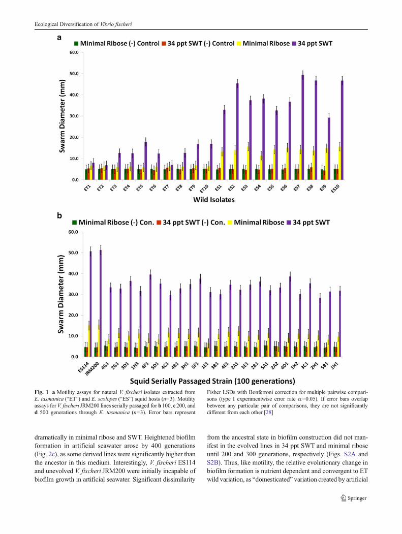
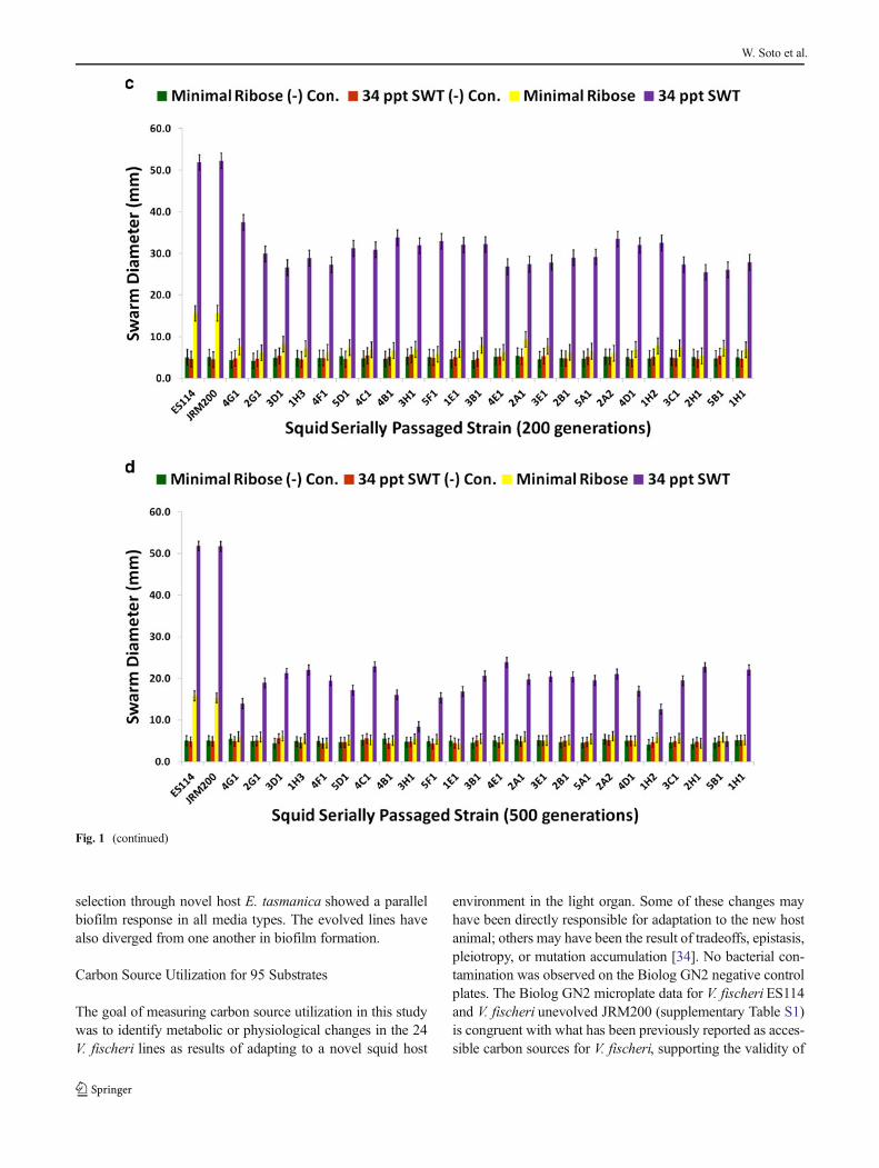
shown to be genetically distinct from one another [7, 8, 33].These field isolates were randomly sampled from theirEuprymna squid hosts and used as a comparison to theevolved clones for motility (Fig. 1a). ES wild strains weremore motile than ET on minimal ribose and 34 ppt SWT.V. fischeri ES114 is chloramphenicol sensitive, whileV. fischeri JRM200 is a V. fischeri ES114 derivative which ischloramphenicol resistant [24]. V. fischeri JRM200 was usedto create 24 lines that were serially transferred through novelsquid host E. tasmanica for 500 generations [7] and used inthis study. All 24 evolved lines improved in colonization ofE. tasmanica relative to ancestor, as no lineage remained thesame or became worse. V. fischeri lines serially passagedthrough E. tasmanica for 500 generations became less motilerelative to ancestor V. fischeriES114 and unevolved V. fischeriJRM200 (zero generations through E. tasmanica) on minimalribose and 34 ppt SWT (Fig. 1b–d for 100, 200, and 500generations; supplementary Figs. S1A and S1B for 300 and400 generations). Unlike many bacterial species, V. fischeri iscapable of motility on minimal media without nutritionalsupplements such as casamino acids [27]. No growth wasobserved on 34 ppt artificial seawater 0.5 % agar plates aswell as on the motility negative control 5.0 % agar plates.Significant decreases in motility were first observed for alllines by 100 generations (Fig. 1b) on motility SWT plates andcontinued to decline throughout the remainder of the selectionexperiment. However, by 500 generations, most derived linesstill remained motile on SWT plates relative to SWT motilitynegative controls (Fig. 1d). Although significant decreases inmotility on minimal ribose is evident by 100 generations(Fig. 1b), a marked drop was noted by 200 generations(Fig. 1c), as most of the derived lines are no longer signifi-cantly more motile than on minimal ribose motility negativecontrol plates. Motility on minimal ribose plates wascompletely lost by 300 generations (Fig. S1A). Thus, thederived lines converged on ET motility on minimal riboseand 34 ppt SWT, yet significant differences (polymorphisms)were seen among the various lineages in motility as a result ofadaptation to novel host E. tasmanica.
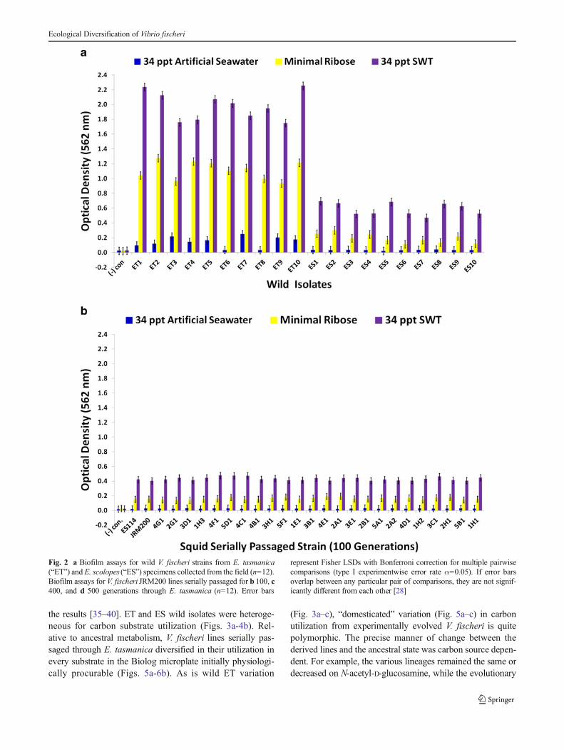
Biofilm Formation
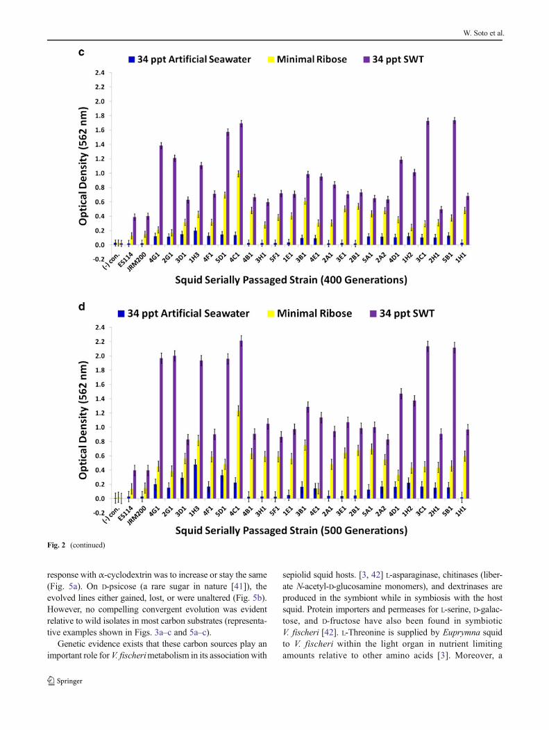
No bacterial contamination was observed in uninoculatednegative control wells for 34 ppt artificial seawater, minimalribose media, or 34 ppt SWT. Biofilm formation is higher forthe ET wild isolates than the ES ones in artificial seawater,minimal ribose, and 34 ppt SWT (Fig. 2a).V. fischeripassagedthrough E. tasmanica increased their biofilm formation capac-ity (Fig. 2b–d for 100, 400, and 500 generations; supplemen-tary Figs. S2A and S2B for 200 and 300 generations). Relativeto the loss of motility, the evolution of elevated biofilmformation was delayed, not appearing in great levels until400 generations (Fig. 2c), when it simultaneously appeared
W. Soto et al.
dramatically in minimal ribose and SWT. Heightened biofilmformation in artificial seawater arose by 400 generations(Fig. 2c), as some derived lines were significantly higher thanthe ancestor in this medium. Interestingly, V. fischeri ES114and unevolved V. fischeri JRM200 were initially incapable ofbiofilm growth in artificial seawater. Significant dissimilarity
from the ancestral state in biofilm construction did not man-ifest in the evolved lines in 34 ppt SWT and minimal riboseuntil 200 and 300 generations, respectively (Figs. S2A andS2B). Thus, like motility, the relative evolutionary change inbiofilm formation is nutrient dependent and convergent to ETwild variation, as “domesticated” variation created by artificial
Fig. 1 a Motility assays for natural V. fischeri isolates extracted fromE. tasmanica (“ET”) and E. scolopes (“ES”) squid hosts (n=3). Motilityassays for V. fischeri JRM200 lines serially passaged for b100, c200, andd 500 generations through E. tasmanica (n=3). Error bars represent
Fisher LSDs with Bonferroni correction for multiple pairwise compari-sons (type I experimentwise error rate α=0.05). If error bars overlapbetween any particular pair of comparisons, they are not significantlydifferent from each other [28]
Ecological Diversification of Vibrio fischeri
selection through novel host E. tasmanica showed a parallelbiofilm response in all media types. The evolved lines havealso diverged from one another in biofilm formation.
Carbon Source Utilization for 95 Substrates
The goal of measuring carbon source utilization in this studywas to identify metabolic or physiological changes in the 24V. fischeri lines as results of adapting to a novel squid host
environment in the light organ. Some of these changes mayhave been directly responsible for adaptation to the new hostanimal; others may have been the result of tradeoffs, epistasis,pleiotropy, or mutation accumulation [34]. No bacterial con-tamination was observed on the Biolog GN2 negative controlplates. The Biolog GN2 microplate data for V. fischeri ES114and V. fischeri unevolved JRM200 (supplementary Table S1)is congruent with what has been previously reported as acces-sible carbon sources for V. fischeri, supporting the validity of
Fig. 1 (continued)
W. Soto et al.
the results [35–40]. ET and ES wild isolates were heteroge-neous for carbon substrate utilization (Figs. 3a-4b). Rel-ative to ancestral metabolism, V. fischeri lines serially pas-saged through E. tasmanica diversified in their utilization inevery substrate in the Biolog microplate initially physiologi-cally procurable (Figs. 5a-6b). As is wild ET variation
(Fig. 3a–c), “domesticated” variation (Fig. 5a–c) in carbonutilization from experimentally evolved V. fischeri is quitepolymorphic. The precise manner of change between thederived lines and the ancestral state was carbon source depen-dent. For example, the various lineages remained the same ordecreased on N-acetyl-D-glucosamine, while the evolutionary
Fig. 2 a Biofilm assays for wild V. fischeri strains from E. tasmanica(“ET”) and E. scolopes (“ES”) specimens collected from the field (n=12).Biofilm assays for V. fischeri JRM200 lines serially passaged for b 100, c400, and d 500 generations through E. tasmanica (n=12). Error bars
represent Fisher LSDs with Bonferroni correction for multiple pairwisecomparisons (type I experimentwise error rate α=0.05). If error barsoverlap between any particular pair of comparisons, they are not signif-icantly different from each other [28]
Ecological Diversification of Vibrio fischeri
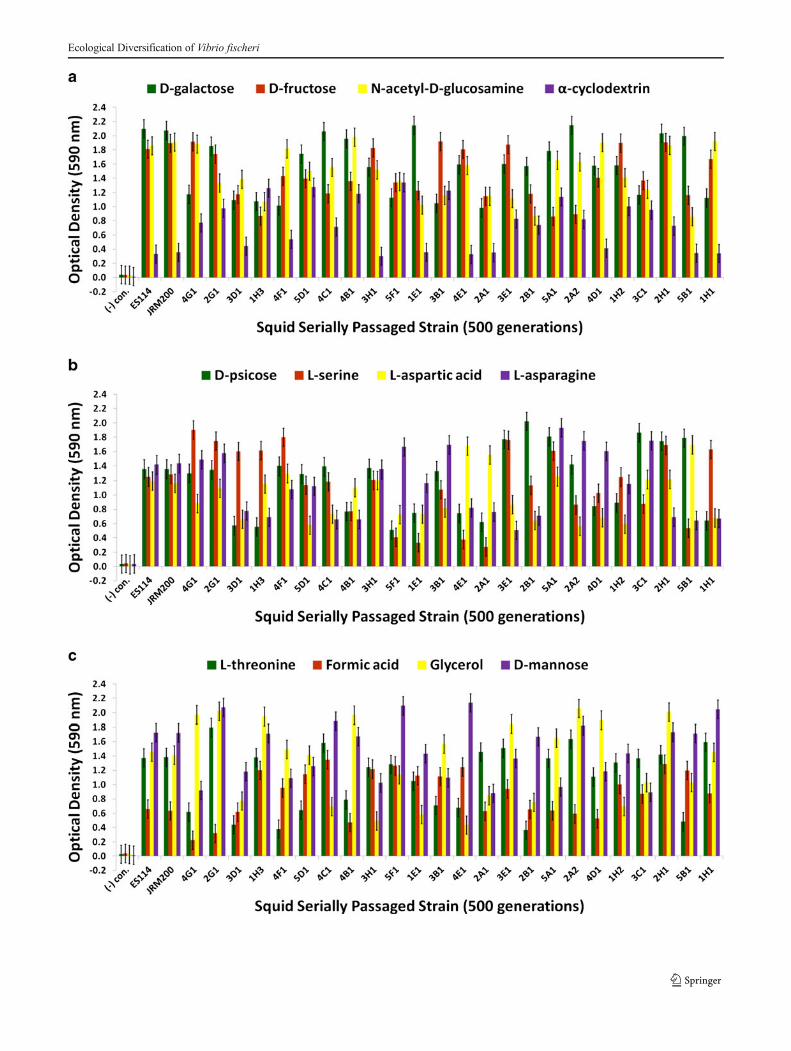
response with α-cyclodextrin was to increase or stay the same(Fig. 5a). On D-psicose (a rare sugar in nature [41]), theevolved lines either gained, lost, or were unaltered (Fig. 5b).However, no compelling convergent evolution was evidentrelative to wild isolates in most carbon substrates (representa-tive examples shown in Figs. 3a–c and 5a–c).
Genetic evidence exists that these carbon sources play animportant role for V. fischerimetabolism in its association with
sepiolid squid hosts. [3, 42] L-asparaginase, chitinases (liber-ate N-acetyl-D-glucosamine monomers), and dextrinases areproduced in the symbiont while in symbiosis with the hostsquid. Protein importers and permeases for L-serine, D-galac-tose, and D-fructose have also been found in symbioticV. fischeri [42]. L-Threonine is supplied by Euprymna squidto V. fischeri within the light organ in nutrient limitingamounts relative to other amino acids [3]. Moreover, a
Fig. 2 (continued)
W. Soto et al.
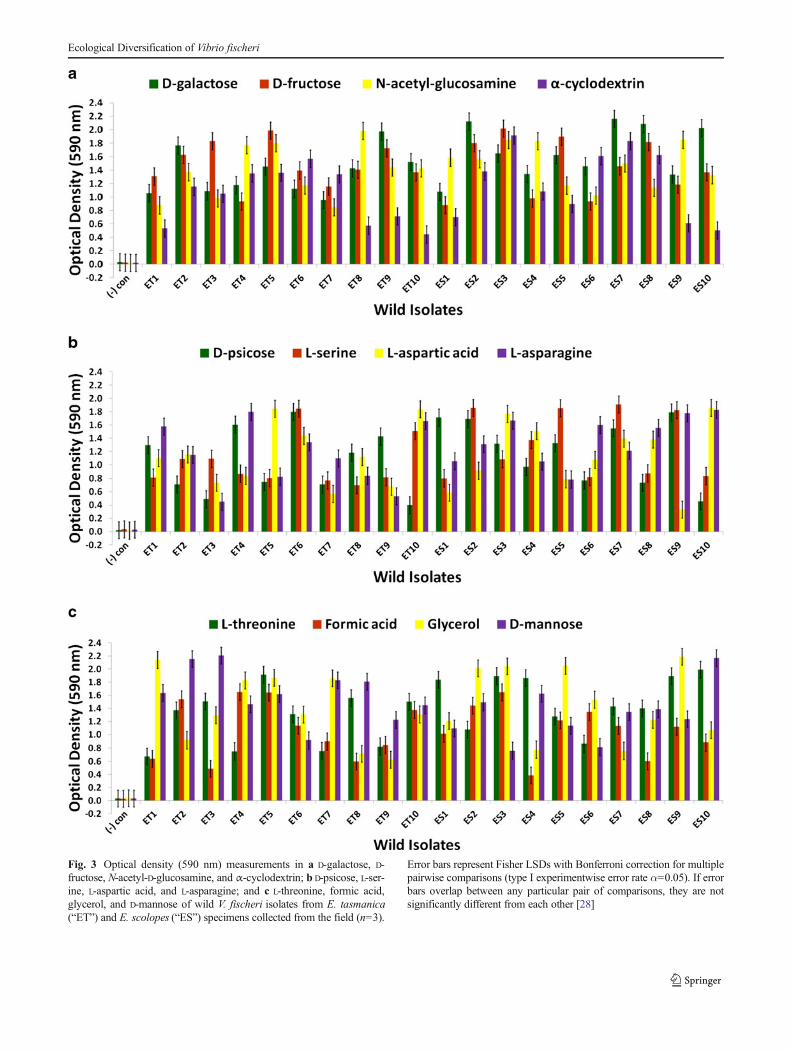
Fig. 3 Optical density (590 nm) measurements in a D-galactose, D-fructose, N-acetyl-D-glucosamine, and α-cyclodextrin; b D-psicose, L-ser-ine, L-aspartic acid, and L-asparagine; and c L-threonine, formic acid,glycerol, and D-mannose of wild V. fischeri isolates from E. tasmanica(“ET”) and E. scolopes (“ES”) specimens collected from the field (n=3).
Error bars represent Fisher LSDs with Bonferroni correction for multiplepairwise comparisons (type I experimentwise error rate α=0.05). If errorbars overlap between any particular pair of comparisons, they are notsignificantly different from each other [28]
Ecological Diversification of Vibrio fischeri
multitude of genes are dedicated to the V. fischeri catabolismof glycerol, formic acid, chitin, and fumaric acid while in thelight organ. Fumaric acid can be made by the deamination ofL-aspartic acid, a reaction carried out by aspartate ammonia-lyase, an enzyme expressed by symbiotic V. fischeri [42]. D-Mannose residues coat the epithelial linings of light organswithin sepiolid squid hatchlings and play a fundamental rolein V. fischeri attachment to squid host eukaryotic cells throughmannose-recognizing adhesins on the bacterial surface [22,43], which potentially serve as a source for “mannose” graz-ing. Prolific variation in catabolism of D-mannose among thederived lines relative to precursor state was evident (Fig. 5c).
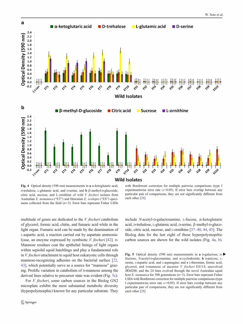
For V. fischeri, some carbon sources in the Biolog GN2microplate exhibit the most substantial metabolic diversity(hyperpolymorphic) known for any particular substrate. They
include N-acetyl-D-galactosamine, L-fucose, α-ketoglutaricacid, D-trehalose, L-glutamic acid, D-serine,β-methyl-D-gluco-side, citric acid, sucrose, and L-ornithine [37–40, 44, 45]. TheBiolog data for the last eight of these hyperpolymoprhiccarbon sources are shown for the wild isolates (Fig. 4a, b).
Fig. 4 Optical density (590 nm) measurements in aα-ketoglutaric acid,D-trehalose, L-glutamic acid, and D-serine, and b β-methyl-D-glucoside,citric acid, sucrose, and L-ornithine of wild V. fischeri isolates fromAustralian E. tasmanica (“ET”) and Hawaiian E. scolopes (“ES”) speci-mens collected from the field (n=3). Error bars represent Fisher LSDs
with Bonferroni correction for multiple pairwise comparisons (type Iexperimentwise error rate α=0.05). If error bars overlap between anyparticular pair of comparisons, they are not significantly different fromeach other [28]
�Fig. 5 Optical density (590 nm) measurements in a D-galactose, D-fructose, N-acetyl-D-glucosamine, and α-cyclodextrin; b D-psicose, L-serine, L-aspartic acid, and L-asparagine; and c L-threonine, formic acid,glycerol, and D-mannose of ancestor V. fischeri ES114, unevolvedJRM200, and the 24 lines evolved through the novel Australian squidhost E. tasmanica for 500 generations (n=3). Error bars represent FisherLSDs with Bonferroni correction for multiple pairwise comparisons (typeI experimentwise error rate α=0.05). If error bars overlap between anyparticular pair of comparisons, they are not significantly different fromeach other [28]
W. Soto et al.
Ecological Diversification of Vibrio fischeri

All ET and no ES strains utilize α-ketoglutaric acid, D-treha-lose, L-glutamic acid, and D-serine, while no ET and all ESisolates metabolize citric acid and sucrose. The ability toutilize seven new carbon sources was gained in someV. fischeri lines serially passaged through E. tasmanica for500 generations—N-acetyl-D-galactosamine, L-fucose, α-ketoglutaric acid, D-trehalose, L-glutamic acid, D-serine, andβ-methyl-D-glucoside, relative to forerunners V. fischeriES114 and unevolved V. fischeri JRM200 (last five shown inFig. 6a, b). Conversely, substrates such as citric acid, sucrose,and L-ornithine were lost in some lines (Fig. 6b). N-acetyl-D-galactosamine has been found to participate in cell–cell at-tachment through lectins, carbohydrate-binding proteins on
extracellular surfaces, including bioluminescent bacteria[46]. Lectins may be involved in biofilm formation and bac-terial microcolony aggregations. Previous research has hy-pothesized that lectins with N-acetyl-D-galactosamine speci-ficity govern symbiosis initiation in bioluminescent bacteriawith marine animals, V. fischeri included [47]. Furthermore, D-serine, citric acid, α-ketoglutaric acid, β-methyl-D-glucoside,and N-acetyl-D-galactosamine have all been listed as carbonsubstrates dispensable for Vibrio cholerae, which are evolu-tionarily discarded and reacquired as necessary for certainniche environments [48]. Metabolic pathways can evolvemodularly in function, some being especially malleable, toaccommodate the niche breadth ecologically necessary for
Fig. 6 Optical density (590 nm) measurements in aα-ketoglutaric acid,D-trehalose, L-glutamic acid, and D-serine, and b β-methyl-D-glucoside,citric acid, sucrose, and L-ornithine of ancestor V. fischeri ES114, un-evolved JRM200, and the 24 lines evolved through the novel Australiansquid host E. tasmanica for 500 generations (n=3). Error bars represent
Fisher LSDs with Bonferroni correction for multiple pairwise compari-sons (type I experimentwise error rate α=0.05). If error bars overlapbetween any particular pair of comparisons, they are not significantlydifferent from each other [28]
W. Soto et al.
Vibrio populations under specific environmental parameters(e.g., free-living versus host-associated lifestyles). Curiously,no wild isolates from E. scolopesand E. tasmanicagrew onN-acetyl-D-galactosamine, L-fucose, and L-ornithine. L-Fucoseresidues may also serve as attachment sites for bacteria toother cells, such as fucose-sensitive hemagglutinin A (fshA)in V. cholerae [49]. In summary, ET convergence was ob-served with α-ketoglutaric acid, D-trehalose, L-glutamic acid,D-serine, β-methyl-D-glucoside, citric acid, and sucrose(Figs. 4a, b and 6a, b). As with motility and biofilm, evolu-tionary distinction is emerging among the lines in carbonsource metabolism.
Bioluminescence
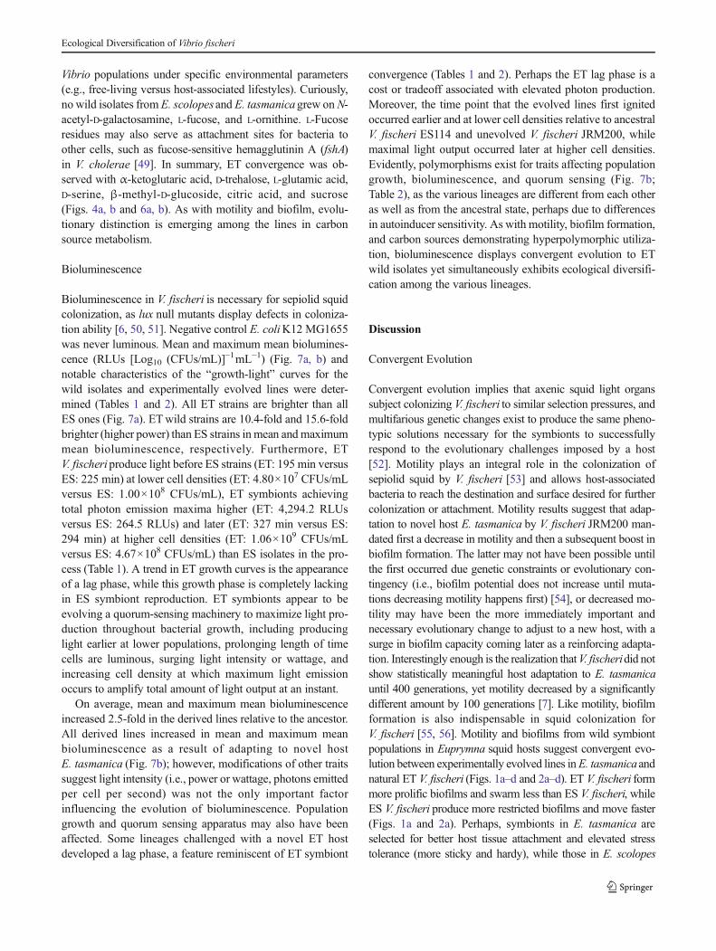
Bioluminescence in V. fischeri is necessary for sepiolid squidcolonization, as lux null mutants display defects in coloniza-tion ability [6, 50, 51]. Negative control E. coliK12 MG1655was never luminous. Mean and maximum mean biolumines-cence (RLUs [Log10 (CFUs/mL)]−1mL−1) (Fig. 7a, b) andnotable characteristics of the “growth-light” curves for thewild isolates and experimentally evolved lines were deter-mined (Tables 1 and 2). All ET strains are brighter than allES ones (Fig. 7a). ETwild strains are 10.4-fold and 15.6-foldbrighter (higher power) than ES strains in mean andmaximummean bioluminescence, respectively. Furthermore, ETV. fischeriproduce light before ES strains (ET: 195 min versusES: 225 min) at lower cell densities (ET: 4.80×10⁷ CFUs/mLversus ES: 1.00×10⁸ CFUs/mL), ET symbionts achievingtotal photon emission maxima higher (ET: 4,294.2 RLUsversus ES: 264.5 RLUs) and later (ET: 327 min versus ES:294 min) at higher cell densities (ET: 1.06×10⁹ CFUs/mLversus ES: 4.67×10⁸ CFUs/mL) than ES isolates in the pro-cess (Table 1). A trend in ET growth curves is the appearanceof a lag phase, while this growth phase is completely lackingin ES symbiont reproduction. ET symbionts appear to beevolving a quorum-sensing machinery to maximize light pro-duction throughout bacterial growth, including producinglight earlier at lower populations, prolonging length of timecells are luminous, surging light intensity or wattage, andincreasing cell density at which maximum light emissionoccurs to amplify total amount of light output at an instant.
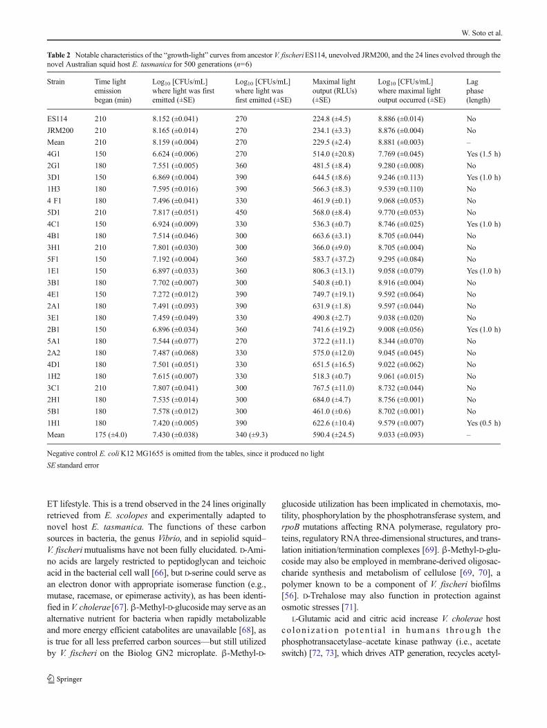
On average, mean and maximum mean bioluminescenceincreased 2.5-fold in the derived lines relative to the ancestor.All derived lines increased in mean and maximum meanbioluminescence as a result of adapting to novel hostE. tasmanica (Fig. 7b); however, modifications of other traitssuggest light intensity (i.e., power or wattage, photons emittedper cell per second) was not the only important factorinfluencing the evolution of bioluminescence. Populationgrowth and quorum sensing apparatus may also have beenaffected. Some lineages challenged with a novel ET hostdeveloped a lag phase, a feature reminiscent of ET symbiont
convergence (Tables 1 and 2). Perhaps the ET lag phase is acost or tradeoff associated with elevated photon production.Moreover, the time point that the evolved lines first ignitedoccurred earlier and at lower cell densities relative to ancestralV. fischeri ES114 and unevolved V. fischeri JRM200, whilemaximal light output occurred later at higher cell densities.Evidently, polymorphisms exist for traits affecting populationgrowth, bioluminescence, and quorum sensing (Fig. 7b;Table 2), as the various lineages are different from each otheras well as from the ancestral state, perhaps due to differencesin autoinducer sensitivity. As with motility, biofilm formation,and carbon sources demonstrating hyperpolymorphic utiliza-tion, bioluminescence displays convergent evolution to ETwild isolates yet simultaneously exhibits ecological diversifi-cation among the various lineages.
Discussion
Convergent Evolution
Convergent evolution implies that axenic squid light organssubject colonizing V. fischeri to similar selection pressures, andmultifarious genetic changes exist to produce the same pheno-typic solutions necessary for the symbionts to successfullyrespond to the evolutionary challenges imposed by a host[52]. Motility plays an integral role in the colonization ofsepiolid squid by V. fischeri [53] and allows host-associatedbacteria to reach the destination and surface desired for furthercolonization or attachment. Motility results suggest that adap-tation to novel host E. tasmanica by V. fischeri JRM200 man-dated first a decrease in motility and then a subsequent boost inbiofilm formation. The latter may not have been possible untilthe first occurred due genetic constraints or evolutionary con-tingency (i.e., biofilm potential does not increase until muta-tions decreasing motility happens first) [54], or decreased mo-tility may have been the more immediately important andnecessary evolutionary change to adjust to a new host, with asurge in biofilm capacity coming later as a reinforcing adapta-tion. Interestingly enough is the realization thatV. fischeridid notshow statistically meaningful host adaptation to E. tasmanicauntil 400 generations, yet motility decreased by a significantlydifferent amount by 100 generations [7]. Like motility, biofilmformation is also indispensable in squid colonization forV. fischeri [55, 56]. Motility and biofilms from wild symbiontpopulations in Euprymna squid hosts suggest convergent evo-lution between experimentally evolved lines inE. tasmanicaandnatural ET V. fischeri (Figs. 1a–d and 2a–d). ET V. fischeri formmore prolific biofilms and swarm less than ES V. fischeri, whileES V. fischeri produce more restricted biofilms and move faster(Figs. 1a and 2a). Perhaps, symbionts in E. tasmanica areselected for better host tissue attachment and elevated stresstolerance (more sticky and hardy), while those in E. scolopes
Ecological Diversification of Vibrio fischeri
are evolutionarily honed for chemotaxis (more nimble locomo-tion and elevated nutrient sensitivity). Biofilm formation ispositively correlated in many host-associated vibrios with colo-nization potential, immunity avoidance, and eukaryotic cellattachment in hosts [57–59]. Biofilms are known to increasebacterial survival against environmental stress [58–61].
The artificial variation generated from the experimentallyevolved lines is congruent with ET natural variation, asV. fischeri “domesticated” in E. tasmanica appear more phe-notypically similar to field ET V. fischeri in motility andbiofilm production and less like their original ES V. fischeri.Figure 2c suggests a threshold event or critical period was
Fig. 7 Mean and maximum mean bioluminescence for awild V. fischeristrains from E. tasmanica (“ET”) and E. scolopes (“ES”) specimenscollected from the field and b ancestor V. fischeri ES114, unevolvedJRM200, and the 24 lines evolved through the novel Australian squidhost E. tasmanica for 500 generations (n=6). Error bars represent Fisher
LSDs with Bonferroni correction for multiple pairwise comparisons (typeI experimentwise error rate α=0.05). If error bars overlap between anyparticular pair of comparisons, they are not significantly different fromeach other [28]. E. coliK12 MG1655 was used as the negative control
W. Soto et al.

reached by 400 generations, since a sharp and sudden expansein biofilm growth precipitously arises across many of thelineages in different media in a manner suggestive of secondmessenger signaling, signal transduction cascades, andquorum-sensing autoinducers [62]. The decrease in motilityis more progressive and continued throughout the 500 gener-ation time period. Not only was convergent evolution ob-served in biofilm development and motility between the de-rived lines and wild isolates but an inverse relationship wasalso noted. The inverse relationship between biofilm develop-ment and motility is known to have a biochemical basis withinthe genus Vibrio, specifically the second messenger cyclicdiguanylate (c-di-GMP) [56, 59] and is a topic for futureresearch. Generally, high intracellular concentrations of sec-ond messenger c-di-GMP correlate with increased biofilmformation, while lower quantities are associated with elevatedmotility (e.g., V. cholerae) [63]. Inverse relationships betweenmotility and sessility in microbial lifestyles along a “c-di-GMP” continuum within hosts is an intriguing topic to par-tially explain competitive dominance and resulting tradeoffsobserved in Vibrio symbionts colonizing Euprymna [9, 10].
Possibly, c-di-GMP intracellular pool levels as a “host” pointof selection may be dissipated by sympatry of several Sepiolahost species [1], not permitting for “c-di-GMP” ecologicalspecialization for swarmer or biofilm ecotypes due to morehomogenization of host environments in a geographical area.A scattering of few mutations affecting biofilm gene expres-sion and c-di-GMP regulation drives convergent evolutionand adaptive radiation in Pseudomonas fluorescens in struc-tured microcosms, while simultaneously provoking astoundingdiversity [64, 65].
As with motility and biofilm formation, some convergentevolution appears to be occurring with carbon metabolism forsubstrates characteristic of evolutionary fluid allocation andredeployment in the V. fischeri physiological repertoire, whenweighted against wild symbiont light organ populations fromfield-caught E. tasmanica and E. scolopes specimens(Figs. 4a, b, and 6a, b) [37–40, 44, 45]. Analyzing wildV. fischeri isolates from Euprymna squid hosts provides com-pelling evidence that citric acid and sucrose metabolic capa-bilities are abandoned in favor of L-glutamic acid,β-methyl-D-glucoside, D-trehalose,α-ketoglutaric acid, and D-serine for an
Table 1 Notable characteristics of the “growth-light” curves from wild V. fischeri strains from E. tasmanica (“ET”) and E. scolopes (“ES”) specimenscollected from the field (n=6)
Strain Time lightemissionbegan (min)
Log10 [CFUs/mL]where light was firstemitted (±SE)
Log10 [CFUs/mL]where light was firstemitted (±SE)
Maximal lightoutput (RLUs)(±SE)
Log10 [CFUs/mL]where maximal lightoutput occurred (±SE)
Lagphase(length)
ET1 210 7.797 (±0.033) 300 3,597.9 (±29.3) 8.754 (±0.046) No
ET2 240 8.111 (±0.116) 360 2,464.5 (±20.9) 9.347 (±0.127) No
ET3 180 7.510 (±0.057) 270 3,835.1 (±23.5) 8.464 (±0.140) No
ET4 150 7.231 (±0.056) 300 3,072.5 (±33.4) 8.696 (±0.053) No
ET5 180 7.492 (±0.134) 300 5,609.8 (±38.1) 8.737 (±0.077) Yes (1.0 h)
ET6 210 7.786 (±0.082) 330 4,570.8 (±25.7) 9.088 (±0.060) No
ET7 210 7.835 (±0.051) 360 4,809.9 (±19.9) 9.398 (±0.146) No
ET8 210 7.815 (±0.086) 330 5,606.6 (±39.5) 9.096 (±0.048) Yes (1.0 h)
ET9 180 7.418 (±0.087) 360 4,963.8 (±29.7) 9.338 (±0.122) Yes (1.0 h)
ET10 210 7.818 (±0.054) 360 4,411.4 (±25.4) 9.317 (±0.092) No
Mean 195 (±5.8) 7.681 (±0.056) 327 (±5.4) 4,294.2 (±162.6) 9.024 (±0.072) –
ES1 210 7.793 (±0.109) 270 191.5 (±0.4) 8.375 (±0.048) No
ES2 240 8.152 (±0.094) 300 316.1 (±5.6) 8.716 (±0.077) No
ES3 240 8.168 (±0.078) 360 226.8 (±3.6) 9.275 (±0.058) No
ES4 270 8.407 (±0.093) 330 229.9 (±4.1) 9.045 (±0.093) No
ES5 240 8.207 (±0.084) 300 250.4 (±2.4) 8.752 (±0.035) No
ES6 180 7.542 (±0.121) 240 306.9 (±2.5) 8.170 (±0.068) No
ES7 240 8.111 (±0.052) 300 305.9 (±1.1) 8.696 (±0.033) No
ES8 240 8.216 (±0.059) 330 345.6 (±0.9) 9.033 (±0.071) No
ES9 210 7.780 (±0.055) 270 242.8 (±1.5) 8.369 (±0.047) No
ES10 180 7.628 (±0.117) 240 228.6 (±2.6) 8.254 (±0.073) No
Mean 225 (±4.8) 8.000 (±0.032) 294 (±6.5) 264.5 (±8.5) 8.669 (±0.099) –
Negative control E. coliK12 MG1655 is omitted from the tables, since it produced no light
SE standard error
Ecological Diversification of Vibrio fischeri
ET lifestyle. This is a trend observed in the 24 lines originallyretrieved from E. scolopes and experimentally adapted tonovel host E. tasmanica. The functions of these carbonsources in bacteria, the genus Vibrio, and in sepiolid squid–V. fischerimutualisms have not been fully elucidated. D-Ami-no acids are largely restricted to peptidoglycan and teichoicacid in the bacterial cell wall [66], but D-serine could serve asan electron donor with appropriate isomerase function (e.g.,mutase, racemase, or epimerase activity), as has been identi-fied inV. cholerae [67].β-Methyl-D-glucosidemay serve as analternative nutrient for bacteria when rapidly metabolizableand more energy efficient catabolites are unavailable [68], asis true for all less preferred carbon sources—but still utilizedby V. fischeri on the Biolog GN2 microplate. β-Methyl-D-
glucoside utilization has been implicated in chemotaxis, mo-tility, phosphorylation by the phosphotransferase system, andrpoB mutations affecting RNA polymerase, regulatory pro-teins, regulatory RNA three-dimensional structures, and trans-lation initiation/termination complexes [69]. β-Methyl-D-glu-coside may also be employed in membrane-derived oligosac-charide synthesis and metabolism of cellulose [69, 70], apolymer known to be a component of V. fischeri biofilms[56]. D-Trehalose may also function in protection againstosmotic stresses [71].
L-Glutamic acid and citric acid increase V. cholerae hostco lon iza t ion po ten t i a l i n humans th rough thephosphotransacetylase–acetate kinase pathway (i.e., acetateswitch) [72, 73], which drives ATP generation, recycles acetyl-
Table 2 Notable characteristics of the “growth-light” curves from ancestor V. fischeriES114, unevolved JRM200, and the 24 lines evolved through thenovel Australian squid host E. tasmanica for 500 generations (n=6)
Strain Time lightemissionbegan (min)
Log10 [CFUs/mL]where light was firstemitted (±SE)
Log10 [CFUs/mL]where light wasfirst emitted (±SE)
Maximal lightoutput (RLUs)(±SE)
Log10 [CFUs/mL]where maximal lightoutput occurred (±SE)
Lagphase(length)
ES114 210 8.152 (±0.041) 270 224.8 (±4.5) 8.886 (±0.014) No
JRM200 210 8.165 (±0.014) 270 234.1 (±3.3) 8.876 (±0.004) No
Mean 210 8.159 (±0.004) 270 229.5 (±2.4) 8.881 (±0.003) –
4G1 150 6.624 (±0.006) 270 514.0 (±20.8) 7.769 (±0.045) Yes (1.5 h)
2G1 180 7.551 (±0.005) 360 481.5 (±8.4) 9.280 (±0.008) No
3D1 150 6.869 (±0.004) 390 644.5 (±8.6) 9.246 (±0.113) Yes (1.0 h)
1H3 180 7.595 (±0.016) 390 566.3 (±8.3) 9.539 (±0.110) No
4 F1 180 7.496 (±0.041) 330 461.9 (±0.1) 9.068 (±0.053) No
5D1 210 7.817 (±0.051) 450 568.0 (±8.4) 9.770 (±0.053) No
4C1 150 6.924 (±0.009) 330 536.3 (±0.7) 8.746 (±0.025) Yes (1.0 h)
4B1 180 7.514 (±0.046) 300 663.6 (±3.1) 8.705 (±0.044) No
3H1 210 7.801 (±0.030) 300 366.0 (±9.0) 8.705 (±0.004) No
5F1 150 7.192 (±0.004) 360 583.7 (±37.2) 9.295 (±0.084) No
1E1 150 6.897 (±0.033) 360 806.3 (±13.1) 9.058 (±0.079) Yes (1.0 h)
3B1 180 7.702 (±0.007) 300 540.8 (±0.1) 8.916 (±0.004) No
4E1 150 7.272 (±0.012) 390 749.7 (±19.1) 9.592 (±0.064) No
2A1 180 7.491 (±0.093) 390 631.9 (±1.8) 9.597 (±0.044) No
3E1 180 7.459 (±0.049) 330 490.8 (±2.7) 9.038 (±0.020) No
2B1 150 6.896 (±0.034) 360 741.6 (±19.2) 9.008 (±0.056) Yes (1.0 h)
5A1 180 7.544 (±0.077) 270 372.2 (±11.1) 8.344 (±0.070) No
2A2 180 7.487 (±0.068) 330 575.0 (±12.0) 9.045 (±0.045) No
4D1 180 7.501 (±0.051) 330 651.5 (±16.5) 9.022 (±0.062) No
1H2 180 7.615 (±0.007) 330 518.3 (±0.7) 9.061 (±0.015) No
3C1 210 7.807 (±0.041) 300 767.5 (±11.0) 8.732 (±0.044) No
2H1 180 7.535 (±0.014) 300 684.0 (±4.7) 8.756 (±0.001) No
5B1 180 7.578 (±0.012) 300 461.0 (±0.6) 8.702 (±0.001) No
1H1 180 7.420 (±0.005) 390 622.6 (±10.4) 9.579 (±0.007) Yes (0.5 h)
Mean 175 (±4.0) 7.430 (±0.038) 340 (±9.3) 590.4 (±24.5) 9.033 (±0.093) –
Negative control E. coliK12 MG1655 is omitted from the tables, since it produced no light
SE standard error
W. Soto et al.
CoA when respiration and central metabolism are backlogged,and post-translational regulation of proteins, illustrating the effectcarbon sources can have especially on animal host colonizationwhen the substrates themselves are main metabolites easilyshuttled or shunted into alternate biochemical pathways (Theacetate switch is present in V. fischeri [74], but acetic acid didexhibit ET convergence.) [72]. L-Glutamic acid has also beenreported to aid bacterial growth during iron limitation [75], whichis noteworthy considering the squid light organ is low in avail-able iron [76]. Furthermore, α-ketoglutaric acid is known tofunction as a substrate for the production of bacterialsiderophores to assist iron acquisition in Vibrio [77]. α-Ketoglutaric acid is also believed to possess value against oxida-tive stress [78], and the squid light organ is known to initiate arespiratory burst via innate host immunity that produces toxicoxygen species [79]. Perhaps, the convergent evolution depictedin biofilm formation and these carbon sources are related, eitherto exploit new resources or increase resistance against unaccus-tomed stressors present in a novel host. Enhancing colonizationand persistence in an unfamiliar host are other possibilities. Howalternate carbon sources may be used by V. fischeri to manageagainst various host stresses (e.g., tenacious immune defenses) isa stimulating prospect for future study. For instance, amino acidsmay bemore useful than monosaccharides for some cells againstosmotic stress, while the former might be more beneficial toregulate high pH stress if nitrogen is not limiting (otherwiseamino sugars such N-acetyl-D-glucosamine could be used ifavailable) [80]. Although not relevant to the squid light organ,carbon source is known to affect bacterial susceptibility to heavymetal toxicity, including V. fischeri [81]. Convergent evolutioncoupling metabolism and stress together has been described inmutualisms between invertebrate hosts and microbial symbionts[82, 83].
For bioluminescence, the derived lines displayed conver-gence to ETwild isolates (Fig. 7a, b; Tables 1 and 2), implyingthat light emission may be associated with biofilm formation,motility, and the convergent carbon sources. luxU links biolu-minescence to biofilm formation in V. fischeri [84], and rpoNcontrols motility, biofilm, luminescence, and squid coloniza-tion ability [85]. Bioluminescence may be tied to biofilmformation, motility, and central metabolism through integratedcircuitries involving quorum sensing and c-di-GMP secondmessenger signaling [86], which is consistent with the simul-taneous increase in luminosity, biofilm formation, and lowermotility. Relative to the ancestor, light emission occurredearlier and at a lower cell density for the derived lines, whilemaximal light output ensued later and at a higher cell density(Table 2). Early quorum sensing at low cell densities and latequorum sensing at high cell densities implicate the AinS andLux signal transduction relays in V. fischeri, respectively [87].AinS quorum sensing has also been associated with the ace-tate switch [74]. Thus, evidence exists adaptation toE. tasmanica and conceivably to novel hosts in general
involves selection at multiple layers of cell signaling (e.g.,GGDEF, EAL, and PilZ proteins), which could account forappearance of a lag phase [14, 88]. Mutations affecting AinSquorum sensing is known to affect growth curves [89]. Previ-ous efforts failed to find bioluminescence differences amongthe evolved lines within E. tasmanica, and several replicateanimals were not examined within a single evolved line (e.g.,using n=3 squid hosts with 3B1 instead of one animal eachwith 3B1, 5B1, and 4F1) [7]. At the time of study, there wereinsufficient E. tasmanica hatchlings available for these exper-iments. However, derived lines that are significantly brighterat 500 generations relative to ancestral V. fischeri ES114 andunevolved V. fischeri JRM200 have recently been observed inE. tasmanica (unpublished data). Prior investigation showedthe derived lines were more dim in the ancestral hostE. scolopes, giving off less photons per log10 (CFUs/mL)mL−1 [7]. E. scolopes hatchlings and adults are smaller (withconcomitantly smaller light organs as well) than E. tasmanicathroughout their entire life cycles. Therefore, E. scolopes in-dividual hosts carry one to two orders of magnitude lesssymbionts than E. tasmanica, regardless of the ontogeneticstage. Hence, tradeoffs may now exist in the ancestral hostE. scolopeswhere V. fischericell densities and quorum sensingare mistimed for motility, biofilm formation, and biolumines-cence due to adaptation of the derived lines to AustralianE. tasmanica, since symbionts can no longer reach the properpopulation levels in the Hawaiian squid at the appropriatetimes of host development, as the symbiont and host lifehistories are no longer accurately synchronized.
Diversification
Diversifying evolution or radiation predominates over conver-gent evolution in morphospace when profuse phenotypesprevail to be evolutionarily fit or successful to a particularselection pressure. A multitude of mutations to attain each ofthese prosperous possibilities in a complex, heterogeneousenvironment with vacant niches provide ecological opportu-nity, an attribute characteristic of island evolution, includingGalapagos finches, Hawaiian silversword alliance, Hawaiianhoneycreepers, Anolis lizards, and cichlids in their lake habitatislands [90–92]. Aside from the carbon sources displayingconvergent evolution, the remaining carbon sources metabo-lizable by V. fischeriwild isolates depicted a general polymor-phism, a tendency also reflected in the derived lines (repre-sentative examples in Figs. 3a–c and 5a–c). These polymor-phic carbon sources are being used distinctly and variedly,both individually and as an assortment. These results arecongruent with other microbial experimental evolution studies[52, 93], including with E. coli experiments with less than 800generations of evolution in homogenous and unstructuredenvironments [94]. This increased “domesticated” variationmay be reflective of diversifying selection in the squid light
Ecological Diversification of Vibrio fischeri

organ with incipient resource partitioning and changes innutrient specificities toward carbon resources, perhaps evenas part of cross-feeding or syntrophy between other V. fischeriecotypes within the light organ of the same squid host indi-vidual [91, 94]. The implication of these results in nature areprofound. Euprymna squid can live up to a year, whichamounts to 1,500–2,000 V. fischerigenerations [7]. Squid lightorgans are microcosms full of convoluted chasms with tre-mendous physical and biochemical complexity and heteroge-neity [95], and a single V. fischeri clone has immense oppor-tunity to evolve cross-feeding with either other V. fischerisubtypes or host cells within a host lifetime. The squid lightorgan microenvironment varies in spatial and temporal carbonsource composition available toV. fischericells (e.g.,N-acetyl-D-glucosamine at night and glycerol in the morning) [42].Moreover, within the light organ of a single individual squidhost, differentV. fischerisubtypeswill specialize inwhere theywill settle and reside, with little mixing of individualV. fischerivariants within the light organ symbiont population despitethe disturbance imposed by daily venting [42, 96]. In turn, thisdissimilarity in the spatial localization and structured regionaloccupancy of the light organ by V. fischeri cells leads todifferential gene expression in the symbionts, which can bea primer for the evolution of ecological differentiation.
An imperative realization is biofilm formation, motility,convergent carbon sources, and bioluminescence are alsoexemplifying ecological diversification. Despite the manifes-tation in convergence to wild ET isolates in these traits, theevolved lines are still becoming divergent from one another.Other laboratories have also proved convergent evolutionfacilitates ecological diversification in separate lineages, in-cluding on other carbon sources [52, 93]. Quorum sensing andbiofilms beget diversity, including the modification of popu-lation growth (e.g., lag phase changes) [97, 98]. Research withV. cholerae verifies alternate carbon sources can be incorpo-rated or substituted into the composition of a biofilm to accessnew ecological niches [99, 100]. Intriguingly, biolumines-cence (i.e., quorum sensing) and trans-genetic regulation ofthe lux operon could function as a generator for V. fischeriadaptive radiation inside this symbiont’s animal hosts, ascAMP receptor protein (a transcriptional regulator) may havea continuum of graded possible effects on lux operon geneexpression due to the presence of different carbon sources invarious light organ microenvironments within sepiolid squidsand monocentrid fishes [62]. A captivating possibility is theutilization of the sepiolid squid-Vibrio symbiosis to investigateevolvability and versatility in V. fischeri. V. fischeri possessesthree biofilm gene clusters, syp, vpsII like, and the celluloseoperon, yielding over 30 genes that can be independentlymodified to stimulate biofilm niche differentiation to exploitsquid light organ microenvironments in novel ways [101].This evolutionary process is quite analogous to the customi-zation of the myriad bony elements in the head region of
cichlids [102]. Additionally, modularity (as seen in convergentcarbon sources, Figs. 4a, b, and 6a, b) increases the evolution-ary diversification potential of a lineage, including for adap-tive radiation [103, 104].
Squids as Host Habitat Islands
The use of biogeography theory to characterize microbialdiversity has been previously applied, including mutualisms,pathogen–host interactions, and microcosms [105–110]. Axe-nic sepiolid squid hatchlings, first emanating from their eggs,essentially serve as sterile and mobile volcanic islands (i.e.,host habitat islands) that subject their bioluminescent symbi-onts to severe genetic bottlenecks during colonization [7, 106,108, 111, 112]. Only 6 to 12 V. fischeri cells of a 100–103
CFUs/mL seawater inoculum initiate symbiosis in an animalthat ultimately reach an adult light organ carrying capacity of108–1011 cells per host [113], providing pioneer V. fischericolonizers with ecological release from the semi-starvationfrequently encountered during the oceanic free-living phase.Such episodes of genetic bottlenecks with ensuing abundantproliferation and population recovery are called “founderflushes” [114], which provide opportunities for populationmovements across valleys via genetic drift to alternative adap-tive peaks [90] and facilitate genetic revolutions, unique ad-aptations, founder effect evolution not normally possible, andnovel independent evolutionary trajectories into vacant niches[e.g., island syndromes such as gigantism in the generaGeochelone and Aepyornis, nanism in Mammuthus exilis,and evolution of woody lifestyles by herbaceous plants] [90,101, 115–118]. Founder flushes have been documented forbacteria, including V. cholerae and Helicobacter pylori, andare of significance in epidemiology [119]. Within the squidhost, founder flushes lead to expanding V. fischeri nichebreadth and ecological diversification (e.g., alternate metabol-ic utilization) from initial tight symbiont bottlenecks contain-ing low genetic diversity, as has been observed in heteroge-neous and complex microcosm environments withP. fluorescens [91, 120]. Moreover, the sepiolid squid lightorgan is an immensely specialized and complex structure [95],where V. fischeri populations can undergo ecological differen-tiation over the lifetime of the cephalopod host [7, 90, 101].These conclusions are consistent with population geneticsconducted with ES, ET, and Sepiolawild isolates of V. fischerifrom sepiolid squid hosts through vast scales of spaceencompassing the world’s oceans and time spanning 20,000generations of symbiont evolution [7, 33, 121, 122]. Thepresence of phenotypic convergent evolution and ecologicaldiversification of V. fischeri serially passaged throughE. tasmanica in motility, biofilm formation, metabolism, andbioluminescence when compared to wild isolates from thisanimal indicate host adaptive landscapes that have bothsmooth and rugged, multi-peaked topographies [123, 124],
W. Soto et al.

providing this bacterium with tremendous ecological oppor-tunities to exploit a vast N-dimensional niche hypervolumespace within animal hosts [125]. Innumerable evolutionarytrajectories and adaptive peaks are available in the fitnesslandscape of a novel host. Simply put, there are many waysV. fischeri can successfully colonize a squid, permitting exten-sive specialization, resource partitioning, and genetically dis-tinct ecotype subpopulations to crystallize within an animalhost—in essence adaptive radiation [120, 126, 127].
Acknowledgments The authors would like to thank the staff at KewaloMarine Laboratory and the Sydney Institute of Marine Sciences for thehelp with collecting and maintaining squids prior to shipment. F. Riverawas supported by the NMSUMinority for Access Careers program (NIHGM07667-35). W. Soto was supported by the NIH RISE program atNMSU (NIH NIGMS R25GM061222). This work was supported byNIH NIAID 1SC1AI081659, NIH NIAIDS 3SC1AI081659-02S1, andNSF IOS 074498 to M.K.N.
References
1. Nishiguchi MK (2000) Temperature affects species distribution insymbiotic populations of Vibrio spp. Appl Environ Microbiol 66:3550–3555
2. Jones BW, Nishiguchi MK (2004) Counterillumination in the bob-tail squid, Euprymna scolopes (Mollusca: Cephalopoda). Mar Biol144:1151–1155
3. Graf J, Ruby EG (1998) Host-derived amino acids support the prolif-eration of symbiotic bacteria. Proc Natl Acad Sci 95:1818–1822
4. McFall-Ngai MJ, Ruby EG (1991) Symbiont recognition and sub-sequent morphogenesis as early events in an animal–bacterial mu-tualism. Science 254:1491–1494
5. Nyholm SV, Nishiguchi MK (2008) The evolutionary ecology of asepiolid squid–Vibrio association: from cell to environment. Vie EtMilieu Life Environ 58:175–184
6. Ruby EG (1996) Lessons from a cooperative, bacterial-animalassociation: the Vibrio fischeri–Euprymna scolopes light organ sym-biosis. Annu Rev Microbiol 50:591–624
7. Soto W, Punke EB, Nishiguchi MK (2012) Evolutionary perspec-tives in a mutualism of sepiolid squid and bioluminescent bacteria:combined usage of microbial experimental evolution and temporalpopulation genetics. Evolution 66:1308–1321
8. Nishiguchi MK, Nair VS (2003) Evolution of symbiosis in theVibrionaceae: a combined approach using molecules and physiolo-gy. Int J Syst Evol Microbiol 53:2019–2026
9. Nishiguchi MK (2002) Host recognition is responsible for symbiontcomposition in environmentally transmitted symbiosis. MicrobEcol 44:10–18
10. Nishiguchi MK, Ruby EG, McFall-Ngai MJ (1998) Competitivedominance during colonization is an indicator of coevolution in ananimal–bacterial symbiosis. Appl EnvironMicrobiol 64:3209–3213
11. Bello G (1995) A key for the identification of the Mediterraneansepiolids (Mollusca: Cephalopoda). In: Boletzky S (ed)Mediterranean Sepiolidae, vol 16, Bulletin de L’Instituteoceanographiique. Monaco, Monaco, pp 41–55
12. O'Shea TM, Klein AH, Geszvain K, Wolfe AJ, Visick KL (2006)Diguanylate cyclases control magnesium-dependent motility ofVibrio fischeri. J Bacteriol 188:8196–8205
13. McCarter ML (2010) Bacterial acrobatics on a surface: swirlingpacks, collisions, and reversals during swarming. J Bacteriol 192:3246–3248
14. Wolfe AJ, Visick KL (2008) Get the message out: cyclic-di-GMPregulates multiple levels of flagellum-based motility. J Bacteriol190:463–475
15. Daniels R, Vanderleyden J, Michiels J (2004) Quorum sensing andswarming migration in bacteria. FEMS Microbiol Rev 28:261–289
16. Kim M-Y, Park R-Y, Choi M-H, Sun H-Y, Kim C-M, Kim S-Y,Rhee J-H, Shin S-H (2006) Swarming differentiation of Vibriovulnificus downregulates the expression of the vvhBA hemolysingene via the LuxS quorum-sensing system. J Microbiol 44:226–232
17. Trimble MJ, McCarter LL (2011) Bis-(3′–5′)-cyclic dimeric GMP-linked quorum sensing controls swarming in Vibrioparahaemolyticus. Proc Natl Acad Sci 108:18079–18084
18. McCarter ML (2004) Dual flagellar systems enable motility underdifferent circumstances. J Mol Microbiol Biotechnol 7:18–29
19. McCarter LL (2001) Polar flagellar motility of the Vibrionaceae.Microbiol Mol Biol Rev 65:445–462
20. Merino S, Shaw JG, Tomás JM (2006) Bacterial lateral flagella: aninducible flagella system. FEMS Microbiol Lett 263:127–135
21. Merino S, Tomás JM (2009) Lateral flagella systems. In: Jarrell KF(ed) Pili and Flagella: current research and future trends. CaisterAcademic Press, Norfolk, UK
22. Ariyakumar DS, Nishiguchi MK (2009) Characterization of twohost-specific genes, mannose-sensitive hemagglutinin (mshA) anduridyl phosphate dehydrogenase (UDPDH) that are involved in theVibrio fischeri-Euprymna tasmanica mutualism. FEMS MicrobiolLett 299:65–73
23. Nealson KH (1978) Isolation, identification, and manipulation ofluminous bacteria. Methods Enzymol 57:153–165
24. McCann J, Stabb EV, Millikan DS, Ruby EG (2003) Populationdynamics of Vibrio fischeri during infection of Euprymna scolopes.Appl Environ Microbiol 69:5928–5934
25. Soto W, Gutierrez J, Remmenga MD, Nishiguchi MK (2009) Salinityand temperature effects on physiological responses of Vibrio fischerifrom diverse ecological niches. Microb Ecol 57:140–150
26. Nair VS, Nishiguchi MK (2009) Vibrio fischeri strains exhibitdifferential biological properties both in their free-living and sym-biotic niches. Vie Et Milieu Life Environ 58:175–184
27. Harshey RM (2003) Bacterial motility on a surface: many ways to acommon goal. Annu Rev Microbiol 57:249–273
28. Sokal RR, Rohlf FJ (1995) Biometry. W.H. Freeman & Company,New York City
29. Christensen GD, Simpson WA, Younger JJ, Baddour LM, BarrettFF, Melton DM, Beachey EH (1985) Adherence of coagulase-negative staphylococci to plastic tissue culture plates: a quantitativemodel for the adherence of staphylococci to medical devices. J ClinMicrobiol 22:996–1006
30. Bochner BR (2009) Global phenotypic characterization of bacteria.FEMS Microbiol Lett 33:191–205
31. Bochner BR (1989) Sleuthing out bacterial identities. Nature 339:157–158
32. Abdi H (2010) The greenhouse–Geisser correction. In: Salkind N(ed) Encyclopedia of research design. SAGE Publications,Thousand Oaks, CA, USA
33. Jones BW, Lopez JE, Huttenburg J, Nishiguchi MK (2006)Population structure between environmentally transmitted vibriosand bobtail squids using nested clade analysis. Mol Ecol 15:4317–4329
34. Elena SF, Lenski RE (2003) Evolution experiments with microor-ganisms: the dynamics and genetic bases of adaptation. Nat RevGenet 4:457–469
35. Hendrie MS, Hodgkiss W, Shewan JM (1971) Proposal that Vibriomarinus (Russell 1891) Ford 1927 be amalgamated with Vibriofischeri (Beijerinck 1889) Lehmann and Neumann 1896. Int J SystBacteriol 21:217–221
36. Ruby EG, Nealson KH (1976) Symbiotic association ofPhotobacterium fischeri with the marine luminous fish
Ecological Diversification of Vibrio fischeri
Monocentris japonica: a model of symbiosis based on bacterialstudies. Biol Bull 151:574–586
37. Bryant TN, Lee JV, West PA, Colwell RR (1986) Numerical clas-sification of Vibrio and related genera. J Appl Bacteriol 61:437–467
38. Sawabe T, Sugimura I, Ohtsuka M, Nakano K, Tajima K, Ezura Y,Christen R (1998) Vibrio halioticoli sp. nov., a non-motilealginolytic marine bacterium isolated from the gut of the abaloneHaliotis discus hannai. Int J Syst Bacteriol 48:573–580
39. Alcaide E (2003) Numerical taxonomy of Vibrionaceae isolatedfrom cultured amberjack (Seriola dumerili) and surrounding water.Curr Microbiol 46:184–189
40. Farmer JJ III, Janda JM, Brenner DJ, Krieg NR, Staley JR (2005)Vibrio. In: Brenner DJ, Krieg NR, Staley JR (eds) Bergey’s manualsystematic bacteriology. Springer-Verlag, New York, pp 494–546
41. Kim H-J, Hyun E-K, Kim Y-S, Lee Y-J, Oh D-K (2006)Characterization of an Agrobacterium tumefaciens d-psicose, 3-epimerase that converts, d-fructose to d-psicose. Appl EnvironMicrobiol 72:981–985
42. Wier AM, Nyholm SV, Mandel MJ, Massengo-Tiassé RP, SchaeferAL, Koroleva I, Splinter-BonDurant S, Brown B,Manzella L, EinatSnir E, Almabrazi H, Scheetz TE, de Fatima BM, Casavant TL,SoaresMB, Cronan JE, Reed JL, Ruby EG,McFall-NgaiMJ (2010)Transcriptional patterns in both host and bacterium underlie a dailyrhythm of anatomical and metabolic change in a beneficial symbi-osis. Proc Natl Acad Sci 107:2259–2264
43. Visick KL, McFall-Ngai MJ (2000) An exclusive contract: speci-ficity in the Vibrio fischeri-Euprymna scolopes partnership. JBacteriol 182:1779–1787
44. Noguerola I, Blanch AR (2008) Identification of Vibrio spp. with aset of dichotomous keys. J Appl Microbiol 105:175–185
45. Thompson FL, Iida T, Swings J (2004) Biodiversity of Vibrios.Microbiol Mol Biol Rev 68:403–431
46. Vydryakova GA, Kirpichenko TV, Lifant'eva AA (2007) Formationof aggregated structures by luminescent bacteria in the presence ofcarbohydrates. Microbiology 2:282–284
47. Vydryakova GA (2006) Carbohydrate specificity of lectins fromluminous bacteria. Appl Biochem Microbiol 42:364–368
48. Keymer DP, Miller MC, Schoolnik GK, Boehm AB (2007) Genomicand phenotypic diversity of coastal Vibrio cholerae strains is linked toenvironmental factors. Appl Environ Microbiol 73:3705–3714
49. Albert MJ, Bhuiyan NA, Talukder KA, Kaisar A, Faruque ASG,Nahar S, Faruque SM, Ansaruzzaman M, Rahman M (1997)Phenotypic and genotypic changes in Vibrio cholerae O139Bengal. J Clin Microbiol 35:2588–2592
50. Soto W, Lostroh CP, Nishiguchi MK (2010) Physiological re-sponses to stress in the Vibrionaceae. In: Seckback J, Grube M(eds) Cooperation and stress in biology: joint ventures in biology,vol 17, Springer. New York, NY, USA, pp 407–426
51. Visick KL, Foster J, Doino J, McFall-Ngai MJ, Ruby EG(2000) Vibrio fischeri lux genes play an important role incolonization and development of the host light organ. JBacteriol 182:4578–4586
52. MacLean RC, Bell G (2003) Divergent evolution during an exper-imental adaptive radiation. Proc R Soc B Biol Sci 270:1645–1650
53. Millikan DS, Ruby EG (2002) Alterations in Vibrio fischeri motilitycorrelate with a delay in symbiosis initiation and are associated withadditional symbiotic colonization defects. Appl Environ Microbiol 68:2519–2528
54. Travisano M, Mongold JA, Bennett AF, Lenski RE (1995)Experimental tests of the roles of adaptation, chance, and historyin evolution. Science 267:87–90
55. Hussa EA, Darnell CL, Visick KL (2008) RscS functions upstreamof SypG to control the syp locus and biofilm formation in Vibriofischeri. J Bacteriol 190:4576–4583
56. Yildiz FH, Visick KL (2009) Vibrio biofilms: so much the same yetso different. Trends Microbiol 17:109–118
57. McDougald D, Kjelleberg S (2006) Adaptive responses of vibrios.In: Thompson FL, Austin B, Swings J (eds) The biology of vibrios.ASM, Washington, D.C, pp 133–155
58. Chavez-Dozal AA, Nishiguchi MK (2011) Variation in biofilmformation among symbiotic and free-living strains ofVibrio fischeri.J Basic Microbiol 51:452–458
59. Chavez-Dozal AA, Hogan D, Gorman C, Quintanal-Villalonga A,Nishiguchi MK (2012) Multiple Vibrio fischeri genes are involvedin biofilm formation and host colonization. FEMS Microbiol Lett81:562–573
60. Landini P (2009) Cross-talk mechanisms in biofilm formation andresponses to environmental and physiological stress in Escherichiacoli. Res Microbiol 160:259–266
61. Chavez-Dozal AA, Gorman C, Erken M, Steinberg PD,McDougald D, Nishiguchi MK (2013) Predation response ofVibrio fischeribiofilms to bacterivorus protists/phagotrophic proto-zoa. Appl Environ Microbiol 79:553–558
62. Miyashiro T, Ruby EG (2012) Shedding light on bioluminescenceregulation in Vibrio fischeri. Mol Microbiol 84:795–806
63. Jude BA, Taylor RK (2008) Genetics of Vibrio cholerae coloniza-tion and motility. In: Faruque SM, Nair GB (eds) Vibrio choleraegenomics and molecular biology. Caister Academic, Norfolk, pp67–79
64. McDonald MJ, Gehrig SM, Meintjes PL, Zhang X-X, Rainey PB(2009) Adaptive divergence in experimental populations ofPseudomonas fluorescens. IV. Genetic constraints guide evolutionarytrajectories in a parallel adaptive radiation. Genetics 183:1041–1053
65. MacLean RC (2005) Adaptive radiation in microbial microcosms. JEvol Biol 18:1376–1386
66. Madigan MT, Martinko JM (2006) Brock biology of microorgan-isms. Pearson Prentice Hall, Upper Saddle River
67. Lam H, Oh D-C, Cava F, Takacs C-N, Clardy J, de Pedro MA,Matthew K, Waldor MK (2009) D-Amino acids govern stationaryphase cell wall remodeling in bacteria. Science 323:1552–1155
68. Tobisch S, Stulke J, Hecker M (1999) Regulation of the lic operonofBacillus subtilisand characterization of potential phosphorylationsites of the licR regulator protein by site-directed mutagenesis. JBacteriol 181:4995–5003
69. Perkins AE, Nicholson WL (2008) Uncovering new metaboliccapabilities of Bacillus subtilis using phenotype profiling ofrifampin-resistant rpoBmutants. J Bacteriol 190:807–814
70. Kennedy EP (1987) Membrane-derived oligosaccharides. In:Neidhardt FC, Ingraham JL, Low KB, Magasanik B, ShaechterM, Umbarger HE (eds) Escherichia coli and Salmonellatyphimurium, vol 1. ASM Press, Washington, D.C, pp 672–679
71. DeLoney CR, Bartley TM, Visick KL (2002) Role for phosphoglu-comutase in Vibrio fischeri–Euprymna scolopes symbiosis. JBacteriol 184:5121–5129
72. Minato Y, Fassio SR, Wolfe AJ, Hase CC (2013) Central metabo-lism controls transcription of a virulence gene regulator in Vibriocholerae. Microbiology 159:792–802
73. Wolfe AJ (2005) The acetate switch. Microbiol Mol Biol Rev 69:12–50
74. Studer SV, Mandel MJ, Ruby EG (2008) AinS quorum sensingregulates the Vibrio fischeri acetate switch. J Bacteriol 190:5915–5923
75. Filiatrault MJ, Stodghill PV, Bronstein PA, Moll S, Lindeberg M,Grills G, Schweitzer P,WangW, Schroth GP, Luo S, Khrebtukova I,Yang Y, Thannhauser T, Butcher BG, Cartinhour S, Schneider DJ(2010) Transcriptome analysis of Pseudomonas syringae identifiesnew genes, noncoding RNAs, and antisense activity. J Bacteriol192:2359–2372
76. Graf J, Ruby EG (2000) Novel effects of a transposon insertion inthe Vibrio fischeri glnD gene: defects in iron uptake and symbioticpersistence in addition to nitrogen utilization. Mol Microbiol 37:168–179
W. Soto et al.
77. Kadi N, Challis GL (2009) Siderophore biosynthesis: a sub-strate specificity assay for nonribosomal peptide synthetase-independent siderophore synthetases involving trapping ofacyl-adenylate intermediates with hydroxylamine. MethodsEnzymol 458:432–457
78. Mizunoe Y, Wai SN, Takade A, Yoshida S (1999) Restoration ofculturability of starvation-stressed and low-temperature-stressedEscherichia coliO157 cells by using H2O2-degrading compounds.Arch Microbiol 172:63–67
79. Ruby EG, McFall-Ngai MJ (1999) Oxygen-utilizing reactions andsymbiotic colonization of the squid light organ by Vibrio fischeri.Trends Microbiol 7:414–420
80. Jaenicke R (1981) Enzymes under extremes of physical conditions.Annu Rev Biophys Bioeng 1:1–67
81. Tekerlekopoulou AG, Tsiamis G, Dermou E, Siozios S, Bourtzis K,Vayenas DV (2010) The effect of carbon source on microbialcommunity structure and Cr(VI) reduction rate. BiotechnolBioeng 107:478–487
82. Chaston J, Goodrich-Blair H (2010) Common trends in mutualismrevealed by model associations between invertebrates and bacteria.FEMS Microbiol Rev 34:41–58
83. Fan L, Reynolds D, Liu M, Stark M, Kjelleberg S, Webster NS,Thomas T (2012) Functional equivalence and evolutionary conver-gence in complex communities of microbial sponge symbionts.Proc Natl Acad Sci 109:E1878–E1887
84. Ray VA, Visick KL (2012) LuxU connects quorum sensing tobiofilm formation in Vibrio fischeri. Mol Microbiol 86:954–970
85. Wolfe AJ, Millikan DS, Campbell JM, Visick KL (2004) Vibriofischeriσ54 controls motility, biofilm formation, luminescence, andcolonization. Appl Environ Microbiol 70:2520–2524
86. Srivastava D, Waters CM (2012) A tangled web: regulatory con-nections between quorum sensing and cyclic di-gmp. J Bacteriol194:4485–4493
87. Lupp C, Ruby EG (2004) Vibrio fischeri LuxS and AinS: compar-ative study of two signal synthases. J Bacteriol 186:3873–3881
88. Visick KL (2005) Layers of signaling in a bacterium–host associa-tion. J Bacteriol 187:3603–3606
89. Lupp C, Ruby EG (2005) Vibrio fischeri uses two quorum-sensingsystems for the regulation of early and late colonization factors. JBacteriol 187:3620–3629
90. Whittaker RJ, Fernandez-Palacios JM (2007) Island biogeography:ecology, evolution, and conservation. Oxford University Press,Oxford
91. Travisano M, Rainey PB (2000) Studies of adaptive radiation usingmodel microbial systems. Am Nat 156:S35–S44
92. Rainey PB, Travisano M (1998) Adaptive radiation in a heteroge-neous environment. Nature 394:69–72
93. Ostrowski EA, Woods RJ, Lenski RE (2008) The genetic basis ofparallel and divergent phenotypic responses in evolving populationsof Escherichia coli. Proc R Soc B Biol Sci 275:277–284
94. Rosenzweig RF, Sharp RR, Treves DS, Adams J (1994) Microbialevolution in a simple unstructured environment: genetic differenti-ation in Escherichia coli. Genetics 137:903–917
95. Sycuro L, Ruby E, McFall-Ngai M (2006) Confocal microscopy ofthe light organ crypts in juvenile Euprymna scolopes reveals theirmorphological complexity and dynamic function in symbiosis. JMorphol 267:555–568
96. Dunn AK, Millikan DS, Adin DM, Bose JL, Stabb EV (2006) Newrfp- and pES213-derived tools for analyzing symbiotic Vibriofischeri reveal patterns of infection and lux expression in situ.Appl Environ Microbiol 72:802–810
97. Ponciano JM, La H-J, Joyce P, Forney LJ (2009) Evolution ofdiversity in spatially structured Escherichia coli populations. JBacteriol 75:6047–6054
98. Milton DL (2006) Quorum sensing in vibrios: complexity for di-versification. Int J Med Microbiol 296:61–71
99. Chatterjee SN, Chaudhuri K (2003) Lipopolysaccharides of Vibriocholerae. I. Physical and chemical characterization. BiochimBiophys Acta 1639:65–79
100. Sozhamannan S, Yildiz FH (2011) Diversity and genetic basis ofpolysaccharide biosynthesis in Vibrio cholerae. In: BhattacharyaSK, Ramamurthy T (eds) Epidemiological and molecular aspectson cholera. Springer, New York
101. Schluter D (2000) Ecology of adaptive radiation. Oxford UniversityPress, Oxford
102. Galis F, Metz JAJ (1998) Why are there so many cichlid species?Trends Ecol Evol 13:1–2
103. Yang AS (2001) Modularity, evolvability, and adaptive radiations: acomparison of the hemi- and holometabolous insects. Evol Dev 3:59–72
104. Rainey PB, Cooper TF (2004) Evolution of bacterial diversity andthe origins of modularity. Res Microbiol 155:370–375
105. Dickerson JE, Robinson JR, Robinson JV (1985) Microcosms asislands: a test of theMacArthur-Wilson equilibrium theory. Ecology66:966–980
106. Holt RD (2000) A biogeographical and landscape perspective onwithin-host infection dynamics. In: Bell CR, BrylinskyM, Johnson-Green P (eds) Proceedings of the 8th international symposium ofmicrobial ecology. Atlantic Canada Society for Microbial Ecology,Halifax, Canada, pp 583–588
107. Papke RT, Ward DM (2004) The importance of physical isolation tomicrobial diversification. FEMS Microbiol Lett 48:293–303
108. Taylor MW, Schupp PJ, De Nys R, Kjelleberg S, Steinberg PD(2005) Biogeography of bacteria associated with the marine spongeCymbastela concentrica. Environ Microbiol 7:419–433
109. Green J, Bohannan B (2006) Spatial scaling of microbial biodiver-sity. Trends Ecol Evol 21:501–507
110. Ramette A, Tiedje JM (2007) Biogeography: an emerging corner-stone for understanding prokaryotic diversity, ecology, and evolu-tion. Microb Ecol 53:197–207
111. Kuris AM, Blaustein AR, Alio JJ (1980) Hosts as islands. Am Nat116:570–586
112. Van der Gast CJ (2008) Islands shaping thought in microbial ecol-ogy. Adv Appl Microbiol 64:167–182
113. Wollenberg MS, Ruby EG (2009) Population structure of Vibriofischeri within the light organs of Euprymna scolopes from twoO’ahu populations. Appl Environ Microbiol 75:193–202
114. Slatkin M (1996) In defense of founder-flush theories of speciation.Am Nat 147:493–505
115. Agenbroad LD (1998) New pygmy mammoth (Mammuthus exilis)localities and radiocarbon dates from San Miguel, Santa Rosa, andSanta Cruz Islands, California. In: Weigand PW (ed) Contributions tothe geology of the northern channel islands southern California.American Association of Petroleum Geologists, Pacific Section,Bakersfield, pp 169–175
116. Yoshida A, Fumihito A, Yamagishi S, Tanida K (2005)Underground temperature change throughout a year measured at acoastal dune bearing sub-fossil egg shell fragments of the elephantbird (Aepyornis) in faux-cap, southern Madagascar. J YamashinaInst Ornithol 36:136–140
117. Templeton AR (2008) The reality and importance of founder speci-ation in evolution. Bioessays 30:470–479
118. Barton NH, Charlesworth B (1984) Genetic revolutions,founder effects, and speciation. Annu Rev Ecol Evol Syst15:133–164
119. Garg P, Aydanian A, Smith D, Morris J, Glenn J, Nair GB, Stine OC(2003) Molecular epidemiology of 0139 Vibrio cholerae: mutation,lateral gene transfer, and founder flush. Emerg Infect Dis 9:810–814
120. MacLean RC, Bell G (2002) Experimental adaptive radiation inPseudomonas. Am Nat 160:569–581
121. Zamborsky DJ, Nishiguchi MK (2011) Phylogeographical patternsamong sympatric populations of sepiolid squids and their Vibrio
Ecological Diversification of Vibrio fischeri

symbionts in the Mediterranean Sea. Appl Environ Microbiol 77:642–649
122. Soto W (2009) Evolutionary ecology of symbionts within free-living and host environments in the sepiolid squid-Vibrio symbiosis.New Mexico State University, Las Cruces
123. Wright S (1982) Character change, speciation, and the higher taxa.Evolution 36:427–443
124. ColegraveN, Buckling A (2005)Microbial experiments on adaptivelandscapes. Bioessays 27:1167–1173
125. Hutchinson GE (1957) Concluding remarks. Cold Spring HarbSymp Quant Biol 22:415–427
126. Habets MGJL, Rozen DE, Hoekstra RF, de Visser JGM (2006) Theeffect of population structure on the adaptive radiation of microbialpopulations evolving in spatially structured environments. Ecol Lett9:1041–1048
127. Hunt DE, David LA, Gevers D, Preheim SP, Alm EJ, Polz MF(2008) Resource partitioning and sympatric differentiation amongclosely related bacterioplankton. Science 320:1081–1085
W. Soto et al.