edinburgh research explorer · edinburgh research explorer ... natalie a. roylea,b,e, ... older age...
TRANSCRIPT

Edinburgh Research Explorer
Correlational structure of ‘frontal’ tests and intelligence testsindicates two components with asymmetrical neurostructuralcorrelates in old age
Citation for published version:Cox, SR, MacPherson, SE, Ferguson, KJ, Nissan, J, Royle, NA, Maclullich, AM, Wardlaw, JM & Deary, IJ2014, 'Correlational structure of ‘frontal’ tests and intelligence tests indicates two components withasymmetrical neurostructural correlates in old age' Intelligence, vol 46, pp. 94-106. DOI:10.1016/j.intell.2014.05.006
Digital Object Identifier (DOI):10.1016/j.intell.2014.05.006
Link:Link to publication record in Edinburgh Research Explorer
Document Version:Publisher's PDF, also known as Version of record
Published In:Intelligence
Publisher Rights Statement:© Cox, S. R., Macpherson, S. E., Ferguson, K. J., Nissan, J., Royle, N. A., Maclullich, A. M., ... Deary, I. J.(2014). Correlational structure of ‘frontal’ tests and intelligence tests indicates two components withasymmetrical neurostructural correlates in old age. Intelligence, 46, 94-106. 10.1016/j.intell.2014.05.006
General rightsCopyright for the publications made accessible via the Edinburgh Research Explorer is retained by the author(s)and / or other copyright owners and it is a condition of accessing these publications that users recognise andabide by the legal requirements associated with these rights.
Take down policyThe University of Edinburgh has made every reasonable effort to ensure that Edinburgh Research Explorercontent complies with UK legislation. If you believe that the public display of this file breaches copyright pleasecontact [email protected] providing details, and we will remove access to the work immediately andinvestigate your claim.
Download date: 18. Jul. 2018

Intelligence 46 (2014) 94–106
Contents lists available at ScienceDirect
Intelligence
Correlational structure of ‘frontal’ tests and intelligence testsindicates two components with asymmetrical neurostructuralcorrelates in old age
Simon R. Cox a,b,c,⁎, Sarah E. MacPherson b,c, Karen J. Ferguson b,d, Jack Nissan b,Natalie A. Royle a,b,e, Alasdair M.J. MacLullich b,d,f, Joanna M. Wardlaw a,b,e, Ian J. Deary b,c
a Brain Research Imaging Centre, Neuroimaging Sciences, University of Edinburgh, UKb Centre for Cognitive Ageing and Cognitive Epidemiology, University of Edinburgh, UKc Department of Psychology, University of Edinburgh, UKd Geriatric Medicine, University of Edinburgh, UKe Scottish Imaging Network, a Platform for Scientific Excellence (SINAPSE) Collaboration, UKf Endocrinology Unit, University of Edinburgh, UK
a r t i c l e i n f o
⁎ Corresponding author at: Department of PsycholoEdinburgh EH8 9JZ, UK. Tel.: +44 131 651 1303; fax:
E-mail address: [email protected] (S.R. Cox).
http://dx.doi.org/10.1016/j.intell.2014.05.0060160-2896/Crown Copyright © 2014 Published by Else
a b s t r a c t
Article history:Received 24 February 2014Received in revised form 5 May 2014Accepted 8 May 2014Available online xxxx
Both general fluid intelligence (gf) and performance on some ‘frontal tests’ of cognition declinewithage. Both types of ability are at least partially dependent on the integrity of the frontal lobes, whichalso deteriorate with age. Overlap between these two methods of assessing complex cognition inolder age remains unclear. Such overlap could be investigated using inter-test correlations alone, asin previous studies, but this would be enhanced by ascertaining whether frontal test performanceand gf share neurobiological variance. To this end, we examined relationships between gf and 6frontal tests (Tower, Self-Ordered Pointing, Simon, Moral Dilemmas, Reversal Learning and FauxPas tests) in 90 healthy males, aged ~73 years. We interpreted their correlational structure usingprincipal component analysis, and in relation to MRI-derived regional frontal lobe volumes(relative to maximal healthy brain size). gf correlated significantly and positively (.24 ≤ r ≤ .53)with themajority of frontal test scores. Some frontal test scores also exhibited shared variance aftercontrolling for gf. Principal component analysis of test scores identified units of gf-common andgf-independent variance. The former was associatedwith variance in the left dorsolateral (DL) andanterior cingulate (AC) regions, and the latterwith variance in the rightDL andAC regions. Thus,weidentify two biologically-meaningful components of variance in complex cognitive performance inolder age and suggest that age-related changes to DL and AC have the greatest cognitive impact.Crown Copyright © 2014 Published by Elsevier Inc. This is an open access article under the CC
BY license (http://creativecommons.org/licenses/by/3.0/).
Keywords:AgeingFrontal lobesIntelligenceNeuropsychologyMRI
1. Introduction
The brain's frontal lobes support a range of complex cognitivefunctions and comprise several densely interconnected, butstructurally heterogeneous sub-regions. They are a majorfocus of interest in both neuropsychology and differential
gy, 7 George Square,+44 131 651 1771.
vier Inc. This is an open access
psychology. Here, we empirically bring together assessmentsfrom these two psychological approaches and relate them toregional volumes from the brain's frontal lobes in older age.
Tasks have been developed in the domain of experimentalneuropsychology to elicit specific frontal brain activationpatterns (from functional imaging) or be sensitive to behav-ioural profiles caused by focal frontal lesions (Stuss & Levine,2002), which we shall call ‘frontal’ tests. The emerging picturefrom neuropsychology, based on lesion studies and functionalneuroimaging, indicates modularity for frontal lobe structure–
article under the CC BY license (http://creativecommons.org/licenses/by/3.0/).
95S.R. Cox et al. / Intelligence 46 (2014) 94–106
function mapping whereby distinct regions, whilst denselyinterconnected, each make discrete contributions to perfor-mance on tests of complex cognition. This has led to a broadsegregation of function between dorsal and ventral frontalregions (MacPherson, Phillips, & Della Sala, 2002; Phillips &Della Sala, 1998; Sarazin et al., 1998; Steele & Lawrie, 2004;Stuss, Shallice, Alexander, & Picton, 1995).
Differential psychology aims to understand the nature andcauses of individual differences in psychological traits andstates, including cognitive abilities. Normal healthy individualswho perform well in one cognitive domain (such as processingspeed, memory and reasoning) also tend to perform wellin another (Carroll, 1993). Current neurobiological modelsof general intelligence (g; a central concept in differentialpsychology) indicate a central role for the functioning ofdorsolateral and cingulate, but not ventral regions of the frontallobes (Duncan, 2010; Jung & Haier, 2007). More recentlyhowever, the contribution of ventral regions to intelligence hasalso been suggested, using voxel-basedmorphometry (Colometal., 2009; Narr et al., 2007) and lesion-based mapping (Barbeyet al., 2012; Gläscher et al., 2009). Therefore, the relationshipsbetween the cognitive tests used in neuropsychology anddifferential psychology are of interest.
The frontal region of the brain is particularly susceptible tothe effects of age. Its gross volume, cortex (volume andthickness) and white matter (volume and diffusion-basedmeasures of integrity) show disproportionate age-relateddecreases compared to other parts of the brain (Burzynska etal., 2012; Driscoll et al., 2009; Fjell et al., 2009; Sullivan &Pfefferbaum, 2007). Increasing age is also accompanied by adecline in complex cognitive functioning indexed by somefrontal tests (Kemp, Després, Sellal, & Dufour, 2012; Lamar &Resnick, 2004; MacPherson et al., 2002) and also general fluidintelligence (gf; Deary et al., 2009; Salthouse, 2004). Despite theinterest that differential psychologists and neuropsychologistsshare in the frontal lobes of the brain and how they age, thereare few comparisons of scores from the tests produced by thesetwo areas of psychology (Davis, Pierson, & Finch, 2011). It isimportant to capture all aspects of cognitive ageing if we are tounderstand its nature and determinants, but two key issues ofvalidity levelled at frontal tests (Rabbitt, Lowe, & Shilling, 2001)have significantly hampered research on this issue in thecognitive ageing literature: vagueness of conceptual boundariesand uniqueness of theoretical construct.
1.1. Vagueness of conceptual boundaries
The cognitive processes that are disrupted by frontallesions or are associated with increased Blood OxygenationLevel-Dependent (BOLD) response in functional magneticresonance imaging (fMRI) studies have been ascribed a widevariety of names andmodels such that, “a common functionaldenominator would appear elusive” (Goldman-Rakic, 1993,p. 13 from Rabbitt et al., 2001). For example, Salthouse(2005) and Davis et al. (2011) both highlight the lack ofconsensus regarding definitions of ‘executive function’ andthe diversity of methods used to assess it. Rabbitt (1997)observed that the common usage of ‘inhibition’ perpetuatesmisleading analogies between potentially unrelated func-tional processes.
1.2. Uniqueness of theoretical construct
Correlations between test scores for the same theoreticalconstruct “should not be explainable in terms of individualdifferences in functional property other than the one they aresupposed to measure” (Rabbitt et al., 2001, p. 11). Potentialconfounders of frontal tests may be that they all measure onesingle construct (e.g. gf; Duncan, Burgess, & Emslie, 1995) andset of neural sub-systems, or that each test taps multiple latentconstructs (also known as task-impurity; Miyake & Friedman,2012; Rabbitt, 1997; Salthouse, 2005) and distinct neuralsub-systems. Moreover, strong lesion-symptom double dissoci-ations in the literature remain the exception rather than therule, and the dense reciprocal connectivity amongst frontalareas has clearly made it difficult to elucidate the specificfunctional contributions that sub-regions might make.Whereasit is plausible that frontal regions make unique processingcontributions to task performance (e.g. Zald, 2007), the currentliterature might suggest that, at worst, an anatomically puretest of frontal sub-regional function is unattainable (Nyhus &Barceló, 2009), and at best, such a task has not yet beendeveloped (e.g. Manes et al., 2002).
When addressing both criticisms, we propose that taking aneurobiological perspective considerably alters our expecta-tions and interpretation of cognitive test covariances. Forexample, the following strongly relate to measures of intelli-gence: putative tests of shifting and working memory (Lehto,Juuarvi, Kooistra, & Pulkkinen, 2003), subtests from the Delis–Kaplan Executive Function System (D-KEFS; Floyd, Bergeron,Hamilton, & Parra, 2010), CANTAB factors of planning andset-shifting (Robbins et al., 1998), Stroop and Tower tests(Crawford, Bryan, Luszcz, Obonsawin, & Stewart, 2000;Salthouse, 2005), a factor of updating (Friedman et al., 2006),and a unitary executive function comprised of inhibition,working memory and shifting tests (Brydges, Reid, Fox, &Anderson, 2012). Salthouse also reported that the age effectsthat were present for the Stroop and Tower tests (Salthouse,2005) and a variant of the Trail Making test (Salthouse, 2011)were entirely explained by the relationship between age andeither reasoning or perceptual speed. The distinct nomenclature(e.g. ‘intelligence’, ‘shifting’, ‘working memory’) sets up anexpectation of several unique theoretical constructs, in opposi-tion to the obvious interpretation of these data (i.e. each appearto broadly measure the same construct). Yet, by consideringthese prior data in light of the proposed neural correlates of gfand frontal tests, reported correlations between some neuro-psychological tests and general fluid intelligence scores are arealistic expectation because both are consistently linked withcommon frontal sub-regions (whereas, for other frontal testsand gf, the converse is true). Dorsolateral prefrontal cortex(DLPFC) and dorsal anterior cingulate cortex (dACC) function-ing are implicated in gf (Duncan, 2010; Jung & Haier, 2007) andperformance on the Tower test (seeMethods), Trail Making test(e.g. Yochim, Baldo, Nelson, & Delis, 2007; Zakzanis, Mraz, &Graham, 2005) and stimulus–response conflict tasks such asthe Stroop (e.g. Peterson et al., 2002) and Simon tasks (seeMethods). By contrast, tests such as the Faux Pas test thought totap other (non-gf-implicated) ventromedial frontal regions suchas the orbitofrontal cortex (OFC) may not be expected to showsuch strong associations with intelligence amongst normal,young, healthy populations.

96 S.R. Cox et al. / Intelligence 46 (2014) 94–106
Nevertheless, inferring functional links based on inter-testcorrelations may be impossible to disconfirm by behaviouralevidence alone (Rabbitt, 2011, p. 787), and the concept of theneural substrates of intelligence being confined to the dorsalareas of the frontal lobes has been challenged by several recentfindings, discussed below. Thus, extending test correlationsto explore whether common variances of ‘frontal’ tasks andintelligence actually have a neurobiological basis in thepopulation under investigation has the potential to moreaccurately corroborate or disconfirm inferences arising frombehavioural evidence. Two studies have taken this approach,both focusing on the correspondence of the loci of brain lesionsto scores on frontal tests and intelligence (Barbey et al., 2012;Roca et al., 2010). Both employed a series of tests includingcomponents of the D-KEFS (trail-making, verbal fluency,card sorting, twenty questions; Barbey et al., 2012) and theWisconsin card sorting test, Iowa Gambling Task, verbal fluencyand Go-No go (Roca et al., 2010). However, Roca and colleaguesarguably included a more diverse range of frontal tasksincluding the Faux Pas, Hotel and Proverbs tests. Both papersreported that deficits on the majority of these tasks can bemainly explained by a loss of general intelligence. Barbey andcolleagues further demonstrated that lesion-symptommappingfor executive and intelligence deficits showed overlappingcorrespondence to left-sided frontal and parietal lesions.However, Faux Pas, Hotel, and Proverbs scoreswere not entirelyexplained by g (Roca et al., 2010), and lower scoreswere relatedto right frontal lesions. Thus, the limited lesion data mightsuggest that the frontal neural correlates of intelligence andsome common frontal tests pertain to the left frontal cortex,whereas some other components of complex cognition may beless related to intelligence and relate to the right. Yet, to theauthors' knowledge, no attempt to relate the correlationalstructure of intelligence and frontal tests to brain structure hasyet been undertaken in generally healthy community-dwellingolder people. Such an examination is potentially informativebecause the pattern of age-related neurostructural changewhich might explain variance amongst cognitive tests is notuniform across the frontal lobes. Rather, the DLPFC and ACChave been identified as particularly susceptible to age-relatedatrophy; changes which are hypothesised to drive someage-related changes in cognitive ability (Hedden & Gabrieli,2004; MacPherson et al., 2002; Tisserand et al., 2002). Thus, theaddition of cerebral data can serve to both partially validatebehavioural findings and also elucidate frontal loci wheresignificant individual differences in atrophy are behaviourallymeaningful.
1.3. Summary & study aims
The frontal lobes of the brain are centrally implicated in bothgeneral cognitive ability and discrete cognitive processes. It isunclear how frontal tests covary with general intelligence inageing populations, but it is important to address this questionusing an appropriate breadth of cognitive tests. Knowledge ofthe biological bases of test performance in ageing populations isessential because these bases may be different when comparedwith younger populations with specific lesions, or indeed inhealthy younger populations in general, as studied withfunctional imaging. Some neuropsychological tests have beencriticised for i) exhibiting blurred conceptual boundaries and
ii) lacking a unique theoretical construct. As discussed above,explanations at the level of cognitive processes may be tooambiguous to interpret the correlational structure of multipletests of complex cognition using behavioural measures alone.Rather, our interpretation of cognitive test relationships isenhanced if we consider the age-related variance amongstneural substrates of these tests.
In a group of healthy, community-dwelling older adults,this study therefore aims to examine whether – and to whatdegree – behavioural performance on a selection of tests(suggested as indices of frontal function) overlaps with ameasure of gf in older age. We also aim to establish howfactors of common test score variance relate to individualdifferences in regional frontal integrity.
Firstly, we examine frontal tests' internal consistency. Teststhought to be sensitive to the functions of the dorsal or ventralfrontal lobes should correlate more strongly with each otherthan with other frontal tests (a test of their conceptualboundaries). Secondly, in order to address the uniqueness oftheoretical constructs, we first aim to consider frontal tests'correlations with a measure of gf, and then examine whetherfrontal lobe tests share any unique variance beyond thataccounted for by gf. Consistent with current models of g, wewould expect gf to correlate more strongly with neuropsycho-logical tests sensitive to the dorsal than ventral frontal lobe.Finally, commonality amongst the cognitive measures isexplored using principal component analyses. Such a latentvariable approach has been suggested as appropriate forpartially alleviating the task-impurity problem (Miyake &Friedman, 2012). The extracted principal components are thencorrelated with frontal lobe sub-regional volumes in order toexamine their possible biological foundations in older age. Bystatistically controlling the volume of frontal lobe sub-regionsfor intra-cranial volume, the resultant measures indicate thedegree to which the raw volume is smaller than would beexpected when the brain was at its maximal size (filling theintra-cranial vault). Thus, correlations between these cerebralmeasures and cognitive components allow us to broadlyestimate the frontal loci in which age-related brain changeshave occurred that are functionally relevant to a particularaspect of cognition.
2. Methods
2.1. Participants
The participants were 90 elderly community-dwellingmales from the Lothian Birth Cohort 1936 (LBC1936). Themembers of this cohort were born in 1936 and sat a validIQ-type test at school in Scotland in 1947 at an average age of11 years. At around 70 years of age, 1091 surviving, healthy,community-dwelling residents in the Edinburgh area whohad taken this initial test were recruited as the LBC1936. Theinitial wave of testing contained this same mental test inaddition to other cognitive and medical tests which aredetailed elsewhere (Deary et al., 2007). Three years later, 866returned for a second follow-up wave of cognitive testing andan MRI scan.
From this secondwave, the participantswere selected on thefollowing criteria: score of 24 or greater on the Mini-MentalState Exam (MMSE; Folstein, Folstein, & McHugh, 1975), score

97S.R. Cox et al. / Intelligence 46 (2014) 94–106
less than 11 on the depression facet of the Hospital Anxiety andDepression Scale (Zigmond & Snaith, 1983), had an MRI scanless than 1.5 years before cognitive testing, and not taking anyantidepressant or glucocorticoid medication. Of the 118 poten-tial participants, 90 agreed to participate. Written informedconsent was obtained from each participant and the study wasconducted in compliance with departmental guidelines onparticipant testing and the Declaration of Helsinki. Ethicalapproval was gained from NHS Lothian Research EthicsCommittee (NREC:07/MRE10/58) and the Philosophy, Psychol-ogy and Language Sciences Research Ethics Committee at theUniversity of Edinburgh.
2.2. Cognitive tests
2.2.1. General cognitive ability factor (gf)A general cognitive ability factor (gf) was derived from
the first unrotated principal component analysis (PCA) of theWechsler Memory Scale — III (WMS-III; Wechsler, 1998 ) UKsubtest (Backward Digit Span) and Wechsler Adult IntelligenceScale — III (WAIS-III; Wechsler, 1998) UK subtests (Letter–Number Sequencing, Matrix Reasoning, Block Design, DigitSymbol and Symbol Search). A fuller description of these tests isavailable in an open-access LBC1936 protocol (Deary et al.,2007).
2.2.2. Tower test (D-KEFS; Delis, Kaplan, & Kramer, 2001)Successful completion of the Tower test (taken from the
D-KEFS) required solving 9 problems, each beginning withwooden discs in a specific configuration on a 3-peg board.Patient studies and functional neuroimaging have implicatedthe dorsal frontal lobe (dorsolateral prefrontal cortex: DLPFC;anterior cingulate cortex: ACC) in the performance on theTower test (Boghi et al., 2006; Cazalis et al., 2003, 2006;Gomez-Beldarrain, Harries, Garcia-Monco, Ballus, & Grafman,2004; Yochim, Baldo, Kane, & Delis, 2009). The objective ofthe task is to move the discs such that the participant createsa wooden tower depicted in a target image in as few movesas possible within a specified time limit (which increases asproblems become more complex, up to 240 s). The partici-pant may only move one disc at a time, can never place alarger disc on top of a smaller one, and these instructions aredisplayed throughout, at the foot of the target stimuli. Themain outcome variable was Total Achievement Score (/30) inaccordance with D-KEFS scoring booklet.
2.2.3. Self-Ordered Pointing Task (SOPT; Petrides & Milner, 1982)The successful SOPT performance has been related to the
integrity of the dorsal frontal lobe (Berlin, Rolls, & Kischka, 2004;de Zubicaray, Chalk, Rose, Semple, & Smith, 1997; Petrides,Alivisatos, Evans, &Meyer, 1993; Petrides &Milner, 1982). In thecurrent study, the task was administered using a computerisedversion, containing a grid of 12 abstract designs (MacPherson etal., 2002) on a touchscreen interface (iiyama ProLite T2250MTS22 in. 1920 x 1080). The participant was required to select eachdesign only once, choosing an item that they have not previouslyselected. Testing continued until 12 selections have been made.Following each choice, the order of some of the items in the gridwas rearranged to ensure that the participants remember thepreviously-chosen images by their appearance rather than theirlocation. The test ended after three trials have been completed;
these trials involved the same images but in different spatialarrangements, and the trials are self-paced. The outcomemeasure was the number of times a previously-chosen itemwas selected and was averaged across the three trials.
2.2.4. Reversal Learning (Rolls, Hornak, Wade, & McGrath,1994)
Patient and lesion data indicate a central role for the ventralfrontal lobe in the performance of this task (Berlin et al., 2004;Cools, Stefanova, Barker, Robbins, & Owen, 2002; Fellows &Farah, 2005a; Ghahremani, Monterosso, Jentsch, Bilder, &Poldrack, 2010; Gläscher, Hampton & O'Doherty, 2009;Kringelbach & Rolls, 2003; O'Doherty, Kringelbach, Rolls,Hornak, & Andrews, 2001; Remijnse, Nielen, Uylings, &Veltman, 2005; Rolls et al., 1994; Wheeler & Fellows, 2008).We used a modified version of a previously-reported neuroim-aging paradigm (Hampton & O'Doherty, 2007). We used adeterministic contingency (given the large amount of trainingrequired for probabilistic versions) and altered the images fromUS cents to British pence. The participants were presented with2 fractal images with the aim of determining which selectionwill allow them to make the most money. One image willalways give a win of 25p, and the other always a loss of 25p.Once the correct image is correctly identified (indicated by 8consecutive correct selections), the stimulus–reward contin-gency was reversed. This pattern continued for 50 trials,allowing a maximum of 5 reversals after the initial contingencyhas been learned. The main outcome variable was the totalnumber of incorrect selections.
2.2.5. Faux Pas test (Gregory et al., 2002; Stone, Baron-Cohen, &Knight, 1998)
This task requires the participants to identify whether aprotagonist said something awkward, or something they shouldnot have said in 20 short stories (10 containing a faux pas).Ventral frontal lesions have been reported to impair Faux Pasperformance (Lee et al., 2010; Stone et al., 1998; Shamay-Tsoory,Tomer, Berger, Goldsher, & Aharon-Peretz, 2005;Shamay-Tsoory, Tomer, Berger, & Aharon-Peretz, 2003). Theparticipants read the stories at their own pace and wereinstructed to tell the experimenter when they had finishedeach one. They were then asked a series of questions about eachstory to determine whether the participant understood that afaux pas had occurred, including 2 factual control questions toensure general understanding of the story. All participantsexhibited a good factual understanding (M = 39.31, SD = 1.3out of a possible 40). The story remained in front of theparticipants at all times. The audio-taped responses weremarked in accordance with scoring guidelines (http://www2.psy.uq.edu.au/~stone/Faux_Pas_Recog_Test.pdf). The main out-come measure was the total number of correct responses toquestions about the Faux Pas stories (out of a possible 50,excluding the empathy question which asked the participants todescribe how the protagonist might feel).
2.2.6. Simon Task (Simon, 1969)We administered a version of the Simon Task reported by
di Pellegrino, Ciaramelli, and Làdavas (2007), translated intoEnglish. The participants were required to respond as quicklyand accurately as possible to the appearance of a red or greensquare on a computer screen by pressing the red or green key

98 S.R. Cox et al. / Intelligence 46 (2014) 94–106
on the keyboard (positioned on the A and L keys respectively ofa QWERTY keyboard). A single square appeared on either theleft or the right of the screen,making the required response for ared square incongruent if it appears on the right. Theparticipants were made explicitly aware if they had made anerror by the appearance of “(!!!!)” on the screen following theirresponse. The dorsal frontal lobe has been particularly impli-cated in the neural response to incongruency effects andpost-error slowing elicited during such stimulus–responseconflict tasks (Botvinick, Cohen, & Carter, 2004; Cohen, Kaplan,Moser, Jenkins, & Wilkinson, 1999; Egner, 2007; Peterson et al.,2002; Swick & Turken, 2002; Ullsperger & von Cramon, 2004).Themain outcome variableswere the Simon Effect (mean RT onincongruent trials/congruent trials), the directional Simon Effect(mean RT on incongruent trials that follow a congruent trial/congruent trials that follow an incongruent trial; di Pellegrinoet al., 2007) and post-error slowing (PES; mean RT on trialsfollowing an error/trials with no error).
2.2.7. Dilemmas Task (Greene, Sommerville, Nystrom, Darley, &Cohen, 2001)
The participants were presented with a series dilemmas;each one was followed by a suggested action in order to resolvethe situation (e.g. “Would you push the stranger on to the tracksin order to save the five workmen?”) to which the participantshad to respond by pressing y (yes) or n (no). Scenarios inwhichthe value of the potential outcome and the personal/moral costof condoning the suggested action are both high are theorised toelicit a high level of conflict. This form of conflict processing isthought to involve the ventral frontal lobe, and lesions to thisregion lead to faster and more utilitarian responses, comparedto non-lesioned controls (Ciaramelli, Muccioli, Làdavas, & diPellegrino, 2007; Koenigs & Tranel, 2007; Moretto, Làdavas,Mattioli, & di Pellegrino, 2010). The task was presented oncomputer and comprised 11 high-conflict scenarios used byKoenigs and Tranel (2007) and initially Greene et al. (2001).Non-moral and low-conflict dilemmas were excluded altogeth-er as 1) the literature suggests that only those containinghi-conflict moral content demonstrates sensitivity to frontallobe damage and 2) many of these scenarios are not dilemmasat all (Kahane & Shackel, 2008). Progress through the task wasself-paced. The main outcome variables were mean time takento arrive at a decision, and the percentage of suggested actionsthat each participant endorsed.
2.3. MRI acquisition
Structural MRI data were obtained from a GE SignaHorizon HDxt 1.5 T clinical scanner (General Electric, Mil-waukee, WI, USA) using a self-shielding gradient set withmaximum gradient strength of 33 m/Tm, and an 8-channelphased-array head coil. A high-resolution T1W volumesequence was acquired in the coronal plane, along thehippocampal long-axis (Wardlaw et al., 2011).
2.4. MRI analysis
Frontal lobe sub-regionsweremanually segmented based ona systematic review of existing protocols and their correspon-dence to the neuropsychological, functional, cytoarchitectonicand hodological literature (Cox et al., 2014). The resultant
protocol was applied by one of the authors (SRC) andwas highlyreproducible (intra-rater Intra-Class Correlation CoefficientsN .96; Shrout & Fleiss, 1979) based on 20 hemispheres measuredat least 2 weeks apart. Frontal lobe regional gyral volumes(which include gyral grey and white matter) were derived forthe following 6 regions per hemisphere: orbitofrontal, dorsolat-eral, medial superior frontal, dorsal and ventral anteriorcingulate, and inferior frontal. These will be abbreviated to OF,DL, MS, dAC, vAC and IF respectively. Movement artefacts werepresent in anterior slices of 2 MRI scans, leaving 88 with frontallobe measures. For detailed reproducibility, boundaries andprocedural notes, see Supplementary material. Intracranialvolume, measured by one of the authors (NAR), included allstructures and CSF inside the dura. The lower limit was the axialslice immediately inferior to the inferior limit of the cerebellartonsils on or above the superior tip of the odontoid process(Valdés Hernández et al., 2012; Wardlaw et al., 2011).
2.5. Statistical analysis
Methods used to determine frontal test internal consistencyare found in the Supplementary material. Pearson's product–moment correlation tests were performed to examine therelationships between test scores for variables that approxi-mated to a normal distribution. Spearman's rank ordercorrelation tests were used for those that were non-normallydistributed. Next, the relationships between frontal tests andgeneral fluid cognitive ability were examined. Tests forsignificant differences between correlations from the samesample (Williams, 1959) using the cocor package in R (http://cran.r-project.org/web/packages/cocor/cocor.pdf)were used totest predictions that dorsal frontal tasks (i.e. Simon task, Towertask and SOPT) would correlate with gf more strongly thanventral frontal tasks (Dilemmas task, Faux Pas task and ReversalLearning). In light of the strong relationship between gf andprocessing speed (r (88) = .78, p b .001), we also sought toexamine the differences in their correlations with frontal tasks.These analyses and the method of deriving a factor score forprocessing speed can be found in the Supplementary material.Though our alpha level of significance was p b .05, weacknowledged the potential for type I error amongst multiplesimultaneous comparisons of inter-correlated variables by alsoconsidering interpretations at p b .01.
Further analyses tested correlations between frontal testscores after partialling out gf to identify unique shared variancebetween frontal test scores that is not in common with gf. Thecorrelational structure of cognitive performance was furtherexamined using two principal component analyses (PCAs) offrontal test scores, firstwithout and thenwith the inclusion of gf,followed by varimax rotation in SPSS 19. Finally, scores werederived for each participant from the rotated principal compo-nents using the PCA that included gf. Thesewere correlatedwithfrontal lobe regional volumes. To test whether these effectswere significantly lateralised, we compared the correlationmagnitudes (Williams, 1959, as above) of principal componentsbetween homotopic frontal regions.
3. Results
Summary statistics for the cognitive scores, includingmeasures of internal consistency for the frontal tests appear

99S.R. Cox et al. / Intelligence 46 (2014) 94–106
in Table 1. In general, the variables exhibited acceptableinternal consistency (ICCs N .75) including the SOPT whichwas the test with the lowest value (Cronbach's alpha = .67).Though no participants obtained a perfect score on any task,there was a markedly skewed distribution for the Faux Pasand Reversal Learning tests, indicative of a ceiling effect. TheSimon Effect and directional Simon Effect means indicatedthat response times were longer for incongruent trials, andfor contingency switches from congruent–incongruent thanincongruent–congruent, as expected.
3.1. Frontal test correlations
Correlations between cognitive test scores are presented inthe lower diagonal of Table 2. Although these indicate a degreeof shared variance between some scores for tests typicallyexpected to tap the same subset of regions, there are also similaramounts of variance shared between tests thought to tapdifferent frontal areas. In general, these correlations are smalland several would not survive amore stringent alpha threshold.Thus, the test scores amongst our participants did not show theexpected correlational patterns that might be predicted fromthe neuropsychological literature.
3.2. Frontal test correlations with gf
Influential neurobiological models of g posit that dorsalbut not ventral frontal regions comprise part of the ‘gnetwork’ (Duncan, 2010; Jung & Haier, 2007). Therefore itwas hypothesised that the Simon task, Tower task and SOPT
Table 1Descriptive statistics of test scores and frontal lobe volumes.
n Mean SD
Tower 90 17.60 3.95SOPT 88 2.56 0.94Faux Pasb 90 39.40 7.35RL Errorsb 87 12.90 7.17Post-error slowing 88 1.28 0.17Simon Effect 89 1.08 0.06SE Direction 89 1.05 0.07Dilemmas MRT (s)b 86 8.70 4.45Dilemmas % Endorsement 86 58.14 23.05g 90 0.03 1.14Left DL 88 25,063.73 5676.50Right DL 88 24,477.3 5441.09Left dAC 88 3117.16 1259.85Right dAC 88 2684.72 968.62Left vAC 88 5026.47 1889.26Right vAC 88 3990.77 1540.82Left IF 88 16,238.06 2962.52Right IF 88 15,715 3128.29Left OF 88 17,580.23 3656.62Right OF 88 16,598.45 3543.02Left MS 88 7492.52 1786.05Right MS 88 7545.03 1680.60
Note. All volumes are given in mm3, Cons.: internal consistency using ICCs except fofor details of analysis, SOPT: mean number of repetitions made on the Self-OrdMRT: mean reaction time, DL: dorsolateral frontal, dAC: dorsal anterior cingulate, vAsuperior frontal gyrus.
a Based on a separate sample of 125 participants, 70–79 years, reported in the Db Non-parametric variable (Wilcoxon method used).c Spearman–Brown corrected.d Mean number of errors = 6.87.e Of the total 250 trials, the participants encountered a mean of 116 variable-rele
would correlate with gf more strongly than ventral frontaltasks (Dilemmas, Faux Pas and Reversal Learning). Thecorrelations are presented in the bottom row of Table 2. TheSimon Effect and Dilemmas endorsements did not correlatesignificantly with gf, and there was a trend-level associationbetween higher gf and greater PES. All other tasks signifi-cantly correlated with g, and with medium to large effectsizes (.32 to .53, p b .05; Cohen, 1988), and with a smalleffect size with faster mean RT during the Dilemmas taskwhich would not survive at p b .01. The correlations betweengf and frontal tests were mostly higher than the inter-frontallobe test correlations reported above. This pattern ofcorrelations was not significantly different when using gderived without measures of processing speed (Digit Symboland Symbol Search from the WAIS-III), or when frontal testswere correlated with a measure of processing speed (TableS2).
3.3. Partial correlations between frontal test measures, control-ling for gf
In order to explore the degree to which shared variancebetween the frontal lobe tests reflects variance shared withgf, correlations between frontal tests were conducted with gfpartialled out (Table 2, upper diagonal). The majority ofpreviously significant correlations amongst frontal tests wereattenuated to non-significance, but there were some in-stances where frontal lobe tests shared a significant propor-tion of variance beyond their mutual relationship with gf.The relationship between Tower score and total Reversal
Min Max Cons. # trials
9 29 78a 90.67 4.67 .67 3
16.09 49 .86c 105 28 .89c 510.91 1.78 .86c d
0.94 1.24 .75c 1200.89 1.20 .81c e
2.75 31.31 .82c 110 100 .91c 11
−2.47 3.17 – –
12,205 43,061 – –
13,254 36,051 – –
1139 6302 – –
1059 5598 – –
1452 9197 – –
1922 9138 – –
9421 23,846 – –
8675 22,070 – –
10,067 27,624 – –
9797 26,924 – –
4299 12,197 – –
3932 12,426 – –
r SOPT for which Cronbach's alpha is reported — see Supplementary materialered Pointing Task, RL Errors: total errors on the Reversal Learning Task,C: ventral anterior cingulate, IF: inferior frontal, OF: orbitofrontal, MS: medial
-KEFS technical manual.
vant trials, SD = 16.07.

Table 2Cognitive test score correlations.
Towerscore
SOPTrepetitions
FauxPasa
RLErrorsa
Post-errorslowing
SimonEffect
SE Direction Dilemmasmean RTb
Dilemmas %Endorsement
Tower score – − .08 .13 − .27⁎⁎ .00 − .20† .11 − .07 − .05SOPT reps. − .33⁎⁎ – − .19 .06 .07 − .15 − .07 .07 .00Faux Pasa .31⁎⁎ − .36⁎⁎ – − .13 − .02 .11 .08 − .18 − .15RL Errorsa − .37⁎⁎⁎ .20† − .24⁎ – − .16 .20† − .15 .01 .07PE Slow .10 − .05 .04 .20† – − .15 − .04 .05 .08Simon Effect − .22⁎ − .07 .05 .23⁎ − .26⁎ – − .00 − .23⁎ .14SE Direction .10 − .07 .07 − .14 − .04 − .01 – − .07 − .13Dilemmas Mean RTb − .16 .16 − .27⁎ .08 − .02 .21† − .06 – − .04Dilemmas % Endorsement − .05 .01 − .15 .08 .07 .14 − .13 − .03 –
g .51⁎⁎⁎ − .53⁎⁎⁎ .43⁎⁎⁎ − .32⁎⁎ .21† − .11 .01 − .24⁎ − .02
Note. Test score correlations before (lower diagonal) and after (upper diagonal) partialling out gf. SOPT = Self-Ordered Pointing Task; RL = Reversal Learning;SE = Simon Effect.
a Non-normally distributed (Spearman method used).b Log-transformed.⁎ p b .05.
⁎⁎ p b .01.⁎⁎⁎ p b .001.
† Trend (.08 b p N .05).
100 S.R. Cox et al. / Intelligence 46 (2014) 94–106
Learning errors remained significant (rho (85) = − .27, p =.002), as did the correlation between the Simon Effect andDilemmas mean RT (r (83) = − .23, p = .031). The SimonEffect also demonstrated gf-independent correlations at atrend level with the Tower score (r (87) = − .19, p = .062)and Reversal Learning Errors (rho (84) = .20, p = .056).However, all but the relationship between Tower andReversal Learning would not be significant at p b .01 andthe remaining partial correlations between all other frontaltests did not reach significance.
3.4. Principal component analysis
An initial PCA, using frontal test scores alone (Table 3), didnot clearly reflect the hypothesised relationship between thefrontal lobe tests and their putative sub-regions. Rather itechoed the correlational structure reported above, withloadings from Tower task, SOPT, Reversal Learning, Faux Pastask and Dilemmas endorsements on component 1 (loadingvalues N .3). The first unrotated component accounted for 24%of the data's variance. The scree plot indicated the extraction
Table 3Principal component analysis loadings without and with gf.
Cognitive test 1st unrotated Rotated components
PC1 PC2
g – – –
Tower score − .69 − .64 .25SOPT .69 .73 .15Faux Pas − .71 − .74 − .06RL Errors .65 .60 − .29PE Slowing − .30 − .25 .60Simon Effect .17 − .01 − .83SE Direction − .16 − .10 − .07Dilemmas MRT (ms) .37 .47 .44Dilemmas % Endorsed .15 .03 − .13Eigenvalue 2.20 2.12 1.45Explained variance (%) 24 24 16Cumulative proportion – 24 40
Note. Loadings N .3 are shown in bold. SOPT: Self-Ordered Pointing Task, RL: Revers
of 3 factors which were extracted using varimax rotation (toforce orthogonal components) and cumulatively explained53% of the variance (also shown in Table 3). The secondcomponent accounted for a further 16%, comprising theSimon Effect and PES and the shared gf-independentvariances amongst frontal tests previously identified inTable 2 (upper diagonal). Component 3 accounted for afurther 13% of the variance and had high loadings for theDilemmas percentage of endorsements (.75) and the direc-tional Simon Effect (− .66); variables which do not correlatewith each other or with any other cognitive score. There wasalso a comparatively lower loading of PES (.36).
Introducing gf in a second PCA provides information aboutthe correlational structure of the frontal tests and theirrelation to g. The distribution of loadings on the firstunrotated component was broadly unchanged, and had ahigh loading from g (.81; Table 3), accounting for 27% of thetotal test variance. The scree plot suggested the extraction of3 components whose loadings are shown in Table 3. Thatpattern and magnitude of the loadings across the threeextracted components were not significantly altered when
1st unrotated Rotated components
PC3 PC1 PC2 PC3
– .81 .81 .09 .07− .15 .69 .66 .23 − .14.01 − .70 − .73 .18 .01.00 .70 .72 − .09 − .01.10 − .57 − .53 − .26 .15.36 .30 .25 .59 .34.15 − .16 − .04 − .83 .14− .66 .11 .08 − .08 − .68− .08 − .37 − .43 .46 − .06.75 − .10 − .04 − .12 .741.18 2.71 2.68 1.43 1.1913 27 27 14 1253 – 27 41 53
al Learning, PE: post-error, SE: Simon Effect, MRT: mean reaction time.

.24* -.41***
-.41*** .41***
-.28*.23*
Principal Component 1 Principal Component 2
Fig. 1. Correlations between frontal lobe regional volumes and principalcomponents 1 (left; blue) and 2 (right; red) derived from frontal test scoresand gf. *p b .05, ***p b .001. (For interpretation of the references to colour inthis figure legend, the reader is referred to the web version of this article.)
101S.R. Cox et al. / Intelligence 46 (2014) 94–106
compared to the previous PCA; gf's loading was strongest onthe first rotated component (.81) and only slightly alteredthe amount of test variance in comparison to the firstPCA. Rotated component 2 showed a low g loading (.09),accounting for 14% of total test variance. The similaritiesbetween the first and second PCAs strongly suggest that testscore variance can be viewed as components of gf-commonand gf-independent test score variance. Dominant loadingson the latter component are by tasks commonly thought totap cognitive conflict or inhibitory processes. Though it isusual to ‘name’ principal components based on what theymight reflect (and for ease of reference in subsequentanalysis), we are mindful of the irony in speculativelyapplying cognitive nomenclature which we have argued ispotentially misleading. Consequently, we shall refer to thecomponents as PC1, PC2 and PC3 for the moment. A factor ofprocessing speed (see Supplementary material) was signifi-cantly correlated with PC1 (r (77) = .672, p b .001), but notwith PC2 (r (77) = .115, p = .311) or PC3 (r (77) = .116,p = .309). This suggests that PC2 and PC3 represent units
Table 4Correlations between frontal lobe regional volumes and principal components deri
DL dAC vAC
L R L R L
PC1 .24⁎ .09 .41⁎⁎⁎ .07 .23⁎
PC2 − .21 − .41⁎⁎⁎ − .05 − .41⁎⁎⁎ .04PC3 .02 .02 − .10 − .05 − .20
Note. PC = principal component; DL = dorsolateral; dAC = dorsal anterior cOF = orbitofrontal gyri; MS = medial superior frontal gyrus; L = left; R = right.⁎ p b .05.
⁎⁎⁎ p b .001.
of variance that are largely independent of both g andprocessing speed.
3.5. Correlations between principal components and frontal loberegions
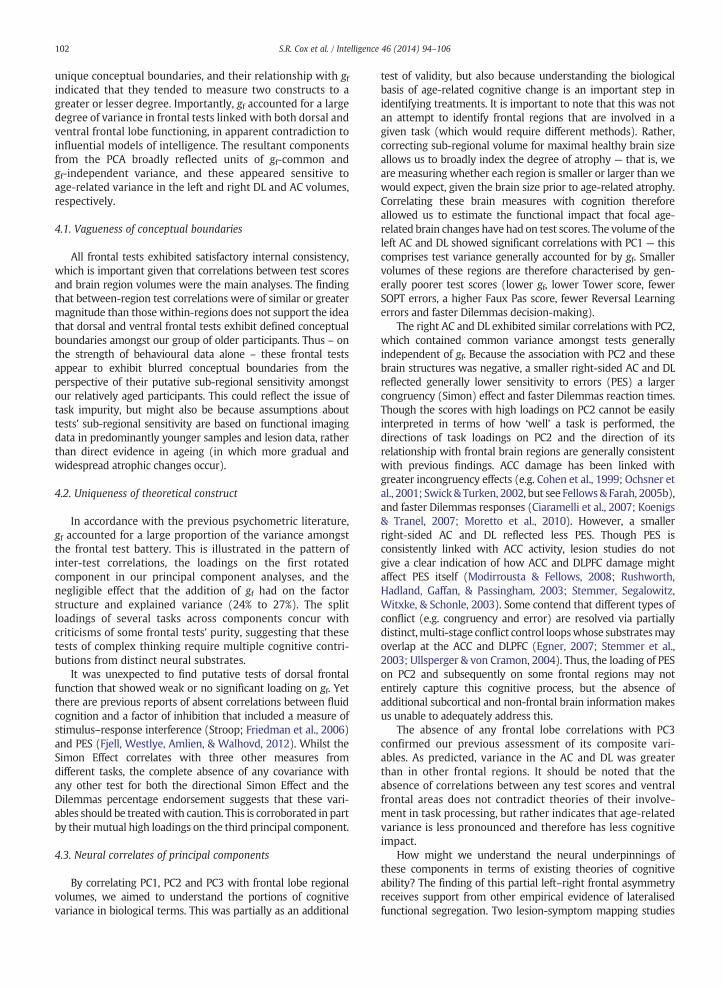
Scores for each of the three rotated components from thesecond PCA (which includes frontal lobe tests and gf) wereextracted and correlated with brain frontal sub-regionalvolumes corrected for ICV (Table 4 & Fig. 1). The significantneural correlates of PC1 were left DL, left dAC and left vAC.PC2 significantly correlated with right DL, right dAC and rightvAC. PC3 did not significantly correlate with any frontalregional volumes. However, bilateral vAC correlations wouldnot reach significance with a more stringent alpha threshold,nor would that between left DL and PC1.
Comparing the magnitudes of the correlations betweenthe left and right homotopic brain regions allowed us to testwhether the neural correlates of PC1 were significantlyleft-lateralised, and those of PC2 were right-lateralised. PC1correlations with dorsal and vAC were significantly greateron the left than the right (t(74) = 3.05, p = .003; t(74) =2.03, p = .046) but the difference between magnitudes forthe left and right DL was non-significant (t(74) = 1.49,p = .141). Correlations between PC2 and brain structurewere significantly right-lateralised for the DL (t(74) = 2.11,p = .039), dAC (t(74) = 3.24, p = .002) and vAC(t(74) =2.54, p = .013). All tests were two-tailed. Correlationsamongst principal components and brain regions uncorrect-ed for ICV can be found in the Supplementary material(Table S3). These are similar to the ICV-corrected associationsreported herein, albeit with slightly larger effect sizes, likelyreflecting the additional contribution of raw head size tocognitive ability.
4. Discussion
So-called ‘frontal’ cognitive tests have been criticised fortheir vagueness of conceptual boundaries, and it is unclear towhat extent individual differences in such test scores areexplained by so-called ‘general intelligence’ differences inolder age. An informative way to proceed, therefore, is tohave tests from both neuropsychological and differentialpsychology traditions, and to have brain information. To thisend, we examined the relationships between frontal testsand general intelligence, and interpreted the correlationalstructure in relation to measures of frontal lobe structurecorrected for ICV. The frontal tests did not generally display
ved from frontal tests and gf.
IF OF MS
R L R L R L R
− .03 .20 .19 .10 .00 − .12 − .11− .28⁎ − .04 − .18 .05 − .15 − .20 − .02− .12 − .08 − .09 .16 − .05 .09 − .03
ingulate; vAC = ventral anterior cingulate; IF = inferior frontal gyrus;
102 S.R. Cox et al. / Intelligence 46 (2014) 94–106
unique conceptual boundaries, and their relationship with gfindicated that they tended to measure two constructs to agreater or lesser degree. Importantly, gf accounted for a largedegree of variance in frontal tests linked with both dorsal andventral frontal lobe functioning, in apparent contradiction toinfluential models of intelligence. The resultant componentsfrom the PCA broadly reflected units of gf-common andgf-independent variance, and these appeared sensitive toage-related variance in the left and right DL and AC volumes,respectively.
4.1. Vagueness of conceptual boundaries
All frontal tests exhibited satisfactory internal consistency,which is important given that correlations between test scoresand brain region volumes were the main analyses. The findingthat between-region test correlations were of similar or greatermagnitude than those within-regions does not support the ideathat dorsal and ventral frontal tests exhibit defined conceptualboundaries amongst our group of older participants. Thus – onthe strength of behavioural data alone – these frontal testsappear to exhibit blurred conceptual boundaries from theperspective of their putative sub-regional sensitivity amongstour relatively aged participants. This could reflect the issue oftask impurity, but might also be because assumptions abouttests' sub-regional sensitivity are based on functional imagingdata in predominantly younger samples and lesion data, ratherthan direct evidence in ageing (in which more gradual andwidespread atrophic changes occur).
4.2. Uniqueness of theoretical construct
In accordance with the previous psychometric literature,gf accounted for a large proportion of the variance amongstthe frontal test battery. This is illustrated in the pattern ofinter-test correlations, the loadings on the first rotatedcomponent in our principal component analyses, and thenegligible effect that the addition of gf had on the factorstructure and explained variance (24% to 27%). The splitloadings of several tasks across components concur withcriticisms of some frontal tests' purity, suggesting that thesetests of complex thinking require multiple cognitive contri-butions from distinct neural substrates.
It was unexpected to find putative tests of dorsal frontalfunction that showed weak or no significant loading on gf. Yetthere are previous reports of absent correlations between fluidcognition and a factor of inhibition that included a measure ofstimulus–response interference (Stroop; Friedman et al., 2006)and PES (Fjell, Westlye, Amlien, & Walhovd, 2012). Whilst theSimon Effect correlates with three other measures fromdifferent tasks, the complete absence of any covariance withany other test for both the directional Simon Effect and theDilemmas percentage endorsement suggests that these vari-ables should be treatedwith caution. This is corroborated in partby their mutual high loadings on the third principal component.
4.3. Neural correlates of principal components
By correlating PC1, PC2 and PC3 with frontal lobe regionalvolumes, we aimed to understand the portions of cognitivevariance in biological terms. This was partially as an additional
test of validity, but also because understanding the biologicalbasis of age-related cognitive change is an important step inidentifying treatments. It is important to note that this was notan attempt to identify frontal regions that are involved in agiven task (which would require different methods). Rather,correcting sub-regional volume for maximal healthy brain sizeallows us to broadly index the degree of atrophy — that is, weare measuring whether each region is smaller or larger than wewould expect, given the brain size prior to age-related atrophy.Correlating these brain measures with cognition thereforeallowed us to estimate the functional impact that focal age-related brain changes have had on test scores. The volume of theleft AC and DL showed significant correlations with PC1 — thiscomprises test variance generally accounted for by gf. Smallervolumes of these regions are therefore characterised by gen-erally poorer test scores (lower gf, lower Tower score, fewerSOPT errors, a higher Faux Pas score, fewer Reversal Learningerrors and faster Dilemmas decision-making).
The right AC and DL exhibited similar correlations with PC2,which contained common variance amongst tests generallyindependent of gf. Because the association with PC2 and thesebrain structures was negative, a smaller right-sided AC and DLreflected generally lower sensitivity to errors (PES) a largercongruency (Simon) effect and faster Dilemmas reaction times.Though the scores with high loadings on PC2 cannot be easilyinterpreted in terms of how ‘well’ a task is performed, thedirections of task loadings on PC2 and the direction of itsrelationship with frontal brain regions are generally consistentwith previous findings. ACC damage has been linked withgreater incongruency effects (e.g. Cohen et al., 1999; Ochsner etal., 2001; Swick& Turken, 2002, but see Fellows& Farah, 2005b),and faster Dilemmas responses (Ciaramelli et al., 2007; Koenigs& Tranel, 2007; Moretto et al., 2010). However, a smallerright-sided AC and DL reflected less PES. Though PES isconsistently linked with ACC activity, lesion studies do notgive a clear indication of how ACC and DLPFC damage mightaffect PES itself (Modirrousta & Fellows, 2008; Rushworth,Hadland, Gaffan, & Passingham, 2003; Stemmer, Segalowitz,Witxke, & Schonle, 2003). Some contend that different types ofconflict (e.g. congruency and error) are resolved via partiallydistinct,multi-stage conflict control loopswhose substratesmayoverlap at the ACC and DLPFC (Egner, 2007; Stemmer et al.,2003; Ullsperger & von Cramon, 2004). Thus, the loading of PESon PC2 and subsequently on some frontal regions may notentirely capture this cognitive process, but the absence ofadditional subcortical and non-frontal brain information makesus unable to adequately address this.
The absence of any frontal lobe correlations with PC3confirmed our previous assessment of its composite vari-ables. As predicted, variance in the AC and DL was greaterthan in other frontal regions. It should be noted that theabsence of correlations between any test scores and ventralfrontal areas does not contradict theories of their involve-ment in task processing, but rather indicates that age-relatedvariance is less pronounced and therefore has less cognitiveimpact.
How might we understand the neural underpinnings ofthese components in terms of existing theories of cognitiveability? The finding of this partial left–right frontal asymmetryreceives support from other empirical evidence of lateralisedfunctional segregation. Two lesion-symptom mapping studies

103S.R. Cox et al. / Intelligence 46 (2014) 94–106
have linked left-sided frontal lesions with intelligence deficits(Barbey et al., 2012; Gläscher et al., 2010) and one has linkedtest score variance not common to g with right sided frontallesions (Roca et al., 2010). Intelligence in older age was alsorecently associated with the left but not right dorsal frontalcortical thickness (Karama et al., 2014). Similarly, the rightfrontolateral activity is related to post-error-related activity(thought to be involved in adaptive post-error behaviour; e.g.Hester, Barre,Mattingley, Foxe, & Garavan, 2007), the resolutionof stimulus–response conflict induced by incongruent trials(Milham, 2003) and moral judgement (Tassy et al., 2012). Ittherefore appears that there is some degree of consistencybetween our findings and the evidence for hemisphericlateralisation of cognition function.
From cognitive psychology, based on numerous lesionand imaging studies, it has been proposed that the left lateralregions are involved in the capacity to form or selecttask-relevant rules (criterion setting) and right lateralregions are tasked with updating contingencies and dynamicfine-tuning performance of ongoing behaviours (monitoring)in order to optimise behaviour (Stuss, 2011; Stuss &Alexander, 2007; Vallesi, 2012). Though cognitive nomencla-ture may result in the blurring of conceptual boundaries, wecautiously observe that the test score loadings on the PCsin our study show a plausible fit. In addition to a highintelligence loading on PC1, performance on the Tower test,SOPT and Reversal Learning all intuitively require the flexibleacquisition and selection of rules. It could be argued that theFaux Pas and Dilemmas tasks also require the participant toorient themselves within each scenario and select theappropriate social/moral rules. Likewise, for PC2, perfor-mance monitoring and contingency-updating are involved inresolving response conflict from competing stimulus cues(Simon Effect) or conflicting moral perspectives (Dilemmas),and in response to feedback regarding errors where perfor-mance may subsequently need optimising (PES).
4.4. Limitations and methodological considerations
Psychometrically, the PCA loadings for PC2 are not particu-larly robust because they show relatively low loadings ingeneral, indicating relatively weak cohesion amongst thecommon test variances orthogonal to PC1. Nevertheless, thesignificant correlation of PC2 with frontal lobe structure mayindicate that this is a plausible cognitive construct which someof the tasks themselves only measure relatively weakly. Inaddition, two tasks (Simon Task and Dilemmas task) contribut-ed more than one variable to the analysis. Multiple variableswere selected because the literature indicated more than onevariable may be related to frontal lobe functioning. However,two of the variables from these tasks (the directional SimonEffect and the percentage of endorsements in the Dilemmastask) had no influence in our analysis and thus their inclusion inthe overall results had little bearing. PES and the Simon Effectwere both derived from the Simon Task, but they were notstrongly correlated, consistent with the view that errordetection and conflict processing may be partially dissociableprocesses, as discussed above. With respect to PC1, it isimportant to note that the subtests used to derive g includedtwo that were explicitly processing speed measures. Thoughsome studies have attempted to empirically separate fluid
intelligence and processing speed (e.g. Conway, Cowan,Bunting, Therriault, & Minkoff, 2002), in the Lothian BirthCohort sample, g appears to be fundamentally predicated uponprocessing speed (Penke et al., 2012). In the current sub-sample,gf measures derived with or without overt tests of processingspeed (Digit Symbol and Symbol Search)were highly correlated(r (88) = .96, p b .0001). Removing these measures from ourmeasure of g did not affect the observed correlational patternwith frontal tests. Likewise, a measure of processing speedshowed the same pattern of results. Moreover, PC1 (heavilyloaded on by gf) was the only component to significantlycorrelatewith processing speed. Onemay therefore argue that itis unclear whether the relevant overlap with frontal tests isprimarily with processing speed or with gf.
Though we aimed to broadly index the degree ofage-related atrophic effects in frontal regions (observed sizerelative to maximal healthy size) on cognition, this is animperfect measure and sub-regional atrophy is impossible togauge accurately in a cross-sectional setting. Moreover,declines in brain size and intelligence do not exhibit perfecttemporal synchrony; the former showing evidence of declinein the mid-teens (Courchesne et al., 2000) and the latter notuntil later in the lifecourse (Hedden & Gabrieli, 2004). Thus,part of the observed difference between raw volume and ICVmay not reflect age-related atrophic influences.
In addition, our sample size is relatively small compared tostudies by Salthouse (2005, 2011), and is limited to a narrowage range and a relatively healthy male-only sample. Thus, ourcurrent design precludes inferences regarding cognitive changeover time, its relation to sub-regional frontal lobe structure overtime and in a female sample. Whilst these are limitations to thegeneralisability of our results, they can be considered strengthswith respect to the omission of important possible confoundersof age and gender. Nevertheless, our self-selecting cohortmembers are likely to represent a relatively restricted sample;they were representative of the overall LBC1936 cohort whosecognitive abilities were 0.78 of a standard deviation higher thantheir age group when first tested in 1947, and the variance wasrestricted by 44% (Johnson et al., 2012). Though Johnson andcolleagues found that range restriction did not substantiallyalter the magnitudes of observed associations, inferences to abroader population should be undertaken with caution. Wewere also unable to relate the principal components of cognitionto other areas of the brain whose structure may be relevant.
Finally, the large number of cognitive tests and frontalbrain regions led to a relatively large number of statisticaltests. We did not use a conventional correction for multiplecomparisons (e.g. Bonferroni), partly due to the exploratorynature of the study and partly because such an approachwould be overly conservative. Given the covariances thatexist amongst the cognitive tests and also amongst the brainregions under examination, each new comparison does notfulfil the criterion of an independent opportunity for type 1error to arise (Nyholt, 2001; Ott, 1999). Nevertheless, it isprudent to consider whether our inferences hold if a morestringent significance threshold (e.g. p b .01) were applied.Our assessment of a generally weak cohesion amongst thefrontal tests and that the majority show some overlap with g(Table 2) would remain unchanged, and this would stillbroadly reflect the loadings observed in PCAs. Regarding thecorrelations amongst PCs and frontal regions, correlations
104 S.R. Cox et al. / Intelligence 46 (2014) 94–106
with the vAC and between PC1 and left DL would beattenuated. Nevertheless, it is not likely for such a patternof asymmetrical results amongst simultaneous comparisonsto arise by chance, and the alpha threshold would notaffect our findings of significant lateralisation. Studies withimproved power to reliably detect small effect sizes overmultiple comparisons are required to corroborate thesefindings.
In summary, our key findings amongst a group of olderadults are as follows: frontal tests exhibited blurred conceptualboundaries, and there was considerable (but not complete)overlap with gf. A large proportion of test score variance wasdescribed by two components which appeared to have discreteneuroanatomical bases in the left and right frontal lobes in olderage, indicative of distinct underlying constructs. These con-structs may plausibly reflect criterion setting and monitoringreferred to in previous reviews of hemispheric lateralisation offunction. However, a closer examination of the direction andnature of loadings of individual cognitive tests on the principalcomponents makes a definitive interpretation of this correla-tional structure challenging, particularly as test scores loadingon PC2 cannot easily be gauged by how ‘well’ the task wasperformed. Whether PC1 represents g or ‘criterion setting’(if such constructs are independent), and PC2 represents‘monitoring’ is conjecture, but our results indicate that thesefrontal tasks may require multiple parts of cognition which canbe partially explained by their split loadings on these twoneuroanatomically distinct frontal components. The addition ofcognitive and neurobiological information in older age thereforecomplements previous lesionwork on this subject. Future workcould combine structural MRI and cognitive measures to largerageing samples, with explicit selection of tasks thought to tapthese two constructs and it could acquire longitudinal brainimaging data to address the functional implications of change inthe same regions amongst individuals over time.
Acknowledgements
We thank the Lothian Birth Cohort 1936 members whotook part in this study, radiographers at the Brain ResearchImaging Centre, Tom Booth for his useful suggestions, andresearch associates who collected and entered some of thecognitive data used in this manuscript. This research andLBC1936 phenotype collection were supported by Age UK(The Disconnected Mind project). It was undertaken in theCentre for Cognitive Ageing and Cognitive Epidemiology(http://www.ccace.ed.ac.uk) – part of the cross councilLifelong Health andWellbeing Initiative –which is supportedby funding from the UK's Biotechnology and BiologicalSciences Research Council, the Economic and Social ResearchCouncil and the Medical Research Council (MR/K026992/1).Brain imaging took place in the University of Edinburgh inthe Brain Research Imaging Centre (http://www.bric.ed.ac.uk) which is part of the SINAPSE collaboration (http://sinapse.ac.uk).
Appendix A. Supplementary data
Supplementary data to this article can be found online athttp://dx.doi.org/10.1016/j.intell.2014.05.006.
References
Barbey, A. K., Colom, R., Solomon, J., Krueger, F., Forbes, C., & Grafman, J.(2012). An integrative architecture for general intelligence and execu-tive function revealed by lesion mapping. Brain, 135(4), 1154–1164.
Berlin, H. A., Rolls, E. T., & Kischka, U. (2004). Impulsivity, time perception,emotion and reinforcement sensitivity in patients with orbitofrontalcortex lesions. Brain, 127(5), 1108–1126.
Boghi, A., Rasetti, R., Avidano, F., Manzone, C., Orsi, L., D'Agata, F., et al.(2006). The effect of gender on planning: An fMRI study using the Towerof London task. NeuroImage, 33(3), 999–1010.
Botvinick, M. M., Cohen, J. D., & Carter, C. S. (2004). Conflict monitoring andanterior cingulate cortex: An update. Trends in Cognitive Sciences, 8(12),539–546.
Brydges, C. R., Reid, C. L., Fox, A. M., & Anderson, M. (2012). A unitaryexecutive function predicts intelligence in children. Intelligence, 40(5),458–469. http://dx.doi.org/10.1016/j.intell.2012.05.006.
Burzynska, A. Z., Nagel, I. E., Preuschhof, C., Gluth, S., Bäckman, L., Li, S. -C.,et al. (2012). Cortical thickness is linked to executive functioning inadulthood and aging. Human Brain Mapping, 33(7), 1607–1620.
Carroll, J. B. (1993). Human cognitive abilities: A survey of factor analyticstudies. New York: Cambridge University Press.
Cazalis, F., Feydy, A., Valabrègue, R., Pélégrini-Issac, M., Pierot, L., & Azouvi, P.(2006). fMRI study of problem-solving after severe traumatic braininjury. Brain Injury, 20(10), 1019–1028.
Cazalis, F., Valabregue, R., Pelegrini-Issac, M., Asloun, S., Robbins, T. W., &Granon, S. (2003). Individual differences in prefrontal cortical activationon the Tower of London planning task: Implication for effortfulprocessing. European Journal of Neuroscience, 17(10), 2219–2225.
Ciaramelli, E., Muccioli, M., Làdavas, E., & di Pellegrino, G. (2007). Selectivedeficit in personal moral judgment following damage to ventromedialprefrontal cortex. Social Cognitive and Affective Neuroscience, 2(2), 84–92.
Cohen, J. (1988). Statistical power analysis for the behavioral sciences (2nd ed.).NJ: Erlbaum: Hillsdale.
Cohen, R., Kaplan, R., Moser, D., Jenkins, M., & Wilkinson, H. (1999).Impairments of attention after cingulotomy. Neurology, 53, 819–824.
Colom, R., Haier, R. J., Head, K., Álvarez-Linera, J., Quiroga, M.Á., & Shih, P. C.(2009). Gray matter correlates of fluid, chrystallised and spatialintelligence: Testing the P-FIT model. Intelligence, 37, 124–135.
Conway, A. R. A., Cowan, N., Bunting, M. F., Therriault, D. J., & Minkoff, S. R. B.(2002). A latent variable analysis of working memory capacity, short-term memory capacity, processing speed, and general fluid intelligence.Intelligence, 30(2), 163–183.
Cools, R., Stefanova, E., Barker, R., Robbins, T., & Owen, M. (2002). Dopaminergicmodulation of high-level cognition in Parkinson's disease: The role of theprefrontal cortex revealed by PET. Brain, 125(3), 584–594.
Courchesne, E., Chisum, H. J., Townsend, J., Cowles, A., Covington, J., Egaas, B.,et al. (2000). Normal brain development and aging: Quantitativeanalysis at in vivo MR imaging in healthy volunteers. Neuroradiology,216, 672–682.
Cox, S. R., Ferguson, K. J., Royle, N. A., Shenkin, S. D., MacPherson, S. E.,MacLullich, A. M., et al. (2014). A systematic review of brain frontal lobeparcellation techniques in magnetic resonance imaging. Brain Structureand Function, 219(1), 1–22.
Crawford, J. R., Bryan, J., Luszcz, M. A., Obonsawin, M. C., & Stewart, L. (2000).The executive decline hypothesis of cognitive aging: Do executivedeficits qualify as differential deficits and do they mediate age-relatedmemory decline? Aging Neuropsychology & Cognition, 7(1), 9–31.
Davis, A. S., Pierson, E. E., & Finch, W. H. (2011). A canonical correlationanalysis of intelligence and executive functioning. AppliedNeuropsychology, 18(1), 61–68.
de Zubicaray, G. I., Chalk, J. B., Rose, S. E., Semple, J., & Smith (1997). Deficitson self ordered tasks associated with hyperostosis frontalis interna.Journal of Neurology, Neurosurgery, and Psychiatry, 63(3), 309–314.
Deary, I. J., Corley, J., Gow, A. J., Harris, S. E., Houlihan, L. M., Marioni, R. E.,et al. (2009). Age-associated cognitive decline. British Medical Bulletin,92, 135–152.
Deary, I. J., Gow, A. J., Taylor, M. D., Corley, J., Brett, C., Wilson, V., et al.(2007). The Lothian Birth Cohort 1936: A study to examine influences oncognitive ageing from age 11 to age 70 and beyond. BMC Geriatrics, 7, 28.
Delis, D. C., Kaplan, E., & Kramer, J. H. (2001). Delis Kaplan ExecutiveFunction System: Technical manual. San Antonio, TX: The PsychologicalCorporation.
di Pellegrino, G., Ciaramelli, E., & Làdavas, E. (2007). The regulation ofcognitive control following rostral anterior cingulate cortex lesion inhumans. Journal of Cognitive Neuroscience, 19(2), 275–286.
Driscoll, I., Davatzikos, C., An, Y., Wu, X., Shen, D., Kraut, M., et al. (2009).Longitudinal pattern of regional brain volume change differentiatesnormal ageing from MCI. Neurology, 72(22), 1906–1913.

105S.R. Cox et al. / Intelligence 46 (2014) 94–106
Duncan, J. (2010). The multiple-demand (MD) system of the primate brain:Mental programs for intelligent behaviour. Trends in Cognitive Sciences,14(4), 172–179.
Duncan, J., Burgess, P., & Emslie, H. (1995). Fluid intelligence after frontallobe lesions. Neuropsychologia, 33, 261–268.
Egner, T. (2007). Congruency sequence effects and cognitive control.Cognitive, Affective & Behavioural Neuroscience, 7(4), 380–390.
Fellows, L. K., & Farah, M. J. (2005). Different underlying impairments indecision-making following ventromedial and dorsolateral frontal lobedamage in humans. Cerebral Cortex, 15(1), 58–63.
Fellows, L. K., & Farah, M. J. (2005). Is anterior cingulate cortex necessary forcognitive control? Brain, 128, 788–796.
Fjell, A. M., Westlye, L. T., Amlien, I., Espeseth, T., Reinvang, I., Raz, N., et al.(2009). High consistency of regional cortical thinning in aging acrossmultiple samples. Cerebral Cortex, 19(9), 2001–2012.
Fjell, A. M., Westlye, L. T., Amlien, I. K., & Walhovd, K. B. (2012). A multi-modal investigation of behavioural adjustment: Post-error slowing isassociated with white matter characteristics. NeuroImage, 61(1),195–205.
Floyd, R. G., Bergeron, R., Hamilton, G., & Parra, G. R. (2010). How doexecutive functions fit with the Cattell–Horn–Carroll model? Someevidence from a joint factor analysis of the Delis–Kaplan ExecutiveFunction System and the Woodcock–Johnson III tests of cognitiveabilities. Psychology in the Schools, 47(7), 721–738.
Folstein, M. F., Folstein, S. E., & McHugh, P. R. (1975). Mini-mental state. Apractical method for grading the cognitive state of patients for theclinician. Journal of Psychiatric Research, 12, 189–198.
Friedman, N. P., Miyake, A., Corley, R. P., Young, S. E., DeFries, J. C., & Hewitt, J.K. (2006). Not all executive functions are related to intelligence.Psychological Science, 17, 172–179.
Ghahremani, D. G., Monterosso, J., Jentsch, J. D., Bilder, R. M., & Poldrack, R. A.(2010). Neural components underlying behavioral flexibility in humanreversal learning. Cerebral Cortex, 20(8), 1843–1852.
Gläscher, J., Hampton, A. N., & O'Doherty, J. P. (2009). Determining a Role forVentromedial Prefrontal Cortex in Encoding Action- Based Value SignalsDuring Reward- Related Decision Making. Cerebral Cortex, 19(2),483–495.
Gläscher, J., Rudrauf, D., Colom, R., Paula, L. K., Tranel, D., Damasio, H., et al.(2010). Distributed neural system for general intelligence revealed bylesion symptom mapping. Proceedings of the National Academy ofSciences, 107(10), 4705–4709.
Gläscher, J., Tranel, D., Paul, L. K., Rudrauf, D., Rorden, C., Hornaday, A., et al.(2009). Lesion mapping of cognitive abilities linked to intelligence.Neuron, 61(5), 681–691.
Gomez-Beldarrain, M., Harries, C., Garcia-Monco, J. C., Ballus, E., & Grafman, J.(2004). Patients with right frontal lesions are unable to assess and useadvice to make predictive judgments. Journal of Cognitive Neuroscience,16(1), 74–89.
Goldman-Rakic, P. S. (1993). Specification of higher cortical functions.Journal of Head Trauma Rehabilitation, 8, 13–23.
Greene, J. D., Sommerville, R. B., Nystrom, L. E., Darley, J. M., & Cohen, J. D.(2001). An fMRI investigation of emotional engagement in moraljudgment. Science, 293(5537), 2105–2108.
Gregory, C., Lough, S., Stone, V., Erzincioglu, S., Martin, L., Baron-Cohen, S.,et al. (2002). Theory of mind in patients with frontal variantfrontotemporal dementia and Alzheimer's disease: Theoretical andpractical implications. Brain, 125(4), 752–764.
Hampton, A. N., & O'Doherty, J. P. (2007). Decoding the neural substrates ofreward-related decision making with functional MRI. PNAS, 104(4),1377–1382.
Hedden, T., & Gabrieli, J. D. E. (2004). Insights into the ageing mind: Aview from cognitive neuroscience. Nature Reviews Neuroscience, 5,87–96.
Hester, R., Barre, N., Mattingley, J. B., Foxe, J. J., & Garavan, H. (2007).Avoiding another mistake: Error and posterror neural activity associatedwith adaptive posterror behaviour change. Cognitive, Affective, &Behavioural Neuroscience, 7(4), 317–326.
Johnson, W., Gow, A. J., Corley, J., Redmond, P., Henderson, R., Murray, et al.(2012). Can we spot deleterious ageing in two waves of data? TheLothian Birth Cohort 1936 from ages 70 to 73. Longitudinal and LifeCourse Studies, 3(3), 312–331.
Jung, R. E., & Haier, R. J. (2007). The Parieto-Frontal Integration Theory (P-FIT) of intelligence: Converging neuroimaging evidence. The Behavioraland Brain Sciences, 30(2), 135–154 (discussion 154–187).
Kahane, G., & Shackel, N. (2008). Do abnormal responses show utilitarianbias? Nature, 452. http://dx.doi.org/10.1038/0678.
Karama, S., Bastin, M. E., Murray, C., Royle, N. A., Penke, L., Muñoz Maniega, S.,et al. (2014). Childhood cognitive ability accounts for associations betweencognitive ability and brain cortical thickness in older age. MolecularPsychiatry, 19, 555–559.
Kemp, J., Després, O., Sellal, F., & Dufour, A. (2012). Theory of Mind in normalageing and neurodegenerative pathologies. Ageing Research Reviews,11(2), 199–219.
Koenigs, M., & Tranel, D. (2007). Irrational economic decision-making afterventromedial prefrontal damage: Evidence from the ultimatum game.The Journal of Neuroscience, 27(4), 951–956.
Kringelbach, M., & Rolls, E. T. (2003). Neural correlates of rapid reversallearning in a simple model of human social interaction. NeuroImage, 20,1371–1383.
Lamar, M., & Resnick, S. M. (2004). Aging and prefrontal functions:Dissociating orbitofrontal and dorsolateral abilities. Neurobiology ofAging, 25(4), 553–558.
Lee, T. M. C., Ip, A. K. Y., Wang, K., Xi, C. -H., Hu, P. -P., Mak, H. K. F., et al.(2010). Faux pas deficits in people with medial frontal lesions as relatedto impaired understanding of a speaker's mental state. Neuropsychologia,48(6), 1670–1676.
Lehto, J. E., Juuarvi, P., Kooistra, L., & Pulkkinen, L. (2003). Dimensions ofexecutive functioning: Evidence from children. British Journal ofDevelopmental Psychology, 21(1), 59–80.
MacPherson, S. E., Phillips, L. H., & Della Sala, S. (2002). Age, executivefunction and social decision making: A dorsolateral prefrontal theory ofcognitive aging. Psychology and Aging, 17(4), 598–609.
Manes, F., Sahakian, B., Clark, L., Rogers, R., Antoun, N., Aitken, M., et al.(2002). Decision-making processes following damage to the prefrontalcortex. Brain, 125(3), 624–639.
Milham, M. (2003). Practice-related effects demonstrate complementaryroles of anterior cingulate and prefrontal cortices in attentional control.NeuroImage, 18(2), 483–493.
Miyake, A., & Friedman, N. P. (2012). The nature and organization ofindividual differences in executive functions: Four general conclusions.Current Directions in Psychological Science, 21(1), 8–14.
Modirrousta, M., & Fellows, L. K. (2008). Dorsal medial prefrontal cortexplays a necessary role in rapid error prediction in humans. Journal ofNeuroscience, 28(51), 14000–14005.
Moretto, G., Làdavas, E., Mattioli, F., & di Pellegrino, G. (2010). Apsychophysiological investigation of moral judgment after ventromedialprefrontal damage. Journal of Cognitive Neuroscience, 22(8), 1888–1899.
Narr, K. L., Woods, R. P., Thompson, P. M., Szeszko, P., Robinson, D.,Dimtcheva, T., et al. (2007). Relationships between IQ and regionalcortical gray matter thickness in healthy adults. Cerebral Cortex, 17(9),2163–2171.
Nyholt, D. R. (2001). Genetic case–control association studies — Correctingfor multiple testing. Human Genetics, 109(5), 564–567.
Nyhus, E., & Barceló, F. (2009). The Wisconsin Card Sorting Test and thecognitive assessment of prefrontal executive functions: A critical update.Brain and Cognition, 71(3), 437–451.
Ochsner, K. N., Kosslyn, S. M., Cosgrove, G. R., Cassem, E. H., Price, B. H.,Nierenberg, A. A., et al. (2001). Deficits in visual cognition and attentionfollowing bilateral anterior cingulotomy. Neuropsychologia, 39, 219–230.
O'Doherty, J., Kringelbach, M. L., Rolls, E. T., Hornak, J., & Andrews, C. (2001).Abstract reward and punishment representations in the humanorbitofrontal cortex. Nature Neuroscience, 4(1), 95–102.
Ott, J. (1999). Analysis of human genetic linkage (3rd ed.). Baltimore, USA:Johns Hopkins University Press.
Penke, L., Muñoz Maniega, S., Bastin, M. E., Valdés Hernández, M. C., Murray, C.,Royle, N. A., et al. (2012). Brain white matter tract integrity as a neuralfoundation for general intelligence.Molecular Psychiatry, 17(10), 1026–1030.
Peterson, B. S., Kane, M. J., Alexander, G. M., Lacadie, C., Skudlarski, P., Leung,H. -C., et al. (2002). An event-related functional MRI study comparinginterference effects in the Simon and Stroop tasks. Cognitive BrainResearch, 13(3), 427–440.
Petrides, M., Alivisatos, B., Evans, A. C., & Meyer, E. (1993). Dissociation ofhuman mid-dorsolateral from posterior dorsolateral frontal cortex inmemory processing. Proceedings of the National Academy of Sciences,90(3), 873–877.
Petrides, M., & Milner, B. (1982). Deficits on subject-ordered tasks afterfrontal- and temporal-lobe lesions in man. Neuropsychologia, 20(3),249–262.
Phillips, L. H., & Della Sala, S. (1998). Aging, intelligence, and anatomicalsegregation in the frontal lobes. Learning and Individual Differences,10(3), 217–243.
Rabbitt, P. M. A. (1997). Methodologies and models in the study of executivefunction. In P. M. A. Rabbitt (Ed.), Methodology of frontal and executivefunction (pp. 1–38). Hove, UK: Psychology Press.
Rabbitt, P. M. A. (2011). Between-individual variability and interpretation ofassociations between neuropsychological and behavioural measures inaging populations: Comment on Salthouse (2011). Psychological Bulletin,137(5), 785–789.
Rabbitt, P. M. A., Lowe, C., & Shilling, V. (2001). Frontal tests and models forcognitive ageing. European Journal of Cognitive Psychology, 13, 5–28.
106 S.R. Cox et al. / Intelligence 46 (2014) 94–106
Remijnse, P. L., Nielen, M. M. A., Uylings, H. B. M., & Veltman, D. J. (2005).Neural correlates of a reversal learning task with an affectively neutralbaseline: An event-related fMRI study. NeuroImage, 26(2), 609–618.
Robbins, T. W., James, M., Owen, A. M., Sahakian, B. J., Lawrence, A. D.,McInnes, L., et al. (1998). A study of performance on tests from theCANTAB battery sensitive to frontal lobe dysfunction in a large sample ofnormal volunteers: Implications for theories of executive functioningand cognitive ageing. Journal of the International NeuropsychologicalSociety, 4, 474–490.
Roca, M., Parr, A., Thompson, R., Woolgar, A., Torralva, T., Antoun, N., et al.(2010). Executive function and fluid intelligence after frontal lobelesions. Brain, 133(1), 234–247.
Rolls, E. T., Hornak, J., Wade, D., & McGrath, J. (1994). Emotion-relatedlearning in patients with social and emotional changes associated withfrontal lobe damage. Journal of Neurology, Neurosurgery & Psychiatry,57(12), 1518–1524.
Rushworth, M. F. S., Hadland, K. A., Gaffan, D., & Passingham, R. E. (2003).The effect of cingulate cortex lesions on task switching and workingmemory. Journal of Cognitive Neuroscience, 15(3), 338–353.
Salthouse, T. A. (2004). What and when of cognitive aging. Current Directionsin Psychological Science, 13(4), 140–144.
Salthouse, T. A. (2005). Relations between cognitive abilities and measuresof executive functioning. Neuropsychology, 19(4), 532–545.
Salthouse, T. A. (2011). What cognitive abilities are involved in trail-makingperformance? Intelligence, 39(4), 222–232.
Sarazin, M., Pillon, B., Giannakopoulos, P., Rancurel, G., Samson, Y., & Dubois,B. (1998). Clinicometabolic dissociation of cognitive functions and socialbehavior in frontal lobe lesions. Neurology, 51(1), 142–148.
Shamay-Tsoory, S. G., Tomer, R., Berger, B. D., & Aharon-Peretz, J. (2003).Characterization of empathy deficits following prefrontal brain damage:The role of the right ventromedial prefrontal cortex. Journal of CognitiveNeuroscience, 15(3), 324–337.
Shamay-Tsoory, S. G., Tomer, R., Berger, B. D., Goldsher, D., & Aharon-Peretz,J. (2005). Impaired “affective theory of mind” is associated with rightventromedial prefrontal damage. Cognitive and Behavioral Neurology,18(1), 55–67.
Shrout, P. E., & Fleiss, J. L. (1979). Intraclass correlations: Uses in assessingrater reliability. Psychological Bulletin, 2, 420–428.
Simon, J. R. (1969). Reactions towards the source of stimulation. Journal ofExperimental Psychology, 81, 174–176.
Steele, J. D., & Lawrie, S. M. (2004). Segregation of cognitive and emotionalfunction in the prefrontal cortex: A stereotactic meta-analysis.NeuroImage, 21(3), 868–875.
Stemmer, B., Segalowitz, S. J., Witxke, W., & Schonle, P. W. (2003). Errordetection in patients with lesions to the medial prefrontal cortex: AnERP study. Neuropsychologia, 42, 118–130.
Stone, V. E., Baron-Cohen, S., & Knight, R. T. (1998). Frontal lobe contributions totheory of mind. Journal of Cognitive Neuroscience, 10(5), 640–656.
Stuss, D. T. (2011). Functions of the frontal lobes: Relation to executivefunctions. Journal of the International Neuropsychological Society, 17,759–765.
Stuss, D. T., & Alexander, M. P. (2007). Is there a dysexecutive syndrome?Philosophical Transactions of the Royal Society of London, B, BiologicalSciences, 362, 901–915.
Stuss, D. T., & Levine, B. (2002). Adult clinical neuropsychology: Lessons fromstudies of the frontal lobes. Annual Review of Psychology, 53, 401–433.
Stuss, D. T., Shallice, T., Alexander, M. P., & Picton, T. W. (1995). Amultidisciplinary approach to anterior attentional functions. Annals ofthe New York Academy of Sciences, 769, 191–211.
Sullivan, E. V., & Pfefferbaum, A. (2007). Neuroradiological characterizationof normal adult ageing. The British Journal of Radiology, 80, S99–S108.
Swick, D., & Turken, A. U. (2002). Dissociation between conflict detectionand error monitoring in the human anterior cingulate cortex. PNAS,99(25), 16354–16359.
Tassy, S., Ouiller, O., Duclos, Y., Coulon, O., Mancini, J., Derulle, C., et al.(2012). Disrupting the right prefrontal cortex alters moral judgement.Scan, 7, 282–288.
Tisserand, D. J., Pruessner, J. C., Arigita, E. J. S., van Boxtel, M. P. J., Evans, A. C.,Jolles, J., et al. (2002). Regional frontal cortical volumes decreasedifferentially in aging: An MRI study to compare volumetric approachesand voxel-based morphometry. NeuroImage, 17(2), 657–669.
Ullsperger, M., & von Cramon, D. Y. (2004). Neuroimaging of performancemonitoring: Error detection and beyond. Cortex, 40, 593–604.
Valdés Hernández, M. C., Royle, N. A., Jackson, M. R., Muñoz Maniega, S.,Penke, L., Bastin, M. E., et al. (2012). Color fusion of magnetic resonanceimaging improves intracranial volume measurement in studies of aging.Open Journal of Radiology, 2(1), 1–9.
Vallesi, A. (2012). Organisation of executive functions: Hemisphericasymmetries. Journal of Cognitive Psychology, 24(4), 367–386.
Wardlaw, J. M., Bastin, M. E., Valdés Hernandez, M. C., Munoz Maniega, S., Royle,N. A., Moris, Z., et al. (2011). Brain aging, cognition in youth and older ageand vascular disease in the Lothian Birth Cohort 1936: Rationale, design andmethodology of the imaging protocol. International Journal of Stroke, 6(6),547–559.
Wechsler, D. (1998). WAIS-IIIUK administration and scoring manual. London,UK: Psychological Corporation.
Wheeler, E. Z., & Fellows, L. K. (2008). The human ventromedial frontal lobeis critical for learning from negative feedback. Brain, 131(5), 1323–1331.
Williams, E. J. (1959). The comparison of regression variables. Journal of theRoyal Statistical Society, Series B, 21, 396–399.
Yochim, B. P., Baldo, J. V., Kane, K. D., & Delis, D. C. (2009). D-KEFS Tower Testperformance in patients with lateral prefrontal cortex lesions: Theimportance of error monitoring. Journal of Clinical and ExperimentalNeuropsychology, 31(6), 658–663.
Yochim, B., Baldo, J., Nelson, A., & Delis, D. C. (2007). D-KEFS Trail MakingTest performance in patients with lateral prefrontal cortex lesions.Journal of the International Neuropsychological Society, 13(4), 704–709.
Zakzanis, K. K., Mraz, R., & Graham, S. J. (2005). An fMRI study of the TrailMaking Test. Neuropsychologia, 43(13), 1878–4886.
Zald, D. H. (2007). Orbital versus dorsolateral prefrontal cortex: Anatomicalinsights into content versus process differentiation models of the prefrontalcortex. Annals of the New York Academy of Sciences, 1121, 395–406.
Zigmond, A. S., & Snaith, R. P. (1983). The Hospital Anxiety and DepressionScale. Acta Psychiatrica Scandanavica, 67(6), 361–370.