edinburgh research · pdf fileaccepted manuscript accepted manuscript 5 phosphatase inhibitor...
TRANSCRIPT
Edinburgh Research Explorer
Mass spectrometry analysis of the oxidation states of the pro-oncogenic protein anterior gradient-2 reveals covalentdimerization via an intermolecular disulphide bond
Citation for published version:Clarke, DJ, Murray, E, Faktor, J, Mohtar, A, Vojtesek, B, MacKay, CL, Smith, PL & Hupp, TR 2016, 'Massspectrometry analysis of the oxidation states of the pro-oncogenic protein anterior gradient-2 revealscovalent dimerization via an intermolecular disulphide bond' Biochimica et biophysica acta-Bioenergetics.DOI: 10.1016/j.bbapap.2016.02.011
Digital Object Identifier (DOI):10.1016/j.bbapap.2016.02.011
Link:Link to publication record in Edinburgh Research Explorer
Document Version:Peer reviewed version
Published In:Biochimica et biophysica acta-Bioenergetics
Publisher Rights Statement:Author's final peer-reviewed manuscript as accepted for publication
General rightsCopyright for the publications made accessible via the Edinburgh Research Explorer is retained by the author(s)and / or other copyright owners and it is a condition of accessing these publications that users recognise andabide by the legal requirements associated with these rights.
Take down policyThe University of Edinburgh has made every reasonable effort to ensure that Edinburgh Research Explorercontent complies with UK legislation. If you believe that the public display of this file breaches copyright pleasecontact [email protected] providing details, and we will remove access to the work immediately andinvestigate your claim.
Download date: 25. May. 2018

�������� ����� ��
Mass spectrometry analysis of the oxidation states of the pro-oncogenicprotein anterior gradient-2 reveals covalent dimerization via an intermoleculardisulphide bond
David J. Clarke, Euan Murray, Jakub Faktor, Aiman Mohtar, Borek Vo-jtesek, C. Logan MacKay, Pat Langridge Smith, Ted R. Hupp
PII: S1570-9639(16)30024-3DOI: doi: 10.1016/j.bbapap.2016.02.011Reference: BBAPAP 39705
To appear in: BBA - Proteins and Proteomics
Received date: 24 November 2015Revised date: 23 January 2016Accepted date: 9 February 2016
Please cite this article as: David J. Clarke, Euan Murray, Jakub Faktor, Aiman Mohtar,Borek Vojtesek, C. Logan MacKay, Pat Langridge Smith, Ted R. Hupp, Mass spec-trometry analysis of the oxidation states of the pro-oncogenic protein anterior gradient-2reveals covalent dimerization via an intermolecular disulphide bond, BBA - Proteins andProteomics (2016), doi: 10.1016/j.bbapap.2016.02.011
This is a PDF file of an unedited manuscript that has been accepted for publication.As a service to our customers we are providing this early version of the manuscript.The manuscript will undergo copyediting, typesetting, and review of the resulting proofbefore it is published in its final form. Please note that during the production processerrors may be discovered which could affect the content, and all legal disclaimers thatapply to the journal pertain.
ACC
EPTE
D M
ANU
SCR
IPT
ACCEPTED MANUSCRIPT
1
Mass Spectrometry analysis of the oxidation states of the pro-oncogenic protein Anterior
Gradient-2 reveals covalent dimerization via an intermolecular disulphide bond.
David J. Clarke*2, Euan Murray1, Jakub Faktor3, Aiman Mohtar1, Borek Vojtesek3, C. Logan
MacKay2, Pat Langridge Smith2, and Ted R. Hupp*1
University of Edinburgh, 1Institute of Genetics and Molecular Medicine, Division of Cancer
Biology; 2School of Chemistry, Edinburgh, Scotland, United Kingdom, EH4 2XR; 3Regional
Centre for Applied Molecular Oncology, Masaryk Memorial Cancer Institute, 656 53 Brno, Czech
Republic;
Running title: Redox regulated AGR2 dimerization interface
Keywords: cancer, p53, Anterior Gradient-2, aptamers, therapeutics, protein mass spectrometry
* correspondence: [email protected] (mass spectrometry) & [email protected] (AGR2
biology)
ACC
EPTE
D M
ANU
SCR
IPT
ACCEPTED MANUSCRIPT
2
SUMMARY
Anterior Gradient-2 (AGR2) is a component of a pro-oncogenic signalling pathway that
can promote p53 inhibition, metastatic cell migration, limb regeneration, and cancer
drug-resistance. AGR2 is in the protein-disulfide isomerase superfamily containing a
single cysteine (Cys-81) that forms covalent adducts with its client proteins. We have
found that mutation of Cysteine-81 attenuates its biochemical activity in its sequence-
specific peptide docking function, reduces binding to Reptin, and reduces its stability
in cells. As such, we evaluated how chemical oxidation of its cysteine affects its
biochemical properties. Recombinant AGR2 spontaneously forms covalent dimers in
the absence of reductant whilst DTT promotes dimer to monomer conversion.
Mutation of Cysteine-81 to alanine prevents peroxide catalyzed dimerization of AGR2
in vitro, suggesting a reactive cysteine is central to covalent dimer formation. Both
biochemical assays and ESI mass spectrometry were used to demonstrate that low
levels of a chemical oxidant promote an intermolecular disulfide bond through
formation of a labile sulfenic acid intermediate. However, higher levels of oxidant
promote sulfinic or sulfonic acid formation thus preventing covalent dimerization of
AGR2. These data together identify the single cysteine of AGR2 as an oxidant
responsive moiety that regulates its propensity for oxidation and its monomeric-
dimeric state. This has implications for redox regulation of the pro-oncogenic
functions of AGR2 protein in cancer cells.

ACC
EPTE
D M
ANU
SCR
IPT
ACCEPTED MANUSCRIPT
3
INTRODUCTION
Oesophageal adenocarcinoma is thought to develop from pre-malignant lesions termed
Barrett’s oesophagus [1]. Selection pressures are known to drive p53 gene mutation early in
oesophageal cancer progression [2]. Whole genome sequencing has determined that
selection pressures for p53 gene mutations are remarkably confined to a highly specific
stage, that being Barrett’s tissue associated with high-grade dysplasia [3]. Barrett’s
epithelium forms a relatively unique microenvironment for the identification of stress activated
p53 modifiers might place selection pressures on the survival of cells with either p53 gene
mutation or in maintenance of the wt-p53 alleles [4-6]. Using a proteomics approach in
Barrett’s epithelium to identify such possible p53 modifiers, a protein named Anterior
Gradient-2 (AGR2) was identified and validated as an abundant and potent inhibitor of p53-
dependent transcription [7, 8]. AGR2 remains upregulated in a large proportion of
oesophageal adenocarcinomas [9]. The recent generation of an AGR2 isogenic cell panel
highlighted p53 pathway suppression as the dominant effect of enhanced AGR2 protein-
dependent remodelling of the proteome [10]. Thus, AGR2 pathway function forms a core
proteomic landscape whose study might shed light on oesophageal adenocarcinoma
development as well as p53 pathway silencing.
AGR2 was originally identified as a secretory protein that is highly expressed in Xenopus
eggs [11]. Apart from its function as a p53 inhibitor [8], subsequent studies have shown a
significant role for AGR2 in a range of biological pathways including cell migration, cellular
transformation, metastasis [12] [9], and limb regeneration in vertebrates [13]. Clinical studies
have also implicated the protein in inflammatory bowel disease [14], hormone-dependent
breast cancers [15] [16], and in predicting poor prognosis in prostate cancers [17]. The
molecular mechanisms underlying these wide-ranging biological pathways trigged by AGR2
are still not completely defined and as the AGR2 gene is confined to vertebrates, the AGR2
gene pathway cannot be dissected using powerful genetic systems like yeast, flies, or worms
[18]. However, emerging functions in the AGR2 pathway focus on (i) identifying its client
proteins as it mediates protein folding in the endoplasmic reticulum [19] and as it stimulates
receptor maturation [20] [21]; (ii) understanding its function in the endoplasmic reticulum in
response to unfolded protein responses [22], (iii) defining the nature of its monomer-dimer
equilibrium in tis chaperone cycle [23]; and (iv) understanding the significance of its highly
specific peptide docking function which is relatively unique for a molecular chaperone [24]
[25].
Endoplasmic reticulum localized molecular chaperones like AGR2 have specific roles that
enable the folding, trafficking, and assembly of complex cysteine-rich transmembrane
ACC
EPTE
D M
ANU
SCR
IPT
ACCEPTED MANUSCRIPT
4
receptors with unique protein folding requirements. Reduced glutathione and its oxidized
counterpart comprise the dominant redox buffer in eukaryotes [26]. A relatively high pro-
oxidizing environment in the endoplasmic reticulum is thought to facilitate the iterative cycles
of enzymatic cysteine reduction and oxidation on cysteine-rich client receptors destined for
the secretory system and transmembrane destinations [27]. Classic thioredoxins have a
conserved thioredoxin fold comprised of the CxxC motif that mediates covalent bond
formation with cysteine containing client proteins followed by resolution through cycles of
reduction-oxidation [28]. AGR2 by contrast is part of the thioredoxin superfamily that contain
CxxS motifs and which lack the ability to exploit a two cysteine redox system that classically
mediates client protein oxidation and reduction cycles [29]. As such, AGR2 single cysteine
oxidation and reduction could form an important rate-limiting step in its reaction cycle with
implications for client protein maturation in human diseases like cancer. In this report, we
detail the effects of single cysteine mutation on the known core biochemical function of
AGR2 and also the nature of AGR2 cysteine oxidation on its oligomerization state using
mass spectrometry (MS). We demonstrate first that cysteine mutation can alter its specific
activity in protein-interaction assays. Secondly, we demonstrate that low levels of cysteine
oxidation can induce covalent dimerization through an unstable sulfenic acid intermediate.
Finally, we show that higher levels of cysteine oxidant yield a sulfinic or sulfonic acid product
that traps AGR2 in the monomeric and oxidized state. These assays will facilitate future
dissection of the reaction mechanism of AGR2 as it mediates protein folding of cysteine rich
client proteins. This may reveal how its oxidation state in the endoplasmic reticulum might
effects its specific activity as an oncogenic chaperone in the development and maintenance
of the transformed phenotype.
EXPERIMENTAL PROCEDURES
General
All reagents were purchased from Sigma unless otherwise stated.
In Vivo Cross-linking of proteins in MCF7 Breast Cancer Cells
MCF7 breast cancer cells were cultured in DMEM supplemented with 10% FCS in a
humidified incubator in 5% CO2 at 37ºC. For cross-linking, cells were grown to ~90%
confluence before exposure to DSS (Pierce 21555) or EGS (Pierce 21565). DSS or EGS
was dissolve in 100% DMSO to a concentration of 100mM before being further diluted to the
appropriate concentration in growth media and added to cells. Cells were incubated for a
further 1 hour at 37ºC before being harvested into PBS and lysed into the following buffer:
150mM NaCl, 50mM Tris HCl pH 8.0, 50mM NaF, 5mM EDTA, 1% NP-40 plus 1:100
ACC
EPTE
D M
ANU
SCR
IPT
ACCEPTED MANUSCRIPT
5
phosphatase inhibitor cocktail (Sigma P5726) and 1:100 protease inhibitor cocktail (Sigma
P8340). AGR2 containing cross-linked complexes were detected by Western blot analysis
using a polyclonal antibody to AGR2 raised in rabbit (Moravian Biotechnology Ltd., Brno
Czech Republic).
Site Directed Mutagenesis of AGR2 at Cysteine 81
Human AGR2 was cloned into Invitrogen’s Gateway expression vectors pDEST17 for
bacterial expression or pDEST12.1 for mammalian expression [8]. Site-directed
mutagenesis was then carried out on these constructs using Stratagene’s QuickChange®
Site-Directed Mutagenesis Kit as per manual. Mutagenic oligonucleotide primers used for
C81A were as follows; site of mutation underlined: forward: 5’-
GATTATTCATCACTTGGATGAGGCCCCACACAGTC,reverse:5’-
GACTGTGTGGGGCCTCATCCAAGTGATGAATAATC. Mutagenic oligonucleotide primers
used for C81S were as follows; site of mutation underlined: forward: 5’-
GATTATTCATCACTTGGATGAGTCTCCACACAGTC,reverse:5’-
GACTGTGTGGAGACTCATCCAAGTGATGAATAATC.
In vivo Cross-linking of Transfected wt and C81A AGR2
H1299 lung carcinomas cells were cultured in RPMI supplemented with 10% FCS in a
humidified incubator in 5% CO2 at 37ºC. One day prior to transfection cells were plated into
6 well plates (2ml media) so that they would be 90-95% confluent the following day. Plasmid
DNA (1μg) encoding either wt or C81A AGR2 and LipofectamineTM 2000 (10 μl) were added
to two separate volumes of RPMI w/o serum (250 μl) and both were incubated for 5 mins at
RT. After this time the two mixtures were combined, gently mixed and incubated at RT for a
further 20 mins at RT before addition to cells. Cells were incubated for 24 hours before
cross-linking was carried out as described above using 1mM DSS.
In vitro Oxidation of Recombinant AGR2 to Form Homodimer
His-tagged AGR2 purified as described previously [8] was incubated in various
concentrations of H2O2 for 4 hours at RT. After this time 4 μg of each reaction was analysed
by SDS-PAGE carried out w/o the addition of DTT to sample loading buffer. The resulting
gel was stained with Coomassie Blue. For mass spectrometry, AGR2 was concentrated to 50
M in ammonium acetate (100 mM, pH 7.2) before reaction with H2O2. Oxidation was
allowed to proceed for various times before the addition of 4-fold H2O:MeOH:HCOOH
(50:45:5) (v/v) quenched the reaction. Reptin was purified as follows; the gene was cloned
in frame with an N-terminal precision protease sequence into pDEST15 vector,

ACC
EPTE
D M
ANU
SCR
IPT
ACCEPTED MANUSCRIPT
6
containing glutathione S-transferase N-terminal tag, expressed into BL21 (DE3)
competent E. coli cells and grown O/N. The cells were subcultured and induced with
1M IPTG after OD 0.4 had been reached. After 3 hours the cells were pelleted and
incubated with lysis buffer (10% sucrose, 50mM Tris (pH 8), 150mM NaCl, 3mg
Lysozyme, 0.5% NP40, 5mM DTT, 1mM Benzamidine, 20g/ml leupeptin, 1g/ml
aprotinin, 2g/ml pepstatin, 10g/ml soybean trypsin inhibitor, 1mM EDTA) for 45
minutes before a 1-minute incubation at 37˚C and sonication on ice. The lysates
were then centrifuged at 4000rpm for 15 minutes and the supernatant added to
glutathione sepharose 4B beads and incubated for 2 hours at 4˚C. The beads were
washed extensively using wash buffer (10 x with 20mM HEPES (pH 7.5), 1mM DTT,
10% glycerol, 150mM NaCl, ten times with 20mM HEPES (pH 7.5), 1mM DTT, 10%
glycerol, 1.0 M NaCl, and two times with 20mM HEPES (pH 7.5), 1mM DTT, 10%
glycerol, 150mM NaCl) before elution buffer (25mM HEPES (pH 7.5), 1mM DTT,
10% glycerol, 150mM NaCl) containing GST-tagged PreScission protease. The
mixture was incubated O/N at 4oC and then eluted Reptin was measured for purity
using an SDS-Coomassie blue stained gel and concentration by Bradford reagent
(Sigma).
Modification of AGR2 with 7-chloro-4-nitrobenzo-2-oxa-1,3-diazole (NDB-Cl). A solution
containing 50 M AGR2 in ammonium acetate (100 mM, pH 7.2) was treated with 1 mM
H2O2 in the presence of 4 mM NDB-Cl. The reaction was allowed to proceed for 45 minutes
in the dark. H2O:MeOH:HCOOH (v/v/v 50:45:5) was then added to quench the reaction and
leave the sample at a final concentration of 10 M for MS analysis. A reaction without H2O2
was used as a control.
Protein mass spectrometry. Before mass spectrometry analysis each sample was denatured
with H2O:MeOH:HCOOH (v/v/v 50:48:2), resulting in a protein concentration of approximately
10 M. Mass spectrometry data was acquired on an apex ultra Qh-FT-ICR mass
spectrometer equipped with a 12 Tesla magnet and an electrospray ion source (Bruker
Daltonics, Billerica, MA). Nano-ESI was performed using a TriVersa Nanomate (Advion
BioSciences, Ithaca, NY) running in infusion mode. Desolvated ions were transmitted to a 6
cm Infinity cell penning trap. Trapped ions were excited (frequency chirp 48-500 kHZ at 100
steps of 25s) and detected between m/z 600 and 3000 for 0.5 s to yield a broadband 512K-
or 1 Mword time-domain data. Each spectrum was the sum of 32 mass analyses. The mass
spectra were externally calibrated using ES tuning mix (Agilent) and analysed using
ACC
EPTE
D M
ANU
SCR
IPT
ACCEPTED MANUSCRIPT
7
DataAnalysis software (Bruker Daltonics). Isotope distributions of specific charge states were
predicted using IsotopePattern software (Bruker Daltonics) from theoretical empirical
formulae. These were overlaid upon the recorded experimental data as scatter plots, with the
theoretical apex of each isotope peak designated by a circle.
Peptide ELISA to Measure Affinity of wt and mutant AGR2 to Peptide Aptamers and Reptin
For measuring AGR2 binding to Reptin; the wells of an ELISA plate were coated with 50 μL
of recombinant Reptin (10 g/mL) overnight at 4⁰C in 0.1M Sodium Carbonate buffer (pH
9.0). The following day the wells were washed six times with 200 μL of PBST and blocked
with 200 μL of 3% BSA in PBST for 1 h. A dilution series of either wt or C81A/K95R
recombinant His-tagged AGR2 was prepared and added to the wells for 1 hr at room
temperature. The wells were then washed six times with PBST and probed for the presence
of AGR2 using polyclonal AGR2 antibody diluted 1:2000 in blocking buffer. The reaction was
detected using swine anti-rabbit HRP monoclonal antibody diluted 1:2000, developed by
ECL, and read using Fluoroskan Ascent FL. For measuring AGR2 binding to synthetic
peptides; the wells of an ELISA plate were coated with 50 μL of streptavidin (2 g/mL in H2O)
overnight at 37 ⁰C. The following day the wells were washed four times with 200 μL of PBST,
50 μL of synthetic peptides (D8, E7, or F3; 0.1 mg/mL) was added for 1 hr at room
temperature, and the wells were washed six times with 200 μL of PBST and blocked with 200
μL of 3% BSA in PBST for 1 h. The sequences of D8, E7, and F3 are, HLPTTIYYGPPG,
YPWHHSWTHTTL, and NTSSMPIFSTNR, respectively. A dilution series of either wt or
C81A/K95R recombinant His-tagged AGR2 was prepared and added to the wells for 1 hr at
room temperature. The wells were then washed six times with PBST and probed for the
presence of AGR2 using polyclonal AGR2 antibody diluted 1:2000 in blocking buffer. The
reaction was detected using swine anti-rabbit HRP monoclonal antibody diluted 1:2000,
developed by ECL, and read using Fluoroskan Ascent FL.
Accumulation of Transfected AGR2 after MG132 Treatment
HCT116 colon cancer cells (wt-p53 and isogenic p53-null cells) were cultured in McCoy’s 5A
media supplemented with 10% FCS in a humidified incubator in 5% CO2 at 37ºC. One day
prior to transfection cells were plated into 6 well plates (2ml media) so that they would be 90-
95% confluent the following day. Transfections were carried out using Attractene
transfection reagent (Qiagene). Either wt or C81A AGR2 or empty vector control (0.6μg) and
Attractene (2.25μl) were added to media w/o serum to a final volume of 500 μl and incubated
at RT for 10 mins before being added to the cells and incubated for 24 hours. After this time
media was removed and replaced with fresh media containing either 0 or 10 μM MG132
ACC
EPTE
D M
ANU
SCR
IPT
ACCEPTED MANUSCRIPT
8
(Calbiochem) and incubated for a further 5 hours. Cells were then harvested, lysed and
AGR2 levels were measured by Western blot as described above.
RESULTS
Cys81 mutation affects the biochemical and cellular stability of AGR2 protein.
Dimer to monomer stability can regulate the specific activity of AGR2 in protein-binding
assays; most notably, deletion of its N-terminal dimer instability domain can increase its
activity [30]. We first aimed to develop an understanding of whether mutation of Cys81
affects the AGR2 dimer-monomer equilibrium of AGR2. The use of amino acid-reactive
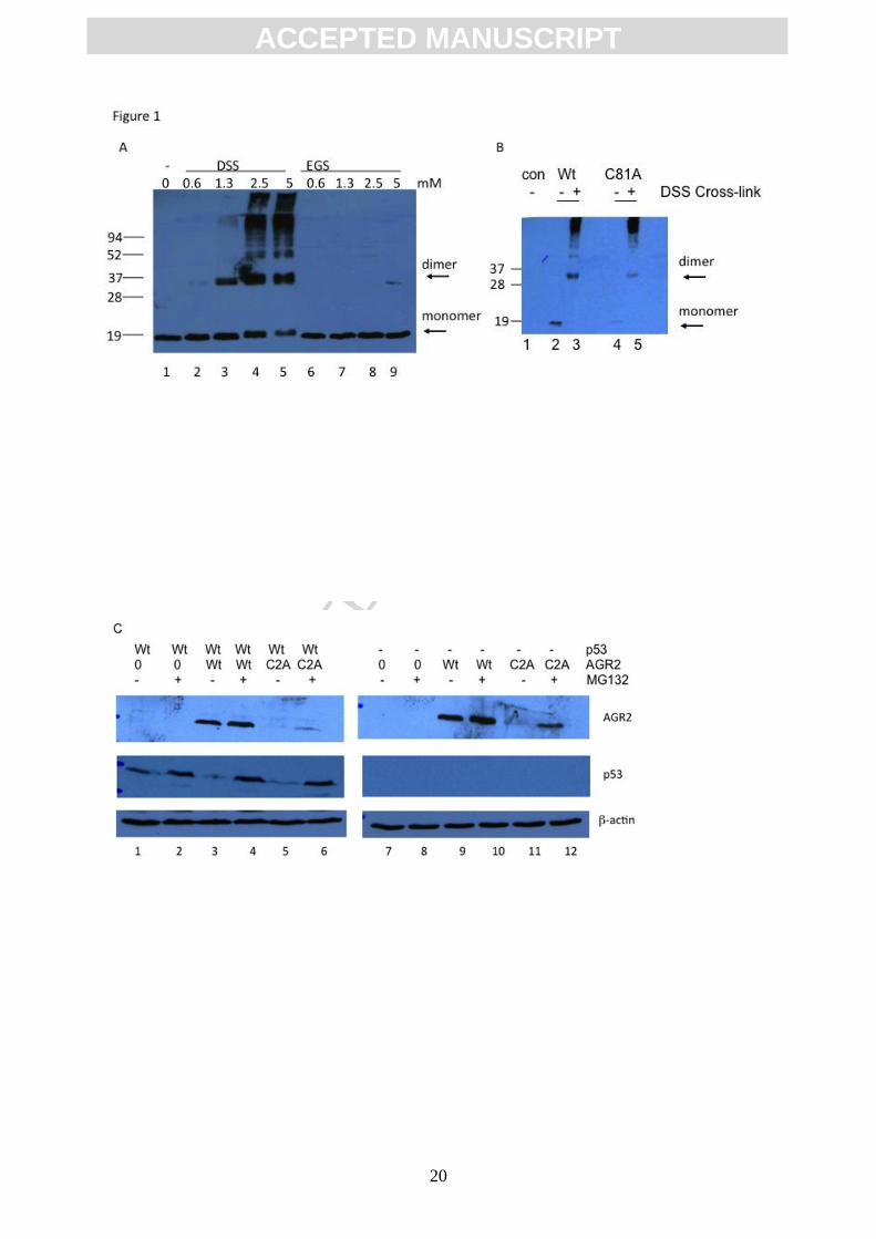
chemicals were initially used as tools to define dimerization determinants. The cell
membrane permeable cross linker DSS (but not EGS) induced a predominant dimerization of
AGR2 in cells (Figure 1A). We observed that this DSS-mediated chemical oxidation occurs
equally well in wild-type AGR2 or the AGR2C81A cysteine mutant (Figure 1B) indicating that
DSS mediated dimerization in cells does not proceed through cysteine oxidation. This is
consistent with previous data showing that in vitro, the DSS reactive cross-link that stabilizes
AGR2 dimerization maps to K95[30]. These data also indicate that mutating Cys81 does not
alter the fixed distance between Lys95 residues in the AGR2 dimer that facilitate DSS-
dependent cross-linking. In addition, we noticed that the AGR2C81A mutant protein exhibited
reduced steady state levels than wt-AGR2 (Figure 1B, lanes). As such we evaluated whether
the instability of the AGR2C81A mutant protein was proteasome dependent; indeed the lower
steady state levels of the AGR2C81A mutant protein were elevated by the proteasome inhibitor
MG132 (Figure 1C). These data suggests that in vivo modification of Cys81 of AGR2 could
alter its steady state levels.
Next we evaluated whether there are any differences in the specific activity of AGR2 as a
function of Cys81 mutation. First, we observed that the AGR2C81A mutant protein is defective
in binding to the chaperone protein Reptin (Figure 1D). Similar loss of binding to Reptin was
observed when using the AGR2K95R mutant protein (Figure 1D). The biochemical activity of
the AGR2C81A mutant protein towards its consensus peptide-docking site was also reduced
compared to wt-AGR2 (Figure 1E). As such, we focused in this study on measuring whether
the more physiologically relevant cysteine oxidation can impact on the dimeric subunit
structure of AGR2.
Oxidation-dependent dimerization of AGR2 dependent upon Cys81
To evaluate the effects of cysteine oxidation on AGR2 dimer stability, we purified his-tagged
AGR2 (larger mass than untagged AGR2 due to the amino acid tag) in the absence of
reducing agent (normally used during purification) that resulted in the accumulation of a

ACC
EPTE
D M
ANU
SCR
IPT
ACCEPTED MANUSCRIPT
9
larger molecular mass species of approximately twice the mass of his-tagged AGR2 (Figure
2A, lane 1). The incubation of the sample with the reducing agent DTT resulted in collapse of
the ~48 kDa species (Figure 2A, lane 2 vs 1) suggesting a cysteine-dependent covalent
dimer was formed under pro-oxidizing conditions. AGR2 protein has one reactive cysteine at
codon 81 (Figure 2B) and mutation of this amino acid to alanine prevented the spontaneous
oxidation-dependent dimerization (Figure 2C, lane 3 vs 1).
In order to determine whether the covalent dimeric form of AGR2 can form through such an
oxidation, we needed to set up an in vitro oxidation system that can be used to monitor
quantitatively covalent dimerization of reduced and monomeric AGR2 protein. A titration in
vitro of H2O2 into a solution with reduced AGR2 protein results in the appearance of dimeric
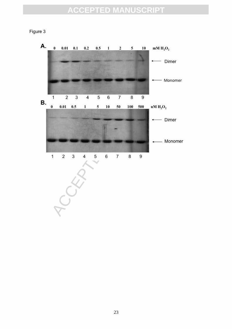
AGR2 (Figure 3A, lane 2 vs 1), whilst at concentrations greater than 200 M the dimerization
was attenuated (Figure 3A, lane 5-9 vs lane 2-4). A titration of H2O2 at reduced
concentrations demonstrates that dimerization is promoted from a concentration of 5-100 M
(Figure 3B, lanes 5-8 vs lanes 1). The use of AGR2C81S and AGR2C81A mutant proteins
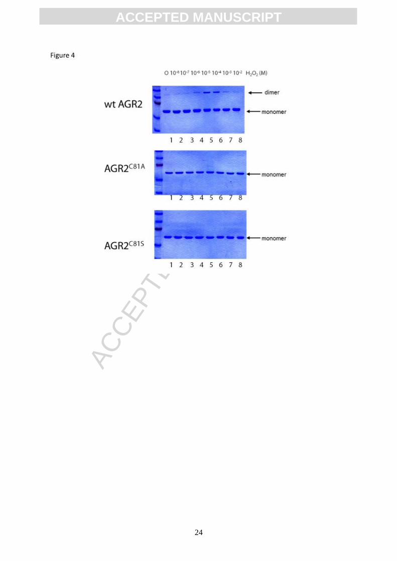
confirmed that the peroxide induced dimerization was dependent upon Cys81 (Figure 4A-C).
These data provide a dynamic assay that can be used to chemically oxidize reduced AGR2
to either promote dimerization at low H2O2 concentrations or to prevent AGR2 dimerization at
elevated concentrations of H2O2. This suppression of AGR2 oxidation at elevated
concentrations of H2O2 suggests a mechanism whereby oxidation levels can regulate the
conversion of AGR2 monomer to dimer. This might prove in future to have effects in cells on
the specific activity of AGR2 in pro-oxidizing environment of the endoplasmic reticulum.
Oxidation-dependent dimerization of AGR2 through direct Cys81 modification
We next set out to determine whether, indeed, chemical evidence can be obtained for direct
Cys81 oxidation through a Cys81-Cys81 cross link but also to explain why higher
concentrations of oxidant prevents AGR2 covalent dimerization. ESI mass spectrometry was
used to determine whether such a reaction mechanism could be developed by analysing
changes in molecular mass of full-length AGR2 as a function of increasing oxidant and
therefore whether evidence could be found for dimerization through a disulfide bond or
through a distinct amino acid.
The AGR2 protein used has an N-terminal histidine-tag (Figure 5A). After rapid replacement
of reduced his-tagged AGR2 protein in ammonium acetate buffer a H2O/MeOH/HCOOH
electrospray make-up solvent was added in order to denature the protein before direct
infusion MS analysis. The protein showed a charge distribution from [M+12H]12+ to
ACC
EPTE
D M
ANU
SCR
IPT
ACCEPTED MANUSCRIPT
10
[M+24H]24+ (Figure 5B and Figure 6) with an nominal average mass of 21,041 Daltons for
the cysteine-reduced protein. Analysis of the isotope distribution of the [M+20H]20+ charge
state is consistent with the theoretical isotope distribution of monomeric AGR2 containing
one reduced cysteine residue [C944H1506N258O277S5]20+ (Figure 5B, insert). Gel filtration
confirmed that AGR2 protein in the reduced state does not form a stable homodimer (data
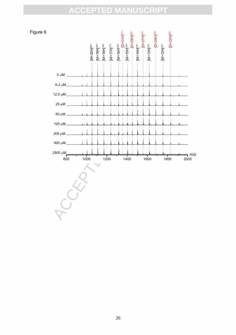
not shown; [30]). A titration of AGR2 with the oxidant H2O2 was performed and monitored by
mass spectrometry, which resulted in the appearance of a second species in the mass
spectrum. This species displayed a charge state distribution from [M+23H]23+ to [M+31H]31+,
and a nominal average mass of 42079 Da; the signals of these ions were elevated in
response to increasing oxidant (Figure 6). This observation is consistent with the formation
of a dimeric AGR2 species, as suggested using gel electrophoresis (Figure 3 and 4). Indeed,
an expanded spectrum of the ion at [M+27H]27+ charge state is consistent with the theoretical
isotope distribution of an AGR2 dimer containing one intermolecular disulfide bond
[C1888H2997N516O554S10]27+(Figure 7A and B). These data together indicate that the reactive
cysteine plays a direct role in covalent dimer formation (Figure 7C).
Interestingly, the additions of higher levels of oxidant prevent dimer formation (Figure 4). To
investigate why dimerization might be suppressed at higher oxidant concentrations, we also
analysed this reaction mechanism using mass spectrometry. Expanded analysis of the ions
isolated with a [M+15H]15+ charge state at higher oxidant concentrations resulted in the loss
of the dimer and accumulation of ions with monomeric mass consistent with the addition of
two oxygen atoms [C944H1501N258O279S5]15+, suggesting oxidation of the cysteine residue
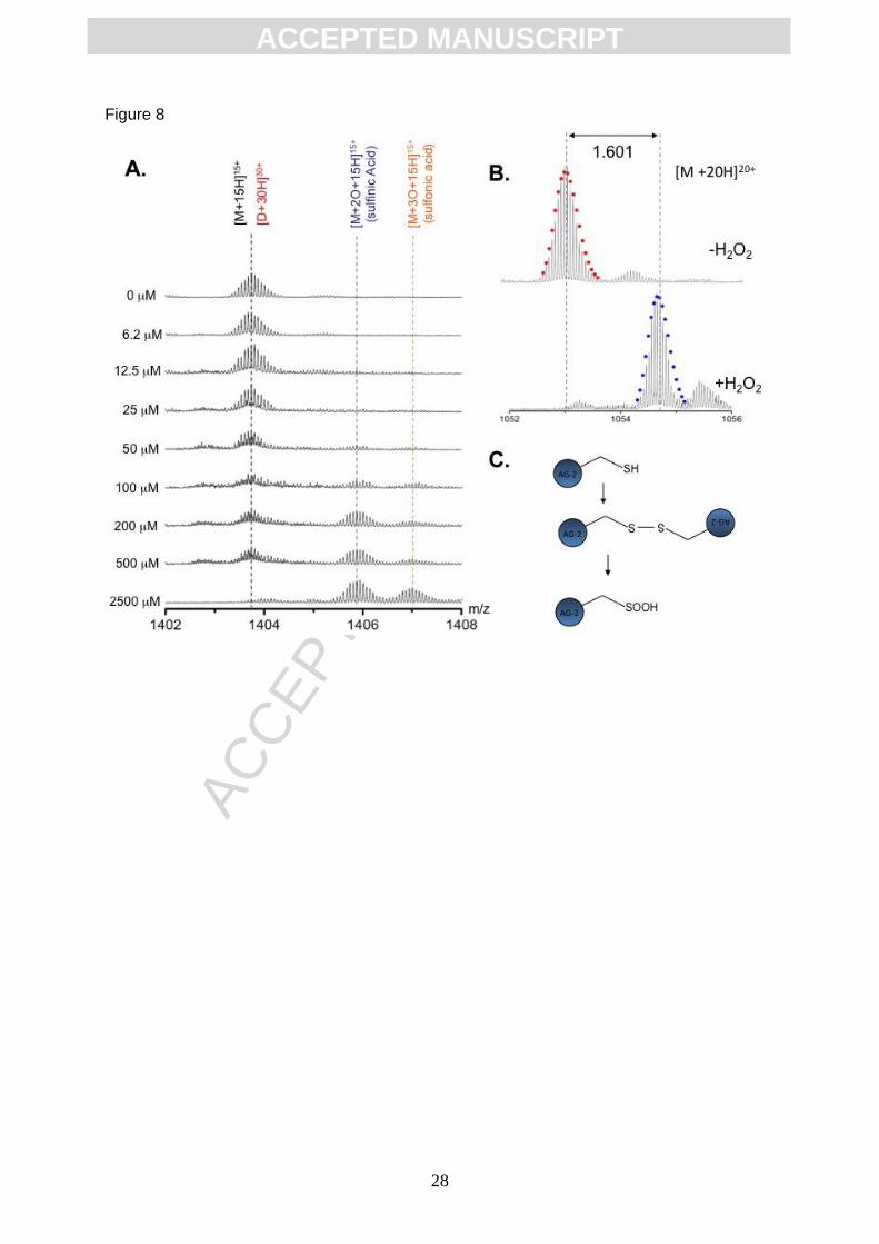
within AGR2 to the sulfinic acid, Cys-SOOH (Figure 8A and B). The highest concentrations of
oxidant also yielded a species with three additional oxygen atoms, [C944H1501N258O280S5]15+,
consistent with formation of sulfonic acid, Cys-SO3H (Figure 8A).
It is clear from the above results that, within AGR2, Cys-81 is susceptible to oxidation by
peroxide. We have demonstrated that with excess peroxide treatment Cys-81 is readily
oxidised to a sulfinic acid. However, under mild peroxide condition (< 200 M), oxidation
favours disulfide bond formation – resulting in an AGR2 dimer covalently coupled via an
intermolecular disulfide bond. Both H2O2 mediated disulfide bond formation and over-
oxidation of cysteine to sulfinic acid are thought to proceed through a labile sulfenic acid
intermediate (Cys-S-OH). Species containing this oxidation state of cysteine are generally
highly reactive and unstable to the ESI process making them unsuitable for MS analysis.
However, it is known that the electrophilic agent NBD-Cl reacts specifically with both Cys-SH
and Cys-S-OH to produce Cys-S-NDB adducts and Cys-S(O)-NDB adducts respectively,
both with mass +163 Da (Figure 9A) [31]. Crucially, upon reaction with NBD-Cl, the oxygen
ACC
EPTE
D M
ANU
SCR
IPT
ACCEPTED MANUSCRIPT
11
atom from a sulfenic acid is retained within the product; producing a mass label that is stable
to ESI-MS. Reduced or oxidized AGR2 protein was incubated with NBD-Cl prior to infusion
for analysis using mass spectrometry. Under these conditions, the protein with charge state
of [M+18H]18+ had an increase in mass of 162.963 Da (Figure 9B) which is consistent with a
single NBD adduct. The oxidized AGR2 mass at this charge state was elevated by 178.982
Da (Figure 9B) which is consistent with the trapping of an unstable sulfenic acid intermediate
(Figure 9B). Together, these MS observations allow us propose a mechanism for peroxide-
mediated dimer formation and propose a mechanism whereby excessive cysteine oxidation
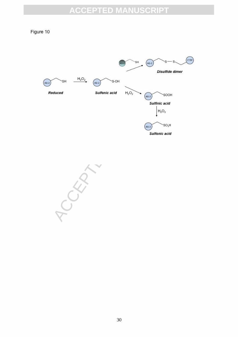
prevents dimer formation (summarized in Figure 10).
DISCUSSION
Our experiments aimed to define how the single cysteine in AGR2 impacts on its protein
binding activity and oxidation-dependent dimerization. In this report, we first developed an in
vivo assay that demonstrates that AGR2 appears to exist in a non-covalent dimeric state as
defined by the ability of DSS to cross link AGR2 into a homo dimer (Figure 1A). This in vivo
cross-link might occur through Lys95-Lys95 adducts (mapped in vitro [30]) and not through
Cys81-Cys81 adducts (Figure 1B). However, the titration of the chemical oxidant peroxide
suggested that AGR2 can assemble into a distinct dimeric structure that is mediated through
a Cys81-Cys81 covalent bond (Figure 4). The discovery that AGR2 forms an oxidation
responsive dimer opens the door to begin to analyse this fundamental property of AGR2
homodimer on its cell physiology. Mutation of the reactive cysteine to alanine reduced the
specific activity of AGR2 in sequence-specific peptide-aptamer binding and in Reptin protein-
binding assay (Figure 1D and E). This mutation also changes the in vivo stability of AGR2
(Figure 1C).
Mass spectrometry is a powerful tool for the characterisation of protein modifications by the
monitoring molecular mass of the intact protein species; for example we have previously
used this technique to define the stoichiometry of methylation and acetylation on native
histones purified from HCT116 colon cancer cells [41] and to characterise reactive cysteine’s
on the p53 tumour suppressor [42]. In this current report, mass spectrometry was used to
define the nature of the covalent modifications on AGR2 as a function of increasing oxidant
concentration. Mass spectrometry has also been previously used to define the mechanism of
sulfonucleotide reduction by sulfonucleotide reductase [43]. We were able to distinguish
between a two models that invoked a reaction mechanism involving the reduction of an inter-
molecular disulfide in a dimeric structure or an intramolecular disulfide bond in a monomeric
form of the enzyme prior to nucleophilic attack on sulfonucleotides to form an enzyme-
thiosulfonate intermediate. Covalent dimerization via disulfide bond formation appears to be
ACC
EPTE
D M
ANU
SCR
IPT
ACCEPTED MANUSCRIPT
12
a increasingly important physiological mechanism involved in regulating signalling proteins.
The adhesion factor E-cadherin can also form a DTT-sensitive dimer with dimerization
driving the degradation of the protein in cells [44]. By contrast, dimerization of AGR2 appears
to have physiological significance since the monomeric mutant protein (C81A) was
significantly de-stabilized through the proteasome pathway in HCT116 colon cancer cells
(Figure 1C). In turn, changes in AGR2 steady state levels in cells might regulate some of the
many biological functions of AGR2 including cell migration, transformation, or p53 inhibition.
Specifically, since the dimer to monomer conversion in AGR2 is responsive to the redox
state of the protein, this suggests that in a pro-oxidizing environment like that observed in
cancer cells and/or in the endoplasmic reticulum, AGR2 oxidation states might yield diverse
regulatory pools of the protein. This has implications for AGR2 binding to Reptin in the
cytosol since the cysteine mutation in AGR2 severely impacts on Reptin Binding (Figure 1).
Indeed, it is interesting that AGR2 is more able to inhibit p53 protein activity after cells have
been subjected to a pro-oxidizing stress of UVC irradiation [8] or cisplatin [10]. Additionally,
AGR2 is also able to mediate export of p53 from the nucleus into the cytoplasm better upon
UVC irradiation [25]. UVC is a known pro-oxidizing stimulus in cells [45, 46] and these data
allow a model to be developed that proposes AGR2 protein activity might be stimulated by
stresses present in a pro-oxidizing environment revealing a potentially physiological role for
dimer assembly in AGR2-mediated oncogenesis.
Lastly, a recent report has confirmed that AGR2 is a PDI that is essential for the production
of the intestinal mucin MUC2, a cysteine-rich glycoprotein that forms the protective mucus
gel lining the intestine [35]. This agrees with other reports suggesting that AGR2 plays a role
in mucus secretion [36] (and European patent WO/2004/056858). This is a major function of
goblet cells and producing this protective mucus layer can have an impact in inflammatory
bowel disease through inhibition of bacterial entry into intestinal cells. The cysteine residue
within the AGR2 thioredoxin-like domain was shown to form mixed disulfide bonds with
cysteines in the N-terminus or C-terminus of MUC2 as the receptor is being processed [35].
Thus we would expect that irreversible oxidation of AGR2 would impact upon mucin
secretion and/or that there exist chemical control mechanisms in the ER that minimize
sulfinic or sulfonic acid formation. This might involve other CxxC containing PDIs that prevent
irreversible oxidation of AGR2. It remains to be determined if AGR2 interacts with such CxxC
containing PDIs. Such control of AGR2 might be important for regulating inflammatory bowel
disease, as AGR2-/- mice are highly susceptible to chemically-induced acute colitis [35].
Further studies are required to precisely determine how dietary chemicals or intracellular
oxidation of AGR2 impacts its ability to interact with cysteine-rich client proteins like MUC2.
ACC
EPTE
D M
ANU
SCR
IPT
ACCEPTED MANUSCRIPT
13
ACKNOWLEDGEMENTS
This work was supported by the Cancer Research UK (C483/A6354), the Medical research
Council-UK, the Breast Cancer Campaign, and the BBSRC RASOR consortium
(BB/C511599/1). DJC would also like to thank the University of Edinburgh for the award of
Chancellor’s Fellowship. This work was partially supported by the project MEYS - NPS-I -
LO1413. BV was supported with GACR P301/11/1678.
REFERENCES
[1] J.M. Weaver, C.S. Ross-Innes, R.C. Fitzgerald, The '-omics' revolution and oesophageal
adenocarcinoma, Nat Rev Gastroenterol Hepatol, 11 (2014) 19-27.
[2] P. Campomenosi, M. Conio, M. Bogliolo, S. Urbini, P. Assereto, A. Aprile, P. Monti, H.
Aste, G. Lapertosa, A. Inga, A. Abbondandolo, G. Fronza, p53 is frequently mutated in
Barrett's metaplasia of the intestinal type, Cancer Epidemiol Biomarkers Prev, 5 (1996) 559-
565.
[3] J.M. Weaver, C.S. Ross-Innes, N. Shannon, A.G. Lynch, T. Forshew, M. Barbera, M.
Murtaza, C.A. Ong, P. Lao-Sirieix, M.J. Dunning, L. Smith, M.L. Smith, C.L. Anderson, B.
Carvalho, M. O'Donovan, T.J. Underwood, A.P. May, N. Grehan, R. Hardwick, J. Davies, A.
Oloumi, S. Aparicio, C. Caldas, M.D. Eldridge, P.A. Edwards, N. Rosenfeld, S. Tavare, R.C.
Fitzgerald, O. Consortium, Ordering of mutations in preinvasive disease stages of esophageal
carcinogenesis, Nat Genet, 46 (2014) 837-843.
[4] T.R. Hupp, Development of physiological models to study stress protein responses,
Methods Mol Biol, 99 (2000) 465-483.
[5] J. Darragh, M. Hunter, E. Pohler, J.F. Dillon, P. Ross, N. Kernohan, T.R. Hupp, The
Calcium Binding Domain of SEP53 is Required for Survival in Response to DCA-Mediated
Stress, The FEBS Journal, 273 (2006) 1930-1947.
[6] T.J. Little, L. Nelson, T. Hupp, Adaptive evolution of a stress response protein, PLoS One,
2 (2007) e1003.
[7] A. Yagui-Beltran, A.L. Craig, L. Lawrie, D. Thompson, S. Pospisilova, D. Johnston, N.
Kernohan, D. Hopwood, J.F. Dillon, T.R. Hupp, The human oesophageal squamous
epithelium exhibits a novel type of heat shock protein response, Eur J Biochem, 268 (2001)
5343-5355.
[8] E. Pohler, A.L. Craig, J. Cotton, L. Lawrie, J.F. Dillon, P. Ross, N. Kernohan, T.R. Hupp,
The Barrett's antigen anterior gradient-2 silences the p53 transcriptional response to DNA
damage, Mol Cell Proteomics, 3 (2004) 534-547.
[9] Z. Wang, Y. Hao, A.W. Lowe, The adenocarcinoma-associated antigen, AGR2, promotes
tumor growth, cell migration, and cellular transformation, Cancer Res, 68 (2008) 492-497.
[10] T.A. Gray, K. Alsamman, E. Murray, A.H. Sims, T.R. Hupp, Engineering a synthetic cell
panel to identify signalling components reprogrammed by the cell growth regulator anterior
gradient-2, Mol Biosyst, 10 (2014) 1409-1425.
[11] F. Aberger, G. Weidinger, H. Grunz, K. Richter, Anterior specification of embryonic
ectoderm: the role of the Xenopus cement gland-specific gene XAG-2, Mech Dev, 72 (1998)
115-130.
ACC
EPTE
D M
ANU
SCR
IPT
ACCEPTED MANUSCRIPT
14
[12] D. Liu, P.S. Rudland, D.R. Sibson, A. Platt-Higgins, R. Barraclough, Human homologue
of cement gland protein, a novel metastasis inducer associated with breast carcinomas, Cancer
Res, 65 (2005) 3796-3805.
[13] A. Kumar, J.W. Godwin, P.B. Gates, A.A. Garza-Garcia, J.P. Brockes, Molecular basis
for the nerve dependence of limb regeneration in an adult vertebrate, Science, 318 (2007)
772-777.
[14] W. Zheng, P. Rosenstiel, K. Huse, C. Sina, R. Valentonyte, N. Mah, L. Zeitlmann, J.
Grosse, N. Ruf, P. Nurnberg, C.M. Costello, C. Onnie, C. Mathew, M. Platzer, S. Schreiber, J.
Hampe, Evaluation of AGR2 and AGR3 as candidate genes for inflammatory bowel disease,
Genes Immun, 7 (2006) 11-18.
[15] D.R. Zweitzig, D.A. Smirnov, M.C. Connelly, L.W. Terstappen, S.M. O'Hara, E. Moran,
Physiological stress induces the metastasis marker AGR2 in breast cancer cells, Mol Cell
Biochem, 306 (2007) 255-260.
[16] A. Mackay, A. Urruticoechea, J.M. Dixon, T. Dexter, K. Fenwick, A. Ashworth, S.
Drury, A. Larionov, O. Young, S. White, W.R. Miller, D.B. Evans, M. Dowsett, Molecular
response to aromatase inhibitor treatment in primary breast cancer, Breast Cancer Res, 9
(2007) R37.
[17] Y. Zhang, S.S. Forootan, D. Liu, R. Barraclough, C.S. Foster, P.S. Rudland, Y. Ke,
Increased expression of anterior gradient-2 is significantly associated with poor survival of
prostate cancer patients, Prostate Cancer Prostatic Dis, 10 (2007) 293-300.
[18] E. Chevet, D. Fessart, F. Delom, A. Mulot, B. Vojtesek, R. Hrstka, E. Murray, T. Gray,
T. Hupp, Emerging roles for the pro-oncogenic anterior gradient-2 in cancer development,
Oncogene, 32 (2013) 2499-2509.
[19] V. Brychtova, A. Mohtar, B. Vojtesek, T.R. Hupp, Mechanisms of anterior gradient-2
regulation and function in cancer, Semin Cancer Biol, 33 (2015) 16-24.
[20] A. Dong, A. Gupta, R.K. Pai, M. Tun, A.W. Lowe, The human adenocarcinoma-
associated gene, AGR2, induces expression of amphiregulin through Hippo pathway co-
activator YAP1 activation, J Biol Chem, 286 (2011) 18301-18310.
[21] A. Gupta, A. Dong, A.W. Lowe, AGR2 gene function requires a unique endoplasmic
reticulum localization motif, J Biol Chem, 287 (2012) 4773-4782.
[22] A. Higa, A. Mulot, F. Delom, M. Bouchecareilh, D.T. Nguyen, D. Boismenu, M.J. Wise,
E. Chevet, Role of pro-oncogenic protein disulfide isomerase (PDI) family member anterior
gradient 2 (AGR2) in the control of endoplasmic reticulum homeostasis, J Biol Chem, 286
(2011) 44855-44868.
[23] P. Patel, C. Clarke, D.L. Barraclough, T.A. Jowitt, P.S. Rudland, R. Barraclough, L.Y.
Lian, Metastasis-Promoting Anterior Gradient 2 Protein Has a Dimeric Thioredoxin Fold
Structure and a Role in Cell Adhesion, J Mol Biol, (2012).
[24] E. Murray, E.O. McKenna, L.R. Burch, J. Dillon, P. Langridge-Smith, W. Kolch, A. Pitt,
T.R. Hupp, Microarray-Formatted Clinical Biomarker Assay Development Using Peptide
Aptamers to Anterior Gradient-2, Biochemistry, 46 (2007).
[25] A. Fourtouna, E. Murray, J. Nicholso, M.M. Maslon, L. Pang, D. Dryden, T. Hupp,
AGR2 as a novel drug target for Activation of the P53 pathway in Cancer: Peptide Aptamers
Targeting AGR2 Stimulate the Nuclear Import of p53, Current Chemical Biology, accepted
for publication (2008).
[26] J.R. Winther, U. Jakob, Redox control: A black hole for oxidized glutathione, Nat Chem
Biol, 9 (2013) 69-70.
[27] M. Delic, C. Rebnegger, F. Wanka, V. Puxbaum, C. Haberhauer-Troyer, S. Hann, G.
Kollensperger, D. Mattanovich, B. Gasser, Oxidative protein folding and unfolded protein
response elicit differing redox regulation in endoplasmic reticulum and cytosol of yeast, Free
Radic Biol Med, 52 (2012) 2000-2012.
ACC
EPTE
D M
ANU
SCR
IPT
ACCEPTED MANUSCRIPT
15
[28] S. Lee, S. Min Kim, J. Dotimas, L. Li, E.P. Feener, S. Baldus, R.B. Myers, W.A.
Chutkow, P. Patwari, J. Yoshioka, R.T. Lee, Thioredoxin-interacting protein regulates protein
disulfide isomerases and endoplasmic reticulum stress, EMBO Mol Med, 6 (2014) 732-743.
[29] S. Persson, M. Rosenquist, B. Knoblach, R. Khosravi-Far, M. Sommarin, M. Michalak,
Diversity of the protein disulfide isomerase family: identification of breast tumor induced
Hag2 and Hag3 as novel members of the protein family, Mol Phylogenet Evol, 36 (2005)
734-740.
[30] T.A. Gray, E. Murray, M.W. Nowicki, L. Remnant, A. Scherl, P. Muller, B. Vojtesek,
T.R. Hupp, Development of a fluorescent monoclonal antibody-based assay to measure the
allosteric effects of synthetic peptides on self-oligomerization of AGR2 protein, Protein Sci,
22 (2013) 1266-1278.
[31] H.R. Ellis, L.B. Poole, Novel application of 7-chloro-4-nitrobenzo-2-oxa-1,3-diazole to
identify cysteine sulfenic acid in the AhpC component of alkyl hydroperoxide reductase,
Biochemistry, 36 (1997) 15013-15018.
[32] A. Fourtouna, E. Murray, J. Nicholson, M.M. Maslon, L. Pang, D.T.F. Dryden, T.R.
Hupp, The Anterior Gradient-2 Pathway as a Model for Developing Peptide-Aptamer Anti-
Cancer Drug Leads that Stimulate p53 Function, Curr Chem Biol, 3 (2009) 124-137.
[33] C.S. Sevier, C.A. Kaiser, Formation and transfer of disulphide bonds in living cells, Nat
Rev Mol Cell Biol, 3 (2002) 836-847.
[34] I. Raykhel, H. Alanen, K. Salo, J. Jurvansuu, V.D. Nguyen, M. Latva-Ranta, L.
Ruddock, A molecular specificity code for the three mammalian KDEL receptors, J Cell Biol,
179 (2007) 1193-1204.
[35] S.W. Park, G. Zhen, C. Verhaeghe, Y. Nakagami, L.T. Nguyenvu, A.J. Barczak, N.
Killeen, D.J. Erle, The protein disulfide isomerase AGR2 is essential for production of
intestinal mucus, Proc Natl Acad Sci U S A, 106 (2009) 6950-6955.
[36] E. Di Valentin, C. Crahay, N. Garbacki, B. Hennuy, M. Gueders, A. Noel, J.M. Foidart,
J. Grooten, A. Colige, J. Piette, D. Cataldo, New asthma biomarkers: lessons from murine
models of acute and chronic asthma, Am J Physiol Lung Cell Mol Physiol, 296 (2009) L185-
197.
[37] H. Zhu, D.C. Lam, K.C. Han, V.P. Tin, W.S. Suen, E. Wang, W.K. Lam, W.W. Cai, L.P.
Chung, M.P. Wong, High resolution analysis of genomic aberrations by metaphase and array
comparative genomic hybridization identifies candidate tumour genes in lung cancer cell
lines, Cancer Lett, 245 (2007) 303-314.
[38] F.R. Fritzsche, E. Dahl, A. Dankof, M. Burkhardt, S. Pahl, I. Petersen, M. Dietel, G.
Kristiansen, Expression of AGR2 in non small cell lung cancer, Histol Histopathol, 22 (2007)
703-708.
[39] E. Missiaglia, E. Blaveri, B. Terris, Y.H. Wang, E. Costello, J.P. Neoptolemos, T.
Crnogorac-Jurcevic, N.R. Lemoine, Analysis of gene expression in cancer cell lines identifies
candidate markers for pancreatic tumorigenesis and metastasis, Int J Cancer, 112 (2004) 100-
112.
[40] G.C. Fletcher, S. Patel, K. Tyson, P.J. Adam, M. Schenker, J.A. Loader, L. Daviet, P.
Legrain, R. Parekh, A.L. Harris, J.A. Terrett, hAG-2 and hAG-3, human homologues of genes
involved in differentiation, are associated with oestrogen receptor-positive breast tumours and
interact with metastasis gene C4.4a and dystroglycan, Br J Cancer, 88 (2003) 579-585.
[41] C.L. Mackay, B. Ramsahoye, K. Burgess, K. Cook, S. Weidt, J. Creanor, D. Harrison, P.
Langridge-Smith, T. Hupp, L. Hayward, Sensitive, Specific, and Quantitative FTICR Mass
Spectrometry of Combinatorial Post-Translational Modifications in Intact Histone H4, Anal
Chem, (2008).
[42] J. Scotcher, D.J. Clarke, S.K. Weidt, C.L. Mackay, T.R. Hupp, P.J. Sadler, P.R.
Langridge-Smith, Identification of two reactive cysteine residues in the tumor suppressor

ACC
EPTE
D M
ANU
SCR
IPT
ACCEPTED MANUSCRIPT
16
protein p53 using top-down FTICR mass spectrometry, J Am Soc Mass Spectrom, 22 (2011)
888-897.
[43] K.S. Carroll, H. Gao, H. Chen, C.D. Stout, J.A. Leary, C.R. Bertozzi, A conserved
mechanism for sulfonucleotide reduction, PLoS Biol, 3 (2005) e250.
[44] M. Trivedi, R.A. Davis, Y. Shabaik, A. Roy, G. Verkhivker, J.S. Laurence, C.R.
Middaugh, T.J. Siahaan, The role of covalent dimerization on the physical and chemical
stability of the EC1 domain of human E-cadherin, J Pharm Sci, (2009).
[45] R.M. Tyrrell, Activation of mammalian gene expression by the UV component of
sunlight- -from models to reality, Bioessays, 18 (1996) 139-148.
[46] H.H. Evans, M.F. Horng, M. Ricanati, J.T. Deahl, N.L. Oleinick, Mutagenicity of
photodynamic therapy as compared to UVC and ionizing radiation in human and murine
lymphoblast cell lines, Photochem Photobiol, 66 (1997) 690-696.
FIGURE LEGENDS
Figure 1. Mutation of AGR2 at Cys81 alters its properties. (A). In vivo treatment of living
cells with cell-membrane permeable homobifunctional N-hydroxysuccimide esters. MCF7
cells grown in media with 10% FCS were incubated with increasing concentrations of the
cell-membrane permeable cross-linkers DSS (lanes 2-5) or EGS (lanes 6-9) for 1 hour at
37oC. Cells were harvested, lysed using a Tris-HCl (pH 8.0) buffer containing 1% NP-40,
lysates were separated by electrophoresis, and protein was immunoblotted to determine
whether intermediary species of AGR2 could be detected under conditions where the
majority of AGR2 was not cross-linked. Higher molecular mass adducts that react with the
AGR2 antibody is highlighted by arrows. (B). Mutation of AGR2 at Cysteine-81 does not
prevent in vivo dimerization induced by DSS. H1299 cells were transfected with expression
vectors including; vector only control (lanes 1), encoding wt-AGR2 (lanes 2 and 3), or
AGR2C81A protein (lanes 4 and 5). Cells were incubated with DSS and lysates were
immunoblotted to examine extent of AGR2 cross-linking. The arrows highlight monomeric
and dimeric AGR2 protein. (C). The C81A mutation in AGR2 sensitizes to proteasome-
dependent degradation in vivo in HCT116 colorectal cancer cells. Vectors expressing wt-
AGR2 or AGR2C81A were transfected into HCT116 cells (lanes 1-6) or isogenic p53-null
HCT116 cells (lanes 7-12). Twenty fours hours later, cells were treated with the proteasome
inhibitor MG132 (as indicated by +) and cells were harvested for immunoblotting: AGR2; p53;
and Actin loading control. (D and E). Effects of C81A mutation of AGR2 on its Reptin and
peptide-binding activity. (D) Fixed amounts of Reptin (500 ng/well) were absorbed onto
ELISA wells or (E) fixed amounts of the indicated biotinylated peptides (D8 and E7 are AGR2
binding peptides and F3 is a non-specific control) were adsorbed onto the streptavidin solid
phase. Next, wt-AGR2, AGR2K95R or AGR2C81A (C2A mutant) proteins were titrated to
measure relative binding affinities of AGR2 towards (D) Reptin or (E) the indicated synthetic
ACC
EPTE
D M
ANU
SCR
IPT
ACCEPTED MANUSCRIPT
17
peptides using anti-AGR2 antibody. The data are plotted as extent of AGR2 binding (RLU) as
a function of AGR2 titration (g).
Figure 2. A reactive cysteine mediates covalent dimerization of AGR2. (A). In vitro
oxidation of AGR2 promotes homodimerization. Recombinant his-tagged AGR2 protein (100
ng) was incubated without reducing agent (DTT; lane 1) or with DTT for 1 hour at 37oC (lane
2) prior to electrophoresis. Samples were immunoblotted for examining changes in the
denatured mass of AGR2 protein. (B). Primary amino acid sequence of AGR2 with Cysteine
81 highlighted. (C). Mutation of Cyteine-81 prevents oxidation induced AGR2 dimerization.
Recombinant his-tagged wt- AGR2 protein (lanes 1, 2, 4 ,and 5) or AGR2C81A protein (lanes
3, 4, 7, and 8) were incubated without or with reducing agent (DTT), as indicated. Samples
were immunoblotted for examining changes in the denatured mass of AGR2 protein.
Figure 3. Oxidant-induced dimerization of AGR2. AGR2 protein was incubated with the
indicated concentrations of hydrogen peroxide ((A) 0-10 mM and (B) 0-500 M]), reactions
were quenched using SDS sample buffer (without DTT), samples were separated by
electrophoresis, and then stained with Coomassie blue. The arrows indicate position of
monomeric and dimeric AGR2.
Figure 4. A reactive cysteine mediates covalent peroxide-induced dimerization of
AGR2. AGR2 protein (wild type, C81A or C81S) were incubated with the indicated
concentrations of hydrogen peroxide (H2O2), reactions were quenched using SDS sample
buffer (without DTT), samples were separated by electrophoresis, and then stained with
Coomassie blue. The arrows indicate position of monomeric and dimeric AGR2
Figure 5. Intact Mass Spectrometry analysis of histidine-tagged AGR2. (A) Amino acid
sequence of AGR2 with the histidine tag (underlined), the theoretical empirical formula
(without N-terminal methionine residue) is highlighted. (B) AGR2 protein affinity purified form
nickel-agarose column was buffer exchanged into 100 mM Ammonium Acetate (pH 7.2) and
the protein concentration was adjusted to 0.5 mg/ml. Aliquots (10 l) were mixed with 20 l of
a electrospray make-up solvent containing 45% methanol and 5% formic acid. Samples were
directly infused and analysed by nESI MS. The data depicts the charge state distribution.
Analysis of the isotope distribution of the [M +20H]20+ charge state is highlighted, insert.
Figure 6. Effects of oxidant titration on the charge state distribution of AGR2 protein.
AGR2 protein (50 M) affinity purified form nickel-agarose column was buffer exchanged into

ACC
EPTE
D M
ANU
SCR
IPT
ACCEPTED MANUSCRIPT
18
100 mM Ammonium Acetate (pH 7.2), incubated with the indicted concentrations of the
oxidant H2O2 (final concentration 6.2 M to 2.5 mM) for 20 minutes before the reaction was
terminated using a 3x volume of electrospray make-up solvent. Samples were directly
infused and analysed by nESI MS. Data depicts the mass-charge distribution (from
[M+20H]20+ to [M+12H]12+) of AGR2 highlighted as a function of H2O2 concentration. Dose
dependent formation of a dimeric species was observed with increasing oxidant
concentration. Charge states corresponding the dimeric species are highlighted in red (from
[D+31H]31+ to [D+23H]23+).
Figure 7. Conversion of AGR2 from monomer and dimer state monitored by mass
sepctrometry. (A) Appearance of the 27+ charge state of AGR2 dimer,[D+ 27H]27+,as a
function of oxidant concentration. Consistent with an AGR2 dimer containing an
intermolecular disulfide bond. (B) Isotope distribution of the [D+ 27H]27+ charge state of
AGR2. The theoretical isotope distribution is overlaid as a scatter plot. (C) Diagram of
disulfide bond formation.
Figure 8. Conversion of AGR2 from reduced monomer to monomeric sulfinic/sulfonic
acid isoform. (A) Expanded mass-charge analysis of the AGR2 [D+30H]30+ /[M+15H]15
charge state; Appearance of species consistent with [M+2O+15H]15+ (sulfinic acid;
highlighted in blue) and [M+3O+H]30+(sulfonic acid; highlighted in orange) are observed at
higher oxidant concentration. (B) Expanded mass-charge analysis of the [M+20H]20+ charge
state highlighting the +32 Da mass shift which occurs upon 2.5 mM H2O2 treatment of AGR2.
(C) This modification is attributed to oxidation of cysteine to sulfinic acid within the AGR2
monomer.
Figure 9. Evidence for sulfenic intermediate in the oxidation of AGR2 protein. (A).
Theoretical effects of NBD-Cl modification of cysteine thiols and cysteine sulfenic acids
which results in a mass of +163 Da. Depicted are the cysteine thiol adduct and the labile
sulfenic acid intermediate adduct. (B). Analysis of the [M+18H]18+ charge state of of AGR2
protein monomers with distinct cysteine NBD-modifications. AGR2 protein (50 M) was
treated with 4 mM NDB-Cl with out without 0.1 mM H2O2. The reaction continued for 45
minutes in the dark prior to quenching with a 3x electrospray make-up solvent containing
45% Methanol and 5% formic acid. The mass/charge spectrum of the [M +18H]18+ charge
state of AGR2 protein highlights the unmodified protein (top panel); the thiol modified protein
(middle panel; 163.963 Da); and a significant amount of the sulfenic-trapped intermediate
(bottom panel; 178.982 Da).

ACC
EPTE
D M
ANU
SCR
IPT
ACCEPTED MANUSCRIPT
19
Figure 10. Distinct isoforms of AGR2 protein can be produced through cysteine
oxidation. Oxidation produces a reactive sulfenic acid intermediate that reacts
intermolecularly with a reduced AGR2 monomer to form a dimeric covalent complex. Higher
levels of oxidant promote the formation of a sulfinic acid that cannot form homodimers or with
the highest level of oxidant leading to the formation of the sulfonic acid. These isoforms
might have distinct specific activities in protein-protein interactions and in mediating client
protein maturation.
ACC
EPTE
D M
ANU
SCR
IPT
ACCEPTED MANUSCRIPT
20
ACC
EPTE
D M
ANU
SCR
IPT
ACCEPTED MANUSCRIPT
21

ACC
EPTE
D M
ANU
SCR
IPT
ACCEPTED MANUSCRIPT
22
ACC
EPTE
D M
ANU
SCR
IPT
ACCEPTED MANUSCRIPT
23
ACC
EPTE
D M
ANU
SCR
IPT
ACCEPTED MANUSCRIPT
24
ACC
EPTE
D M
ANU
SCR
IPT
ACCEPTED MANUSCRIPT
25
Figure 5
ACC
EPTE
D M
ANU
SCR
IPT
ACCEPTED MANUSCRIPT
26

ACC
EPTE
D M
ANU
SCR
IPT
ACCEPTED MANUSCRIPT
27
ACC
EPTE
D M
ANU
SCR
IPT
ACCEPTED MANUSCRIPT
28
Figure 8

ACC
EPTE
D M
ANU
SCR
IPT
ACCEPTED MANUSCRIPT
29
Figure 9
ACC
EPTE
D M
ANU
SCR
IPT
ACCEPTED MANUSCRIPT
30
ACC
EPTE
D M
ANU
SCR
IPT
ACCEPTED MANUSCRIPT
31
Highlights
AGR2 is an ER resident chaperone that mediates cysteine-rich receptor maturation
Mutation of its single cysteine attenuates its protein-protein interaction function
AGR2 dimerizes by two distinct mechanisms, one of which is through cysteine oxidation
Oxidation dimerizes AGR2 through a labile sulfenic acid intermediate
High level oxidation induces a sulfinic or sulfonic acid preventing dimerization
Differential AGR2 oxidation has the potential to impact on its chaperone function