edition edition for immunocytochemical staining. the kit is for 50 tests (100 slides). ... 231 (0),...
TRANSCRIPT

6 'Dako
HercepTestTM for the Dako AutostainerCode K5207
1st edition
For immunocytochemical staining.
The kit is for 50 tests (100 slides).
(xx) K520721-5/US/KDH/2010.10.19 p. 1/50

Contents Page
Intended Use 4Summary and Explanation - Breast 5
Background 5Characteristics 5
Principle of Procedure - Breast 5Reagents - Breast 6
Materials provided 6Materials required but not provided 7
Storage - Breast 7Specimen Preparation - Breast 8
Paraffin-embedded sections 8Treatment of tissues prior to staining 8
Precautions - Breast 8INSTRUCTIONS FOR USE - Breast 10A. Reagent Preparation 10
A.1 EpitopeRetrieval Solution 10A.2 Wash Buffer 10A.3 Substrate-Chromogen Solution (DAB) 10A.4 Counterstain 10A.5 Mounting Medium 10
B. Staining Procedure Performed on the Autostainer 10B.1 Procedural notes 10B.2 Staining protocol 11
Quality Control - Breast 13Interpretation of Staining - Breast 15Limitations - Breast 18
General limitations 18Product-specific limitations 18
Performance Characteristics - Breast 19Background 19Comparison studies 19Comparison to the Clinical Trial Assay (CTA) 19Accuracy 20Reproducibility 21Immunoreactivity 22
Troubleshooting - Breast 23Summary and Explanation - Gastric 26
Background 26Characteristics 26
Principle of Procedure - Gastric 26Reagents - Gastric 27
Materials provided 27Materials required but not provided 28
Storage - Gastric 28Specimen Preparation - Gastric 29
Paraffin-embedded sections 29Treatment of tissues prior to staining 29
Precautions - Gastric 29INSTRUCTIONS FOR USE - Gastric 31A. Reagent Preparation 31
A.1 Epitope Retrieval Solution 31A.2 Wash Buffer 31A.3 Substrate-Chromogen Solution (DAB) 31A.4 Counterstain 31A.5 Mounting Medium 31
B. Staining Procedure Performed on the Autostainer 32B.1 Procedural notes 32B.2 Staining protocol 32
Quality Control - Gastric - 34
(xn) K520721-5/US/KDH/2010.10.19 p. 2150

Interpretation of Staining - Gastric 35Limitations - Gastric 39
General limitations 39Product-specific limitations 40
Performance Characteristics - Gastric 40Background 40Reproducibility 42Immunoreactivity 44
Troubleshooting - Gastric 45References 48Explanation of symbols 50
(xx) K520721-5/US/KDHI2010.10.19 p. 3150

Intended UseFor in vitro diagnostic use.
HercepTest TM is a semi-quantitative immunocytochemical assay to determine HER2protein overexpression in breast cancer tissues routinely processed for histologicalevaluation and formalin-fixed, paraffin-embedded cancer tissue from patients withmetastatic gastric or gastroesophageal junction adenocarcinoma. HercepTest TM isindicated as an aid in the assessment of patients for whom Herceptin® (trastuzumab)treatment is being considered (see Herceptin® package insert).
NOTE for breast cancer only: All of the patients in the Herceptin® clinical trials were selectedusing an investigational immunocytochemical clinical trial assay (CTA). None of the patients inthose trials were selected using the HercepTest TM. The HercepTest TM was compared to theCTA on an independent set of samples and found to provide acceptably concordant results.The actual correlation of the HercepTest TM to Herceptin® clinical outcome has not beenestablished.
NOTE for gastric cancer only: All of the patients in the phase III B018255 (ToGA) studysponsored by Hoffmann-La Roche were selected using Dako HercepTest TM (IHC) and DakoHER2 FISH pharmDxTM Kit (FISH). However, enrollment in the B018255 study was limited topatients whose tumors were HER2 protein overexpressing (IHC 3+) or gene amplified (FISH+;HER2/CEN-17 ratio - 2.0). No patients were enrolled whose tumors were not gene amplifiedbut HER2 protein weakly to strongly overexpressing [FISH(-)/IHC 2+], therefore it is unclear ifpatients whose tumors are not gene amplified but HER2 protein overexpressing [i.e., FISH(-),IHC 2+ or 3+] will benefit from Herceptin® treatment. The study also demonstrated that geneamplification and protein overexpression (IHC) are not as Correlated as with breast cancer,therefore a single method should not be used to determine HER2 status.
For the present kit, Code K5207, reagent volumes have been adjusted especially for use withthe Autostainer.
Gastric or gastroesophageal junction adenocarcinoma is also referred to as gastric cancer inthis document.
For breast cancer application, please refer to pages 3-22.
For gastric cancer application, please refer to pages 23-44.
Important: Please note for breast cancer tissue and gastric cancer tissue differencesespecially in the Interpretation of Staining Sections.
(x) K520721-5/US/KDH/2010.10.19 p. 4150 '31

Breast Cancer
Summary and Explanation - Breast
BackgroundThe human HER2 gene (also known as ERBB2 or N'EU) encodes a protein often referred to asHER2 protein or p1 85HER2 . The HER2 protein is a membrane receptor tyrosine kinase withhomology to the epidermal growth factor receptor (EGFR or HER1) (1-8). The HER2 protein isa normal component expressed by a variety of epithelial cell types (8).
In a fraction of patients with breast cancer, the HER2 protein is overexpressed as part of theprocess of malignant transformation and tumor progression (9). Overexpression of the HER2protein on the surface of breast cancer cells suggested that it could be a target for an antibodytherapeutic. Herceptin® (trastuzumab) is a humanized monoclonal antibody (1 0) that binds withhigh affinity to the HER2 protein and has been shown to inhibit the proliferation of human tumorcells that overexpress HER2 protein in vitro and in vivo (1 1-1 3).
CharacteristicsHercepTeSt TM was developed to provide an alternative to the investigational CTA used in theHerceptin® clinical studies. The performance of HercepTeSt TM for determination of HER2protein overexpression was evaluated in an independent study comparing the results of theHercepTesttm to the CTA on 548 breast tumor specimens, none of which were obtained frompatients in the Herceptin® clinical studies. The results indicated a 79% concordance betweenthe results from the two assays on these tissue specimens.
The concordance data also indicates that a 3+ reading with HercepTeSt TM was highly likely tocorrespond with a positive reading on the CTA which would have met the entry criteria for thetrial (2+ or 3+). A finding of 2+ on HercepTeSt Tm did not correlate as well with the CTA results.Approximately 42% (53/1 26) of HercepTesttm 2+ results were negative by CTA (0 - 1+) whichwould not have allowed entry into the Herceptin® clinical trials.
HercepTesttm is interpreted as negative for HER2 protein overexpression (0 and 1 + stainingintensity), weakly positive (2+ staining intensity), and strongly positive (3+ staining intensity).HercepTeSt TM is not intended to provide prognostic information to the patient and physician andhas not been validated for that purpose.
Principle of Procedure - BreastThe HercepTeSt TM contains reagents required to complete a two-step immuno-cytochemicalstaining procedure for routinely processed, paraffin-embedded specimens. Followingincubation with the primary rabbit antibody to human HER2 protein, this kit employs a ready-to-use Visualization Reagent based on dextran technology. This reagent consists of bothsecondary goat anti-rabbit immunoglobulin molecules and horseradish peroxidase moleculeslinked to a common dextran polymer backbone, thus eliminating the need for sequentialapplication of link antibody and peroxidase-conjugated antibody. Cross-reaction of theVisualization Reagent with human immunoglobulins and fetal calf serum has been removed bysolid-phase absorption. The enzymatic conversion of the subsequently added chromogenresults in formation of a visible reaction product at the antigen site. The specimen may then becounterstained and coverslipped. Results are interpreted using a light microscope.Control Slides containing three formalin-fixed, paraffin-embedded human breast cancer celllines with staining intensity scores of 0, 1+, and 3÷ are provided to validate staining runs.The staining intensity of these cell lines has been correlated to the number of receptorsper cell.
HercepTeSt TM, Code K(5207, is applicable for automated staining using the Autostainer.
0(,a K520721-5/US/KDH/2010.1O.19 p. 5(50

Breast Cancer
Reagents - Breast
Materials providedThe materials listed below are sufficient for 50 tests (50 slides incubated with Primary Antibodyto HER2 Protein and 50 slides incubated with the corresponding Negative Control Reagent).The number of tests is based on the use of 200 pL per slide of vials Nos. 1, 2, 3 and 4, and ofthe Substrate-Chromogen Solution (DAB). The kit provides materials sufficient for a maximumof 15 individual staining runs.
Vial No. Quantity Description
I 2 x 11 mLPeroxidase-Blocking Reagent: 3% hydrogen peroxidecontaining 15 mmol/L sodium azide (NaN 3).
2 1x 12 mLRabbit Anti-Human HER2 Protein: Ready-to-use affinity-isolatedantibody. Supplied in 0.05 mol/L Tris/HCI, 0.1 mol/L NaCI,15 mmol/L. NaN 3, pH 7.2, containing stabilizing protein.
Immunogen: Synthetic C-terminal fragment (intracytoplasmicpart) of the HER2 protein coupled to keyhole limpethemocyanin.
Specificity: HER2 protein.
Purification method: The antibody is affinity isolated by using animmobilized HER2 protein peptide.
3 2 x 11 mL vsSUi.AON
Visualization Reagent: Dextran polymer conjugated withhorseradish peroxidase and affinity-isolated goat anti-rabbitimmunoglobulins. Supplied in Tris/HCI buffer containingstabilizing protein and an antimicrobial agent.
4 1 x 12 mL [ ANCONTROL
Negative Control Reagent: Immunoglobulin fraction of normalrabbit serum at an equivalent protein cbncentration as theantibody to HER2 protein. Supplied in 0.05 mol/L Tris/HCI,0.1 mol/L NaCI, 15 mmol/L NaN3, pH 7.2, containing stabilizingprotein.
5 15 x 1 1 mLEDAB Buffered Substrate: Substrate buffer solution, pH 7.5,containing <0.1% hydrogen peroxide, stabilizers, enhancers,and an antimicrobial agent.
6 3 x 3 mLDAB Chromogen: 5% 3,3'-diaminobenzidine tetrahydrochloridechromogen solution.
7 1 x 1 L I EPITOPE RE" IE SOLU"ON X IL.
Epitope Retrieval Solution (x 10): 0.1 mol/L citrate buffer with anantimicrobial agent.
8 2 x 1 LL suFFR.~Wash Buffer (lOx): Tris/HCI buffer with a detergent and anantimicrobial agent.
3 x 5 slides LsUDEsControl Slides: Each slide contains sections of three formalin-
(xx) K520721-5/US/KDH/2010.10.19 p. 6/50

Breast Cancerfixed, paraffin-embedded breast carcinoma cell linesrepresenting different levels of HER2 protein expression: MDA-231 (0), MDA-175 (1+), and SK-BR-3 (3+). The Control Slideshave been heat treated for better adherence of sections to glassslides. Any additional heat treatment of Control Slidesperformed to improve the adherence of sections to glass slidesmay compromise staining results.
NOTE: All reagents, including Epitope Retrieval Solution and Wash Buffer, areformulated specifically for use with this test. For the test to perform as specified, nosubstitutions should be made except for the Wash Buffer, where Dako Code S3006may be used.
Materials required but not providedAmmonium hydroxide, 15 mol/L diluted to 37 mmol/L
Counterstain: Hematoxylin, such as water-based Mayer's Hematoxylin, Dako Code S3301(see INSTRUCTIONS FOR USE, A.4)Coverslips
Distilled or deionized water (Washing Water)
Drying oven, capable of maintaining 60 0C or less
Ethanol, absolute and 95%
Light microscope (4-40x objective magnification)
Mounting medium, such as Dako Faramount, Code S3025, or Glycergel TM , Code C0563
Positive and Negative Tissues to use as process controls (see Quality Control Section)
Slides, SuperFrost Plus, poly-L-lysine-coated slides, or Dako Silanized Slides, Code S3003,(see Specimen Preparation)
Staining jars or baths
Timer (capable of 2-40 minute intervals)
Water bath with lid (capable of maintaining Epitope Retrieval Solution at 95-99 °C)
Xylene, toluene, or xylene substitutes
K5207 has been tailored for use with the Autostainer Immunostaining System, Code S3400.Please refer to the Autostainer User Guide for necessary Autostainer Components.
Storage - BreastStore at 2-8 °C.
Do not use the kit after the expiration date stamped on the outside package. If reagents arestored under any conditions other than those specified in this package insert, they must bevalidated by the user (14a, 14b). Note that Control Slides must also be stored at 2-8 °C.
There are no obvious signs to indicate instability of this product. Therefore, Positive andNegative Controls should be run simultaneously with patient specimens. If unexpected stainingis observed, which cannot be explained by variations in laboratory procedures, and a problemwith HercepTest TM is suspected, immediately contact Dako's Technical Services.
0(X) K520721-5/US/KDH/2010.10.19 p. 7/50

Breast Cancer
Specimen Preparation - BreastSpecimens from biopsy must be handled to preserve the tissue for immunocytochemicalstaining. Standard methods of tissue processing should be used for all specimens (15).
Paraffin-embedded sectionsTissues preserved in neutral buffered formalin or Bouin's fixative for routine processing andparaffin embedding are suitable for use. For example, specimens from the biopsy should beblocked into a thickness of 3 or 4 mm and fixed for 18-24 hours in neutral buffered formalin.The tissues are then dehydrated in a series of alcohols and xylene, followed by infiltration bymelted paraffin held at no more than 60 0C. Properly fixed and embedded tissues expressingthe HER2 protein will keep indefinitely prior to sectioning and slide mounting if stored in a coolplace (15-25 °C) (15, 16). In the USA, the Clinical Laboratory Improvement Act of 1988requires in 42 CFR 493.1259(b) that "The laboratory must retain stained slides at least tenyears from the date of examination and retain specimen blocks at least two years from the dateof examination" (16).
Tissue specimens should be cut into sections of 4-5 jim, mounted onto slides and air-dried atroom temperature for a minimum of 12 hours (or until dry) or at 37 °C overnight or at 60 °C forone hour. CAUTION: Excessive heating for more than one hour at >60 °C may cause asignificant decrease or loss of the specific membrane-associated HER2 immunoreactivity (17).
To preserve antigenicity, tissue sections mounted onto slides (SuperFrost Plus, poly-L-lysine orsilanized slides) should be stained within 4-6 weeks of sectioning when stored at roomtemperature (20-25 °C) (18). The slides required for HER2 protein evaluation and verificationof tumor presence should be prepared at the same time. A minimum of 5 slides isrecommended, 1 slide for tumor presence, 2 slides for HER2 protein evaluation (1 forincubation with vial No. 2 and 1 for incubation with vial No. 4), and 2 slides for back-up.
The use of HercepTestTM on decalcified tissues has not been validated and is notrecommended.
Consult Dako's "Education Guide: Immunohistochemical Staining Methods" (19) andreferences 15 and 16 for further details on specimen preparation.
Treatment of tissues prior to stainingA specific epitope retrieval method in 10 mmol/L citrate buffer, must be used for optimal assayperformance. The Epitope Retrieval Solution is supplied in the HercepTest TM kit. This methodinvolves heating of tissue sections mounted on slides that are immersed in 10 mmol/L citratebuffer (20) in a calibrated water bath capable of maintaining the Epitope Retrieval Solution atthe required temperature (95-99 0C). Laboratories located at higher elevations shoulddetermine the best method of maintaining the required water bath temperature. The epitoperetrieval must be performed in a water bath. Other methods of heating have been tested anddo not give reproducible results. Immediately after epitope retrieval, commence the stainingprocedure. Deviation from the described procedure may affect results.
Precautions - Breast1. For in vitro diagnostic use.
2. For professional users.
3. Vial 1, Peroxidase-Blocking Reagent, contains 3% hydrogen peroxide. Safety data sheetavailable for professional users on request.
4. Vial 6, DAB Chromogen, contains 1-5% 3,3'-diaminobenzidine tetrahydrochloride (biphenyl-3,3',4,4'-tetrayltetraammonium tetrachloride) and is labeled:Harmful.R40 Limited evidence of a carcinogenic effect.R43 May cause sensitization by skin contact.
00) K520721-5/US/KDHI2010.10.19 p. 8/50

Breast Cancer
R68 Possible risk of irreversible effects.S35 This material and its container must be disposed of in a safe way.S36/37 Wear suitable protective clothing and gloves.As a general rule, persons under 18 years of age are not allowed to work with this product.Users must be carefully instructed in the proper work procedure, the dangerous propertiesof the product and the necessary safety instructions (per European Union Directive94/33/EC). Please refer to the Material Safety Data Sheet (MSDS) for additionalinformation.
USA: 3,3'-diaminobenzidine (DAB) may be harmful by inhalation, in contact with skin and ifswallowed. Material is irritating to eyes and skin. If skin contact should occur, rinse affectedareas with soap and water.
NOTE: Although diaminobenzidine is structurally related to benzidine, there is no evidencefor the carcinogenicity of diaminobenzidine. Consult Federal, State, or local regulations fordisposal.
5. Vial 8, Wash Buffer, contains 5-bromo-5-nitro-1,3-dioxane that may produce an allergicreaction. Safety data sheet available for professional users on request.
6. This product contains sodium azide (NaN3), a chemical highly toxic in pure form. At productconcentrations, though not classified as hazardous, sodium azide may react with lead andcopper plumbing to form highly explosive build-ups of metal azides. Upon disposal, flushwith large volumes of water to prevent metal azide build-up in plumbing (21, 22).
7. Vial 2, 3 and 4 contain material of animal origin. As with any product derived from biologicalsources, proper handling procedures should be used.
8. Control slides and specimens, before and after fixation, and all materials exposed to them,should be handled as if capable of transmitting infection, and disposed of with properprecautions (23). Never pipette reagents by mouth and avoid contacting the skin andmucous membranes with reagents and specimens. If reagents come in contact withsensitive areas, wash with copious amounts of water.
9. Minimize microbial contamination of reagents to avoid non-specific staining.
10. Incubation times, temperatures, or methods other than those specified may give erroneousresults. Excess drying at -60 °C for more than one hour may cause a significant decreaseor loss of the specific membrane-associated HER2 immunoreactivity (17).
1 1. Reagents have been optimally diluted. Further dilution may result in loss of antigenstaining.
12. All reagents, including Epitope Retrieval Solution and Wash Buffer, are formulatedspecifically for use with this test. In order for the test to perform as specified, nosubstitutions should be made except for the Wash Buffer, where Code S3006 may be used.
13. The Visualization Reagent and DAB Chromogen may be affected adversely if exposed toexcessive light levels. Do not store system components or perform staining in strong light,such as direct sunlight.
14. Wear appropriate personal protective equipment to avoid contact with eyes and skin. Referto the Material Safety Data Sheet (MSDS) for.additional information.
(xn) K520721-5/US/KDH/2010.10.19 p. 9/50

Breast Cancer
INSTRUCTIONS FOR USE - Breast
A. Reagent PreparationIt is convenient to prepare the following reagents prior to staining:
A.1 Epitope Retrieval SolutionDilute a sufficient quantity of Vial 7 (Epitope Retrieval Solution x 10) 1:10 using distilled ordeionized water for the staining procedure that is planned. Unused diluted solution may bestored at 2-8 0C for one month. Discard diluted solution if cloudy in appearance.
A.2 Wash BufferDilute a sufficient quantity of Vial 8 (Wash Buffer x 10) 1:10 using distilled or deionized waterfor the wash steps. Unused diluted buffer may be stored at 2-8 °C for one month.Discard buffer if cloudy in appearance.
The Autostainer is programmed to rinse tissue sections after the Peroxidase-Blocking Reagentand Substrate-Chromogen Solution. Note that for these rinse steps either distilled (ordeionized) water or Wash Buffer may be used. All other rinse steps require the use of WashBuffer.
A.3 Substrate-Chromogen Solution (DAB)Prepare Substrate-Chromogen Solution by adding 11 drops (25-30 EL per drop) of DABChromogen from Vial 6 to one Vial 5 containing DAB Buffered Substrate (11 mL) and mix.Prepared Substrate-Chromogen Solution (DAB) is stable for approximately 5 days when storedat 2-8 OC. This solution should be mixed thoroughly prior to use. Any precipitate developing inthe solution does not affect staining quality.
NOTE: The color of the DAB Chromogen in Vial 6 may vary from clear to light lavender-brown.This will not affect the performance of this product. Please dilute according to the guidelines inthis package insert. Addition of excess DAB Chromogen to the DAB Buffered Substrate willresult in deterioration of the positive signal.
A.4 CounterstainThe colored end-product of the DAB staining reaction is alcohol and water insoluble. Use ahematoxylin counterstain and adjust the hematoxylin staining intensity to a similar level shownin Dako's "HercepTest M Interpretation Manual - Breast Cancer". Hematoxylin, either alcoholor water-based, such as Dako Mayer's Hematoxylin, Code S3301, may be used.Follow hematoxylin counterstaining with a thorough rinse in distilled or deionized water, thenimmerse tissue slides into a bath of 37 mmol/L ammonia water (see Section B.2, Step 3).Ammonia water (37 remol/L) is prepared by mixing 2.5 mL of 15 mol/L (concentrated)ammonium hydroxide with 1 liter of distilled or deionized water. Unused 37 mmol/L ammoniawater may be stored at room temperature (20-25 0C) in a tightly capped bottle for up to12 months.
A.5 Mounting MediumNon-aqueous, permanent mounting medium is recommended. However, aqueous mounting isalso acceptable. Dako Faramount Aqueous Mounting Medium, Ready-to-Use, Code S3025, orGlycergel TM Mounting Medium, Code C0563, is recommended for aqueous mounting. LiquefyGlycergel TI by warming to approximately 40 (± 5) 0C prior to use.
B. Staining Procedure Performed on the Autostainer
B.1 Procedural notesThe user should read these instructions carefully and become familiar with all components andinstrumentation prior to use (see Precautions).
(xx) K520721-5/USKDH/2010.10.19 p. 10/50

Breast Cancer
All reagents should be equilibrated to room temperature (20-25 0C) prior to immunostaining.Likewise, all incubations should be performed at room temperature.
Do not allow tissue sections to dry while loading slides on the Autostainer and during thestaining procedure. Dried tissue sections may display increased non-specific staining.
If the staining procedure is interrupted, slides may be kept in a buffer bath following incubationof the primary antibody for up to one hour at room temperature (20-25 °C) without affecting thestaining performance.
Deparaffinization and rehydration: Prior to staining, tissue slides must be deparaffinized toremove embedding medium and rehydrated. Avoid incomplete removal of paraffin. Residualembedding medium will result in increased non-specific staining. This step should beperformed at room temperature (20-25 °C).
1. Place slides in a xylene bath and incubate for 5 (±1) minutes. Change baths andrepeat once.
2. Tap off excess liquid and place slides in absolute ethanol for 3 (±1) minutes. Change bathsand repeat once.
3. Tap off excess liquid and place slides in 95% ethanol for 3 (±1) minutes. Change bathsand repeat once.
4. Tap off excess liquid and place slides in distilled or deionized water for a minimum of30 seconds. Commence staining procedure as outlined in Section B.2, Step 1,Epitope retrieval.
Xylene and alcohol solutions should be changed after 40 slides. Toluene or xylene substitutes,such as Histoclear, may be used in place of xylene.
NOTE: The reagents and instructions supplied in this kit have been designed for optimalperformance. Further dilution of the reagents or alteration of incubation temperatures may giveerroneous or discordant results. Differences in tissue processing and technical procedures inthe user's laboratory may invalidate the assay results for use in selecting patients forHerceptin® therapy.
B.2 Staining protocol
Performed at room temperature, 20-25 'C.
Step 1: Epitope retrievalFill staining jars, e.g. Coplin jars, with the diluted Epitope Retrieval Solution (seeINSTRUCTIONS FOR USE, Section A.1). Place staining jars containing EpitopeRetrieval Solution in water bath. Heat water bath and the Epitope Retrieval Solution to95-99 0C. Cover jars with lids to stabilize the temperature and avoid evaporation.
Immerse the room temperature deparaffinized sections in the preheated EpitopeRetrieval Solution in the staining jars. BRING TEMPERATURE OF THE WATERBATH AND THE EPITOPE RETRIEVAL SOLUTION BACK TO 95-99 °C. Incubatefor 40 (±1) minutes at 95-99 OC.
Remove the entire jar with slides from the water bath. Allow the slides to cool in theEpitope Retrieval Solution for 20 (±1) minutes at room temperature.
Decant the Epitope Retrieval Solution and rinse sections in the Wash Buffer(see INSTRUCTIONS FOR USE, Section A.2).For optimal performance, soak sections in Wash Buffer for 5-20 minutes after epitoperetrieval and prior to staining.
NOTE: The Epitope Retrieval Solution is designed for single use application only.Do not re-use.
(X) . K520721-5/US/KDH/2010.10.19 p. 11/50

Breast Cancer
Step 2: Autostainer procedure1. Use the Autostainer-generated map on the HercepTest TM autoprogram for
required program times and reagent volumes (see point 4 below for specificvolumes).
2. Place the Autostainer reagent vials in the Autostainer reagent rack according tothe computer-generated reagent map.
3. Load the slides onto the Autostainer according to the computer- generatedslide map.
4. Set Program and begin the HercepTest TM program. The following is an outline ofthe program run:* Rinse
* 200 pL Peroxidase-Blocking Reagent - 5 minutes
* Rinse
* 200 pL Primary Anti-HER2 Protein (or Negative Control Reagent) - 30 minutes
* Rinse
* 200 pL Visualization Reagent - 30 minutes
* Rinse
* Rinse
* Switch
* 200 pL Substrate-Chromogen Solution (DAB) - 10 minutes
Rinse slides in deionized water after the substrate-chromogen step
NOTE: Autostainer Hardware version 01 rinses slides in buffer. Therefore, the slidesmust be rinsed with deionized water after they have been removed from theAutostainer.
Step 3: Counterstain (instructions are for hematoxylin)Remove slides from the Autostainer and counterstain in hematoxylin as describedbelow.
Immerse slides in a bath of hematoxylin. Incubate for 2-5 minutes, depending on thestrength of hematoxylin used.
Rinse gently in a distilled or deionized water bath. Ensure all residual hematoxylin hasbeen cleared.Optional: Dip slides 10 times into a bath of 37 mmol/L ammonia water (see SectionA.4).Rinse slides gently in a bath of distilled or deionized water for 2-5 minutes.
NOTE: Depending on the incubation length and potency of the hematoxylin used,counterstaining will result in a pale to dark blue coloration of the cell nuclei. Excessiveor incomplete counterstaining may compromise proper interpretation of results.
Step 4: MountingNon-aqueous, permanent mounting medium is recommended. Otherwise, aqueousmounting medium is also acceptable. Specimens may be mounted and coverslippedwith a water-based mounting medium such as Dako Faramount, Code S3025, orGlycergel T M, Code C0563.
NOTE: Slides may be read when convenient. However, some fading may occur ifslides are coverslipped with an aqueous mounting medium and exposed to strong lightover a period of one week. To minimize fading, store slides in the dark at roomtemperature (20-25 °C).
(xx) K520721-5/US/KDH/2010.10.19 p. 12/50

Breast Cancer
Quality Control - BreastDifferences in tissue fixation, processing and embedding in the user's laboratory may producesignificant variability in results, necessitating regular performance of in-house controls inaddition to the Control Slides supplied by Dako. In the USA, consult the quality controlguidelines of the College of American Pathologists (CAP) Certification Program forImmunohistochemistry, see also CLSI (formerly NCCLS) Quality Assurance forImmunocytochemistry, Approved Guideline (24), and reference 25 for additional information.
Table 1. The purpose of daily quality control.
Non-Specific Antibody' or BufferTissue: Fixed and Processed Like Specific Antibody Plus Same Secondary Antibody asPatient Sample and Secondary Antibody Used with Specific Antibody
Positive Control: Controls all steps of the analysis, Detection of non-specific backgroundTissue or cells containing target Validates reagent and procedures used stainingantigen to be detected (could be for HER2 protein staininglocated in patient tissue). The idealcontrol is weakly positive stainingtissue as this is most sensitive toantibody or antigen degradation
Negative Control: Detection of unintended antibody Detection of non-specific backgroundTissues or cells expected to be cross-reactivity to cells/cellular stainingnegative (could be located in patient componentstissue or positive control tissue)
Patient tissue Detection of specific staining Detection of non-specific backgroundstaining
Control Slide supplied by Dako Controls staining procedure only
* Serum from same species as the specific antibody, but not directed against the same target antigen. To detect non-specific antibody binding, e.g. binding of Fc portion of antibody by the tissue.
Control Slide (provided): Each of the supplied Control Slides contains three pelleted,formalin-fixed, paraffin-embedded human breast cancer cell lines with staining intensity scoresof 0, 1+ and.3+. One slide should be stained in each staining run. The evaluation of the ControlSlide cell lines supplied by Dako indicates the validity of the staining run.
Positive Control Tissue: Controls should be fresh autopsy/biopsy/surgical specimens fixed,processed and embedded as soon as possible in the same manner as the patient sample(s).Positive tissue controls are indicative of correctly prepared tissues and proper stainingtechniques. One Positive Control Tissue for each set of test conditions should be included ineach staining run.
The Positive Control Tissues should give weak positive staining so they can detect subtlechanges in the primary antibody sensitivity. The Control Slides supplied with this kit orspecimens processed differently from the patient sample(s) validate reagent performance onlyand do not verify tissue preparation. Use previously determined HER2 protein 2+overexpressing invasive (infiltrating) human breast carcinoma tissue for the ideal PositiveControl Tissue.
NOTE: Known positive control tissue should only be utilized for monitoring the correctperformance of processed tissues and test reagents, NOT as an aid in formulating a specificdiagnosis of patient samples. If the Positive Control Tissue fails to demonstrate appropriatepositive staining, results with the patient specimens should be considered invalid.
Negative Control Tissue: Use a negative control tissue (known to be HER2 protein negative)fixed, processed and embedded in a manner identical to the patient sample(s) with eachstaining run to verify the specificity of the primary antibody and to provide an indication of
(xx) K520721-5/US/KDH/2010.10.19 p. 13150

Breast Cancer
specific background staining. Colon, liver or thyroid are appropriate for negative control tissue.The variety of different cell types present in most tissue sections offers internal negative controlsites (this should be verified by the user). Normal breast ducts can serve as internal negativecontrols.If specific staining occurs in the Negative Control Tissue or in the internal negative controltissue, results with the patient specimens should be consideredinvalid and the test re-run.
Non-Specific Negative Control Reagent: Use the supplied Negative Control Reagent inplace of the primary antibody with a section of each patient specimen to evaluate non-specificstaining and allow better interpretation of specific staining at the antigen site. The incubationperiod for the Negative Control Reagent should correspond to that of the primary antibody.
Assay verification: Prior to initial use of an antibody or staining system in a diagnosticprocedure, the user should verify the antibody's specificity by testing it on a series of in-housetissues with known immunocytochemical performance characteristics representing knownpositive and negative tissues. Refer to the quality control procedures previously outlined in thissection of the product insert and to the quality control requirements of the CAP CertificationProgram for Immunohistochemistry and/or CLSI (formerly NCCLS) Quality Assurance forImmunocytochemistry, Approved Guideline (24). These quality control procedures should berepeated for each new antibody lot, or whenever there is a change in assay parameters. Breastcarcinomas with known HER2 protein staining intensities from 0 - 3+ and negative tissues,e.g. colon, liver or thyroid are suitable for assay verification.
Oc<) K520721-51US/KDH/2010.10.19 p. 14/50

Breast Cancer
Interpretation of Staining - BreastFor the determination of HER2 protein overexpression, only the membrane staining intensityand pattern should be evaluated using the scale presented in Table 2. Slide evaluation shouldbe performed by a pathologist using a light microscope. For evaluation of theimmunocytochemical staining and scoring, an objective of 1Ox magnification is appropriate.The use of a 20-40x objective magnification is useful in confirmation of the score. Cytoplasmicstaining should be considered non-specific staining and is not to be included in the assessmentof membrane staining intensity (8). To aid in the differentiation of 0, 1+, 2+ and 3+ staining, referto Dako's "HercepTest TM Interpretation Manual - Breast Cancer" for representative pictures ofthe staining intensities.
Only specimens from patients with invasive breast carcinoma should be scored. In cases withcarcinoma in situ and invasive carcinoma in the same specimen, only the invasive componentshould be scored.Table 2. Cell membrane staining intensity criteria.
HER2 ProteinOverexpression
Staining pattern Score Assessment(Report to treating physician) (Report to treating physician)
No staining is observed or membrane staining is observed in less 0 Negativethan 10% of the tumor cells
A faint/barely perceptible membrane staining is detected in morethan 10% of the tumor cells. The cells are only stained in part of 1+ Negativetheir membrane
A weak to moderate complete membrane staining is observed in 2+ Weakly positivemore than 10% of the tumor cells
A strong complete membrane staining is observed in more than10% of the tumor cells 3+ Strongly positive
HercepTest TM is interpreted as negative for HER2 protein overexpression (0 and 1 + stainingintensity), weakly positive (2+ staining intensity), and strongly positive (3+ staining intensity).HercepTest TM is not intended to provide prognostic information to the patient and physician andhas not been validated for that purpose.
For each staining run, slides should be examined in the order presented in Table 3 to determinethe validity of the staining run and enable semi-quantitative assessment of the staining intensityof the sample tissue.
(xx) K520721-51USIKDHI2010.10.19 p. 15/50

Breast CancerTable 3. Order of slide evaluation.
Slide Reading Order Rationale
1. Control Slide containing the three cell lines Presence of 3+ brown cell membrane staining (rimming) in the3+ Control Cell Line SK-BR-3, partial brown rimming in the 1+Control Cell Line MDA-1 75, and no staining in the 0 Control CellLine MDA-231 indicates a valid assay
Punctate and discontinuous membrane staining is present in asmall to moderate number of the cells in the weakly positive 1 +Control Cell Line MDA-175. Also dot-like immunostaining of theGolgi region of the cytoplasm can be observed in this cell line
Presence of brown staining in the 0 Control Cell Line MDA-231(negative for HER2 protein staining) indicates that there wasnon-specific staining during the assay. The assay results maybe invalid due to overstaining
2. Positive Control Tissue'Slide Presence of brown membrane staining should be observed.Staining of the cytoplasm and negative tissues should not bemore than 1+
3. Negative Control Tissue Slide The ABSENCE of specific staining in the Negative ControlTissue Slide confirms the lack of kit cross-reactivity tocells/cellular components. If specific membrane staining occursin the Negative Control Tissue Slide, results with the patientspecimen should be considered invalid
4. Patient tissue slide stained using the Negative Control Absence of specific membrane staining verifies the specificReagent labeling of the target antigen by the primary antibody
Other tan or brown staining occurning in the cytoplasm of thespecimen treated with the Negative Control Reagent, such as inconnective tissue, leukocytes, erythrocytes, or necrotic tissue,should be considered non-specific background staining andshould be reported under the comments section of the dataspreadsheet
5. Patient tissue slide stained using the primary antibody When HER2 protein overexpression is detected in thespecimen, it will appear as brown rimming localized on the cellmembrane of tumor cells treated with the primary antibody
1. Control Slide (provided): The Control Slide stained with HercepTest TM should be examinedfirst to ascertain that all reagents are functioning properly. The presence of a brown (3,3'-diaminobenzidine, DAB) reaction product at the cell membrane is indicative of positivereactivity.
Presence of circumferential brown cell membrane staining (rimming) in the 3+ Control Cell LineSK-BR-3, partial brown rimming in the 1+ Control Cell Line MDA-175, and no staining in the 0Control Cell Line MDA-231 indicates a valid assay. If any of the Control Cell Lines performoutside of these criteria, all results with the patient specimens should be considered invalid.
2. Positive Control Tissue: The Positive Control Tissue Slide should be examined next.This slide verifies that the fixation method and epitope retrieval process are effective.Use intact cells for interpretation of staining results because necrotic or degenerated cells oftenstain non-specifically (26). Staining should be observed in tumor tissue as brown, cellmembrane staining. Brown staining of the cytoplasm and negative tissues within the specimenshould not be more than corresponding to 1 + staining intensity score.
3. Negative Control Tissue: The Negative Control Tissue Slide should be examined after thePositive Control Tissue to verify the specificity of the labeling of the target antigen by theprimary antibody. The absence of specific staining in the Negative Control Tissue confirms thelack of kit cross-reactivity to cells/cellular components. If specific staining occurs in theNegative Control Tissue, results with the patient specimen should be considered invalid.Alternatively, negative portions of the Positive Control Tissue may serve as the NegativeControl Tissue, but this should be verified by the user. Note that a weak reaction (0 - 1 +
(xx) K520721-5/USIKDH/2010.10.19 p. 16/50

Breast Cancer
staining intensity) can be observed in most normal epithelial tissue. Possible negative controltissues include: colon, liver and thyroid.
Non-specific staining, if present, will be of a diffuse appearance. Sporadic staining ofconnective tissue may also be observed in sections from excessively formalin-fixed tissues.
4 + 6. Patient Tissue: Examine patient specimens stained with HercepTest TM last. Positivestaining intensity should be assessed within the context of any non-specific backgroundstaining of the Negative Control Reagent. As with any immunocytochemical test, a negativeresult means that the antigen was not detected, not that the antigen was absent in thecells/tissue assayed. Refer to Summary and Explanation, Limitations, and PerformanceCharacteristics for specific information regarding HercepTest TM immunoreactivity.
Additional Recommendations for Interpretation of HercepTestTM StainingMost metastatic breast carcinomas tested for HER2 protein overexpression are given a scoreof 0 or 3+. While the majority of these cases are clear-cut, a small percentage of the remaining1+ and 2+ samples may be more difficult to interpret. Use the following guidelines forinterpretation of HercepTest TM staining in your laboratory.* Evaluate the Control Cell Lines to validate the assay performance.
* Evaluate the Positive and Negative Control Slides.
* A hematoxylin and eosin (H&E) staining of the tissue specimen is recommended for the firstevaluation. (The tumor may not be obvious when looking at the sample stained withHercepTest TM . An H&E stained slide is required from the pathologist to verify the presenceof the tumor). The HercepTest T Mshould be performed on a paired section (serial section)from the same paraffin block of the specimen.
* Evaluate the sections stained for HER2 protein overexpression at low power first.The majority of positive cases will be obvious at low power magnification.
* Well-preserved and well-stained areas of the specimen should be used to make adetermination of the percent positive tumor cells.
* In general, the score of cases should be obvious at low magnification. If determinationbetween 1 +/2+ borderline cases is difficult at low magnification, the score is usually 1+.
* To verify membrane staining, use 20-40x objective magnification.
* If a majority of tumor cells demonstrate complete membrane staining, the staining is either2+ or 3+. Go to 20-40x objective magnification to confirm score.
* In a majority of the 3+ cases, at least 80% of the tumor cells are stained and the membranestaining is intense.
* If the specimen is near the cut off of 10% tumor cells positive, it is recommended that aminimum of 100 tumor cells be counted to determine the percentage of stained cells.
* If there is complete membrane staining at a weak to moderate intensity in greater than 10%of the tumor cells, the score of the specimen is 2+. This is usually accompanied byincomplete membrane staining of the majority of the remaining tumor cells.
* If less than 10% of the tumor cells have complete circumferential membrane staining,although other tumor cells may demonstrate an incomplete membrane staining, the score is1+.
* If less than 10% of the tumor cells have complete or incomplete circumferential membranestaining, the score is 0.
(XX) K520721-5/USIKDH/2010.10.19 p. 17/50

Breast Cancer
Limitations - Breast
General limitations1. Immunocytochemistry is a multi-step diagnostic process that requires specialized training
in the selection of the appropriate reagents; tissue selection, fixation, and processing;preparation of the immunocytochemistry slide; and interpretation of the staining results.
2. Tissue staining is dependent on the handling and processing of the tissue prior to staining.Improper fixation, freezing, thawing, washing, drying, heating, sectioning, or contaminationwith other tissues or fluids may produce artifacts, antibody trapping, or false-negativeresults. Inconsistent results may be due to variations in fixation and embedding methods,or to inherent irregularities within the tissue.
3. Excessive or incomplete counterstaining may compromise proper interpretation of results.
4. The clinical interpretation of any positive staining or its absence must be evaluated withinthe context of clinical presentation, morphology and other histopathological criteria.The clinical interpretation of any staining, or its absence, must be complemented bymorphological studies and proper controls as well as other diagnostic tests. It is theresponsibility of a qualified pathologist, who is familiar with the antibodies, reagents andmethods used, to interpret the stained preparation. Staining must be performed in acertified, licensed laboratory under the supervision of a pathologist who is responsible forreviewing the stained slides and assuring the adequacy of positive and negative controls.
5. Tissues from persons infected with hepatitis B virus and containing hepatitis B surfaceantigen (HBsAg) may exhibit non-specific staining with horseradish peroxidase (27).
6. Reagents may demonstrate unexpected reactions in previously untested tissue types.The possibility of unexpected reactions even in tested tissue types cannot be completelyeliminated due to biological variability of antigen expression in neoplasms, or otherpathological tissues (28). Contact Dako's Technical Services with documentedunexpected reaction.
7. False-positive results may be seen due to non-immunological binding of proteins orsubstrate reaction products. They may also be caused by pseudoperoxidase activity(erythrocytes) and endogenous peroxidase activity (cytochrome C) (28).
8. The staining procedure should be performed at ambient temperature of 20-25 OC.
Product-specific limitations1. The antigen present in the 1+ Control Cell Line MDA-175 is subject to degradation over
time. Assess the Control Slide results in connection with the expiration date of the ControlSlide. Negative staining of the MDA-175 cells may only indicate that the Control Slide hasdegraded. The Control Slides must be stored at 2-8 0C.
2. False-negative results could be caused by degradation of the antigen in the tissues overtime. Specimens should be stained within 4-6 weeks of mounting of tissues on slides whenstored at room temperature (20-25 °C) (29).
3. For optimal and reproducible results, the HER2 protein requires heat-induced epitoperetrieval when tissues are routinely fixed (neutral buffered formalin or Bouin's fixative) andparaffin embedded. This pre-treatment needs to be completed at the beginning of theentire staining process. See the "Specimen Preparation Section, Treatment of tissues priorto staining" for instructions.
4. Heat-induced epitope retrieval of the HER2 protein should only be done using a calibratedwater bath. Other methods of heating have been tested and do not give reproducibleresults.
5. Do not replace kit reagents with reagents carrying other lot numbers or with reagents fromother manufacturers. The only exception is the Wash Buffer that may be replaced withDako Wash Buffer, Code S3006.
(xx) K520721-5/US/KDH/2010.10.19 p. 18/50

Breast Cancer
6. False results could be obtained from evaluation of cytoplasmic staining. Consider only theintensity of cell membrane staining when interpreting results.
7. Stained Control Slides should be used only for validation of the staining run and should notbe used as a guide to score the staining reaction in tissue sections.
8. Strong focal staining (3+), i.e. "hot spots," may occasionally be seen. This may be theresult of uneven fixation and/or processing of tissue. Immunostaining of a second tissueblock from the same specimen is recommended.
9. Use of HercepTest TM on specimens fixed in fixatives other than neutral buffered formalin orBouin's fixative has not been validated.
10. Normal epithelium in breast tissue should stain between 0 and 1+. If higher than 1 +staining of the normal epithelium is observed, the test should be repeated. Note thatnormal tonsil and esophageal epithelia may stain up to 2+ intensity.
Performance Characteristics - Breast
BackgroundThe clinical trial assay (CTA) used to identify eligible patients for the Herceptin® clinical studieswas for investigational use and is no longer available. The HercepTest TM was developed toprovide a comparable alternative to the CTA.
The safety and effectiveness of Herceptin® were evaluated in a randomized controlled clinicaltrial and a large, open-labelled trial (See Herceptin® package insert). All patients selected forthe Herceptin® clinical trials demonstrated overexpression of HER2 protein byimmunocytochemistry testing performed with the CTA at a central laboratory. Patients wereeligible for Herceptin® treatment if their tumor had 2+ or 3+ levels of HER2 proteinoverexpression (based on a 0-3+ scale, where 3+ represented the highest level).
Subgroup analysis of the results from these studies suggests that patients whose tissues arestrongly positive (3+) for HER2 protein overexpression may benefit more from Herceptin® thanpatients whose tissues are weakly positive (2+). The degree of HER2 protein overexpression ispotentially an important predictor of the effect of Herceptin® treatment. Because none of thepatients in the Herceptin® studies were selected using the HercepTest® the correlationbetween the degree of positivity and the likelihood of clinical benefit from Herceptin® treatmentis unknown.
Comparison studiesTwo studies were performed to characterize the HercepTest TM .
1) Comparison to the Clinical Trial Assay (CTA).2) Accuracy when compared with five additional assays.
Comparison to the Clinical Trial Assay (CTA)The HercepTestTM was compared to the CTA used to identify eligible patients for Herceptin®therapy using 274 HER2 protein positive (2+ or 3+) and an equal number of HER2 proteinnegative breast cancer tissue specimens. Table 4 shows the results in a 2 x 2 diagram where 0and 1 + were considered to be negative and 2+ and 3+ were positive.
(xx) K520721-5/US/KDH/2010.10.19 p. 19/50 ('6~

Breast CancerTable 4. A 2 x 2 concordance of the HercepTest"- to the Clinical Trial Assay (number of specimens).
Clinical Trial Assay
Positive Negative Total
HercepTest TM Positive 216 59 275
Negative 58 215 273
Total 274 274 548
Concordance: 79% (76-82%) 95% confidence interval.
The overall binary concordance of the HercepTeStTM to the OTA was 79% (4311548), with a 2-sided 95% confidence interval of 76-82%. Twenty one per cent (21 %) of the results werediscordant between these two methods.The HercepTest Tm results are reported on a 0-3+ scale interpreted as negative for HER2protein overexpression (0 and 1 + staining intensity), weakly positive (2+ staining intensity), andstrongly positive (3+ staining intensity).
Table 5. A 3 x 3 concordance for Hercep-restT" and Clinical Trial Assay.
Clinical Trial Assay
3+ 2+ 0 -1+ Total
Hercep~rest tm 3+ 107 36 6 149
2+ 16 57 53 126
0 -1+ 8 50 215 273
Total 131 143 274 548
This 3 x 3 presentation of the concordance study indicates that a 3+ reading on theHercepTestWr is highly likely to correspond with a positive result on the CTA, which would havemet the entry criteria for the Herceptin® trial (2+ or 3+). A finding of 2+ on HercepTeSt Tm didnot correlate as well with the CTA results. Approximately 42% (53/1 26) of HercepTeStTm 2+results were negative by OTA (0 - 1 +) which would not have allowed entry into the Herceptin®clinical trials.
AccuracyHercepTesttm was also tested on 2 microscope slides containing paraffin-embedded tissuesections from 168 breast tumors. These tumors had been previously characterized by fivedifferent methods of determining HER2 gene amplification and overexpression of HER2protein, including in-house Southern blot, fluorescence in situ hybridization (FISH) foramplification of DNA, Northern blot RNA analysis, Western blot, and immunocytochemistry (ICC)on frozen tissues (29). The results are presented in Table 6.
Table 6. Comparison of HercepTest1" to combined results (C1E) from gene amplification and HER2 protein overexpression tests.
Reference OE Classification
+ - Total
Hercop~restw + 43 0 43
26 99 125
Total 69 99 168
Positive agreement: 43169 = 62%Negative agreement: 99/99 = 100%
The results indicated an 85% (142/1 68) level of agreement (95% confidence interval of78-89%) between the positivity (2+ and 3+) and negativity (0 and 1 +) staining intensity by the
(XX) K520721-5/US/KDH/201 0.10.19 p. 20/50

Breast Cancer
HercepTest TM. None of the samples negative by the 5 different methods were positive by theHercepTest TM, whereas the combined results for the 5 different methods showed a highernumber of positive cases.
ReproducibilityIntra-run reproducibility: Intra-run reproducibility was tested in one laboratory with5 specimens of different ICC intensity staining scores. Each specimen was run in triplicate in amasked randomized format. This protocol was used with automated staining. All specimensgave 100% reproducible results.
Inter-run reproducibility: Inter-run reproducibility was tested at three laboratories over 4 dayswith 5 specimens of different ICC intensity staining scores randomized and masked usingautomated methodology. Excellent reproducibility was seen for positive versus negative results(0 and 1 + versus 2+ and 3+) with the exception of two samples in one laboratory that variedbetween 1 + and 2+. There was 100% reproducibility for the 2+ and 3+ samples.
Inter-laboratory reproducibility: Inter-laboratory reproducibility was tested at threegeographically separated laboratories with 40 identical randomized and masked specimens ofvarious ICC staining intensity scores. Freshly cut slides were forwarded to each testinglaboratory for automated staining and evaluation by a pathologist. Inter-laboratory percentagreement ranged from 83% to 90% for a dichotomous positive/negative determination where0 and 1 + were negative and 2+ and 3+ were positive for HER2 protein overexpression.Compared to results obtained at the reference laboratory that had performed the CTA, 12.5%(15/120) comparative results were discrepant between negative (0 or 1+) and positive (2+ or3+) determinations. An additional 10% (12/120) were discrepant between 2+ and 3+ scores.
0(n K520721-5/US/KDH/2010.10.19 p. 21/50 (q

Breast Cancer
ImimuncoreactivityTable 7 summarizes HercepTeSt TM immunoreactivity with the recommended panel of normaltissues. All tissues were formalin fixed and paraffin embedded and stained with HercepTesttmaccording to the instructions in the package insert.
Table 7. Summary of Hercep~rest"~ normal tissue reactivity.
Tissue Type Positive Tissue Element Staining and(No. Tested) Staining Pattern
Adrenal (3) NoneBone marrow (3) NoneBrain/Cerebellum (3) NoneBrainlCerebrum (3) NoneBreast (3) Mammary gland (1 + staining intensity)Cervix uteri (3) NoneColon (3) Columnar epithelium, surface (1-. staining intensity)Esophagus (3) Squamous epithelia (113 tissues, 2+ staining intensity)Heart (3) NoneKidney (3) Tubule (1+ staining intensity)Liver (3) NoneLung (3) NoneMesothelial cells (3) NoneOvary (3) NonePancreas (3) Langerhans cells, cytoplasmic (3+ staining intensity)Parathyroid (3) NonePeripheral nerve (3) NonePituitary (3) Endocrine cells, cytoplasmic (3+ staining intensity)Prostate (3) Prostate gland (2+ staining intensity)Salivary gland (3) NoneSkeletal muscle (3) NoneSkin (3) NoneSmall intestine (3) Columnar epithelium, surface (1 + staining intensity)Spleen (3) NoneStomach (3) Epithelium (1/3 tissues, 1 + staining intensity)Testis (3) NoneThymus (3) NoneThyroid (3) NoneTonsil (3) Squamous epithelia (2+ staining intensity)Uterus (3) Endometrium (1/3 tissues, 1+ staining intensity)
Reported staining in all tissues was membrane, unless otherwise noted. All threespecimens of each tissue type had the same staining intensity unless otherwise noted.
(xx) ~~~~~~~~~~~~~~~~~K520721-5/US/KDH/2010.10.19 p. 22150

Breast Cancer
Troubleshooting - BreastRefer to the Troubleshooting section in Dako's previously referenced Handbook (19) forremedial action, or contact Dako's Technical Service Department to report unusual staining.
Problem Probable Cause Suggested Action
1. No staining of la. Programming error. Reagents la. Check programming grid to verifyslides not used in proper order that the staining run was
programmed correctly
lb. Reagent vials were not loaded lb. Check the Reagent Map to verifyin the correct locations in the the proper location of reagentreagent racks vials
ic. Insufficient reagent in ic. Ensure that enough reagent isreagent vial loaded into the reagent vials prior
to commencing the run. Refer toReagent Map for volumes required
ld. Sodium azide in Wash Solution id. Use fresh preparation of WashBuffer provided in the kit
le. Excessive heating of mounted le. Air dry the tissue sections attissue sections prior to room temperature for a minimumdeparaffinization and heat- of 12 hours or until dry.induced antigen retrieval may Alternatively, dry at 37 °Clead to loss of visible HER2 overnight or dry at 60 0C for aimmunoreactivity. maximum of one hour. Drying of
tissue sections at elevatedtemperatures must only beperformed in a calibrated ovenwith uniform heat distribution (17).
2. Weak staining of 2a. Inadequate epitope retrieval 2a. Verify that Epitope Retrievalslides Solution reaches 95-99 °C for a
full 40 minutes and is allowed tocool for an additional 20 minutes
2b. Inadequate reagent incubation 2b. Review Staining Proceduretimes instructions
2c. Inappropriate fixation 2c. Ensure that patient tissue is notmethod used over-fixed or that an alternative
fixative was not used
2d. Excessive heating of mounted 2d. Air dry the tissue sections attissue sections prior to room temperature for a minimumdeparaffinization and heat- of 12 hours or until dry.induced antigen retrieval may Alternatively, dry at 37 °Ccause a significant decrease in overnight or dry at 60 0C for avisible HER2 immunoreactivity maximum of one hour. Drying of
tissue sections at elevatedtemperatures must only beperformed in a calibrated ovenwith uniform heat distribution (17).
3. Excessive 3a. Paraffin incompletely removed 3a. Use fresh clearing solutions andbackground follow procedure as outlined instaining of slides Section B.1
3b. Starch additives used in 3b. Avoid using starch additives formounting sections to slides adhering sections to glass slides.
(xx) ' K520721-5/US/KDH/2010.10.19 p. 23/50

Breast CancerMany additives are immuno-reactive
3c. Slides not thoroughly rinsed 3c. Ensure that the Autostainer isproperly primed prior to running.Check to make sure thatadequate buffer is provided forentire run. Use fresh solutions ofbuffers and washes
3d. Sections dried during staining 3d. Verify that the appropriateprocedure volume of reagent is applied to
slides. Make sure the Autostaineris run with the hood in the closedposition and is not exposed toexcessive heat or drafts
3e. Sections dried while loading 3e. Ensure sections remain wet withthe Autostainer buffer while loading and prior to
initiating run
3f. Inappropriate fixation 3f. Ensure that approved fixativemethod used was used. Alternative fixative
may cause excessive back-ground staining
3g. Non-specific binding of 3g. Check fixation method of thereagents to tissue specimen and presence of
necrosis
4. Tissue detaches 4a. Use of incorrect slides 4a. Use silanized slides, such asfrom slides Dako Silanized Slides, Code
S3003, SuperFrost Plus or poly-L-lysine coated slides
5. Excessively strong 5a. Inappropriate fixation method 5a Ensure that only approved fixativesspecific staining used and fixation methods are used
5b. Use of improper heat source for 5b. Ensure that only a water bath isepitope retrieval, e.g. steamer, used for the epitope retrieval stepmicrowave oven or autoclave
5c. Reagent incubation times 5c. Review Staining Proceduretoo long instructions
5d. Inappropriate wash solution 5d. Use only the Wash Buffer that isused recommended for the kit
6. Weak staining of 6a. Incorrect epitope retrieval 6a . Immerse the slides in the pre-the 1+ Control protocol followed heated Epitope RetrievalSlide Cell Line . Solution. Bring temperature of
the Epitope Retrieval Solutionback to 95-99 "C and pre-treatfor a full 40 minutes
6b. Lack of reaction with Substrate- 6b. Ensure that the full 10-minuteChromogen Solution (DAB) incubation time is used. Ensure
that only one drop of DABChromogen was added to 1 mLof DAB Buffered Substrate
6c. Degradation of Control Slide 6c. Check kit expiration date and kitstorage conditions on outside ofpackage
(xx) K520721-51USIKDH/2010.10.19 p. 24/50

Breast CancerNOTE: If the problem cannot be attributed to any of the above causes, or if the suggestedcorrective action fails to resolve the problem, please call Dako's Technical Services for furtherassistance.Additional information on staining techniques and specimen preparation can be found in Dako'spreviously referenced Handbook (19) (available from Dako), Atlas of Immunohistology (30) andImmunoperoxidase Techniques. A Practical Approach to Tumor Diagnosis (31)
(xx) K520721-51USIKDHI201O.10.19 p. 25/50

Gastric Cancer
Summary and Explanation - Gastric
BackgroundThe human HER2 gene (also known as ERBB2 or NEU) encodes a protein often referred to asHER2 protein or p185HER2. The HER2 protein is a membrane receptor tyrosine kinase withhomology to the epidermal growth factor receptor (EGFR or HER1) (1-8). The HER2 protein isa normal component expressed by a variety of epithelial cell types (8).
Overexpression of the HER2 protein and amplification of the HER2 gene in gastric cancer havebeen shown in a large number of studies (32). HER2 positivity can be detected inapproximately 20% of the patients by either IHC or FISH (32). Preclinical in vitro and in vivostudies have demonstrated that trastuzumab (Herceptin®) is effective in different gastric cancermodels thus leading to the initiation of several clinical studies (32-36).
All of the patients in the phase III B018255 (ToGA) study sponsored by Hoffmann-La Rochewere selected using Dako HercepTest TM (IHC) and Dako HER2 FISH pharmDxTM Kit (FISH)with HER2 positivity defined as IHC 3+ and/or FISH+ (HER2/CEN 17 > 2.0). The studydemonstrated the clinical utility of both HercepTest TM (IHC) and HER2 FISH pharmDx TM Kit(FISH) for the assessment of HER2 status in patients with advanced gastric orgastroesophageal junction adenocarcinoma for whom trastuzumab treatment is beingconsidered (37, 38). Trastuzumab is a humanized monoclonal antibody that binds with highaffinity to the HER2 protein and has been shown to inhibit the proliferation of human tumorcells that overexpress HER2 protein in vitro and in vivo (33-36).
CharacteristicsIn the B018255 (ToGA) study, the HER2 status of all patients was determined for both HER2protein overexpression by IHC (HercepTest TM, Dako) and HER2 gene amplification by FISH(HER2 FISH pharmDx TM Kit, Dako) with HER2 positivity defined as IHC 3+ or FISH+(HER2/CEN-17 Ž 2.0). The results from this study showed that -22% of the patients withadvanced gastric or gastroesophageal junction adenocarcinoma were HER2-positive (38).
No patients were enrolled whose tumors were not gene amplified but HER2 protein weakly tostrongly overexpressing [FISH(-)/IHC 2+] therefore it is unclear if patients whose tumors arenot gene amplified but HER2 protein overexpressing [i.e., FISH(-), IHC 2+ or 3+] will benefitfrom Herceptin® treatment. The study also demonstrated that gene amplification (FISH) andprotein overexpression (IHC) are not as correlated as with breast cancer, therefore a singlemethod should not be used to determine HER2 status.
For use of the HercepTest tm in the assessment of patients for whom trastuzumab treatment isbeing considered, please see the package insert for Herceptin®for more information.
Principle of Procedure - GastricThe HercepTest TM contains reagents required to complete a two-step immuno-cytochemicalstaining procedure for routinely processed, paraffin-embedded specimens. Followingincubation with the primary rabbit antibody to human HER2 protein, this kit employs a ready-to-use Visualization Reagent based on dextran technology. This reagent consists of bothsecondary goat anti-rabbit immunoglobulin molecules and horseradish peroxidase moleculeslinked to a common dextran polymer backbone, thus eliminating the need for sequentialapplication of link antibody and peroxidase-conjugated antibody. Cross-reaction of theVisualization Reagent with human immunoglobulins and fetal calf serum has been removed bysolid-phase absorption. The enzymatic conversion of the subsequently added chromogenresults in formation of a visible reaction product at the antigen site. The specimen may then becounterstained and coverslipped. Results are interpreted using a light microscope.Control Slides containing three formalin-fixed, paraffin-embedded human breast cancer celllines with staining intensity scores of 0, 1+, and 3+ are provided to validate staining runs.
(x) K520721-5/USIKDH/2010.10.19 p. 26/50

Gastric CancerThe staining intensity of these cell lines has been correlated to the number of receptors percell.
HercepTest TM , Code K5207, is applicable for automated staining using the Autostainer.
Reagents - Gastric
Materials providedThe materials listed below are sufficient for 50 tests (50 slides incubated with Primary Antibodyto HER2 Protein and 50 slides incubated with the corresponding Negative Control Reagent).The number of tests is based on the use of 200 pL per slide of vials Nos. 1, 2, 3 and 4, and ofthe Substrate-Chromogen Solution (DAB). The kit provides materials sufficient for a maximumof 15 individual staining runs.
Vial No. Quantity Description
1 2 x 11 mL ['E"L°Peroxidase-Blocking Reagent: 3% hydrogen peroxidecontaining 15 mmol/L sodium azide (NaN 3).
2 1 x 12 mLRabbit Anti-Human HER2 Protein: Ready-to-use affinity-isolatedantibody. Supplied in 0.05 mol/L Tris/HCI, 0.1 mol/L NaCI,15 mmol/L NaN3, pH 7.2, containing stabilizing protein.Immunogen: Synthetic C-terminal fragment (intracytoplasmicpart) of the HER2 protein coupled to keyhole limpethemocyanin.Specificity: HER2 protein.
Purification method: The antibody is affinity isolated by using animmobilized HER2 protein peptide.
3 2 x 11 mLVisualization Reagent: Dextran polymer conjugated withhorseradish peroxidase and affinity-isolated goat anti-rabbitimmunoglobulins. Supplied in Tris/HCI buffer containingstabilizing protein and an antimicrobial agent.
4 1 x 12 mL [".c° "T "°L
Negative Control Reagent: Immunoglobulin fraction of normalrabbit serum at an equivalent protein concentration as theantibody to HER2 protein. Supplied in 0.05 mol/L Tris/HCI,0.1 mol/L NaCI, 15 mmol/L NaN 3, pH 7.2, containing stabilizingprotein.
5 15 x 11 mLDAB Buffered Substrate: Substrate buffer solution, pH 7.5,containing <0.1% hydrogen peroxide, stabilizers, enhancers,and an antimicrobial agent.
6 3 x 3 mLDAB Chromoqen: 5% 3,3'-diaminobenzidine tetrahydrochloridechromogen solution.
7 1 x 1 L [ EPITOPEKRIIEVAL SOLUMON (XI0I
Epitope Retrieval Solution (x 10): 0.1 mol/L citrate buffer with anantimicrobial agent.
(xx) K520721-5/US/KDH/2010.10.19 p. 27150

Gastric Cancer8 2 x 1 L
Wash Buffer (1Ox): Tris/HCI buffer with a detergent and anantimicrobial agent.
3 x 5 slides CONTROLSIrDES
Control Slides: Each slide contains sections of three formalin-fixed, paraffin-embedded breast carcinoma cell linesrepresenting different levels of HER2 protein expression: MDA-231 (0), MDA-175 (1+), and SK-BR-3 (3+). The Control Slideshave been heat treated for better adherence of sections to glassslides. Any additional heat treatment of Control Slidesperformed to improve the adherence of sections to glass slidesmay compromise staining results.
NOTE: All reagents, including Epitope Retrieval Solution and Wash Buffer, areformulated specifically for use with this test. For the test to perform as specified, nosubstitutions should be made except for the Wash Buffer, where Dako Code S3006may be used.
Materials required but not providedAmmonium hydroxide, 15 mol/L diluted to 37 mmol/L
Counterstain: Hematoxylin, such as water-based Mayer's Hematoxylin, Dako Code S3301 (seeINSTRUCTIONS FOR USE, A.4)
Coverslips
Distilled or deionized water (Washing Water)
Drying oven, capable of maintaining 60 °C or less
Ethanol, absolute and 95%
Light microscope (4-40x objective magnification)
Mounting medium, such as Dako Faramount, Code S3025, or Glycergel M, Code C0563
Positive and Negative Tissues to use as process controls (see Quality Control Section)
Slides, SuperFrost Plus, poly-L-lysine-coated slides, or Dako Silanized Slides, Code S3003,(see Specimen Preparation)
Staining jars or baths
Timer (capable of 2-40 minute intervals)
Water bath with lid (capable of maintaining Epitope Retrieval Solution at 95-99 °C)
Xylene, toluene, or xylene substitutes
K5207 has been tailored for use with the Autostainer Immunostaining System, Code S3400.Please refer to the Autostainer User Guide for necessary Autostainer Components.
Storage - GastricStore at 2-8 °C.
Do not use the kit after the expiration date stamped on the outside package. If reagents arestored under any conditions other than those specified in this package insert, they must bevalidated by the user (14a, 14b). Note that Control Slides must also be stored at 2-8 OC.
There are no obvious signs to indicate instability of this product. Therefore, Positive andNegative Controls should be run simultaneously with patient specimens. If unexpected stainingis observed, which cannot be explained by variations in laboratory procedures, and a problemwith HercepTest TM is suspected, immediately contact Dako's Technical Services.
(xx) K520721-5/USIKDH/2010.10.19 p. 28/50

Gastric Cancer
Specimen Preparation - GastricGastric or gastroesophageal junction adenocarcinoma specimens from biopsies, excisions, orresections must be handled correctly to preservethe tissue for immunocytochemical staining.Standard methods of tissue processing should be used for all specimens (15). When testingsmall biopsy specimens, ascertain intact tumor morphology and the presence of sufficienttumor cells for IHC evaluation. If HercepTest TM analysis is performed on a biopsy specimen,multiple (7-8) evaluable biopsy specimens from different regions of the tumor should beanalysed to ensure reliable determination of HER2 status.
Paraffin-embedded sectionsTissues preserved in neutral buffered formalin and paraffin embedding are suitable for use.Specimens should e.g. be cut into blocks of a thickness of 3 or 4 mm and fixed for 18-24 hoursin neutral-buffered formalin. Biopsy specimens were fixed for 6-8 hours in the ToGA trial (forstudy reference, refer to (37)). The tissues are then dehydrated in a series of alcohols andxylene, followed by infiltration by melted paraffin held at no more than 60 0C. Properly fixed andembedded tissues expressing the HER2 protein will keep indefinitely prior to sectioning andslide mounting if stored in a cool place (15-25 'C) (15, 16). In the USA, the Clinical LaboratoryImprovement Act of 1988 requires in 42 CFR 493.1259(b) that "The laboratory must retainstained slides at least ten years from the date of examination and retain specimen blocks atleast two years from the date of examination" (16).
Tissue specimens should be cut into sections of 4-5 pm, mounted onto slides and air-dried atroom temperature for a minimum of 12 hours (or until dry) or at 37 0C overnight or at 60 0C forone hour. CAUTION: Excessive heating for more than one hour at >60 0C may cause asignificant decrease or loss of the specific membrane-associated HER2 immunoreactivity (17).
To preserve antigenicity, tissue sections mounted onto slides (SuperFrost Plus, poly-L-lysine orsilanized slides) should be stained within 4-6 weeks of sectioning when stored at roomtemperature (20-25 0C) (18). The slides required for HER2 protein evaluation and verificationof tumor presence should be prepared at the same time. A minimum of 5 slides isrecommended, 1 slide for tumor presence, 2 slides for HER2 protein evaluation (1 forincubation with vial No. 2 and 1 for incubation with vial No. 4), and 2 slides for back-up.
The use of HercepTest TM on decalcified tissues has not been validated and is notrecommended.
Consult Dako's "Education Guide: Immunohistochemical Staining Methods" (19) andreferences 15 and 16 for further details on specimen preparation.
Treatment of tissues prior to stainingA specific epitope retrieval method in 10 mmol/L citrate buffer, must be used for optimal assayperformance. The Epitope Retrieval Solution is supplied in the HercepTest TM kit. This methodinvolves heating of tissue sections mounted on slides that are immersed in 10 mmol/L citratebuffer (20) in a calibrated water bath capable of maintaining the Epitope Retrieval Solution atthe required temperature (95-99 °C). Laboratories located at higher elevations shoulddetermine the best method of maintaining the required water bath temperature. The epitoperetrieval must be performed in a water bath. Other methods of heating have been tested anddo not give reproducible results. Immediately after epitope retrieval, commence the stainingprocedure. Deviation from the described procedure may affect results.
Precautions - Gastric1. For in vitro diagnostic use.
2. For professional users.
3. Vial 1, Peroxidase-Blocking Reagent, contains 3% hydrogen peroxide. Safety data sheetavailable for professional users on request.
(xx) K520721-5/US/KDH/2010.10.19 p. 29150

Gastric Cancer
4. Vial 6, DAB Chromogen, contains 1-5% 3,3'-diaminobenzidine tetrahydrochloride (biphenyl-3,3',4,4'-tetrayltetraammonium tetrachloride) and is labeled:Harmful.R40 Limited evidence of a carcinogenic effect.R43 May cause sensitization by skin contact.R68 Possible risk of irreversible effects.S35 This material and its container must be disposed of in a safe way.S36/37 Wear suitable protective clothing and gloves.As a general rule, persons under 18 years of age are not allowed to work with this product.Users must be carefully instructed in the proper work procedure, the dangerous propertiesof the product and the necessary safety instructions (per European Union Directive94/33/EC). Please refer to the Material Safety Data Sheet (MSDS) for additionalinformation.
USA: 3,3'-diaminobenzidine (DAB) may be harmful by inhalation, in contact with skin and ifswallowed. Material is irritating to eyes and skin. If skin contact should occur, rinse affectedareas with soap and water.
NOTE: Although diaminobenzidine is structurally related to benzidine, there is no evidencefor the carcinogenicity of diaminobenzidine. Consult Federal, State, or local regulations fordisposal.
5. Vial 8, Wash Buffer, contains 5-bromo-5-nitro-1,3-dioxane that may produce an allergicreaction. Safety data sheet available for professional users on request.
6. This product contains sodium azide (NaN 3), a chemical highly toxic in pure form. At productconcentrations, though not classified as hazardous, sodium azide may react with lead andcopper plumbing to form highly explosive build-ups of metal azides. Upon disposal, flushwith large volumes of water to prevent metal azide build-up in plumbing (21, 22).
7. Vial 2, 3 and 4 contain material of animal origin. As with any product derived from biologicalsources, proper handling procedures should be used.
8. Control slides and specimens, before and after fixation, and all materials exposed to them,should be handled as if capable of transmitting infection, and disposed of with properprecautions (23). Never pipette reagents by mouth and avoid contacting the skin andmucous membranes with reagents and specimens. If reagents come in contact withsensitive areas, wash with copious amounts of water.
9. Minimize microbial contamination of reagents to avoid non-specific staining.
10. Incubation times, temperatures, or methods other than those specified may give erroneousresults. Excess drying at Ž60 °C for more than one hour may cause a significant decreaseor loss of the specific membrane-associated HER2 immunoreactivity (17).
11. Reagents have been optimally diluted. Further dilution may result in loss of antigenstaining.
12. All reagents, including Epitope Retrieval Solution and Wash Buffer, are formulatedspecifically for use with this test. In order for the test to perform as specified, nosubstitutions should be made except for the Wash Buffer, where Code S3006 may be used.
13. The Visualization Reagent and DAB Chromogen may be affected adversely if exposed toexcessive light levels. Do not store system components or perform staining in strong light,such as direct sunlight.
14. Wear appropriate personal protective equipment to avoid contact with eyes and skin. Referto the Material Safety Data Sheet (MSDS) for additional information.
15. For accurate interpretation of HercepTest T results on stained biopsy samples from gastricor gastroesophageal junction adenocarcinoma, a cluster of at least 5 stained tumor cells isrecommended.
(xx) K520721-5/US/KDH/2010.10.19 p. 30/50

Gastric Cancer16. Due to the heterogeneous nature of gastric cancer biopsy specimens it is important to
perform HER2 IHC testing on multiple pieces (7-8) of biopsy from different regions of thetumor to obtain a reliable result.
17. HER2 protein overexpression and HER2 gene amplification are not as well correlated ingastric cancer as with breast cancer, therefore a single method should not be used todetermine HER2 status.
INSTRUCTIONS FOR USE - Gastric
A. Reagent PreparationIt is convenient to prepare the following reagents prior to staining:
A.1 Epitope Retrieval SolutionDilute a sufficient quantity of Vial 7 (Epitope Retrieval Solution x 10) 1:10 using distilled ordeionized water for the staining procedure that is planned. Unused diluted solution may bestored at 2-8 0C for one month. Discard diluted solution if cloudy in appearance.
A.2 Wash BufferDilute a sufficient quantity of Vial 8 (Wash Buffer x 10) 1:10 using distilled or deionized waterfor the wash steps. Unused diluted buffer may be stored at 2-8 0C for one month.Discard buffer if cloudy in appearance.
The Autostainer is programmed to rinse tissue sections after the Peroxidase-Blocking Reagentand Substrate-Chromogen Solution. Note that for these rinse steps either distilled (ordeionized) water or Wash Buffer may be used. All other rinse steps require the use of WashBuffer.
A.3 Substrate-Chromogen Solution (DAB)Prepare Substrate-Chromogen Solution by adding 11 drops (25-30 OL per drop) of DABChromogen from Vial 6 to one Vial 5 containing DAB Buffered Substrate (11 mL) and mix.Prepared Substrate-Chromogen Solution (DAB) is stable for approximately 5 days when storedat 2-8 0C. This solution should be mixed thoroughly prior to use. Any precipitate developing inthe solution does not affect staining quality.
NOTE: The color of the DAB Chromogen in Vial 6 may vary from clear to light lavender-brown.This will not affect the performance of this product. Please dilute according to the guidelines inthis package insert. Addition of excess DAB Chromogen to the DAB Buffered Substrate willresult in deterioration of the positive signal.
A.4 CounterstainThe colored end-product of the DAB staining reaction is alcohol and water insoluble. Use ahematoxylin counterstain and adjust the hematoxylin staining intensity to a similar level shownin Dako's "Guidelines for Scoring HercepTestt - Gastric Cancer". Hematoxylin, either alcoholor water-based, such as Dako Mayer's Hematoxylin, Code S3301, may be used.Follow hematoxylin counterstaining with a thorough rinse in distilled or deionized water, thenimmerse tissue slides into a bath of 37 mmol/L ammonia water (see Section B.2, Step 3).Ammonia water (37 mmol/L) is prepared by mixing 2.5 mL of 15 mol/L (concentrated)ammonium hydroxide with 1 liter of distilled or deionized water. Unused 37 mmol/L ammoniawater may be stored at room temperature (20-25 0C) in a tightly capped bottle for up to12 months.
A.5 Mounting MediumNon-aqueous, permanent mounting medium is recommended. However, aqueous mounting isalso acceptable. Dako Faramount Aqueous Mounting Medium, Ready-to-Use, Code S3025, orGlycergel TM Mounting Medium, Code C0563, is recommended for aqueous mounting. LiquefyGlycergel TM by warming to approximately 40 (± 5) QC prior to use.
(:c<) K520721-5/US/KD /2010.10.19 p. 31/50

Gastric Cancer
B. Staining Procedure Performed on the Autostainer
B.1 Procedural notesThe user should read these instructions carefully and become familiar with all components andinstrumentation prior to use (see Precautions).
All reagents should be equilibrated to room temperature (20-25 0C) prior to immunostaining.Likewise, all incubations should be performed at room temperature.
Do not allow tissue sections to dry while loading slides on the Autostainer and during thestaining procedure. Dried tissue sections may display increased non-specific staining.
If the staining procedure is interrupted, slides may be kept in a buffer bath following incubationof the primary antibody for up to one hour at room temperature (.20-25 0C) without affecting thestaining performance.
Deparaffinization and rehydration: Prior to staining, tissue slides must be deparaffinized toremove embedding medium and rehydrated. Avoid incomplete removal of paraffin. Residualembedding medium will result in increased non-specific staining. This step should beperformed at room temperature (20-25 OC).
1. Place slides in a xylene bath and incubate for 5 (±1) minutes. Change baths andrepeat once.
2. Tap off excess liquid and place slides in absolute ethanol for 3 (±1) minutes. Change bathsand repeat once.
3. Tap off excess liquid and place slides in 95% ethanol for 3 (±1) minutes. Change baths andrepeat once.
4. Tap off excess liquid and place slides in distilled or deionized water for a minimum of30 seconds. Commence staining procedure as outlined in Section B.2, Step 1,Epitope retrieval.
Xylene and alcohol solutions should, be changed after 40 slides. Toluene or xylene substitutes,such as Histoclear, may be used in place of xylene.NOTE: The reagents and instructions supplied in this kit have been designed for optimalperformance. Further dilution of the reagents or alteration of incubation temperatures may giveerroneous or discordant results. Differences in tissue processing and technical procedures inthe user's laboratory may invalidate the assay results for use in selecting patients forHerceptin®) therapy.
B.2 Staining protocolPerformed at room temperature, 20-25 'C.
Step 1: Epitope retrievalFill staining jars, e.g. Coplin jars, with the diluted Epitope Retrieval Solution (seeINSTRUCTIONS FOR USE, Section Al1). Place staining jars containing EpitopeRetrieval Solution in water bath. Heat water bath and the Epitope Retrieval Solution to95-99 OC. Cover jars with lids to stabilize the temperature and avoid evaporation.
Immerse the room temperature deparaffinized sections in the preheated EpitopeRetrieval Solution in the staining jars. BRING TEMPERATURE OF THE WATERBATH AND THE EPITOPE RETRIEVAL SOLUTION BACK TO 95-99 OC. Incubatefor 40 (±1) minutes at 95-99 OC.
Remove the entire jar with slides from the water bath. Allow the slides to cool in theEpitope Retrieval Solution for 20 (±1) minutes at room temperature.Decant the Epitope Retrieval Solution and rinse sections in the Wash Buffer (seeINSTRUCTIONS FOR USE, Section A.2).
(xx) 1(~~~~~~~~~~~~~~~~~520721 -5/USIKDHI201O.10.1 9 p. 32/50

Gastric CancerFor optimal performance, soak sections in Wash Buffer for 5-20 minutes after epitoperetrieval and prior to staining.
NOTE: The Epitope Retrieval Solution is designed for single use application only.Do not re-use.
Step 2: Autostainer procedure1. Use the Autostainer-generated map on the HercepTest TM autoprogram for required
program times and reagent volumes (see point 4 below for specific volumes).
2. Place the Autostainer reagent vials in the Aufostainer reagent rack according to thecomputer-generated reagent map.
3. Load the slides onto the Autostainer according to the computer- generatedslide map.
4. Set Program and begin the HercepTest TM program. The following is an outline ofthe program run:
* Rinse
* 200 pL Peroxidase-Blocking Reagent - 5 minutes
a Rinse
* 200 pL Primary Anti-HER2 Protein (or Negative Control Reagent) - 30 minutes
* Rinse
a 200 pL Visualization Reagent - 30 minutes
* Rinse
* Rinse
* Switch
200 pL Substrate-Chromogen Solution (DAB) - 10 minutes
Rinse slides in deionized water after the substrate-chromogen step
NOTE: Autostainer Hardware version 01 rinses slides in buffer. Therefore, the slidesmust be rinsed with deionized water after they have been removed from theAutostainer.
Step 3: Counterstain (instructions are for hematoxylin)Remove slides from the Autostainer and counterstain in hematoxylin as describedbelow.
Immerse slides in a bath of hematoxylin. Incubate for 2-5 minutes, depending on thestrength of hematoxylin used.
Rinse gently in a distilled or deionized water bath. Ensure all residual hematoxylin hasbeen cleared.
Optional: Dip slides 10 times into a bath of 37 mmol/L ammonia water (see SectionA.4).Rinse slides gently in a bath of distilled or deionized water for 2-5 minutes.
NOTE: Depending on the incubation length and potency of the hematoxylin used,counterstaining will result in a pale to dark blue coloration of the cell nuclei. Excessiveor incomplete counterstaining may compromise proper interpretation of results.
Step 4: MountingNon-aqueous, permanent mounting medium is recommended. Otherwise, aqueousmounting medium is also acceptable. Specimens may be mounted and coverslippedwith a water-based mounting medium such as Dako Faramount, Code S3025, orGlycergelTM, Code C0563.
(xx) K520721-5/USM/DHI20110.19 p. 33150
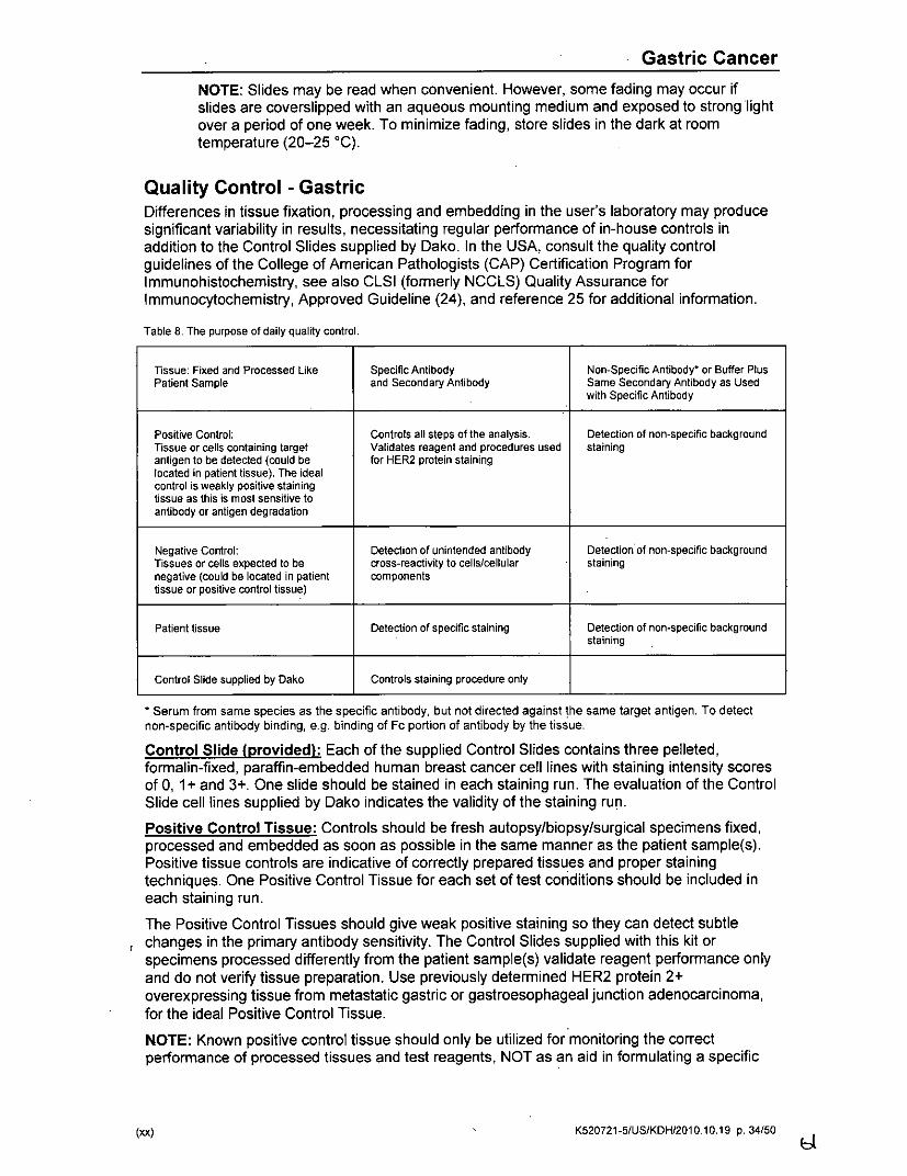
Gastric Cancer
NOTE: Slides may be read when convenient. However, some fading may occur ifslides are coverslipped with an aqueous mounting medium and exposed to strong lightover a period of one week. To minimize fading, store slides in the dark at roomtemperature (20-25 0C).
Quality Control - GastricDifferences in tissue fixation, processing and embedding in the user's laboratory may producesignificant variability in results, necessitating regular performance of in-house controls inaddition to the Control Slides supplied by Dako. In the USA, consult the quality controlguidelines of the College of American Pathologists (CAP) Certification Program forImmunohistochemistry, see also CLSI (formerly NCCLS) Quality Assurance forImmunocytochemistry, Approved Guideline (24), and reference 25 for additional information.
Table 8. The purpose of daily quality control.
Tissue: Fixed and Processed Like Specific Antibody Non-Specific Antibody' or Buffer PlusPatient Sample and Secondary Antibody Same Secondary Antibody as Used
with Specific Antibody
Positive Control: Controls all steps of the analysis. Detection of non-specific backgroundTissue or cells containing target Validates reagent and procedures used stainingantigen to be detected (could be for HER2 protein staininglocated in patient tissue). The idealcontrol is weakly positive stainingtissue as this is most sensitive toantibody or antigen degradation
Negative Control: Detection of unintended antibody Detection of non-specific backgroundTissues or cells expected to be cross-reactivity to cells/cellular stainingnegative (could be located in patient componentstissue or positive control tissue)
Patient tissue Detection of specific staining Detection of non-specific backgroundstaining
Control Slide supplied by Dako Controls staining procedure only
* Serum from same species as the specific antibody, but not directed against the same target antigen. To detectnon-specific antibody binding, e.g. binding of Fc portion of antibody by the tissue.
Control Slide (provided): Each of the supplied Control Slides contains three pelleted,formalin-fixed, paraffin-embedded human breast cancer cell lines with staining intensity scoresof 0, 1 + and 3+. One slide should be stained in each staining run. The evaluation of the ControlSlide cell lines supplied by Dako indicates the validity of the staining run.
Positive Control Tissue: Controls should be fresh autopsy/biopsy/surgical specimens fixed,processed and embedded as soon as possible in the same manner as the patient sample(s).Positive tissue controls are indicative of correctly prepared tissues and proper stainingtechniques. One Positive Control Tissue for each set of test conditions should be included ineach staining run.
The Positive Control Tissues should give weak positive staining so they can detect subtlechanges in the primary antibody sensitivity. The Control Slides supplied with this kit orspecimens processed differently from the patient sample(s) validate reagent performance onlyand do not verify tissue preparation. Use previously determined HER2 protein 2+overexpressing tissue from metastatic gastric or gastroesophageal junction adenocarcinoma,for the ideal Positive Control Tissue.
NOTE: Known positive control tissue should only be utilized for monitoring the correctperformance of processed tissues and test reagents, NOT as an aid in formulating a specific
(xx) K520721-5/US/KDH/2010.10.19 p. 34150
Gastric Cancer
diagnosis of patient samples. If the Positive Control Tissue fails to demonstrate appropriatepositive staining, results with the patient specimens should be considered invalid.
Negative Control Tissue: Use a negative control tissue (known to be HER2 protein negative)fixed, processed and embedded in a manner identical to the patient sample(s) with eachstaining run to verify the specificity of the primary antibody and to provide an indication ofspecific background staining. Colon, liver or thyroid are appropriate for negative control tissue.The variety of different cell types present in most tissue sections offers internal negative controlsites (this should be verified by the user).
If specific staining occurs in the Negative Control Tissue, results with the patient specimensshould be considered invalid and the test re-run.
Non-Specific Negative Control Reagent: Use the supplied Negative Control Reagent inplace of the primary antibody with a section of each patient specimen to evaluate non-specificstaining and allow better interpretation of specific staining at the antigen site. The incubationperiod for the Negative Control Reagent should correspond to that of the primary antibody.
Assay verification: Prior to initial use of an antibody or staining system in a diagnosticprocedure, the user should verify the antibody's specificity by testing it on a series of in-housetissues with known immunocytochemical performance characteristics representing knownpositive and negative tissues. Refer to the quality control procedures previously outlined in thissection of the product insert and to the quality control requirements of the CAP CertificationProgram for Immunohistochemistry and/or CLSI (formerly NCCLS) Quality Assurance forImmunocytochemistry, Approved Guideline (24). These quality control procedures should berepeated for each new antibody lot, or whenever there is a change in assay parameters.Gastric or gastroesophageal junction adenocarcinoma, with known HER2 protein stainingintensities from 0 - 3+ and negative tissues, e.g. colon, liver or thyroid are suitable for assayverification.
Interpretation of Staining - GastricFor determination of HER2 protein expression, only the membrane staining intensity andpattern should be evaluated using the scale presented in Table 9. Slide evaluation should beperformed by a pathologist using a light microscope. For evaluation of theimmunohistochemical staining and scoring, an objective of 1OX magnification is appropriate.The use of a 5-40x objective magnification is useful in confirmation of the score. Cytoplasmicstaining should be considered non-specific staining and is not to be included in the assessmentof membrane staining intensity (8). To aid in the differentiation of 0, 1+, 2+ and 3+ staining,refer to Dako's "Guidelines for Scoring HercepTest TM - Gastric" for representative pictures ofthe staining intensitites and patterns.
Only specimens from patients with gastric or gastroesophageal junction adenocarcinoma,should be scored. In cases with intestinal metaplasia and adenocarcinoma in the samespecimen, only the adenocarcinoma component should be scored. For interpretation ofHercepTest TM stained biopsies a cluster of at least 5 stained tumor cells is recommended.
0o0') K520721-5/US/KDH/2010.10.19 p. 35/50

Gastric CancerTable 9. Interpretation and scoring of HER2 immunohistochemical staining
Surgical Specimen - Biopsy Specimen - HER2 OverexpressionScore Staining Pattern Staining Pattern Assessment
No reactivity or membranous reactivity No reactivity or no membranous0 in < 10% of tumor cells reactivity in any (or < 5 clustered) tumor Negative
cell
Faint/barely perceptible membranous Tumor cell cluster (Ž5 cells) with areactivity in a 10% of tumor cells; cells faint/barely perceptible membranousare reactive only in part of their reactivity irrespective of percentage ofmembrane tumor cells stained
Weak to moderate complete, Tumor cell cluster (Ž 5 cells) with a weakbasolateral or lateral membranous to moderate complete, basolateral or
2+ reactivity in -> 10% of tumor cells lateral membranous reactivity Equivocalirrespective of percentage of tumor cellsstained
Strong complete, basolateral or lateral Tumor cell cluster (_ 5 cells) with a3+ membranous reactivity in Ž 10% of strong complete, basolateral or lateral Positive
tumor cells membranous reactivity irrespective ofpercentage of tumor cells stained
Guidelines based on Hofmann et al. (39).
HercepTest TM is interpreted as negative for HER2 protein overexpression (0 and 1+ score),equivocal (2+ score), and positive (3+ score). HercepTest TM is not intended to provideprognostic information to the patient and physician and has not been validated for that purpose.
(xx) K520721-5/US/KDH/2010,10.19 p. 36/50
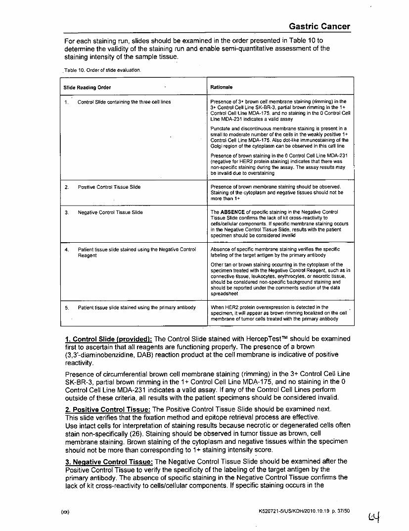
Gastric CancerFor each staining run, slides should be examined in the order presented in Table 10 todetermine the validity of the staining run and enable semi-quantitative assessment of thestaining intensity of the sample tissue.
Table 1 0. Order of slide evaluation.
Slide Reading Order .Rationale
1 . Control Slide containing the three cell lines Presence of 3+ brown cell membrane staining (rimming) in the3+ Control Cell Line SK-BR-3. partial brown rimming in the 1+-Control Cell tine MIDA-1 75, and no staining in the 0 Control CellLine MDA-231 indicates a valid assayPunctate and discontinuous membrane staining is present in asmall to moderate number of the cells in the weakly positive 1.+Control Cell Line MDA-175. Also dot-like immunostaining of theGolgi region of the cytoplasm can be observed in this cell line
Presence of brown staining in the 0 Control Cell Line MDA-231(negative for HER2 protein staining) indicates that there wasnon-specific staining during the assay. The assay results maybe invalid due to overstaining
2. Positive Control Tissue Slide Presence of brown membrane staining should be observed.Staining of the cytoplasm and negative tissues should not bemore than 1.
3. Negative Control Tissue Slide The ABSENCE of specific staining in the Negative ControlTissue Slide confirms the lack of kit cross-reactivity tocells/cellular components. If specific membrane staining occursin the Negative Control Tissue Slide, results with the patientspecimen should be considered invalid
4. Patient tissue slide stained using the Negative Control Absence of specific membrane staining verifies the specificReagent labeling of the target antigen by the primary antibody
Other tan or brown staining occurring in the cytoplasm of thespecimen treated with the Negative Control Reagent, such as inconnective tissue, leukocytes, erythrocytes, or necrotic tissue,should be considered non-specific background staining andshould be reported under the comments section of the dataspreadsheet
5. Ptiet tssu slie saind uing he rimry ntilbody When HER2 protein overexpression is detected in thespecimen, it will appear as brown rimming localized on the cellmembrane of tumor cells treated with the primnary antibody
1. Control Slide (Drovided): The Control Slide stained with HercepTeSt TM should be examinedfirst to ascertain that all reagents are functioning properly. The presence of a brown(3,3'-diaminobenzidine, DAB) reaction product at the cell membrane is indicative of positivereactivity.Presence of circumferential brown cell membrane staining (rimming) in the 3+ Control Cell LineSK-8R-3, partial brown rimming in the 1 + Control Cell Line MDA-1 75, and no staining in the 0Control Cell Line MDA-231 indicates a valid assay. If any of the Control Cell Lines performoutside of these criteria, all results with the patient specimens should be considered invalid.2. Positive Control Tissue: The Positive Control Tissue Slide should be examined next.This slide verifies that the fixation method and epitope retrieval process are effective.Use intact cells for interpretation of staining results because necrotic or degenerated cells oftenstain non-specifically (26). Staining should be observed in tumor tissue as brown, cellmembrane staining. Brown staining of the cytoplasm and negative tissues within the specimenshould not be more than corresponding to 1I+ staining intensity score.3. Negative Control Tissue: The Negative Control Tissue Slide should be examined after thePositive Control Tissue to verify the specificity of the labeling of the target antigen by theprimary antibody. The absence of specific staining in the Negative Control Tissue confirms thelack of kit cross-reactivity to cells/cellular components. If specific staining occurs in the
(,a) ~~~~~~~~~~~~~~~~~~~~~~K520721-51USIKDH/2010.10.1 9 p. 37/50 L

Gastric CancerNegative Control Tissue, results with the patient specimen should be considered invalid.Alternatively, negative portions of the Positive Control Tissue may serve as the NegativeControl Tissue, but this should be verified by the user. Note that a weak reaction (0 - 1 +staining intensity) can be observed in most normal epithelial tissue. Possible negative controltissues include: colon, liver and thyroid.
Non-specific staining, if present, will be of a diff use appearance. Sporadic staining ofconnective tissue may also be observed in sections from excessively formalin-fixed tissues.
4 + S. Patient Tissue: Examine patient specimens stained with HercepTestt m last. Positivestaining intensity should be assessed within the context of any non-specific backgroundstaining of the Negative Control Reagent. As with any immunocytochemnical test, a negativeresult means that the antigen was not detected, not that the antigen was absent in thecells/tissue assayed. Refer to Summary, and Explanation, Limitations, and PerformanceCharacteristics for specific information regarding HercepTeStTM immunoreactivity.Additional Recommendations for Interpretation of HercepTesttm StainingGastric or gastroesophageal junction adenocarcinoma tested for HER2 protein overexpressionare scored from 0 to 3+. While the 0 and 3+ cases are clear-cut, a small percentage of theremaining 1+ and 2+ samples may be more difficult to interpret. Use the following guidelines forinterpretation of HercepTesttm staining in your laboratory.* Evaluate the Control Cell Lines to validate the assay performance.
* Evaluate the Positive and Negative Control Slides.
* A hemnatoxylin and eosin (H&E) staining of the tissue specimen is recommended for the firstevaluation. (The tumor may not be obvious when looking at the sample stained withHercepTest'". An H&E stained slide is required from the pathologist to verify the presenceof the tumor). The HercepTeStTM should be performed on a paired section (serial section)from the same paraffin block of the specimen.
• Evaluate the sections stained for HER2 protein overexpression at low power first.The majority of positive cases will be obvious at low power magnification.
* For 1+ cases, use 40x objective magnification to verify membrane staining.
* For 2+ cases, use lOx-20x objective magnification to verify membrane staining.
Surgical specimen
* Well-preserved and well-stained areas of the specimen should be used to make a.determination of the percent of positive stained tumor cells.
* If a majority of tumor cells demonstrate complete, basolateral or lateral membrane staining,the staining is either 2+ or 3+.
* If there is complete, basolateral or lateral membrane staining at a strong intensity in equal toor more than 1 0% of the tUmor cells in surgical specimens, the score of the specimen is 3-'.
* If there is complete, basolateral or lateral membrane staining at a weak to moderateintensity in equal to or more than 1 0% of the tumor cells in surgical specimens, the score ofthe specimen is 2+.
* If equal to or more than 1 0% of the tumor cells in surgical specimens, stained only in part oftheir membrane, have a faint/barely perceptible intensity, the score of the specimen is 1 +.
* If no staining is observed the score of the surgical specimen isO0.
* If less than 1 0% of the tumor cells in surgical specimens have staining, irrespective of thestaining pattern (e.g. complete, basolateral, lateral or part of their membrane), the score is 0.
(W K520721-5/USiKDH/2010.10.19 p. 38/50

Gastric Cancer
Biopsy specimen* If there is a tumor cell cluster of at least 5 stained tumor cells with a strong complete,
basolateral or lateral membrane staining, the score of the biopsy specimen is 3+,irrespective of percentage of tumor cells stained.
* If there is a tumor cell cluster of at least 5 stained tumor cells with a weak to moderatecomplete, basolateral or lateral membrane staining, the score of the biopsy specimen is 2+,irrespective of percentage of tumor cells stained.
* If there is a tumor cell cluster of at least 5 stained tumor cells with a faint/barely perceptiblemembrane staining and cells are stained only in part of their membrane, the score of thebiopsy specimen is 1+, irrespective of percentage of tumor cells stained.
* If no staining is observed the score of the biopsy specimen is 0.
* If membrane staining (irrespective of staining intensity) is observed in less than 5 clusteredtumor cells, the score of the biopsy specimen is 0.
Limitations - Gastric
General limitations1. Immunocytochemistry is a multi-step diagnostic process that requires specialized training in
the selection of the appropriate reagents; tissue selection, fixation, and processing;preparation of the immunocytochemistry slide; and interpretation of the staining results.
2. Tissue staining is dependent on the handling and processing of the tissue prior to staining.Improper fixation, freezing, thawing, washing, drying, heating, sectioning, or contaminationwith other tissues or fluids may produce artifacts, antibody trapping, or false-negativeresults. Inconsistent results may be due to variations in fixation and embedding methods, orto inherent irregularities within the tissue.
3. Excessive or incomplete counterstaining may compromise proper interpretation of results.
4. The clinical interpretation of any positive staining or its absence must be evaluated withinthe context of clinical presentation, morphology and other histopathological criteria.The clinical interpretation of any staining, or its absence, must be complemented bymorphological studies and proper controls as well as other diagnostic tests. It is theresponsibility of a qualified pathologist, who is familiar with the antibodies, reagents andmethods used, to interpret the stained preparation. Staining must be performed in acertified, licensed laboratory under the supervision of a pathologist who is responsible forreviewing the stained slides and assuring the adequacy of positive and negative controls.
5. Tissues from persons infected with hepatitis B virus and containing hepatitis B surfaceantigen (HBsAg) may exhibit non-specific staining with horseradish peroxidase (27).Reagents may demonstrate unexpected reactions in previously untested tissue types.The possibility of unexpected reactions even in tested tissue types cannot be completelyeliminated due to biological variability of antigen expression in neoplasms, or otherpathological tissues (28). Contact Dako's Technical Services with documented unexpectedreaction.
6. False-positive results may be seen due to non-immunological binding of proteins orsubstrate reaction products. They may also be caused by pseudoperoxidase activity(erythrocytes) and endogenous peroxidase activity (cytochrome C) (28).
7. The staining procedure should be performed at ambient temperature of 20-25 0C.
(n) K520721-5/US/KDH/2010,10.19 p. 39150

Gastric Cancer
Product-specific limitations1. The antigen present in the 1+ Control Cell Line MDA-175 is subject to degradation over
time. Assess the Control Slide results in connection with the expiration date of the ControlSlide. Negative staining of the MDA-1 75 cells may only indicate that the Control Slide hasdegraded. The Control Slides must be stored at 2-8 0C.
2. False-negative results could be caused by degradation of the antigen in the tissues overtime. Specimens should be stained within 4-6 weeks of mounting of tissues on slides whenstored at room temperature (20-25 °C) (29).
3. For optimal and reproducible results, the HER2 protein requires heat-induced epitoperetrieval when tissues are routinely fixed (neutral-buffered formalin) and paraffin embedded.This pre-treatment needs to be completed at the beginning of the entire staining process.See the "Specimen Preparation Section, Treatment of tissues prior to staining" forinstructions.
4. Heat-induced epitope retrieval of the HER2 protein should only be done using a calibratedwater bath. Other methods of heating have been tested and do not give reproducibleresults.
5. Do not replace kit reagents with reagents carrying other lot numbers or with reagents fromother manufacturers. The only exception is the Wash Buffer that may be replaced withDako Wash Buffer, Code S3006.
6. False results could be obtained from evaluation of cytoplasmic staining. Consider only theintensity of cell membrane staining when interpreting results.
7. Stained Control Slides should be used only for validation of the staining run and should notbe used as a guide to score the staining reaction in tissue sections.
8. Strong focal staining (3+), i.e. "hot spots," may occasionally be seen. This may be the resultof uneven fixation and/or processing of tissue. Immunostaining of a second tissue blockfrom the same specimen is recommended.
9. Use of HercepTestTM on specimens fixed in fixatives other than neutral buffered formalinhas not been validated.
10. Note that normal tonsil and esophageal epithelia may stain up to 2+ intensity.
11. Use of crushed gastric cancer specimens and interpretation of artifactual staining around abiopsy edge should be avoided.
Performance Characteristics - Gastric
BackgroundThe clinical safety and efficacy of trastuzumab (Herceptin®) has been demonstrated in theB018255 study (the ToGA trial) (36, 37). The study was designed as an open labeled,randomized, multicenter, phase III study in HER2-positive patients with advanced gastric orgastroesophageal junction adenocarcinoma. In this study HER2 positivity was defined as beingeither IHC-positive (3+)(HercepTest TM, Dako) or positive by HER2 FISH (HER2/CEN-17 > 2.0)(HER2 FISH pharmDx Kit, Dako). After inclusion in the study the patients were randomized toreceive chemotherapy [cisplatin and a fluoropyrimidine (capecitabine or 5 fluorouracil); FC] orchemotherapy plus trastuzumab (FC+H).In the BO18255 study a total of 594 patients were enrolled, of whom 594 (296 patients in thechemotherapy arm, 298 patients in the chemotherapy plus trastuzumab arm) received studymedication.The main efficacy outcome measure was overall survival (OS) analyzed by stratified log ranktest. The final OS analysis based on 351 deaths was statistically significant (nominalsignificance level of 0.0193). An updated OS analysis was also conducted at one year after thefinal analysis. The median overall survival of 13.5 months on the Herceptin® plus
(xx) K520721-51USIKDHI2010.10.19 p. 40/50

Gastric Cancer
chemotherapy arm was significantly longer compared to 11.0 month median overall survival onthe chemotherapy alone arm. The efficacy results of both the definitive and the updated (final)analyses are summarized in Table II and Figure 1.
Table 11. Overall Survival in intention to Treat (ITTP oulationFC Arm FC + H ArmN= 296 N=298
Final Overall SurvivalNo. Deaths (%4) 184 (62.2%) 167 (56.0%)Median 11.0 13.595% Cl (mos.) (9.4,12.5) (11,7,15.7)
Hazard Ratio 0.7395% CI (0.60. 0.91)p-value*, Iwo-sided 0.0038*
Updated Overall SurvivalNo. Deaths (%) 227 (76.7%) 221 (74.2%)Median 11.7 13.195% Cl (mos.) (10.3.13.0) (11.9, 15.1)
Hazard Ratio 0.8095% ci (0.67, 0.97)Comparing with the nominal significance level of 0.0193
I'~~~~~~~~~~~~~~~~~II
26 27 1 0 3 14 3 2 0290 232 168I 95A-1 0
Fluoropyrimnidine tCiSplatin --- Fluoropyuimidina * isplatin .Trasuua
Figure 1. Updated Overall Survival in Patients with Metastatic Gastric Cancer
(xx) K520721-5/US/KDH/2010.10.19 p. 41/50

Gastric CancerAn exploratory analysis of OS-based on gene amplification (FISH) and protein overexpression(IHC) testing is summarized in Table 12.
Table 12. Exploratory Analyses by HER2 Status Usin the U pdated Overall Survival Results.FC FC+H
N=296' N=298 b
FISH+ / IHC 0, 1+ subgroup (N=133)No. Deaths / n (%) 57/71 (80.3%) 56162 (90.3%)Median OS Duration (mos.) 8.8 8.395% Cl (mos.) (6.4, 11.7) (6.2, 10.7)
Hazard ratio (95% CI) 1.33 (092, 1.92)FISH+ I IHC2+ subgroup (N=160)
No. Deaths / n (%) 65/80 (81%) 64180 (80%)Median OS Duration (mos.) 10.8 12.395% CI (mos.) (6.8,12.8) (9,5, 15.7)
Hazard ratio (95% CI) 0.78 (0.55,1.10)FISH+ or FISH-/IHC3+0subgroup (N=294)
No. Deaths/n (%) 104/143 (73%) 96/151 (64%)Median OS Duration (mos.) 13.2 18.095% CI (mos.) (11.5. 15.2) (15.5, 21.2)
Hazard ratio (95% CI) 0.66 (0.5, 0.87)
Median survival was estimated from Kaplan-Meier curves.Two patients on. FC arm who were FISH+ but IHC status unknown were excluded fromthe analyses.
b Five patients on Herceptin® arm who were FISH+ but IHC status unknown wereexcluded from the analyses.
c Includes 6 patients on chemotherapy arm, 10 patients on Herceptin® arm with FISH-,IHC3+ and 8 patients on chemotherapy arm, 8 patients on Herceptin® arm with FISHstatus unknown, IHC3+.
ReproducibilityHercepTest TM analysis of 60 different gastric cancer specimens obtained from stomach orgastroesophageal junction areas representing surgical resections and biopsies were performedon five non-consecutive days at three study sites. The 60 specimens in the study were equallydistributed in the three HER2 status categories. A total of 2040 HER2 scorings were performedby six pathologists.
Day-to-day agreement (negative, equivocal, positive) ranged from 83.1% to 98.3%. In 47 of 60possible comparisons the agreements were at 90.0% or above. In Table 13, specific examplesof the day-to-day comparisons are shown with average agreements at 91.2%, 92.5% and92.5% for the three sites.
Site-to-site agreements were 82.7%, 75.0% and 88.0%, respectively for pairwise sitecomparison (see Table 14). According to Fisher's exact test; the results were not differentbetween sites.
Observer-to-observer agreements between pathologists at each site were 88.0%, 83.6% and81.0% for the three study sites, respectively (Table 15).In conclusion HercepTestTM analysis of gastric cancer specimens at three study sitesdemonstrated good agreement between observations with respect to days, sites andobservers.
(xx) K520721-5/US/KDH/2010.10.19 p. 42/50 6:,

Gastric CancerTable 13. Day-to-Day Overall Percent Agreements - A Subset of 12 out of 60 comparisons
observer 1 Observer 2 Average_________________ Agreement 0195 ILL
4 Agreement C195 LL' Agreement
Site 1 Day 1 vs Day 2 85.0 74.4 93.3 84.9 9
Day 3vs Day 4 93.3 84.9 93.3 84.9Site 2 Day 1 vs Day 2 95.0 87.3 83.1 72.0 9
___________________ ~~~~~~~~~~~~92.5Day 3vs Day 4 96.7 89.7 91.7 82.7
1C195 LL: 95% lower limit confidence interval.
Table 14. Site-to-site Overall Percent Agreements
Agreemet C195LL1 AverageAgreement 0195 ~Agreement
Site 1 vs Site 2 Day 1 vs Day 1 83.3 72.4
Day 2 vs Day 2 85.0 74.4Day 3 vs Day 3 85.0 74482.7Day 4vs Day 4 81.7 70.5
_____ _____ _____ ____ Day 5 vs Day 5 78.3 66.7
Site 1 vs Site 3 Day 1 vs Day 1 80.0 68.6
Day 2 vs Day 2 73.3 61.2Day 3 vs Day 3 78.3 66.7 75.0Day 4 vs Day 4 68.3 55.9Day 5 vs Day 5 75.0 63.0
Shte?2 vs Site 3 flay 1 vs Day 1 68.3 78.5
Day 2 vs Day 2 86.7 76.4Day 3 vs Day 3 90.0 80.5 88.0Day 4 vs Day 4 86.7 76.4
_____ _____ _____ ____ Day 5 vs Day 5 88.3 78.5
C0195 LE: 95% lower limit confidence interval.
Table 15. Observer-to-Observer Agreements
Agreement CI 95 LIL' Average Agreement
Site 1 Day 1 91.7 82.7
Day 2 91.7 82.7.Day 3 93.3 84.9 88.0Day 4 83.3 72.4
_______ _______ ____ _ Day Y5 80.0 68.6
Site 2 Day 1 86.4 76.0Day 2 83.3 72.4Day 3 83.3 72.4 83.6Day 4 83.3 72.4
_________ _________ Day 5 81.7 70.5
Site 3 Day 1 80.0 68.6Day 2 78.3 66.7-DayS3 80.0 68.6 81.0DaYy 4 78366.7
______ ______ ______ _ Day5 90.0 80.5
01I95 IL: 95% lower limit confidence interval.
(xx) . ~~~~~~~~~~~~~~~~K520721-5/usIKrDHI2010.10.19 p. 43150

Gastric Cancer
ImmunoreactivityTable 16 summarizes HercepTest TM immunoreactivity with the recommended panel of normaltissues. All tissues were formalin-fixed and paraffin-embedded and stained with HercepTest TM
according to the instructions in the package insert.
Table 16. Summary.of HercepTest TM normal tissue reactivity.
Tissue Type Positive Tissue Element Staining and(No. Tested) Staining Pattern
Adrenal (3) NoneBone marrow (3) NoneBrain/Cerebellum (3) NoneBrain/Cerebrum (3) NoneBreast (3) Mammary gland (1+ staining intensity)Cervix uteri (3) NoneColon (3) Columnar epithelium, surface (1 + staining intensity)Esophagus (3) Squamous epithelia (1/3 tissues, 2+ staining intensity)Heart (3) NoneKidney (3) Tubule (1+ staining intensity)Liver (3) NoneLung (3) NoneMesothelial cells (3) NoneOvary (3) NonePancreas (3) Langerhans cells, cytoplasmic (3+ staining intensity)Parathyroid (3) NonePeripheral nerve (3) NonePituitary (3) Endocrine cells, cytoplasmic (3+ staining intensity)Prostate (3) Prostate gland (2+ staining intensity)Salivary gland (3) NoneSkeletal muscle (3) NoneSkin (3) NoneSmall intestine (3) Columnar epithelium, surface (1+ staining intensity)Spleen (3) NoneStomach (3) Epithelium (1/3 tissues, 1+ staining intensity)Testis (3) NoneThymus (3) NoneThyroid (3) NoneTonsil (3) Squamous epithelia (2+ staining intensity)Uterus (3) Endometrium (1/3 tissues, 1+ staining intensity)
Reported staining in all tissues was membrane, unless otherwise noted, All threespecimens of each tissue type had the same staining intensity unless otherwise noted.
(xx) K520721-5/US/KDH/2010.10.19 p. 44/50 -

Gastric Cancer
Troubleshooting - GastricRefer to the Troubleshooting section in Dako's previously referenced Handbook (19) forremedial action, or contact Dako's Technical Service Department to report unusual staining.
Problem Probable Cause Suggested Action
1. No staining of la. Programming error. Reagents la. Check programming grid to verifyslides not used in proper order that the staining run was
programmed correctly
lb. Reagent vials were not loaded lb. Check the Reagent Map to verifyin the correct locations in the the proper location of reagentreagent racks vials
ic. Insufficient reagent in ic. Ensure that enough reagent isreagent vial loaded into the reagent vials prior
to commencing the run. Refer toReagent Map for volumes required
ld. Sodium azide in Wash Solution ld. Use fresh preparation of WashBuffer provided in the kit
le. Excessive heating of mounted le. Air dry the tissue sections attissue sections prior to room temperature for a minimumdeparaffinization and heat- of 12 hours or until dry.induced antigen retrieval may Alternatively, dry at 37 °Clead to loss of visible HER2 overnight or dry at 60 0C for aimmunoreactivity. maximum of one hour. Drying of
tissue sections at elevatedtemperatures must only beperformed in a calibrated oven withuniform heat distribution (17).
2. Weak staining of 2a. Inadequate epitope retrieval 2a. Verify that Epitope Retrievalslides Solution reaches 95-99 °C for a
full 40 minutes and is allowed tocool for an additional 20 minutes
2b. Inadequate reagent incubation 2b. Review Staining Proceduretimes instructions
2c. Inappropriate fixation method 2c. -Ensure that patient tissue is notused over-fixed or that an alternative
fixative was not used
2d. Excessive heating of mounted 2d. Air dry the tissue sections attissue sections prior to room temperature for a minimumdeparaffinization and heat- of 12 hours or until dry.induced antigen retrieval may Alternatively, dry at 37 OCcause a significant decrease in overnight or dry at 60 00 for avisible HER2 immunoreactivity maximum of one hour. Drying of
tissue sections at elevatedtemperatures must only beperformed in a calibrated oven withuniform heat distribution (17).
3. Excessive 3a. Paraffin incompletely removed 3a. Use fresh clearing solutions andbackground follow procedure as outlined instaining of slides Section B.1
3b. Starch additives used in 3b. Avoid using starch additives for
(xx) K520721-5/US/KDH/2010.10.19 p. 45/50 7L

Gastric Cancermounting sections to slides adhering sections to glass slides.
Many additives are immuno-reactive
3c. Slides not thoroughly rinsed 3c. Ensure that the Autostainer isproperly primed prior to running.
-Check to make sure thatadequate buffer is provided forentire run. Use fresh solutions ofbuffers and washes
3d. Sections dried during staining 3d. Verify that the appropriateprocedure volume of reagent is applied to
slides. Make sure the Autostaineris run with the hood in the closedposition and is not exposed toexcessive heat or drafts
3e. Sections dried while loading 3e. Ensure sections remain wet withthe Autostainer buffer while loading and prior to
initiating run
3f. Inappropriate fixation method 3f. Ensure that approved fixativeused was used. Alternative fixative
may cause excessive back-ground staining
3g. Non-specific binding of 3g. Check fixation method of thereagents to tissue specimen and presence of
necrosis
4. Tissue detaches 4a. Use of incorrect slides 4a. Use silanized slides, such asfrom slides Dako Silanized Slides, Code
S3003, SuperFrost Plus or poly-L-lysine coated slides
5. Excessively strong 5a. Inappropriate fixation method 5a -Ensure that only approvedspecific staining used fixatives and fixation methods
are used
Sb. Use of improper heat source S1b. Ensure that only a water bath isfor epitope retrieval, e.g. used for the epitope retrieval stepsteamer, microwave oven orautoclave
Sc. Reagent incubation times too 5c. Review Staining Procedurelong instructions
Sd. Inappropriate wash solution 5d. Use only the Wash Buffer that isused recommended for the kit
6. Weak staining of 6a. Incorrect epitope retrieval 6a. Immerse the slides in the pre-the 1 + Control protocol followed heated Epitope RetrievalSlide Cell Line Solution. Bring temperature of
the Epitope Retrieval Solutionback to 95-99 0C and pre-treatfor a full 40 minutes
6b. Lack of reaction with 6b. Ensure that the full 1 0-minuteSubstrate-Chromogen Solution incubation time is used. Ensure(DAB) that only one drop of DAB
Chromnogen was added to 1 mLof DAB Buffered Substrate
Oa) ~~~~~~~~~~~~~~~~~~K520721-5/USIKDH/201 0.10.19 p. 46/50

Gastric Cancer6c. Degradation of Control Slide 6c. Check kit expiration date and kit
storage conditions on outside ofpackage
NOTE: If the problem cannot be attributed to any of the above causes, or if the suggestedcorrective action fails to resolve the problem, please call Dako's Technical Services for furtherassistance.Additional information on staining techniques and specimen preparation can be found in Dako'spreviously referenced Handbook (19) (available from Dako), Atlas of Immunohistology (30) andImmunoperoxidase Techniques. A Practical Approach to Tumor Diagnosis (31).
(xx) <K520721-51USIKDHI2010.10.19 p. 47/50

References1. Coussens L, Yang-Feng TL, Liao Y-C, Chen E, Gray A, McGrath J, et al. Tyrosine kinase receptor
with extensive homology to EGF receptor shares chromosomal location with neu oncogene.Science 1985;230:1132-9.
2. King CR, Kraus MH, Aaronson SA. Amplification of a novel v-erbB-related gene in a humanmammary carcinoma. Science 1985;229:974-6.
3. Semba K, Kamata N, Toyoshima K, Yamamoto T. A v-erbB-related protooncogene, c-erbB-2, isdistinct from the c-erbB-l/epidermal growth factor-receptor gene and is amplified in a humansalivary gland adenocarcinoma. Proc Natl Acad Sci USA 1985;82:6497-501.
4. Yamamoto T, Ikawa S, Akiyama T, Semba K, Nomura N, Miyajima N, et al. Similarity of proteinencoded by the human c-erb-B-2 gene to epidermal growth factor receptor. Nature 1986;319:230-4.
5. Schechter AL, Hung M-C, Vaidyanathan L, Weinberg RA, Yang-Feng TL, Francke U, et al. Theneu gene: an erbB-homologous gene distinct from and unlinked to the gene encoding the EGFreceptor. Science 1985;229:976-8.
6. Bargmann Cl, Hung M-C, Weinberg RA. The neu oncogene encodes an epidermal growth factorreceptor-related protein. Nature 1986;319:226-30.
7. Natali PG, Nicotra MR, Bigotti A, Venturo I, Slamon DJ, Fendly BM, et al. Expression of the p185encoded by HER2 oncogene in normal and transformed human tissues. Int J Cancer 1990;45:457-61.
8. Press MF, Cordon-Cardo C, Slamon DJ. Expression of the HER-2/neu proto-oncogene in normalhuman adult and fetal tissues. Oncogene 1990;5:953-62.
9. Lonardo F, Di Marco E, King CR, Pierce JH, Segatto 0, Aaronson SA, et al. The normal erbB-2product is an atypical receptor-like tyrosine kinase with constitutive activity in the absence ofligand. New Biologist 1990;2:992-1003.
10. Carter P, Presta L, Gorman CM, Ridgway JBB, Henner D, Wong WLT, et al. Humanization of ananti-p185 HER2 antibody for human cancer therapy. Proc Natl Acad Sci USA 1992;89:4285-9.
11. Hudziak RM, Lewis GD, Winget M, Fendly BM, Shepard HM, Ullrich A. p185HER2 monoclonalantibody has antiproliferative effects in vitro and sensitizes human breast tumor cells to tumornecrosis factor. Mol Cell Biol 1989;9:1165-72.
12. Lewis GD, Figari I, Fendly B, Wong WL, Carter P, Gorman C, et al. Differential responses ofhuman tumor cell lines to anti-p185 HER2 monoclonal antibodies. Cancer Immunol Immunother1993;37:255-63.
13. Baselga J, Norton L, Albanell J, Kim Y-M, Mendelsohn J. Recombinant humanized anti-HER2antibody (Herceptin TM) enhances the antitumor activity of paclitaxel and doxorubicin againstHER2/neu overexpressing human breast cancer xenografts. Cancer Res 1998;58:2825-31.
14. a. Medical Laboratories - Particular requirements for quality and competence, ISO 15189:2003.b. Clinical Laboratory Improvement Amendments of 1988: Final Rule, 57CFR7163, February 28,1992.
15. Sheehan DC, Hrapchak BB. Theory and practice of histotechnology. St. Louis: The C. V. MosbyCompany; 1980.
16. Kiernan JA. Histological and histochemical methods: theory & practice. New York: PergamonPress; 1981.
17. Lundgaard Hansen B, Winther H, Moller K. Excessive section drying of breast cancer tissue priorto deparaffinisation and antigen retrieval causes a loss in HER2 immunoreactivity,Immunocytochemistry 2008;6,119-22.
18. DakoCytomation California Inc., Data on file.19. Key M. Education Guide: Immunohistochemical Staining Methods. Fifth Edition. Dako, Carpinteria,
California; 2009.20. Leong AS-Y, Milios J, Leong FJ. Epitope retrieval with microwaves. A comparison of citrate buffer
and EDTA with three commercial retrieval solutions. Appl Immunohistochem 1996;4:201-7.21. "Procedures for decontamination of plumbing systems containing copper and/or lead azides",
Department of Health, Education, and Welfare, National Institute for Occupational Safety andHealth, Rockville, MD, 1976.
22. "Decontamination of laboratory sink drains to remove azide salts." Center for Disease ControlManual Guide: Safety Management, No. CDC-22, Atlanta, GA, April 30, 1976.
23. Clinical and Laboratory Standards Institute (formerly NCCLS). Protection of Laboratory WorkersFrom Occupationally Acquired Infections; Approved Guideline - Third Edition. CLSI documentM29-A3 [ISBN 1-56238-567-4]. Clinical and Laboratory Standards Institute, 940 West ValleyRoad, Suite 1400, Wayne, Pennsylvania 19087-1898 USA, 2005.
(xx). K520721-5/US/KDH/2010.10.19 p. 48/50 is5

24. Clinical and Laboratory Standards Institute (formerly NCCLS). Quality assurance forimmunocytochemistry; Approved guideline. CLSI document MM4-A (1-56238-396-5) CLSI, 940West Valley Road, Suite 1400, Wayne, Pennsylvania 19087-1898 USA; 1999.
25. Elias JM, Gown AM, Nakamura RM, Wilbur DC, Herman GE, Jaffe ES, et al. Special report:quality control in immunohistochemnistry. Am J Clin Pathol 1989;192:836-43.
26. Nadji M, Morales AR. Immunoperoxidase: Part 1. The technique and its pitfalls. Lab Med1983;14: 767-7 1.
27. Omata M, Liew C-T, Ashcavai M, Peters RL. Nonimmunologic binding of horseradish peroxidaseto hepatitis B surface antigen: a possible source of error in irnmunohistochemistry. Am J Clin Path1 980;73:626-32.
28. Herman GE, Elfont EA. The taming of immunohistochemistry: the new era of quality control.Biotech & Histochem 1991; 66:194-9.
29. Press MF, Hung G, Godolphin W, Slamon DJ. Sensitivity of HER-2lneu antibodies in archivaltissue samples: potential source of error in immunohistochemnical studies of oncogene expression.Cancer Res 1994;54:2771-7.
30. Tubbs RR, Gephardt GN, Petras RE. Specimen processing and quality assurance. In: Atlas ofimmunohistology. Chicago: Amer Soc Clin Pathol Press; 1986:16-27.
31. Nadji M, Morales AR. Immunoperoxidase techniques. A practical approach to tumor diagnosis.Chicago: Amer Soc Olin Pathol Press; 1986.
32. Jorgensen JT. Targeted HER2 Treatment in Advanced Gastric Cancer. Oncology 201 0; 78:26-33.33. Tanner M, Hollmen M, Junttila TT, Kapanen Al, Tomola 5, Soini Y, et al. Amplification of HER-2 in
gastric carcinoma: association with Topoisomerase II alpha gene amplification, intestinal type,poor prognosis and sensitivity to trastuzumab. Ann Oncol 2005:16:273-8.
34. Matsui Y, Inomnata M, Tojigamori M, Sonoda K, Shiraishi N, Kitano S. Suppression of tumor growthin human gastric cancer with H-ER2 overexpression by an anti-HER2 antibody in a murine model.Int J Oncol 2005; 27: 681-5.
35. Fujimoto-Ouchi K, Sekiguchi F, Yasuno H, Moriya Y, Mor K, Tanaka Y. Antitumor activity oftrastuzumab in combination with chemotherapy in human gastric cancer xenograft models.Cancer Chemnother Pharmacol 2007; 59: 795-805.
36. Kim SY, Kim HP, Kim YJ, Oh DY, Im S-A, Lee D, et al. Trastuzumab inhibits the growth of humangastric cancer cell lines with HER2 amplification synergistically with cisplatin. Int J Oncol 2008;32:89-95.
37. Bang YJ, Van Cutsem E, Feyereislova A, Chung HC, Shen L, Sawaki A et al. Trastuzumab incombination with chemotherapy versus chemotherapy alone for treatment of HER2-positiveadvanced gastric or gastro-esophageal junction cancer (ToGA): a phase 3, open-label,randomized controlled trial. Lancet 201 0; 00I: 10. 1016/S0140-6736(10)61 121-x.
38. Bang Y, Chung J, Xu J, Lordick F, Sawaki A, Lipatov 0, et al. Pathological features of advancedGastric Cancer (GC): Relationship to human epidermal growth factor receptor 2 (HER2) positivity inthe global screening programme of the ToGA trial. J Clin Oncol 2009; 27:15s (suppl; abstr 4556).
39. Hofmann M, Stoss 0, Shi D, BUttner R, van de Vijver M, Kim W, et al. Assessment of a HER2scoring system for gastric cancer: results from a validation study. Histopath 2008;52:797-805.
(W) K520721-5IUSIKDH/201 0 0.1O.9 p. 49150

Explanation of symbols
RF Caargu ... m.ber 2 8
Temperatlure miliotO Ba X Ham
medical device FaiefadewicrFIVD3 In vifro d.egriostic Us by
L~ Consult instruc.tos for Contains suffident for
Produced by: Distributed in the USA by:
Dako Denmark A/S Dako North America, Inc.Produktionsvej 42 6392 Via RealDK-2600 Glostrup Carpinteria, California 93013, USADenmark/ Danemark/ D~nemark Tel. 805/566-6655Tel. +45 44 85 95 00 Toll Free 800/235-5743Fax +45 44 85 95 95 Ordering Information
Tel. 800/235-5763Fax 805/566-6688Technical ServicesTel. 800/424-0021
HercepTest TM and Herceptin® are trademarks of Genentech, Inc. subject to licenses held by DakoDenmark ANS and F. Hoffmann-La Roche Ltd.
. K520721-51USIKDH/2010.10.19 p. 50/50