effect luminal sodium concentration bicarbonate absorption in...
TRANSCRIPT
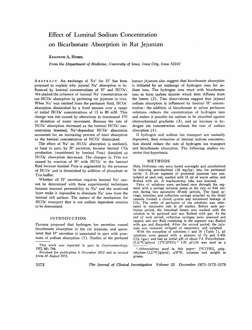
Effect of Luminal Sodium Concentration
on Bicarbonate Absorption in Rat Jejunum
KENNETHA. HUBEL
From the Department of Medicine, University of Iowa, Iowa City, Iowa 52242
A B S T R A C T An exchange of Na' for H' has beenproposed to explain why jejunal Na' absorption is in-fluenced by luminal concentrations of H' and HCO8-.Westudied the influence of luminal Na' concentration onnet HCO03 absorption by perfusing rat jejunum in vivo.When Na' was omitted from the perfusion fluid, HCOs-absorption diminished by a fixed amount over a rangeof initial HCO03 concentrations of 15 to 80 mM. Thischange was not caused by alterations in transmural PDor direction of water movement. Because the rate ofHCO3- absorption decreased as the luminal HCOa- con-centration lessened, Na'-dependent HCO3- absorptionaccounted for an increasing percent of total absorptionas the luminal concentration of HCO3- diminished.
The effect of Na' on HCO3- absorption is mediated,at least in part, by H' secretion, because luminal C02production (manifested by luminal Pco2) dimished asHCOi- absorption decreased. The changes in Pco2 arecaused by reaction of H+ with HCO3- in the luminalfluid because luminal Pco2 is augmented by the presenceof HCO3- and is diminished by addition of phosphate orTris buffer.
Whether all H+ secretion requires luminal Na+ can-not be determined with these experimental techniquesbecause mucosal permeability to Na+ and the unstirredlayer make it impossible to eliminate Na+ ions from theluminal cell surface. The nature of the mechanism forHCO3- transport that is not sodium dependent remainsto be determined.
INTRODUCTIONParsons proposed that hydrogen ion secretion causedbicarbonate absorption in the rat jejunum, and specu-lated that H+ secretion is associated in part with proc-esses of sodium absorption (1). Studies of the perfused
This work was reported in part in Gastroenterology.1972. 62: 764.
Received for publication 6 December 1972 and in revisedform 14 August 1973.
human jejunum also suggest that bicarbonate absorptionis initiated by an exchange of hydrogen ions for so-dium ions. The hydrogen ions react with bicarbonateions to form carbon dioxide which then diffuses fromthe lumen (2). Two observations suggest that jejunalsodium absorption is influenced by luminal H+ concen-tration: the addition of bicarbonate to saline perfusionsolutions reduces the concentration of hydrogen ionsand makes it possible for sodium to be absorbed againstelectrochemical gradients (3), and an increase in hy-drogen ion concentration reduces the rate of sodiumabsorption (4).
If hydrogen and sodium ion transport are mutuallydependent, then reduction of luminal sodium concentra-tion should reduce the rate of hydrogen ion transportand bicarbonate absorption. The following studies ex-amine that hypothesis.
METHODSMale Holtzman rats were fasted overnight and anesthetizedby injecting pentobarbital (50 mg/kg) into the peritonealcavity. A 25-cm segment of proximal jejunum was can-nulated at each end, washed with 15 ml of warm saline, andflushed with air. A tracheostomy tube was inserted.
Pairs of solutions were perfused once through the seg-ment with a syringe infusion pump at the rate of 0.41 ml/min during two successive 30-min periods. The input sy-ringe, intestine, and collection syringe attached to the distalcannula formed a closed system and minimized leakage ofC02. The order of perfusion of the solutions was alter-nated in successive rats in all studies. Before each per-fusion period, the intestinal lumen was washed with thesolution to be perfused and was flushed with gas. At theend of each period, collection syringes were removed andcapped, and any fluid remaining in the segment was flushedwith gas and discarded. After the second period, the jeju-num was removed, stripped of mesentery, and weighed.
With the exception of solutions 1 and 10 (Table I), allsolutions were gassed with a mixture of 02 and 5-6%C02 (gas) and had an initial pH of about 7.4. Polyethylene-[1,2-'4C]glycol ('4C]PEG) 1 1.25 ,uCi/dl was used as a
'Abbreviations used in this paper: ["4C] PEG, poly-ethylene- [1,2-'4C]glycol; gWW, jejunum wet weight ingrams.
The Journal of Clinical Investigation Volume 52 December 1973 3172-31793172

TABLE IComposition of Perfusion Fluids (mM)
Solution Na K Choline Cl HCO3 Iseth. Mann. Tris P04
1 145 5 125 25 252 25 5 120 125 25 303 145 5 - 125 25 - 804 145 5 5 25 120 255 140 5 -- 130 15 - 346 15 130 130 15 417 30 120 125 25 - 308 140 5 65 80 379 80 65 65 80 37
10 145 5 - 15011 145 5 - 130 20 - 2512 145 5 70 80 2813 140 5 125 20 5 2014 140 5 - 125 20 - 3515 140 5 105 20 57 20
nonabsorbable marker to permit calculation of net watermovement (5). Except for the hypertonic solution, theosmolality of all perfusion solutions was similar to that ofrat plasma, 303-305 mosmol/kg H20.
The pH and Pco2 of the luminal fluid were determinedsoon after collection with a capillary pH electrode andPco2 electrode designed for small samples (InstrumentationLaboratory, Inc., Lexington, Mass.). Bicarbonate was cal-culated with the Henderson-Hasselbalch equation using apK' of 6.1. Sodium and potassium concentrations weremeasured with a flame photometer and chloride was de-termined with a coulometric chloridometer. The concen-tration of ['4C] PEG (counts per minute/milliliter) wasmeasured with a scintillation counter. The osmolality (milli-osmoles/kilogram) of perfusion fluids was measured by themethod of freezing point depression with an AdvancedOsmometer (Advanced Instruments, Needham Heights,Mass.).
"Initial" concentrations of ions and of ['4C] PEG weremeasured in samples of fluid obtained from the input sy-ringes and samples of fluid for "final" determinations wereobtained from the collection syringes after each collectionperiod. Fluid remaining in the jejunum was discarded.
Net fluxes of ions and water were calculated as follows:
=y* PEGSJnetiOn = V\ionf PEG1 - ion,,gW -'
JnetH20 = V -PEGi_ 1)ggw-1.
Ions and iont are the concentrations of the ion (micro-moles/milliliter) measured in the initial and final samples;PEG4 and PEG, are the specific activities of ["C] PEG(counts per minute/milliliter) measured in the initial andfinal samples. V is the volume (milliliter) of perfusion fluidpumped into the segment in 30 min; gWWis the wetweight of the jejunum in grams. Thus, Jnetion is ex-pressed as micromoles X (30 min)' X gWW-', and J..tH2Ois expressed as milliliters X (30 min)' X gWW-'.
Changes in the Pco% of the luminal fluid are expressedas A Pco2 and were calculated by subtracting the initial
Pco2 from the final Pco2, i.e., A Pco2 = final Pco2 - initialPco2.
Three different groups of six rats each were used to de-termine transmural electrical potential difference (PD)when the following pairs of solutions were perfused (TableI): Na' 145 vs. Na' 25 (solutions 1 and 2), isotonic vs.hypertonic (solutions 1 and 3), and Na+ 145 vs. Na+ ise-thionate (solution 1 and 4). A group of eight rats wasused to determine PD when solutions with and withoutbicarbonate were perfused (solutions 1 and 10). Bridgesof saturated KCl in agar were used. One bridge contactedthe fluid perfusing the intestinal lumen and the other wasplaced in the peritoneal cavity. Electrical contact with asensitive voltmeter was made through calomel half-cells.The voltmeter was read every 3 min for 30 min and themean of the 10 values was calculated. The studies of PDand transport were not performed concurrently.
The signs preceding the net flux measurements indicatemovement into (+) or out of (-) the lumen.
The statistical significance of differences between meanswas determined with the t test for paired samples.
RESULTS
Study 1: effect of reduction in sodium concen-tration of perfusion fluid (Table II)Perfusion solutions 1 and 2 (Table I). N= 6. To
determine the effect of luminal sodium concentration onbicarbonate absorption, the jejunum was perfused withtwo solutions whose mean measured sodium concentra-tions were 143 mMand 30 mM. When the sodium con-
centration was reduced, less HC03- was absorbed,' pHdid not fall as far, and Pco2 rose less. Sodium was se-creted instead of absorbed, chloride secretion increased,and potassium absorption was enhanced. There was alsoan apparent absorption of anions in excess of cations,
'In this discussion, absorption denotes a net loss fromthe lumen, and secretion denotes a net increase in thelumen.
Luminal Sodium Concentration and Jejunal Bicarbonate Absorption 3173

TABLE I IEffect of Low Luminal Sodium Concentration on the Net
Movement of Electrolytes and Water, and the Mag-nitude of Luminal Pco, and Transmural
Electrical Potential Difference(PD) (N = 6)
Mean Na+ concentration ofperfusion fluid
Dependentvariable 143 mM 30 mM P
HCO3-*4umol -105±a13 -84 ±22 <0.05mM 20.4±0.7 21.9±0.8 <0.05
pHInitial 7.42 ±0.02 7.44±i0.01 <0.01A -0.30±0.02 -0.24±0.02 <0.01
Pco2Initial mmHg 3841 3841 NSA mmHg 15±2 11±3 <0.05
H20ml -0.2840.20 0.2440.26 <0.001
PDmV -5.2±1.6 3.5±1.0 <0.001
Nabpumol -102±15 119421 <0.001mM 143±1 30±1 <0.001
K+pmol 0.47±1.75 -7.28±1.98 <0.01mM 5.1±0.1 4.7±10.1 <0.001
Cl-jumol 24.1±26.5 109±27.3 <0.001mM 12842 128±1 NS
* For measurements of net movement and electrolytes andwater, signs indicate movement into (+) or out of (-) thelumen. Rates of net transport of water and electrolytes areexpressed as the quantity X (30 min)-' X (gram wet wtof intestine)-. The mean concentration of ions for the per-fusion period is expressed in millimoles/liter. Values shown arethe mean±SD. The sign of the PD is the polarity of thejejunal lumen.
and this is probably because choline is not accounted forin the table. The charge balances suggest that appreci-able amounts of choline were absorbed.
When the luminal Nat concentration was reduced, twofactors that could influence net movement of HCOs-and H' changed significantly: the transmural PD became8.7 mVmore positive in the lumen, and the net move-ment of water was into the lumen. The influence of thesetwo factors on bicarbonate absorption was estimated instudies 2 adl(l 3.
Study 2: effect of direction of water movement(Table III)
Perfusion solutions 1 and 3 (Table I). N= 6. Whenthe osmolality of the perfusion we increased from 305mosmol/kg to 350 mosmol/kg by adding mannitol, watermoved into rather than out of the lumen, and the meanrate of movement of water into the lumen was greaterthan when the low sodium solution was perfused in study1. However, the direction of water movement did notsignificantly affect the net movement of ions, the PD,Or the final Pco2 of the perfusion fluid.
Study 3: effect of transmural PD (Table IV)Perfusion solutions 1 and 4 (Table I). N= 6. When
the solution containing sodium isethionate was per-fused, the luminal PD was 7.5 mVmore negative than
TABLE IIIEffect of Direction of W47ater Movement on the Net Movement
of Electrolytes and W1ater, and the Magnitude of LuminalPco, and Transmural Electrical Potential Difference(PD). (N = 6) Initial Osmolalities: Isotonic = 305
mosmol/kg; Hypertonic = 350 mosmol/kg
Perfusion fluidDependent
variable Isotonic Hypertonic P
H20*ml -0.18±0.22 0.49±40.18 <0.001
HCO8-JAmol -130±421 -125-±19 NSmM 21.4±1.3 21.2 ±i0.8 NS
pHInitial 7.47±i=0.01 7.46±4-0.01 NSA -0.33-0.06 -0.33±-0.07 NS
Pco2Initial mmHg 37±1 37±-1 NSA mmHg 14±3 13±-5 NS
PDmV - 6.2±-2.1 -6.0±M2.3 NS
Na+/Amol -80.8-39.0 - 63.4 -±34.7 NSmM 143--1 140±1 <0.001
Kitpmol 1.26±1.15 2.56--1.34 NSmM 5.1 ±-0.1 5.0±-0.1 <0.05
Cl-1umol 37.9±-33.4 46.5±429.7 NSmM 127±-1 124±-1 <0.001
* See footnote to Table I1.
3174 K. A. Hiubel

when sodium chloride was used. Sodium absorption de-creased, chloride was secreted, and water moved intothe lumen. However, the change in PD did not alternet movement of HCOs-, the change in pH, or the APco2.
Study 4: how omission of Na+ from luminal fluidaffects bicarbonate absorption when initial HCO3-concentration of luminal fluid is varied (TableVA, B. and C)Perfusion solutions 5 and 6, 1 and 7, 8 and 9 (Table
I). N=8 for each pair of perfusion fluids. When so-dium was omitted from the perfusion fluid, sodium dif-fused into the lumen and raised the concentration inluminal fluid to about 10 mMwhether the initial concen-tration of HC0s- was 15 mM (Table VA), 25 mM(Table VB), or 80 mM(Table VC). Regardless of theinitial HCO- concentration, HC03- absorption decreased
TABLE IVEffect of Change in Transmural Electrical Potential Differences
Magnitude of Luminal Pco2. (N = 6) PDwas Changedby Substituting Isethionate for Chloride
in the Perfusion Fluid
Perfusion fluidDependent
variable Chloride Isethionate P
PDmV -6.9±2.1 -14.4±2.7 <0.02
HCO3-*,Amol -125±9 -125±17 NSmM 21.8±1.9 23.1±1.9 <0.001
pHInitial 7.43±0.01 7.46±0.02 <0.02A -0.33±0.04 -0.33±0.05 NS
PcO2Initial mmHg 41±2 41±3 NSA mmHg 15±3 14±3 NS
H20ml -0.34±0.24 0.10±0.12 <0.01
Na+AumoI -98.6±39.9 -2.4±16.1 <0.001mM 143 ±1 144±1 <0.05
K+
gimol 0.47±2.25 4.54±3.57 NSmM 5.140.1 5.1 i0.1 NS
Cl-4umol -22.7±25.0 134±21 <0.001mM 126±1 5.6±1 <0.001
* See footnote to Table II.
TABLE VEffect of Low Luminal Sodium Concentration on the Net
Movement of Bicarbonate, and on the Change in pHand Pco2 of Perfusion Fluid When the Initial
HCO3- Concentration was 15 mM(A),25 mM(B), and 80 mM(C)
(A)(N = 8) Mean luminal Na concentration
Dependentvariable 138±2 mM 9.3±1.4 mM a P
HCO-*.smol -71.6 11 .6 -40.8 A8.3 30.8 <0.001mean mM 12.440.5 13.340.4 0.9 <0.01
pHInitial 7.1940.02 7.2040.01 - NSA -0.28 ±0.05 -0.20 ±0.04 0.08 <0.001
PCO2
Initial mmHg 40.4±0.9 40.5±0.7 - NSA mmHg 9.54±1.4 7.6 41.6 1.9 <0.05
(B)Mean luminal Na concentration
143 41 mM 8.9 ±1.4 mMHCO3-
pmol -124±-27 -94±-19 30 <0.05mean mM 20.9 ±0.9 22.9±0.8 2 <0.001
pHInitial 7.43±+0.01 7.46±10.01 - <0.02a -0.34±0.07 -0.27±0.04 0.07 <0.01
PCO2Initial mmHg 39.0 ±0.7 39.1 ±0.6 - NSA mmHg 16.5 ±4.8 13.7 ±4.9 2.8 <0.01
(C)Mean luminal Na concentration
13841 mM 11.1 ±2 mMHCO-
JAmol -174±36 -145432 29 <0.05Mean mM 73.5±1.9 74.7 ±2.2 1.2 <0.05
pHInitial 7.86 ±0.01 7.88 ±0.01 - <0.01A -0.14±0.02 -0.12 ±0.03 0.02 <0.05
Pco2Initial mmHg 42.1 ±1.3 42.4±0.6 - NSA mmHg 6.54±2.0 5.4 ±1.8 1.1 <0.05
* See footnote to Table II.
by a constant amount, about 30 /mmol X (30 min)-' XgWW-1. In addition, the A Pco2 and A pH were less inthe solutions from which Na+ was omitted.
When the initial lumen HC03- concentration was 25mM(Table VB), the A Pco2 was larger than when theinitial concentration of HC03- was either 15 mM(Ta-ble VA) or 80 mM(Table VC). The reasons for thisare not clear, but they do not affect the validity of theconclusions which are based on data with groupsrather than between groups.
Luminal Sodium Concentration and Jejunal Bicarbonate Absorption 3175
TABLE VIEffect of 25 mMHCO3- in Perfusion Fluid on the
Magnitude of Luminal Pco2 (N = 8)
Initial HCOa- concentrationDependent
variable 0 mM 25 mM P
PCo2
Initial mmHg Nil NilA mmHg 30±2 36i2 <0.01
Study 5: effect of HCO8- in perfusion fluid onPco2 and PD (Table VI)Perfusion solutions 1 and 10 (Table I). N = 8. When
the perfusion fluid contained an initial HCO3r concentra-tion of 25 mM, the increase in Pco2 was 6 mmHg largerthan when HCO3- was omitted. The mean PD (+SD)in eight rats when solutions with and without HCO3-were perfused were - 4.7 mV (±2.5), and - 5.4 mV(±2.3). The differences were not significant.
Study 6: effect of enhanced HCO3- absorption onPco2 (Table VII)Perfusion solutions 11 and 12 (Table I). N= 8.
When the initial concentration of HCOs was increasedfrom 20 mMto 80 mM, net bicarbonate absorption morethan doubled. However, there was no significant differ-ence in the A Pco2 in the two solutions.
Study 7: effect of buffering on HCOj- absorptionand Pco2 (Tables VIII and IX)
Perfusion solutions Tris study 13 and 14 (Table I).N =8. Phosphate study 14 and 15. N= 12. As ex-pected, when the perfusion fluid was buffered with Tris(Table VIII) or phosphate (Table IX), the pH fellless than in the unbuffered fluid. Buffering with Trisreduced HCO3- absorption by 18%, but the reductionwas not significant when phosphate was used. In bothstudies, however, buffering significantly reduced theA Pco2.
DISCUSSIONOur studies demonstrate that a reduction in the con-centration of sodium in fluid perfusing the lumen of therat jejunum decreases HCO3- absorption by a constantamount, and that this decrease is not caused by con-current changes in PD or water movement. The asso-ciated changes in Pco2 of the perfusion fluid suggestthat the rate of HCO3- absorption decreases because therate of H5 secretion diminishes when the luminal Na5concentration is reduced. The data supporting these con-clusions are discussed below.
Effect of sodium, net water movement, and PD.When luminal sodium concentration was reduced, hy-drogen ions accumulated in the lumen at a reduced rate,and bicarbonate absorption diminished (Table II). How-ever, there were significant changes in two other fac-tors that could have influenced the net movement ofH' and HCO3-: the PD became 8.7 mVmore positivein the lumen, and water moved into, rather than out of,the lumen. To estimate the influence of net water move-ment on transport of HCO3-, a hypertonic solution wascirculated through the lumen (Table III). Althoughthe hypertonic solution caused a change in water move-ment greater than that induced by the low sodium solu-tions, the net movement of HCOa- was unaffected.Hence, the net movement of water could not explain thereduction in HCO3- absorption when the luminal sodiumconcentration was decreased. The influence of PD wasmore difficult to determine because it was not possibleto induce PD changes of similar polarity and magni-tude without reducing the intraluminal sodium concen-tration. Wecould, however, study the effect of changesof similar magnitude but opposite polarity. When thelumen was perfused with a solution containing sodiumisethionate instead of sodium chloride, the lumen be-came more negative by 7.5 mV (vs. 8.7 mVmore posi-tive with low sodium solution). The net movement ofpotassium, a cation that is transported passively, re-flected these changes. Net movement of potassium into thelumen diminished when the lumen became positive (TableII), and increased when the lumen became negative(Table IV). However, net movement of HCO3- did notchange significantly when the lumen became morenegative, so the effect on HCO03 movement was notcaused by changes in water movement or PD; the low lu-minal sodium concentration itself must have directlyaffected the processes that govern HCO3- absorption.
Omitting Na' from the perfusion fluid diminished therate of HCO3- absorption by a constant amount of about30 Amol X (30 min)- regardless of whether the initial
TABLE VI IEffect of Increased Net Movement of HCO3- on the Magnitude
of Luminal Pce2 (N = 8)
Initial HCO3- concentrationDependent
variables 20 mM 80 mM P
HCO3-*ysmol -78.5±4 7.8 -167 ±46.8 <0.01
PCo2
Initial mmHg 42±1 41±1 NSAmmHg 8+3 11±2 NS
* See footnote to Table II.
3176 K. A. Hubel

luminal HC03- concentration was 15, 25, or 80 mM(Table V). However, the percent of HCO3- absorptionthat was Na+ dependent increased from 17% to 43%as the initial HCO3- concentration was reduced from 80mMto 15 mMand overall HCOs- absorption rate di-minished. Thus, the mechanism of HCO3- absorptionthat requires Na+ becomes relatively more important asthe concentration of HC03- in the lumen decreases be-low that of the plasma. Is 30 ALmol X (30 min)' XgWW' the maximal rate of Na+-dependent HCO3- ab-sorption? Probably not, because the unidirectional fluxof Na+, (JsmNa'), into the unstirred layer of fluid ad-jacent to the mucosa makes it impossible to create aluminal environment that is sodium-free. If sodiumcould be eliminated completely from the lumen, the re-duction in HCOs3 absorption might be greater.
Could alterations in chloride movement have influ-enced the movement of hydrogen or bicarbonate ionswhen the luminal concentration of sodium was re-duced? Mechanisms that could link the transport ofchloride to the movement of H+ or HCO3- are: (a)transport of H+ and Cl- into the lumen, or (b) exchangeof Cl- for luminal HCO3-. When the concentration of so-dium in the perfusion fluid was lowered, the blood-lumenconcentration gradient of Cl- remained the same; hencethe unidirectional movement of Cl- into the lumen couldhave been influenced only by a change in PD (TableII). Increased electropositivity of the lumen should haveincreased Cl- flux into the lumen and the net absorptionof HCO3-. Bicarbonate absorption diminished, however,despite the increased net movement of chloride into thelumen. It seems unlikely that Cl- transport influencedHCO3- absorption, but the possibility cannot be en-tirely excluded.
Mechanism of action of sodium. By what mechanismdid the reduction in sodium concentration alter the
TABLE VIIIEffect of Buffering with Tris on Net Movement of HCO-,
and on Changes in Luminal pH and Pco2 (N = 8)
Tris TrisDependent
variables Not added Added P
HCO3-*,umol -82.0±12.7 -67.0±15.6 <0.05
pHInitial 7.29± 0.01 7.29i0.01 NSa -0.26+0.04 -0.19i0.05 <0.01
PCo2Initial mmHg 41.6+0.8 42.6-±0.8 NSA mmHg 8.1±2.4 5.5i1.7 <0.05
* See footnote to Table II.
TABLE IXEffect of Buffering with Phosphate on Net Movement of HCUs-
and on Changes in Luminal pH and PCO2 (N = 8)
Phosphate PhosphateDependent
variables Not added Added P
HC03-*,umol -87.1±9.8 -77.4413.5 NS
pHInitial 7.27±0.0 7.31±0.0 <0.001A -0.2740.04 -0.22±0.04 <0.02
PCo2
Initial mmHg 42.3±0.1 43.1+0.1 <0.05A mmHg 13.3±2.9 10.1±2.4 <0.05
* See footnote to Table II.
movement of hydrogen- and bicarbonate ions ? Wasthis caused primarily by a reduction in net movementof bicarbonate ions from the lumen, or by a reduction innet movement of hydrogen ions into the lumen? If bi-carbonate leaves the lumen as the HCOi- ion rather thanas dissolved C02, the increase in luminal Pco2 should beless in the solution that has the higher rate of bicarbo-nate loss, because removal of HC03- ions forces thereaction, OH- + COO=~HC03-, to the right. The APCO2 was higher in the solution that had the higher rateof bicarbonate loss, however, implying that secretion ofhydrogen ions into the lumen caused the net loss ofbicarbonate, i.e., H' ions reacted with HCO&- to formC02 which diffused from the lumen (Tables II and V).
Alternative explanations for changes in A Pco2. Couldsome other process have caused these changes in APco2? If HCOs- ions were transported from the lumeninto tissue fluids that were more acid than those of thelumen, the Pcon of the tissue fluid would increase at arate faster than Pco2 decreased in the lumen. The tissueC02 might then diffuse back into the lumen and increaseluminal Pco2. If such a process is important, enhance-ment of the mucosa-to-serosa flux of HCO3- (JmsHCOs-)should increase A Pco2. Assuming that the serosa-to-mucosa flux of HCO8- (JsmHCO8-) remains constantwhen the HCOs- concentration of the perfusion fluid isincreased from 20 mMto 80 mM, an increase in HCO3-absorption must be caused by an augmented JmsHCO3,.When net HCO3- absorption increased from 78.5 to 167/Amol X (30 min)1 X gWW-1, A Pco2 did not changesignificantly (Table VII). These findings demonstratethat it is possible to increase HCO3- absorption (andJmsHCO.3) without increasing A Pco2, and suggest thatluminal Pco2 is not affected significantly by reactions ofHCO3- in mucosal tissue fluid. They support the viewthat the difference in A Pco2 seen when low Na solutions
Luminal Sodium Concentration and Jejunal Bicarbonate Absorption 3177
are perfused arises because of reactions in the intestinallumen, and not because of reactions of HCOs- in cellsor interstitial fluid.
The effect of intraluminal buffering provides addi-tional evidence that C02 is generated in the lumen ratherthan in the surrounding tissues. Tris buffer reduced theabsorption of HCO3- (Table VIII). When H' ionsmoved into the luminal fluid, they could interact witheither Tris or HC03-. Because fewer H' ions reactedwith HCO3- in the Tris-containing solution, less C02was generated, and less HCO3- was absorbed. Phosphatebuffer had a similar effect on A Pco2, but did not de-crease HCO3- absorption significantly (Table IX). Thereduction in A Pco2 differs from the results of studiesin man by Turnberg, Fordtran, Carter, and Rector (2)who found that the A Pco2 increased when the perfusionsolution was buffered with phosphate. In both studiesthe Pco2 changes are cited as evidence that hydrogenions are secreted into the luminal fluid. The reasons forthe difference are not clear. Because, in my studies, thereaction of H' with HCOs- should have come to equi-librium in most of the luminal fluid by the time it en-tered the distal cannula, I believe that the above explana-tions for the changes in Pco2 are applicable.
Our view that the differences in A Pco2 were causedby intraluminal reactions of HCO3- differs from that ofHamilton, Dawson, and Webb, who concluded fromtheir studies in anesthetized dogs that luminal Pco2, "isnot the result of chemical interaction postulated in previ-ous studies, but rather of separate factors of tissue per-fusion, C02 diffusion, and C02 production" (6). It isclear that in the absence of chemical reactions in thelumen, the Pco2 of luminal fluid will equilibrate withthat of the adjacent mucosa; hence, factors of tissueperfusion, COa production, and diffusion will deter-mine luminal Pco2. It is equally clear, however, thatreaction of HCOs- with H+ in the lumen could con-tribute significantly, because luminal Pco2 does notequilibrate instantaneously with tissue Pco2. In thestudies of Hamilton, et al., after gas mixtures with aPco2 higher than steady-state mucosal Pco2 were in-fused into the canine intestinal lumen, luminal Pco2values declined at an exponential rate to steady-statevalues with a half-time of about 5 min (6). Thus, lu-minal Pco2 in the steady state might be considerablyhigher than that of the basal Pco2 of surrounding tis-sues if C02 were being generated in the lumen. In ourstudy (Table VI) and that of Turnberg et al. (2), theaddition of bicarbonate to jejunal fluid augmented APco2. If that additional C02 was not generated in thelumen, it must have been caused by an enhanced rate ofaerobic metabolism of mucosal cells, a reduced rate ofCO2 removal by the blood, or reaction of HCOrs with
H+ in the fluid of the mucosal cells or interstitium. Wehave shown that the last possibility is unlikely (TableVII). The contrasting effects on luminal Pco2 of bi-carbonate and the two buffers strongly suggest that thedifferences in A Pco2 were not caused by changes intissue perfusion or cell metabolism, because it is unlikelythat tissue perfusion is decreased by bicarbonate and in-creased by phosphate and Tris, or that cell metabolismis enhanced by bicarbonate and diminished by phosphateand Tris. The changes in A Pco2 are readily understood,however, if the reactions that influence luminal Pco2occur in the lumen.
Contribution of H' secretion to HCOs- absorption.What percent of HC08- absorption is caused by H+secretion? The answer to this question may depend onthe luminal concentration of HCO3-. When the initialHCO3- concentration of luminal fluid was increasedfrom 20 to 80 mM, the rate of HC03s absorption morethan doubled, but A Pco2 did not increase significantly(P> 0.1). This suggests that H+ secretion is not theonly mechanism of HC08- absorption at the higher lu-minal HC03- concentration, for, if it were, the A Pco2might have been greater in the perfusion fluid with thehigher rate of HCO- absorption. It is clear that H+ se-cretion causes some HCOs- absorption at higher luminalconcentrations of HCO3 because omission of Na+ fromthe perfusion solution reduces C02 production (A Pco2)and absorption of HCOs (Table VC). When the initialluminal HCO3- concentration was 80 mM, omission ofNa+ from the perfusion fluid reduced HCO3- absorptionby 17%. Hence, at least 17% of HCOs absorption iscaused by H+ secretion (assuming that the effect of Na+omission is mediated entirely by a reduction in H+ se-cretion). With the available data, there is no way toestimate the maximal contribution of H+ secretion toHC03- absorption. In what other ways might HCOs- beabsorbed? Passive diffusion of HCOs- may contribute toabsorption when HC03- concentration in the lumenexceed those of plasma, but the failure of changes in PDto affect the HCO3 movement provides some evidenceagainst it. Perhaps the magnitude of change in PD wastoo small (about 8 mV) to effect a change in HCOs-net movement that could be detected in our studies.These questions cannot be answered with the availabledata.
The addition of HCO3- to a saline perfusion solutioncaused no significant change in PD, implying that theprocesses that cause HCOs& absorption do not generate aPD. What are examples of such a system? No PDwould be generated if Cl- accompanies H+ into the lu-men, or if Na+ leaves as H+ enters. Our studies and thoseof Turnberg et al. (2) support the latter mechanism,but they do not exclude the possibility that secretion of
3178 K. A. Hubel

H' and C1- accounts for a fraction of HCO0- absorptionthat may not be Na' dependent.
CONCLUSIONS
(a) When the concentration of HCO8 in the lumenis equal to or less than plasma, H' secretion causessome, and perhaps all, HCOs absorption.
(b) Some H' secretion requires Na' in the lumen.Perhaps all H' secretion requires luminal Na', but thiscannot be determined with the techniques used in thisstudy because the diffusion of Na' into the lumen makesit impossible to create a luminal fluid that is free of Na'.
(c) The rate of HCOs absorption that is Na' de-pendent is constant regardless of whether the initialHCOi- concentration of the perfusion fluid is smaller,larger, or equal to that of plasma. However, as theinitial HCOs- concentration of perfusion fluid is re-duced, the percent of HCO3- absorption that is Na' de-pendent increases because the rate of total HCOs- ab-sorption decreases.
(d) The nature of the mechanism for HCOs- trans-port that is not Na' dependent remains to be determined.
ACKNOWLEDGMENTSI gratefully acknowledge the skilled assistance of Mrs. MaryDenton.
This work was supported in part by U. S. Public HealthService Grants AM09022 and AM5390 from the NationalInstitute of Arthritis and Metabolic Disease.
REFERENCES1. Parsons, D. S. 1956. The absorption of bicarbonate-
saline solutions by the small intestine and colon of thewhite rat. Q. J. Exp. Physiol. Cogn. Med. Sci. 41: 410.
2. Tumberg, L. A., J. S. Fordtran, N. W. Carter, and F. C.Rector, Jr. 1970. Mechanism of bicarbonate absorptionand its relationship to sodium transport in the humanjejunum. J. Clin. Invest. 49: 548.
3. Fordtran, J. S., F. C. Rector, Jr., and N. W. Carter.- 1968. The mechanisms of sodium absorption in thehuman small intestine. J. Clin. Invest. 47: 884.
4. McHardy, G. J. R., and D. S. Parsons. 1957. The ab-sorption of water and salt from the small intestine ofthe rat. Q. J. Exp. Physiol. Cogn. Med. Sci. 42: 33.
5. Miller, D. L., and H. P. Schedl. 1970. Total recoverystudies of nonabsorbable indicators in the rat small in-testine. Gastroenterology. 58: 40.
6. Hamilton, J. D., A. M. Dawson, and J. P. W. Webb.1968. Observations upon small gut "mucosal" PO2 andPCO2 in anesthetized dogs. Gastroenterology. 55: 52.
Luminal Sodium Concentration and Jejunal Bicarbonate Absorption 3179