effect of aging on the flexural strength...
TRANSCRIPT
Abdul-Monem et al. Mechanical properties of fiber-reinforced composites
Alexandria Dental Journal. (2016) Vol.41 Pages:328-335 328
EFFECT OF AGING ON THE FLEXURAL STRENGTH
AND FRACTURE TOUGHNESS OF
A FIBER REINFORCED COMPOSITE RESIN VERSUS
TWO NANOHYBRID COMPOSITE RESINS Mohamed M. Abdul-Monem1 BDS, Ibrahim L. El-Gayar2 PhD, Fayza H. Al-Abbassy3 PhD
ABSTRACT
INTRODUCTION: It is known that posterior composite restorations have high failure rates and high frequency of replacement as shown by
studies. This may be attributed to the inability of conventional fillers to withstand the forces of mastication in the posterior region. New methods
of reinforcement such as glass fibers are being used to increase the mechanical properties of dental composites.
OBJECTIVES: was to compare the effect of aging in distilled water at 37°C for 1 day, 3 months and 6 months on the flexural strength and
fracture toughness of a fiber reinforced composite (EverX posterior, GC, Europe), a nano-hybrid ceramic filled composite (IPS Empress Direct,
Ivoclar Vivadent, Lieschtenstein) and a nano-hybrid zirconia filled composite (Z250 XT, 3M ESPE, USA).
MATERIALS AND METHODS: For each test, twenty-one specimens were fabricated from each of the three composites and were then
subdivided into three subgroups of seven specimens each according to the aging period in distilled water. After each aging period, the specimens
were fractured in a Universal testing machine and the results were analyzed using ANOVA and post hoc test (Fisher's LSD) at p<0.05
significance level. Following each test, the fractured surfaces of the 6 months aged specimens were examined using SEM.
RESULTS: The flexural strength and fracture toughness of the fiber-reinforced composite was the highest with a statistical significance in the three
aging periods followed by the nano-hybrid zirconia filled composite and the least was the nano-hybrid ceramic filled composite.SEM imaging findings
were consistent with the results.
CONCLUSIONS: The fiber-reinforced composite had the highest flexural strength and fracture toughness after each of the three aging periods
Although aging in water decreased the mechanical properties of the fiber-reinforced composite, it still remained higher than the two nano-
hybrid composites which ensures its ability to withstand forces of mastication in the posterior region.
KEYWORDS: Fiber-reinforced, aging, flexural strength, fracture toughness, nano-hybrid.
1- Instructor at the Dental Biomaterials Department, Faculty of Dentistry, Alexandria University, Alexandria, Egypt. 2- Professor of Operative Dentistry, Faculty of Dentistry, Alexandria University, Alexandria, Egypt.
3- Professor of Dental Biomaterials, Faculty of Dentistry, Alexandria University, Alexandria, Egypt.
INTRODUCTION Composite restorative materials represent a success of
modern biomaterials research, since they replace biological
tissue in both appearance and function (1). Their indications
have been extended to direct anterior and posterior fillings,
indirect inlays, onlays, veneers, crowns and partial fixed
bridges (2).
Composite resin is composed of three distinct phases,
each with its own role in dictating material properties: resin
matrix, fillers, and the filler-resin interface (1). The filler
type, shape and amount, as well as the efficient coupling of
fillers and resin matrix, contribute to the material
performance. Properties such as compressive strength,
flexural strength, hardness and elastic modulus improve as
the filler content increases (3).
According to Van Noort, it is possible to classify dental resin
composites according to filler size into: macro-filled, micro-
filled, hybrid, small particle hybrid and nano-filled resin dental
composites (4). Nanocomposites are the premises of new
materials that can be applied in many fields due to their
improved mechanical properties, light weight, and light-
conducting properties (5). Such materials are available as
nanofill types, containing both discrete nanomer and
nanocluster particles, and as nano-hybrid compounds
containing milled glass fillers and discrete nanoparticles (40-
50 nm). Nanocomposites are claimed to combine the good
mechanical strength of the hybrids and the superior polish of
the microfills (6).
Short fiber reinforced composite was introduced as a
dental restorative composite resin. The composite resin is
intended to be used in high stress bearing areas especially
in molars. The results of the laboratory mechanical tests
revealed substantial improvements in the load bearing
capacity, the flexural strength and fracture toughness of
dental composite resin reinforced with short E-glass fiber
fillers in comparison with conventional particulate filler
restorative composite resin (7).
The function of bulk short fiber composite substructure
is based on supporting the surface particulate filler
composite layer and working as crack stopper layer.
Reinforcing effect of the fiber fillers is based on stress
transfer from polymer matrix to fibers but also behavior of
individual fiber as a crack stopper (8).
It has been reported that damage to composite materials
may result from deterioration of the matrix and fillers or is
due to mechanical and environmental loads, interfacial
debonding, microcracking or filler particle fracture, which
may reduce the survival probability of composite
restorations in vivo (9).
When comparing the aging behavior of different dental
materials, the clinical aging process is most commonly
simulated in vitro, using defined artificial aging protocols
(10). One of the laboratoy tests most used to evaluate the
mechanical behavior of resin composites is the flexural
strength test. It simulates the complex forces that develop in
areas of stress concentration, since it induces compressive

Abdul-Monem et al. Mechanical properties of fiber-reinforced composites
Alexandria Dental Journal. (2016) Vol.41 Pages:328-335 329
and tensile forces simultaneously, close to and opposite
from the point of loading, respectively (11).
Dental materials researchers regard the fracture
toughness of a material as a more accurate predictor than
traditional compressive and tensile testing of how a material
will perform under various occlusal and masticatory
stresses (12). Taken together both flexural strength and
fracture toughness determine the bulk characteristics, as
opposed to a surface characteristic, of the resin composite
material (13).
The hypothesis to be tested in this study is that there will
be a difference in the fracture toughness and flexural
strength of a fiber- reinforced composite, a nano-hybrid
ceramic filled composite and a nano-hybrid zirconia filled
composite after aging in distilled water for different periods
of time.
MATERIALS AND METHODS Three different types of composites with different types of
fillers were used,Group I: a fiber-reinforced composite
(EverX Posterior,GC, Europe) , Group II :a nano-hybrid
ceramic filled composite (IPS Empress Direct, Ivoclar
Vivadent, Lieschtenstein) and Group III :a nano-hybrid
zirconia filled composite (Z250 XT ,3M ESPE,USA). The
compositions of the three composite materials are listed in
(Table 1).
Table 1: The composition of the three composite materials used in
the study.
Z250 XT
IPS
Empress
Direct
EverX
Posterior Composite
3M ESPE Ivoclar
Vivadent GC Europe Manufacturer
Nanoh-ybrid
zirconia filled
Composite
Nano-
hybrid
ceramic
filled
composite
Fiber-
Reinforced
composite
Type
Bis-GMA
UDMA
TEGDMA
Bis-EMA
PEGDMA
Bis-GMA.
UDMA.
Bis-GMA.
TEGDMA.
PMMA.
Resin Matrix
Surface-modified
zirconia/silica
(≤3 µm).
Non-
agglomerated/non-
aggregated
surface-modified
silica (20 nm).
Barium Al-
fluorosilicate
glass. (0.7
µm)
Prepolymer
(1-10 µm).
Yetterbium
trifloride
(100 nm).
Spherical
mixed
oxide
(150 nm).
E-glass
fibers
o length (1-
2mm).
o diameter (17
µm)
Barium
borosilicate
glass
(0.1-2.2 µm).
Fillers
82 %
79 %
77% Filler loading
(by weight)
68% 59% 53.6% Filler loading
(by volume)
A- Flexural strength test
A total of sixty-three specimens were prepared for this test.
Twenty-one specimens were prepared from each type of
composite under investigation. The specimens were
fabricated according to ISO standard 4049/2000 (14) using a
teflon split mold (15) with dimensions 25x2x2 mm (length x
width x height) (11). A teflon split mold was used to ensure
that the composite material will not stick to it and to ensure that
no force was required to remove the cured specimens from the
mold to prevent any internal or external stresses.
Composite material was inserted in bulk using compules
and a gun for ease of insertion. Composite was covered with
a celluloid strip on top of which a glass slide was placed to
ensure a smooth surface. Photoactivation was carried out
with three non-overlapping 20 seconds exposures using a
LED curing unit (Elipar S10, 3M ESPE, USA) of 1200
mW/cm2 light intensity and 10 mm tip diameter to cover the
entire length of the specimen in three exposures. The mold
was clearly marked to avoid overlapping of the exposures.
After photoactivation, the top portion of the specimen was
identified with a marker and excess material was removed with
a scalpel blade number 11 (11).
The specimens of each group were subdivided into three
subgroups of seven specimens each according to the aging time
in distilled water and stored in glass beakers in the incubator
(MLW BST 5020, Germany) at 37°C for 1 day as subgroup
(a), for 3 months as subgroup (b) and for 6 months as subgroup
(c).
After each aging period, each specimen was dried and
aligned horizontally in the lower platform of the universal
testing machine (Comten Industries, USA) with the light
cured side of the specimen, identified with a marker, facing
upwards.
The load was applied to the central part of the specimen
with a knife-edge indenter with a span of 20 mm between
the supports at a crosshead speed of 0.5mm/sec till it
fractured. The maximum fracture load (L in Newtons) of
each specimen was recorded, and the flexural strength (FS)
in MPa, was calculated as follows (11):
FS =𝟑𝐋𝐃
𝟐𝐰𝐡𝟐
Where: L=Maximum fracture load in Newtons, D=Distance
between the supports (20 mm), w=Specimen width (2 mm),
h=Specimen height (2 mm).
B- Fracture toughness test:
A total of sixty-three specimens were prepared for this test.
Twenty-one specimens were prepared from each type of
composite under investigation. Specimens were prepared
according to the American Society for Testing Materials
(ASTM) guidelines for the single-edge notched beam
specimen (Standard E-399) (16) using a custom made
rectangular teflon split mold (17) and using a sharp stainless
steel razor blade to produce a centrally placed notch. A
teflon split mold was used to ensure that the composite material
will not stick to it and to ensure that no force was required to
remove the cured specimens from the mold to prevent any
internal or external stresses.
The dimensions of the specimens were 5 x2.5x 20 mm
(width x thickness x length) with a 2.5 mm notch on one
side (18). Composite was inserted into the mold in two
increments, each increment covering the entire length of the
mold. Photoactivation was done for the first increment using a
LED curing unit (Elipar S10, 3M ESPE, USA) in two non-
overlapping exposures for 20 seconds each. The second
increment was inserted into the mold and then the razor blade
was put into the composite to produce a centrally placed notch
The top layer was covered with a celluloid strip on top of which
a microscopic glass slide was placed to ensure a smooth surface
Abdul-Monem et al. Mechanical properties of fiber-reinforced composites
Alexandria Dental Journal. (2016) Vol.41 Pages:328-335 330
and then light cured. Excess material was removed with a
scalpel blade number 11.
The specimens of each group were subdivided into three
subgroups of seven specimens each according to the aging time
in distilled water and stored in glass beakers in the incubator
(MLW BST 5020, Germany) at 37°C for1 day as subgroup (a),
for 3 months as subgroup (b) and for 6 months as subgroup (c).
After each storage period, each specimen was dried and
placed horizontally in the lower platform of the universal
testing machine (Comten Industries, USA) with the notch
facing downwards.The load was applied to the centre of the
specimen with a knife-edge indenter with a span of 15mm
between the supports at a crosshead speed of 0.5mm/sec till
it fractured. Visual examination of the fractured parts was
performed to ensure that the fracture plane was through the
notch and that it was perpendicular to the vertical and
horizontal planes through the center of the specimens (19).
Fracture toughness (KIC) in MPa.m1/2 was calculated
according to the equation (19):
KIC =
5.1bw
PL
w
a
Where,
w
a=
25.0
7.293.315.2199.13
w
a
w
a
w
a
w
a
w
a
and α = 2(1 + 2 a/w) (1 – a/w)1.5
Where,
kIC = stress intensity factor, P= load at fracture, L = span
distance between the supports, w= width of the specimen,
b= thickness of the specimen, a= crack length.
Scanning electron microscope (SEM) examination
For both flexural strength and fracture toughness tests, after
the aging period of 6 months, a specimen from each of the
three composites was randomly selected and the fractured
surfaces were viewed using the scanning electron
microscope (JEOL JSM-5300, USA). The specimens were
mounted on stubs, gold sputtered and viewed at a
magnification of (1000x). SEM images of the fractured
surfaces were obtained with an accelerating voltage of 15
kV.
STATISTICAL ANALYSIS Calculation of flexural strength and fracture toughness
using the mathematical equations was done and descriptive
statistics were calculated as means and standard deviations.
Comparison of mean flexural strength and mean fracture
toughness at different aging periods was done using analysis
of variance (ANOVA) at p<0.05 significance level followed
by Fisher's LSD test for pair wise multiple comparisons.
Statistical analysis was done using SPSS (Statistical
Package for Social Science, SPSS Inc., Chicago, IL, USA)
program version 16.0.
RESULTS
A- Flexural strength
Statistical analysis showed significant difference between
the flexural strength of the three composite materials at the
different aging periods. The results are summarized in
(Table 2) and represented graphically using simple bar chart
(Figure 1).
Flexural strength of the fiber-reinforced composite was
the highest in all the three aging periods with a statistical
significant difference compared to the two nano-hybrid
composites. The least was related to the nano-hybrid
ceramic filled composite with a flexural strength half that of
the fiber-reinforced composite at the different aging
periods.
For the fiber- reinforced composite the flexural strength
significantly decreased by about 17% after 3 months aging,
from (172.67 ± 21.34 MPa) to (142.47 ± 14.97MPa) and
23% after 6 months aging, from (172.67 ± 21.34 MPa) to
(123.87 ± 9.25 MPa). For the nano-hybrid ceramic filled
composite, the flexural strength significantly decreased by
about 31 % after 3 months aging, from (108.80 ± 3.65 MPa)
to (79.85 ± 11.07 MPa) and 35% after 6 months aging, from
(108.80 ± 3.65 MPa) to (69.31 ± 5.65 MPa). For the nano-
hybrid zirconia filled composite, the flexural strength
significantly decreased by about 33% after 3 months aging,
from (157.34 ± 5.89 MPa) to (106.04 ± 3.41 MPa) and 40
% after 6 months aging, from (157.34 ± 5.89 MPa) to (92.57
± 3.29 MPa) but still remained at higher values than the
nano-hybrid ceramic filled composite.
Table 2: Flexural strength results of the three composite materials
at the three aging periods.
Flexural
strength test
(MPa)
EverX
Posterior
(n= 7)
IPS
Empress
Direct
(n= 7)
Z 250 XT
(n= 7)
F test
value
1 Day
46.383* Mean ± SD. 172.67 ±
21.34
108.80 ±
3.65
157.34 ±
5.89
Significance
between groups
p1<0.001*, p2= 0.040*, p3<0.001*
3 Months
57.990* Mean ± SD. 142.47 ±
14.97
79.85 ±
11.07
106.04 ±
3.41
Significance
between groups
p1<0.001*, p2<0.001*, p3<0.001*
6 Months
122.638* Mean ± SD. 123.87 ±
9.25
69.31 ±
5.65
92.57 ±
3.29
Significance
between groups
p1<0.001*, p2<0.001*, p3<0.001*
Significance between aging periods
EverX
Posterior
p4= 0.002*, p5<0.001*, p6= 0.043* 16.651*
IPS Empress
Direct
P4<0.001*, p5<0.001*, p6= 0.017* 52.337*
Z 250 XT
p4<0.001*, p5<0.001*, p6<0.001* 428.972*
p1: p value for comparing between EverX Posterior and IPS Empress
Direct p2: p value for comparing between Everx Posterior and Z 250 XT
p3: p value for comparing between IPS Empress Direct and Z 250 XT
p4: p value for comparing between 1 day and 3 months p5: p value for comparing between 1 day and 6 months
p6: p value for comparing between 3 months and 6 months
*: Statistically significant at p ≤ 0.05
Flexural strength Scanning electron microscope (SEM)
imaging results
After aging in distilled water for 6 months, the scanning
electron microscope images of the flexural strength

Abdul-Monem et al. Mechanical properties of fiber-reinforced composites
Alexandria Dental Journal. (2016) Vol.41 Pages:328-335 331
specimens' fractured surface revealed cohesive fractures in
the resin matrix. In case of the fiber-reinforced composite,
the fracture goes around the fibers and the fibers remained
intact (Figure 2A) while in case of the nano-hybrid ceramic
filled composite and the nano-hybrid zirconia filled
composite the fracture goes through the resin matrix and the
fillers (Figure 2B and 2C).
Figure 1: Comparison between the different composite materials
in the same aging period and its effect on the flexural strength.
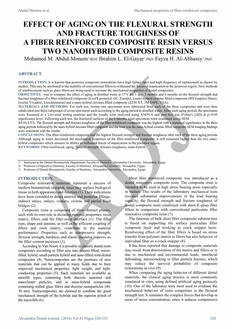
B- Fracture toughness
Statistical analysis showed significant difference between the
fracture toughness of the three composite materials at the
different aging periods. The results are summarized in (Table 3)
and represented graphically using simple bar chart (Figure 3).
Fracture toughness of the fiber-reinforced composite was
the highest in all the three aging periods with a statistical
significant difference compared to the two nano-hybrid
composites. The least was the nano-hybrid ceramic filled
composite. The fracture toughness of the fiber-reinforced
composite was about 4 times that of the nano-hybrid ceramic
filled composite and 3 times that of the nano-hybrid zirconia
filled composite at the different aging periods.
For the fiber-reinforced composite, the fracture toughness
significantly decreased by about 37% after 3 months aging,
from (5.21 ± 0.79 MPa.m1/2) to (3.23 ± 0.19 MPa.m1/2) and
42% after 6 months aging, from (5.21 ± 0.79 MPa.m1/2) to
(2.98 ± 0.27 MPa.m1/2). For the nano-hybrid ceramic filled
composite, the fracture toughness significantly decreased by
about 35% after 3 months aging, from (1.19 ± 0.04 MPa.m1/2)
to (0.87 ± 0.16 MPa.m1/2) and 50% after 6 months aging, from
(1.19 ± 0.04 MPa.m1/2) to (0.57 ± 0.12 MPa.m1/2). For the
nano-hybrid zirconia filled composite, the fracture toughness
decreased by about 16% after 3 months aging, from (1.27 ±
0.18 MPa.m1/2) to (1.11 ± 0.17 MPa.m1/2) and 26 % after 6
months aging, from (1.27 ± 0.18 MPa.m1/2) to (0.93 ± 0.15
MPa.m1/2) but still remained at lower values than the fiber-
reinforced composite.
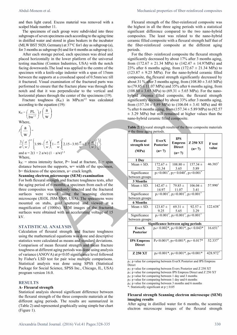
Fracture toughness Scanning electron microscope
(SEM) imaging results
After aging in distilled water for 6 months, the scanning
electron microscope images of the fracture toughness
specimens' fractured surface revealed that the fibers in the
fiber-reinforced composite acted as micro-crack deflectors
(Figure 4A) while the ceramic fillers in the nano-hybrid
ceramic filled composite and the zirconia fillers in the nano-
hybrid zirconia filled composite (Z250 XT, 3M ESPE,
USA) failed to act as micro-crack deflectors (Figure 4B and
4C).
Figure 2: SEM images of flexural strength specimens after
aging for 6 months. The red arrows show cohesive fractures in
the resin matrix. (A)In the fiber-reinforced composite, the fracture
goes around the glass fibers with the glass fibers remaining intact.
(B) Nano-hybrid ceramic filled composite and (C) Nano-hybrid
zirconia filled composite, show a clear cohesive fracture in the
resin matrix and the ceramic fillers and zirconia fillers
respectively.
Abdul-Monem et al. Mechanical properties of fiber-reinforced composites
Alexandria Dental Journal. (2016) Vol.41 Pages:328-335 332
Figure 3: Comparison between the different composite materials
in the same aging period and its effect on the fracture toughness.
Table 3: Fracture toughness results of the three composite
materials at the three aging periods.
Fracture
toughness
test (MPa.m1/2)
EverX
Posterior
(n= 7)
IPS
Empress
Direct
(n= 7)
Z 250 XT
(n= 7)
F test
value
1 Day
170.542* Mean ± SD. 5.21 ± 0.79 1.19 ± 0.04 1.27 ± 0.18
Significance
between groups
p1<0.001*, p2<0.001*, p3= 0.752
3 Months
391.692* Mean ± SD. 3.23 ± 0.19 0.87 ± 0.16 1.11 ± 0.17
Significance
between groups
p1<0.001*, p2= 0.018*, p3= 0.018*
6 Months
327.283* Mean ± SD. 2.98 ± 0.27 0.57 ± 0.12 0.93 ± 0.15
Significance
between groups
p1<0.001*, p2< 0.001*, p3= 0.002*
Significance between aging periods
EverX
Posterior
p4<0.001*, p5<0.001*, p6= 0.349 43.101*
IPS Empress
Direct
p4<0.001*, p5<0.001*, p6<0.001* 50.044*
Z 250 XT
p4= 0.086, p5= 0.001*, p6= 0.056 7.450*
p1: p value for comparing between EverX Posterior and IPS Empress
Direct p2: p value for comparing between Everx Posterior and Z 250 XT
p3: p value for comparing between IPS Empress Direct and Z 250 XT
p4: p value for comparing between 1 day and 3 months p5: p value for comparing between 1 day and 6 months
p6: p value for comparing between 3 months and 6 months
*: Statistically significant at p ≤ 0.05
DISCUSSION A- Flexural strength test
One of the laboratory tests most used to evaluate the mechanical
behavior of resin composites is the flexural strength test. It
simulates the complex forces that develop in areas of stress
concentration, since it induces compressive and tensile forces
simultaneously, close to and opposite from the point of loading,
respectively (11). The test serves as a predictor of the mechanical
behavior of a resin composite used clinically as stated by
Reinhardt et al (1994) (20).
Figure 4: SEM images of fracture toughness specimens after
aging for 6 months. The red arrows show micro-cracks in the resin
matrix.(A)In the fiber-reinforced composite , the fibers acted as micro-
crack deflectors .(B)Nano-hybrid ceramic filled composite and (C)
Nano-hybrid zirconia filled composite, the ceramic fillers and zirconia
fillers failed to act as micro-crack deflectors respectively.
Multiple factors influence the flexural strength test one of
which is the test specimen dimensions. In this study
specimens were prepared according to ISO standard 4049
with dimensions 25x2x2 mm (length x width x height)
Abdul-Monem et al. Mechanical properties of fiber-reinforced composites
Alexandria Dental Journal. (2016) Vol.41 Pages:328-335 333
which is the most commonly used specimen dimensions in
literature (11).
Another factor that influences flexural strength test is the
storage condition. Long-term water storage leads to reduced
mechanical properties. This accelerates hydrolytic
degradation of both the resin matrix and the silane layer
covering the inorganic fillers (21).
The temperature is another important factor affecting flexural
strength results. It has been argued that all mechanical properties
must be evaluated at 37ºC to bear clinical significance, since that
is the temperature at which the materials will be used in the oral
cavity (22).
Flexural strength results of this study showed that the fiber-
reinforced composite had the highest flexural strength
compared to the two nano-hybrid composites in all the three
aging periods. This agrees with Abouelleil et al 2015 (23) who
did a study to compare the mechanical properties of a newly
developed fiber -reinforced composite (EverX Posterior, GC,
Europe) to other commercially available bulk fill composites.
They found that after 48 hours’ storage in water, the fiber-
reinforced composite (EverX Posterior, GC, Europe) had the
highest flexural strength among the tested materials.
This also agrees with Goracci et al 2014 (24) who
compared the flexural strength of the fiber-reinforced
composite (EverX Posterior, GC, Europe) with other low-
stress behavior dental composites. They found that the
fiber-reinforced composite had the highest flexural strength
among the tested materials after 24 hours’ storage in water.
This is due to the fact that the fibers in the fiber-reinforced
composite being longer (2 mm) than the critical fiber length of
E-glass fibers (0.5-1.6 mm) with Bis-GMA polymer matrix,
allows for stress transmission from matrix to fibers, thus
producing an effective reinforcement. The physical
explanation of the strengthening and stiffening mechanism is
that since the matrix has a much lower modulus than the fiber,
the matrix strains more. The critical fiber length is therefore the
minimum length at which the center of the fiber reaches its
ultimate tensile strength when the matrix reaches its maximum
shear strength (25).
Additionally, the so-called Semi-Interpenetrating
Polymer Network (Semi-IPN); a resin matrix containing
Bis-GMA, TEGDMA, and PMMA in the form of net-poly
(methyl methacrylate)-inter-net-poly (bis-glycidyl-A-
dimethacrylate) increases the mechanical properties (26).
Moreover, as shown in previous works, the filler volume
percentage is closely related to the flexural strength and
flexural modulus values. Interestingly, the fiber-reinforced
composite with the lowest filler volume percentage (53.6%)
had the highest flexural strength, showing the role of the fibers
in increasing the material stiffness and resistance to bending
force during testing and probably during function (23).
The results of this study showed that the flexural strength of
the fiber-reinforced composite decreased significantly by about
17% after 3 months aging and 23% after 6 months aging. This
may be due to plasticization of the resin matrix and loss of
interfacial bond strength. A partial hydrolysis of the silane
bonds formed between the glass fibers and the matrix may
explain this result. Furthermore, the degradation of the glass
fiber itself cannot be ruled out, because glasses also are
susceptible to hydrolytic degradation. The mechanism of
hydrolytic decomposition is based on the dilution of boric
oxide, a compound which forms glass, from the surface of the
glass fiber (27).
Hydrolysis is known to be an autocatalytic reaction, but it is
possible that hydrolysis of the silane-promoted adhesion could
also be reversible. This is due to silane coupling agent on the
glass surface reforming the original bond by a recondensation
reaction of the hydrolysis products, i.e., silanols to make a new
bond with adjacent groups (27).
The primary mechanism for the ingress of water is diffusion
and some absorption is facilitated by the polarity of polymer
chains. Water molecules penetrate into the spaces between
polymer chains and occupy positions between the chains, and
thus, the polymer chains are forced apart. Water molecules act
as a plasticizer and the polymer chains generally become more
mobile and as a result, the flexural strength is reduced (27).
In this study, the nano-hybrid ceramic filled composite had
the lowest flexural strength among the three tested composite
materials in all the three aging periods. This may be due to the
brittle nature of the ceramic fillers used (Yetterbium
trifluoride) which can't withstand bending and break
immediately without permanent deformation.
In addition to that, the resin matrix of the nano-hybrid
ceramic filled composite is made of Bis-GMA and
UDMA.BIS-GMA, despite its high intrinsic reactivity, the
presence of hydroxyl groups on the backbone and the pi-pi
bond interactions given by the aromatic rings increase the
initial viscosity to a point that the polymer typically does
not reach high conversion which leads to increased water
sorption and consequently low flexural strength values (28).
UDMA is also characterized by high solubility,
hydrophilicity and low degree of conversion (28) which
may explain why the flexural strength of IPS Empress
Direct was lower than the other two composites. In the
literature it is also reported that UDMA based composites
undergo softening in water or oral simulating fluids easier
than Bis-GMA resins (29).
The nano-hybrid zirconia filled composite had the second
highest flexural strength after the fiber-reinforced
composite (EverX Posterior, GC, Europe). The fillers in the
nano-hybrid zirconia filled composite are surface-modified
zirconia/silica with filler volume (68%) which is higher than
that of nano-hybrid ceramic filled composite (52-59%) and
the resin matrix is a blend of 5 polymers; Bis-GMA,
UDMA, Bis-EMA, TEGDMA and PEGDMA. This
explains its superiority over (IPS Empress Direct, Ivoclar
Vivadent, Lieschtenstein) but still it was inferior to the
fiber-reinforced composite (EverX posterior, GC, Europe)
with its fibers.
The flexural strength results are consistent with the
scanning electron microscope (SEM) imaging findings.
SEM images revealed cohesive fractures in the resin
matrix.The fibers remained intact in the fiber-reinforced
composite while in the two nano-hybrid composites, the
fracture line passed through the matrix and fillers. These
findings emphasize the role of fibers in increasing the
flexural strength of dental composites.
B- Fracture toughness test
Dental materials researchers regard the fracture toughness
of a material as a more accurate predictor than traditional
compressive and tensile testing of how a material will
perform under various occlusal & masticatory stresses (12).
Fracture toughness (KIC) is a measure of the stress
intensity at the tip of a crack or flaw from which a crack
propagates through a material in an unstable manner. The
subscript (I) refers to the case when the crack is propagated
in mode I or tensile opening. This property has been related
Abdul-Monem et al. Mechanical properties of fiber-reinforced composites
Alexandria Dental Journal. (2016) Vol.41 Pages:328-335 334
to the ability of the material to resist both crack propagation
and wear in the oral environment (19).
The pre-crack is required in fracture toughness testing,
because it simulates a sharp, natural flaw in the interior of a
material. Since the stress concentration is highest when the
notch or crack is sharpest, it was believed that the most accurate
evaluation of fracture toughness for dental composites would
be achieved by testing specimens with an extremely sharp
flaw, i.e., one made by propagating a crack from a sharp notch.
The most commonly used fracture toughness test in literature
is the single-edge notch test with precracks and has recorded
the most accurate and predictable fracture toughness values
(30).
Results of this study showed that the fiber-reinforced
composite had the highest fracture toughness compared to
the two nano-hybrid composites in all the three aging
periods. The results are in agreement with Abouelleil et al
2015 (23) and Goracci et al 2014 (24).
Reinforcing effect of the fiber fillers is based on stress
transfer from polymer matrix to fibers. Random fiber
orientation and the polymer matrix by the semi-IPN structure
likely had a significant role in mechanical properties. In
addition to the toughening mechanism by fibers, the linear
polymer chains of PMMA in the cross-linked matrix of
BisGMA-TEGDMA plasticize the polymer matrix to some
extent and increase the fracture toughness of the composite
resin (26).
In this study, the nano-hybrid ceramic-filled composite
showed the least fracture toughness at all the three aging
periods. This is attributed to the ceramic fillers and their brittle
nature and their failure to act as crack stoppers. Also the
hydrophilicity of its UDMA based resin matrix reduced its
fracture toughness.
The majority of studies are in general agreement that the
fracture toughness of composites increases as filler volume
fraction is increase The presence of reinforcing particles
distributes the propagating nominal force into many
components, causes the crack front to curve or dissipate
between particles, and becomes energetically unfavorable
for a crack to grow (31).
The results of the study showed that the nano-hybrid
zirconia filled composite with filler volume (68%) had
higher fracture toughness than the nano-hybrid ceramic
filled composite with filler volume (59%). But although the
fiber-reinforced composite had the lowest filler volume
(53.6%), it showed the highest fracture toughness.This
shows the ability of fibers to act as crack deflectors rather
than ceramic or zirconia fillers.
Besides the matrix-filler interaction, crack pinning, crack
branching, crack deflection, or micro crack-induced
toughening are seen as main mechanisms of increasing
fracture toughness values by filler particles in resin
composites. The mechanism of increasing toughness by
matrix-filler interaction seems to play an important function
especially in composites with decreased filler size, like the
nano-hybrid materials.The decreased filler size is able to
change the organic matrix between the particles, improving the
mechanical properties, as a consequence of decreasing inter-
particle distances (31).
The fracture toughness results are consistent with the
scanning electron microscope (SEM) imaging findings.
SEM images revealed that the fibers acted as crack
deflectors in the fiber-reinforced composite while in the two
nano-hybrid composites, the ceramic and zirconia fillers
failed to act as crack deflectors. These findings emphasize
the role of fibers in increasing the fracture toughness of
dental composites.
CONCLUSIONS From the results of this study, the following could be
concluded:
The fiber-reinforced composite (EverX
posterior,GC,Europe) had the highest flexural strength and
fracture toughness compared to the two nano-hybrid
composites (IPS Empress Direct, Ivoclar Vivadent,
Lieschtenstein) and (Z250 XT,3M ESPE ,USA) in all the three
aging periods. Although aging in water decreased the
mechanical properties of the fiber-reinforced composite, but it
still remained higher than the two nano-hybrid composites
which ensures its ability to withstand forces of mastication in
the posterior region.
CONFLICT OF INTERESTS The authors declare that they have no conflict of interests.
REFERENCES 1. Cramer NB, Stansbury JW, Bowman CN. Recent advances
and developments in composite dental restorative materials.
J Dent Res .2011; 90: 402-16.
2. Mota EG. Relationship between filler content and selected
mechanical properties of six microhybrid composites. J
Dent Sci .2011; 26: 151-5.
3. Masouras K, Silikas N, Watts DC. Correlation of filler
content and elastic properties of resin-composites. Dent
Mater J. 2008; 24: 932-9
4. Van Noort R. Introduction to dental materials. 3rd ed.
London, UK: Elsevier, 2007:99-126
5. Colceriu A, Moldovan M, Prejmerean C, Buruiana T,
Buruiana EC, Furtos G, et al. Nanocomposite used in
dentistry. Eur Cells Mater J .2005; 10: 10-19.
6. Suzuki T, Kyoizumi H, Finger WJ, Kanehira M, Endo T,
Utterodt A, et al. Resistance of nanofill and nanohybrid resin
composites to toothbrush abrasion with calcium carbonate
slurry. Dent Mater J .2009; 28: 708-16.
7. Garoushi S, Vallittu P, Lassila LV. Fracture toughness,
compressive strength and load-bearing capacity of short
glass fiber-reinforced composite resin. Chin J Dent Res
.2011; 14: 15-9.
8. Garoushi S, Tanner J, Vallittu PK, Lassila L. Preliminary
clinical evaluation of short fiber-reinforced composite resin
in posterior teeth: 12-months report. Open Dent J. 2012; 6:
41-5.
9. Drummond JL. Degradation, fatigue, and failure of resin
dental composite materials. J Dent Res .2008; 87: 710-19.
10. Hahnel S, Henrich A, Bürgers R, Handel G, Rosentritt M.
Investigation of mechanical properties of modern dental
composites after artificial aging for one year. Oper Dent J.
2010; 35: 412-9.
11. Calheiros FC, Pfeifer CS, Brandão LL, Agra CM, Ballester
RY. Flexural properties of resin composites: Influence of
specimen dimensions and storage conditions. Dent Mater J
.2013; 32: 228-32.
12. Medina Tirado JI, Nagy WW, Dhuru VB, Ziebert AJ. The
effect of thermocycling on the fracture toughness and
hardness of core build up materials. J Prosth Dent. 2001; 86:
474-80.
Abdul-Monem et al. Mechanical properties of fiber-reinforced composites
Alexandria Dental Journal. (2016) Vol.41 Pages:328-335 335
13. Combe EC, Burke FJT, Douglas WH. Dental biomaterials.
London: Kluwer Academic Publishers, 1999. p 17.
14. International Organization for Standardization. ISO
4049/2000 – Dentistry – Polymer-based filling, restorative
and luting materials. Switzerland: ISO; 2000.
15. Garapati SN, Priyadarshini, Raturi P, Shetty D, Srikanth
KV. An in vitro evaluation of diametral tensile strength and
flexural strength of nanocomposite vs hybrid and minifill
composites cured with different light sources (QTH vs
LED). J Contemp Dent Pract .2013; 14: 84-9.
16. American Dental Association Council on Dental Materials
Instruments and Equipment: Status report on posterior
composites. J Am Dent Assoc .1983;107:74-6.
17. Knobloch LA, Kerby RE, Seghi R, Berlin JS, Clelland
N.Fracture toughness of packable and conventional
composite materials. J Prosthet Dent .2002;88:307-13.
18. Fujishima A, Ferracane JL. Comparison of four modes of
fracture toughness testing for dental composites. Dent
Mater J .1996; 12: 38-43.
19. Bonilla ED, Mardirossian G, Caputo AA. Fracture
toughness of posterior resin composites. Quintessence Int.
2001; 32: 206-10.
20. Reinhardt JW, Boyer DB, Stephens NH. Effects of
secondary curing on indirect posterior composite resins.
Oper Dent J .1994; 19: 217-20.
21. Ferracane JL. Hygroscopic and hydrolytic effects in
polymer networks. Acad Dent Mater .2004; 18: 118-28.
22. Musanje L, Darvell BW. Effects of strain rate and
temperature on the mechanical properties of resin
composites. Dent Mater J .2004; 20: 750-65.
23. Abouelleil H, Pradelle N, Villat C, Attik N, Colon P,
Grosgogeat B.Comparison of mechanical properties of a
new fiber reinforced composite and bulk filling composites.
Restor Dent Endod. 2015; 40:262-270.
24. Goracci C, Cadenaro M, Fontanive L, Giangrosso G,
Juloski J, Vichi A. Ferrari M.Polymerization efficiency and
flexural strength of low-stress restorative composites. Dent
Mater J.2014; 30: 688-94.
25. Petersen RC. Discontinuous fiber-reinforced composites
above critical length. J Dent Res .2005; 84:365-70.
26. Garoushi S, Säilynoja E, Vallittu PK, Lassila L.Physical
properties and depth of cure of a new short fiber reinforced
composite. Dent Mater J. 2013; 29:835-41.
27. Lassila LV, Nohrstrom T, Vallittu PK. The influence of
short-term water storage on the flexural properties of
unidirectional glass fiber-reinforced composites. J Biomat.
2002; 23:2221–29.
28. Gajewski VE, Pfeifer CS, Fróes-Salgado NR, Boaro LC,
Braga RR. Monomers used in resin composites: degree of
conversion, mechanical properties and water
sorption/solubility. Braz Dent J .2012; 23:508-14.
29. Ho CT, Vijayaraghavan TV, Lee SY, Tsai A, Huang HM,
Pan LC. Flexural behaviour of post-cured composites at
oral-simulating temperatures. J Oral Rehabil 2001; 28:658–
67.
30. Ferracane JL,Antonio RC, Matsumoto H.Variables
affecting the fracture toughness of dental composites. J
Dent Res.1987; 66:1140-5.
31. Ilie N, Hickel R, Valceanu AS, Huth KC. Fracture
toughness of dental restorative materials. Clin Oral Investig
.2012; 16: 489-98.