effect of cyclooxygenase blockade blood flowthrough well-developed coronary...
TRANSCRIPT

1091
Effect of Cyclooxygenase Blockade on BloodFlow Through Well-Developed Coronary
Collateral VesselsJohn Altman, Daniel Dulas, and Robert J. Bache
Collateral vessels that develop after coronary artery occlusion demonstrate perivascular inflammation,subintimal hyperplasia, and endothelial proliferation. This study was performed to test the hypothesisthat these abnormalities are associated with evidence for increased production of vasodilator prostaglan-dins. Eight dogs were studied 4-6 months after occlusion of the anterior descending coronary artery hadbeen performed to stimulate collateral vessel growth. At the time of study, the anterior descendingcoronary artery was cannulated at the site of occlusion to allow measurement of retrograde blood flow as
an index of interarterial collateral flow. Injection of radioactive microspheres during the retrograde flowcollection allowed determination of continuing tissue flow in the collateral-dependent zone as an index ofintramural microvascular collateral flow. Retrograde and tissue flows were measured before and 20minutes after 5 mg/kg i.v. indomethacin, a dose that caused 95±3% inhibition of the coronary
vasodilation in response to a 500 jig intracoronary bolus of arachidonic acid. Heart rate and mean aorticpressure were not significantly altered by indomethacin, and blood flow to the normally perfusedmyocardial region was not changed by administration of indomethacin. However, indomethacin caused a
40±7% decrease in retrograde flow (p<0.01), and microvascular collateral flow to the dependentmyocardium decreased by 20±10%o (p<0.05). These data indicate that, unlike the normal coronary
circulation, well-developed coronary collateral vessels are under the tonic influence of vasodilatorprostaglandins. (Circulation Research 1992;70:1091-1098)KEY WoRDs * coronary vessels * coronary occlusion * myocardial blood flow * prostaglandins
* indomethacin
A lthough the coronary vessels are responsive tolocally produced vasoactive derivatives of ara-chidonic acid,' most evidence does not support
an important role for prostaglandins in regulation ofcoronary blood flow during physiological conditions.Thus, Owen et a12 and Harlan et a13 reported thatcyclooxygenase blockade with indomethacin did notalter baseline blood flow or the increase in flow thatoccurred during reactive hyperemia or in response tothe increased myocardial oxygen demands produced byisoproterenol in open-chest dogs. Similarly, indometh-acin did not alter coronary blood flow or myocardialoxygen extraction in intact awake dogs either at rest orduring graded treadmill exercise.4 These reports thusfail to support a substantial role for products of cyclo-oxygenase metabolism in regulation of the normal cor-onary circulation.
From the Department of Medicine, University of MinnesotaMedical School, Minneapolis.
Supported by US Public Health Science grants HL-20598 andHL-32427 from the National Heart, Lung, and Blood Institute.J.A. was supported by a Medical Student Research Fellowshipfrom the American Heart Association.Address for correspondence: Robert J. Bache, MD, Cardiovas-
cular Division, Department of Medicine, University of MinnesotaMedical School, Box 508 UMHC, 420 Delaware Street SE, Min-neapolis, MN 55455.
Received September 16, 1991; accepted January 27, 1992.
Collateral vessels that develop in response to coro-nary artery occlusion have a well-developed muscularmedia and are capable of active vasomotion, which maymodulate blood flow to the dependent myocardium.5,6Unlike normal arterial vessels, however, ultrastructuralexamination of these collateral vessels has demon-strated perivascular inflammation, monocyte adherenceto the endothelium, subintimal hyperplasia, and endo-thelial proliferation.78 Although these histological ab-normalities are most prominent during collateral growthearly after coronary occlusion, Schaper et a17 foundendothelial proliferation in collateral vessels 1 yearafter coronary occlusion. Because these histologicalabnormalities may be associated with elevated localprostaglandin production,9 this study was performed toexamine the effect of cyclooxygenase blockade on cor-onary collateral blood flow.
Materials and MethodsThese studies were performed in accordance with the
position of the American Heart Association on researchanimal use adopted November 11, 1984, and under thesupervision of the Animal Care Committee of theUniversity of Minnesota. Two groups of adult mongreldogs were studied. Group 1 consisted of five unoccludeddogs that were used to document the degree of cyclo-oxygenase blockade that could be achieved by measur-ing the response of coronary blood flow to intracoro-nary arachidonic acid before and after treatment with
by guest on July 12, 2018http://circres.ahajournals.org/
Dow
nloaded from

1092 Circulation Research Vol 70, No 6 June 1992
indomethacin. Group 2 consisted of eight dogs withchronic coronary artery occlusion that were used toexamine the effect of cyclooxygenase blockade on cor-onary collateral blood flow.
Group 1Five adult mongrel dogs of either sex were anesthe-
tized with sodium pentobarbital (30-35 mg/kg i.v.),intubated, and ventilated with a respirator (HarvardApparatus, South Natick, Mass.) with room air andsupplemental oxygen. Under sterile conditions, a leftthoracotomy was performed in the fifth intercostalspace. A heparin-saline-filled polyvinyl catheter (3.0mm o.d.) was inserted into the left internal thoracicartery and advanced into the ascending aorta. Thepericardium was opened, and a heparin-filled catheterwas introduced into the left ventricle near the apicaldimple. The proximal left circumflex coronary arterywas mobilized and a 5-MHz Doppler flowmeter probewas fitted around the artery. A heparin-filled Silasticcatheter (0.3 mm o.d.) was inserted into the circumflexartery distal to the flowmeter probe.10 The pericardiumwas loosely closed, the catheters and electrical leadswere tunneled subcutaneously to exit in the interscap-ular area, and the chest was closed in layers. Cathetersand electrical leads were protected by a nylon vest theanimals had been trained to wear. Catheters wereflushed with heparin-saline daily to maintain patency.
Studies were performed seven to 10 days after sur-gery. Aortic and left ventricular pressures were mea-sured with Statham P231D pressure transducers. Cir-cumflex blood flow was measured with a Dopplerflowmeter (Craig Hartley, Houston, Tex.). Data wererecorded on an eight-channel recorder (Coulbourn In-struments Inc., Lehigh Valley, Pa.). Injections of ara-chidonic acid or its vehicle were made into the coronaryartery, and the flow responses were observed. Arachi-donic acid was dissolved in a solution of 10% ethanol in100 mM Na2CO3 under nitrogen. This solution wasdiluted with isotonic saline to a final concentration of 1mg/ml and used within 3 hours after preparation. Theresponse to intracoronary arachidonic acid or vehicle(0.5 ml) was observed in triplicate at 5-minute intervalsto ensure that a consistent response was achieved.Indomethacin (5 mg/kg) was then administered as anintravenous bolus. Twenty minutes after the adminis-tration of indomethacin, the hemodynamic and coro-nary flow responses to intracoronary arachidonic acidand its vehicle were again observed. The response tovehicle was subtracted from the arachidonic acid re-sponse, and the excess flow produced by arachidonicacid was compared with the corresponding measure-ment obtained after administration of indomethacin.
Group 2Development of collateral vessels. Collateral vessel
development was induced by catheter embolization ofthe left anterior descending coronary artery with ahollow plug in eight adult mongrel dogs as previouslydescribed.6 Animals were anesthetized with sodiumpentobarbital (25 mg/kg i.v.), intubated, and ventilatedwith a respirator. The right carotid artery was isolatedunder sterile surgical conditions. After administration
coronary artery catheter was introduced into the leftcoronary ostium and directed toward the anterior de-scending artery. A 0.014-in. angioplasty guide wire waspassed through the catheter into the distal anteriordescending artery. Nitroglycerin (100 ,g i.c.) was given.The coronary catheter was removed without disturbingthe guide wire, and a hollow stainless-steel plug (2.3-2.7mm o.d., 1.1 mm i.d., 4 mm in length) was pushed alongthe guide wire with a length of flanged PE-90 tubinguntil the plug was firmly wedged in the coronary artery.The guide wire was removed, and the position andpatency of the plug were confirmed by fluoroscopyduring intracoronary injection of 5 ml of 60% diatri-zoate meglumine.
Surgical preparation. The animals were returned tothe laboratory 4-6 months after coronary embolization,premedicated with morphine sulfate (1 mg/kg s.c.),anesthetized with a-chloralose (100 mg/kg i.v. followedby 10 mg/kg per hour), intubated, and ventilated with arespirator. Supplemental oxygen was used to maintainarterial Po2 within the physiological range. Two 7F NIHcatheters were introduced into the femoral arteries andpositioned in the ascending aorta for blood samplingand for pressure monitoring. A similar catheter wasintroduced into the left carotid artery and advancedinto the left ventricle for pressure measurement. A leftthoracotomy was performed in the fifth intercostalspace. A pneumatic cuff occluder was fitted around thedescending thoracic aorta to allow control of proximalaortic pressure. The heart was suspended in a pericar-dial cradle and a polyvinyl chloride catheter (3.0 mmo.d.) was inserted into the left atrium through the atrialappendage. The coronary artery plug was located bypalpation, and the anterior descending artery was dis-sected free for 1.0-1.5 cm proximal and distal to theplug. After administration of sodium heparin (200units/kg i.v. followed by 1,000 units/hr), the artery wasoccluded proximally, and a longitudinal arteriotomy wasperformed. The plug was extracted, and the artery wasallowed to bleed freely to dislodge any residual throm-bus. The artery was then cannulated with a thin-wallstainless-steel cannula (4.0 mm o.d.). Resistance of thecoronary cannula determined from the pressure dropproduced by passing measured flows of blood throughthe cannula was 0.097 mm Hg/ml per minute of flow.Pressure at the cannula tip was measured with a 23-gauge tube incorporated into the wall of the cannula.
Myocardial blood flow. Myocardial blood flow wasmeasured with 15-,um-diameter microspheres labeledwith 1251, 5"Cr, 85Sr, 95Nb, 4'Sc, or `4lCe (NEN Co., Boston,Mass.; and 3M Co., St. Paul, Minn.). Microspheres wereobtained as 1.0 mCi in 10 ml of 10% low molecularweight dextran. Microspheres were agitated in an ultra-sonic bath for at least 10 minutes before injection. Foreach intervention, 3X 10⁶ microspheres were injectedinto the left atrium, and a reference sample of arterialblood was withdrawn from the aortic catheter at aconstant rate of 15 ml/min with a peristaltic pump.Reference sampling was begun at the time of micro-sphere injection and continued for 90 seconds.
Experimental protocol. Aortic, left ventricular, andcoronary cannula pressures were measured withStatham P231D pressure transducers. Left ventricularpressure was recorded at normal and high gain for
of heparin sodium (6,000 units i.v.), an 8F Judkins right measurement of end-diastolic pressure. Left ventricular
by guest on July 12, 2018http://circres.ahajournals.org/
Dow
nloaded from

Altman et al Indomethacin and Coronary Collateral Blood Flow 1093
dP/dt was obtained by electronic differentiation of thepressure signal. Data were recorded on an eight-chan-nel direct-writing recorder (Coulbourn Instruments).Interarterial collateral blood flow was measured bycollecting retrograde flow from the coronary arterycannula into a graduated cylinder for 30-second inter-vals while the cannula tip was maintained at the level ofthe heart. The level of the cannula tip was maintainedconstant throughout the study. Control measurementsof retrograde blood flow were repeated until consistentmeasured collections were obtained. Measurementswere thereafter performed in triplicate, and the resultswere averaged. To assess microvascular collateral flowthat continued during diversion of coronary arterycannula flow, an injection of microspheres was admin-istered to six of the animals during control conditionssimultaneously with a retrograde flow collection.
After completion of the control measurements, indo-methacin was administered as a single intravenous bolusdose of 5 mg/kg. Hemodynamic variables were recordedcontinuously, and retrograde blood flow collectionswere performed in duplicate at 20 minutes after theadministration of indomethacin. A second injection ofmicrospheres was administered simultaneously with thefinal retrograde flow collection 20 minutes after theadministration of indomethacin. In five of the animals,hemodynamic measurements and retrograde flow col-lections were repeated 60 minutes after indomethacinadministration.To allow determination of total collateral tissue flow,
the shadow technique of Patterson and Kirk" was usedto delineate the collateral-dependent myocardial re-gion. For this procedure, microspheres were adminis-tered into the left atrium while the coronary cannulawas perfused with nonradioactive arterial blood from apressurized reservoir. Cannula tip pressure was main-tained 10 mm Hg above mean aortic pressure during themicrosphere injection. In this way, the myocardiumdistal to the site of occlusion was perfused with nonra-dioactive blood to distinguish it from the remainder ofthe heart, which was marked with microsphere-contain-ing blood.
Tissue preparation. The heart was excised and fixed in10% buffered formalin, and the left ventricle was di-vided into four transverse rings parallel to the mitralvalve ring. The rings were then sectioned radially into 16segments, which were divided into epicardial and en-docardial halves. The resultant specimens were weighedon an analytical balance and placed into vials forcounting. Myocardial and blood reference samples werecounted in a gamma spectrometer with a multichannelanalyzer (model 5912, Packard Instrument Co., Inc.,Downers Grove, Ill.) at window settings correspondingto the peak energies of each radionuclide. The activityin each window was corrected for background andoverlapping counts between isotopes with a digitalcomputer. Blood flow to each myocardial specimen(Qm) was computed using the formula Qm=QrxCm/Cr,where Q, is reference blood flow rate (in milliliters perminute), Cm is counts per minute of myocardial speci-men, and Cr is counts per minute of reference bloodspecimen. Blood flows were expressed as milliliters perminute per gram of myocardium.Data analysis. Total flow to the collateral tissue was
TABLE 1. Response of Hemodynamic Variables and CoronaryBlood Flow to Intra-arterial Administration ofArachidonic Acid toFive Unoccluded Dogs (Group 1) During Control Conditions and20 Minutes After Administration of Indomethacin
Mean aortic CoronaryHeart rate pressure blood flow(bpm) (mm Hg) (ml/min)
CON INDO CON INDO CON INDO
Baseline 141+14 125+15* 104+3 107+2 40.5±5.6 42.8+6.8AA 141±16 130±14 101±3 107+2 70.9+9.0t 43.9+7.9*
Values are mean+±SEM. bpm, Beats per minute; CON, controlconditions; INDO, 20 minutes after administration of 5 mg/kg i.v.indomethacin; AA, bolus dose of 500 jig arachidonic acid.
*p<0.05 in comparison with CON.tp<0.05 in comparison with baseline.
myocardial samples in the collateral-dependent regionas identified by the shadow technique. Heart rate andpressures were measured directly from the strip-chartrecordings. Hemodynamic data were analyzed usinganalysis of variance for repeated measures. A value ofp<0.05 was required for statistical significance. Com-parisons of retrograde blood flows and microsphereflows between control and intervention were analyzedwith the Wilcoxon signed rank test and independent ttest. All data are expressed as mean+SEM.
ResultsGroup 1The response to intracoronary bolus injection of
arachidonic acid is shown in Table 1. Administration ofarachidonic acid resulted in no change in heart rate oraortic pressure but produced a 75 +8% peak increase incoronary artery blood flow. The coronary vasodilationproduced by arachidonic acid was brief in duration, withflow returning to the control level within 1-2 minutes.Indomethacin resulted in a nonsignificant increase inmean aortic pressure with a significant decrease of heartrate from 141 to 125 beats per minute (p<0.05).Indomethacin produced 95±3% inhibition of the coro-nary vasodilation produced by arachidonic acid(p<O.Ol).Group 2Hemodynamic data. Hemodynamic measurements
before and after administration of indomethacin areshown in Table 2. Mean aortic pressure tended toincrease and heart rate tended to decrease after indo-methacin administration, but these changes did notachieve statistical significance. Left ventricular end-di-astolic pressure was not significantly changed after theadministration of indomethacin. Left ventricular dP/dtwas slightly decreased after indomethacin administra-tion (p<0.02). Distal coronary pressure with the can-nula closed tended to decrease after indomethacinadministration, although this did not achieve statisticalsignificance. However, the tendency for distal coronarypressure to decrease in conjunction with a trend towardincreased aortic pressure resulted in a 65% increase inthe aortic to coronary pressure gradient after the ad-ministration of indomethacin (p<0.03).
Retrograde blood flow. Retrograde flow measure-ments from the cannulated left anterior descending
computed as the sum of absolute blood flow to all coronary artery are shown in Figure 1. During control
by guest on July 12, 2018http://circres.ahajournals.org/
Dow
nloaded from

1094 Circulation Research Vol 70, No 6 June 1992
TABLE 2. Hemodynamic Data for Eight Dogs With Chronic Coronary Artery Occlusion (Group 2) During ControlConditions and After Administration of Indomethacin
Mean aortic LV end-diastolic Mean coronary Aortic-coronaryHeart rate pressure pressure LV dP/dt pressure pressure gradient(bpm) (mm Hg) (mm Hg) (mm Hg/sec) (mm Hg) (mm Hg)
Control 138±11 95±4 5±1 2300±200 78±5 17±2INDO 130±12 99±4 7±2 2000±200* 71±5 28±3*
Values are mean±SEM. bpm, Beats per minute; LV, left ventricular; INDO, after administration of 5 mg/kg i.v.indomethacin.
*p<0.05 in comparison with control.
conditions, retrograde flow ranged from 11 to 71 ml/min(mean, 41±7 ml/min). Twenty minutes after indometh-acin administration, retrograde blood flow was de-creased in every animal studied (mean decrease,40±7%;p<0.01). In five animals, retrograde blood flowmeasurements were monitored for 1 hour after theadministration of indomethacin. In these animals, ret-rograde flow remained depressed throughout the 60-minute observation period after the administration ofindomethacin, so that retrograde flow 60 minutes afterindomethacin administration (29±9 ml/min) was notdifferent from retrograde flow 20 minutes after indo-methacin administration (31±9 ml/min).
Collateral zone tissue flow. Myocardial blood flowmeasurements obtained with the shadow techniquedemonstrated a sharp boundary between collateral-dependent and adjacent normally perfused myocar-dium. Collateral-dependent myocardium ranged from12 to 29 g (mean, 18±2 g). Total left ventricular massranged from 99 to 161 g (mean, 125±9 g). Collateral-dependent myocardium represented an average of15±2% of the left ventricle.
Tissue blood flows measured with microspheres areshown in Table 3. During control conditions, blood flow
7A
c
E
E
3:
ar-
Q
0aam
0
CDa
50.
25
n
CON INDOFIGURE 1. Plot showing individual responses (open circles)of retrograde blood from the anterior descending coronary
cannula of eight dogs during control conditions (CON) and20 minutes after administration of 5 mg/kg i.v. indomethacin(INDO). Mean±SEM blood flow is indicated by solid circlewith bars. *p<0.01 vs. CON.
in the normal zone was 0.78+0.08 ml/min per gram, andflow to the subendocardium (ENDO) was significantlygreater than flow to the subepicardium (EPI) (ENDO/EPI=1.29+0.08). Normal zone blood flow and theENDO/EPI ratio were not altered by indomethacin.Blood flows to the collateral-dependent myocardiumshown in Table 3 and Figure 2 were made with thecoronary cannula open to atmospheric pressure, so theyrepresent continuing microvascular collateral flow notdiverted during the retrograde flow collection. As shownin Figure 2, there was considerable variability in bloodflow to the collateral-dependent region during controlconditions with the cannula open; animals appeared tofall into two groups of three having relatively high flows(mean, 0.71±0.06 ml/min per gram) and three havingrelatively low flows (mean, 0.41±0.02 ml/min per gram).Of all measured variables, only mean aortic pressurewas different between these two groups; values were106±5 mm Hg in the high flow group and 87±1 mm Hgin the low flow group (p<0.02). Indomethacin de-creased mean tissue flow in the collateral-dependentregion by 20±10% (p<0.05); the decrease in flow wasmore prominent in the high flow group, so that flows inall animals were tightly grouped after the administrationof indomethacin. No hemodynamic change could befound that might explain the more prominent decreasein response to indomethacin in the high flow group.Other measures of collateral function, including retro-grade flow and the pressure gradient from aorta tocoronary artery with the cannula closed, were notdifferent between the two groups, suggesting that theresponse to indomethacin was not determined by thedegree of collateral development. Total collateral bloodflow obtained as the sum of retrograde flow and collat-eral zone tissue flow is shown in Table 4. Total collateralblood flow decreased 31% after cyclooxygenase block-ade (p<0.01).
DiscussionCyclooxygenase blockade with indomethacin caused
no significant change in blood flow to normally perfusedmyocardium. This is in agreement with previous studiesin normal hearts which have found little evidence thatprostaglandins participate in the control of coronaryblood flow during physiological conditions.2-4 In con-trast to the lack of effect on perfusion of normalmyocardium, indomethacin caused a substantial de-crease of collateral blood flow. This finding suggests thatthe prostaglandins exert tonic vasodilator activity oncoronary collateral vessels. This new finding, as well asthe method for determination of collateral blood flow,will be discussed in detail.
+ *I
0
0
0
0
%
V '
by guest on July 12, 2018http://circres.ahajournals.org/
Dow
nloaded from

Altman et al Indomethacin and Coronary Collateral Blood Flow
TABLE 3. Myocardial Tissue Flow Measured With Microspheres During Control Conditions and After Administrationof Indomethacin in Dogs With Chronic Coronary Artery Occlusion (Group 2)
Normal zone Collateral zone
Myocardial blood flow Myocardial blood flow(m/min per gram) (mllmin per gram)
ENDO EPI Mean ENDO/EPI ENDO EPI Mean ENDO/EPI
CON 0.89±0.10 0.70±0.08 0.78±0.08 1.29±0.08 0.56±0.09 0.55±0.08 0.56±0.07 1.02±0.11
INDO 0.83±0.11 0.70±0.10 0.74±0.10 1.18±0.09 0.37±0.04* 0.45±0.04 0.42±0.02* 0.85±0.11
Values are mean+SEM. ENDO, subendocardium; EPI,administration of 5 mg/kg i.v. indomethacin.
*p<0.05 in comparison with CON.
In the present study, opening the coronary cannula toatmospheric pressure did not abolish tissue flow in thecollateral-dependent region. For this reason both retro-grade and tissue flows were measured to account fortotal collateral flow. Continuing tissue flow during ret-rograde diversion indicates that some collaterals enterthe recipient vessel distal to a site of significant resis-tance, so that antegrade flow encounters less resistancethan flow back into the arterial cannula. Collaterals maydevelop at several levels in the coronary arterial vascu-lature; at each level the fraction of flow diverted whenthe cannula is opened is determined by the relative ratioof antegrade versus retrograde resistance.12 Scheel etal13 reported that embolizing the collateral-dependentmyocardium with microspheres up to 50 ,um in diameterincreased retrograde flow, whereas embolization with80-,um microspheres decreased retrograde flow, sug-gesting that the smallest collaterals entered recipientvessels between 50 and 80 ,um in diameter. Downey etal14 found that in well-collateralized hearts 84-,um mi-crospheres embolized into the distal vasculature weredislodged by a subsequent period of retrograde flow,
1 n
01
c08
L-
_
¢ 0.6
01 0.4.mLLI
V)V)1- 0.2
CON INDOFIGURE 2. Plot showing tissue blood flow measured withmicrospheres in the collateral-dependent myocardial region ofsix dogs (each represented by an open circle) during controlconditions (CON) and 20 minutes after administration of 5mg/kg i.v. indomethacin (INDO). Mean +SEM blood flow isindicated by solid circle with bars. *p<0.05 vs. CON.
subepicardium; CON, control conditions; INDO, after
indicating that some collaterals entered recipient ves-sels less than 84 ,um in diameter. In normal caninehearts, Harrison et al1" determined that mean stempressure at the origin of the collateral vessels was within7 mm Hg of aortic pressure, whereas collaterals arisingfrom vessels with a pressure of 45 mm Hg could accountfor no more than 25% of total collateral flow. In thepresent study, antegrade tissue flow constituted approx-imately 20% of total collateral flow. If these data can beextrapolated to the normal hearts studied by Harrisonet al, the microsphere flow could be accounted for bycollaterals originating from arterial vessels with pres-sures of approximately 45 mm Hg. Thus, approximatelyhalf of the vascular resistance would be proximal to theorigin of these microvascular collaterals. Since Chilianet all6 demonstrated that approximately 45% of coro-nary resistance resides in vessels larger than 100 ,um indiameter, the findings are consistent with the previousreports suggesting that the smallest collateral vessels arelikely to be in the range of 80 ,gm in diameter.1314However, the increased flow in the well-collateralizedheart would increase the pressure in the recipientvessel, so that it is likely that collaterals larger than thiscould result in sufficient pressure to perfuse the distalvascular bed. In addition, the resistance offered by thecannula would cause increased pressure in the recipientartery and encourage antegrade flow. However, cannularesistance was relatively low and would have causedonly a minimal increase in pressure in the recipientartery. It should be noted that residual flow within theshadow region was uniform and not preferentially de-livered to the border region. This indicates that even thesmallest collateral vessels entered the recipient arterialsystem sufficiently proximal to cause uniform distribu-tion of flow in the recipient vascular bed.
TABLE 4. Retrograde Blood Flow, Tissue Flow to the Collateral-Dependent Region, and Total Collateral Flow in Dogs WithChronic Coronary Artery Occlusion (Group 2)
Collateral zone
Retrograde Tissue flow Total Totalblood flow (mUmin tissue flow collateral flow(ml/min) per gram) (m/min) (miin)
CON 41±7 0.56±0.07 10.4+2.4 51± 10INDO 27+6* 0.42±0.02* 7.6±1.2* 35+9*
Values are mean+SEM. CON, control conditions; INDO, afteradministration of 5 mg/kg i.v. indomethacin. Total collateral flowwas determined as the sum of retrograde and tissue flow.
*p<0.05 in comparison with CON.
lr.
9
%..v
1095
by guest on July 12, 2018http://circres.ahajournals.org/
Dow
nloaded from

1096 Circulation Research Vol 70, No 6 June 1992
Since the resistance to collateral blood flow includesnot only the collateral vessels but also the coronaryarterial segment proximal to the origin of the collateralvessels, epicardial artery constriction could have con-tributed to the observed decrease in collateral flow inresponse to indomethacin. However, Lane and Bove,17using quantitative angiography in anesthetized dogs,found that indomethacin (5 mg/kg i.v.) caused nochange in the cross-sectional area of normal coronaryartery segments. This finding indicates that the de-creased collateral flow observed in the present studycould not be ascribed to indomethacin-induced vaso-constriction of coronary arteries proximal to the originof the collateral vessels. Collateral flow can also beinfluenced by changes in normal zone flow. Thus, agentsthat cause vasodilation of resistance vessels in normalmyocardial regions increase the pressure drop acrossthe proximal coronary artery segment, thereby decreas-ing the pressure available at the origin of the collateralvessels.18 In the present study, blood flow in the normalregion did not change in response to indomethacinadministration. Thus, indomethacin did not producechanges in either the coronary arteries or the coronaryresistance vessels in the normal myocardium that wouldbe expected to cause a decrease in collateral blood flow.The observed decrease in tissue blood flow in the
collateral-dependent region in response to indometh-acin could have resulted from vasoconstriction of eitherthe collateral vessels or the resistance vessels distal tothe site of entry of the collateral channels into therecipient arterial vasculature. Indomethacin did notcause generalized constriction of the coronary resis-tance vessels, since blood flow in the normal myocar-dium was not decreased. However, small-vessel respon-siveness may be altered in vascular beds perfused bycollateral vessels. Thus, Sellke et al19 found that endo-thelium-dependent dilation was depressed in coronaryarterioles chronically perfused through collateral ves-sels. It is possible that an abnormality of endothelium-derived vasodilator prostaglandins could also exist insmall vessels perfused through collateral channels.
Although no previous studies are available examiningthe effect of cyclooxygenase blockade on well-devel-oped coronary collateral vessels, several investigatorshave reported the effects of these agents on blood flowthrough native collateral vessels after acute coronaryartery occlusion. These studies demonstrated that indo-methacin, ibuprofen, or flurbiprofen did not alter bloodflow through native collateral vessels after acute coro-nary occlusion.20-22 Capurro et a123 reported that aspirin(600 mg i.v.) caused a small but significant increase inblood flow to the subepicardium of the acutely ischemicregion between 5 minutes and 4 hours after coronaryocclusion in open-chest dogs. However, small increasesin collateral flow have been shown to occur spontane-ously during the first hours after acute coronary arteryocclusion in the dog,24 so that it is unclear whether thesmall increase in collateral flow was the result of a drugeffect. Thus, the available data do not document aconsistent effect of cyclooxygenase blockade on thenative collateral vessels that exist at the time of acutecoronary occlusion.
Collateral vasoconstriction produced by indometh-acin is likely mediated through inhibition of local pros-
concentrated in the endothelial cells.25 In ultrastruc-tural studies of well-developed canine collateral vesselsexamined 8-52 weeks after ameroid placement,Schaper et a17 found increased numbers of endothelialcells per unit inner vascular space when compared withnormal vessels. The present study suggests that thisendothelial cell proliferation is associated with in-creased production of vasodilator prostaglandins in thecollateral vessels. Prostacyclin is the principal enzymat-ically generated prostaglandin in vascular endothelialcells and is a potent vasodilator of coronary arteries andresistance vessels.26 Several investigators have examinedthe effect of prostacyclin on blood flow through therudimentary collateral vessels that exist at the time ofacute coronary artery occlusion, but the results areconflicting. Jugdutt et a127 found that prostacyclin re-duced infarct size in awake dogs subjected to acutecoronary artery occlusion and that this effect was asso-ciated with an increase in blood flow to the acutelyischemic myocardium. Smith et a128 also found thatprostacyclin exerted a protective effect on acutely isch-emic myocardium, but this effect occurred with nochange in collateral blood flow. In contrast to theequivocal response of native collateral vessels,Scholtholt et al,29 using isolated perfused canine hearts5 weeks after ameroid constrictor implantation, foundthat prostacyclin (100 ,ug/min i.c.) caused a 50% de-crease in transcollateral resistance. This finding indi-cates that moderately well-developed coronary collat-eral vessels are responsive to the vasodilator effects ofprostacyclin and is consonant with the concept ex-pressed in the present study that interruption of endog-enous prostacyclin production decreases tonic vasodila-tor activity.
In addition to causing inhibition of prostacyclin pro-
duction, cyclooxygenase blockade has potential for in-creasing leukotriene production by diversion of arachi-donic acid from prostaglandin synthesis into thelipoxygenase pathway.30 Arachidonic acid is cleaved by5-lipoxygenase to form 5-HPETE, which is then con-verted to numerous products including leukotriene C4and D4, which are potent coronary vasoconstrictors.31Leukotriene production was first demonstrated in leu-kocytes32 but has been shown to occur in rabbit hearts33and in isolated human and canine epicardial coronaryartery segments.34 The potential for indomethacin-in-duced leukotriene production thus exists in coronaryvessels, but Lane and Bove17 demonstrated that normalcanine epicardial arteries do not undergo vasoconstric-tion in response to indomethacin. Developing collateralvessels show perivascular leukocyte infiltration,78 whichcould enhance local leukotriene production, but theseabnormalities are evident principally during early col-lateral development and are absent after 8 weeks ofcollateral growth. The present study was carried out 4-6months after coronary artery occlusion, well after thetime that leukocyte infiltration would have subsided. Inaddition, Ezra et a135 demonstrated that tolerance to thevasoconstricting effect of intracoronary leukotriene D4infusion developed within 2-4 minutes, whereas thedecrease in retrograde blood flow in the present studyremained stable over the 60-minute observation period.For these reasons, it is unlikely that leukotriene produc-tion contributed substantially to indomethacin-induced
tanoid production. Vascular prostaglandin production is collateral constriction.
by guest on July 12, 2018http://circres.ahajournals.org/
Dow
nloaded from
Altman et al Indomethacin and Coronary Collateral Blood Flow 1097
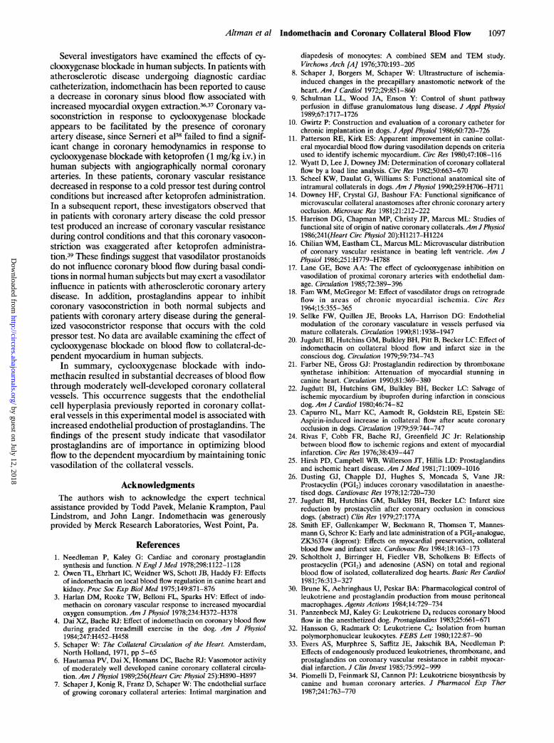
Several investigators have examined the effects of cy-clooxygenase blockade in human subjects. In patients withatherosclerotic disease undergoing diagnostic cardiaccatheterization, indomethacin has been reported to causea decrease in coronary sinus blood flow associated withincreased myocardial oxygen extraction.36,37 Coronary va-soconstriction in response to cyclooxygenase blockadeappears to be facilitated by the presence of coronaryartery disease, since Semeri et a138 failed to find a signif-icant change in coronary hemodynamics in response tocyclooxygenase blockade with ketoprofen (1 mg/kg i.v.) inhuman subjects with angiographically normal coronaryarteries. In these patients, coronary vascular resistancedecreased in response to a cold pressor test during controlconditions but increased after ketoprofen administration.In a subsequent report, these investigators observed thatin patients with coronary artery disease the cold pressortest produced an increase of coronary vascular resistanceduring control conditions and that this coronary vasocon-striction was exaggerated after ketoprofen administra-tion.39 These findings suggest that vasodilator prostanoidsdo not influence coronary blood flow during basal condi-tions in normal human subjects but may exert a vasodilatorinfluence in patients with atherosclerotic coronary arterydisease. In addition, prostaglandins appear to inhibitcoronary vasoconstriction in both normal subjects andpatients with coronary artery disease during the general-ized vasoconstrictor response that occurs with the coldpressor test. No data are available examining the effect ofcyclooxygenase blockade on blood flow to collateral-de-pendent myocardium in human subjects.
In summary, cyclooxygenase blockade with indo-methacin resulted in substantial decreases of blood flowthrough moderately well-developed coronary collateralvessels. This occurrence suggests that the endothelialcell hyperplasia previously reported in coronary collat-eral vessels in this experimental model is associated withincreased endothelial production of prostaglandins. Thefindings of the present study indicate that vasodilatorprostaglandins are of importance in optimizing bloodflow to the dependent myocardium by maintaining tonicvasodilation of the collateral vessels.
AcknowledgmentsThe authors wish to acknowledge the expert technical
assistance provided by Todd Pavek, Melanie Krampton, PaulLindstrom, and John Langr. Indomethacin was generouslyprovided by Merck Research Laboratories, West Point, Pa.
References1. Needleman P, Kaley G: Cardiac and coronary prostaglandin
synthesis and function. N Engl J Med 1978;298:1122-11282. Owen TL, Ehrhart IC, Weidner WS, Schott JB, Haddy FJ: Effects
of indomethacin on local blood flow regulation in canine heart andkidney. Proc Soc Exp Biol Med 1975;149:871-876
3. Harlan DM, Rooke TW, Belloni FL, Sparks HV: Effect of indo-methacin on coronary vascular response to increased myocardialoxygen consumption. Am J Physiol 1978;234:H372-H378
4. Dai XZ, Bache RJ: Effect of indomethacin on coronary blood flowduring graded treadmill exercise in the dog. Am J Physiol1984;247:H452-H458
5. Schaper W: The Collateral Circulation of the Heart. Amsterdam,North Holland, 1971, pp 5-65
6. Hautamaa PV, Dai X, Homans DC, Bache RJ: Vasomotor activityof moderately well developed canine coronary collateral circula-tion. Am J Physiol 1989;256(Heart Circ Physiol 25):H890-H897
7. Schaper J, Konig R, Franz D, Schaper W The endothelial surfaceof growing coronary collateral arteries: Intimal margination and
diapedesis of monocytes: A combined SEM and TEM study.Virchows Arch [A] 1976;370:193-205
8. Schaper J, Borgers M, Schaper W: Ultrastructure of ischemia-induced changes in the precapillary anastomotic network of theheart. Am J Cardiol 1972;29:851-860
9. Schulman LL, Wood JA, Enson Y: Control of shunt pathwayperfusion in diffuse granulomatous lung disease. J Appl Physiol1989;67:1717-1726
10. Gwirtz P: Construction and evaluation of a coronary catheter forchronic implantation in dogs. JAppl Physiol 1986;60:720-726
11. Patterson RE, Kirk ES: Apparent improvement in canine collat-eral myocardial blood flow during vasodilation depends on criteriaused to identify ischemic myocardium. Circ Res 1980;47:108-116
12. Wyatt D, Lee J, Downey JM: Determination of coronary collateralflow by a load line analysis. Circ Res 1982;50:663-670
13. Scheel KW, Daulat G, Williams S: Functional anatomical site ofintramural collaterals in dogs. Am J Physiol 1990;259:H706-H711
14. Downey HF, Crystal GJ, Bashour FA: Functional significance ofmicrovascular collateral anastomoses after chronic coronary arteryocclusion. Microvasc Res 1981;21:212-222
15. Harrison DG, Chapman MP, Christy JP, Marcus ML: Studies offunctional site of origin of native coronary collaterals.Am JPhysiol1986;241(Heart Circ Physiol 20):H1217-H1224
16. Chilian WM, Eastham CL, Marcus ML: Microvascular distributionof coronary vascular resistance in beating left ventricle. Am JPhysiol 1986;251:H779-H788
17. Lane GE, Bove AA: The effect of cyclooxygenase inhibition onvasodilation of proximal coronary arteries with endothelial dam-age. Circulation 1985;72:389-396
18. Fam WM, McGregor M: Effect of vasodilator drugs on retrogradeflow in areas of chronic myocardial ischemia. Circ Res1964;15:355-365
19. Sellke FW, Quillen JE, Brooks LA, Harrison DG: Endothelialmodulation of the coronary vasculature in vessels perfused viamature collaterals. Circulation 1990;81:1938-1947
20. Jugdutt BI, Hutchins GM, Bulkley BH, Pitt B, Becker LC: Effect ofindomethacin on collateral blood flow and infarct size in theconscious dog. Circulation 1979;59:734-743
21. Farber NE, Gross GJ: Prostaglandin redirection by thromboxanesynthetase inhibition: Attenuation of myocardial stunning incanine heart. Circulation 1990;81:369-380
22. Jugdutt BI, Hutchins GM, Bulkley BH. Becker LC: Salvage ofischemic myocardium by ibuprofen during infarction in consciousdog. Am J Cardiol 1980;46:74-82
23. Capurro NL, Marr KC, Aamodt R, Goldstein RE, Epstein SE:Aspirin-induced increase in collateral flow after acute coronaryocclusion in dogs. Circulation 1979;59:744-747
24. Rivas F, Cobb FR, Bache RJ, Greenfield JC Jr: Relationshipbetween blood flow to ischemic regions and extent of myocardialinfarction. Circ Res 1976;38:439-447
25. Hirsh PD, Campbell WB, Willerson JT, Hillis LD: Prostaglandinsand ischemic heart disease. Am J Med 1981;71:1009-1016
26. Dusting GJ, Chapple DJ, Hughes S, Moncada S, Vane JR:Prostacyclin (PGI2) induces coronary vasodilatation in anaesthe-tised dogs. Cardiovasc Res 1978;12:720-730
27. Jugdutt BI, Hutchins GM, Bulkley BH, Becker LC: Infarct sizereduction by prostacyclin after coronary occlusion in consciousdogs. (abstract) Clin Res 1979;27:177A
28. Smith EF, Gallenkamper W, Beckmann R, Thomsen T, Mannes-mann G, Schror K: Early and late administration of a PG12-analogue,ZK36374 (iloprost): Effects on myocardial preservation, collateralblood flow and infarct size. Cardiovasc Res 1984;18:163-173
29. Scholtholt J, Birringer H, Fiedler VB, Scholkens B: Effects ofprostacyclin (PGI2) and adenosine (ASN) on total and regionalblood flow of isolated, collateralized dog hearts. Basic Res Cardiol1981;76:313-327
30. Brune K, Aehringhaus U, Peskar BA: Pharmacological control ofleukotriene and prostaglandin production from mouse peritonealmacrophages. Agents Actions 1984;14:729-734
31. Panzenbeck MJ, Kaley G: Leukotriene D4 reduces coronary bloodflow in the anesthetized dog. Prostaglandins 1983;25:661-671
32. Hansson G, Radmark 0: Leukotriene C4: Isolation from humanpolymorphonuclear leukocytes. FEBS Lett 1980;122:87-90
33. Evers AS, Murphree S, Saffitz JE, Jakschik BA, Needleman P:Effects of endogenously produced leukotrienes, thromboxane, andprostaglandins on coronary vascular resistance in rabbit myocar-dial infarction. J Clin Invest 1985;75:992-999
34. Piomelli D, Feinmark SJ, Cannon PJ: Leukotriene biosynthesis bycanine and human coronary arteries. I Pharmacol Exp Ther1987;241:763-770
by guest on July 12, 2018http://circres.ahajournals.org/
Dow
nloaded from

1098 Circulation Research Vol 70, No 6 June 1992
35. Ezra D, Feuerstein G, Czaja JF, Laurindo FRM, Finton CK, YehGC, Goldstein RE: Unique coronary vasodilator induction byleukotriene D4. Am J Physiol 1985;249(Heart Circ Physiol18):H698-H702
36. Friedman PL, Brown EJ, Gunther S, Alexander RW, Barry WH,Mudge GH, Grossman W: Coronary vasoconstrictor effect ofindomethacin in patients with coronary-artery disease. N Engi JMed 1981;305:1171-1175
37. Pacold I, Hwang MH, Lawless CE, Diamond P, Scanlon PJ, LoebHS: Effects of indomethacin on coronary hemodynamics, myocar-
dial metabolism and anginal threshold in coronary artery disease.Am J Cardiol 1986;57:912-915
38. Semeri GGN, Gensini GF, Abbate R, Castellani S, Bonechi F,Dagianti A, Arata L, Fedele F, lacoboni C, Prisco D: Physiologicrole of coronary PGI2 and PGE2 in modulating coronary vascularresponse to sympathetic stimulation.Am Heart J 1990;1 19:848-855
39. Serneri GGN, Gensini GF, Abbate R, Castellani S, Bonechi F,Carnovali M, Rostagno C, Dabizzi P, Dagianti A, Arata L, FedeleF, lacoboni C, Prisco D: Defective coronary prostaglandin modu-lation in anginal patients. Am Heart J 1990;120:12-21
by guest on July 12, 2018http://circres.ahajournals.org/
Dow
nloaded from

J Altman, D Dulas and R J Bachecollateral vessels.
Effect of cyclooxygenase blockade on blood flow through well-developed coronary
Print ISSN: 0009-7330. Online ISSN: 1524-4571 Copyright © 1992 American Heart Association, Inc. All rights reserved.is published by the American Heart Association, 7272 Greenville Avenue, Dallas, TX 75231Circulation Research
doi: 10.1161/01.RES.70.6.10911992;70:1091-1098Circ Res.
http://circres.ahajournals.org/content/70/6/1091World Wide Web at:
The online version of this article, along with updated information and services, is located on the
http://circres.ahajournals.org//subscriptions/
is online at: Circulation Research Information about subscribing to Subscriptions:
http://www.lww.com/reprints Information about reprints can be found online at: Reprints:
document. Permissions and Rights Question and Answer about this process is available in the
located, click Request Permissions in the middle column of the Web page under Services. Further informationEditorial Office. Once the online version of the published article for which permission is being requested is
can be obtained via RightsLink, a service of the Copyright Clearance Center, not theCirculation Researchin Requests for permissions to reproduce figures, tables, or portions of articles originally publishedPermissions:
by guest on July 12, 2018http://circres.ahajournals.org/
Dow
nloaded from