effect of electron demand on sensing behavior of
TRANSCRIPT

Effect of Electron Demand on Sensing Behavior of
Carbazolopyridinophanes
by
Grace Abban
Bachelor of Science, Chemistry, University of Cape Coast
Master of Science, Chemistry, East Tennessee State University
A dissertation submitted to the
College of Engineering and Science
at Florida Institute of Technology
in partial fulfillment of the requirements
for the degree of
Doctor of Philosophy
in
Chemistry
Melbourne, FL
August 2020

Copyright 2020 Grace Abban
All Rights Reserved
The author grants permission to make single copies

Effect of Electron Demand on Sensing Behavior of
Carbazolopyridinophanes
a dissertation by Grace Abban.
Approved as to style and content
Alan B. Brown, Ph.D., Committee Chairperson
Associate Professor, Chemistry
Yi Liao, Ph.D.
Professor, Chemistry
Rudolf Wehmschulte, Ph.D. rer. nat.
Professor, Chemistry
Andrew Palmer, Ph.D.
Associate Professor, Ocean Engineering and Marine Sciences
Daniel Kirk, Ph.D.
Professor and Head
Biomedical and Chemical Engineering and Sciences

iii
ABSTRACT
Effect of Electron Demand on Sensing Behavior of
Carbazolopyridinophanes
by
Grace Abban
Major Advisor: Alan B. Brown, Ph.D.
Hydrazine (HZ), monomethylhydrazine (MMH), and 1,1-
dimethylhydrazine (UDMH) are widely used as high energy rocket
propellants in bipropellant rocket engines. However, hydrazine and its
derivatives are highly flammable and carcinogenic. Currently, hydrazine
sensors used for detection are irreversible which makes them costly. Two
proposed carbazolopyridinophanes containing intramolecular hydrogen
bonds were synthesized to detect hydrazine as real-time reversible sensors.
However, their thresholds for detection (100 and 25 ppb for CP and
22OMeCP respectively) were above the regulatory limit of 10 ppb due to
background fluorescence. It was established that the quenching of carbazole
in the presence of the CP system has a direct correlation to the relative
strength of the intramolecular hydrogen bond to the pyridine subunit. Our
goal was to synthesize a carbazolopyridinophane with an electron donating
dimethylamino group with the aim of decreasing both the background
fluorescence and the detection limit. (CH3)2NCP was synthesized and

iv
purified for photophysical studies. Butylamine was chosen as the analyte to
mimic HZ without producing any toxic element for initial analysis. The
absorption and emission spectra of (CH3)2NCP were similar in morphology
to those of CP and 22OMeCP. (CH3)2NCP’s background fluorescence was
reduced to 0.7 % and its fluorescence was restored in the presence of 1 ppb
n-butylamine, ethylenediamine and triethylamine as well as 4 ppb ammonia.
It can be concluded that the addition of the electron donating group to the
quencher of the phane decreased the background fluorescence and increased
their sensitivity by improving the detection limit.

v
Table of Contents
List of Figures ..................................................................................................... vii
List of Tables ........................................................................................................ ix
List of Keywords .................................................................................................. x
List of Compounds .............................................................................................. xi
Acknowledgement ............................................................................................. xiii
Chapter 1: Introduction ..................................................................................... 1
1.1 Fluorescence Quenching ............................................................ 1
1.2 The Role of Hydrogen-Bonding in Quenching ........................ 6
1.3 N-Heterocyclic Systems in Fluorescence Quenching Studies . 8
1.4 Intramolecular Emitter/Quencher Systems ........................... 11
1.5 Hydrazine: Applications, Health Concerns and Methods of
Detection.......................................................................................... 14
1.6 Heterocyclophanes and Fluorescence Quenching ................... 16
1.7 Application of Early CP results to Hydrazine Detection ...... 21
1.8 Modification of CP ................................................................... 23
1.9 Research Goal and Experimental Design .............................. 24
Chapter 2: Synthesis.......................................................................................... 27
2.1 An Overview of Phane Synthesis ............................................ 27
2.2 Diphenylamine and Carbazole Synthesis ............................... 30
vi
2.3 Synthesis of Pyridine Subunit ................................................. 34
2.4 Phane Synthesis ........................................................................ 35
2.5 Synthesis Conclusion ................................................................ 36
2.6 Experimental Methods .................................................................. 37
Chapter 3: Physical Studies ............................................................................. 45
3.1 Introduction to Physical Studies ............................................. 45
3.2 General Methods ...................................................................... 46
3.3 2D 15N NMR Experiments ....................................................... 48
3.4 Molecular Modeling ................................................................. 50
3.5 Absorption and Fluorescence Spectra .................................... 52
3.6 Quenching Assays of Emitter/Quencher Pairs ...................... 59
3.7 Restoration of Fluorescence in (CH3)2NCP ........................... 61
Chapter 4: Conclusion and Future Studies.................................................. 68
References ............................................................................................................ 69
Appendix A NMR Spectra .............................................................................. 75
Appendix B Gaussian XY Data ...................................................................... 89

vii
List of Figures
1. Jablonski diagram ………………………………………………….1
2. The proposed mechanism of quenching ……………………………4
3. Some N-heterocyclic emitters and quenchers ……………………...8
4. Illustration of the charge transfer complex ……………………….10
5. 7H- and 13H-dibenzocarbazoles ………………………………….10
6. Syn and Anti conformers of pyridylindole ……………………….13
7. Bridged pyridylindole …………………………………………….13
8. Pyridocarbazole …………………………………………………...14
9. Originally proposed heterocyclophane compounds ……………....19
10. Proposed modified heterocyclophane compounds ……………….23
11. Phane synthesized in this work …………………………………...25
12. Synthesis of CP …………………………………………………...27
13. Carbazole synthetic scheme ………………………………………28
14. Pyridine synthetic scheme ………………………………...………29
15. Overall phane synthetic scheme ………………………………….35
16. 15N-1H HMBC spectra of (CH3)2NCP ……………………………50
17. CBZ absorption spectra (2.88E-6 M) …………………………….52
18. (CH3)2NCP absorption spectra (1.732E-5 M) ……………………53
19. Comparison of CBZ and (CH3)2NCP absorption spectra .……….53
20. CBZ emission spectra ……………………………………………55
21. (CH3)2NCP emission spectra …………………………………….55

viii
22. Comparison of CBZ and (CH3)2NCP emission spectra ………….56
23. Relationship between background fluorescence and Hammett
parameters…………………………………………………………58
24. Emission spectra of CBZ/PYR system …………………………...59
25. Emission spectra of CBZ/DMAP system ………………………...60
26. Restoration of (CH3)2NCP fluorescence in n-heptane by butylamine
…………………………………………………………………….63
27. Restoration of (CH3)2NCP fluorescence in n-heptane by butylamine
starting at 1 ppb................................................................................63
28. Restoration of (CH3)2NCP fluorescence in n-heptane by
ethylenediamine ………………………………………………….64
29. Restoration of (CH3)2NCP fluorescence in n-heptane by
ethylenediamine starting at 1 ppb …………………………………65
30. Restoration of (CH3)2NCP fluorescence in n-heptane by
triethylamine ………………………………………………………65
31. Restoration of (CH3)2NCP fluorescence in n-heptane by
triethylamine starting at 1 ppb..........................................................66
32. Restoration of (CH3)2NCP fluorescence in n-heptane by ammonia
…………………………………………………………………….67

ix
List of Tables
1. Crude 1H NMR results for the optimization of Åkermark cyclization
reaction……………………………………………………………31
2. B3LYP/6-31+G (d, p) stabilities of the hydrogen bonded complexes.
CB represents carbazole…………………………………………...51
3. S/R data for CBZ and (CH3)2NCP at 350 nm……………………57
x
List of Keywords
Åkermark Cyclization
Carbazole
Carbazolopyridinophane
Emission
Emitter
Fluorescence
Fluorescence Quenching
Hartwig-Buchwald amination
Heterocyclophane
Hydrogen bonding
Mitsunobu reaction
Palladium (II) acetate
Pyridine
Quencher
Reduction
Sensing
UV-visible

xi
List of Compounds
Carbazole (CBZ)
Diethyl-1,8-Carbazoledicarboxylate (10)
1,8-Carbazoledimethanol (11)
1,8-Carbazolebis(methanethiolacetate) (12)
Carbazolopyridinophane (CP)
22-Chloro-Carbazolopyridinophane (22ClCP)
Dimethylamino-Carbazolopyridinophane ((CH3)2NCP)
22-Methoxy-Carbazolopyridinophane (22OMeCP)
Chelidamic acid (1)
Ethyl-2-aminobenzoate (8)
Ethyl-2-bromobenzoate (7)
Diethyl-2,2’-iminodibenzoate (9)
4-Chloropyridine-2,6-dicarboxylic acid (2)
4-Dimethylamino-2,6-bis(bromomethyl)pyridine (6)
4-Dimethylaminopyridine-2,6-dicarboxylic acid (3)
4-Dimethylamino-2,6-bis(carbomethoxy)pyridine (4)
4-Dimethylamino-2,6-bis(hydroxymethyl)pyridine (5)

xii
Lithium aluminum hydride (LAH)
Tetrahydrofuran (THF)
Sodium borohydrate (NaBH4)
Dimethyl sulfoxide (DMSO)

xiii
Acknowledgement
Thanks to God Almighty for his protection, care, abundant grace and mercy
that has seen me through my course successfully. I thank my family
especially my parents, uncles, aunties and all my siblings (Sarah, Ruth,
John, and Dorcas) for their support and words of encouragement through
my program. To my husband Selorm Joy Fanah, I really appreciate you for
listening to all my numerous complaints and motivating me every step of
the way. To my lovely daughter Selikem, you give me a reason to soar
higher.
My sincere gratitude goes to my major advisor Dr. Alan B. Brown for his
guidance and support throughout this project, I say God richly bless you. I
also thank Dr. Palmer, Dr. Liao, and Dr. Wehmschulte for gladly accepting
to sit on my committee and their contributions to this project. Thanks to my
group members who helped with this research: Eric Ziegler, Will
Henderson and Dr. Mohamed Ramadan. Special thanks to the Florida
Institute of Technology and the Department of Biomedical and Chemical
Engineering and Sciences and its staff for giving me the opportunity to be in
the graduate program as well as their financial support. Finally, to my
friends Dr. Salomey Sasu, and Dr. Damilola Ajadi thank you for helping in
diverse ways.

1
Chapter 1: Introduction
1.1 Fluorescence Quenching
Molecules are often excited to a higher state of energy when light is
shined on them. Some of the excited molecules may return to the ground
state either through kinetically or thermally controlled de-excitation
pathways. The energy lost from the higher singlet excited states to the lower
one results from vibrational relaxation and collision of solvent molecules.
Once at the first excited singlet state (S1), further energy can be lost through
a number of mechanisms as shown in Figure 1 below.1-5
Figure 1: Jablonski diagram
Generally, the energy that is released during molecular collisions
and vibrations is minimized in the presence of high viscosity solvents and at

2
temperatures below 298 K causing other deactivation pathways to occur.1-5
Fluorescence, which is a radiative decay of excited molecules from the first
excited singlet (S1) to the ground state (S0), is an example of such a
kinetically-controlled de-excitation pathway.1-5 Another form of de-
excitation pathway is internal conversion (IC), which is a competing non-
radiative relaxation from S1 to S0 resulting from energy released through
vibrational motions.1-7 The ability for fluorescence or internal conversion to
occur during de-excitation depends on their respective rate constants, rate
constant of fluorescence kf and rate constant of internal conversion ki.1-5
Intersystem crossing (ISC) is also another form of non-radiative de-
excitation that occurs when there is sufficient overlap between the lowest
singlet excited state (S1) and a triplet state (T). For intersystem crossing to
occur its rate constant kisc should be greater than kf or ki.1-5 A radiative de-
excitation from the lowest triplet state (T1) to S0 known as phosphorescence
can also occur if the rate of phosphorescence is greater than the rate of
intersystem crossing (kiscp).1-5 The lifetime of T1 (10-4 to 100 s) is longer
compared to that of S1 (10-9 to 10-6 s) hence ISC is a forbidden process.4
Phosphorescence is rarely observed at room temperature compared to
fluorescence.1-5
Fluorescence quenching is the act of decreasing the intensity of
fluorescence observed at a particular wavelength.1-5 The extent to which
fluorescence occurs can be controlled by making modifications to the

3
environment of the fluorophore (fluorescing molecule). A way of favoring
fluorescence over thermal de-excitations is by introducing low temperatures
and high viscosity solvents such as propanol. Similarly, phosphorescence
can be favored over thermal de-excitations by moving to a less viscous
solvent (such as hexane) and higher temperatures.1-5 Biological systems can
also cause quenching by changing the electronic states of the emitter. Some
of the changes that biological systems cause include altered ionization
potentials, electron affinities, or an increase or decrease of acid or base
character in the excited state of the molecule.8-10
Some emitters (fluorescent molecules) and quenchers (molecules
that decrease emitter fluorescence) have received significant attention
because quenching may occur as a result of hydrogen-bonding.11-13 The role
of the hydrogen bonding differs greatly depending on the emitter/quencher
pair being studied. However, it provides a powerful basis that can be used to
change the electronic properties of a molecule and control its fluorescence.
1-5
Quenching of fluorescence can be considered as occurring through a
specific mechanism, but this reduction of fluorescence occurs because kf has
been decreased or ki (or the rate of any other non-radiative relaxation) has
been increased as a result of that mechanism.1-5 In the past, hydrogen-
bonded facilitated quenching of numerous heterocyclic systems have been
studied.14 These quenching studies were able to explain the mechanism of

4
quenching, the specific role of the hydrogen bond and how they result in the
various de-excitation pathways for every heterocyclic aromatic
emitter/quencher pair. Emitter/quencher pairs of N-heterocyclic aromatics
are among the best characterized systems and are the foundation for this
current work.13
Fluorescence quenching within hydrogen-bonded heterocyclic
aromatic emitter/quencher pairs have generally been given to four principal
mechanisms as shown in figure 2 below.1-3
Figure 2: The proposed mechanisms of quenching.11, 13
Charge transfer (CT), proton transfer (PT), and H-atom transfer
(HT) can in principle be differentiated from energy transfer (ET) by the
observation of the intermediate species.1, 3 During quenching, CT results in
partially charged species, or if a complete electron transfer occurs ion pairs
are formed but the donor molecule is not deprotonated. PT produces an
anionic donor and a protonated cationic acceptor while HT produces radical
species.1-3

5
In an ET mechanism, the donor returns to the ground state as energy
is transferred to the acceptor from the donor across the hydrogen bond.1-5
Phosphorescence or fluorescence can be observed at longer wavelength
(lower energy) from the acceptor during ET depending on the acceptor and
energy states involved in the transfer.5 The donor and acceptor molecules in
ET remain neutral, and neither proton or H-atom transfer is observed. ET
mechanisms can occur between singlet or triplet excited states on the
condition that the acceptor molecule has energy states of the same
multiplicity which are of lower energy compared to that of the donor.1-5 ET
mechanisms are similar to that of internal conversion in that they both
involve energy transfer from states of the same multiplicity. This is quite
different from intersystem crossing where energy can be transferred
between states of different multiplicity (i.e. from singlet to triplet).
However, ISC can occur first within the donor molecule, and then ET will
proceed as a result of energy transfer from the triplet state of the donor to
the corresponding lower energy triplet state of the acceptor.1-5 For example,
singlet-singlet ET occurs for the acridine facilitated quenching of carbazole
because S1 (lower lying singlet excited state) of carbazole is higher in
energy than the S1 of acridine. On the other hand, quenching of carbazole
by quinoline occurs as a result of ET between the triplet states of the two
molecules because S1 of carbazole is lower in energy than the S1 of

6
quinolone, but the T1 of carbazole is higher in energy than that of
quinolone. 1, 13
1.2 The Role of Hydrogen-Bonding in Quenching
Generally, the mechanisms of fluorescence quenching between
heterocyclic aromatic compounds require hydrogen-bonding.1, 3 How active
or passive the hydrogen bond within the two compounds is in the context of
quenching depends greatly on the mechanism involved. In PT and HT,
because there is transfer of a proton or an H-atom from the donor to the
acceptor molecule their hydrogen bond is considered as active.1, 3 However,
in CT and ET the quenching mechanisms appear to involve hydrogen bonds
in a more passive role. CT depends strongly on distance so the
intermolecular interaction favored by the hydrogen bonding may help to
bring the emitter and quencher into close enough proximity for quenching
to occur.1, 3 The probability that either a charge or an energy transfer will
occur between the donor or acceptor species is also increased because the
hydrogen bonding can lower the ΔE (change in energy) between the excited
states of the emitter/quencher pair.
It has been observed that if both the donor and acceptor have
conjugated π-electron systems and if the hydrogen bond complex formed
between the two is directly connected with both π-systems, then
fluorescence might be strongly quenched.18 In theory the hydrogen bond
creates a connection between the electronic states of both compounds when

7
dealing with hydrogen bonded quenching between an emitter and quencher
with conjugated π-systems.14 Previous research has shown that a hydrogen
bond (H-bond) with a π-system proton donor causes H-bond-facilitated
quenching for aromatic fluorescent chemicals that are proton acceptors.15
Quenching occurred for acridine n-oxide when H-bonded to phenol or
naphthol, but no quenching occurred when it is H-bonded to ethanol or
benzyl alcohol.17 Quenching was observed when aromatic acceptors with
H-bonding capability to the conjugated π system, like pyridine, were tested
with aromatic donor fluorescent substances such as carbazole.17 Quenching
was, however, not observed with non-conjugated π system H-bonding
molecules such as triethylamine.17 This information indicates that
facilitation of H-bond quenching requires the conjugation of the two
conjugated π-systems.17
The electron density between the π-systems of the emitter and
quencher can delocalize through H-bonding, re-distributing the charge
density across both molecules, decreasing the energy difference (ΔE)
between their individual electronic states and therefore lowering the overall
energy of the H-bonded excited state complex.1, 3, 12, 18 The formation of
non-radiative excited state complexes with higher rates of internal
conversion may also result from lowering ΔE between the excited and
ground states of the emitter.1, 3, 12, 18,

8
1.3 N-Heterocyclic Systems in Fluorescence Quenching Studies
The best characterized systems for fluorescence quenching
applications are those comprised of N-heterocyclic aromatic emitters and
quenchers as shown in Figure 3 below.
Figure 3: Some N-heterocyclic emitters: carbazole (CBZ) and indole
(IND); and quenchers: pyridine (PYR), quinoline (QIN), and acridine
(ACR).
Some emitter/quencher pairs have been studied more extensively
than others. The focus of this work will be on the carbazole/pyridine system
because the quenching mechanism has been carefully studied. 18 It is not
possible to have either singlet or triplet energy transfer from carbazole to
pyridine because the S1 and T1 states of the pyridine quencher are much
higher in energy than those in carbazole.15 This effectively rules out ET as a
potential quenching mechanism for the pair since the singlet or triplet

9
energy transfer from carbazole to pyridine is forbidden. However, the ease
of both the electron loss that is predicted by the ionization potentials19 and
the electron gain predicted by the half-wave reduction potentials19 are all in
line with charge or electron transfer from the emitter to the quencher;
especially since H-bonding and excitation are expected to promote electron
loss from the emitter. In turn, the transfer of the charge should increase the
ease of proton transfer from a more positive donor to a more negative
acceptor. Early work done by Ermolaev19 showed a charge-transfer
mechanism occurring in the CBZ/PYR system. Mataga18 later determined
that ki from S1 to S0 was significantly increased in the CBZ/PYR complex
compared to CBZ, resulting in the quenching of carbazole fluorescence.
Martin and Ware19 reported the value for ki, ruled out HT or PT
mechanisms as the quenching methods, and suggested that the ki was
increased as a function of the hydrogen-bonded complex between carbazole
and pyridine.
They also determined that the equilibrium constant for the H-bonded
complex between carbazole and pyridine in the ground state, Kg, was
smaller than Kex for the excited state hydrogen-bonded complex.19 These
studies suggest that the quenching mechanism specific to CBZ/PYR is
likely a charge transfer mechanism through the hydrogen bond. Mataga
later conducted picosecond laser studies that indicated that hydrogen-
bonded systems of CBZ/PYR undergo a charge transfer mechanism.19 The

10
quenching specifically occurs due to the formation of a non-fluorescent
charge transfer state in the excited hydrogen bonded pair.19 This is shown in
Figure 4 below.
Figure 4: Illustration of the charge transfer complex
Further studies using 7-H and 13-H dibenzocarbazoles (Figure 5)
gave consistent results that support the charge transfer mechanism within
the H-bonded complex with pyridine.19
Figure 5: 7H- and 13H-Dibenzocarbazoles
Other carbazole emitter/N-heterocyclic pairs have been studied.
Studies done by Mataga16 on the carbazole/quinoline pair shows that their

11
quenching mechanism is the result of ET in the hydrogen-bonded complex.
In this system, quinoline quenches carbazole fluorescence and
phosphorescence, and sensitized phosphorescence is emitted from quinoline
itself as a result of triplet-triplet energy transfer to quinoline.16 Studies done
on carbazole/acridine emitter/quencher pairs showed that no radiative
emission occurred at a longer wavelength, indicating the quenching
mechanism is likely not due to an ET process.20, 22 It was proposed that
carbazole/acridine quenching is the result of the formation of a charge or
electron transfer complex enabled by the hydrogen bond.20, 24
Other emitter/quencher N-heterocyclic pairs that have been studied
include naphthylamine/pyridine16, naphthol/pyridine16, pyrazine/indole16,
carbazole/benzophenone20, aminopyrene/pyridine21, pyrenol/pyridine20 and
pyrene/indole20. Other examples are the more recent work done within our
group13, 22-23 on diphenylamine/pyridine, carbazole/acridine,
diphenylamine/acridine and carbazole/methoxypyridine. These
diphenylamine studies were the first time quenching studies have been
performed with this molecule.15, 22
1.4 Intramolecular Emitter/Quencher Systems
Various intramolecular emitter/quencher systems have been studied
in the past to provide deeper insight into the role of the hydrogen bond and
the influence of the conformational freedom in fluorescence quenching.
Herbich24 used 2-(2’-pyridyl) indoles (Figure 6 and 7) and

12
pyridocarbazoles25 (Figure 8) to learn more about the fluorescence
quenching of aza-aromatic systems containing indolyl (or carbazolyl) and
pyridyl subunits facilitated by hydrogen bonding. It was observed that the
alcohols formed cyclic hydrogen bonded complexes with the proton
donor/acceptor moieties except for alkylated donor amines (e.g. indolyl or
carbazolyl species). The mechanism for the quenching was proposed to be
because of an increase in the rate of internal conversion (ki) due to proton
movement within the cyclic hydrogen-bonded complex.24
Two distinct ground states for pyridylindole: syn (H-bonded) and anti (non-
H-bonded) were observed (Figure 6). These observations supported the
hypothesis that two different conformations of the compound exist in
solution.24 This conformational freedom was shown to influence the
quenching of the pyridylindole emission.24 The results showed that addition
of an increased amount of alcohol to the pyridylindole/hexane solution
caused an increased population of anti-conformers.24 It is hypothesized that
greater than 90 % of the molecules exist in the anti-conformation in pure
alcohol solution.24 Bridging the molecule at positions 3 and 3’ (Figure 7)
gave rise to the syn conformation with only one ground state.24 Quenching
of the bridged indolyl fluorescence is observed in alcohol, with the decay
time of the bridged systems corresponding to the decay time of the syn
conformer in the non-bridged pyridolylindoles.24 When pyridine or
quinoline was used instead of alcohols, fluorescence was also quenched.24-26

13
The fluorescence quenching mechanism induced by the aromatics appeared
to differ from that observed in the presence of alcohols.24
Figure 6: Syn and Anti conformations of pyridylindole
Figure 7: Bridged pyridylindole
Extensive studies have also been done on the hydrogen-bonded
quenching of pyridocarbazole/alcohol pairs.25 This system experiences
excited state single proton transfer between the emitter and the hydrogen-
bonded alcohol.24, 25 Pyridocarbazoles experience fluorescence quenching
14
as a result of an increased rate of internal conversion ki within the hydrogen-
bonded complex.25
Figure 8: Pyridocarbazole
It is interesting to note that N-methylation of the carbazolyl or
indolyl nitrogen prevents the pyridocarbazole and pyridoindole systems
from acting as proton donor.25 Since hydrogen bonding to the donor is
impossible, bridging via the cyclic hydrogen bond cannot be accomplished,
and both the alcohol and aromatic amine quenchers do not quench the
systems. This observation is consistent with other experimental results that
N-alkylated carbazoles are not quenched in the presence of pyridine,
acridine, or quinoline.14, 16, 18
1.5 Hydrazine: Applications, Health Concerns and Methods of
Detection
Hydrazine (HZ), monomethylhydrazine (MMH), and 1,1-
dimethylhydrazine (UDMH) are widely used as high energy rocket
propellants in bipropellant rocket engines.26 They are hypergolic with

15
various oxidizers such as nitrogen tetroxide (N2O4) and nitric acid (HNO3).
At 1 atm and 373 K, the vapors of HZ and MMH have lower limits of
flammability in air of 4.7% and 2.5% respectively.26-28 Hydrazine,
however, does not have any upper limit of flammability, with its flammable
range extending all the way to 100% vapor concentration.26 In addition to
flammability, they have toxic effects such as skin burns, eye damage27 and
liver damage starting at concentrations in air well below the olfactory
thresholds.26 The threshold limit values (TLVs) for hydrazine and its
derivatives have been set at 10 ppb (v/v).29 Since exposure limits are low
and well below olfactory senses, leak detection for these chemicals is a
critical component of workplace safety.
Specifications for a useful hydrazine sensor vary with the
application. A sensor for the detection of hydrazine and its derivatives
ideally should be ppb-sensitive, re-useable and real time responsive. An
ideal sensor must also be simple and have low-cost reliability. Continuous
gaseous detection systems are needed for monitoring workplace safety to
ensure that areas are non-toxic.26 Electrical devices should not be employed
to avoid possible combustion, given the vapors’ inherent flammability.
There are a number of limitations to sensor systems which are
currently used: real-time detection is halted by irreversible chemical
reactions, time dependent concentrations for cumulative response, and
significant replacement costs of sensor components.30 The current

16
colorimetric dosimeter badges which are based on vanillin and either p-
dimethyl-aminobenzaldehyde31 or 2,4-dinitrobenzaldehyde32, are sensitive
and react quickly, but the chemistry involves formation of hydrazones or
azines, and is effectively irreversible; thus, the badges can be used only
once.33 In passive mode the detection of UDMH = 10 ppb occurs within 1.4
hours.32 Other existing colorimetric sensors are based on a reaction with
pH-sensitive triphenylmethane dyes.32
Electrochemical sensors are well designed for analysis of aqueous
hydrazine, however, the hydrazine vapors must be dissolved first before
they can be electrochemically analyzed, which limits the speed of the
detection.34 Gold-doped tungsten oxide and titanium dioxide solid state
sensors have been proposed, but they are undesirable for combustible gas
detection due to high temperature requirements (100-800 oC).34 Sample
handling for monitoring of multiple locations remote from a central analysis
station is usually minimized by an array of sensors attached to the central
point by a fiber optic network. These fiber optics attached sensors involve
colorimetry of phosphomolybdic acid, and again their chemistry is
irreversible, however, their detection limits are 103 times higher than the
TLVs.35
1.6 Heterocyclophanes and Fluorescence Quenching
The use of bridged or fused emitter/quencher pair heterocycles
supports the study of fluorescence quenching within constrained

17
intramolecular systems. Emitter/quencher pairs which had been known were
used in the study of the bridged/fused systems.25 In some of the systems
such as pyridocarbazole25, similar quenching mechanisms were observed
for both the constrained and free emitter/quencher pairs while other fused
pairs (pyridylindole, dipyridocarbazole) 24, 25 resulted in the observation of
new emission characteristics due to changes in the structural features of the
compound itself.
Our interest in the fluorescence quenching of these fused/bridged
systems led to the design of an experimental system based on
heterocyclophanes. Heterocyclophanes are macrocyclic structures that
contain heteroaromatic subunits linked with a bridging element (e.g.
aliphatic chains). The first cyclophanes were synthesized as a way to study
steric and electronic effects on aromaticity.36, 37 Later heterocyclophanes
were synthesized in order to study their ability to chelate metal ions as well
as host-guest interactions with heteroatoms.37
Our research group’s initial goal was to find out the extent to which
fluorescence quenching required the hydrogen bond within the N-
heterocyclic emitter/quencher pairs.11 Gibson11, 12 produced
emitter/quencher pairs (including CBZ/PYR) within a macrocycle in a way
that restricted conformational freedom and therefore limiting the relative
hydrogen bond distance. The use of a bimolecular heterocyclophane
structure eliminated issues related to concentration or dynamic quenching

18
since a 1:1 mole ratio of the emitter to quencher was used for the synthesis.
Fluorescence should be quenched within the heterocyclophane because the
emitter and quencher are oriented to facilitate hydrogen bonding. In
principle, the fluorescence should be restored if the hydrogen bond is
interrupted.
Carbazolopyridinophane (CP), reverse CP phane (rCP),
acridinocarbazolophane (AC) and reverse AC phane (rAC) are the four
different heterocyclophanes (Figure 9) that were proposed by Gibson.11 The
synthetic approach to CP and AC was such that carbazole was bis-
functionalized at the 1 and 8 positions so that coupling with 2,6-
bis(bromomethyl)pyridine or 4,5-bis(bromomethyl)acridine would result in
the nitrogen atom of the quencher locked facing the carbazole N-H.11 This
resulted in the formation of hydrogen bonding within the heterocyclophanes
and therefore being quenched.11 The synthetic approach for the reverse
phanes were similar to that of CP and AC, however, the carbazole was bis-
functionalized at the 3 and 6 positions and coupled to the same quenchers
mentioned before.11 This resulted in the N-H groups on the carbazole facing
outwardly from the nitrogen of the quencher subunits. Hydrogen bonding
was not present in this orientation and therefore would not result in
quenching.11

19
Figure 9: Originally proposed heterocyclophane compounds
Synthetic problems made it impossible to obtain AC and rCP, but
only CP was synthesized, successfully purified and studied.11 Fluorescence
quenching did occur in CP, although a background fluorescence of 20 %
was observed.11, 12 Fluorescence restoration of CP was determined by
titrating with various concentrations of protic solvents (i.e. alcohols),
alkylamines, aprotic solvents (acetonitrile), and water in n-heptane.12 These
fluorescence restoration studies of CP were measured against the
fluorescence intensity of free carbazole (parent emitter). Fluorescence was
observed to be restored by ammonia, primary amines, secondary
alkylamines and hydrazine.12 Water, alcohols and protic solvents failed to

20
restore fluorescence of CP.12 The restoration of fluorescence was observed
to be reversible because once the alkylamines or hydrazine evaporated from
the solution, the fluorescence of CP was quenched.12
Interestingly, carbazole fluorescence was not quenched by
triethylamine in non-polar or polar protic solvents.38, 39 However
pyridocarbazole (Figure 8) and pyridylindole (Figure 6 and 7) derivatives
which also contain intramolecular proton donor and acceptor, were shown
to experience quenching in the presence of alcohols.24, 25 Quenching in these
systems was attributed to transfer of excited state proton between the
emitter and the hydrogen bonded alcohol.24, 25 The quenching mechanism
that occurred when aromatic amines like pyridine and quinoline are paired
with pyridylindole and pyridocarbazole in the presence of alcohols,
appeared to involve a charge transfer process in the hydrogen-bonded
excited state.24, 25
Triethylamine interacts with carbazole in non-polar solvent resulting
in a red-shift and broadening of the emission spectra, however, it does not
quench fluorescence.38 Fluorescence quenching of carbazole by
trimethylamine in acetonitrile has been reported with the resulting emission
having a red-shift signifying changes to the excited state of carbazole.38 The
fluorescence restoration of CP by alkylamines is thought to be the result of
an interaction between the amine and the cyclophane causing the disruption
of the internal hydrogen bond.11, 12

21
1.7 Application of Early CP results to Hydrazine Detection
As seen in section 1.5 above, a sensor for detecting hydrazine and its
derivatives should be ppb-sensitive, real time responsive and re-usable. A
rapid response detection mechanism is usually required for real-time
detection. The interaction or reaction that produces the measurable response
must have a high rate. Hydrogen bond formation is a good example of a
non-covalent intermolecular interaction and is quick in a room time
environment (˂˂ 1 s).39 Hence a sensor design that incorporates a hydrogen
bond disruption by hydrazine for detection is expected to afford real time
detection. A sensor that uses non-covalent intermolecular interactions for
detection may be reusable because of the inherent reversibility of the
interactions.39 There has been some reports on hydrogen-bond-facilitated
quenching of organic compounds in the past.11-19
Fluorescence based sensors are very attractive due to their
exceptional detection limit.1 A fluorescent sensor design would be optimal
if it gives off a pre-determined wavelength in the presence of hydrazine.
Carbazole is a well-known stable fluorescent organic compound that has an
emission maximum of 335 nm.40 Research has shown that carbazole can be
quenched by pyridine through hydrogen bonding.16, 41 The fluorescence
quenching of carbazole by pyridine is a radiationless process19, which is a
desired property for fluorescent sensor design because there will be no

22
emission wavelength or inherent background radiation until the hydrogen
bond is disrupted.
A molecule, 2,18-dithia[3](1,8)carbazole[3](2,6)pyridinophane or
CP (Figure 9), in which pyridine quenches carbazole intramolecularly
except in the presence of hydrazine, was conceived from these data. It was
hypothesized that the intramolecular hydrogen bond required for the
mechanism of quenching carbazole emission could be used as an “off/on”
switch for hydrazine detection.12 The results from the experiment showed
that CP would work as a reversible, fluorescence based hydrazine sensor.
Although the concept of a real-time, reversible sensor was proven, the
detection limit threshold was 100 ppb, an order of magnitude too high for
application.12 A background fluorescence of 20 % was also observed for the
original CP.12 Carbazole and 1,8-dimethylcarbazole both have emission at
350 nm when they are excited at λmax = 234 nm.11 According to Gibson
although a fully quenched CP system should not have emission from
excitation at λmax, a weak emission was observed at 330-400 nm when CP
was irradiated at λmax.11 This background fluorescence made the threshold
for detection above the needed limit.12 Initially, the background
fluorescence was thought to be due to the presence of impurities but
attempts by Hernberg to purify it proved that was not the case.13 It has also
been hypothesized that certain conformers of CP can weaken the hydrogen
bond between the emitter and quencher.12

23
1.8 Modification of CP
It was observed that the pyridine plane was twisted 53o from the
carbazole plane when looking down the N-H…N bond of the crystal
structure of CP.12 Quenching and emission processes are usually rate
competitive, and a weakened hydrogen bond may slow the quenching
versus the emission pathways.19 It was established from this result that the
quenching of carbazole in the presence of the CP system has a direct
correlation to the relative strength of the intramolecular hydrogen bond to
the pyridine subunit.12
Based on this hypothesis Vorisek proposed four different modified
CP with various pyridine subunits (Figure 10).23 It was proposed that the
modified subunits will have increased aromatic π-electron density by
substituting with electron donating groups such as amino, chloro and
methoxy.42 In addition to these, nitro an electron withdrawing group was
proposed to be tested as a control.23
Figure 10: Proposed modified heterocyclophane compounds
24
Problems associated with the synthesis made it impossible for the amino
and nitro subunits to be synthesized, however 22OMeCP and 22ClCP were
successfully synthesized.23 22OMeCP was successfully characterized and
studied because 22ClCP degraded upon purification by column
chromatograpy.23 22OMeCP and CP had very similar photophysical
characteristics in that they both exhibited a quenched state that was not
altered by either a non-protic solvent or protic solvent. Fluorescence was
restored for both CP and 22OMeCP in the presence of butyl amine but not
in ethylene diamine and triethylamine.23 The sensitivity levels for
22OMeCP were also similar to that of CP, but there was decreased
background fluorescence in 22OMeCP (~ 15 % less) compared to CP.
Vorisek concluded that the addition of an electron donating moiety to the
quencher (pyridine) molecule decreases the background fluorescence in the
heterocyclophane CP thereby lowering the threshold for detection (100 ppb
to 25 ppb).23 The enhanced quenching in 22OMeCP was attributed to the
change in the electron density of its quencher subunit.23 These observations
led to the research being discussed herein.
1.9 Research Goal and Experimental Design
The fundamental goal of this research is to synthesize a modified
heterocyclophane (CH3)2NCP (Figure 11) by incorporating an electron
donating dimethylamino group in the quencher subunit. We hope to
decrease the background fluorescence observed in the previously
25
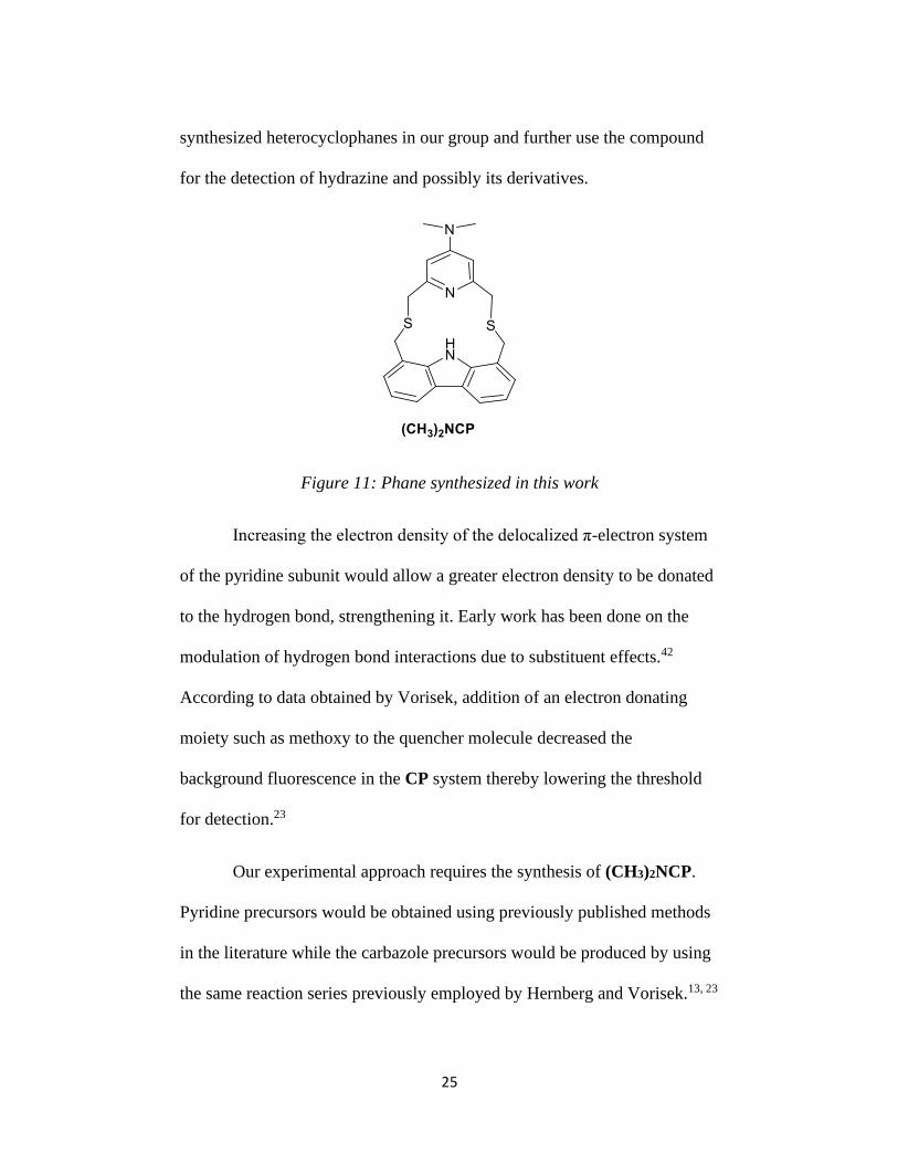
synthesized heterocyclophanes in our group and further use the compound
for the detection of hydrazine and possibly its derivatives.
Figure 11: Phane synthesized in this work
Increasing the electron density of the delocalized π-electron system
of the pyridine subunit would allow a greater electron density to be donated
to the hydrogen bond, strengthening it. Early work has been done on the
modulation of hydrogen bond interactions due to substituent effects.42
According to data obtained by Vorisek, addition of an electron donating
moiety such as methoxy to the quencher molecule decreased the
background fluorescence in the CP system thereby lowering the threshold
for detection.23
Our experimental approach requires the synthesis of (CH3)2NCP.
Pyridine precursors would be obtained using previously published methods
in the literature while the carbazole precursors would be produced by using
the same reaction series previously employed by Hernberg and Vorisek.13, 23

26
The coupling method employed by Hernberg would also be used to obtain
(CH3)2NCP.13 The absorption and emission spectra of the proposed
heterocyclophane will be done in a non-polar solvent, n-heptane. The
excitation spectra will be compared to that of the parent system CP to better
understand the effects of the intramolecular interactions effect on
quenching. Quenching assays of the heterocyclophane will be done at room
temperature in solvent phase. This will help obtain data that will allow for
better comparison of the parent CP and 22OMeCP, as well as to determine
the threshold of detection of a chosen analyte. The analyte chosen (butyl
amine) will mimic HZ without producing any toxic element for initial
analysis. Titrating against butyl amine will provide the necessary data
required to observe the diminishing of the quenching event and eventually
restoration of fluorescence.

27
Chapter 2: Synthesis
2.1 An Overview of Phane Synthesis
The synthesis of the carbazolopyridinophane CP, involves the direct
coupling of a mercaptide nucleophile with a halogenated electrophile
through conditions that favor the SN2 type of mechanism.11-13, 43 This is
because CP is made up of a thioether bridge and a plane of symmetry along
the nitrogen atoms of both the pyridine and carbazole moieties.11-13 This
approach has been used successfully by Gibson,11, 12 Hernberg13 and
Vorisek23 to synthesize several carbazolopyridinophanes including CP itself
by coupling 1,8-carbazolebis-(methanethiolacetate), 12 and 2,6-
bis(bromomethyl)pyridine, 14 (Figure 12).
Figure 12: Synthesis of CP
28
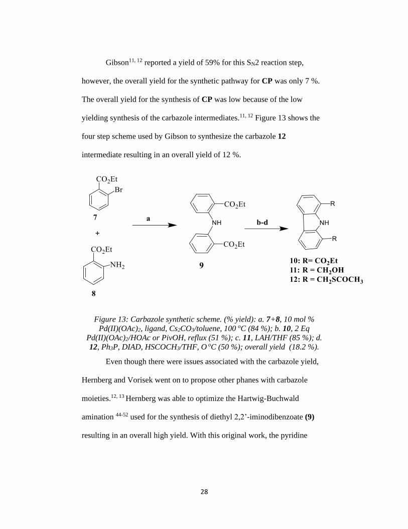
Gibson11, 12 reported a yield of 59% for this SN2 reaction step,
however, the overall yield for the synthetic pathway for CP was only 7 %.
The overall yield for the synthesis of CP was low because of the low
yielding synthesis of the carbazole intermediates.11, 12 Figure 13 shows the
four step scheme used by Gibson to synthesize the carbazole 12
intermediate resulting in an overall yield of 12 %.
Figure 13: Carbazole synthetic scheme. (% yield): a. 7+8, 10 mol %
Pd(II)(OAc)2, ligand, Cs2CO3/toluene, 100 oC (84 %); b. 10, 2 Eq
Pd(II)(OAc)2/HOAc or PivOH, reflux (51 %); c. 11, LAH/THF (85 %); d.
12, Ph3P, DIAD, HSCOCH3/THF, O oC (50 %); overall yield (18.2 %).
Even though there were issues associated with the carbazole yield,
Hernberg and Vorisek went on to propose other phanes with carbazole
moieties.12, 13 Hernberg was able to optimize the Hartwig-Buchwald
amination 44-52 used for the synthesis of diethyl 2,2’-iminodibenzoate (9)
resulting in an overall high yield. With this original work, the pyridine

29
subunit synthesis (Figure 14) was successful with no challenging issues,
however, low intermediate yield for the carbazole part was still observed.
Figure 14: Pyridine synthetic scheme. (% yield): e. 1, POCl3, reflux, 20 hrs
(79 %); f. 2, 40 % (CH3)2NH, reflux, 3 days (79 %); g. 3, methanol, H2SO4,
reflux, 7 days (95 %); h. 4, ethanol, NaBH4, reflux, 2 days (83 %); i. 5, 48
% HBr, reflux, 24 hrs (77 %); overall yield (37.9 %).

30
2.2 Diphenylamine and Carbazole Synthesis
The first step in the carbazole synthesis is the diphenylamine
synthesis which is based on the Hartwig-Buchwald cross coupling
amination reaction.48 Gibson11, 12 and Hernberg15 both did quite extensive
work on this step. While Gibson used the Ullmann coupling53 with a yield
of 45 %, every attempt by Hernberg to reproduce this resulted in 10 % yield
and was therefore considered unfavorable for the synthesis of 9. Hernberg
later replaced the Ullman coupling with a modified Hartwig-Buchwald
amination.13 This method involves the reaction of an aryl sulfonate or aryl
bromide with an amine (primary or secondary cyclic, acyclic, or aryl) in the
presence of catalytic palladium (II), a base usually sodium tert-butoxide, a
bulky phosphine ligand and a solvent (toluene, dioxane or
tetrahydrofuran).49 The modification done by Hernberg13 resulted in
reducing the reaction time from 17 hours to 1 hour and also an increased
yield (76 % compared to 45 %). This modified synthetic step was used in
this work to produce 9 in 84 % yield, which eventually helped to address
the low overall yield of Gibson’s11, 12 work.
The second step in the synthesis of carbazolophanes is the
cyclization of the diphenylamine 9 to give diethyl 1,8-
carbazoledicarboxylate 10 using the Åkermark54 cyclization.11-13 In
Gibson11, 12 and Hernberg’s13 work, this was the limiting step in terms of the
overall yield. The limitation of this step was reported to be due to the

31
presence of the electron-withdrawing groups which slow the reaction and
thereby require the use of larger amounts of the palladium (II) catalyst.54
Åkermark later showed that the presence of a co-oxidant will let the Pd (0)
be re-oxidized back to Pd (II), creating a catalytic cycle.53 However,
attempts by our group to use varying amounts of copper (II) co-oxidant to
optimize this step proved futile.12 As part of this work, various solvents,
oxidant and base were explored to know the best reaction conditions that
would lead to the formation of 10 in good yield. The best condition only
yielded about 52 % (entries 2 and 3 of Table 1) of the product with the rest
being the starting material 9. The best solvent was pivalic acid (trimethyl
acetic acid) which yielded more of the product. Increasing the reaction time
did not improve the percentage of the product formed.
Table 1: Crude 1H NMR results for the optimization of Åkermark
cyclization reaction.
Entry Solvent Oxidant Base Time
(Hr.)
Product:
Reactant
Isolated yield
(%)
1 PivOH Cu(OAc)2/O2 24 0.3510 35.1
2 PivOH O2 24 1.0556 52.0
3 PivOH O2 48 1.0556 52.0
4 AcOH Cu(OAc)2/O2 24 0.3700 37.0
5 PivOH Cu(OAc)2/O2 K2CO3 24 0.0787 7.87
6 THF O2 K2CO3 24 0.0635 6.35

32
7 AcOH Cu(OAc)2/O2 K2CO3 24 0.2316 23.2
8 AcOH O2 24 0.4120 41.2
9 AcOH O2 48 0.4301 43.0
Research56-60 has led to two general conclusions: a) low to moderate
yields associated with palladium based carbazole cyclizations are common
and b) the lowest yields are mostly related to the diphenylamine precursors
bearing electron-withdrawing substituents. Several reports on the
cyclization mechanism show that the Pd (II) inserts into the C-H bonds of
the aryl rings and then forms a palladacycle intermediate.57, 58 Whether there
is a change in oxidation state during the formation of the palladacycle is not
known, however, the resulting C-C bond is formed via oxidative process,
which indicates that there is reductive elimination of the palladium. The
formation of Pd (0) is supported by work done by Hernberg13, Åkermark54
and Watanabe60 where Pd (0) could only be oxidized back to Pd (II) by
using a co-oxidant via reductive elimination. Based on the mechanism, it is
possible that both the type and position of the functional groups could affect
the formation of the palladacycle intermediate. Hernberg13 proposed that:
“An electron-rich ring system would likely promote the electrophilic
substitution of the Pd (II) to form the palladacycle, and would also facilitate
the reductive elimination step to form the C-C. However, electron-
withdrawing groups such as the ethyl ester moieties on the diphenylamine
diester 9 would de-activate the rings, inhibiting the formation of the

33
palladacycle. Once formed, the lowered electron density in the rings would
also slow the reductive elimination step, lowering the yield of the desired
carbazole and likely promoting the formation of other side products.”13
Purification of 10 was another problem encountered with the
Åkermark cyclization. Pure 10 was obtained by using two columns
followed by recrystallization which led to low yields. This was because not
all the starting material 9 was consumed in the cyclization.
Hydride reduction of 10 to produce 1,8-carbazoledimethanol 11 via
lithium aluminum hydride (LAH) was the third step in the synthesis of the
carbazole subunit. The reaction mixture was quenched with sodium
potassium tartrate solution and then extracted with excess chloroform,
which was later placed on a hot plate to reduce the volume. Pure 11
crystallized from solution when left to cool to room temperature as either
thin needle crystals or fine powder. This work was able to reproduce the
average yield of 70-80 % observed by Hernberg.13 It appears that the
formation of the reduced product 11 depends on the dryness of the starting
material. At the initial stages of this work the yields were very low,
however, it changed once 10 was dried for a period of 24 hrs under vacuum
prior to the synthesis.
In the final step of the carbazole synthesis, a modified Mitsunobu
reaction was used to convert 11 to 1,8-carbazolebis(methanethiolacetate)
12. Hernberg13 reported an average yield of 70 % for this step, however,

34
this work did not reproduce that (50 %). This may be due to the purification
process since flash chromatography followed by preparative TLC was
needed to produce pure 12.
2.3 Synthesis of Pyridine Subunit
Chelidamic acid 1, a commercially available material was the
precursor for the substituted pyridines. A modified1, 45, 46 method by Will
Henderson (our past undergraduate group member) was used for the
conversion of 1 to 4-dimethylamino-2,6-bis(bromomethyl)pyridine 6. In the
first step, chelidamic acid 1 was refluxed with POCl3 to produce 4-
chloropyridine-2,6-dicarboxylic acid 2 with a yield of 79 % yield. 4-
Chloropyridine-2,6-dicarboxylic acid 2 was then refluxed with 40 %
aqueous dimethylamine to afford 4-dimethylaminopyridine-2,6-
dicarboxylic acid 3 in 79 %. 4-Dimethylamino-2,6-
bis(carbomethoxy)pyridine 4 was obtained in 95 % yield by refluxing 3 in
the presence of methanol. In the next step, 4-dimethylamino-2,6-
bis(carbomethoxy)pyridine 4 was reduced using NaBH4 to give 85 % of 4-
dimethylamino-2,6-bis(hydroxymethyl)pyridine 5. Finally, 4-
dimethylamino-2,6-bis(bromomethyl)pyridine 6 was obtained in 77 % yield
by refluxing 5 with 48 % HBr.

35
2.4 Phane Synthesis
A revised version of Mitchell and Zhang’s method43 was used by
Gibson11 for the synthesis of CP in 59 % yield. Gibson’s change required
dissolving the two precursors 6 and 12 in benzene and then delivering the
starting materials to an alcoholic base solution over a period of 18 hours.
Even though he had success with this method, there were a few issues
associated with the solubility of the carbazole. This affected the amount of
precursor delivered to the reaction vessel since it would slowly precipitate
on the walls of the syringe.
Figure 15: Overall phane synthetic scheme. (% yield): a. 7+8, 10 mol %
Pd(II)(OAc)2, ligand, Cs2CO3/toluene, 100 oC (84 %); b. 10, 2 Eq
Pd(II)(OAc)2/HOAc or PivOH, reflux (51 %); c. 11, LAH/THF (85 %); d.
12, Ph3P, DIAD, HSCOCH3/THF, O oC (50 %); overall yield to give (18.2
%); e. 12+6, 0.02 M KOH/ethanol solution, high dilution factor, RT, 72 hrs.
Due to these issues, Hernberg13 tried in situ reduction of the
methanethiolacetate 12 with diisobutylaluminium hydride (DIBAL-H) to
form the thiol followed by addition of the pyridine precursor 6. Although it

36
produced one of the phanes described in her work, there was no
improvement in the yield. Hernberg13 decided to use the modified Mitchell
and Zhang43 coupling approach used by Gibson11 with a few changes. Her
modification included changing the solvent from benzene to THF which is
also good for SN2-type reactions and manual delivery over a long period of
time instead of using a syringe pump13. These modifications improved the
yields of the coupling reaction. Vorisek23 also used this modified method
with minor changes to produce the phanes. Vorisek increased the relative
dilution factor by 2 to facilitate a slower reaction because at higher dilution
the probability of collisions is decreased. The speed for adding the
precursors was also changed to 5-6 drops every 30 seconds to reflect the
dilution factor change. By doing this precipitation of the precursors in the
syringe was decreased, however, the addition time was greatly increased.
Vorisek’s23 modification was used in this work to produce (CH3)2NCP with
40 % yields.
Synthesis of (CH3)2NCP showed there were no impurities in the
aromatic region of the 1H NMR so recrystallization of the crude material
was done using hexanes as solvent. A problem encountered is the presence
of hexane residual peaks in the 1H NMR of the phane.
2.5 Synthesis Conclusion
All the intermediates for (CH3)2NCP were synthesized and
characterized. Changes were made to the purification of the carbazole

37
intermediates to afford pure products. The optimizations described in
Hernberg3 and Vorisek’s23 work were also tested. This work replicated the
increased yields observed in Hernberg’s work as well as the changes made
to Mitchell and Zhang’s43 coupling method used by both Hernberg and
Gibson11. The purification of the phane posed a lot of issues which
prolonged the time for this work.
2.6 Experimental Methods
General procedures, solvents, and reagents.
All air or moisture sensitive reactions were performed under N2
atmosphere unless otherwise stated. The NMR spectroscopic studies were
carried on a Bruker AV400 spectrometer with a multi-nuclei probe at 400
MHz for 1H, 100 MHz for 13C and 40.55 MHz for 15N. The NMR chemical
shifts and coupling constants were quoted in parts per million (ppm) and as
a ‘J’ value in Hz respectively with tetramethyl silane (TMS) as the internal
standard. The resonance splitting patterns were described as singlet (s),
doublet (d), triplet (t), quartet (q) or multiplet (m). The mass spectrometric
studies were performed using a Joel DART-AccuTOF mass spectrometer
while melting points were obtained using a Mel-temp II apparatus. Solvents
used were bought from Fisher Scientific Company, Aldrich Chemical or
VWR. Reagents were also purchased from Acros Organics, Alfa Aesar,
Aldrich Chemical, Fisher Scientific or Strem. The reagents were used as
received unless otherwise stated.

38
4-Chloropyridine-2,6-dicarboxylic acid (2)
Chelidamic acid 1 (5.00 g, 27.3 mmol) was added to a 100 mL
round bottom flask. About 45 mL of POCl3 was added and then refluxed at
90 oC for a period of 20 hours. The reaction mixture was then cooled in an
ice bath and water was slowly added until there was no visible reaction. The
resulting slurry was filtered and recrystallized from water to give 2 (4.33 g,
79 %) as light brown needles. MP = 211-212 oC; 1H NMR (d6-DMSO):
8.29 (2H, s); 13C NMR (CDCl3) 164.7, 150.1, 145.4 and 127.4; DART MS
(Peak voltage 1000 V, Detection voltage 2500 V, temp 300 oC) m+1
(relative intensity) Calculated C7H4ClO4 MW = 201, observed 202.
4-Dimethylaminopyridine-2,6-dicarboxylic acid (3)
4-Chloropyridine-2,6-dicarboxylic acid 2 (4.00 g, 19.0 mmol) was
combined with 31.4 mL of 40 % aqueous dimethylamine in a 100 mL round
bottom flask. The solution was refluxed at 90 oC for 3 days. The solution
was cooled and acetic acid was added until white precipitates were formed.
The crystals were filtered and dried under vacuum to yield 3 (3.15 g, 79 %)
as white solid. MP = 264 oC, 1H NMR (d6-DMSO): 7.25 (2H, s) and 3.90
(6H, s); DART MS (Peak voltage 1000 V, Detection voltage 2500 V, temp
300 oC) m+1 (relative intensity) Calculated C9H10NO4 MW = 210.0640,
observed 210.0642.

39
4-Dimethylamino-2,6-bis(carbomethoxy)pyridine (4)
36 ml of methanol was added to 4-dimethylamino-2,6-
pyridinedicarboxylic acid 3 (3.00 g, 14.3 mmol) in a 100 mL round bottom
flask forming a slurry. About 2.4 mL of H2SO4 was added which resulted in
dissolution of the slurry. The solution was then refluxed at 85 oC for 7 days.
The reaction mixture was then cooled in an ice bath and a saturated solution
of NaHCO3 was added to make it basic. White precipitates were formed and
filtered to yield 4 (3.24 g, 95 %) as a grey powder. MP = 171-172 oC; 1H
NMR (d6-DMSO): 7.51 (2H, s), 3.99 (6H, s) and 3.14 (6H, s); 13C NMR
(CDCl3) 165.97, 148.14, 146.23, 109.96, 52.85 and 39.31; DART MS (Peak
voltage 1000 V, Detection voltage 2500 V, temp 300 oC) m+1 (relative
intensity) Calculated C11H14NO4 MW = 238.0954, observed 238.0957.
4-Dimethylamino-2,6-bis(hydroxymethyl)pyridine (5)
75 mL of anhydrous ethanol was added to a 100 mL round bottom
flask containing 4-dimethylamino-2,6-bis(carbomethoxy)pyridine 4 (3.12 g,
13.1 mmol). The solution was chilled in an ice bath and about 1.98 g of
NaBH4 was added. The reaction was stirred for 30 mins in an ice bath, then
allowed to warm to room temperature and finally heated to reflux for 2
days. The solvent was evaporated, and the residue was dissolved with a
saturated solution of NaHCO3. The resulting mixture was then extracted
with CHCl3, dried with Na2SO4, filtered, and evaporated in vacuo to yield 5
(1.98 g, 83 %) as a white solid. MP = 139-140 oC, 1H NMR (CDCl3): 6.38

40
(2H, s), 4.62 (4H, s), 4.04 (2H, s) and 3.01 (6H, s); 13C NMR (CDCl3)
158.9, 155.8, 102.1, 64.9 and 39.5; DART MS (Peak voltage 1000 V,
Detection voltage 2500 V, temp 300 oC) m+1 (relative intensity) Calculated
C9H14NO2 MW = 183.1131, observed 182.1029.
4-Dimethylamino-2,6-bis(bromomethyl)pyridine (6)
4-Dimethylamino-2,6-bis(hydroxymethyl)pyridine 5 (100 mg, 0.549
mmol) was added into a 5 mL conical flask. About 1 mL of 48 % HBr was
added and the resulting mixture was heated to reflux for 24 hours. The
solution was then cooled to 0 oC and saturated aqueous KOH solution was
added dropwise until it became basic. White solids were formed, filtered
and dried under vacuum to yield 6 (130 mg, 76.5 %). MP = 160 oC; 1H
NMR (CDCl3): 6.55 (2H, s), 4.45 (4H, s), and 3.03 (6H, s); 13C NMR
(CDCl3) 156.67, 155.86, 105.45, 39.37 and 34.47; DART MS (Peak voltage
1000 V, Detection voltage 2500 V, temp 300 oC) m+1 (relative intensity)
Calculated C9H12Br2 MW = 308.9403, observed 309.9351.
Diethyl-2,2’-iminodibenzoate (9)
Pd(OAc)2 (142.0 mg, 0.63 mmol), 2-(di-cyclohexylphosphino)-
2’,4’,6’-triisopropylbiphenyl (450.0 mg, 0.94 mmol) and Cs2CO3 (2.87g,
8.8mmol) were added into a 100 mL round bottom flask containing a stir
bar. Ethyl-2-bromobenzoate 7 (1.44 g, 6.29 mmol), ethyl-2-aminobenzoate
8 (1.12 g, 6.76 mmol) and toluene (1.5 mL) were then added sequentially

41
via a syringe. The initially orange-red homogenous solution was refluxed
for 2 hours at 100 oC with stirring under nitrogen gas protection. The color
deepened to cherry red, then brown and finally greenish brown. After 4
hours, the reaction mixture was cooled to room temperature in a water bath.
The reaction mixture was diluted with 30 mL of CHCl3 and 40 mL of 2M
NH4Cl and then was extracted 3 times with 30 mL portions of CHCl3. The
organic layers were combined, washed with brine, and then dried over
anhydrous Na2SO4. The solvent was removed in vacuo after filtration. The
crude compound was purified by column chromatography (1:10 ethyl
acetate: hexanes) followed by recrystallization in hexanes to yield 3 (1.649
g, 83.70 %) as a yellow solid. MP = 65-66 oC; 1H NMR (CDCl3): 10.99
(1H, s), 8.01 (2H, d, J = 8.0), 7.54 (2H, d, J = 8.6), 7.37 (2H, t, J = 9.0), 6.91
(2H, t, J = 7.6), 4.44 (4H, q, J = 7.3) and 1.60 (6H, t, J = 7.0); DART MS
(Peak voltage 1000 V, Detection voltage 2500 V, temp 300 oC) m+1
(relative intensity) Calculated C18H19NO4 MW = 313.35, observed 314.267
(100).
Diethyl-1,8-carbazoledicarboxylate (10)
9 (1.00 g, 3.2 mmol), Pd(OAc)2 (1.59 g, 6.80 mmol) and 50 mL of
pivalic acid or glacial acetic acid were added into a 100 mL round bottom
flask. The reactions were refluxed at 160 oC for pivalic acid or 120 oC for
acetic acid for 24 hours. The reaction mixture was then allowed to cool to
room temperature and quenched with saturated NaHCO3 and then extracted

42
3 times with 30 mL portions of CHCl3. The organic layers were combined,
washed with brine, and then dried over anhydrous Na2SO4. The solvent was
removed in vacuo after filtration. The crude was purified by column
chromatography (1:7 ethyl acetate: hexane followed by 3:2 chloroform:
hexane) followed by recrystallization in hexanes to yield 10 (0.51 g, 51.2
%) as a whitish yellow solid. MP = 108-110 oC; 1H NMR (CDCl3): 11.24
(1H, s), 8.28 (2H, d, J = 7.3), 8.16 (2H, d, J = 7.9), 7.32 (2H, t, J = 8.0), 4.56
(4H, q, J = 7.0) and 1.43 (6H, t, J = 7.3); DART MS (Peak voltage 2000 V,
Detection voltage 2700 V, temp 300 oC) m+1 (relative intensity) Calculated
C18H17NO4 MW = 311.33, observed 313.13813 (71), 311.13126 (100),
313.14662 (14), 266.10360 (100) and 267.10208 (14).
1,8-Carbazoledimethanol (11)
LAH (414.3 mg, 10.9 mmol) was stirred in 6 mL of THF at room
temperature. A solution of 10 (371.0 mg, 1.19 mmol) dissolved in 6 mL of
THF was added dropwise via syringe to this suspension and it turned
yellowish-green. After about 0.5 h, the reaction solution turned whitish
gray. The resulting solution was then quenched with 60 mL of saturated
sodium potassium tartrate solution and extracted with CHCl3 (6 х 30 mL).
The organic layers were then washed over brine, heated up to reduce the
volume to 10-20 mL and allowed to cool to room temperature for crystals to
form. The solution was chilled on ice and the crystals were filter under
vacuum to yield 11 (0.230 g, 85 %) as white crystalline needles. MP = 196-

43
200 oC; 1H NMR (CD3OD): 7.99 (d, 2H, J = 8.4), 7.35 (d, 2H, J = 7.4),
7.15 (t, 2H, J = 7.4 ) , 5.01 (s, 4H); DART MS (Peak voltage 2000 V,
Detection voltage 2700 V, temp 300 oC) m+1 (relative intensity) Calculated
C18H17NO4 MW = 227.26, observed 227.07752 (17), 210.08369 (100).
1,8-Carbazolebis(methanethiolacetate) (12)
Triphenylphosphine (45.1 mg, 1.72 mmol) dissolved in 2 mL THF
was stirred at 0 oC for about 5 minutes. To this suspension was added
diisopropylazodicarboxylate (358.0 mg, 0.35 mL, 1.77 mmol) to give a
whitish yellow solid. A solution of 1,8-carbazoledicarboxylate 11 (98.0 mg,
0.43 mmol) and thiolacetic acid (128 mg, 0.10 mL, 3.90 mmol) dissolved in
6 mL of THF was added dropwise via a syringe. After 1 hour the ice bath
was removed and the reaction mixture was allowed to warm back to room
temperature for another 1 hour. The solvent was then removed in vacuo to
give a viscous golden oil, which was subjected to flash column
chromatography (7:2 ethyl acetate: hexanes) followed by preparative TLC
(toluene). This yielded 12 (73.5 mg, 50 %) as brown solid. 1H NMR
(CDCl3) 9.39 (s, 1H), 7.97 (d, 2H, J =7.8), 7.32 (d, 2H, J = 7.4), 7.17 (t, 2H,
J = 7.4), 4.49 (s, 4H), 2.42 (s, 6H); DART MS (peak voltage 2200,
detection voltage 2700, temp = 375 °C), M++ l (relative intensity)
Calculated C18H17NO2S2 MW = 343.46, observed 344.05411 (69),
268.06632 (100).

44
Dimethylamino-carbazolopyridinophane (CH3)2NCP
20 mL of 0.02 M KOH/ethanol solution were put into a 100 mL two
neck flask equipped with a stir bar. A solution of 1,8-
carbazolebis(methanethiolacetate) 12 (36 mg, 0.105 mmol) and 4-
dimethylamino-2,6-bis(bromomethyl)pyridine 6 (32.5 mg, 0.105 mmol)
dissolved in about 6 mL of THF was added slowly via a syringe at a rate of
3-5 drops per second. The reaction mixture was allowed to stir at room
temperature for 72 hours under N2 atmosphere. An off-white precipitate
formed on the side walls of the flask. The liquid phase was then poured out
into a separate round bottom flask. The precipitate was dissolved with
CH2Cl2 and combined with the liquid phase. The solvent was then removed
in vacuo. The resulting solid was re-dissolved in dichloromethane and
partitioned with saturated brine solution. The combined organic layers were
dried over anhydrous Na2SO4, filtered and the solvent removed in vacuo.
Recrystallization in hexanes gave (CH3)2NCP (18.0 mg, 42.4 %) as a cream
solid. 1H NMR (CDCl3) 13.28 (s, 1H), 7.98 (d, 2H, J = 7.6), 7.30 (d, 2H, J =
7.4), 7.15 (t, 2H, J = 7.0), 6.48 (s, 2H), 4.24 (s, 4H), 3.96 (s, 4H) and 3.06
(s, 6H); 13C NMR (CDCl3) 158.15, 156.04, 139.89, 125.92, 123.54, 121.81,
119.54, 118.81, 104.73, 39.31, 37.33 and 33.01.

45
Chapter 3: Physical Studies
3.1 Introduction to Physical Studies
The characterization for the photophysical processes that occur
within the emitter/quencher parent compound and the phane, as well as
some 2-D NMR studies, are discussed here. This chapter provides the
studies done, accompanied with a discussion of their results in each
individual section.
The phane was characterized by using NMR studies. Both the
absorption and emission spectra of pyridine, 4-dimethylamino pyridine,
carbazole and (CH3)2NCP were collected in n-heptane. Quenching studies
were done on carbazole by the addition of pyridine in incremental amount
to the dilute solution. The fluorescence intensity of the phane was compared
to that of the parent emitter (carbazole) alone in n-heptane. The
fluorescence restoration of (CH3)2NCP was studied by exposing the phane
in n-heptane solutions to n-butylamine in increasing concentrations in order
to know the concentration of analyte that produces increased fluorescence.
The same method employed by Vorisek23 was used for the fluorescence
restoration studies. This was done by applying the signal/reference (S/R)
method at constant wavelength analysis (λexc = 298 nm; λmax = 350 nm) for
fluorimetry in order to correct for any drift in the machine. The S/R data
collected are then used to show increasing fluorescence intensity as a
function of analyte concentration.

46
3.2 General Methods
Potassium hydroxide (KOH) was placed into a flask containing
pyridine (PYR) and allowed to stand for 48 hours. The solution was then
decanted and fractionally distilled in a nitrogen atmosphere, and the
resulting distillate was sealed in a flask containing 4 A molecular sieves
until used. Carbazole (CBZ, 95 % purity) received from Alfa Aesar was
recrystallized once from toluene and dried under high vacuum overnight.15
The resulting crystal of CBZ was subjected to TLC analysis and it showed
the presence of no impurities. (CH3)2NCP was purified by recrystallizing
from dichloromethane: distilled hexanes solution prior to analysis. TLC of
(CH3)2NCP showed no impurities after the purification. Spectroscopic
grade 1-butylamine was used as received from Aldrich. Emission and
fluorescence spectra were taken in spectroscopic grade n-heptane as
received. Solvents were checked for any fluorescing or absorbing impurities
before being used.
2D 15N-1H NMR studies were performed on a Bruker AV400 NMR
equipped with a multi-nuclei probe. The 2D 15N-1H heteronuclear single
quantum correlation (HSQC) and heteronuclear multiple bond correlation
(HMBC) programs used by Vorisek41 were implemented for this work.
Gaussian 09 software was used for obtaining the theoretical models.
The calculations were first optimized using molecular mechanics
calculations and later subjected to DFT calculations. The DFT calculations

47
were done to determine the lowest energy conformer in the gas phase. All
the DFT calculations done utilized the B3LYP functional with a 6-31G⃰⃰ ⃰
basis set. Hydrogen-bond distances and hydrogen-bond energies were
determined from these results.
The absorption spectra were collected on a Jasco Model V-650 UV-
Vis spectrophotometer. The parameters used for the measurement were set
as follows: band width 0.5 nm, scanning speed 100 nm/min, data pitch 0.2
nm, observed range 500-250 nm and source change 350 nm. Quartz
cuvettes of 1.0 cm path length were used for all the samples.
The emission and excitation spectra were obtained from a Jobin
Yvon Horiba Fluoromax 3 fluorimeter with a xenon arc lamp excitation
source. Emission spectra for CBZ and (CH3)2NCP were collected by
exciting them at 318 nm. Scans were usually collected from 328 nm to 600
nm. A slit width of 3.00 nm for both emission and excitation, increment of
1.00 nm and integration time of 1.00 s are the parameters utilized for each
scan. S signal channel was used for the emission spectra. For constant
wavelength analysis, S/R signals were set at 318 nm for excitation and the
excitation data were collected at 350 nm for comparison. These same
parameters were used for the fluorescence spectra.
Quenching assays were obtained to determine the ability of PYR
and 4-(dimethylamino)pyridine (DMAP) to quench CBZ. The same
excitation wavelength and parameters described above were used.

48
Incremental concentrations (0 mM-1.0 mM) of PYR and DMAP were
added to a fixed concentration of CBZ and allowed to equilibrate over 30
mins and were tested to observe their quenching behavior.
Fluorescence restoration studies of (CH3)2NCP were performed by
adding incremental concentrations (ppb) of n-butylamine to a 10-6 M
solution of the phane in n-heptane. The S/R method of collection was used
for these studies as well. The new observed intensities were divided by the
initial S/R data collected at the zero point to obtain the relative intensities of
each data point, I/Io. In the case of these experiments I is the current
collected S/R average for a concentration while Io is the S/R data collected
in the absence of any amine analyte. Selection of n-butylamine is based on
the results of CP and 22OMeCP reported by Gibson, Hernberg and
Vorisek.11-13, 23
3.3 2D 15N NMR Experiments
2D 15N HMBC and HSQC were used to determine the 15N chemical
shifts of the phane since the natural abundance of 15N is very low (0.36 %).
The main focus was to determine if there were any changes in the nitrogen
chemical shift for the phane as a result of the hydrogen bonding and
modification of the pyridine subunit just as was reported by Vorisek.23, 61
The acquisition parameters used for each of the two experiments were 32
number of scans and spectral width of 550.000 ppm. The complete 15N-1H
HSQC and HMBC spectra for (CH3)2NCP obtained can be found in

49
Appendix A, pages A10 and A11. Characterization of the phane by these
methods was somewhat successful because the HMBC yielded good results;
but the HSQC did not yield any observable correlation above the noise
threshold even after performing the experiment for longer number of scans
in different NMR solvents (CDCl3 and DMSO-d6).
15N HMBC NMR (Figure 16) of (CH3)2NCP showed a correlation
between the hydrogens in the thioether bridge at 3.9 ppm and a nitrogen at
263 ppm. This result for the nitrogen is in line with the full data obtained by
Vorisek41 (CP 299 ppm and 22OMeCP 279 ppm) for the PYR subunit. The
change in shift observed in the PYR subunit is because of the modification
done to it. The CBZ subunit remains the same for all the phanes. 15N HSQC
of (CH3)2NCP did not yield any results even though the experiment was
run for longer number of scans. Vorisek23 observed similar issues with the
characterization of 22OMeCP, however CP yielded results for both
experiments. These results may be due to the modification done to the PYR
subunits.

50
Figure 16: 15N-1H HMBC spectra of (CH3)2NCP
3.4 Molecular Modeling
DFT calculations were carried out on several conformers of each
phane without the sulfur bridges to know if they are capable of hydrogen
bonding and if so how strong they may be. All molecular modeling was
carried out using Gaussian 09 software at the B3LYP/6-31+G (d, p) level.
The systems studied are hydrogen-bonded complexes pyridine: carbazole,
p-methoxypyridine: carbazole62, p-dimethylaminopyridine: carbazole, p-
bromopyridine: carbazole, p-nitropyridine: carbazole, p-cyanopyridine:
carbazole and p-ethylpyridine: carbazole. The geometries were optimized
with frequency analysis at the same level of theory to ensure these were true
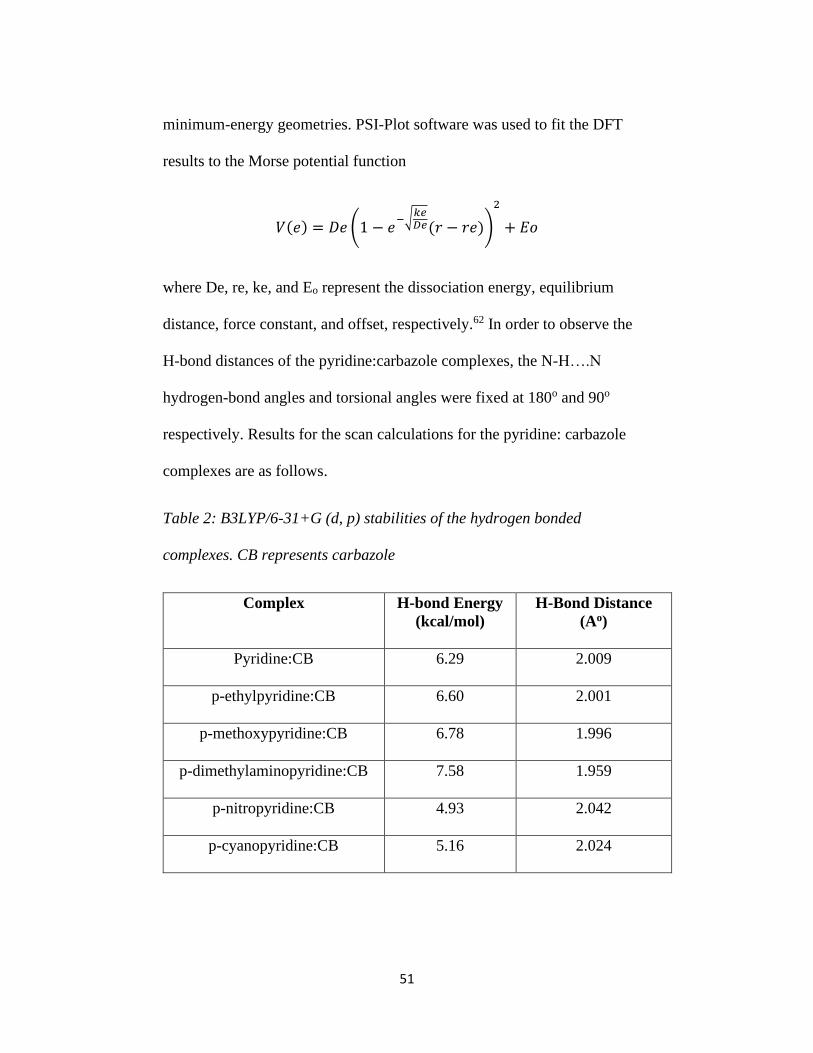
51
minimum-energy geometries. PSI-Plot software was used to fit the DFT
results to the Morse potential function
𝑉(𝑒) = 𝐷𝑒 (1 − 𝑒−√
𝑘𝑒𝐷𝑒(𝑟 − 𝑟𝑒))
2
+ 𝐸𝑜
where De, re, ke, and Eo represent the dissociation energy, equilibrium
distance, force constant, and offset, respectively.62 In order to observe the
H-bond distances of the pyridine:carbazole complexes, the N-H….N
hydrogen-bond angles and torsional angles were fixed at 180o and 90o
respectively. Results for the scan calculations for the pyridine: carbazole
complexes are as follows.
Table 2: B3LYP/6-31+G (d, p) stabilities of the hydrogen bonded
complexes. CB represents carbazole
Complex
H-bond Energy
(kcal/mol)
H-Bond Distance
(Ao)
Pyridine:CB
6.29 2.009
p-ethylpyridine:CB
6.60 2.001
p-methoxypyridine:CB
6.78 1.996
p-dimethylaminopyridine:CB
7.58 1.959
p-nitropyridine:CB
4.93 2.042
p-cyanopyridine:CB
5.16 2.024
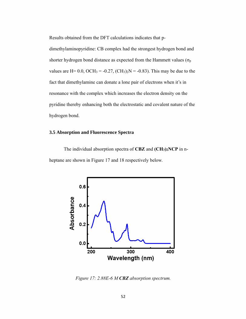
52
Results obtained from the DFT calculations indicates that p-
dimethylaminopyridine: CB complex had the strongest hydrogen bond and
shorter hydrogen bond distance as expected from the Hammett values (σp
values are H= 0.0, OCH3 = -0.27, (CH3)2N = -0.83). This may be due to the
fact that dimethylamine can donate a lone pair of electrons when it’s in
resonance with the complex which increases the electron density on the
pyridine thereby enhancing both the electrostatic and covalent nature of the
hydrogen bond.
3.5 Absorption and Fluorescence Spectra
The individual absorption spectra of CBZ and (CH3)2NCP in n-
heptane are shown in Figure 17 and 18 respectively below.
Figure 17: 2.88E-6 M CBZ absorption spectrum.

53
Figure 18: 1.732E-5 M (CH3)2NCP absorption spectrum.
The combined absorption spectra of CBZ and (CH3)2NCP collected
in n-heptane are shown in Figure 19 below.
Figure 19: Comparison of CBZ and (CH3)2NCP absorption spectra.
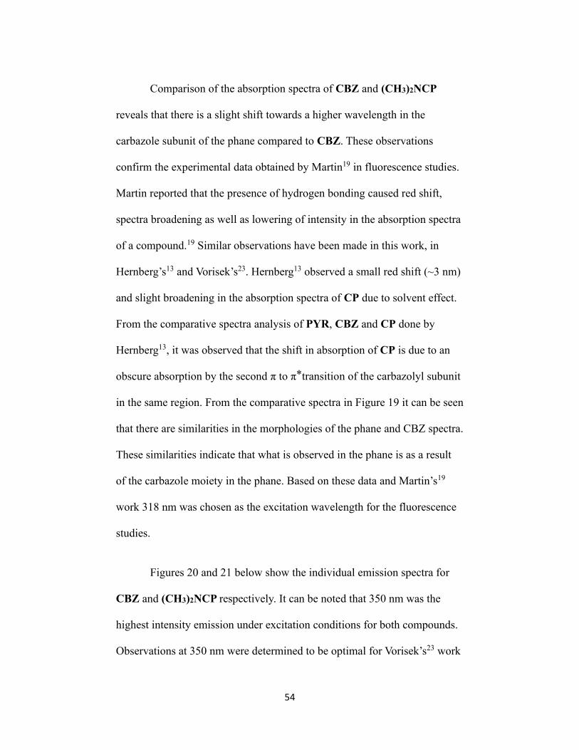
54
Comparison of the absorption spectra of CBZ and (CH3)2NCP
reveals that there is a slight shift towards a higher wavelength in the
carbazole subunit of the phane compared to CBZ. These observations
confirm the experimental data obtained by Martin19 in fluorescence studies.
Martin reported that the presence of hydrogen bonding caused red shift,
spectra broadening as well as lowering of intensity in the absorption spectra
of a compound.19 Similar observations have been made in this work, in
Hernberg’s13 and Vorisek’s23. Hernberg13 observed a small red shift (~3 nm)
and slight broadening in the absorption spectra of CP due to solvent effect.
From the comparative spectra analysis of PYR, CBZ and CP done by
Hernberg13, it was observed that the shift in absorption of CP is due to an
obscure absorption by the second π to π ⃰ transition of the carbazolyl subunit
in the same region. From the comparative spectra in Figure 19 it can be seen
that there are similarities in the morphologies of the phane and CBZ spectra.
These similarities indicate that what is observed in the phane is as a result
of the carbazole moiety in the phane. Based on these data and Martin’s19
work 318 nm was chosen as the excitation wavelength for the fluorescence
studies.
Figures 20 and 21 below show the individual emission spectra for
CBZ and (CH3)2NCP respectively. It can be noted that 350 nm was the
highest intensity emission under excitation conditions for both compounds.
Observations at 350 nm were determined to be optimal for Vorisek’s23 work
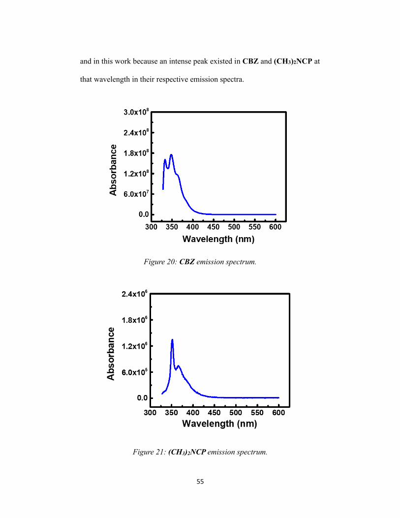
55
and in this work because an intense peak existed in CBZ and (CH3)2NCP at
that wavelength in their respective emission spectra.
Figure 20: CBZ emission spectrum.
Figure 21: (CH3)2NCP emission spectrum.

56
The combined fluorescence spectra of CBZ and (CH3)2NCP
collected in n-heptane are shown in Figure 22 below.
Figure 22: Comparison of CBZ and (CH3)2NCP emission spectra.
The emission spectra above show a significantly lower intensity background
radiation in (CH3)2NCP compared to CBZ. These data support our
hypothesis that the fluorescence of CBZ is quenched in the phanes. From
the overall spectra it can be inferred that (CH3)2NCP displays ~0.76 %
background fluorescence based on their relative intensities. Results obtained
for CP and 22OMeCP by Vorisek23 showed a background fluorescence of
~20 % and ~5 % respectively. These data support the hypothesis that
introduction of more electron donating group to the phane reduces the
background fluorescence.

57
Comparison of the S/R data obtained for (CH3)2NCP at 350 nm in
Table 3 also shows a background fluorescence of 0.7 %. However, CP and
22OMeCP showed different background fluorescence (37 % and 13 %
respectively) according to the data obtained for their S/R at 350 nm
compared to that of their overall spectra intensities.23
Table 3: S/R data for CBZ and (CH3)2NCP at 350 nm.
Compound S/R I/Io
CBZ 169889242.2 1
(CH3)2NCP 1227385.26 0.007
The reduction of the background fluorescence of the modified CP’s
correlates with their Hammett values (σp values are H= 0.0, OCH3 = -0.27,
(CH3)2N = -0.83). As shown in Figure 23 below there is a linear relationship
between the σp values and the background fluorescence observed in the
phanes.

58
Figure 23: Relationship between background fluorescence and Hammett
parameters.
The observation of the background fluorescence in the phane is due
to part of the phane not existing as hydrogen-bonded at room temperature in
the excited state.13 This interpretation was previously proposed by Hernberg
and Vorisek as well.13, 23 At room temperature there is sufficient thermal
energy present to allow conformational changes within the phane. This
would cause bending or twisting between the carbazole and pyridine
subunits of the phane resulting in disruption of the internal hydrogen bond.
1-5 Hernberg proposed that an easy test of this hypothesis was to take low
temperature acquisition of emission spectra for the phanes. Hernberg
proposed that emission acquisition at 77 K should be able to drop the
background fluorescence observed in CP and possibly all phanes produced

59
by our group as there will be decrease in energy available to sustain
conformation freedom at that temperature.11-13
3.6 Quenching Assays of Emitter/Quencher Pairs
Figure 24 shows emission spectra of CBZ quenched with increasing
concentrations of PYR. Figure 25 shows emission spectra of CBZ
quenched with increasing concentrations of DMAP. Concentration of CBZ
used for both studies is 5.5E-5 M to mimic results from Martin’s19 work.
Figure 24: Emission spectra of CBZ/PYR system.

60
Figure 25: Emission spectra of CBZ/DMAP system.
Figures 24 and 25 above demonstrate that 350 nm is the optimal
choice for the S/R acquisition for the fluorescence quenching studies for the
phane. Comparison of Figures 24 and 25 show that there is decreased
quenching of the CBZ system by DMAP. This data supports the hypothesis
that increasing the basicity of the pyridine quencher through electron
donation at the para position should promote increased fluorescence.
The absorption and emission spectra obtained for CBZ and
(CH3)2NCP in this work are similar in morphology and show the same
characteristic features as CP and 22OMeCP obtained by Gibson11, 12,
Hernberg13 and Vorisek23. This indicates that fluorescence restoration of
(CH3)2NCP should be observed at a similar wavelength to CBZ/PYR and
61
CP. The reduced background fluorescence observed in the emission spectra
of (CH3)2NCP supports the hypothesis stated earlier in this work that
increased electron donation to the quencher will promote hydrogen-bond
facilitated quenching mechanism.
3.7 Restoration of Fluorescence in (CH3)2NCP
It has been reported that the fluorescence of CP was restored in the
presence of hydrazine, hydrazine derivatives (MMH and UDMH), ammonia
and primary alkylamines, but not in triethylamine or piperidine.11-13, 22
Gibson’s results with CP suggested that it may be a viable fluorescence
based sensor for hydrazine and its derivatives, however, its detection limit
was higher than the threshold for detection. (CH3)2NCP has thus far
demonstrated that it shares photophysical properties with CP. Experiments
to determine whether the fluorescence of (CH3)2NCP could be restored
using n-butylamine, ethylenediamine and triethylamine were performed.
Butylamine was selected as our representative “restorative” control
based on previous work done by Gibson, Hernberg, Gagnon and Vorisek.12,
13, 22, 23 A 1.73E-5 M solution of (CH3)2NCP was prepared in n-heptane.
The solution was prepared by dissolving the corresponding mass of
(CH3)2NCP in 100 mL of n-heptane. This resulted in a homogenous sample
after stirring on a stir plate with the aid of a magnetic stir bar for about 1
hour at room temperature. To each 2 mL of phane solution was added a
volume of n-butylamine corresponding to their individual concentrations.

62
Concentrations of n-butylamine tested for the fluorescence restoration
studies ranged from 0 ppb to 20 ppb as shown in Figure 25. The solutions
were then topped with n-heptane to the 5 mL mark on the volumetric flask.
The solutions were shaken and allowed to stand at room temperature for 30
minutes. Emission spectra were then collected using the S/R method with
an excitation wavelength of 318 nm and observing the changes at 350 nm.
Fluorescence restoration was then assessed for the phane by comparing the
intensity of the solution with 0 ppb n-butylamine with the rest of solutions
with increased n-butylamine concentration. This was done by finding the
ratio of I/Io for each analyte concentration, with “Io” being 0 ppb in each
case and “I” being the new intensity.
The fluorescence of (CH3)2NCP was restored in the presence of 1
ppb n-butylamine. Figure 26 shows the results for (CH3)2NCP emission
changes as a result of titrating against n-butylamine. Figure 27 shows the
same data starting at the 1 ppb titration point, demonstrating a much better
linear regression for the fluorescence restoration of (CH3)2NCP. The
experiment was repeated twice, and the same results were obtained in each
case. The data obtained for (CH3)2NCP are comparable to data obtained for
the parent CP and 22OMeCP fluorescence restoration by n-butylamine in
Vorisek, Gibson and Hernberg’s work. 12, 13, 22, 23

63
Figure 26: Restoration of (CH3)2NCP fluorescence in n-heptane by
butylamine.
Figure 27: Restoration of (CH3)2NCP fluorescence in n-heptane by
butylamine starting at 1 ppb.

64
Triethylamine and ethylenediamine, which are a tertiary amine and a
bulky primary amine respectively, were also tested for fluorescence
restoration even though they did not restore fluorescence of the parent CP
or 22OMeCP.12, 13, 22, 23 The fluorescence restoration studies were done
using the same conditions as that of n-butylamine. Results obtained showed
that they both restore the fluorescence of (CH3)2NCP at 1 ppb analyte
concentration. To ensure that these results were consistent, the experiments
were repeated for each analyte and the same results were obtained. The
figures below show the data obtained for each of the analytes.
Figure 28: Restoration of (CH3)2NCP fluorescence in n-heptane by
ethylenediamine.

65
Figure 29: Restoration of (CH3)2NCP fluorescence in n-heptane by
ethylenediamine starting at 1 ppb.
Figure 30: Restoration of (CH3)2NCP fluorescence in n-heptane by
triethylamine.
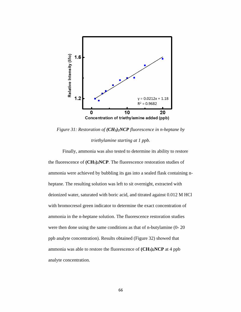
66
Figure 31: Restoration of (CH3)2NCP fluorescence in n-heptane by
triethylamine starting at 1 ppb.
Finally, ammonia was also tested to determine its ability to restore
the fluorescence of (CH3)2NCP. The fluorescence restoration studies of
ammonia were achieved by bubbling its gas into a sealed flask containing n-
heptane. The resulting solution was left to sit overnight, extracted with
deionized water, saturated with boric acid, and titrated against 0.012 M HCl
with bromocresol green indicator to determine the exact concentration of
ammonia in the n-heptane solution. The fluorescence restoration studies
were then done using the same conditions as that of n-butylamine (0- 20
ppb analyte concentration). Results obtained (Figure 32) showed that
ammonia was able to restore the fluorescence of (CH3)2NCP at 4 ppb
analyte concentration.

67
Figure 32: Restoration of (CH3)2NCP fluorescence in n-heptane by
ammonia.
These results demonstrate that (CH3)2NCP is also receptive to the
fluorescence restorative effect exerted by primary alkylamines on our phane
systems.

68
Chapter 4: Conclusion and Future Studies
1H NMR obtained for (CH3)2NCP showed a peak in the ~ 13 ppm
range which is indicative of a hydrogen-bonded hydrogen attached to a
nitrogen atom. This chemical shift was not seen in any 1H NMR of the
carbazole precursors. The photophysical characteristics of (CH3)2NCP are
very similar to those of both CP and 22OMeCP obtained by Vorisek.23
(CH3)2NCP showed a background fluorescence of 0.7 % at room
temperature and was quenched with respect to the emission of CBZ. The
fluorescence of (CH3)2NCP was restored when in the presence of
butylamine, ethylenediamine, triethylamine and ammonia at relatively low
concentrations. The detection limits for the analytes are 1 ppb for
butylamine, ethylenediamine, and triethylamine. That of ammonia was 4
ppb. These results validate previous reported results within our group that
the addition of an electron donating group to the phanes decreased the
detection limit.11-13, 23 The observed diminished background fluorescence in
(CH3)2NCP compared to CP and 22OMeCP can be attributed to the fact
that addition of a more electron donating group to the pyridine subunit
strengthens the internal hydrogen bond.44
Future studies should include threshold limits for detection of
hydrazine and possibly its derivatives to determine whether (CH3)2NCP can
detect the set TLVs for hydrazine and its derivatives.

69
References
1. Valeur, B. Molecular Fluorescence: Principles and Applications, 2002,
Wiley-VCH.
2. McHale, J. L. Molecular Spectroscopy, 1999, Prentice Hall.
3. Becker, R. S. Theory and Interpretation of Fluorescence and
Phosphorescence, 1969, John Wiley & Sons, Inc.
4. Silbey, R. J.; Alberty, R. A.; Bawendi, M. G. Physical Chemistry’, 4th
ed., 2004, John Wiley & Sons, Inc.
5. Kuhn, H.; Forsterling, H.-D.; Waldeck, D. H Principles of Physical
Chemistry, 2nd Ed., 2009, John Wiley & Sons.
6. Engel, T.; Reid, P. Thermodynamics, Statistical Thermodynamics, and
Kinetics, 2005, Prentice Hall.
7. Engel, T.; Reid, P. Quantum Chemistry and Spectroscopy, 2005,
Prentice Hall.
8. Pfister, G.; Williams, D. J. J. Chem. Phys. 1973, 59, 2683-2689
9. Johnson, G. E. J. Phys. Chem. 1980, 84, 2940-2946.
10. Zelent, B.; Durocher, G. J. Org. Chem. 1981, 46, 1496-1499.
11. Gibson, T. L. Ph.D. Dissertation, Florida Institute of Technology,
1999.
12. Brown, A. B.; Gibson, T. L.; Baum, J. C.; Ren, T.; Smith, T. M.
Sensors and Actuators B 2005, 110, 8-12.
70
13. Hernberg, E. A. Ph.D. Dissertation, Florida Institute of Technology,
2011.
14. Mataga, N.; Kaifu, Y. J. Chem. Phys. 1962, 36, 2804-2805.
15. Berlman, I. B. Handbook of Fluorescence Spectra of Aromatic
Molecules, 1965, Academic Press.
16. Tanaka, F.; Kato, M.; Mataga, N. Z. Phys. Chem. (N.F.) 1970, 70, 104-
112.
17. Mataga, N.; Kubota, T. Molecular Interactions and Electronic Spectra,
Marcel Dekker, New York, 1970, pp 346-351.
18. Mataga, N.; Tanaka, F.; Kato, M. Acta Phys. Pol. 1968, 34, 733-745
19. Martin, M. M.; Ware, W. R. J. Phys. Chem. 1978, 26, 2770-2776.
20. Altamirano, M. S.; Bohorquez, M. del V.; Previtali, C. M.; Chesta, C.
A. J. Phys. Chem. A. 2008, 112, 589-593.
21. Ikeda, N.; Miyasaka, H.; Okada, T.; Mataga, N. J. Am. Chem. Soc.
1983, 105, 5206-5211.
22. Gagnon, D. B.S. Undergraduate Thesis, Florida Institute of
Technology, 2012.
23. Vorisek, D. P. Ph.D. Dissertation, Florida Institute of Technology,
2014.
24. Herbich, J.; Hung, C-Y.; Thummel, R. P.; Waluk, J. J. Am. Chem. Soc.
1996, 118, 3508-3518

71
25. Marks, D.; Zhang, H.; Borowicz, P.; Waluk, J.; Glasbeek, M. J. Phys.
Chem. A 2000, 104, 7167-7175.
26. Schmidt, E.W. Hydrazine and Its Derivatives, 2nd ed., 2 vols, Wiley-
Interscience, New York, 2001, pp 820-1632, pp 559-670.
27. Mallinckrodt Baker, Inc., Materials Safety Data Sheet for Hydrazine.
1999.
28. Olin Corporation, Materials Safety Data Sheet for
Monomethylhydrazine. 1991.
29. 2000 TLVs and BEIs: Threshold Lim it Values for Chemical Substances
and Physical Agents, and Biological Exposure Indices. American
Conference of Governmental Industrial Hygienists, Cincinnati, Ohio,
2000.
30. Meneghelli, B. J. CPIA Publ. 1999, 687, 117-127.
31. Crossman, K. P.; Carver, P. T.; Wyatt, J. R.; Rose-Pehrsson, S. L.;
Thurow, A., U.S. Government Report 1990, NRL-MR-6668 [Chem.
Abstr. 1991, 115, 14364b],
32. Brenner, K. P.; Rose-Pehrsson, S. L. U.S. Government Report 1994,
NRL/MR/6110-94-7492 [Chem. Abstr. 1995, 123, 294981r]
33. Carter, M. T.; Smith, J. R.; Mowry, D. R.; Patel, J. G. Proc. SPIE ~ Int.
Soc. Opt. Eng. 1998, 3540, 123-133.
34. Klimcak, C.; Radhakrishnan, G.; Delcamp, S.; Chan, Y.; Jaduszliwer,
B.; Moss, S. Proc. SPIE -Int. Soc. Opt. Eng. 1994, 2293, 209-219.

72
Klimcak, C.; Radhakrishnan, G.; Jaduszliwer, B. Ibid. 1995, 2367, 80-
88. Jaduszliwer, B.; Klimcak, C. M. (to Aerospace Corp.), U.S. Patent
1996, 5,567,622 [Chem. Abstr. 1996, 125, 315737z]. Klimcak, C.;
Chan, Y.; Jaduszliwer, B. Proc. SPIE -Int. Soc. Opt. Eng. 1999, 3540,
183-190.
35. Carter, M. T.; Schwartz, M. (to Eltron Research), U.S. Patent 2001,
6,328,932 [Chem. Abstr. 2002, 136, 14852t], Andrawis, A. S.;
Santiago, J.; Young, R. C.; Baum, J. C. Proc. SPIE -Int. Soc. Opt. Eng.
2004, 5502, 239-242.
36. Cram, D. J.; Steinberg, H. J. Am. Chem. Soc. 1951, 73, 5691-5704.
37. Cram, D. J.; Allinger, N. L.; Steinberg, H. J. Am. Chem. Soc. 1954, 76,
6132-6141.
38. Bortolus, P.; Monti, S.; Galiazzo, G.; Gennari, G. Chem. Phys. 1997,
223, 99-108.
39. Schneider, H.-J.; Yatsimirsky, A. K. Principles and Methods in
Supramolecular Chemistry Wiley, Chichester, 2000, pp 69-l17, 259-
265.
40. Van Duuren, B. L. Anal. Chem. 1960, 32, 1436-1442.
41. Mataga, N.; Torihashi, Y.; Kaifu, Y. Z. Phys. Chem 1962, 34, 379-392.
42. Baum, J. C. J. Am. Chem. Soc. 1980, 102, 716-719.
43. Mitchell, R. H.; Zhang, J. Tetrahedron Lett. 1995, 36, 1177-1180.

73
44. Guram, A. S.; Rennels, R. A.; Buchwald, S. L. Angew. Chem., Int. Ed.
Engl. 1995, 34, 1348-1350.
45. Cochran, J. E. Ph.D. Dissertation, University of Wisconsin - Madison,
1993.
46. Wolfe, J. P.; Singer, R. A.; Yang, B. H.; Buchwald, S. L. J. Am. Chem.
Soc. 1999, 121, 9550-9561.
47. Wolfe, J. P.; Buchwald, S. L. J. Org. Chem. 2000, 65, 1144-1157.
48. Wolfe, J. P.; Tomori, H.; Sadighi, J. P.; Yin, J.; Buchwald, S. L. J. Org.
Chem. 2000, 65, 1158-1174.
49. Prashad, M.; Hu, B.; Lu, Y.; Draper, R.; Har, D.; Repic, O.; Blacklock,
T. J. J. Org. Chem. 2000, 65, 2612-2614.
50. Tomori, H.; Fox, J. M.; Buchwald, S. L. J. Org. Chem. 2000, 65, 5334-
5341.
51. Harris, M. C.; Huang, X.; Buchwald, S. L. Org. Lett. 2002, 4, 2885-
2888.
52. Surry, D. S.; Buchwald, S. L. J. Am. Chem. Soc. 2007, 129, 10354-
10355.
53. Åkermark, B.; Eberson, L.; Jonsson, E.; Pettersson, E. J. Org. Chem.
1975, 40, 1365-1367.
54. Åkermark, B.; Oslob, J. D.; Heuschert, U. Tetrahedron Letters 1995,
36, 1325-1326.

74
55. Jordan-Hore, J. A.; Johansson, C. C. C.; Gulias, M.; Beck, E. M.;
Gaunt, M. J. J. Am. Chem. Soc. 2008, 130, 16184-16186.
56. Oliveira-Campos, A. M. F.; Querioz, M.-J. R. P.; Raposo, M. M. M;
Shannon, P. V. R. Tetrahedron Letters 1995, 36, 133-134.
57. Iwaki, T.; Yasuhara, A.; Sakamoto, T. J. Chem. Soc., Perkin Trans. 1
1999, 1505-1510.
58. Bedford, R. B.; Betham, M. J. Org. Chem. 2006, 71, 9403-9410.
59. Sissouma, D.; Maingot, L.; Collet, S.; Guingant, A. J. Org. Chem.
2006, 71, 8384-8389.
60. Watanabe, T.; Ueda, S.; Inuki, S.; Oishi, S.; Fujii, N.; Ohno, H. Chem.
Commun. 2007, 4516-4518.
61. Martin, G. E. Ann. Rep. NMR Spec. 2002, 46, 37-100.
62. Ziegler, E. W.; Baum, J. C.; Brown, A. B.; Blaustein, G. S. Int. J.
Quantum Chem. 2017, 117, e25326.

75
Appendix A NMR Spectra
A1
Diethyl-2,2’-Iminodibenzoate (9) 1H NMR
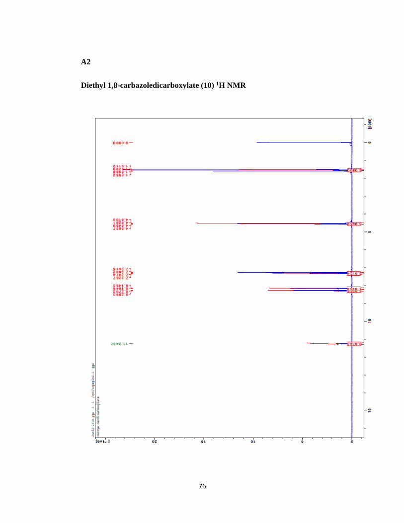
76
A2
Diethyl 1,8-carbazoledicarboxylate (10) 1H NMR

77
A3
1,8-Carbazoledimethanol (11) 1H NMR

78
A4
1,8-Carbazoledimethanol (11) aromatic region 1H NMR

79
A5
1,8-Carbazolebis(methanethioacetate) (12) 1H NMR

80
A6
4-Dimethylamino-2,6-bis(bromomethyl)pyridine (6) 1H NMR

81
A7
4-Dimethylamino-2,6-bis(bromomethyl)pyridine (6) 13C NMR

82
A8
Dimethylamino-carbazolopyridinophane (CH3)2NCP 1H NMR

83
A9
Dimethylamino-carbazolopyridinophane (CH3)2NCP 13C NMR

84
A10
Dimethylamino-carbazolopyridinophane (CH3)2NCP 15N HMBC NMR

85
A11
Dimethylamino-carbazolopyridinophane (CH3)2NCP 15N HSQC NMR

86
A12
Dimethylamino-carbazolopyridinophane (CH3)2NCP COSY NMR

87
A13
Dimethylamino-carbazolopyridinophane (CH3)2NCP HMBC NMR

88
A14
Dimethylamino-carbazolopyridinophane (CH3)2NCP HSQC NMR

89
Appendix B Gaussian XY Data
B1
Pyridine: Carbazole Complex
Scan of Total Energy
X-Axis: Scan Coordinate
Y-Axis: Total Energy (Hartree)
X Y
1.5000000000 -765.8102802240
1.6500000000 -765.8158533520
1.8000000000 -765.8186838920
1.9500000000 -765.8197124590
2.1000000000 -765.8196447240
2.2500000000 -765.8189723690
2.4000000000 -765.8180229030
2.5500000000 -765.8169968540
2.7000000000 -765.8160067410
2.8500000000 -765.8151057510
3.0000000000 -765.8143122150
3.1500000000 -765.8136225100
3.3000000000 -765.8130273820
3.4500000000 -765.8125152610
3.6000000000 -765.8120761870
3.7500000000 -765.8117018390
3.9000000000 -765.8113816730
4.0500000000 -765.8111521750
4.2000000000 -765.8109160670
4.3500000000 -765.8107115250
4.5000000000 -765.8105324320
4.6500000000 -765.8103736870
4.8000000000 -765.8102336980
4.9500000000 -765.8101097200
5.1000000000 -765.8099988760
5.2500000000 -765.8098997030
5.4000000000 -765.8098110200
5.5500000000 -765.8097313850
5.7000000000 -765.8096595370
5.8500000000 -765.8095945140
6.0000000000 -765.8095357040

90
B2
Para-Ethylpyridine: Carbazole Complex
Scan of Total Energy
X-Axis: Scan Coordinate
Y-Axis: Total Energy (Hartree)
X Y
1.4999999171 -844.4506925770
1.6499999171 -844.4560804650
1.7999999171 -844.4587815390
1.9499999171 -844.4597151380
2.0999999171 -844.4595767110
2.2499999171 -844.4588498250
2.3999999171 -844.4578554960
2.5499999171 -844.4567905570
2.6999999171 -844.4557655560
2.8499999171 -844.4548327630
2.9999999171 -844.4540100840
3.1499999171 -844.4532944950
3.2999999171 -844.4526772480
3.4499999171 -844.4521469170
3.5999999171 -844.4516911410
3.7499999171 -844.4513012660
3.8999999171 -844.4509666720
4.0499999171 -844.4506804960
4.1999999171 -844.4504766840
4.3499999171 -844.4502637600
4.4999999171 -844.4500750560
4.6499999171 -844.4499074730
4.7999999171 -844.4497594440
4.9499999171 -844.4496281990
5.0999999171 -844.4495105830
5.2499999171 -844.4494050220
5.3999999171 -844.4493104850
5.5499999171 -844.4492255210
5.6999999171 -844.4491488290
5.8499999171 -844.4490792170
5.9999999171 -844.4490160820
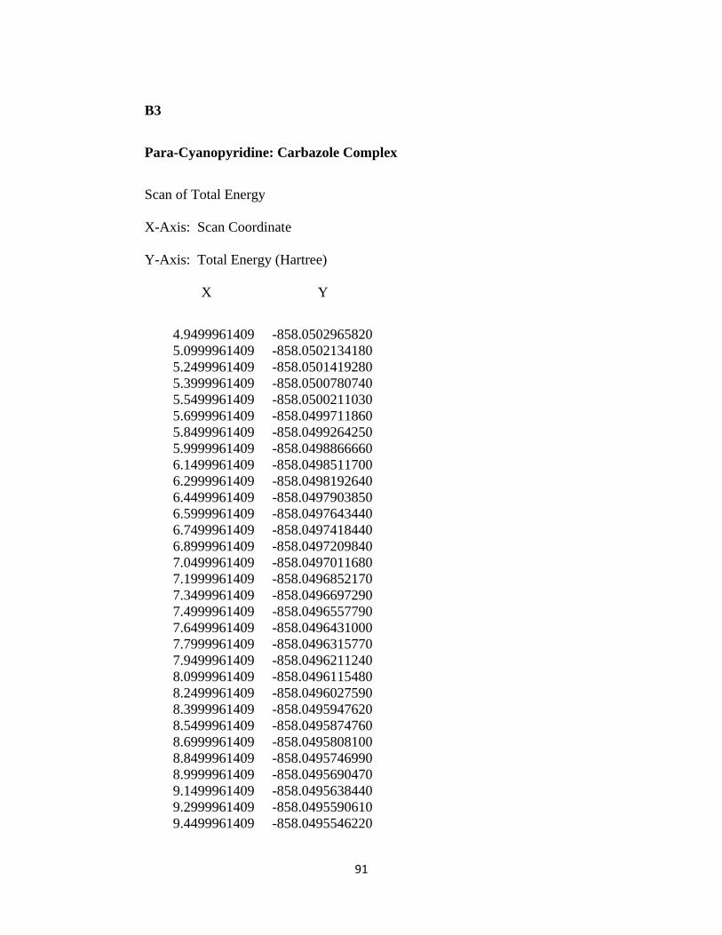
91
B3
Para-Cyanopyridine: Carbazole Complex
Scan of Total Energy
X-Axis: Scan Coordinate
Y-Axis: Total Energy (Hartree)
X Y
4.9499961409 -858.0502965820
5.0999961409 -858.0502134180
5.2499961409 -858.0501419280
5.3999961409 -858.0500780740
5.5499961409 -858.0500211030
5.6999961409 -858.0499711860
5.8499961409 -858.0499264250
5.9999961409 -858.0498866660
6.1499961409 -858.0498511700
6.2999961409 -858.0498192640
6.4499961409 -858.0497903850
6.5999961409 -858.0497643440
6.7499961409 -858.0497418440
6.8999961409 -858.0497209840
7.0499961409 -858.0497011680
7.1999961409 -858.0496852170
7.3499961409 -858.0496697290
7.4999961409 -858.0496557790
7.6499961409 -858.0496431000
7.7999961409 -858.0496315770
7.9499961409 -858.0496211240
8.0999961409 -858.0496115480
8.2499961409 -858.0496027590
8.3999961409 -858.0495947620
8.5499961409 -858.0495874760
8.6999961409 -858.0495808100
8.8499961409 -858.0495746990
8.9999961409 -858.0495690470
9.1499961409 -858.0495638440
9.2999961409 -858.0495590610
9.4499961409 -858.0495546220

92
B4
Para-Dimethylaminopyridine: Carbazole Complex
Scan of Total Energy
X-Axis: Scan Coordinate
Y-Axis: Total Energy (Hartree)
X Y
1.4999995245 -899.7965766140
1.6499995245 -899.8013803190
1.7999995245 -899.8036793760
1.9499995245 -899.8043171150
2.0999995245 -899.8039478710
2.2499995245 -899.8030335410
2.3999995245 -899.8018835320
2.5499995245 -899.8006874540
2.6999995245 -899.7995506880
2.8499995245 -899.7985212650
2.9999995245 -899.7976139290
3.1499995245 -899.7968237040
3.2999995245 -899.7961397760
3.4499995245 -899.7955494770
3.5999995245 -899.7950411880
3.7499995245 -899.7946053130
3.8999995245 -899.7942299630
4.0499995245 -899.7939075640
4.1999995245 -899.7936274290
4.3499995245 -899.7934262440
4.4999995245 -899.7932107000
4.6499995245 -899.7930186620
4.7999995245 -899.7928483970
4.9499995245 -899.7926970300
5.0999995245 -899.7925609040
5.2499995245 -899.7924381480
5.3999995245 -899.7923275690
5.5499995245 -899.7922279610
5.6999995245 -899.7921377250
5.8499995245 -899.7920555310
5.9999995245 -899.7919805170

93
B5
Para-Nitropyridine: Carbazole Complex
Scan of Total Energy
X-Axis: Scan Coordinate
Y-Axis: Total Energy (Hartree)
X Y
1.9500000047 -970.3211505830
2.1000000047 -970.3214181200
2.2500000047 -970.3210207740
2.4000000047 -970.3203036860
2.5500000047 -970.3194770810
2.7000000047 -970.3186598170
2.8500000047 -970.3179098640
3.0000000047 -970.3172489100
3.1500000047 -970.3166763660
3.3000000047 -970.3161844960
3.4500000047 -970.3157642920
3.6000000047 -970.3154084330
3.7500000047 -970.3151085300
3.9000000047 -970.3148559630
4.0500000047 -970.3146442670
4.2000000047 -970.3145081630
4.3500000047 -970.3143544360
4.5000000047 -970.3142213610
4.6500000047 -970.3141049940
4.8000000047 -970.3140040800
4.9500000047 -970.3139158670
5.1000000047 -970.3138383190
5.2500000047 -970.3137702640
5.4000000047 -970.3137104280
5.5500000047 -970.3136575150
5.7000000047 -970.3136106220
5.8500000047 -970.3135690630
6.0000000047 -970.3135322530
6.1500000047 -970.3134994330
6.3000000047 -970.3134701000
6.4500000047 -970.3134438030