effect of experimental infections of various tembusu virus
TRANSCRIPT
Polish Journal of Veterinary Sciences Vol. 21, No. 2 (2018), 389–396
DOI 10.24425/122613
Original article
T. Xu 1,2,a, X. Liu 1,2,a, Q. Liu1, K. Han1, Y. Liu1, J. Yang1, X. Huang1, D. Zhao1, K. Bi1, W. Sun2, Y. Li 1
1 Institute of Veterinary Medicine, Jiangsu Academy of Agricultural Sciences, Key Laboratory of Veterinary Biological Engineering and Technology,
Ministry of Agriculture, National Center for Engineering Research on Veterinary Bio-products, Nanjing, Jiangsu Province, China
2 Nanjing Agricultural University, Nanjing, Jiangsu Province, China
Correspondence to: W Sun, e-mail address: [email protected] (Weidong Sun) Y Li, e-mail address: [email protected] (Yin Li) a These authors contributed equally to this paper
Abstract
In order to compare the pathogenicity of different Tembusu virus (TMUV) strains from geese, ducks and chickens, 56 5-day-old Cherry Valley ducklings which were divided into 7 groups and infected intramuscularly with 7´105 PFU/ml per duck of six challenge virus stocks. The clin-ical signs, weight gain, mortality, macroscopic and microscopic lesions, virus loads in sera of 1, 3, 5, 7, 11 and 14 dpi and serum antibody titers were examined. The results showed that these virus-es could make the young ducks sick, but the clinical signs differed with the different species-orig-inal strains. All the experimental groups lose markedly in weight gain compared to the control, but there were no obvious distinctions in weight gains, as well as macroscopic and microscopic lesions of dead ducks between the infected groups. However, the groups of waterfowl-derived strains (from geese and ducks) showed more serious clinical signs and higher relative expressions of virus loads in sera than those from chicken-derived. The mortality of waterfowl groups was 37.5%, and the greatest mortality of chicken groups was 12.5%. The serum antibodies of the geese-species group JS804 appeared earlier and were higher in the titers than others. Taken toghter, the pathogenicity of waterfowl-derived TMUV was more serious than chicken-derived TMUV and JS804 could be chosen as one TMUV vaccine strain to protect from the infection.
Key words: goose-derived, duck-derived, chicken-derived, Tembusu virus, pathogenicity
Effect of experimental infections of various Tembusu virus strains
isolated from geese, ducks and chickens on ducklings

390 T. Xu et al.
Introduction
Tembusu virus (TMUV), a member of the Ntaya virus (NTAV) group of the genus Flavivirus in the fam-ily Flaviviridae (Cao et al. 2011), was first identified and isolated from a seriously-diseased duck farm in April 2010, China. The clinical symptoms of the infected ducks are generally characterized by retarded growth, acute anorexia, neurological dysfunction and an obvi-ous decline in egg production. The infections cause up to 90% morbidity and 5-15% mortality (Su et al. 2011). The disease was previously designated as duck hemor-rhagic ovaritis (DHO); and the virus was named as duck egg-drop syndrome virus (DEDSV) or Baiyang-dian virus (BYDV) (Liu et al., 2012), and is now typed as Tembusu virus (TMUV). Infections have occurred in most of the waterfowl-breeding areas of China, with especially serious outbreaks in the southern regions, and have caused massive economic loss in the Chinese waterfowl industry (Cao et al. 2011, Yan et al. 2011).
The virus can infect not only ducks, but also geese, chickens and sparrows (Liu et al. 2012, Huang et al. 2013, Tang et al. 2013). However, it is not clear whether TMUV strains originating from different poultry have different pathogenicity. We experimentally infected 5-day-old Cherry Valley ducks with these viruses and compared the same pathogenic characteristics in the outcome of the infections.
Materials and Methods
Virus preparation
Virus Strains
Six TMUV strains used in this study were isolated from diseased fowls with neurological symptoms in Jiangsu Province, China and preserved at -70°C in the laboratory. Viruses JS804 and SQ260 were from geese; JS808 and YC508 from ducks, and NJ705 and WJ901 from chickens.
Breeding viruses
In order to increase the titers of the viruses, three-day-old suckling mice were used to propagate the virus. Breeder mice of the ICR strain were purchased from the Comparative Medicine Centre of Yangzhou Uni-versity, and divided into 7 groups: 6 groups reproducing viruses and one as control. All the groups were fed sepa- rated in the air isolated conditions during whole experi- mental period. The mice were infected with 15 µl of virus suspension of the TMUV strains by intracerebral (i.c.) injection as previously described (Huang et al.
2013). Brain tissues of the mice showing clinical signs were collected and TMUV genome RNA was mea-sured by RT-PCR (Huang et al. 2013). Meanwhile, the mouse infection passages were continued until the viruses reproduced well and six TMUV strains showed similar amount of viral genome RNA. All the viruses were kept at -70°C for use.
Measuring virus titers
Virus titers were determined by plaque assay on DF-1 cells (Okuno et al., 1984). Briefly, monolayer cul-tures of DF-1 cells (1´105/well) grown in 6-well plates were incubated with 10-fold serial dilutions of virus for 1 h at 37°C, followed by an overlay of infected cells with 2 ml of DMEM containing 1.5% methyl cellulose and 2% fetal bovine serum and incubation at 37°C, under 5% CO2 atmosphere for 72 h. The cells were fixed with 1 ml of 10% formaldehyde for 30 min, washed with PBS (pH=7.0), and stained with methylene blue tetrahy-drate solution to visualize the plaques and measured using a microscope.
Animal experiments
After measurement assessment as TMUV-RNA negative with real-time RT-PCR and no TMUV anti-bodies with serum neutralization test (SNT), 56 healthy Cherry Valley ducks (5-day-old) were divided into 7 groups and fed in different isolators equally and ran-domly. Each of the six groups was infected intramuscu-larly (i.m) with 2.8×10⁵ PFU of the six virus strains, respectively. The control was inoculated with 0.4 ml sterile phosphate-buffered saline (PBS).
The ducks were observed and recorded daily over 14 days. Serum samples were collected from every duck at 1, 3, 5, 7, 11 and 14 dpi, and then stored at -70°C. The ducks were weighed at 0, 3, 5, 9, 11 and 14 dpi. Dead ducks were collected and the remaining ducks were necropsied at the end of the experiment. The tis-sues and organs such as hearts, spleens, pancreas and livers of all the ducks were harvested and kept in 10% neutral buffered formalin solution for histopathologi-cal examination. The animal experiments were strictly carried out in accordance with the established humane procedures and bio-security guidelines.
Detection of viral loads with Real-time PCR
Total RNA was obtained using an AxyPrepTM Body Fluid Viral DNA/RNA Miniprep Kit 250-prep (AxyGEN, PR China). First strand cDNA was synthe-sized from 13 µl total RNA using a PrimeScript 1st strand cDNA Synthesis Kit (TaKaRa, DaLian, China) following the manufacturer’s instructions.
391Effect of experimental infections of various Tembusu virus ...
Real time PCR based on Green I SYBR was per-formed in 96-well plates using the Applied Biosystems 7500 Fast Real-Time PCR System (Applied Biosystems, CA). Primers for duck Tembusu E gene and GAPDH (glyceraldehyde-3-phosphate dehydrogenase), are listed in Table 1. The amplification systems contained the fol-lowing components: 2.0 µl of cDNA templates, 10 µl 2×SYBR Green PCR Master Mix (QIAGEN, Beijing, China), 0.1µl QN ROX Reference Dye, 0.56 µl each of the primers, and 6.78 µl RNase-Free water. The opera-tion profile involved initial PCR activation at 95°C for 5 min, followed by 40 cycles of denaturing at 95°C for 10 s and annealing at 60°C for 30 s. Each sample was done in triplicate. Quantitative analysis of the data was carried out in a relative quantification (2-DDCT) study model. The relative expression of control was as 1 and GAPDH as an internal reference gene.
Evaluation of serum antibodies against TMUV
The purified TMUV was serially diluted with PBS (0.05M, pH9.6 ) to 1:400, and placed into coated 96-well microplates at 100 µl/well for 1 h at 37°C or overnight at 4°C. After three washes with PBST, the plates were blocked with 1% bovine serum albumin (BSA) in PBST for 2 h at 37°C. Serum samples inacti-vated at 56°C for 1 h were diluted from 1:2 to 1:128 serially in PBST, and added (100 µl/well) to the plates for 1h at 37°C. They were then incubated with 100 µl/well HRP-conjugated goat anti-mouse IgG (H&L) sera (1:1000 dilution) at 37°C for 1 h. Finally, 100 µl of 3,3’, 5,5’- tetramethylbenzidine (TMB) (Promega, WI, USA) was reacted for 5min at room temperature, and stopped with 50 µl of 2 M H2SO4. The plates were read at 450 nm using an ELISA reader (Bio-Rad, CA, USA).
Results
Clinical signs
In the groups infected with duck-original TMUV stains (JS808 and YC508), the clinical observation is shown in Table 2, and the ducks displayed depression,
inappetence, polydipsia and green loose stool as early as 1 dpi, and showed neurological signs characterized by ataxia and paralysis at 3 dpi. One duck died every day from dpi 4 to dpi 6 in group JS808; while only 1 duck died in group YC508 at 7 dpi. Group JS808 recovered at 7 dpi and group YC508 at 8 dpi.
Ducks in the groups infected with geese-original TMUV stains (JS804 and SQ260), displayed depres-sion, inappetence and green loose stool at 2 dpi as shown in Table 2. In Group JS804, 3 ducks showed atax-ia and paralysis at 3 dpi, died at 5 dpi and the others improved from at 6 dpi. In Group SQ260, 4 ducks developed neurological signs at 4 dpi. 1 died and the other were still weak and had ataxia at 5 dpi. 2 ducks dead at 6 dpi; and the living improved from 7 dpi.
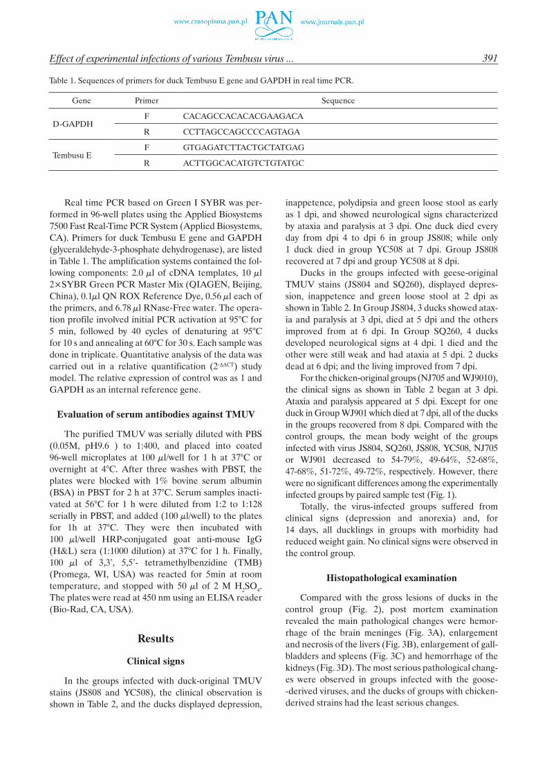
For the chicken-original groups (NJ705 and WJ9010), the clinical signs as shown in Table 2 began at 3 dpi. Ataxia and paralysis appeared at 5 dpi. Except for one duck in Group WJ901 which died at 7 dpi, all of the ducks in the groups recovered from 8 dpi. Compared with the control groups, the mean body weight of the groups infected with virus JS804, SQ260, JS808, YC508, NJ705 or WJ901 decreased to 54-79%, 49-64%, 52-68%, 47-68%, 51-72%, 49-72%, respectively. However, there were no significant differences among the experimentally infected groups by paired sample test (Fig. 1).
Totally, the virus-infected groups suffered from clinical signs (depression and anorexia) and, for 14 days, all ducklings in groups with morbidity had reduced weight gain. No clinical signs were observed in the control group.
Histopathological examination
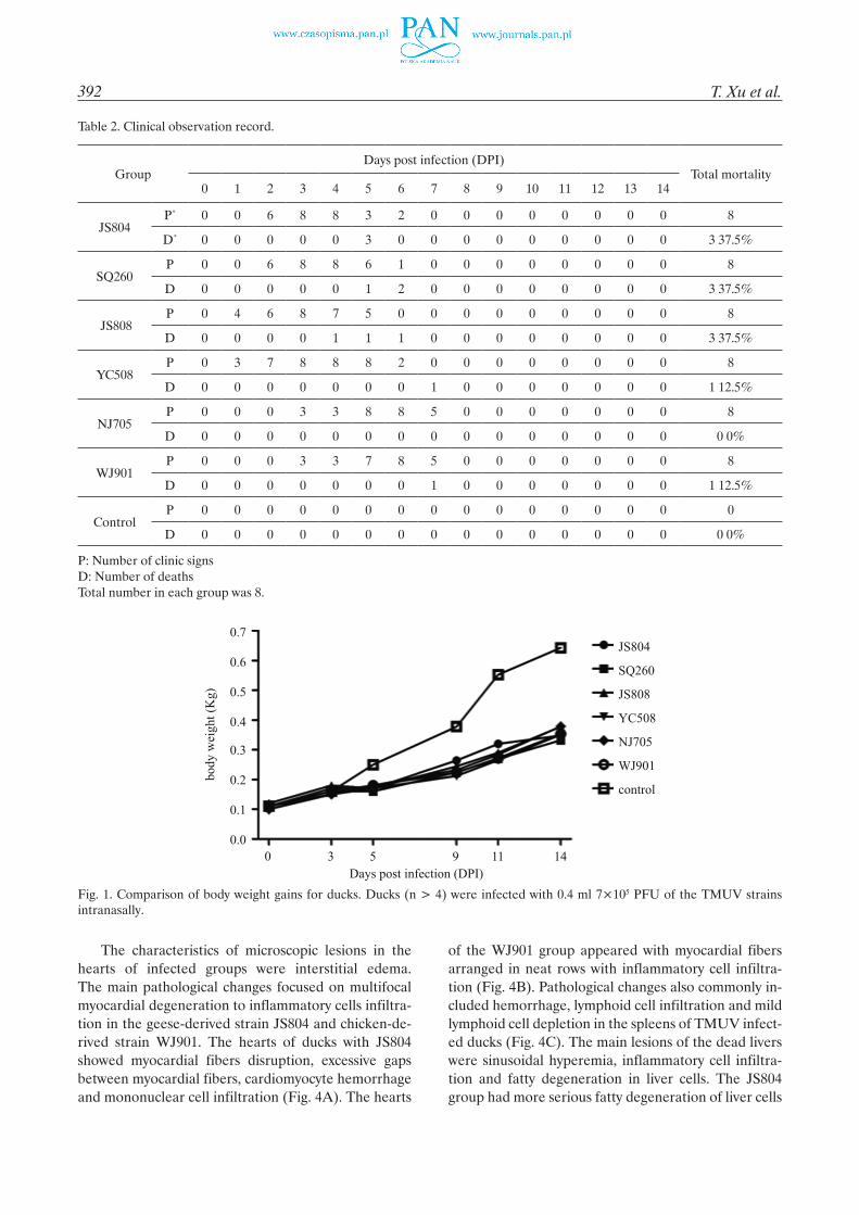
Compared with the gross lesions of ducks in the control group (Fig. 2), post mortem examination revealed the main pathological changes were hemor-rhage of the brain meninges (Fig. 3A), enlargement and necrosis of the livers (Fig. 3B), enlargement of gall-bladders and spleens (Fig. 3C) and hemorrhage of the kidneys (Fig. 3D). The most serious pathological chang-es were observed in groups infected with the goose- -derived viruses, and the ducks of groups with chicken- derived strains had the least serious changes.
Table 1. Sequences of primers for duck Tembusu E gene and GAPDH in real time PCR.
Gene Primer Sequence
D-GAPDHF CACAGCCACACACGAAGACA
R CCTTAGCCAGCCCCAGTAGA
Tembusu EF GTGAGATCTTACTGCTATGAG
R ACTTGGCACATGTCTGTATGC
392 T. Xu et al.
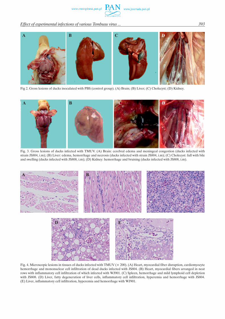
The characteristics of microscopic lesions in the hearts of infected groups were interstitial edema. The main pathological changes focused on multifocal myocardial degeneration to inflammatory cells infiltra-tion in the geese-derived strain JS804 and chicken-de-rived strain WJ901. The hearts of ducks with JS804 showed myocardial fibers disruption, excessive gaps between myocardial fibers, cardiomyocyte hemorrhage and mononuclear cell infiltration (Fig. 4A). The hearts
of the WJ901 group appeared with myocardial fibers arranged in neat rows with inflammatory cell infiltra-tion (Fig. 4B). Pathological changes also commonly in-cluded hemorrhage, lymphoid cell infiltration and mild lymphoid cell depletion in the spleens of TMUV infect-ed ducks (Fig. 4C). The main lesions of the dead livers were sinusoidal hyperemia, inflammatory cell infiltra-tion and fatty degeneration in liver cells. The JS804 group had more serious fatty degeneration of liver cells
Table 2. Clinical observation record.
GroupDays post infection (DPI)
Total mortality 0 1 2 3 4 5 6 7 8 9 10 11 12 13 14
JS804P* 0 0 6 8 8 3 2 0 0 0 0 0 0 0 0 8
D* 0 0 0 0 0 3 0 0 0 0 0 0 0 0 0 3 37.5%
SQ260P 0 0 6 8 8 6 1 0 0 0 0 0 0 0 0 8
D 0 0 0 0 0 1 2 0 0 0 0 0 0 0 0 3 37.5%
JS808P 0 4 6 8 7 5 0 0 0 0 0 0 0 0 0 8
D 0 0 0 0 1 1 1 0 0 0 0 0 0 0 0 3 37.5%
YC508P 0 3 7 8 8 8 2 0 0 0 0 0 0 0 0 8
D 0 0 0 0 0 0 0 1 0 0 0 0 0 0 0 1 12.5%
NJ705P 0 0 0 3 3 8 8 5 0 0 0 0 0 0 0 8
D 0 0 0 0 0 0 0 0 0 0 0 0 0 0 0 0 0%
WJ901P 0 0 0 3 3 7 8 5 0 0 0 0 0 0 0 8
D 0 0 0 0 0 0 0 1 0 0 0 0 0 0 0 1 12.5%
ControlP 0 0 0 0 0 0 0 0 0 0 0 0 0 0 0 0
D 0 0 0 0 0 0 0 0 0 0 0 0 0 0 0 0 0%
P: Number of clinic signsD: Number of deathsTotal number in each group was 8.
Fig. 1. Comparison of body weight gains for ducks. Ducks (n > 4) were infected with 0.4 ml 7×10⁵ PFU of the TMUV strains intranasally.
393Effect of experimental infections of various Tembusu virus ...
Fig 2. Gross lesions of ducks inoculated with PBS (control group). (A) Brain; (B) Liver; (C) Cholecyst; (D) Kidney.
Fig. 3. Gross lesions of ducks infected with TMUV. (A) Brain: cerebral edema and meningeal congestion (ducks infected with strain JS804, i.m); (B) Liver: edema, hemorrhage and necrosis (ducks infected with strain JS804, i.m); (C) Cholecyst: full with bile and swelling (ducks infected with JS808, i.m); (D) Kidney: hemorrhage and bruising (ducks infected with JS808, i.m).
Fig. 4. Microscopic lesions in tissues of ducks infected with TMUV (´ 200). (A) Heart, myocardial fiber disruption, cardiomyocyte hemorrhage and mononuclear cell infiltration of dead ducks infected with JS804. (B) Heart, myocardial fibers arranged in neat rows with inflammatory cell infiltration of which infected with WJ901. (C) Spleen, hemorrhage and mild lymphoid cell depletion with JS808. (D) Liver, fatty degeneration of liver cells, inflammatory cell infiltration, hyperemia and hemorrhage with JS804. (E) Liver, inflammatory cell infiltration, hyperemia and hemorrhage with WJ901.

394 T. Xu et al.
and inflammatory infiltration than that of group WJ901 (Fig. 4D and 4E). To summarize, microscopic lesions from dead ducks in each treated groups were as fol-lows: the lesions of the ducks infected with TMUV JS804 strain were more prominent than those of dead ducks infected with other TMUV strains.
Viral loads in the sera
The sera of the TMUV infected ducks collected at 1, 3, 5, 7, 11 and 14 dpi were examined with qPCR as the viral comparative expression of viral loads as shown in Fig. 5: the virus loads of the groups infected with waterfowl-derived strains began to increase at 1 dpi
(Fig. 5). The comparative expression numbers of all in-fected ducks were higher; groups JS804. SQ260, JS808, and YC508 were very significantly higher and Group JS808 was the highest at 3 dpi (Fig.5). At 5 dpi the viruses declined, while in the groups infected with chicken-original strains, the highest viral numbers appeared at 7 dpi (Fig. 5).
Evaluation of serum antibodies against TMUV
Serum samples of six infected groups were tested by ELISA simultaneously and there were no specific anti-body titers at 5 dpi. At 7 dpi, only ducks in Group JS804 developed a little positive antibody response. At 14 dpi,
Fig. 5. Relative quantity of viral loads in sera of ducks infected with TMUV. qPCR was conducted to detect viral RNA in serum samples collected in 1, 3, 5, 7, 11, 14 dpi. Quantities were normalized to GAPDH and plotted relative to control group (=1). y axis shows the fold changes in viral RNA as determined by the comparative CT method. qPCR was performed in triplicate. Error bars indicate the ± SD of three replicates.
Fig. 6. The ELISA results of serum antibodies against TMUV at 14 dpi. Each sample was detected in triplicate. Antibody titers were expressed as the reciprocal of log2. Data were expressed as mean ± SD (n = 3).

395Effect of experimental infections of various Tembusu virus ...
all the surviving ducks in the infected groups serocon-verted to TMUV; and ducks in Group JS804 developed significantly more antibodies than the others (Fig. 6). The antibody titers selected from highest to lowest are: JS804 > JS808 > SQ260 = YC508 > NJ705 > WJ901(Fig. 6).
Discussion
In April 2010, TMUV infections were first found in China and spread rapidly throughout the main duck-producing regions (Cao et al. 2011, Wan et al. 2012). The virus can infect not only ducks and cause serious diseases, but can also infect geese, chickens and sparrows (Huang et al. 2011, Tang et al. 2011, Liu et al. 2012). Studies on the pathogenicity of TMUV strains isolated from chickens or ducks have been separately reported by different researchers (Sun et al. 2014, Li et al. 2015, Han et al. 2016). However, there were no reports comparing the infecting capability of the viruses isolated from geese, ducks and chickens on young ducks simultaneously. Ducks are reared in large numbers in China, and the younger ducks (5-day-old) were more susceptible to virus infections (Sun et al., 2014). In the present study, 5-day-old Cherry Valley ducklings were infected with an almost equal amount of TMUV strains isolated from geese, ducks and chickens, and the results indicated that all of the viruses could cause poultry diseases. The viruses from waterfowl were more infec-tious than those from chicken, leading to earlier clinical signs, and more prominent and serious clinical signs such as acute anorexia and neurological disorders. The groups infected with the waterfowl-derived viruses demonstrated high mortality, close to 37.5%, while the chicken strains were 0 or 12.5%. These results were similar to reports on the pathogenicity of duck-origin TMUV in ducks (Sun et al. 2014) and chicken-origin TMUV in ducks (Chen et al. 2014). It is clear that the all TMUV strains derived from domestic poultry caused the younger ducks diseases and the viruses from water-fowl were more infectious and pathogenic.
The viruses of the Flavivirus family, such as Japa-nese encephalitis virus, dengue virus, Yellow fever virus, etc. infect sensitive animals and humans, and neurological symptoms and brain lesions are major sig-nificant clinical signs and pathological damage (Kalita et al. 2000, Baruah 2002, Deng et al. 2014). When TMUV, one member of the family, infected the young-er ducklings in the experiments, the initial and obvious symptoms were depression, inappetence and polydip-sia; These were soon followed by ataxia and paralysis. Gross lesions of the dead ducks were characteristic hemorrhage of meningitis (Fig. 3). There were no ob-
servably different gross lesions observed in the dead ducks from all the infected groups. These observations were consistent with Sun’s reports (Sun et al. 2014). This indicate that TMUV could infect and damage the nervous systems of the infected sensitive poultry and the brains might be one of the target organs for the vi-ruses in addition to livers, spleens and hearts. It would be very useful to study in detail how the virus effects the nervous systems and causes the lesions.
Viral loads could be detected in most of the sera of the infected ducks at 1 dpi (Fig. 5), indicating that TMUVs were able to cause systemic infections in a short time. The results were very similar to other studies (Li et al. 2015, Ti et al. 2015). Viral replication of waterfowl-derived strains reached the peak at 3 dpi and decreased later (Fig. 5). The higher viral loads and severe gross lesions were occurred synchronously (Table 2, Fig. 2). This indicates that there was a correla-tion between the comparative expression of viral load in serum and the outcome of TMUV infection. From the studies, it was interesting to note that the difference in the viral loads was between the waterfowl-derived and the chicken-derived strains, and the chicken-origi-nal viruses could produce a larger number at 3 dpi and declined at 5 dpi, while the largest virus loads were at 7 dpi (Fig. 5). Combined with the clinical signs and pathological lesions of the infected ducks, this shows that the chicken-derived viruses can infect ducklings, but the viruses take time to grow in the new host fowl. The chicken-original viruses had different growth capa-bility and also less pathogenicity to ducks. The reasons might be that the viruses change in their genomic com-position when infecting chickens and/or that chickens have different defense styles against the infections, causing the viruses to evolve. There was one report that the chickens-derived strain CJD05 has highly similar genomic sequences to waterfowl TMUV strains, and no deletion or insertion events were detected (Chen et al. 2014). This should help us to analyze these viral genome sequences and understand the difference of the viruses in large numbers.
All six different poultry-derived viruses could in-duce immune responses in the infected ducks to pro-duce specific antibodies (Fig. 6). However, the antibody of JS804 infected ducks was detected earlier and also showed the highest titer compared to the other 5 infect-ed groups (Fig.6). It was also shown that JS804 could produce fewer virus loads compared to the other 3 waterfowl-derived strains in the infections (Fig. 5). If the specific antibodies have NT activities against TMUV infections, the virus strain JS804 would be one ideal candidate for inactivated vaccine virus selection; and this might be one universally choosen method in vaccine preparation. Therefore, the chicken-origin

396 T. Xu et al.
strains would be other better candidates for TMUV attenuated live vaccine strain selection. And now we are trying to choose the viruses as TMUV vaccine strains for ducks to carry out protection studies against infection.
In conclusion, all the TMUV strains could infect 5-day-old ducklings no matter which kind of poultry the viruses were isolated from. The infected ducklings showed varying clinical symptoms and pathological lesions, and also produced specific antibody responses in those still survived. The chicken-origin viruses had different replication curves and less pathogenic capa-bility to younger ducks. The JS804 strain and chick-en-origin strains might be ideal candidates for viral vac-cine selections against future infections.
Acknowledgements
This work was supported by the National Natural Science Foundation of China (No. 31172345; No. 31502101), Jiangsu Provincial Agricultural Science and Technology Innovation Foundation [No. cx (14) 2091] and the Natural Science Foundation of Jiangsu Province (No. BK20160064).
References
Baruah HC, Biswas D, Patgiri D, Mahanta J (2002) Clinical outcome and neurological sequelae in serologically con-firmed cases of Japanese encephalitis patients in Assam, India. Indian Pediatr 39: 1143-1148.
Cao Z, Zhang C, Liu Y, Liu Y, Ye W, Han J, Ma G, Zhang D, Xu F, Gao X, Tang Y, Shi S, Wan C, Zhang C, He B, Yang M, Lu X, Huang Y, Diao Y, Ma X, Zhang D (2011) Tembusu virus in ducks, china. Emerg Infect Dis 17: 1873-1875.
Chen H, Qu Q, Tang Y, Gao X, Wu L, Xue C, Yu C, Cui J, Diao Y (2014) Development and evaluation of a DAS-ELISA for rapid detection of Tembusu virus using monoclonal antibodies against the envelope protein. PloS One. 9: e96366.
Chen S, Wang S, Li Z, Lin F, Cheng X, Zhu X, Wang J, Chen S, Huang M, Zheng M (2014) Isolation and charac-terization of a Chinese strain of Tembusu virus from Hy-Line Brown layers with acute egg-drop syndrome in Fujian, China. Arch Virol 159: 1099-1107.
Deng J, Pei J, Gou H, Ye Z, Liu C, Chen J (2014) Rapid and simple detection of Japanese encephalitis virus by reverse transcription loop-mediated isothermal amplification combined with a lateral flow dipstick. J Virol Methods 213: 98-105.
Han K, Zhao D, Liu Y, Liu Q, Huang X, Yang J, An F, Li Y (2016) Quantitative Proteomic Analysis of Duck Ovarian Follicles Infected with Duck Tembusu Virus by Label-Free LC-MS. Front Microbiol 7: 463.
Huang X, Han K, Zhao D, Liu Y, Zhang J, Niu H, Zhang K, Zhu J, Wu D, Gao L, Li Y (2013) Identification and molecular characterization of a novel flavivirus isolated from geese in China. Res Vet Sci 94: 774-780.
Kalita J, Misra UK (2000) Markedly severe dystonia in Japa-nese encephalitis. Mov Disord 15: 1168-1172.
Liu M, Chen S, Chen Y, Liu C, Chen S, Yin X, Li G, Zhang Y (2012) Adapted Tembusu-like virus in chickens and geese in China. J Clinl Microbiol 50: 2807-2809.
Li N, Lv C, Yue R, Shi Y, Wei L, Chai T, Liu S (2015) Effect of age on the pathogenesis of duck tembusu virus in Cherry Valley ducks. Front Microbiol 6: 581.
Okuno Y, Igarashi A, Fukunaga T, Tadano M, Fukai K (1984) Electron microscopic observation of a newly isolated flavi-virus-like virus from field-caught mosquitoes. J Gen Virol 65: 803-807.
Su J, Li S, Hu X, Yu X, Wang Y, Liu P, Lu X, Zhang G, Hu X, Liu D, Li X, Su W, Lu H, Mok N, Wang P, Wang M, Tian K, Gao F (2011) Duck egg-drop syndrome caused by BYD virus, a new Tembusu-related flavivirus. PLoS One 6: e18106.
Sun X, Diao Y, Wang J, Liu X, Lu A, Zhang L, Ge P, Hao D (2014) Tembusu virus infection in Cherry Valley ducks: the effect of age at infection. Vet Microbiol 168: 16-24.
Tang Y, Diao Y, Yu C, Gao X, Ju X, Xue C, Liu X, Ge P, Qu J, Zhang D (2013) Characterization of a Tembusu virus isolated from naturally infected house sparrows (Passer domesticus) in Northern China. Transbound Emerg dis 60: 152-158.
Ti J, Zhang L, Li Z, Zhao D, Zhang Y, Li F, Diao Y (2015) Effect of age and inoculation route on the infection of duck Tembusu virus in Goslings. Vet Microbiol 181: 190-197.
Wan C, Huang Y, Fu G, Shi S, Cheng L, Chen H (2012) Com-plete Genome Sequence of Avian Tembusu-Related Virus Strain WR Isolated from White Kaiya Ducks in Fujian, China. J Virol 86: 10912.
Yan P, Zhao Y, Zhang X, Xu D, Dai X, Teng Q, Yan L, Zhou J, Ji X, Zhang S, Liu G, Zhou Y, Kawaoka Y, Tong G, Li Z (2011) An infectious disease of ducks caused by a newly emerged Tembusu virus strain in mainland China. Virology 417: 1-8.