effect of immediate and delayed dentin sealing on the fracture strength, failure type and weilbull...
TRANSCRIPT
Eocv
MMa
Hb
P
a
A
R
R
1
A
K
A
B
C
C
D
I
L
V
Cf
h0
d e n t a l m a t e r i a l s 3 2 ( 2 0 1 6 ) e73–e81
Available online at www.sciencedirect.com
ScienceDirect
jo ur nal home p ag e: www.int l .e lsev ierhea l th .com/ journa ls /dema
ffect of immediate and delayed dentin sealingn the fracture strength, failure type and Weilbullharacteristics of lithiumdisilicate laminateeneers
arco M.M. Gresnigta,∗, Marco S. Cunea, Joanne G. de Roosa,utlu Özcanb
University Medical Center Groningen, The University of Groningen, Groningen, Center for Dentistry and Oralygiene, Department of Fixed and Removable Prosthodontics, The NetherlandsUniversity of Zurich, Dental Materials Unit, Center for Dental and Oral Medicine, Clinic for Fixed and Removablerosthodontics and Dental Materials Science, Zurich, Switzerland
r t i c l e i n f o
rticle history:
eceived 5 February 2015
eceived in revised form
1 August 2015
ccepted 15 January 2016
eywords:
dhesion
onding
ementation
eramic
entin
mmediate dentin sealing
aminate
eneer
a b s t r a c t
Objectives. Adhesion on dentin is less reliable than on enamel, which could affect the durabil-
ity of laminate veneers (LV). Immediate dentin sealing (IDS) is suggested instead of delayed
dentin sealing (DDS) to overcome hypersensitivity and prevent debonding from dentin. This
study evaluated the effect of IDS and DDS on the durability of Li2Si2O5 laminate veneers in
vitro.
Methods. Window preparations were made on the labial surfaces of sound maxillary cen-
tral incisors (N = 50). They were randomly divided into five groups: Group 1: Enamel
only + H3PO4 + Adhesive (control); Group 2: <1/4 dentin + H3PO4 + DDS (2 weeks later); Group
3: Complete dentin + H3PO4 + DDS (2 weeks later); Group 4: <1/4 dentin + H3PO4 + IDS; Group
5: Complete dentin + H3PO4 + IDS. Li2Si2O5 laminate veneers (e.max Press) were bonded to
the labial surfaces of the teeth with adhesive resin cement (Variolink Veneer). IDS layers
were silicacoated (CoJet System) and silanized (ESPE-Sil). The teeth with their bonded lami-
nates were thermocycled (10.000× cycles) and then subjected to static loading (1 mm/min).
Failure type and location after debonding were classified. Data were analyzed using ANOVA
and Tukey’s post hoc test ( ̨ = 0.05). Two-parameter Weibull distribution values including the
Weibull modulus, scale (m) and shape (0), values were calculated.
Results. Mean fracture strength (N) per group in descending order was as follows: Group
5 (576 ± 254), Group 4 (478 ± 216), Group 1 (473 ± 159), Group 2 (465 ± 186), and Group 3
(314 ± 137). The presence of complete dentin exposure sealed with DDS after 2 weeks on
the bonded surface (Group 3) resulted in significantly lower fracture strength results than
those in group 5 with IDS (p = 0.034). Weibull distribution presented higher shape (0) for Group
1 (3.67), than those of other groups (2.51–2.89). Failure types were predominantly adhesive
failure between the cement and the laminate veneer in Groups 1, 2, 4 whereas Group 3
∗ Corresponding author at: Department of Fixed and Removable Prosthodontics, Center for Dentistry and Oral Hygiene, University Medicalenter Groningen, The University of Groningen, Antonius Deusinglaan 1, 9713 AV, Groningen, The Netherlands. Tel.: +31 50 363 2608;
ax: +31 50 363 2696.E-mail address: [email protected] (M.M.M. Gresnigt).
ttp://dx.doi.org/10.1016/j.dental.2016.01.001109-5641/© 2016 Academy of Dental Materials. Published by Elsevier Ltd. All rights reserved.
e74 d e n t a l m a t e r i a l s 3 2 ( 2 0 1 6 ) e73–e81
presented more often complete adhesive failures between the cement and dentin. In Group
5, failures showed some IDS and cement with or without ceramic fracture attached on the
tooth.
Significance. When laminate veneers are bonded to a large dentin substrate, application of
immediate dentin sealing improves adhesion and thereby, the fracture strength of Li2Si2O5
laminate veneers.© 2016 Academy of Dental Materials. Published by Elsevier Ltd. All rights reserved.
junction.
1. Introduction
Laminate veneers in particular entail minimal tooth prepara-tion of only 0.3–0.9 mm, which is highly conservative whencompared to their full-coverage crown alternative. Althoughpreparation for laminate veneers could be achieved withinthe vicinity of enamel, some dentin exposure, especially atthe cement–enamel junction or below in the cervical area, issometimes unavoidable [1–3]. Freehand preparation of suchrestorations, without the use of putty indices or guidinggrooves of depth may yield to deeper preparations with higheramount of dentin exposure [2]. Preparation depth may in facthave consequences on the final fracture strength of minimalinvasive restorations, in that lower fracture strength resultswere reported for laminate veneers when bonded to dentincompared to enamel [4]. Unfortunately, clinical studies onsurvival of laminate veneers do not often report whetherpreparations were solely in enamel or dentin. Yet, availableevidence from clinical studies that reported dentin exposureafter tooth preparation, also reported higher incidence of fail-ures [5–8]. Recently, a review on the clinical evaluation oflaminate veneers bonded to dentin concluded that the sur-vival rate diminished when such restorations were bonded todentin [9].
In order to prevent micro-leakage and hypersensitivity,sealing of the dentin prior to impression taking for the indi-rect restorations was advocated in early 1990s [10]. In addition,other studies concluded that adhesive strength of restora-tions was improved when dentin was sealed [11–15]. Adhesivestrength after this so called immediate dentin sealing (IDS)was compared with the conventional adhesive cementation,delayed dentin sealing (DDS), which is a common procedurefor cementation of fixed dental prosthesis. In these stud-ies, bond strength results employing DDS varied between 2and 12 MPa, whereas application of IDS resulted in signifi-cantly higher mean bond strength results between 15 and58 MPa depending on the test method [12,14–16]. Apparently,application of the adhesive resin on freshly cut dentin and fur-ther polymerization of the adhesive resin over time improvedadhesion of bonded restorative materials [17,18]. Furthermore,it was also postulated that application of IDS results in asmooth surface that also improves the adaptation of the indi-rect restorations [19].
Clinical studies on the survival rate of laminate veneersbonded onto teeth with existing resin composite restorations
did not show encouraging results, providing that the sub-strate surfaces were not conditioned [6–8]. However, in an invitro study, ceramic laminate veneers bonded to a completecomposite surface presented higher fracture strength resultsthan those bonded onto enamel [20]. Similarly, clinical sur-vival rate of laminate veneers bonded onto teeth with existingcomposite restorations after the latter was tribochemical sil-icacoated, was not less than those bonded on enamel/dentinup to 40 months of evaluation [21]. Thus, it can be anticipatedthat the presence of adhesive resin would also not impair thebond strength of laminate veneers on the IDS.
The objectives of this study therefore were to (a) comparethe fracture strength of laminate veneers with and withoutIDS application, (b) evaluate the influence of the size of theexposed dentin and (c) failure types after loading until frac-ture. The first hypothesis tested was that the presence of IDSwould positively contribute to the fracture strength of thelaminate veneer compared to conventional adhesive cemen-tation (DDS). The second hypothesis tested was that the sizeof exposed dentin would not decrease the fracture strength ofthe laminate veneers.
2. Material and methods
2.1. Specimen preparation
The brands, types, main chemical compositions, manufac-turers and batch numbers of the materials used for theexperiments are listed in Table 1. Schematic description of theexperimental design is presented in Fig. 1.
Sound human central incisors (N = 50) of similar size, freeof restorations and root canal treatment were selected from apool of recently extracted teeth. All teeth were screened onthe presence of cracks by blue light and those with crackswere eliminated and replaced with new teeth. Before a lam-inate veneer preparation was made, impressions were madeusing a high precision condensation silicone (Provil Novo puttyfast set, Heraeus, Hanau, Germany) in order to obtain moldsfor the provisionals. Window type of tooth preparations with-out incisal overlap, were made with a depth-cutting bur (801201SC Swiss Dental Products, Intensiv Grancia, Switzerland),with this preparation type adhesion of the laminate did notrely on the macro-mechanical retention as in the case ofoverlap preparations. After the depth cuts of 0.3 mm weremade, preparation was finalized using a round-ended tapereddiamond chamfer bur (Swiss Dental Products, FG-2309).The preparations ended 1 mm above the cement–enamel
The amount of dentin exposure was controlled by etch-ing the prepared surface for 5 s and rinsing with water thatresulted in a white, dull enamel surface. Thereafter photos of
d e n t a l m a t e r i a l s 3 2 ( 2 0 1 6 ) e73–e81 e75
Table 1 – The brands, types, chemical compositions, manufacturers and batch numbers of the materials used for theexperiments.
Materials Type Chemical composition Manufacturer Batch number
Total-etch Etching agent 37% phosphoric acid Ivoclar Vivadent,Schaan, Liechtenstein
P14739
OptiBond FL Adhesive resin Primer: Hydroxyethyl methacrylate,Glycerolphophate dimethacrylate, phathalicacid monoethyl methacrylate, ethanol, water,photo-initiatorAdhesive: Triethylene glycol dimethacrylate,Urethane dimethacrylate, Glycerolphophatedimethacrylate, Hydroxyethyl methacrylate,bis-phenol A glycol dimethacrylate, filler,photo initiator
Kerr, Orange, CA, USA 3661962
ESPE-Sil Silane coupling agent Ethyl alcohol, methacryloxypropyl,trimethoxysilane
3M ESPE, St. Paul,Minnesota, USA
1311011
IPS Empressetching gel
Ceramic etching gel <5% Hydrofluoric acid Ivoclar Vivadent P14739
CoJet-Sand One componentprimer
Aluminum trioxide particles coated withsilica, particle size: 30 �m
3M ESPE 442859
Monobond Plus Ethanol,3-trimethoxysilsylpropylmethacrylate,methacrylated phosphoric acid ester
Ivoclar Vivadent N37750
Syntac Primer Primer Water, acetone, maleic acid, dimethacrylate Ivoclar Vivadent P17329Syntac Adhesive Adhesive resin Water, gluteraldehyde, maleic acid,
poly-ethyleneglycodi-methacrylateIvoclar Vivadent P15364
Heliobond Adhesive resin Bis-phenol A glycol dimethacrylate,dimethacrylate, initiators and stabilizers
Ivoclar Vivadent P06157
Variolink Veneer Light curing resincement (Medium
Urethane dimethacrylate, inorganic fillers,ytterbium trifluoride, initiators, stabilizers,
Ivoclar Vivadent N64556
tmBrmu3
2
T
G
G
G
G
Value 0) pigments
he teeth were analysed and surface area of exposed dentineasured using a custom-made image program (Plaqeval,
ME BioMedical Engineering, University of Groningen). Prepa-ation margins remained in enamel in all groups. Smooth
argins were created to prevent stress concentration zonessing finishing discs (Sof-Lex Contouring and Polishing Discs,M ESPE, St Paul, Minnesota, USA).
.2. Experimental groups, IDS and DDS layers
he teeth were than randomly divided into 5 groups.
roup 1: Preparation was made only in enamel. This groupacted as the control group.
roup 2: In this group, next to enamel, <1/4 of the cervicaldentin surface was exposed. Two weeks later, DDSwas created.
roup 3: In this group, dentin was exposed on the completesurface. DDS was created as in Group 2 after 2 weeks.
roup 4: In this group, next to enamel, <1/4 of the cer-vical dentin surface was exposed. The IDS wasachieved immediately after tooth preparation.Dentin was etched with 37% H3PO4 (Total-etch,Ivoclar Vivadent, Schaan, Liechtenstein) for 10 sfollowed by 30 s of rinsing with copious water.
Then, primer and adhesive resin (Optibond FL, Kerr,Orange, USA) was applied, air-thinned and photo-polymerized for 10 s using an LED polymerizationdevice (Bluephase, Ivoclar Vivadent) from a distanceof 2 mm. The output of the polymerization devicewas 1000 mW/cm2 throughout the experiment(Bluephasemeter, Ivoclar Vivadent). After applica-tion of glycerine gel, the surface was again photopolymerized for 40 s. IDS layer was controlled onpresence of voids and excess adhesive resin wasremoved under the microscope (Opmipico, Zeiss,Oberkochen, Germany).
Group 5: In this group, dentin was exposed on the completesurface. The IDS was achieved as in Group 4.
Impressions of the preparations were made using a highprecision silicon impression material (Prestige light, VaniniDental Industry, Grassina, Italy). Then provisional laminates(Protemp 4, 3M ESPE, St Paul, Minnesota, USA) were made andapplied using a spot etch technique where etching was per-formed for 10 s in the middle of the preparation. In Groups4 and 5 spot etching was performed at the enamel marginsand glycerine gel was applied in order to prevent adhesionbetween de IDS and the provisional restoration. After adjus-ting the temporary restorations using polishing discs (Sof-LexCountouring and Polishing Disks, 3M ESPE), specimens werestored in distilled water at 37 ◦C for 2 weeks.
One dental technician fabricated lithium disilicate(Li2Si2O5) laminate veneers (IPS e.max Press, Ivoclar Vivadent)
according to the instructions of the manufacturer. Veneerswere first sintered in a ceramic oven (Programat P3000, IvoclarVivadent) and glazed. The total thickness of the laminateveneers was 0.6 mm.
e76 d e n t a l m a t e r i a l s 3 2 ( 2 0 1 6 ) e73–e81
enta
Fig. 1 – Flow-chart showing experim2.3. Adhesive cementation
A photo-polymerizing resin cement (Variolink Veneer, IvoclarVivadent) was used for cementation of the ceramic laminateveneers. A three-step bonding procedure with separate con-ditioning of the IDS layer was employed to ensure adhesion.Before cementation, provisional restoration was removed;tooth was cleaned with pumice and the fit of ceramic laminateveneers controlled under optical microscope (Zeiss Supra V50,Carl Zeiss, Oberkochen, Germany) (10×).
Cementation surfaces of the ceramic veneers were con-ditioned using hydrofluoric acid (Ceramic etching gel <5%hydrofluoric acid, Ivoclar Vivadent) for 20 s, rinsed and ultra-sonically cleaned (Emag, Valkenswaard, The Netherlands) indistilled water for 5 min. They were then silanized (MonobondPlus, Ivoclar Vivadent), adhesive resin was applied (Heliobond,Ivoclar Vivadent).
In Groups 1–3, teeth were etched with 37% H3PO4 (Total-etch, Ivoclar Vivadent), where enamel was etched for 30 s anddentin for 10 s followed by rinsing with copious water. Primer(Syntac Primer, Ivoclar Vivadent) was applied on the dentinand adhesive resin on the whole preparation (Syntac Adhesive
and Heliobond, Ivoclar Vivadent).In Groups 4 and 5, IDS layer was silica coated (CoJet,3M, ESPE) using a chairside air-abrasion device (Dento-PrepTM, RØNVIG A/S, Daugaard, Denmark) from a distance of
l sequence and allocation of groups.
10 mm, angle of 45 degrees at 2 bar pressure until the surfacebecame matt. Then enamel was etched with 37% H3PO4 for30 s and rinsed. Silane (ESPE-Sil, 3M, ESPE) was applied at thesilica-coated IDS surfaces, followed by adhesive resin appli-cation (Syntact Adhesive and Heliobond, Ivoclar Vivadent) onthe whole preparation.
Laminate veneers were cemented using photo-polymerizing cement (Variolink Veneer, Ivoclar Vivadent).Excess cement was removed using microbrushes, glycerinegel was applied at the margins of the laminate veneersand photo-polymerized for 40 s from labial, lingual andincisal (≥1000 mW/cm2, Bluephase, Ivoclar Vivadent). Cementinterface at the margins was polished using rubber points(Astropol, Ivoclar Vivadent).
2.4. Aging and fracture test
All specimens were thermocycled (Willytec, Munich,Germany) for 10.000 times between 5 ◦C and 55 ◦C with a dwelltime of 30 s in each bath. After aging, digital photos of thespecimens were made. The teeth with the cemented laminate
veneers were embedded perpendicularly in polymethyl-methacrylate (Autoplast, Condular, Wager, Switzerland) upto the cemento-enamel junction in the middle of the plasticrings (PVC, diameter: 2 cm, height: 1 cm).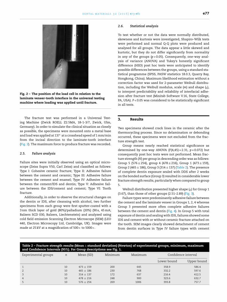
d e n t a l m a t e r i a l s 3 2
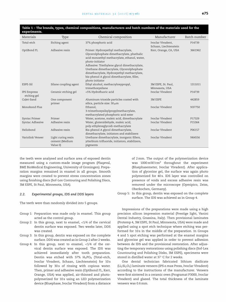
Fig. 2 – The position of the load cell in relation to thelaminate veneer-tooth interface in the universal testingm
iGaaf(
2
FsTbbbuf
ts3Bc4m
exposure of dentin and sealing with IDS, failures showed some
achine where loading was applied until fracture.
The fracture test was performed in a Universal Test-ng Machine (Zwick ROELL Z2.5MA, 18-1-3/7, Zwick, Ulm,ermany). In order to simulate the clinical situation as closelys possible, the specimens were mounted onto a metal basend load was applied at 137◦ at a crosshead speed of 1 mm/minrom the incisal direction to the laminate-tooth interfaceFig. 2). The maximum force to produce fracture was recorded.
.5. Failure analysis
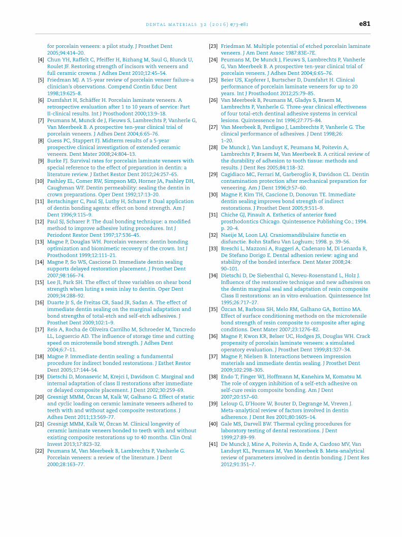
ailure sites were initially observed using an optical micro-cope (Zeiss Supra V50, Carl Zeiss) and classified as follows:ype I: Cohesive ceramic fracture; Type II: Adhesive failureetween the cement and ceramic; Type III: Adhesive failureetween the cement and enamel; Type IV: Adhesive failureetween the cement/IDS and dentin; Type V: Adhesive fail-re between the IDS/cement and cement; Type VI: Toothracture.
Additionally, in order to observe the structural changes onhe dentin or IDS, after cleansing with alcohol, two furtherpecimens from each group were first sputter-coated with a
nm thick layer of gold (80%)/palladium (20%) (90 s, 45 mA;alzers SCD 030, Balzers, Liechtenstein) and analyzed using
old field emission Scanning Electron Microscope (SEM) (LEO40, Electron Microscopy Ltd, Cambridge, UK). Images wereade at 25 kV at a magnification of 500× to 5000×.Table 2 – Fracture strength results (Mean ± standard deviation)
and Confidence Intervals (95%). For Group descriptions see Fig.
Experimental groups n Mean (SD) Minimum
1 10 473 ± 159 200
2 10 465 ± 186 230
3 10 314 ± 137 172
4 10 478 ± 216 248
5 10 576 ± 254 269
( 2 0 1 6 ) e73–e81 e77
2.6. Statistical analysis
To test whether or not the data were normally distributed,skewness and kurtosis were investigated, Shapiro–Wilk testswere performed and normal Q-Q plots were produced andanalysed for all groups. The data appear a little skewed andkurtotic, but they do not differ significantly from normalityin any of the groups (p > 0.05). Consequently, one-way anal-ysis of variance (ANOVA) and Tukey’s honestly significantdifference (HSD) post hoc tests were anticipated to identifypossible differences between the groups, using a standard sta-tistical programme (SPSS, PASW statistics 18.0.3, Quarry Bay,Hongkong, China). Maximum likelihood estimation without acorrection factor was used for 2-parameter Weibull distribu-tion, including the Weibull modulus, scale (m) and shape (0),to interpret predictability and reliability of interfacial adhe-sion after fracture test (Minitab Software V.16, State College,PA, USA). P < 0.05 was considered to be statistically significantin all tests.
3. Results
Two specimens showed crack lines in the ceramic after thethermocycling process. Since no delamination or debondingoccurred, these specimens were not excluded from the frac-ture strength test.
Group means nearly reached statistical significance asdetermined by one-way ANOVA (F(4,45) = 2.31, p = 0.072) butconsequently post hoc tests were not performed. Mean frac-ture strength (N) per group in descending order was as follows:Group 5 (576 ± 254), group 4 (478 ± 216), Group 1 (473 ± 159),Group 2 (465 ± 186), Group 3 (314 ± 137) (Table 2). The presenceof complete dentin exposure sealed with DDS after 2 weekson the bonded surface (Group 3) resulted in considerable lowerfracture strength results, particularly when compared to group5.
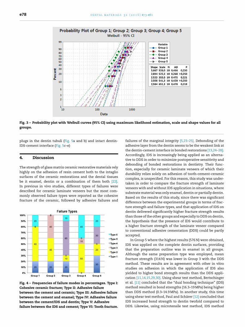
Weibull distribution presented higher shape (0) for Group 1(3.67), than those of other groups (2.51–2.89) (Fig. 3).
Failure types were predominantly adhesive failure betweenthe cement and the laminate veneer in Groups 1, 2, 4 whereasGroup 3 presented more often complete adhesive failuresbetween the cement and dentin (Fig. 4). In Group 5 with total
IDS and cement with or without ceramic fracture attached onthe tooth. SEM images clearly showed detachment of cementfrom dentin surfaces in Type IV failure types with cement
(Newton) of experimental groups, minimum, maximum 1.
Maximum Confidence interval
Lower bound Upper bound
645 358.9 586.4768 332.2 597.6637 216.4 412.5900 323.6 632.7
1006 393.8 757.7
e78 d e n t a l m a t e r i a l s 3 2 ( 2 0 1 6 ) e73–e81
Fig. 3 – Probability plot with Weibull curves (95% CI) using maximum likelihood estimation, scale and shape values for all
groups.plugs in the dentin tubuli (Fig. 5a and b) and intact dentin-IDS-cement interface (Fig. 5c–e)
4. Discussion
The strength of glass matrix ceramic restorative materials relyhighly on the adhesion of resin cement both to the intagliosurfaces of the ceramic restorations and the dental tissuesbe it enamel, dentin or a combination of them both [22].In previous in vivo studies, different types of failures were
described for ceramic laminate veneers but the most com-monly observed failure types were reported as the cohesivefracture of the ceramic, followed by adhesive failures andFig. 4 – Frequencies of failure modes in percentages. Type I:Cohesive ceramic fracture; Type II: Adhesive failurebetween the cement and ceramic; Type III: Adhesive failurebetween the cement and enamel; Type IV: Adhesive failurebetween the cement/IDS and dentin; Type V: Adhesivefailure between the IDS and cement; Type VI: Tooth fracture.
failures of the marginal integrity [5,23–25]. Debonding of theadhesive layer from the dentin seems to be the weakest link atthe dentin-cement interface in bonded restorations [13,24–28].Accordingly, IDS is increasingly being applied as an alterna-tive to DDS in order to minimize postoperative sensitivity anddebonding of bonded restorations in dentistry. Their func-tion, especially for ceramic laminate veneers of which theirdurability relies solely on adhesion of tooth-cement-ceramiccomplex, is unspecified. For this reason, this study was under-taken in order to compare the fracture strength of laminateveneers with and without IDS application in situations, wheresubstrate material was only enamel, dentin or partially dentin.Based on the results of this study, since there was significantdifference between the experimental groups in terms of frac-ture strength and failure types, and that application of IDS ondentin delivered significantly higher fracture strength resultsthan those of the other groups and especially to DDS on dentin,the hypothesis that the presence of IDS would contribute toa higher fracture strength of the laminate veneer comparedto conventional adhesive cementation (DDS) could be partlyaccepted.
In Group 5 where the highest results (576 N) were obtained,IDS was applied on the complete dentin surfaces, providingthat the preparation outline was in enamel in all groups.Although the same preparation type was employed, meanfracture strength (314 N) was lower in Group 3 with the DDSmethod. These results are in agreement with other in vitrostudies on adhesion in which the application of IDS alsoyielded to higher bond strength results than the DDS appli-cation [11,14,15,29,30]. Using shear test method, Bertschingeret al. [11] concluded that the “dual bonding technique” (IDS)method resulted in bond strengths (16.3–19 MPa) being higher
than DDS method (0.3–14.9 MPa). In another study, this timeusing shear test method, Paul and Schärer [12] concluded thatIDS increased bond strength to dentin twofold compared toDDS. Likewise, using microtensile test method, IDS method
d e n t a l m a t e r i a l s 3 2 ( 2 0 1 6 ) e73–e81 e79
Fig. 5 – (a) Typical Type IV failure from a specimen in Group 3 and (b) the corresponding SEM image (5000×). Note cementplugs in the dentin tubuli after detachment of the laminate veneer; (c) Typical Type IV failure from a specimen in Group 5with total exposure of dentin and sealing with IDS, and (d) the corresponding SEM image (5000×) with or (e) withoutceramic fracture attached on the tooth. Note the intact dentin-IDS-cement interface (K: Ceramic, C: Cement and IDS, D:D
wFptscp
tisasas
entin, I: IDS) (500×).
ith the same adhesive resin used in this study (OptibondL) resulted in significantly higher results (59.1–66.6 MPa) com-ared to DDS (11.6 MPa) [14]. Higher bond strength results byhe application of IDS may be explained by the optimal adhe-ion to freshly prepared dentin that is not exposed to anyontamination through the temporary cement used for therovisional restoration [10].
In this study, it was also of interest to investigate whetherhe amount of exposed dentin on the substrate surface had anmpact on the fracture strength of laminate veneers. Formertudies showed that removal of only 0.5 mm in the cervical
rea could already result dentin exposure [1,2]. Dentin expo-ure, is mostly seen in the cervical area of the preparationnd contains often a quarter of the entire labial preparationurface [3]. Thus, it was suggested that preparations havingenamel between 50 to 70% on the entire tooth surface wouldensure durable adhesion [3,31]. Built on this statement, inour study, 1/4 dentin exposure was considered as the criti-cal amount of dentin during preparations. Yet, the amount fordentin did not significantly affect the mean fracture strength,therefore the second hypothesis could be partially accepted. Inthis in vitro study, the cervical margin ended in enamel. Thiswill probably have an influence on the fracture strength com-pared when the cervical preparation margin ended in dentin.
It is not an easy task to state whether the obtained resultsin this study would sustain chewing forces. The average biteforces in human range between 20 and 1000 N but during
actual chewing, the forces do not exceed 270 N [32]. Fur-thermore, the forces in the anterior region of the mouthare reported to be less than in the posterior region ranging
l s 3
r
e80 d e n t a l m a t e r i a
between 155 and 200 N. The fracture strengths in this studyvaried between minimum 200 and maximum 1006 N. Conse-quently, the obtained results fall under this range. However,patients with signs of bruxism express higher masticatoryforces. Thus, large excessive preparations in dentin cannotbe recommended without IDS, as these forces were signifi-cantly lower (314 ± 137 N) than the ones with IDS (576 ± 254 N).Nonetheless, considering the Weilbull parameters, character-istics of adhesion still seems to be the most reliable whenceramic laminate veneers were bonded onto surfaces entirelyin enamel supported by the lower variability of data comparedto dentin exposure with varying amounts with and withoutIDS or DDS. This aspect needs to be verified in higher numberof specimens in future studies.
In addition to fracture strength results, analysis of the fail-ures also provide important information. While in Group 3 inwhich DDS was applied on the preparation entirely in dentinshowed mainly adhesive failures between cement and dentin,in Group 5 where IDS was applied on the preparation entirelyin dentin, showed less adhesive failures. In other words, theuse of IDS layer decreased the amount of adhesive failures atthe cement–dentin interface. Similarly, according to SEM find-ings, cement–dentin interface was the weakest link even inthe IDS applied specimens.
The adhesive failures from dentin accompanied with lowfracture strength results such as in Group 3 can be explainedby the theory that during the application of the compositecement and veneers, the pressure could have collapsed thecollagen network [14,33]. The polymerized adhesive resin (IDS)of Group 5 with full infiltration into the hybrid layer couldhave prevented the collapse of the dentinal collagen structure[13,14,33,34]. Furthermore, water sorption could also decreaseadhesive strength, as improper infiltration of the collagennetwork by the adhesive resin would result in hydrolyti-cal degradation and decrease resin–resin adhesion [35]. Mostfailures of ceramic laminate veneers were observed at theadhesive interface of the substrate where the highest tensileforces are observed [36]. Due to placement of the load cell atthe incisal area, stress concentration was concentrated at theinterface but when adhesive strength is sufficient, the failureoccurs cohesive in the ceramic itself. Report of clinical failuresand their location should verify these findings.
In this study, an interaction was observed between the IDSlayer and the provisional veneer material (Protemp). The pro-visional veneers placed on the IDS applied groups were moredifficult to remove compared with the control and DDS appliedgroups. The presence of flaws in the IDS layer of all speci-mens was analyzed with the aid of microscopy and IDS layerwas found to be still intact after the removal of the provi-sional veneers. In fact, IDS was isolated using glycerine gel.This aspect has not been studied in the dental literature butmost probably, polymerization inhibited layer of IDS layer withthe glycerine and cleaning with pumice was not sufficient.Thus, air-blocking with glycerine gel did not eliminate theoxygen inhibition layer completely [37]. The interaction anddifficult removal of the provisional could be attributed to this
phenomenon [38].All specimens in this study were aged by means of ther-mocycling. Due to this aging process, the interface betweenthe composite matrix and the silica coated inorganic fillers
2 ( 2 0 1 6 ) e73–e81
were expected to hydrolytically degrade mainly at the adhe-sive interface [35]. This aging method was performed to mimicthe oral conditions with intraoral temperature alterations byintake of food and beverages. This method has been used inmany in vitro studies, however published reports on thermo-cycling are contradictory. In a meta-analysis thermocyclingshowed no significant effect on mean shear bond strengthbetween 9.6 (no thermocycling) and 10.3 MPa (with thermo-cycling) [39]. Thus, the guidelines of ISO requiring 500 cyclesmay not be sufficient to have an aging especially in speci-mens prepared for macroshear bond tests [40,41]. This couldapply also to the large area in bonded laminate veneers. How-ever, it has to be emphasized that in this study internal crackswere observed in the ceramic in two specimens after thermo-cycling, indicating that some kind of aging took place in theceramic. Since there was no delamination or cohesive frac-tures both specimens were not excluded in the fracture test.The internal crack lines due to thermocycling did not showinfluence the on ultimate strength of these specimens. How-ever, the effect of aging parameters at longer durations on thelong-term stability of IDS and DDS should be studied in futurestudies.
5. Conclusions
From this study, the following could be concluded:
1. When ceramic laminate veneers are bonded to large sur-faces of exposed dentin, application of an immediatedentin sealing improves the adhesion and thereby the frac-ture strength of veneers. Small areas of dentin exposureless than 1/4 of the bonding surface did not benefit fromIDS application.
2. Considering Weilbull parameters, characteristics of adhe-sion seems to be the most reliable when ceramic laminateveneers are bonded onto surfaces entirely in enamel com-pared to dentin exposure with varying amounts with andwithout IDS or DDS.
Acknowledgements
The authors acknowledge Mr. Stephan van der Made, Kwali-dent Dental Laboratory, Beilen, The Netherlands, for hismeticulous work in fabricating the ceramic laminate veneers,and extend their gratitude to Ivoclar Vivadent, Schaan, Liech-tenstein and Kerr, Orange, CA, USA for generous provision ofsome of the materials used in this study.
e f e r e n c e s
[1] Ferrari M, Patroni S, Balleri P. Measurement of enamelthickness in relation to reduction for etched laminateveneers. Int J Periodont Rest Dent 1992;12:407–13.
[2] Nattress BR, Youngson CC, Patterson CJ, Martin DM, Ralph JP.
An in vitro assessment of tooth preparation for porcelainveneer restorations. J Dent 1995;23:165–70.[3] Cherukara GP, Davis GR, Seymour KG, Zou L,Samarawickrama DY. Dentin exposure in tooth preparations
3 2
d e n t a l m a t e r i a l sfor porcelain veneers: a pilot study. J Prosthet Dent2005;94:414–20.
[4] Chun YH, Raffelt C, Pfeiffer H, Bizhang M, Saul G, Blunck U,Roulet JF. Restoring strength of incisors with veneers andfull ceramic crowns. J Adhes Dent 2010;12:45–54.
[5] Friedman MJ. A 15-year review of porcelain veneer failure-aclinician’s observations. Compend Contin Educ Dent1998;19:625–8.
[6] Dumfahrt H, Schäffer H. Porcelain laminate veneers. Aretrospective evaluation after 1 to 10 years of service: PartII-clinical results. Int J Prosthodont 2000;13:9–18.
[7] Peumans M, Munck de J, Fieuws S, Lambrechts P, Vanherle G,Van Meerbeek B. A prospective ten-year clinical trial ofporcelain veneers. J Adhes Dent 2004;6:65–76.
[8] Guess PC, Stappert FJ. Midterm results of a 5-yearprospective clinical investigation of extended ceramicveneers. Dent Mater 2008;24:804–13.
[9] Burke FJ. Survival rates for porcelain laminate veneers withspecial reference to the effect of preparation in dentin: aliterature review. J Esthet Restor Dent 2012;24:257–65.
[10] Pashley EL, Comer RW, Simpson MD, Horner JA, Pashley DH,Caughman WF. Dentin permeability: sealing the dentin incrown preparations. Oper Dent 1992;17:13–20.
[11] Bertschinger C, Paul SJ, Luthy H, Scharer P. Dual applicationof dentin bonding agents: effect on bond strength. Am JDent 1996;9:115–9.
[12] Paul SJ, Scharer P. The dual bonding technique: a modifiedmethod to improve adhesive luting procedures. Int JPeriodont Restor Dent 1997;17:536–45.
[13] Magne P, Douglas WH. Porcelain veneers: dentin bondingoptimization and biomimetic recovery of the crown. Int JProsthodont 1999;12:111–21.
[14] Magne P, So WS, Cascione D. Immediate dentin sealingsupports delayed restoration placement. J Prosthet Dent2007;98:166–74.
[15] Lee JI, Park SH. The effect of three variables on shear bondstrength when luting a resin inlay to dentin. Oper Dent2009;34:288–92.
[16] Duarte Jr S, de Freitas CR, Saad JR, Sadan A. The effect ofimmediate dentin sealing on the marginal adaptation andbond strengths of total-etch and self-etch adhesives. JProsthet Dent 2009;102:1–9.
[17] Reis A, Rocha de Oliveira Carrilho M, Schroeder M, TancredoLL, Loguercio AD. The influence of storage time and cuttingspeed on microtensile bond strength. J Adhes Dent2004;6:7–11.
[18] Magne P. Immediate dentin sealing: a fundamentalprocedure for indirect bonded restorations. J Esthet RestorDent 2005;17:144–54.
[19] Dietschi D, Monasevic M, Krejci I, Davidson C. Marginal andinternal adaptation of class II restorations after immediateor delayed composite placement. J Dent 2002;30:259–69.
[20] Gresnigt MMM, Özcan M, Kalk W, Galhano G. Effect of staticand cyclic loading on ceramic laminate veneers adhered toteeth with and without aged composite restorations. JAdhes Dent 2011;13:569–77.
[21] Gresnigt MMM, Kalk W, Özcan M. Clinical longevity ofceramic laminate veneers bonded to teeth with and withoutexisting composite restorations up to 40 months. Clin Oral
Invest 2013;17:823–32.[22] Peumans M, Van Meerbeek B, Lambrechts P, Vanherle G.Porcelain veneers: a review of the literature. J Dent2000;28:163–77.
( 2 0 1 6 ) e73–e81 e81
[23] Friedman M. Multiple potential of etched porcelain laminateveneers. J Am Dent Assoc 1987:83E–7E.
[24] Peumans M, De Munck J, Fieuws S, Lambrechts P, VanherleG, Van Meerbeek B. A prospective ten-year clinical trial ofporcelain veneers. J Adhes Dent 2004;6:65–76.
[25] Beier US, Kapferer I, Burtscher D, Dumfahrt H. Clinicalperformance of porcelain laminate veneers for up to 20years. Int J Prosthodont 2012;25:79–85.
[26] Van Meerbeek B, Peumans M, Gladys S, Braem M,Lambrechts P, Vanherle G. Three-year clinical effectivenessof four total-etch dentinal adhesive systems in cervicallesions. Quintessence Int 1996;27:775–84.
[27] Van Meerbeek B, Perdigao J, Lambrechts P, Vanherle G. Theclinical performance of adhesives. J Dent 1998;26:1–20.
[28] De Munck J, Van Landuyt K, Peumans M, Poitevin A,Lambrechts P, Braem M, Van Meerbeek B. A critical review ofthe durability of adhesion to tooth tissue: methods andresults. J Dent Res 2005;84:118–32.
[29] Cagidiaco MC, Ferrari M, Garberoglio R, Davidson CL. Dentincontamination protection after mechanical preparation forveneering. Am J Dent 1996;9:57–60.
[30] Magne P, Kim TH, Cascione D, Donovan TE. Immediatedentin sealing improves bond strength of indirectrestorations. J Prosthet Dent 2005;9:511–9.
[31] Chiche GJ, Pinault A. Esthetics of anterior fixedprosthodontics Chicago. Quintessence Publishing Co.; 1994.p. 20–4.
[32] Naeije M, Loon LAJ. Craniomandibulaire functie endisfunctie. Bohn Stafleu Van Loghum; 1998. p. 39–56.
[33] Breschi L, Mazzoni A, Ruggeri A, Cadenaro M, Di Lenarda R,De Stefano Dorigo E. Dental adhesion review: aging andstability of the bonded interface. Dent Mater 2008;24:90–101.
[34] Dietschi D, De Siebenthal G, Neveu-Rosenstand L, Holz J.Influence of the restorative technique and new adhesives onthe dentin marginal seal and adaptation of resin compositeClass II restorations: an in vitro evaluation. Quintessence Int1995;26:717–27.
[35] Özcan M, Barbosa SH, Melo RM, Galhano GA, Bottino MA.Effect of surface conditioning methods on the microtensilebond strength of resin composite to composite after agingconditions. Dent Mater 2007;23:1276–82.
[36] Magne P, Kwon KR, Belser UC, Hodges JS, Douglas WH. Crackpropensity of porcelain laminate veneers: a simulatedoperatory evaluation. J Prosthet Dent 1999;81:327–34.
[37] Magne P, Nielsen B. Interactions between impressionmaterials and immediate dentin sealing. J Prosthet Dent2009;102:298–305.
[38] Endo T, Finger WJ, Hoffmann M, Kanehira M, Komatsu M.The role of oxygen inhibition of a self-etch adhesive onself-cure resin composite bonding. Am J Dent2007;20:157–60.
[39] Leloup G, D’Hoore W, Bouter D, Degrange M, Vreven J.Meta-analytical review of factors involved in dentinadherence. J Dent Res 2001;80:1605–14.
[40] Gale MS, Darvell BW. Thermal cycling procedures forlaboratory testing of dental restorations. J Dent1999;27:89–99.
[41] De Munck J, Mine A, Poitevin A, Ende A, Cardoso MV, VanLanduyt KL, Peumans M, Van Meerbeek B. Meta-analyticalreview of parameters involved in dentin bonding. J Dent Res2012;91:351–7.