effect of microneedle geometry and supporting substrate on
TRANSCRIPT

RESEARCH ARTICLE – Pharmaceutics, Drug Delivery, and Pharmaceutical Technology
Effect of Microneedle Geometry and Supporting Substrateon Microneedle Array Penetration into Skin
JASPREET SINGH KOCHHAR,1 TEN CHEER QUEK,2 WEI JUN SOON,2 JAEWOONG CHOI,2 SHUI ZOU,3 LIFENG KANG1
1Department of Pharmacy, National University of Singapore, Singapore, 1175432NUS High School of Mathematics and Science, Singapore, 1299573Department of Chemistry, National University of Singapore, Singapore, 117543
Received 9 July 2013; revised 9 August 2013; accepted 15 August 2013
Published online in Wiley Online Library (wileyonlinelibrary.com). DOI 10.1002/jps.23724
ABSTRACT: Microneedles are being fast recognized as a useful alternative to injections in delivering drugs, vaccines, and cosmeticstransdermally. Owing to skin’s inherent elastic properties, microneedles require an optimal geometry for skin penetration. In vitro studies,using rat skin to characterize microneedle penetration in vivo, require substrates with suitable mechanical properties to mimic humanskin’s subcutaneous tissues. We tested the effect of these two parameters on microneedle penetration. Geometry in terms of center-to-center spacing of needles was investigated for its effect on skin penetration, when placed on substrates of different hardness. Both hard(clay) and soft (polydimethylsiloxane, PDMS) substrates underneath rat skin and full-thickness pig skin were used as animal models andhuman skins were used as references. It was observed that there was an increase in percentage penetration with an increase in needlespacing. Microneedle penetration with PDMS as a support under stretched rat skin correlated better with that on full-thickness humanskin, while penetration observed was higher when clay was used as a substrate. We showed optimal geometries for efficient penetrationtogether with recommendation for a substrate that could better mimic the mechanical properties of human subcutaneous tissues, whenusing microneedles fabricated from poly(ethylene glycol)-based materials. C© 2013 Wiley Periodicals, Inc. and the American PharmacistsAssociation J Pharm SciKeywords: microneedles; microneedle geometry; skin substrate stiffness; polydimethylsiloxane
INTRODUCTION
Transdermal drug delivery systems offer numerous benefitsover the delivery of drugs using parenteral or oral route, as theyare painless and obviate the first-pass effects and possible enzy-matic degradation through the oral route.1,2 Microneedles havebeen used as an effective means of enhancing skin permeationwith over 350 published studies highlighting their utility in de-livering different low molecular weight drugs, biotherapeuticsand vaccines, including published human studies with a num-ber of small molecules, protein drugs, and vaccines.3 Micronee-dles have been fabricated from a range of materials, using dif-ferent techniques,3,4 forming arrays of different dimensions,variable shapes, and geometries. This diversity in microneedledesign has called for an investigation of the effect of micronee-dle design on their ability to enhance transdermal permeation.A number of studies have reported the effect of microneedleshape,5 tip diameter,6 length,7 force of insertion,8 velocity ofinsertion,9 and density of microneedle arrays,9 etc. These fac-tors, together with materials used for microneedle fabricationand force applied to an array will determine microneedle in-sertion and fracture properties. Another important parameterto be considered is the skin’s inherent elasticity, which wouldpose a challenge to microneedle penetration. Previous studieshave reported folding of skin around microneedle arrays, either
Correspondence to: Lifeng Kang (Telephone: +65-6516-7519; Fax: +65-6779-1554; E-mail: [email protected])
Jaspreet Singh Kochhar and Ten Cheer Quek contributed equally to thiswork.
This article contains supplementary material available from the authors uponrequest or via the Internet at http://onlinelibrary.wiley.com/.
Journal of Pharmaceutical SciencesC© 2013 Wiley Periodicals, Inc. and the American Pharmacists Association
preventing penetration or allowing for suboptimal penetration,especially for microneedles of lengths shorter than 300 :m.10,11
The depth of microneedle penetration has been correlated to theforce of application,12 which in turn counters skin’s elasticity.
Because microneedles are usually fabricated as arrays, it isof particular interest to study how force is distributed acrossa microneedle array, with density of microneedles influencingthe penetration. A number of simulation studies have been re-ported in the literature, modeling breaching of soft tissues onneedle application.13,14 Modeling studies on skin tissues havealso been carried out to better mimic microneedle insertionproperties. Olatunji et al. recently published their findings onthe effect of interspacing between microneedles and applicationforce, reporting that the force required for insertion was depen-dant on the interspacing between microneedles, irrespectiveof microneedles on an array.15 The study included simulationexperiments to characterize microneedle bending force, buck-ling force, and insertion force against the tip interspacing ofmicroneedles. While they demonstrated that there should bean optimal number of microneedles on an array to enhancedrug delivery, too many needles will form a “bed of nails”, re-ducing penetration and efficiency of microneedle penetration.Apart from this, a few other simulation studies have also beenconducted for analyzing the effect of microneedle geometry onskin penetration.16–18 However, a comprehensive in vitro studyon skin models, coanalyzing various aspects of microneedle ge-ometry, including base diameter, tip diameter, center-to-centerspacing, and insertion force is still lacking in literature.
On the other hand, in vitro experiments using micronee-dles have been carried out using a variety of animal skinmodels. To mimic the mechanical properties of human skin’sinherent subcutaneous fatty tissues, researchers have used
Kochhar et al., JOURNAL OF PHARMACEUTICAL SCIENCES 1
2 RESEARCH ARTICLE – Pharmaceutics, Drug Delivery, and Pharmaceutical Technology
clay,19 layers of Kimwipes,20 dental wax,15 soft sponge,7 andfilter paper soaked with phosphate-buffered saline (PBS) lay-ered above a corkboard.21 While these materials widely varyin hardness, none of them has been demonstrated to possessmechanical properties similar to human subcutaneous tissuesand hence, the extent of microneedle penetration observed mayvary greatly when different substrates are used. This requiresthe careful calibration of penetration properties of micronee-dles under substrates of varying stiffness and comparing themto full-thickness skin models from humans to derive better cor-relation.
In this study, we address the two lacunas highlighetd earlier.Microneedle arrays of various base and tip diameters and vary-ing spacings were fabricated and tested for their extent of pene-tration on excised rat skin model on two different substrates, ahard (clay) and a soft (polydimethylsiloxane, PDMS) substratewere used to study the influence of substrate stiffness on mi-croneedle penetration. These substrates were then compared tomicroneedle penetration on full-thickness pig and human skinsamples. To our knowledge, this is the first study elucidatingthe effects of microneedle geometry as well as substrate stiff-ness on the extent of microneedle penetration through the skin,and hence could serve as a platform for the design of optimizedmicroneedle array based products as well as testing platforms.
EXPERIMENTAL
Materials
Poly(ethylene glycol) diacrylate, PEGDA (Mn = 258), 2-hydroxy-2-methyl-propiophenone (HMP), lidocaine hydrochlo-ride (LH), and trypan blue were purchased from Sigma–Aldrich(St Louis, MO, USA). All other materials were of the reagentgrade and were used as received. PDMS (Sylgard 184 SiliconeElastomer Kit) was obtained from Dow Corning (Midland, MI,USA).
Fabrication of Microneedles
Microneedles were fabricated using a photolithography-basedmethod involving UV light based photopolymerization, previ-ously developed in our lab.19 Briefly, prepolymer solution (amixture of PEGDA with 0.5% v/v HMP) was filled into a cav-ity formed by supporting glass coverslips on a base glass slide.This prepolymer solution is then exposed to high power UVlight source (EXFO OmniCure R© S200-XL, Canada) with a UVfilter (320–500 nm). This resulted in the fabrication of a thinfilm termed as “microneedle backing layer”. Subsequently, thisfilm was placed on a similar substrate and exposed to UV lightthrough a patterned photomask, forming “microneedle shafts”.Photomasks (Infinite Graphics, Singapore) of different patternswere prepared to govern the geometry of microneedles withbase diameter of 200, 300, and 400 :m and a center-to-centerspacing varying between 2× and 5× base diameter. Fabricatedmicroneedles were washed with purified water from MilliporeDirect-Q R© (Molsheim, France) to remove the excess residualprepolymer solution and let to air dry before being removedfrom the coverslip and used for experiments. Microneedleswere imaged using Nikon SMZ 1500 stereomicroscope (Nikon,Japan), to quantify the microneedle geometric characteristics.The base-to-tip ratio was defined as base diameter to tip diam-eter ratio.
Microneedle Penetration in Rat Skin Using a Hard Substrate (Clay)
Rat abdominal skins were obtained from rat cadavers from theNational University of Singapore (NUS) Animal Center. Hairon the skin was first removed using a pair of scissors. To ob-tain a uniform flat piece of skin for penetration studies, sub-cutaneous fat was evenly excised with scissors and scalpel andthereafter fixed on a stable platform prior to penetration ex-periments. Two different substrates were used. As a model forhard substrate, paper clay (SESCO Art and Craft, Singapore)was dried overnight and used as a tissue-like mechanical sup-port underneath the stretched and defatted rat skin. The claysupport measured 7 mm in thickness. Fabricated microneedleswere placed on the skin surface and force was applied with aforce gauge (Dillon GL-500, Fairmont, MN, USA) for a minuteto determine the force required for microneedle insertion. Thepoint of penetration was identified as a sudden drop in the forceapplied (and therefore skin resistance) and was recorded digi-tally on the force gauge; the force at the point of penetration wasrecorded for calculation of force per needle at the point of pene-tration. Force per needle was calculated by dividing the force atpenetration by the total number of needles on an array. Becausemicroneedle arrays of different needle spacing were fabricatedand array area was kept consistent at 1.44 cm2, force per needlevaried between different arrays.
The area of insertion was then immediately stained witha hydrophobic dye, trypan blue (0.4% solution) for 5–10 min.Trypan blue specifically stains sites of stratum corneum per-foration. The excess stain was washed away with purified wa-ter followed by 70% ethanol solution, leaving blue circles onthe skin demonstrating stratum corneum perforation. Excesshair on the skin was removed with hair removal cream Veet(Reckitt Benckiser, Poland) or a manual razor. The premise be-hind using the hair removal cream after trypan blue stainingwas to potentially avoid the damage to the skin and exposureof hydrophobic layers, which are then stained by trypan blue.Subsequently, areas stained with the dye were viewed usingImage Soft hand-held microscope (Eikona, People’s Republic ofChina) and the number of perforations was quantified. Spotson the skin which appear in the shape of the microneedle arrayrelative to other spots were counted as penetration spots. Thesame procedure was repeated for needles of all base diametersand spacing. Penetration into rat skin was also confirmed byhistological sectioning of liquid nitrogen frozen skin samplesfollowed by haematoxylin and eosin staining.19 All animal ex-periments were approved by the Institutional Animal Care andUse Committee, NUS.
Microneedle Penetration in Rat Skin Using a Soft Substrate(PDMS)
A PDMS substrate was fabricated by casting a silicone elas-tomer base and a silicone elastomer curing agent in the ratioof 9:1 as per the manufacturer’s protocol. This mixture wasthen poured in the cover of a 100 × 20 mm petri dish (Greiner,Germany) and degassed in a vacuum chamber for 30 min fol-lowed by heat curing at 70◦C for 2 h. The fabricated solid filmmeasuring 6 mm in thickness was easily withdrawn from thepetri dish cover and used as a skin substrate. Rat skin wasplaced over this substrate and microneedle samples were sim-ilarly inserted by using a force gauge to determine the force ofinsertion; the percentage of microneedle insertion was deter-mined by trypan blue staining method.
Kochhar et al., JOURNAL OF PHARMACEUTICAL SCIENCES DOI 10.1002/jps.23724

RESEARCH ARTICLE – Pharmaceutics, Drug Delivery, and Pharmaceutical Technology 3
Microneedle Penetration in Full-Thickness Pig Ear Skin andHuman Skin
The microneedle arrays of each base diameter that attainedhighest penetration percentages in rat skin from both hardand soft substrates were then inserted into pig ear skin andhuman skin in a manner as described earlier. This is carriedout to ascertain the closeness of mechanical properties of bothsubstrates to full-thickness pig and human skin model. Pigskin was obtained from NUS Animal Center. Full-thickness ca-daver human skin was obtained from Science Care (Phoenix,AZ, USA). The use of human skin for this study has been ap-proved by the National University of Singapore InstitutionalReview Board (no. 13-167E). The human skin tissues were ex-cised from the back of a white, female cadaver, who died ofdebility at the age of 75 years. Similarly, post microneedle in-sertion, trypan blue staining method was used to ascertain thepercentage of microneedles on an array that successfully pene-trated the skin.
In Vitro Drug Release from Microneedles
Lidocaine hydrochloride was encapsulated in situ within boththe microneedle shafts and the backing layer of the most op-timized geometry of microneedles. This was achieved by dis-solving LH in the prepolymer solution at a concentration of3.47% w/w and 4.31% w/w. The fabricated microneedles werethen tested for in vitro release of LH from the microneedles.The microneedles were immersed in 15 mL of 1 × PBS in anincubator maintained at 37◦C. The release solutions from LHwere sampled at 1, 3, 6, 18, and 24 h. At each sampling point,all release solution was withdrawn and replaced with 15 mL offresh 1 × PBS. Release samples were stored at 4◦C until anal-ysis. In the analysis of LH, each sample was transferred to aquartz cuvette was and analyzed by absorbance measurementsat 254 nm with an UV–visible spectrophotometer (U-1900,Hitachi, Tokyo, Japan). Cumulative release and cumulativepercentage release were calculated.
In Vitro Permeation Assay with Horizontal Diffusion Cell
To study the ability of optimized geometry to enhance trans-dermal permeation, microneedle array encapsulated with 4.3%w/w LH was inserted into excised rat abdominal skin. Eachmicroneedle array contained 2.51 ± 0.229 mg of LH. The hor-izontal diffusion cell (TK-6H1, Shanghai Kai Kai Science andTechnology Company Ltd., People’s Republic of China) was runwith circulating water maintained at 37◦C for both donor andreceptor solutions to mimic the body temperature. 4.5 mL of1 × PBS with 0.005% w/v sodium azide was added to the re-ceptor compartment. The permeated solutions for LH were pe-riodically sampled for a period of 48 h. At each sampling point,1 mL of the receptor solution was withdrawn and replaced with1 mL of fresh 1×PBS with 0.005% w/v sodium azide.
The amount of LH permeated was determined by usingHitachi L2000 LaChrome Elite HPLC system with a Hyper-sil ODS C18 reverse column (ODS hypersil, Thermo Scientific;4.6 × 250 mm, 5 :m). The C18 reverse column and the sol-vents were maintained at ambient temperature. The mobilephase consisted of water and acetonitrile (30:70% v/v) contain-ing 5.5% triethylamine and the flow rate was set at 0.7 mL/min.The injection volume was 20 :L for each sampling and ultra-violet detection was performed at a wavelength of 254 nm.22
The standard LH solutions were prepared by series dilution.
Various standard curves were prepared to bracket all possibleconcentrations from the permeation studies. Under these con-ditions, the peak of LH appeared at 5.8 min. The permeationsamples were analyzed by comparing peaks with standardspeaks.
Statistical Analysis
Results were used to compute mean and standard deviation andall experiments performed were at least triplicates. Student’st test was used to analyze the statistical significance betweenspecific pairs of data. For analyzing multiple groups of data orstatistical differences, one-way analysis of variance was used.Results with p value of less than 0.05 were considered to bestatistically significant. PASW Statistics 18 (SPSS) softwarewas used for computing statistical results.
RESULTS
Geometric Properties of Microneedles
Microneedles of varying lengths, base diameters, and spacingcould be conveniently fabricated by changing the fabricationsetup and photomask patterns. It was observed that as thespacing was increased, a relative larger dose of UV light wasrequired to fabricate microneedles with sufficicent strengthfor subsequent penetration studies (Supporting Information,SI 1 in the Supplementary Material). Figures 1a–1c show themicroneedles of 200, 300, and 400 :m base diameter with acenter-to-center spacing of 6× base diameter. When fabricatedin scaffold of particluar spacer thickness,19 mcironeedle lengthfor different base diameters (200–400 :m) ranged between827.9 ± 7.3 and 873.3 ± 12.3 :m (p > 0.05), while the tip diam-eter increased proportionally with the base diameter, rangingbetween 131.3 ± 11.8 and 276.0 ± 26.0 :m (Fig. 1d). For allmicroneedles fabricated via this method, a constant base-to-tipratio of approximately 1.5 was observed across all base diam-eters (Fig. 1e). The fabricated mcironeedles also showed a rel-atively constant needle length across all base diameters and arelatively constant tip diameter amongst same base diameters(SI 2 and SI 3 in the Supplementary Material).
Microneedle Penetration in Rat Skin Using a Hard Substrate (Clay)
We observed a consistent increase in the percentage of mi-croneedles penetrating the skin with the increase in spacing,and the penetration percentage increased with the increase inmicroneedle base diameter [Figs. 2ai–2ci and SI 4 (in the Sup-plementary Material)]. For 200 :m base diameter, penetrationvaried between 5% and 44%, for 300 :m base diameter, pen-etration varied between 9% and 64%, and for 400 :m basediameter, it varied between 12% and 79%, with the increase inspacing between microneedles spread over an array of constantarea of 1.44 cm2.
To explore this further, force per needle was calculated bydividing the force applied on the microneedle array by the num-ber of microneedles on an array. As the number of microneedleson an array decreased with increased spacing, an incrementalforce per needle and higher percentage of penetration were ob-served (Figs. 2aii–2cii). Force per needle increased from 0.19 to1.33 N (200 :m base diameter), 0.24–4.46 (300 :m base diame-ter), and 0.84–7.17 N (400 :m base diameter) with the increasein spacing. The percentage of penetration was thus linearly cor-related to force per needle as observed in Figures 2aiii–2ciii.
DOI 10.1002/jps.23724 Kochhar et al., JOURNAL OF PHARMACEUTICAL SCIENCES

4 RESEARCH ARTICLE – Pharmaceutics, Drug Delivery, and Pharmaceutical Technology
Figure 1. (a–c) images of 200, 300, and 400 :m base diameter needles, respectively, with a center-to-center spacing of 6× base diameter.Relationship between base diameter and (d) tip diameter, and (e) base-to-tip ratio of needle which is the ratio of base diameter to tip diameterremained consistent with no significant differences between different base diameters (p > 0.05).
Histological sectioning of the skin confirmed microneedle pen-etration in skin (SI 6 in the Supplementary Material).
Microneedle Penetration in Rat Skin Using a Soft Substrate(PDMS)
In an attempt to characterize the effect of substrate stiffness, asofter substrate was used for rat skin for in vitro microneedlepenetration experiments. Although a similar trend of increasedpenetration with increased spacing and force per needle wasobserved across all geometries, the penetration percentage waslower than what was observed when clay was used as a sub-strate for rat skin. Penetration percentage ranged from 2% to5% for 200 :m base diameter, 1%–52% for 300 :m base diam-eter, and 9%–37% for 400 :m base diameter [Figs. 3ai–3ci andSI 5 (in the Supplementary Material)]. The penetration forceper needle increased from 0.07 to 0.37 N for 200 :m base diam-eter, 0.13–1.10 N for 300 :m base diameter, and 0.29–1.59 N for400 :m base diameter microneedles (Figs. 3aii–3cii). A similarcorrelation was observed between penetration percentage andforce per needle data (Figs. 3aiii –3ciii).
Microneedle Penetration in Full-Thickness Pig Ear Skin andHuman Skin
With different substrates under rat skin resulting in differentextents of penetration, we further explored to find the optimalsubstrate with close approximation of mechanical propertiesto full-thickness pig ear and human skin. This would enablethe use of a substrate that best estimates microneedle pene-tration in in vitro penetration experiments with microneedles.We chose the microneedle geometries with highest penetrationcapabilities from both substrates. Thus microneedles with basediameters of 200, 300, and 400 :m with center-to-center spac-ing of 1200, 1800, and 2400 :m, respectively, were inserted
on stretched samples of full-thickness pig ear and human skinusing a similar method used for rat skin with two differentsubstrates. Although it has been widely reported in literaturethat pig skin has similar mechanical properties to human skin,we observed that percentage of penetration of microneedles onhuman skin was slightly higher than on pig skin (Figs. 4 and5). On the other hand, because rat skin is most often used as asubstitute, we found that PDMS as a substrate under rat skinmore closely approximates the penetration in human skin ascompared to the use of clay as a substrate (Fig. 6). This indi-cates that PDMS has suitable biomechanical properties similarto subcutaneous tissues.
In Vitro Drug Release from Microneedles
Microneedles measuring 300 :m base diameter and 1800 :mcenter-to-center spacing were used for studying drug encap-sulation purposes. LH was used as a model drug compound.The release of LH in PBS over 24 h resulted in a 93.9% release(3.04 mg) for 3.47% w/w LH-containing microneedles and 91.3%release (3.77 mg) for 4.31% w/w LH-containing microneedles atthe end of 24 h (SI 7 in the Supplementary Material). An ini-tial burst release was observed for the first 3 h, with a gradualincrease over time and relatively constant release thereafteruntil 24 h.
In Vitro Permeation Assay with Horizontal Diffusion Cell
Microneedles containing 4.31% w/w of LH were tested fortheir ability to enhance permeation across rat skin as com-pared to a propylene glycol solution containing 4.31% w/wLH. 320.8 ± 147.5 :g of LH permeated through rat skin af-ter a period of 48 h, with a 78.2-fold increase in permeationamount over control (SI 8 in the Supplementary Material). Mi-croneedles delivered a consistently higher amount of LH, while
Kochhar et al., JOURNAL OF PHARMACEUTICAL SCIENCES DOI 10.1002/jps.23724

RESEARCH ARTICLE – Pharmaceutics, Drug Delivery, and Pharmaceutical Technology 5
Figure 2. The first row shows percentage penetration for needles against centre-to-centre spacing of 2×/3×/4×/5×/6× of base diameters forbase diameters of (ai) 200, (bi) 300, and (ci) 400 :m. The second row shows graphs of needle penetration force against centre-to-centre spacingof 2×/3×/4×/5×/6× for base diameters of (aii) 200, (bii) 300, and (cii) 400 :m. The third row shows linear regressions of percentage penetrationagainst force per needle for base diameters of (aiii) 200, (biii) 300, and (ciii) 400 :m.
passive diffusion resulted in a much lower permeation of thedrug. While the amounts released from microneedles in PBSwere much higher, due to inherent barrier properties of skinand interaction of drugs with skin components, the permeatedamounts were lower.
DISCUSSION
Mechanically, skin behaves like a viscoelastic tissue,23 and be-haves more as an elastic tissue with light applied loads.24 Mi-croneedle geometry is critical to ensure efficent penetrationthrough skin. It has been observed previously that microneedleinsertion in skin led to skin retraction and a 1080 :m inser-tion in skin using single microneedles caused indentations onthe skin with only 100–300 :m penetrating the skin.10 Whilethis effect was observed for single microneedles, the situationis even more complex when using a microneedle array. At thesame time, geometrical dimensions of microneedle shafts playa critical role in efficient penetration through the skin. These
factors motivated us to study microneedle geometric propertiesin detail.
Using our photolithographical approach,19 we fabricated mi-croneedles possessing a tapered vertical profile, which could beexplained through the loss of energy as the UV light travelsa distance through the setup. The photopolymerization is gov-erned by the inverse square law of light, where the intensityof photons emitted from the light source is inversely related tothe square of the distance from the light source.25 However, weobserved that irrespective of the base diameter of microneedles,the decrease in intensity and hence the tapering of micronee-dles shafts remained consistent, giving a uniform base-to-tipratio (Fig. 1).
In this study, microneedle geometry in terms of varyingcenter-to-center spacing has been shown to be a crucial fac-tor in the penetration profile into skin. An increase in force perneedle was seen with increased center-to-center spacing of nee-dles for all base diameters; suggesting that needles with highercenter-to-center spacing are able to penetrate the skin better.This correlates well with the findings by Olatunji et al.15 where
DOI 10.1002/jps.23724 Kochhar et al., JOURNAL OF PHARMACEUTICAL SCIENCES

6 RESEARCH ARTICLE – Pharmaceutics, Drug Delivery, and Pharmaceutical Technology
Figure 3. The first row shows percentage penetration for needles against centre-to-centre spacing of 2×/3×/4×/5×/6× of base diameters forbase diameters of (ai) 200, (bi) 300, (ci) 400 :m. The second row shows graphs of needle penetration force against centre-to-centre spacing of2×/3×/4×/5×/6× for base diameters of (aii) 200, (bii) 300, and (cii) 400 :m. The third row shows linear regressions of percentage penetrationagainst force per needle for base diameters of (aiii) 200, (biii) 300, and (ciii) 400 :m.
an increase in needle interspacing results in lower resistancefor penetration in skin, which could result in higher needlepenetration rates, and thus, a greater quantity of drug deliv-ery. Apart from center-to-center spacing, 300 and 400 :m basediameter needles showed the most promising penetration pro-files with high penetration rates, while 200 :m base diameterneedles had a low penetration rate, which could be attributed toits thin needle profile that results in a weaker structure whichbuckles more easily upon insertion onto the surface of the skinthan for needles of larger base diameters. Microneedles with300 :m base diameter, 1800 :m spacing, and 400 :m base di-ameter, 2400 :m spacing gave consistent and relatively highpercentage of microneedles penetrating the skin. In addition,because sharper needles have a larger length-to-base aspectratio in our study, needles with a smaller length-to-base aspectratio penetrate skin better. (In this study, all needles have thesame length and a sharper needle has a smaller base diameter).
It has been previously reported that decrease in the tip diam-eter enhanced microneedle penetration irrespcetive of center-to-center spacing.26,27 This is true for microneedles fabricatedfrom robust materials like silicon and metals. However, for poly-meric microneedles, it has been reported that decreased tip
diamater led to microneedle shaft weakness and easy fracture,which may potentially impact microneedle insertion.28 In ourstudy, we also observed that for lower tip diameters, micronee-dle shafts were weaker and hence lower extent of penetrationwas observed for the microneedles with 200 :m base diameter(Figs. 2 and 3).
At the same time, our study highlights the effect of mi-croneedles made from Poly(ethylene glycol) -based material,and hence the mechanical properties of harder materials mayinfluence microneedle penetration differently. While hardermaterials like silicon and metal are not influenced much by mi-croneedle geometry, polymeric microneedles require a carefulcalibration of geometrical properties to enhance their penetra-tion efficacy.
The availability of human skin samples for research pur-poses has been limited, prompting the use of rat skin as a sub-stitute for human skin. As these human skin suurogates do notmimic the mechanical properties, it is essential to develop su-urogate models to be used with rat skin. To our knowledge, thisis the first such study to investigate the extent to which certainsubstrates placed under excised rat skin mimic the mechanicaltissue support of human skin.
Kochhar et al., JOURNAL OF PHARMACEUTICAL SCIENCES DOI 10.1002/jps.23724
RESEARCH ARTICLE – Pharmaceutics, Drug Delivery, and Pharmaceutical Technology 7
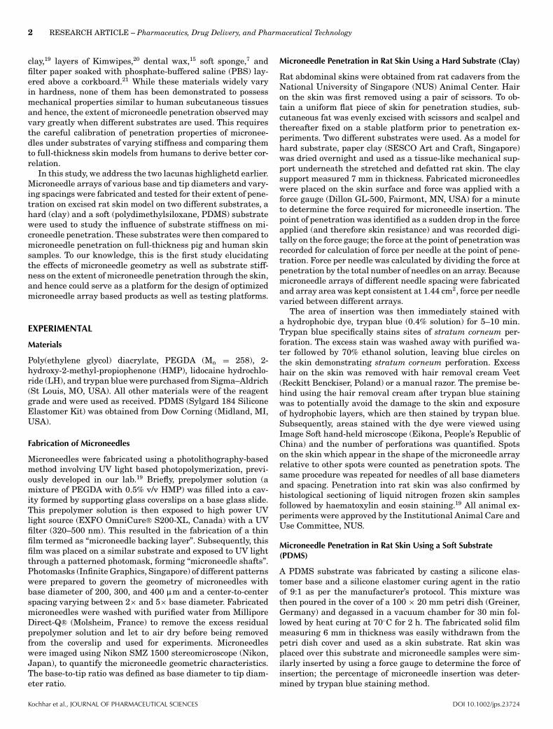
Figure 4. Microneedle penetration performed on pig ear skin for (a)200 :m base diameter, 1200 :m center-to-center spacing, (b) 300 :mbase diameter, 1800 :m center-to-center spacing, and (c) 400 :m basediameter, 2400 :m center-to-center spacing microneedles. (d) the per-centage of needles penetrated into pig ear skin for each needle basediameter. Triplicates were used for each base diameter.
Due to factors such as different mechanical properties andelasticity of various layers of skin as well as the force of ap-plication, microneedle arrays may penetrate to different extentduring in vitro studies. We observed the hard substrate (clay)used resulted in an overestimation of the penetration abilityof the microneedles (Fig. 2), while the soft substrate (PDMS)(Fig. 3) and full-thickness pig skin (Fig. 4) gave a slight un-
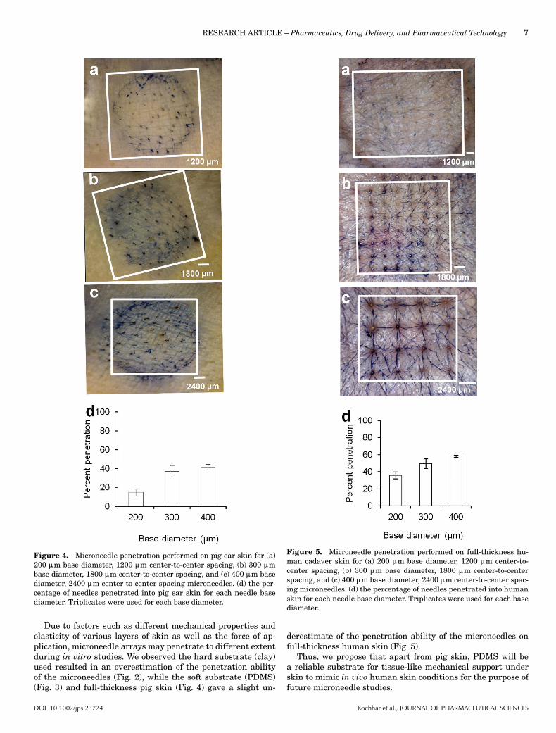
Figure 5. Microneedle penetration performed on full-thickness hu-man cadaver skin for (a) 200 :m base diameter, 1200 :m center-to-center spacing, (b) 300 :m base diameter, 1800 :m center-to-centerspacing, and (c) 400 :m base diameter, 2400 :m center-to-center spac-ing microneedles. (d) the percentage of needles penetrated into humanskin for each needle base diameter. Triplicates were used for each basediameter.
derestimate of the penetration ability of the microneedles onfull-thickness human skin (Fig. 5).
Thus, we propose that apart from pig skin, PDMS will bea reliable substrate for tissue-like mechanical support underskin to mimic in vivo human skin conditions for the purpose offuture microneedle studies.
DOI 10.1002/jps.23724 Kochhar et al., JOURNAL OF PHARMACEUTICAL SCIENCES

8 RESEARCH ARTICLE – Pharmaceutics, Drug Delivery, and Pharmaceutical Technology
Figure 6. Comparison of percentage of microneedle penetration forvarious base diameter of microneedles on different animal skin modelsand human skin.
In the drug release and skin permeation study, the micronee-dles were effective in releasing LH into PBS, with nearly alldrug (93.9% and 91.3%) released after 24 h. While previousstudies at our lab observed a slower release of rhodamine Bencapsulated in polymeric microneedles into PBS which re-ported nearly 60% release after a week,19 possible reasons forhigh LH could be due to the greater hydrophilicity of LH (logP = −1.41)29 as compared to rhodamine B (log P = 2.43). Thisdemonstrates that bolus delivery or sustained delivery could bea possibility with such transdermal microneedle patches. Theoptimized geometries of microneedles were also effective in de-livering LH through rat skin, showing a 78.2-fold higher per-meation amount than control. This demonstrated the potentialof the polymeric microneedles to deliver drugs for therapeuticuse through skin.
CONCLUSIONS
We demonstrated that microneedle geometry can be controlledby using PEGDA macromers through a photolithographicalprocess. Geometry was shown to be critical for efficient pen-etration through the skin, and an increase in spacing betweenmicroneedles results in an increased force of application perneedle, resulting in higher penetration through the skin. Inaddition, we showed the importance of substrate stiffness forin vitro skin permeation studies using substitutes like rat skin.PDMS, possibly possessing mechnical properties in close re-semblance to inherent subcutaneous tissues, offered a bettermodel than previously used harder alternatives, which over-estimated microneedle penetrtaion. The optimized geometrieswere shown to enhance the permeation of a model drug LH,potentially making them applicable for clinical applications.
ACKNOWLEDGMENT
The authors would like to thank the staff of SBIC-Nikon Imag-ing Centre (Singapore) for the assistance provided in imagingthe samples. This study was supported by a National ResearchFoundation Grant NRF2012NRF-POC001-043 and a NationalResearch Foundation University Innovation Fund through In-novation and Entrepreneurship Practicum Grant.
REFERENCES
1. Kalia YN, Merino V, Guy RH. 1998. Transdermal drug delivery. Clin-ical aspects. Dermatol Clin 16(2):289–299.2. Ahad A, Aqil M, Kohli K, Chaudhary H, Sultana Y, Mujeeb M, Tale-gaonkar S. 2009. Chemical penetration enhancers: A patent review.Expert Opin Ther Pat 19(7):969–988.3. Kim YC, Park JH, Prausnitz MR. 2012. Microneedles for drug andvaccine delivery. Adv Drug Deliv Rev 64(14):1547–1568.4. Boehm RD, Miller PR, Singh R, Shah A, Stafslien S, Daniels J,Narayan RJ. 2012. Indirect rapid prototyping of antibacterial acid an-hydride copolymer microneedles. Biofabrication 4(1):1–9.5. Aggarwal P, Johnston CR. 2004. Geometrical effects in mechanicalcharacterizing of microneedle for biomedical applications. Sens Actua-tors B Chem 102(2):226–234.6. Olatunji O, Das DB, Nassehi V. 2012. Modelling transdermal drugdelivery using microneedles: Effect of geometry on drug transport be-haviour. J Pharm Sci 101(1):164–175.7. Yan G, Warner KS, Zhang J, Sharma S, Gale BK. 2010. Evaluationneedle length and density of microneedle arrays in the pretreatment ofskin for transdermal drug delivery. Int J Pharm 391(1–2):7–12.8. Davis SP, Landis BJ, Adams ZH, Allen MG, Prausnitz MR. 2004.Insertion of microneedles into skin: Measurement and predictionof insertion force and needle fracture force. J Biomech 37(8):1155–1163.9. Verbaan FJ, Bal SM, van den Berg DJ, Dijksman JA, van Hecke M,Verpoorten H, van den Berg A, Luttge R, Bouwstra JA. 2008. Improvedpiercing of microneedle arrays in dermatomed human skin by an impactinsertion method. J Control Release 128(1):80–88.10. Martanto W, Moore JS, Couse T, Prausnitz MR. 2006. Mechanismof fluid infusion during microneedle insertion and retraction. J ControlRelease 112(3):357–361.11. Martanto W, Moore JS, Kashlan O, Kamath R, Wang PM, O’NealJM, Prausnitz MR. 2006. Microinfusion using hollow microneedles.Pharm Res 23(1):104–113.12. Donnelly RF, Garland MJ, Morrow DIJ, Migalska K, Singh TRR,Majithiya R, Woolfson AD. 2010. Optical coherence tomography is avaluable tool in the study of the effects of microneedle geometry on skinpenetration characteristics and in-skin dissolution. J Control Release147(3):333–341.13. Okamura AM, Simone C, O’Leary MD. 2004. Force modeling forneedle insertion into soft tissue. IEEE Trans Biomed Eng 51(10):1707–1716.14. DiMaio SP, Salcudean SE. 2003. Needle insertion modeling andsimulation. IEEE Trans Robot 19(5):864–875.15. Olatunji O, Das DB, Garland MJ, Belaid L, Donnelly RF. 2013.Influence of array interspacing on the force required for successfulmicroneedle skin penetration: Theoretical and practical approaches.J Pharm Sci 102(4):1209–1221.16. Al-Qallaf B, Das DB. 2008. Optimization of square micronee-dle arrays for increasing drug permeability in skin. Chem Eng Sci63(9):2523–2535.17. Al-Qallaf B, Das DB. 2009. Optimizing microneedle arrays fortransdermal drug delivery: Extension to non-square distribution of mi-croneedles. J Drug Target 17(2):108–122.18. Al-Qallaf B, Das DB. 2009. Optimizing microneedle arrays to in-crease skin permeability for transdermal drug delivery. Ann N Y AcadSci 1161:83–94.19. Kochhar JS, Goh WJ, Chan SY, Kang L. 2013. A simple method ofmicroneedle array fabrication for transdermal drug delivery. Drug DevInd Pharm 39(2):299–309.20. Park JH, Allen MG, Prausnitz MR. 2004. Biodegradable polymermicroneedles: Fabrication, mechanics and transdermal drug delivery.Conf Proc IEEE Eng Med Biol Soc 4:2654–2657.21. Badran MM, Kuntsche J, Fahr A. 2009. Skin penetration enhance-ment by a microneedle device (Dermaroller) in vitro: Dependency onneedle size and applied formulation. Eur J Pharm Sci 36(4–5):511–523.
Kochhar et al., JOURNAL OF PHARMACEUTICAL SCIENCES DOI 10.1002/jps.23724
RESEARCH ARTICLE – Pharmaceutics, Drug Delivery, and Pharmaceutical Technology 9
22. Liawruangrath S, Liawruangrath B, Pibool P. 2001. Simulta-neous determination of tolperisone and lidocaine by high perfor-mance liquid chromatography. J Pharm Biomed Anal 26(5–6):865–872.23. Pailler-Mattei C, Debret R, Vargiolu R, Sommer P, ZahouaniH. In vivo skin biophysical behaviour and surface topography asa function of ageing. J Mech Behav Biomed Mater In press. doi10.1016/j.jmbbm.2013.04.008.24. Pailler-Mattei C, Bec S, Zahouani H. 2008. In vivo measurementsof the elastic mechanical properties of human skin by indentation tests.Med Eng Phys 30(5):599–606.25. Dunne SM, Millar BJ. 2008. Effect of distance from curing light tipto restoration surface on depth of cure of composite resin. Prim DentCare 15(4):147–152.
26. Gill HS, Denson DD, Burris BA, Prausnitz MR. 2008. Effect ofmicroneedle design on pain in human subjects. Clin J Pain 24(7):585–594.27. Khanna P, Luongo K, Strom JA, Bhansali S. 2010. Sharpeningof hollow silicon microneedles to reduce skin penetration force. J Mi-cromech Microeng 20(4):1–8.28. Gittard SD, Chen B, Xu H, Ovsianikov A, Chichkov BN, Monteiro-Riviere NA, Narayan RJ. 2013. The effects of geometry on skin pen-etration and failure of polymer microneedles. J Adhes Sci Technol27(3):227–243.29. Masson M, Sigurdardottir VB, Matthiasson K, Loftsson T. 2005.Investigation of drug cyclodextrin complexes by a phase distributionmethod: Some theoretical and practical considerations. Chem PharmBull 53(8):958–964.
DOI 10.1002/jps.23724 Kochhar et al., JOURNAL OF PHARMACEUTICAL SCIENCES