effect of oh substitution in 3-benzylchroman-4-ones
TRANSCRIPT

RSC Advances
PAPER
Ope
n A
cces
s A
rtic
le. P
ublis
hed
on 0
4 Ju
ne 2
021.
Dow
nloa
ded
on 1
/19/
2022
1:1
5:01
PM
. T
his
artic
le is
lice
nsed
und
er a
Cre
ativ
e C
omm
ons
Attr
ibut
ion-
Non
Com
mer
cial
3.0
Unp
orte
d L
icen
ce.
View Article OnlineView Journal | View Issue
Effect of OH sub
aDepartment of Atomic and Molecular Ph
Manipal Academy of Higher Education, M
[email protected]; Tel: +91-8147966458bXRD Lab, University of Mysore, Mysuru 570cDepartment of Atomic and Molecular Physi
Manipal Academy of Higher Education, MandDepartment of Chemistry, Manipal Institu
Higher Education, Manipal 576 104, IndiaeSchool of Allied Healthcare & Sciences, Ja
Bangaluru – 560 069, India
† Electronic supplementary information1834992. For ESI and crystallographic datDOI: 10.1039/d1ra02245h
‡ Current address: Department of ChemisGothenburg, Medicinaregatan 9C, 413 90
Cite this: RSC Adv., 2021, 11, 20123
Received 21st March 2021Accepted 22nd May 2021
DOI: 10.1039/d1ra02245h
rsc.li/rsc-advances
© 2021 The Author(s). Published by
stitution in 3-benzylchroman-4-ones: crystallographic, CSD, DFT, FTIR, Hirshfeldsurface, and energy framework analysis†
Abdul Ajees Abdul Salam, *a Shilpa T.,ae Madan Kumar S.,‡b Aseefhali Bankapur,c
Rajeev K. Sinha, c Lalitha Simond and Santhosh Chidangil c
3-Benzylchroman-4-ones (homoisoflavanones) are oxygen-containing heterocycles with a sixteen-carbon
skeleton. They belong to the class of naturally occurring polyphenolic flavonoids with limited occurrence in
nature and possess anti-inflammatory, antibacterial, antihistaminic, antimutagenic, antiviral, and
angioprotective properties. Recently, we reported the synthesis and anticancer activity studies of fifteen
3-benzylchroman-4-one molecules, and most of them were proven to be effective against BT549 and
HeLa cells. In this work, we report the single-crystal X-ray crystallographic studies of two molecules 3-
[(2-hydroxyphenyl)methyl]-3,4-dihydro-2H-1-benzopyran-4-one and 3-[(2,4-dimethoxyphenyl)methyl]-
3,4-dihydro-2H-1-benzopyran-4-one. The single crystals were grown using a novel laser-induced
crystallization technique. We observed that the 3-benzylchroman-4-one derivative bearing OH
substitution at the 20 position adopted different conformation due to formation of dimers through
O–H/O, and C–H/O intermolecular hydrogen bondings. The role of OH substitution in the
aforementioned conformational changes was evaluated using density functional theory (DFT), Hirshfeld
surface, energy framework and FTIR spectroscopy analysis. In addition, we have carried out a Cambridge
Structural Database (CSD) study to understand the conformational changes using five analogue
structures. X-ray crystallographic, computational, and spectroscopic studies of 3-benzylchroman-4-ones
provided an insight into the role of substitution at benzyl moieties in stabilizing the three-dimensional
(3D) structures.
1. Introduction
Homoisoavanones belong to the class of naturally occurringpolyphenolic avonoids. Homoisoavanones have been extrac-ted from several owering plants such as Eucomis, Muscari, andBellevalia in the family of Hyacinthaceae, Liliaceae, Agavaceae,Fabaceae, and Polygonaceae.1,2 3-Benzylchroman-4-ones area family of homoisoavanones, which have the chemical
ysics, Centre for Applied Nanosciences,
anipal 576 104, India. E-mail: abdul.
006, India
cs, Centre of Excellence for Biophotonics,
ipal 576 104, India
te of Technology, Manipal Academy of
in (Deemed to be University), Whiteeld,
(ESI) available. CCDC 1834972 anda in CIF or other electronic format see
try and Molecular Biology, University of, Goteborg, Sweden.
the Royal Society of Chemistry
structure of a oxygen-containing heterocyclic ring, and twophenyl rings with a sixteen-carbon skeleton. They possess anti-inammatory, antibacterial, antihistaminic, antimutagenic,antiviral, and angioprotective properties.3–5 For example, thecaesalpinanone and 6-O-methylcaesalpinianone isolated fromFabaceae inhibited glutathione S-transferase and were shown tohave antioxidant, anti-inammatory effects.6 Sappanone Ainhibited cisplatin-induced kidney injury,7 and homoisopogon A(1) exhibited potent cytotoxicity against human lung cells.8
In a recent work, we have synthesized 15 derivatives of 3-benzylchroman-4-one and identied them as a potential anti-cancer inhibitor against BT549 and HeLa cells.9 We have alsoidentied that they have an affinity with p53 protein. To date,crystal structure of ve 3-benzylchroman-4-one derivatives havebeen reported in the literature.9–12 In this work, we report thesingle-crystal X-ray crystallographic studies of two 3-benzylchroman-4-one derivatives named as 3-[(2-hydrox-yphenyl)methyl]-3,4-dihydro-2H-1-benzopyran-4-one (HIF-4)and 3-[(2,4-dimethoxyphenyl)methyl]-3,4-dihydro-2H-1-benzopyran-4-one (HIF-13). The results show that the confor-mation of HIF-13 is similar to its analogue structures. However,the conformation of HIF-4 is strikingly deviant from all otherpreviously reported structures, including HIF-13. A better
RSC Adv., 2021, 11, 20123–20136 | 20123
RSC Advances Paper
Ope
n A
cces
s A
rtic
le. P
ublis
hed
on 0
4 Ju
ne 2
021.
Dow
nloa
ded
on 1
/19/
2022
1:1
5:01
PM
. T
his
artic
le is
lice
nsed
und
er a
Cre
ativ
e C
omm
ons
Attr
ibut
ion-
Non
Com
mer
cial
3.0
Unp
orte
d L
icen
ce.
View Article Online
understanding of the conformational changes has beenacquired through DFT calculations on HIF-4 and HIF-13 andFTIR spectroscopy on HIF-4. The CSD study was used tounderstand the conformational changes of various 3-benzylchroman-4-one derivatives. Besides Hirshfeld surfaceand energy frameworks studies were also conducted for seven 3-benzylchroman-4-one structures. The theoretical and experi-mental studies revealed the mechanism behind the deviation ofHIF-4 structure compared to other 3-benzylchroman-4-onederivatives.
2. Materials and methods2.1 Synthesis and characterization
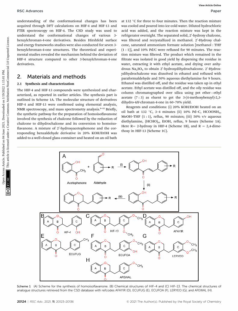
The HIF-4 and HIF-13 compounds were synthesized and char-acterized, as reported in earlier articles. The synthesis part isoutlined in Scheme 1A. The molecular structure of derivativesHIF-4 and HIF-13 were conrmed using elemental analysis,NMR spectroscopy, and mass spectrometry analysis.9–12 Briey,the synthetic pathway for the preparation of homoisoavanoneinvolved the synthesis of chalcone followed by the reduction ofchalcone to dihydrochalcone and its conversion to homoiso-avanone. A mixture of 20-hydroxyacetophenone and the cor-responding benzaldehyde derivative in 20% KOH/EtOH wasadded to a well-closed glass container and heated on an oil bath
Scheme 1 (A) Scheme for the synthesis of homoisoflavanone. (B) Cheanalogue structures retrieved from the CSD database with refcodes AFA
20124 | RSC Adv., 2021, 11, 20123–20136
at 132 �C for three to four minutes. Then the reaction mixturewas cooled and poured into ice-cold water. Diluted hydrochloricacid was added, and the reaction mixture was kept in therefrigerator overnight. The separated solid, 20-hydroxy chalcone,was ltered and recrystallized in methanol. 20-Hydroxy chal-cone, saturated ammonium formate solution [methanol : THF(1 : 1)], and 10% Pd/C were reuxed for 90 minutes. The reac-tion mixture was ltered. The product which remained in theltrate was isolated in good yield by dispersing the residue inwater, extracting it with ethyl acetate, and drying over anhy-drous Na2SO4 to obtain 20-hydroxydihydrochalcone. 20-Hydrox-ydihydrochalcone was dissolved in ethanol and reuxed withparaformaldehyde and 50% aqueous diethylamine for 9 hours.Ethanol was distilled off, and the residue was taken up in ethylacetate. Ethyl acetate was distilled off, and the oily residue wascolumn chromatographed over silica using pet ether : ethylacetate (7 : 3) as eluent to get the 3-(4-methoxybenzyl)-2,3-dihydro-4H-chroman-4-one in 60–70% yield.
Reagents and conditions: (i) 20% KOH/EtOH heated on anoil bath at 132 �C, 3–4 minutes (ii) 10% Pd–C, HCOONH4,MeOH–THF (1 : 1), reux, 90 minutes; (iii) 50% v/v aqueousdiethylamine, (HCHO)n, EtOH, reux, 9 hours (Scheme 1A).Here R¼ 2-hydroxy in HIF-4 (Scheme 1B), and R ¼ 2,4-dime-thoxy in HIF-13 (Scheme 1C).
mical structures of HIF-4 and (C) HIF-13. The chemical structures ofYIR (D), ECUFUG (E), ECUFOA (F), LERYEO (G), and AFEWAL (H).
© 2021 The Author(s). Published by the Royal Society of Chemistry

Fig. 1 Schematic diagram of laser-induced crystallization setup.Components involved in the setup are marked, and the HIF-4 and HIF-13 crystals grown using the laser-induced crystallization techniquewere also shown at the bottom.
Paper RSC Advances
Ope
n A
cces
s A
rtic
le. P
ublis
hed
on 0
4 Ju
ne 2
021.
Dow
nloa
ded
on 1
/19/
2022
1:1
5:01
PM
. T
his
artic
le is
lice
nsed
und
er a
Cre
ativ
e C
omm
ons
Attr
ibut
ion-
Non
Com
mer
cial
3.0
Unp
orte
d L
icen
ce.
View Article Online
2.2 Laser-induced crystallization
Our initial attempts to obtain the crystals of HIF-4 usingconventional crystallization methods failed. Thus, the singlecrystals of HIF-4 (Scheme 1B) and HIF-13 (Scheme 1C) wereobtained using the laser-induced crystallization technique asdescribed.13–15 Fig. 1 shows the schematic diagram of the laser-induced crystallization setup used in this study. The HIF-4 andHIF-13 compounds were dissolved in an equal volume ofethanol and methanol (1 : 1) mixture followed by heating it to40 �C. Each dissolved solution was taken into a coverslip (50 ml)separately, and the nucleant material coir was added to thecrystallization solutions individually as described.13 The
Fig. 2 The thermal ellipsoid diagrams of HIF-4 (A) and HIF-13 (B) drawn
© 2021 The Author(s). Published by the Royal Society of Chemistry
crystallization solution was placed on an inverted microscope(M). A continuous-wave laser beam (l ¼ 1064 nm) was used asa source of excitation with an output power of 60 mW. Amanualbeam expander (BE) to a size of 9 mm was used to expand thelaser beam. The expanded laser beam, a dichroic mirror (M)with high reectivity at 1064 nm, was used via a 1 : 1 telescopicarrangement to the back aperture of the microscope objective(10�, NA ¼ 0.3). A CCD camera was accomplished to visualizeand record the crystallization process (Nikon DS-Fi1c, Japan)attached to the microscope at 50 interlaced frames per second.A linear x–y translational stage was used to control the samplefocusing (Fig. 1).13 When the laser light was focused on thenucleant material coir, bubble formation was observed, fol-lowed by the Brownian motion in the solution. Tiny crystalsstarted to appear within three minutes of laser exposure (shownas insets in Fig. 1). These tiny crystals were used as seed andtransferred into a 5 mL beaker containing crystallization solu-tions for further growth by slow evaporation. The crystal growthwas monitored every 12 hours using a microscope. Diffractionquality crystals with a size of 0.35 � 0.28 � 0.19 mm3 weregrown within 32 hours. A similar crystallization experiment wasconducted for the HIF-13 molecule, and we took the crystal sizeof 0.30 � 0.25 � 0.18 mm3 for X-ray diffraction studies.
2.3 Single crystal data collection, single crystal structuredetermination, renement, and analysis
Single-crystal X-ray diffraction experiments were carried out forthe compounds HIF-4 and HIF-13 (Scheme 1B and C). The
with a 50% probability level with atom numbering.
RSC Adv., 2021, 11, 20123–20136 | 20125

Table 2 Hydrogen bonds for HIF-4a
D–H/A d(D–H)/A d(H–A)/A d(D–A)/A D–H–A/�
O3–O3H/O2#1 0.94(3) 1.83(3) 2.768(2) 174(2)C50–H50/O1#2 0.93(4) 2.671(3) 3.492(2) 148(2)C2–H2A/Cg#3 0.97 2.84 3.494(2) 125
a Symmetry transformation used to generate equivalent atoms: #11 � X,2 � Y, 1 � Z, #21 � X, 1/2 + Y, 1/2 � Z, #31 �X, 1/2 + Y, 1/2 � Z.
RSC Advances Paper
Ope
n A
cces
s A
rtic
le. P
ublis
hed
on 0
4 Ju
ne 2
021.
Dow
nloa
ded
on 1
/19/
2022
1:1
5:01
PM
. T
his
artic
le is
lice
nsed
und
er a
Cre
ativ
e C
omm
ons
Attr
ibut
ion-
Non
Com
mer
cial
3.0
Unp
orte
d L
icen
ce.
View Article Online
preliminary cell determination and the 3D data collection forHIF-4 and HIF-13 were carried out in a Rigaku Saturn 72+single-crystal X-ray diffractometer using graphite mono-chromatized MoKa radiation (l ¼ 0.71075 A). The cell param-eters were rened by the least-squares method in the q range of2–31� for HIF-4 and 3–25� for HIF-13. A complete data set wasprocessed using CrystalClear soware.16 Structure solution bydirect methods using the SHELXS97 17 exposed the positions ofall non-hydrogen atoms of HIF-4 and HIF-13. The initial struc-tures were rened by the least-squares method until conver-gence. Carbon-bound H atoms were placed geometrically, withC–H ¼ 0.93 A, and 0.96 A (methyl) forced to ride on their parentatoms with Uiso(H) ¼ 1.2Ueq(C) or Uiso(H) ¼ 1.5Ueq(Cmethyl). Thehydroxy H atom was located in a difference Fourier map andrened with Uiso(H) ¼ 1.2Ueq(O). The nal renementconverged to an R-value of 0.065 for HIF-4 and 0.053 for HIF-13.The Drmax, Drmin (e A�3) being 0.16 and �0.19 for HIF-4 and0.18 and �0.18 for HIF-13. These calculations were carried outusing the packages SHELXL18 and WinGX.19 ORTEP-3 was usedto prepare the thermal ellipsoid diagrams of HIF-4 and HIF-13(Fig. 2).20 Tabulation of atomic and thermal parameters wasdone using the soware CIFTAB.18 The characterization of ringswas done from puckering parameters' values,21 and symmetryparameters were obtained using PARST97.22 Molecular packingdiagrams were drawn using Mercury.23 The relevant crystallo-graphic data of title compounds were deposited at CambridgeCrystallographic Data Centre (CCDC) with CCDC no. 1834972
Table 1 Crystal data and structure refinement details for HIF-4 and HIF
Identication code HIF-4
Empirical formula C16H14O3
Formula weight 254.27Temperature/K 293(2)Crystal system MonoclinicSpace group P21/ca/A 12.0638(14)b/A 8.9307(9)c/A 12.9341(14)a/� 90b/� 108.465(12)g/� 90Volume/A3 1321.8(3)Z 4rcalc./g cm�3 1.278m/mm�1 0.088F(000) 536.0Crystal size/mm3 0.350 � 0.280 � 0.190Radiation MoKa (l ¼ 0.71073)2q range for data collection/� 5.642 to 62.356Index ranges �17 # h # 16, �12 # k # 12Reections collected 17 821Independent reections 3945 [Rint ¼ 0.0520, Rsigma ¼Data/restraints/parameters 3945/0/176Goodness-of-t on F2 1.029Final R indexes [I $ 2s(I)] R1 ¼ 0.0647, wR2 ¼ 0.1302Final R indexes [all data] R1 ¼ 0.1217, wR2 ¼ 0.1632Largest diff. peak/hole/e A�3 0.16/�0.19CCDC deposition no. 1834972
20126 | RSC Adv., 2021, 11, 20123–20136
(HIF-4), and 1834992 (HIF-13).† Crystal data and structurerenement details were summarized in Table 1. Hydrogenbonds were listed in Tables 2 and 3 for HIF-4, and HIF-13,respectively. The atomic coordinates of all the non-hydrogenatoms with their equivalent isotropic, anisotropic displace-ments parameters, the positional and isotropic displacement ofthe hydrogen atoms for HIF-4, HIF-13 were given in ESI data(Tables S1–S6†).
2.4 Cambridge structural database (CSD)
CSD provides the opportunity to study the conformationalbehavior in analogue structures. We investigated similaritiesand differences of HIF-4 and HIF-13 with respect to analoguestructures retrieved from the CSD. Five analogue structurespossessing the 3-benzylchroman-4-one skeleton (CSD refcodes:AFAYIR (rac-3-(4-hydroxybenzyl)chroman-4-one),10 ECUFOA (3-
-13
HIF-13
C18H18O4
298.34293(2)MonoclinicP21/n7.385(7)27.33(3)8.364(8)90115.630(11)901522(3)41.3020.091632.00.300 � 0.250 � 0.180MoKa (l ¼ 0.71075)6.168 to 50.778
, �17 # l # 18 �8 # h # 8, �31 # k # 32, �10 # l # 911 860
0.0457] 2768 [Rint ¼ 0.0759, Rsigma ¼ 0.0775]2768/0/2010.925R1 ¼ 0.0533, wR2 ¼ 0.1090R1 ¼ 0.1171, wR2 ¼ 0.14050.18/�0.181834992
© 2021 The Author(s). Published by the Royal Society of Chemistry
Paper RSC Advances
Ope
n A
cces
s A
rtic
le. P
ublis
hed
on 0
4 Ju
ne 2
021.
Dow
nloa
ded
on 1
/19/
2022
1:1
5:01
PM
. T
his
artic
le is
lice
nsed
und
er a
Cre
ativ
e C
omm
ons
Attr
ibut
ion-
Non
Com
mer
cial
3.0
Unp
orte
d L
icen
ce.
View Article Online
(3-hydroxy-4-methoxybenzyl)chroman-4-one), ECUFUG (3-(4-methylbenzyl)chroman-4-one),9 AFEWAL (3-(4-methoxybenzyl)-2,3-dihydro-4H-chromen-4-one),12 and LERYEO (3-(3,4-dime-thoxybenzyl)chroman-4-one))11 were extracted from the CSD.
2.5 Hirshfeld surface and energy frameworks
The Hirshfeld surface (HS) analysis, including two-dimensional(2D) ngerprint (FP) plots and electrostatic energy frameworksof HIF-4 and HIF-13, along with other ve other analoguestructures, were generated using the CrystalExplorer (version17.5), and corresponding CIF les were used as the inputles.24,25
2.6 Density functional theory (DFT)
The DFT calculations on HIF-4 and HIF-13 were performedusing the hybrid functional Becke 3 parameter combined withthe Lee–Yang–Parr correlation functional (B3LYP) witha Gaussian type basis 6-31+G(d,p). The B3LYP method has beenproven as reliable and relatively accurate for smaller molecularsystems. In this work, geometry optimization was performed onpossible isomers of HIF-4 and HIF-13 molecules. Following it,optimization was also performed on homodimer of all lowenergy structures. All the calculations were performed underthe methanol solvent environment to follow the crystallizationsolvent condition. Gaussian 09 (Rev. D) program suite was usedfor all the calculations.26 The ball-and-stick model gures weremade using PyMOL (https://www.pymol.org).27
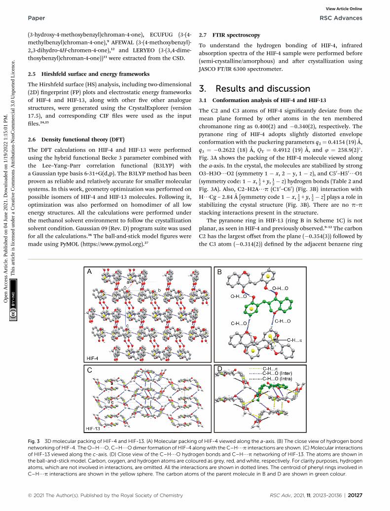
Fig. 3 3D molecular packing of HIF-4 and HIF-13. (A) Molecular packingnetworking of HIF-4. TheO–H/O, C–H/Odimer formation of HIF-4 aloof HIF-13 viewed along the c-axis. (D) Close view of the C–H/O hydrogthe ball-and-stick model. Carbon, oxygen, and hydrogen atoms are colouatoms, which are not involved in interactions, are omitted. All the interactC–H/p interactions are shown in the yellow sphere. The carbon atoms
© 2021 The Author(s). Published by the Royal Society of Chemistry
2.7 FTIR spectroscopy
To understand the hydrogen bonding of HIF-4, infraredabsorption spectra of the HIF-4 sample were performed before(semi-crystalline/amorphous) and aer crystallization usingJASCO FT/IR 6300 spectrometer.
3. Results and discussion3.1 Conformation analysis of HIF-4 and HIF-13
The C2 and C3 atoms of HIF-4 signicantly deviate from themean plane formed by other atoms in the ten memberedchromanone ring as 0.400(2) and �0.340(2), respectively. Thepyranone ring of HIF-4 adopts slightly distorted envelopeconformation with the puckering parameters q2¼ 0.4154 (19) A,q3 ¼ �0.2622 (18) A, QT ¼ 0.4912 (19) A, and 4 ¼ 258.9(2)�.Fig. 3A shows the packing of the HIF-4 molecule viewed alongthe a-axis. In the crystal, the molecules are stabilized by strongO3–H3O/O2 (symmetry 1 � x, 2 � y, 1 � z), and C50–H50/O1(symmetry code: 1 � x, 12 + y, 12 � z) hydrogen bonds (Table 2 andFig. 3A). Also, C2–H2A/p (C10–C60) (Fig. 3B) interaction withH/Cg – 2.84 A [symmetry code 1 � x, 12 + y, 12 � z] plays a role instabilizing the crystal structure (Fig. 3B). There are no p–p
stacking interactions present in the structure.The pyranone ring in HIF-13 (ring B in Scheme 1C) is not
planar, as seen in HIF-4 and previously observed.9–12 The carbonC2 has the largest offset from the plane (�0.354(3)) followed bythe C3 atom (�0.314(2)) dened by the adjacent benzene ring
of HIF-4 viewed along the a-axis. (B) The close view of hydrogen bondngwith the C–H/p interactions are shown. (C) Molecular interactionsen bonds and C–H/p networking of HIF-13. The atoms are shown inred as grey, red, and white, respectively. For clarity purposes, hydrogenions are shown in dotted lines. The centroid of phenyl rings involved inof the parent molecule in B and D are shown in green colour.
RSC Adv., 2021, 11, 20123–20136 | 20127

Table 3 Hydrogen bonds for HIF-13a
D–H/A d(D–H)/A d(H–A)/A d(D–A)/A D–H–A/�
C3–H3/O20 0.98 2.58 3.144(4) 116.8C12–H12C/O2#1 0.96 2.67 3.564(4) 155.1C13–H13B/O20#2 0.96 2.70 3.369(4) 127.1C2–H2B/Cg#3 0.97 2.72 3.668(5) 167.0C8–H8/Cg#4 0.93 2.76 3.623(5) 156.0C12–H12A/Cg#5 0.96 3.00 3.903(5) 158.0
a Symmetry transformation used to generate equivalent atoms: #1X � 1,Y, Z, #2X� 1/2, Y� 1/2, Z + 1/2, #32� X� 2, 1� Y, 1� Z, #41� X, 1� Y, 1� Z, #5X � 1/2, 1/2 � Y, Z � 1/2.
RSC Advances Paper
Ope
n A
cces
s A
rtic
le. P
ublis
hed
on 0
4 Ju
ne 2
021.
Dow
nloa
ded
on 1
/19/
2022
1:1
5:01
PM
. T
his
artic
le is
lice
nsed
und
er a
Cre
ativ
e C
omm
ons
Attr
ibut
ion-
Non
Com
mer
cial
3.0
Unp
orte
d L
icen
ce.
View Article Online
(ring A in Scheme 1C). The pyronone ring of HIF-13 adoptsa distorted envelope conformation with ring puckeringparameters q2 ¼ 0.354 (3) A, q3 ¼ 0.274(3) A, QT ¼ 0.447(3) A,and 4¼ 84.1(4)�. The packing of the HIF-13 is stabilized by a 3Dnetwork of weak intramolecular C3–H3/O20, and C12–H12C/O2, C13–H13B/O20 intermolecular interactions, which serve tolink inversion-related sheets (Fig. 3C). It should be noted thatHIF-13 is the only structure that forms an intramolecular
Fig. 4 CSD survey of HIF-4 and HIF-13. (A) Bond lengths of HIF-4, HIF-1average structures. The values of HIF-4 (black), HIF-13 (blue and italics),
Table 4 Selected bond lengths (A) of the pyranone ring of HIF structcalculated for the disordered atoms are indicated in * mark. For exampl
Bond HIF-4 HIF-13 AFAYIR EUCFU
O1–C2 1.436(2) 1.423(3) 1.388 1.444O1–C2B* — — 1.536 —C2–C3 1.508(2) 1.511(4) 1.393 1.500C2–C3B* — — 1.254 —C3–C4 1.503(2) 1.509(3) 1.512 1.507C4–C3B* — — 1.512 —C4–C10 1.471(2) 1.463(4) 1.457 1.474C10–C9 1.393(2) 1.393(4) 1.388 1.395O1–C9 1.357(2) 1.358(3) 1.359 1.357O2–C4 1.228(2) 1.216(3) 1.218 1.216C3–C11 1.550(2) 1.520(3) 1.507 1.533C3B–C11* — — 1.458 —C11–C10 1.506(2) 1.501(3) 1.510 1.502
20128 | RSC Adv., 2021, 11, 20123–20136
C–H/O hydrogen bond comparison with HIF-4 and the otherve analogue structures retrieved from CSD, as discussed inSection 2.4. In addition to C–H/O hydrogen bonds, there arethree C2–H2B/p (C5–C10), C8–H8/p (C10–C60), C12–H12A/p (C10–C60) interactions stabilize the 3D structure of HIF-13(Table 3 and Fig. 3D).
3.2 CSD studies of 3-benzylchroman-4-ones
The geometric parameters of HIF-4 and HIF-13 are comparablewith the analogue structures AFAYIR,10 ECUFOA,9 ECUFUG,9
AFEWAL,12 and LERYEO11 reported earlier. The bond lengths ofHIF-4 and HIF-13 pyranone rings are compared with the veanalogue structures, and the results are shown in Fig. 4A. Theresults show that except C2–C3, all other bond lengths aresimilar (Fig. 4 and Table 4). The average C2–C3 bond length is1.433 A, much smaller than HIF-4 and HIF-13 because this bondis oen disordered in most of the reported structures. Out ofve analogue structures, three of them (AFAYIR, AFEWAL,EUCFOA) have either C2 or C3 atoms disordered in O1–C2, C2–C3 bonds. In the case of AFAYIR, AFEWAL, both C3 and C4atoms are disordered in the C3–C4 bond. Hence, the C2–C3
3, and the average structures. (B) Bond angles of HIF-4, HIF-13, and theand the CSD average (red) are marked.
ures and analogue structures retrieved from CSD. The bond lengthse, O1–C2B*, in which the C2 atom is disordered
G AFEWAL ECUFOA LERYEO Average
1.437 1.423 1.418 1.422(22)1.487 1.502 — 1.508(25)1.342 1.434 1.496 1.433(68)1.218 1.273 — 1.248(28)1.530 1.514 1.510 1.515(9)1.543 — — 1.528(22)1.466 1.460 1.468 1.465(7)1.379 1.373 1.389 1.385(9)1.354 1.363 1.355 1.358(4)1.212 1.227 1.213 1.217(6)1.502 1.486 1.507 1.507(17)1.522 — — 1.490(45)1.500 1.503 1.509 1.505(4)
© 2021 The Author(s). Published by the Royal Society of Chemistry

Table 5 Selected bond angles of HIF structures and analogue structures retrieved from CSD. The bond angles that correspond to either one ortwo disordered atoms are marked as *. For example, in C9–O1–C2B, the C2 atom is disordered, and C4–C3B–C2B, the C3 and C2 atoms aredisordered
Bond angle HIF-4 HIF-13 AFAYIR ECUFUG AFEWAL ECUFOA LERYEO Average
C9–O1–C2 114.8(1) 115.6(2) 114.6 114.4 115.2 114.6 115.0 114.8(0.3)C9–O1–C2B* — — 115.3 — 112.8 113.0 — 113.7(1.4)O1–C2–C3 111.5(1) 113.3(2) 119.9 112.3 124.0 115.2 112.9 116.9(5.0)O1–C2B–C3 — — 127.2 — 126.3 120.5 — 124.7(3.6)C4–C3–C2 108.3(1) 108.6(2) 113.1 109.4 116.7 111.4 108.4 111.8(3.3)C4–C3B–C2B* — — 123.0 — 117.0 116.0 — 118.7(3.8)C10–C4–C3 115.3(2) 115.1(2) 116.4 115.4 114.3 115.8 114.3 115.2(0.9)C10–C4–C3B* — — 114.1 — 114.5 — — 114.3(0.3)C9–C10–C4 119.6(2) 120.1(2) 120.4 120.2 120.3 119.9 120.1 120.2(0.2)O1–C9–C10 122.8(2) 123.0(2) 122.6 122.5 122.5 122.8 122.9 122.7(0.2)C11–C3–C2 112.9(2) 112.6(2) 109.5 112.1 112.8 123.4 114.3 114.4(5.3)C11–C3B–C2B* — — 116.1 — 115.1 129.0 — 120.1(7.8)C11–C3–C4 109.7(1) 112.8(2) 113.2 112.9 109.4 114.2 112.3 112.4(1.8)C11–C3B–C4 — — 116.0 — 111.2 — — 113.6(3.4)C10–C11–C3 112.4(1) 114.6(2) 113.4 113.7 115.0 116.3 114.0 114.5(1.2)C10–C11–C3B* — — 121.6 — 117.0 — — 119.3(3.3)O2–C4–C3 123.0(2) 122.9(2) 123.5 122.9 122.6 121.6 122.9 122.7(0.7)O2–C4–C3B* — — 118.3 — 121.3 — — 119.8(2.1)O2–C4–C10 121.6(2) 121.9(3) 122.2 121.7 122.3 122.6 122.8 122.3(0.4)
Paper RSC Advances
Ope
n A
cces
s A
rtic
le. P
ublis
hed
on 0
4 Ju
ne 2
021.
Dow
nloa
ded
on 1
/19/
2022
1:1
5:01
PM
. T
his
artic
le is
lice
nsed
und
er a
Cre
ativ
e C
omm
ons
Attr
ibut
ion-
Non
Com
mer
cial
3.0
Unp
orte
d L
icen
ce.
View Article Online
bond disorder is observed frequently; the respective averagebond length has a considerable deviation from HIF-4 and HIF-13 (Table 4). Similarly, the C3–C11 and C11–C10 bond lengths ofHIF-4 and HIF-13 are comparable with the analogue structures.While C11–C10 bond length of HIF-4 (1.506 A) and HIF-13 (1.501A) is closer to the average bond length (1.505 A), the C3–C11bond length of HIF-4 (1.550 A) is longer than HIF-13 (1.520 A),and the average of analogue structures (1.507 A) (Fig. 4A).
The bond angles of pyranone rings of HIF-4 and HIF-13,along with the average bond angles of analogue structures,are shown in Fig. 4B, and the details are summarized in Table 5.Almost all bond angles of HIF-4 and HIF-13 have good agree-ment with the average bond angle values. The O1–C2–C3 of HIF-4 (111.5�) and HIF-13 (113.3�) are slightly lower than the averagevalue (116.9�), and it may be due to the disordered behaviourobserved in the analogue structures. The O1–C2–C3 and C11–C3–C2 bond angles are slightly higher than the normal for the
Table 6 Selected torsion angles of HIF structures and analogue structuangles contains disordered atoms. For example, torsion angle O1–C2B–
Torsion angle HIF-4 HIF-13 AFAYIR
O1–C2–C3–C11 61.7(2) �177.5(2) �171.8O1–C2B–C3B–C11* — — �174.9C2–C3–C11–C10 60.4(2) 69.6(3) 61.2C2B–C3B–C11–C10* — — �26.7C3–C11–C10–C20 81.4(2) 74.7(3) 87.3C3B–C11–C10–C20* — — 62.7C3–C11–C10–C60 �98.3(2) �106.1(3) �117.9C3B–C11–C10–C60* — — �93.3O2–C4–C3–C11 88.9(2) 18.5(3) 17.3O2–C4–C3B–C11* — — �33.4C4–C3–C11–C10 �178.8(1) �167.1(2) �174.2
© 2021 The Author(s). Published by the Royal Society of Chemistry
disordered structures AFAYIR, AFEWAL, and ECUFOA (Table 5and Fig. 4B).
The selected six torsion angles around the pyranone ring andphenyl ring C10–C60 of HIF-4, HIF-13, and their analoguestructures are summarized in Table 6. The C2–C3–C11–C10
torsion angle adopts either �synclinal or +synclinal conforma-tion (Table 6), and HIF-13 has the maximum (69.6(3)�). The C3–C11–C10–C20 torsion angle adopts +synclinal (from +30� to +90�)conformation, except ECUFUG (97.7�), and AFEWAL (94.6�),which adopt +anticlinal conformation. The C3–C11–C10–C60
(�anticlinal) and C4–C3–C11–C10 (�antiperiplanar) torsionangles of all seven structures adopt similar conformations. Thetorsion angle O1–C2–C3–C11 adopts two different orientations.The HIF-13 and its ve analogue structures adopt anti-periplanar (�150� to �180�) conformation, whereas HIF-4(61.7(2)�) adopt +synclinal conformation. Similarly, O2–C4–C3–C11 of HIF-4 (88.9�) also adopts +synclinal conformation,
res retrieved from CSD. The * mark represents the calculated torsionC3B–C11* has two (C2B and C3B) disordered atoms
ECUFUG AFEWAL ECUFOA LERYEO
176.9 �177.0 167.4 �174.6— 172.4 152.5 —�66.2 �50.8 65.5 54.3— 50.0 �12.3 —97.7 94.6 74.8 70.3— 66.3 — —�81.3 �114.2 �104.5 �108.8— �85.9 — —�24.2 26.0 9.71 16.4— �27.5 — —169.8 170.3 178.9 178.3
RSC Adv., 2021, 11, 20123–20136 | 20129
RSC Advances Paper
Ope
n A
cces
s A
rtic
le. P
ublis
hed
on 0
4 Ju
ne 2
021.
Dow
nloa
ded
on 1
/19/
2022
1:1
5:01
PM
. T
his
artic
le is
lice
nsed
und
er a
Cre
ativ
e C
omm
ons
Attr
ibut
ion-
Non
Com
mer
cial
3.0
Unp
orte
d L
icen
ce.
View Article Online
and all other structures adopt synperiplanar (�0� to �30�)conformation. Thus, it is clear that HIF-4 adopts an entirelydifferent conformation than HIF-13 and ve analogue struc-tures extracted from CSD.
3.3 Conformational exibilities of 3-benzylchroman-4-ones
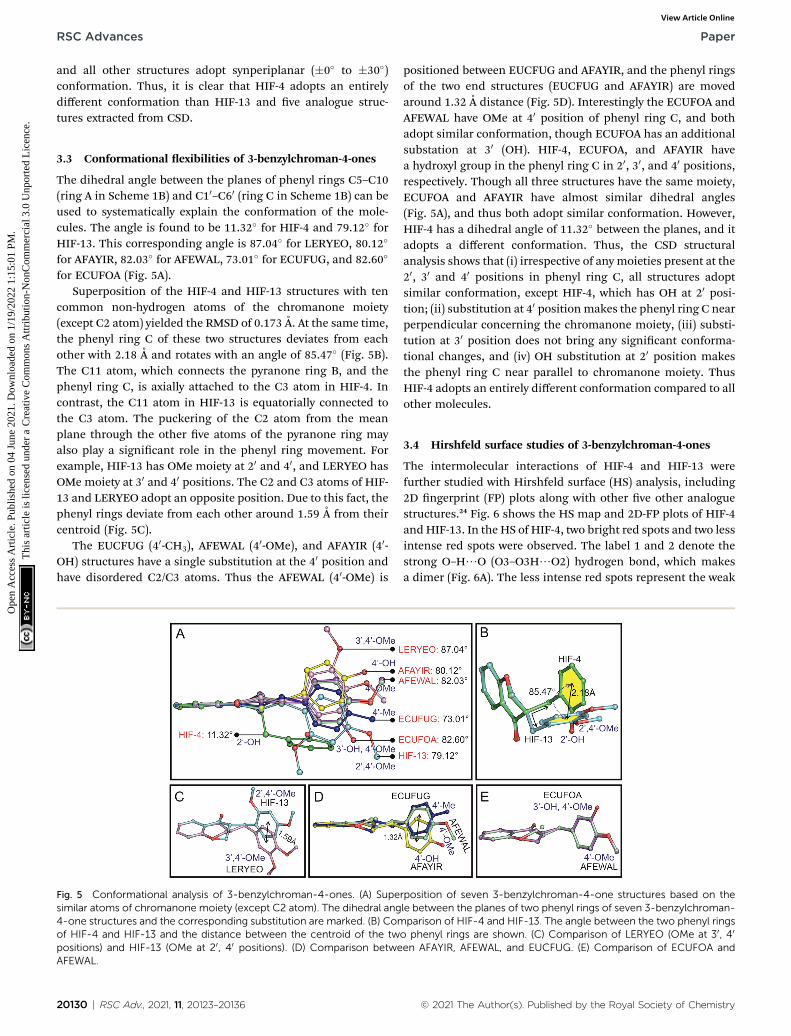
The dihedral angle between the planes of phenyl rings C5–C10(ring A in Scheme 1B) and C10–C60 (ring C in Scheme 1B) can beused to systematically explain the conformation of the mole-cules. The angle is found to be 11.32� for HIF-4 and 79.12� forHIF-13. This corresponding angle is 87.04� for LERYEO, 80.12�
for AFAYIR, 82.03� for AFEWAL, 73.01� for ECUFUG, and 82.60�
for ECUFOA (Fig. 5A).Superposition of the HIF-4 and HIF-13 structures with ten
common non-hydrogen atoms of the chromanone moiety(except C2 atom) yielded the RMSD of 0.173 A. At the same time,the phenyl ring C of these two structures deviates from eachother with 2.18 A and rotates with an angle of 85.47� (Fig. 5B).The C11 atom, which connects the pyranone ring B, and thephenyl ring C, is axially attached to the C3 atom in HIF-4. Incontrast, the C11 atom in HIF-13 is equatorially connected tothe C3 atom. The puckering of the C2 atom from the meanplane through the other ve atoms of the pyranone ring mayalso play a signicant role in the phenyl ring movement. Forexample, HIF-13 has OMe moiety at 20 and 40, and LERYEO hasOMe moiety at 30 and 40 positions. The C2 and C3 atoms of HIF-13 and LERYEO adopt an opposite position. Due to this fact, thephenyl rings deviate from each other around 1.59 A from theircentroid (Fig. 5C).
The EUCFUG (40-CH3), AFEWAL (40-OMe), and AFAYIR (40-OH) structures have a single substitution at the 40 position andhave disordered C2/C3 atoms. Thus the AFEWAL (40-OMe) is
Fig. 5 Conformational analysis of 3-benzylchroman-4-ones. (A) Supesimilar atoms of chromanone moiety (except C2 atom). The dihedral ang4-one structures and the corresponding substitution are marked. (B) Comof HIF-4 and HIF-13 and the distance between the centroid of the twpositions) and HIF-13 (OMe at 20, 40 positions). (D) Comparison betweAFEWAL.
20130 | RSC Adv., 2021, 11, 20123–20136
positioned between EUCFUG and AFAYIR, and the phenyl ringsof the two end structures (EUCFUG and AFAYIR) are movedaround 1.32 A distance (Fig. 5D). Interestingly the ECUFOA andAFEWAL have OMe at 40 position of phenyl ring C, and bothadopt similar conformation, though ECUFOA has an additionalsubstation at 30 (OH). HIF-4, ECUFOA, and AFAYIR havea hydroxyl group in the phenyl ring C in 20, 30, and 40 positions,respectively. Though all three structures have the same moiety,ECUFOA and AFAYIR have almost similar dihedral angles(Fig. 5A), and thus both adopt similar conformation. However,HIF-4 has a dihedral angle of 11.32� between the planes, and itadopts a different conformation. Thus, the CSD structuralanalysis shows that (i) irrespective of any moieties present at the20, 30 and 40 positions in phenyl ring C, all structures adoptsimilar conformation, except HIF-4, which has OH at 20 posi-tion; (ii) substitution at 40 positionmakes the phenyl ring C nearperpendicular concerning the chromanone moiety, (iii) substi-tution at 30 position does not bring any signicant conforma-tional changes, and (iv) OH substitution at 20 position makesthe phenyl ring C near parallel to chromanone moiety. ThusHIF-4 adopts an entirely different conformation compared to allother molecules.
3.4 Hirshfeld surface studies of 3-benzylchroman-4-ones
The intermolecular interactions of HIF-4 and HIF-13 werefurther studied with Hirshfeld surface (HS) analysis, including2D ngerprint (FP) plots along with other ve other analoguestructures.24 Fig. 6 shows the HS map and 2D-FP plots of HIF-4and HIF-13. In the HS of HIF-4, two bright red spots and two lessintense red spots were observed. The label 1 and 2 denote thestrong O–H/O (O3–O3H/O2) hydrogen bond, which makesa dimer (Fig. 6A). The less intense red spots represent the weak
rposition of seven 3-benzylchroman-4-one structures based on thele between the planes of two phenyl rings of seven 3-benzylchroman-parison of HIF-4 and HIF-13. The angle between the two phenyl rings
o phenyl rings are shown. (C) Comparison of LERYEO (OMe at 30, 40
en AFAYIR, AFEWAL, and EUCFUG. (E) Comparison of ECUFOA and
© 2021 The Author(s). Published by the Royal Society of Chemistry
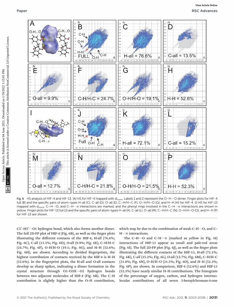
Fig. 6 HS analysis of HIF-4 and HIF-13. (A) HS for HIF-4 mapped with dnorm. Labels 1 and 2 represent the O–H/O dimer. Finger plots for HIF-4full (B) and the specific pairs of atom-types H-all (C), C-all (D), O-all (E), C–H/H–C (F), O–H/H–O (G), and H–H (H) for HIF-4. (I) HS for HIF-13mapped with dnorm. C–H/O, and C–H/p interactions are marked, and the phenyl rings involved in the C–H/p interactions are shown inyellow. Finger plots for HIF-13 full (J) and the specific pairs of atom-types H-all (K), C-all (L), O-all (M), C–H/H–C (N), O–H/H–O (O), and H–H (P)for HIF-13 are shown.
Paper RSC Advances
Ope
n A
cces
s A
rtic
le. P
ublis
hed
on 0
4 Ju
ne 2
021.
Dow
nloa
ded
on 1
/19/
2022
1:1
5:01
PM
. T
his
artic
le is
lice
nsed
und
er a
Cre
ativ
e C
omm
ons
Attr
ibut
ion-
Non
Com
mer
cial
3.0
Unp
orte
d L
icen
ce.
View Article Online
C50–H50/O1 hydrogen bond, which also forms another dimer.The full 2D-FP plot of HIF-4 (Fig. 6B), as well as the nger plotsillustrating the different contacts of the HIF-4, H-all (76.6%;Fig. 6C), C-all (13.5%; Fig. 6D), O-all (9.9%; Fig. 6E), C–H/H–C(24.7%; Fig. 6F), O–H/H–O (19.1; Fig. 6G), and H–H (52.6%;Fig. 6H), are shown. According to divided ngerprints, thehighest contribution of contacts received by the HIF-4 is H–H(52.6%). In the ngerprint plots, the H-all and O-all contactsdevelop as sharp spikes, indicating a dimer formation in thecrystal structure through O3–O3H/O2 hydrogen bondsbetween two adjacent molecules of HIF-4 (Fig. 6B). The C–Hcontribution is slightly higher than the O–H contribution,
© 2021 The Author(s). Published by the Royal Society of Chemistry
which may be due to the combination of weak C–H/O, and C–H/p interactions.
The C–H/O and C–H/p (marked as yellow in Fig. 6I)interactions of HIF-13 appear as small and pale-red areas(Fig. 6I). The full 2D-FP plot (Fig. 6J), as well as the nger plotsillustrating the different contacts of the HIF-13, H-all (72.1%;Fig. 6K), C-all (15.2%; Fig. 6L), O-all (12.7%; Fig. 6M), C–H/H–C(21.8%; Fig. 6N), O–H/H–O (21.5%; Fig. 6O), and H–H (52.3%;Fig. 6P), are shown. In comparison, HIF-4 (52.6%) and HIF-13(52.3%) have nearly similar H–H contributions. The histogramof the percentage of oxygen, carbon, and hydrogen intermo-lecular contributions of all seven 3-benzylchroman-4-one
RSC Adv., 2021, 11, 20123–20136 | 20131

RSC Advances Paper
Ope
n A
cces
s A
rtic
le. P
ublis
hed
on 0
4 Ju
ne 2
021.
Dow
nloa
ded
on 1
/19/
2022
1:1
5:01
PM
. T
his
artic
le is
lice
nsed
und
er a
Cre
ativ
e C
omm
ons
Attr
ibut
ion-
Non
Com
mer
cial
3.0
Unp
orte
d L
icen
ce.
View Article Online
structures shows that LERYEO and ECUFOA have the mostconsiderable oxygen contribution (12.6%), and ECUFOG (8.5%)has the lowest contribution (ESI Fig. S1H†). In terms of carbon,HIF-4 (13.5%) and HIF-13 (13.2%) have the highest contributionthan analogue structures. In general, hydrogen contributesaround 77% on average to make intermolecular interactions forall 3-benzylchroman-4-one structures. In which, ECUFOA(75.6%), HIF-13 (75.8%) are the lowest, and ECUFUG (79.1%)contributes higher than all other structures (ESI Fig. S1H†). Outof seven structures, HIF-4, ECUFOA, and AFAYIR have OHsubstitution at 20, 30, and 40 positions in the phenyl ring C. The2D-FP plot of the analogue structures shows that the AFAYIRand ECUOFA contribution of O–H is almost similar to HIF-4(ESI Fig. S1A–G†). Then what makes the HIF-4 different thanother structures? To understand the fact, along with Hirshfeldsurface, the O–H/O intermolecular interactions are scruti-nized further for HIF-4, ECUFOA, and AFAYIR structures (ESIFig. S1I–K†). HIF-4 is the only structure that forms a dimer viaO–H/O hydrogen bonds among all three structures. In termsof distance (hydrogen-acceptor: 1.83 A) and the angle (174(2)�),HIF-4 is stronger than the other two structures (ESI Fig. S1I†).Though AFAYIR also forms a strong O–H/O hydrogen bond,the hydrogen bond pattern is different than HIF-4 (ESIFig. S1J†). In addition, the oxygen atom in the C]O moiety ofthe pyranone ring act as an acceptor atom for both HIF-4 and
Fig. 7 Energy frameworks analysis for HIF-4. The molecular arrangemen(C, F, and I) directions. The models are shown in ball-and-stick. The totaenergy terms (G–I) are shown in blue, red, and green, respectively. Theelectron-density functions. The energies betweenmolecular pairs are reppairs of molecules.
20132 | RSC Adv., 2021, 11, 20123–20136
AFAYIR structures. In ECUCOA, the OMe moiety in phenyl ringC acts as an acceptor, and the pyranone ring is not involved inthe O–H/O hydrogen bond interactions. The hydrogen bondstrength of the ECUFOA is comparatively weaker than HIF-4 andAFAYIR (ESI Fig. S1K†). Thus, it is evident that the intermolec-ular interactions, especially O–H/O dimer formation in HIF-4,play a signicant role in adopting different conformation due tothe OH substitution at 20 position in phenyl ring C.
3.5 Energy framework analysis of 3-benzylchroman-4-ones
Energy framework analysis helps to understand the crystalpacking and visualize the interaction topologies based onelectrostatic, polarization, dispersion, and exchange-repul-sion.28 The interaction energies (kJ mol�1) calculated fromenergy framework calculations using CrystalExplorer aresummarized in ESI Table S7† for all seven HIF-structures. HIF-4(�100.5 kJ mol�1) has the highest electrostatic energy than allother structures, followed by AFAYIR (�56.8 kJ mol�1) andECUFOA (�38.0 kJ mol�1). It may be because all three structureshave OHmoiety present in the phenyl ring C. It should be notedthat the HIF-4 and AFAYIR structures have only OH substitutionat the phenyl ring C, and their electrostatic energies are higherthan any other structures. Interestingly AFAYIR(�61.7 kJ mol�1) has the highest dispersion energy, and HIF-4has the lowest (�31.2 kJ mol�1). In terms of total energy, HIF-
t of HIF-4 is viewed along with the a (A, D, and G), b (B, E, and H), and cl interaction energies (A–C), electrostatic terms (D–F), and dispersionenergy framework interactions are calculated using B3LYP/6-31G(d,p)resented as cylinders with a scale size of 150 by joining the centroids of
© 2021 The Author(s). Published by the Royal Society of Chemistry
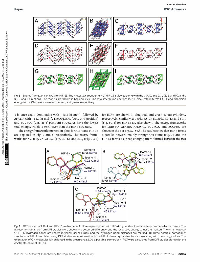
Fig. 8 Energy framework analysis for HIF-13. Themolecular arrangement of HIF-13 is viewed along with the a (A, D, and G), b (B, E, and H), and c(C, F, and I) directions. The models are shown in ball and stick. The total interaction energies (A–C), electrostatic terms (D–F), and dispersionenergy terms (G–I) are shown in blue, red, and green, respectively.
Paper RSC Advances
Ope
n A
cces
s A
rtic
le. P
ublis
hed
on 0
4 Ju
ne 2
021.
Dow
nloa
ded
on 1
/19/
2022
1:1
5:01
PM
. T
his
artic
le is
lice
nsed
und
er a
Cre
ativ
e C
omm
ons
Attr
ibut
ion-
Non
Com
mer
cial
3.0
Unp
orte
d L
icen
ce.
View Article Online
4 is once again dominating with �85.5 kJ mol�1 followed byAFAYIR with �54.3 kJ mol�1. The AFEWAL (OMe at 40 position)and ECUFUG (CH3 at 40 position) structures have the lowesttotal energy, which is 50% lower than the HIF-4 structure.
The energy framework interaction plots for HIF-4 and HIF-13are depicted in Fig. 7 and 8, respectively. The energy frame-works for Etot (Fig. 7A–C), Eele (Fig. 7D–E), and Edisp (Fig. 7G–I)
Fig. 9 DFT models of HIF-4 and HIF-13. (A) Isomers of HIF-4 superimpofive isomers obtained from DFT studies were shown and coloured differeO–H/O hydrogen bonds are shown in yellow dashed lines, and the hstructures of HIF-4 calculated using DFT studies superimposed with theorientation of OHmolecules is highlighted in the green circle. (C) Six posscrystal structure of HIF-13.
© 2021 The Author(s). Published by the Royal Society of Chemistry
for HIF-4 are shown in blue, red, and green colour cylinders,respectively. Similarly, Etot (Fig. 8A–C), Eele (Fig. 8D–E), and Edisp(Fig. 8G–I) for HIF-13 are also shown. The energy frameworksfor LERYEO, AFAYIR, AFEWAL, ECUFOA, and ECUFUG areshown in the ESI Fig. S2–S6.† The results show that HIF-4 formsa parallel network mainly through OH atoms (Fig. 7), and theHIF-13 forms a zig-zag energy pattern formed between the two
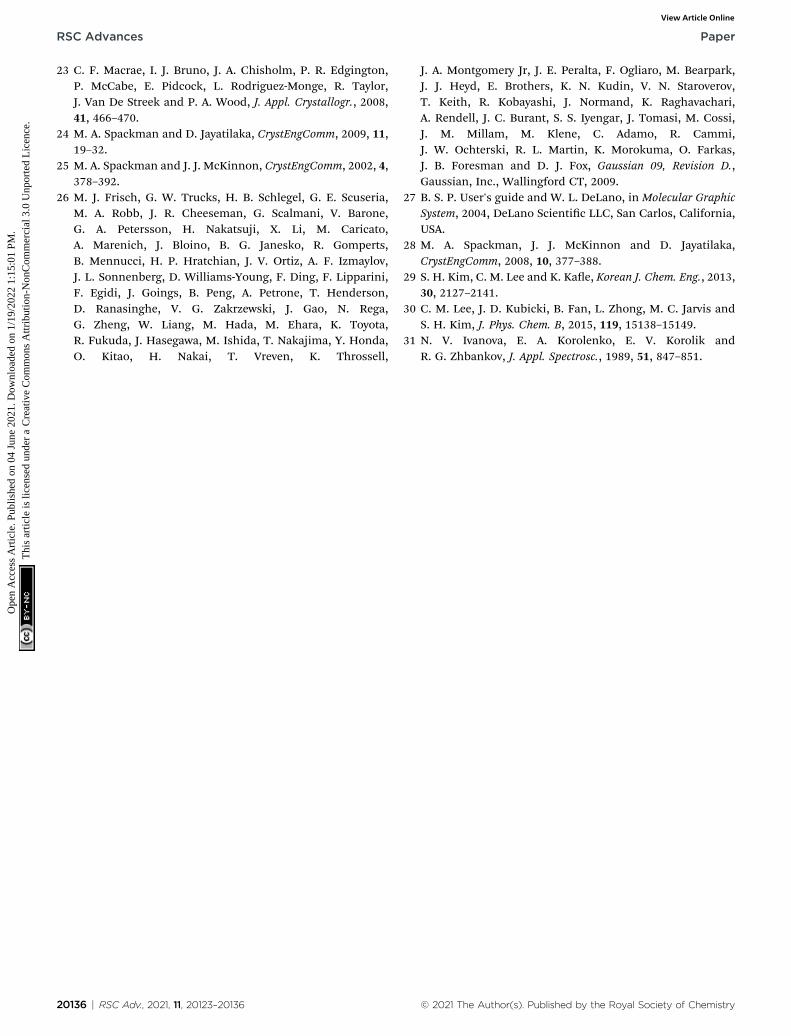
sed with HIF-4 crystal structure based on chroman-4-one moiety. Thently, and the respective energy values are marked. The intramolecularydrogen bond distances are marked. (B) Three possible homodimerHIF-4 dimer crystal structure shown along with the energy values. Theible isomers of HIF-13 were calculated from DFT studies along with the
RSC Adv., 2021, 11, 20123–20136 | 20133

RSC Advances Paper
Ope
n A
cces
s A
rtic
le. P
ublis
hed
on 0
4 Ju
ne 2
021.
Dow
nloa
ded
on 1
/19/
2022
1:1
5:01
PM
. T
his
artic
le is
lice
nsed
und
er a
Cre
ativ
e C
omm
ons
Attr
ibut
ion-
Non
Com
mer
cial
3.0
Unp
orte
d L
icen
ce.
View Article Online
molecules of HIF-13 (Fig. 8). In HIF-4, the electrostatic termsdominate the energy pattern due to the OH substation at the 20
position. In HIF-13, the energy pattern is dominated by thedispersion energies due to the OMe substitutions at 20 and 40
positions in phenyl ring C. The remaining structures LERYEO(ESI Fig. S2†), AFAYIR (ESI Fig. S3†), AFEWAL (ESI Fig. S4†),EUCFOA (ESI Fig. S5†), and ECUFUG (ESI Fig. S6†) form parallelzig-zag sheet energy networks mainly through C3–C11 and C11–C10 bonds, and are comparable to each other. However, asdiscussed earlier, the network pattern of HIF-4 is unique anddifferent than all other structures due to highly dominatedelectrostatic energies.
3.6 DFT studies of HIF-4 and HIF-13
The structures of HIF-4 and HIF-13 molecules were furtherinvestigated using DFT calculations. Although there are severalgeometries possible, Fig. 9A shows the top ve low energymodels of HIF-4. Three isomers, isomer-1 (0.0 kJ mol�1),isomer-2 (2.7 kJ mol�1), and isomer-3 (10.0 kJ mol�1) adoptdifferent conformation than HIF-4 crystallographic structure.Isomer-1 appeared as the most stable conformation withO–H/O (1.703 A) intramolecular hydrogen bond. The isomer-2of HIF-4 is similar to isomer-1, except the phenyl ring is slightlyoriented away with a higher O–H/O (1.732 A) hydrogen bondlength. The isomer-3 adopts a similar conformation as HIF-13.The isomer-4 (10.92 kJ mol�1) and isomer-5 (16.96 kJ mol�1)adopt similar conformation like HIF-4. The only structuraldifference between isomer-4 and isomer-5 is in the orientationof the hydrogen atom in 20-OH (Fig. 9A).
The formation of a molecular dimer is a critical process tostart the nucleation of the molecular system towards the crystal.Because of this, optimizations of homodimers of monomerswere performed in the methanol environment. Fig. 9B showsthe energy-optimized structures of HIF-4 dimers with theirrelative energies. The intermolecular O–H/O (O3–O3H/O2)hydrogen bond stabilizes the dimer structure of isomer-1 withthe distance of 1.765 A and adopts the most stable conforma-tion. Interestingly this lowest energy dimer structure is similarto the crystal structure of HIF-4. In the dimer of isomer-2, theintermolecular O–H/O (O3–O3H/O2) hydrogen bond isformed with the length of 1.833 A. In isomer-3, the
Fig. 10 FTIR spectra of HIF-4. (A) FTIR spectra of HIF-4 in the O–H stretc(B) and crystal (C) of HIF-4.
20134 | RSC Adv., 2021, 11, 20123–20136
intermolecular O–H/O (O3–O3H/O2) hydrogen bond wasintact with a distance of 1.699 A. In addition, a weak C5–H5/O3 (2.582 A) intermolecular hydrogen bond was also observed.Except for one of the monomers of isomer-3, the orientation ofpolar hydrogen O3H is similar for all the isomers and closelyresembles the HIF-4 dimer structure. Though the isomer-2(14.12 kJ mol�1) and isomer-3 (19.65 kJ mol�1) form strongerO–H/O hydrogen bonds, their energies do not favour theformation of 3D networks. Thus, the DFT studies support thecrystal structure of HIF-4.
In the case of HIF-13, six low-energy optimized structures areproduced (Fig. 9C). Isomer-1, isomer-2 (0.44 kJ mol�1), andisomer-3 (1.48 kJ mol�1) are the most stable models. Therefore,in the solution phase at room temperature, all the three struc-tures with energy #2 kJ mol�1 are possible and can formdimers. Both isomer-1 (0.0 kJ mol�1) and isomer-3(1.48 kJ mol�1) are similar to HIF-13 and are superimposable.Except that the C13 atom of isomer-1 (0.0 kJ mol�1), whichoccupy an opposite position in comparison with HIF-13 andisomer-3. The isomer-2 (0.44 kJ mol�1), and isomer-4(2.07 kJ mol�1) adopt a conformation similar to HIF-4, whileisomer-5 (13.43 kJ mol�1), and isomer-6 (20.49 kJ mol�1) adoptentirely different conformation. Thus, the DFT study suggeststhat the two different conformations of HIF-4 and HIF-13 arepossible. The substitution position, dimer formation, andelectrostatic energies of substituted atoms play a signicantrole in determining the conformation of these molecules.
3.7 FTIR spectroscopy studies of HIF-4
Infrared spectroscopy has been widely used to understand theinuence of hydrogen bonding on O–H stretching frequen-cies.29,30 It has been observed that the O–H stretching frequencyshows red-shi when involved in a hydrogen bond formation.30
To understand the hydrogen bonding of HIF-4, FTIR spectrawere recorded on semi-crystalline/amorphous and crystal.Fig. 10A shows the overlaid spectra in the O–H stretchingregion. It is clear from the gure that the O–H vibrations in thecrystalline HIF-4 are shied to the lower frequency side andbecome slightly narrower. These O–H bands can be deconvo-luted into four peaks each at 3201.7, 3252.0, 3314.5 and3410.6 cm�1 for amorphous (Fig. 10B) and 3205.0, 3248.9,
hing region. Deconvolution of the O–H stretching bands of amorphous
© 2021 The Author(s). Published by the Royal Society of Chemistry

Paper RSC Advances
Ope
n A
cces
s A
rtic
le. P
ublis
hed
on 0
4 Ju
ne 2
021.
Dow
nloa
ded
on 1
/19/
2022
1:1
5:01
PM
. T
his
artic
le is
lice
nsed
und
er a
Cre
ativ
e C
omm
ons
Attr
ibut
ion-
Non
Com
mer
cial
3.0
Unp
orte
d L
icen
ce.
View Article Online
3290.3 and 3365.7 cm�1 for crystalline HIF-4 (Fig. 10C). Theresults obtained by FTIR study on HIF-4 suggest that, both interand intra molecular hydrogen bonding exist in amorphous orsemi crystalline powder sample of HIF-4. Thus, there is bandspread at the higher frequency edge and is contributed by theeffect of intra-molecular hydrogen bonding on O–H stretching.It is assumed that the intermolecular hydrogen bonds areshorter and therefore have lower O–H stretching frequenciesthan intramolecular hydrogen bonds.31 The intramolecularhydrogen bonding contribution to O–H stretching has vanishedwhen crystal is formed (Fig. 10C).
4. Conclusions
Efforts to obtain the crystals of HIF-4 using conventional crys-tallization techniques are not successful. Thus, we have usedthe laser-induced crystallization technique to grow the micro-crystals. Later those microcrystals were used as a seed todevelop good quality crystals for single-crystal X-ray diffractionanalysis. Therefore laser-induced crystallization technique maybe explored futher to crystallize organic, inorganic, and bio-logical molecules.13 Single crystal X-ray crystallographic studiesrevealed that the 3-benzylchroman-4-one derivatives tend tocrystallize in a particular conformation. However, the HIF-4,a molecule bearing OH at 20 position in phenyl ring C, adopteda different conformation. DFT, FTIR, Hirshfeld, energy frame-works, and CSD studies are also used to analyze the confor-mational changes of 3-benzylchroman-4-ones. All these studiescomplemented each other to understand the effect of substi-tutions at 20, 30; and 40 positions in phenyl ring C, and role ofhydrogen bonds in conformation of 3-benzylchroman-4-ones.In HIF-13, OMe moiety is present at the 20 and 30 positions,and it adopts a similar conformation as other 3-benzylchroman-4-ones. Some of the reported structures, AFAYIR, ECUFOA haveOH substitution at phenyl ring C in 30 or 40 position, and theyalso adopt similar conformation like HIF-13. Whereas the CSD,energy frameworks, DFT studies revealed that OH substitutionat 20 position activates the dimer formation via strong O–H/Ohydrogen bonds, which facilitate the conformational changes inHIF-4. Our earlier studies have reported homoisoavanones asan effective inhibitor for cancer exclusively for tumorsuppressor protein p53.9 Therefore the new reported moleculeswith different conformations may be an excellent candidate tobe explored further. Further experimental studies may becarried out to understand the anticancer properties of deviatedHIF-4 structure, which may provide insight into anticancer drugmolecule development.
Conflicts of interest
Authors declare no conict of interest.
Acknowledgements
Abdul Ajees Abdul Salam acknowledges the MAHE intramuralresearch grant (MAHE/DREG/PhD/IMF/2019). We gratefullyacknowledge support for this work from the Department of
© 2021 The Author(s). Published by the Royal Society of Chemistry
Biotechnology (BT/PR6413/MED/14/802/2005) and the Depart-ment of Science and Technology's FIST program (SR/FST/PSI-174/2012).
References
1 S. N. Lopez, M. G. Sierra, S. J. Gattuso, R. L. Furlan andS. A. Zacchino, Phytochemistry, 2006, 67, 2152–2158.
2 A. Ata, E. M. Gale and R. Samarasekera, Phytochem. Lett.,2009, 2, 106–109.
3 K. Du Toit, E. E. Elgorashi, S. F. Malan, S. E. Drewes, J. VanStaden, N. R. Crouch and D. A. Mulholland, Bioorg. Med.Chem., 2005, 13, 2561–2568.
4 F. Fusi, A. Ferrara, C. Koorbanally, N. R. Crouch,D. A. Mulholland and G. Sgaragli, J. Pharm. Pharmacol.,2008, 60, 489–497.
5 A. Gaspar, M. J. Matos, J. Garrido, E. Uriarte and F. Borges,Chem. Rev., 2014, 114, 4960–4992.
6 S. Hur, Y. S. Lee, H. Yoo, J. H. Yang and T. Y. Kim, J. Dermatol.Sci., 2010, 59, 163–169.
7 L. Kang, H. Zhao, C. Chen, X. Zhang, M. Xu and H. Duan, Int.Immunopharmacol., 2016, 38, 246–251.
8 N. H. Dang, N. D. Chung, H. M. Tuan, N. T. Hiep andN. T. Dat, Chem. Pharm. Bull., 2017, 65, 204–207.
9 L. Simon, A. A. Abdul Salam, S. Madan Kumar, T. Shilpa,K. K. Srinivasan and K. Byrappa, Bioorg. Med. Chem. Lett.,2017, 27, 5284–5290.
10 S. Shalini, C. R. Girija, L. Simon, K. K. Srinivasan andT. V. Venkatesha, Acta Crystallogr., Sect. E: Struct. Rep.Online, 2013, 69, o1011–o1012.
11 S. Shalini, C. R. Girija, L. Simon, K. K. Srinivasan,T. V. Venkatesha and M. M. Jotani, Acta Crystallogr., Sect.E: Struct. Rep. Online, 2013, 69, o241.
12 L. Simon, S. Shalini, C. R. Girija, K. K. Srinivasan andT. V. Venkatesha, Eur. Sci. J., 2013, 9, 366–373.
13 S. Thippeshappa, S. D. George, A. Bankapur, S. Chidangil,D. Mathur and A. A. Abdul Salam, Sci. Rep., 2018, 8, 16018.
14 T. Shilpa, S. D. George, A. Bankapur, S. Chidangil,A. K. Dharmadhikari, D. Mathur, S. Madan Kumar,K. Byrappa and A. A. Abdul Salam, Photochem. Photobiol.Sci., 2017, 16, 870–882.
15 T. Shilpa, S. G. Bhat, V. R. Rodrigues, S. George,A. K. Dharmadhikari, C. Santhosh and A. A. Ajees, Proc.Indian Natl. Sci. Acad., 2015, 81, 517–523.
16 CrystalClear Soware User’s Guide, Molecular StructureCorporation, Rigaku Corporation, 1999, pp. 1718–1725.
17 G. M. Sheldrick, SHELXS-97 and SHELXL-97, Program forCrystal Structure Solution and Renement, University ofGottingen, Gottingen, 1997.
18 G. M. Sheldrick, Acta Crystallogr., Sect. A: Found. Crystallogr.,2008, 64, 112–122.
19 L. J. Farrugia, J. Appl. Crystallogr., 2012, 45, 849–854.20 L. J. Farrugia, J. Appl. Crystallogr., 1997, 30, 565.21 D. Cremer and J. A. Pople, J. Am. Chem. Soc., 1975, 97, 1354–
1358.22 M. Nardelli, J. Appl. Crystallogr., 1995, 28, 659.
RSC Adv., 2021, 11, 20123–20136 | 20135
RSC Advances Paper
Ope
n A
cces
s A
rtic
le. P
ublis
hed
on 0
4 Ju
ne 2
021.
Dow
nloa
ded
on 1
/19/
2022
1:1
5:01
PM
. T
his
artic
le is
lice
nsed
und
er a
Cre
ativ
e C
omm
ons
Attr
ibut
ion-
Non
Com
mer
cial
3.0
Unp
orte
d L
icen
ce.
View Article Online
23 C. F. Macrae, I. J. Bruno, J. A. Chisholm, P. R. Edgington,P. McCabe, E. Pidcock, L. Rodriguez-Monge, R. Taylor,J. Van De Streek and P. A. Wood, J. Appl. Crystallogr., 2008,41, 466–470.
24 M. A. Spackman and D. Jayatilaka, CrystEngComm, 2009, 11,19–32.
25 M. A. Spackman and J. J. McKinnon, CrystEngComm, 2002, 4,378–392.
26 M. J. Frisch, G. W. Trucks, H. B. Schlegel, G. E. Scuseria,M. A. Robb, J. R. Cheeseman, G. Scalmani, V. Barone,G. A. Petersson, H. Nakatsuji, X. Li, M. Caricato,A. Marenich, J. Bloino, B. G. Janesko, R. Gomperts,B. Mennucci, H. P. Hratchian, J. V. Ortiz, A. F. Izmaylov,J. L. Sonnenberg, D. Williams-Young, F. Ding, F. Lipparini,F. Egidi, J. Goings, B. Peng, A. Petrone, T. Henderson,D. Ranasinghe, V. G. Zakrzewski, J. Gao, N. Rega,G. Zheng, W. Liang, M. Hada, M. Ehara, K. Toyota,R. Fukuda, J. Hasegawa, M. Ishida, T. Nakajima, Y. Honda,O. Kitao, H. Nakai, T. Vreven, K. Throssell,
20136 | RSC Adv., 2021, 11, 20123–20136
J. A. Montgomery Jr, J. E. Peralta, F. Ogliaro, M. Bearpark,J. J. Heyd, E. Brothers, K. N. Kudin, V. N. Staroverov,T. Keith, R. Kobayashi, J. Normand, K. Raghavachari,A. Rendell, J. C. Burant, S. S. Iyengar, J. Tomasi, M. Cossi,J. M. Millam, M. Klene, C. Adamo, R. Cammi,J. W. Ochterski, R. L. Martin, K. Morokuma, O. Farkas,J. B. Foresman and D. J. Fox, Gaussian 09, Revision D.,Gaussian, Inc., Wallingford CT, 2009.
27 B. S. P. User's guide and W. L. DeLano, in Molecular GraphicSystem, 2004, DeLano Scientic LLC, San Carlos, California,USA.
28 M. A. Spackman, J. J. McKinnon and D. Jayatilaka,CrystEngComm, 2008, 10, 377–388.
29 S. H. Kim, C. M. Lee and K. Kae, Korean J. Chem. Eng., 2013,30, 2127–2141.
30 C. M. Lee, J. D. Kubicki, B. Fan, L. Zhong, M. C. Jarvis andS. H. Kim, J. Phys. Chem. B, 2015, 119, 15138–15149.
31 N. V. Ivanova, E. A. Korolenko, E. V. Korolik andR. G. Zhbankov, J. Appl. Spectrosc., 1989, 51, 847–851.
© 2021 The Author(s). Published by the Royal Society of Chemistry