effect of plan growtt regulatorh on properties … · the stag oe localisatiof o sitef n cas bne...
TRANSCRIPT
EFFECT OF PLANT GROWTH REGULATORS ON PROPERTIES OF RAT LIVER MITOCHONDRIA
(Studies with jS-naphthoxy acetic acid and substituted phenoxy acids)
A THESIS SUBMITTED TO THE A L I G A R H M U S L I M U N I V E R S I T Y FOR
THE D E G R E E OF DOCTOR O f PHILOSOPHY
BY
B. VENKAIAH, M.Sc ( B i o c h e m i s t r y )
CENTRAL FOOD TECHNOLOGICAL RESEARCH INSTIfUTE
M Y S O R E - 2 A
1 9 7 0
T943

ACKNOWLEDGEMENT
The author is grate ful to Dr. M.V.Patwardhan,
Discipline of Biochemistry, for his constant advice
and valuable guidance during the course of this
invest igat ion. He is thankful to Dr.R. Radhakrishnamurty
f o r helpful discussions.
He wishes to express his gratitude to
•Dr. A.M. Siddiqui, Reader, Department of Chemistry,
Aligarh Muslim University, Al igarh, f o r his interest
in this work.
Thanks are due to Dr. H.A.B. Parpia, Director,
Central Food Technological Research Ins t i tu te , Mysore,
f o r his keen interest and encouragement.
This work was carried out during the tenure
of a Junior Research Fellowship awarded by the
Council of Sc i en t i f i c and Industrial Research.
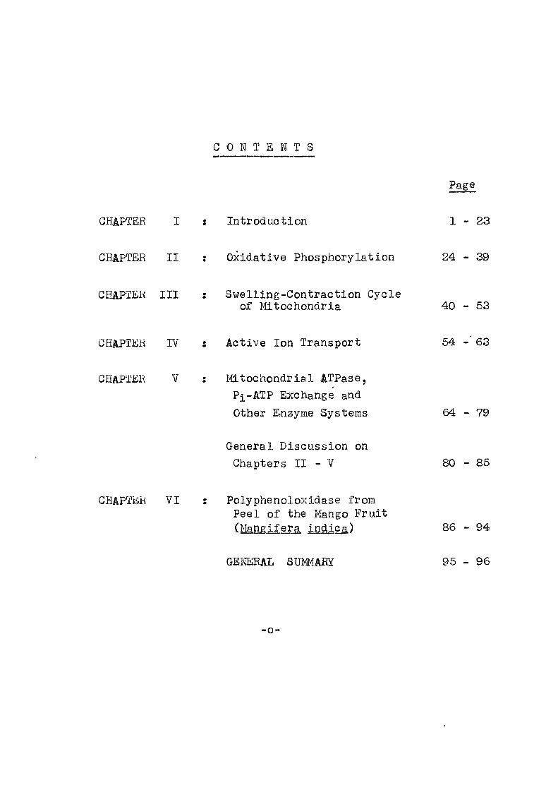
C O N T E N T S
CHAPTER
CHAPTER I I
Introduction
Oxidative Phosphorylation
Page
1 - 2 3
24 - 39
CHAPTER I I I : Swelling-Contraction Cycle of Mitochondria
CHAPTER IV
CHAPTER V
Active Ion Transport
Mitochondrial ATPase, Pi-ATP Exchange and Other Enzyme Systems
General Discussion on Chapters I I - V
40 - 53
64 - '63
6 4 - 7 9
80 - 85
CHAPTER VI Polyphenoloxidase from Peel of the Mango Fruit (Mangifera indica)
GENERAL SUMMARY
86
95
94
96
-o-

CHAPTER I
I N T R O D U G T I O N
I
CHAPTER I
lOTRODUGTlON
A process by which the energy of an oxidisable
substrate i s trapped in the form of high energy bond
phosphate molecule is ca l l ed oxidative phosphorylation.
This is the mechanism by which the c e l l stores the
energy made available by b io l og i ca l oxidations. In i t s
simplest form, the reaction can be written as f o l l ows :
m^-^ B 4- ADP + Pi > A+ BH2 + ATP 4- HgO
The energy is stored in the form of adenosinetriphos-
phate (ATP) a compound f i r s t described by Lohman^. I t
can be said at the outset that in spite of i t s funda-
mental importance, this mechanism is not yet completely
understood,
2 Hacker in one of his lectures has divided
the history of oxidative phosphorylation in four stages -
(1) Recognition of the phenomenon (2) Quantitative
evaluations, (3) Local isat ion of s i tes and (4) Resolution
and reconstruction.
Although the f i r s t demonstration of ATP 3
synthesis coupled to respirat ion wasty Engelhardt in 4
1930, i t was Kalckar who showed in 1939 that phos-
phorylation of AMP took place when c e l l f ree horaogenates
of kidney and other tissues oxidised c i t r a t e , glutamate, g
fumarate or malate, Lipmann about the same time showed
that pyruvate oxidation by Bacterium gul f ruch i i i s coupled
to phosphorylation, He put forward a generalised scheme of
ce l lu lar energy transfer by means of high energy phosphate
compounds.
Next contribution in this study of oxidative 6
phosphorylation was made by Ochoa and Be l i t zer and 7
Tsibakoy who demonstrated that more than one atom of
phosphorus was e s t e r i f i e d for every atom of oxygen consumed
during respirat ion.
The interpretation of phosphate e s t e r i f i e d was
complicated in the early period with two side reactions in
the tissue preparation used-one was the action of adenosine
triphosphate and the other was the action of kinases both of
which could u t i l i s e the ATP that was formed during the
react ion. Experimental procedures have now been devised
to get over the d i f f i c u l t y . The amount of phosphate e s t e r i f i ed
i s determined either by measuring the amount of phosphate
disappearing from the reaction medium (and evidently being
incorporated into ATP) or the amount of e s t e r i f i e d phosphate
formed is estimated enzymatically by coupling with other 8 9 reactions' •
The stage of loca l isat ion of s i tes can be said to have
started with Lehninger's ident i f i ca t i on of mitochondria as the
sub-oellular structures in which oxidative phosphorylation
took place. Although mitochondrion is the' exciusive s i t e
of oxidative phosphorylation there are reports about nucleus
also exhibiting such a propertj-^. Work in laboratories e
of "Lehninger, Lardy, Green, Hunter and Kiel^y have given
vaij-ues for P/0 rat ios of d i f f e r en t substrates as given in
Table l a . Slater^^, however, has cautioned against
laying too much emphasis on getting rat ios of theoret ical
values. There are reports in l i terature where rat ios
higher than theoret ical are mentioned-^'^, Lynn and
Brown have reported values more than 4 for succinate.
The values are yet to be explained.
Table l a ; P/0 rat ios of oxidations catalysed by iso lated mitochondria (Slater ) "
Substrate Observed P/0 ra t i o
o(-ketoglutarate 3-4
/3-hydroxybutyrate 2-3
Succinate 1-2
d -glycerophosphate 1-2
Pyruvate 2-3
Glutamate 2-3
13
Friedkin and Lehninger had made the important
observation that the phosphorylation i s linked to oxidation
of DPNH to oxygen in the absence of intermediates of
4
Krebs oycle. Rates upto 2,6 - 3,0 were obtained when
DPNH was oxidised with mitochondria that were exposed to
hypotonic solutions. Otherwise, DPNH, externally added
did not penetrate the mitochondria.
Oxidative phosphorylation process thus was shown to
consist of two reactions. One i s the oxidation or substrate,
i . e . , dehydrogenation, and second is the concomitant phos-
phorylation. The f i r s t reaction is now come to be known
as electron transport and the set of the enzymes that carry
out this reaction is ca l led electron transport chain.
For the f i r s t property exhibited i t i s not necessary that
the mitochondrial structure be intact , Keil in-Hartree
f ract ion carries out oxidation of succinate whereas fo r
phosphorylation to be obtained the mitochondrial membrane
structure has to be par t ia l l y inctact .
Electron Transport Chain
When a sub-cellular component l ike mitochondrion was
shown to be connected with such an important function l ike
energy conservation, i t was but natural that attempts fo r a
more detai led study of the mechanism of energy production
were soon undertaken. Work in Green's laboratory has mainly
contributed towards the understanding of the constituents of
the electron transport chain. The main aim in these studies
was to separate the electron transport chain into i t s compo-
nents in such a way that the or ig ina l a c t i v i t y could be
restored by recombination. As the enzyme systems connected

were found to be particulate in nature a var iety of t r ea t -
ments both physical and chemical in nature were necessary
before the various components could be separated retaining
considerable or ig ina l a c t i v i t y . Treatments employed the
use of sonicator for mechanical disruption and osmotic
l ys i s , digitonin and deoxycholate, Triton x 100 and some-
times organic chemicals l ike butanol and acetone have been
employed. The accompanying scheme diagramatically represents
the condensed procedure f o r iso lat ion of the complexes of the 14 electron transfer system from mitochondria •
With d i f f e rent procedures employed fol lowing four
eomplexes have been iso la ted ,
(1 ) Reduced NAD G© Q reductase
(2 ) Succinate Gfcy Q reductase
(3) Reduced Go JJ cytochrome G reductase and
(4 ) Gytochrome G oxidase,
Kach of the complexes i s enzymatically act ive in the
isolated form and under proper conditions could be recombined
In the study of these complexes use of a r t i f i c i a l electron
acceptors has been extremely useful. Among the compounds
employed are 2,6 dichlorophenol indophenol, ferr icyanide
and phenazine methosulphate, On a number of occasions,
a c t i v i t i e s towards these dyes undergo considerable variat ion
due to some changes in the protein structure of the enzyme
during iso lat ion procedures. Hence the interpretat ion
of data obtained with these dyes is rather complicated.

MitoGhondria
Ideoxycholate I KGl
I I
Red supernatant
I I I
deoxycholate ammonium acetate
i I
NADH - Gy t oc hr ome reductase
I Gholate ammonium sul fate
5 I i I
$ i
NADH-GO q reductase
( I )
" 1 i
I i Gytochrome G
reductase
i I
Supernatant
I i I I X
1 I
( I I I )
SuGClnate-GO q
reductase
( I I )
1 I
Green residue
i £ I I f I 5 I I I I I I I V I I $
Deoxy-cholate KGl Ammonium^ sul fate
Gytochrome oxidase
(IV)
TEEATMENT OF MITOGHONDRIA FOR ISOLATION OF GOMPLEXES
Besides tjiese lour complexes that are isolated
from the elementary par t i c l e , phospholipid, structural
protein and nonheme iron are the other components of this
electron transport chain.
In mitochondria ra t i o of l i p i d : protein is about
0.4 : Major parts of this l i p i d i s phospholipid.
The fa t t y acid components are highly unsaturated making them
susceptible to peroxidation by oxygen . When these phospho-
l ip ids are extracted with acetone, the elementary par t i c l e
of the mitochondria loses i t s ac t i v i t y that can be restored
by adding back the phospholipid^^. The function
of these phospholipids i s assumed to be providing a non-
aqueous medium for the linking of electron f l o y t o ATP
synthesis.
The structural protein which constitutes between
50-70% of mitochondrial protein was f i r s t iso lated in 20
Green's laboratory . I t i s a small molecular weight
protein (mol. wt, about 22,000) and has no enzymatic
a c t i v i t y . I t can interact with phospholipid involving
hydrophobic bonds. I t can also combine with cytochromes.
This structural protein phospholipid complex i s the one
in which other functional complexes and components of the
electron transfer chain are embedded. •
I t has been also possible recently to separate
mitochondria into inner and outer membrane f rac t ions . The
electron transport machinery i s found to be located in the
7
inner membrane of the mitochondria. The exact orientation
of the enzymes, involved in ox ida t i ve^ phosphorylation, on
these two membrane fract ions i s s t i l l not very cearly
sottlei^^'
Besides these studies on separation and reconstruc-
tion a detai led picture of the composition, structural as wel l
as functional of the respiratory chain was possible due to
some very ingeneous experimental techniques developed by
Chance and his colleagues. With the use of an extremely
sensit ive double beam spectrophotometer, they were able to
record d i f f e r en t spectra of the electron carr iers in intact
mitochondria. As is known NAD, MDF, f lavin^s)and cyto-
chromes are the sequential electron carr iers in the mito-
chondria. By enzymatically reducing the respiratory
chain of the intact mitochondria with the help of an ox i -
disable substrate, Chance and his co-workers recorded
di f ference spectra of the respiratory car r i e rs . They also
calculated the absolute concentrations of each of the
carrier in intact mitochondria and found that the cyto-
chromes, f lavin^s)were in simple molar ra t ios to each other.
Chance has also deduced functional sequence of the respiratory
carr iers . As these carr iers in s i tu are in dynamic steady
state, from the state of these respiratory carr iers in
presence of excess substrate ADP-phosphate and oxygen, their
re la t ive oxidation reduction state agrees with the gradient
of the normal oxidation reduction potent ia l of the car r i e rs .

Progress in the understanding of the phosphorylating
machinery has however been slow to come by when compared to
the studies of respiratory chain. The amount of ATP formed,
the s i te of i t s formation and the mechanism of formation are
some of the problems involved.
Sites of phosphorylation
Location of phosphorylation s i tes has been achieved
by independent work in the laboratories of Lehninger and
Chance. With the help of ascorbate and D-/3-hydroxy
butyrate;, jLehninger advocated one phosphorylation s i t e a f t e r
cytochrome C and two between NAD and cytochrome G, With
the help of crossover theorem ennunciated by Chance and
Williams^^ they were able to locate the three s i tes of
phosphorylation as f i r s t between NADH and f lavoprote in , second
between cytochrome b and c and third between cytochrome a
and oxygen. Recently the f i r s t s i te has been located
between two f lavoproteins which (the f lavoprote ins ) pp
according to Chance et a l function in the respiratory
chain with amytol and rotenone s i tes between them.
Just as in the case of respiratory chain,
studies with subraitochondrial systems that could catalyse
oxidative phosphorylation have contributed to paiJti&l po
'resolution, Pinchot working with bacter ia l systems
demonstrated the f i r s t par t ia l resolution of oxidative
phosphorylation. Racker and his co-\vorkers over the last
10 years have isolated number of protein coupling factors
24 soluble in nature that they designate as F^, F^, Fg 25 and F4 which participate in oxidative phosphorylation.
The f i r s t soluble coupling factor was resolved by shaking 26
mitochondria with glass beads in a Nossal Shaker . This
factor catalyses hydrolysis of ATP, These factors accord-s '
ing to Kacker ' have a dual function participating as
catalyst and also as structural blocks of mitochondrial
membrane.
Theories of oxidative phosphorylation
T i l l a few years back the only theory put
forward f o r the mechanism of formation of ATP was the
chemical one which envisaged the formation of a high 28,29,30,31
energy intermediate. In spite of number of attempts
i t is worthwhile noting that no undisputed iso lat ion
of such an intermediate either phosphorylated or non- 32 phosphorylated has been reported in l i t e ra ture . Slater
has discussed the recent status of these Intermediates.
While these attempts were continuing^ Mitche
introduced and developed an al ternat ive theory now known
as chemiosmotic theory explaining the mechanism of
oxidative phosphorylation. In i t s simplest form the theory
based on proton translocation phenomenon postulates that
ATP is synthesized by reversal of ATPase which is
situated in the membrane^^. The membrane i s supposed
to have highly speci f ic permeability characterist ics
10
towards H and 0H~, thus allowing the ions
resulting ATP synthesis to escape into two d i f f e rent
direct ions. Looking at both the above theories rather
cr i t i ca l ly^ i t becomes apparent that they are not so
very d i f f e r en t . On the other handthe chemiosmotic
theory i s in a position to propose a mechanism of
^ 1 formation that i s vaguely described in the
chemical theory. Chance has recently given his
c r i t i c a l assessment of the two theor ies.
The third alternate theory which i s currently
being postulated is based on conformational changes 37 38 in the mitochondria. Boyer and Lehninger (Mito-
chondrion^ for sometime introduced this notion as an
alternate to the chemical high energy intermediate 39,40,41
theory. Green and his co-workers have
recently with the help of electron microscopy put
forward evidence for conformational basis of energy
conservation. The fol lowing scheme summarises the
energy transformations carried out by the inner membrane
of the mitochondria.
I I I
Electron transfer ^ Energised state'^r^ ATP
Work performances l ike ion translocation swel l ing-contraction, e t c .
17
Aocording to this theory mitochondria oan
exist in three configurational states - non-energised,
energised and energised twisted, Electron transfer and
hydrolysis of ATP are the two enzymic means of generat-
ing the energised s ta te .
In spite of the three theories mentioned
above no one particular mechanism of energy conservation
has been adequately demonstrated to warrant the exclusion
of the other two. I t i s quite possible that a l l the
three may be operative either simultaneously or under
d i f f e ren t conditions.
Other energy dependent mitochondrial reactions
Although formation of ATP is a mkjor pathway
of conserving energy released dnring electron transport
this energy is also u t i l i s ed for other reactions l ike
active accumulation of anions and divalent cations,
swelling contraction phenomenon,
( i ) After the ear l i e r observations that ions + 2.+
l ike K and Mg can be retained by the isolated mito-
chondria, so long as respirat ion i s continuing, considera-4 9 43
ble amount of work by Bartley , Vasington and others have, established that act ive ion uptake can be brought
about by energy produced either during substrate oxidation
or ATP hydro lys i s^ . This Ga ^ uptake requires the
u
presence of anion phosphate in the suspending medium
and inside the mitoohondria Lehninger has demonstrated
that deposition of trioalcium phosphate occurs and
there i s a constant r a t i o of the amount of Ga and
phosphate accumulated at a value of about 1,6, I
Although most of the work on act ive ion uptake i s done
with Ga "*" , studies with Mg^^, Sr'^^' Mn^and
K accumulation are also reported, Ultrastructural
changes in mitochondria a f te r cation uptake are An, AO also described^''* . This ion uptake is shown to be
49 60 accompanied by proton e ject ion by mitochondria » *
( i i>.. The other property connected with the
respiratory state of the mitochondria i s the swelling
contraction exhibited by mitochondria in v i t r o under
certain experimental conditions. Mitochondrial swelling
can be induced by a variety of chemical compounds l ike
inorganic phosphate^^, Ga " , ferrous ion^^, thyroxine^^, 51 54 glutathione f ree f a t t y acids and hormones l ike
oxytocin, vassopressin, parathyroid hormone^^ and soma-
totrophin. Mitochondria swollen under d i f f e r en t conditions 51
can be made to contract in presence of ATP or under some
conditions in presence of substrates l ike succinate which
of course denotes the energy dependent nature of this
phenomenon. During swelling some internal components
leach out of the mitochondria, tehninger has characterised

13
some of them^^ and designated G * and M^ factors which
play a role in the phenomenon.
( i i i ) During the study of oxidative phosphoryla-
tion some exchange reactions l ike P^-ATP exchange, IDP-ATP 1R 59 n c
exchange Hg- ^O-Pji exchange Hg- O - ATP exchange have given
some valuable information. Pi-ATP exchange reaction v/as 60
f i r s t observed by Boyer and his coworkers . These reactions
take place in v i t ro in absence of net electron transport.
Hence to show i t s re lat ion to oxidative phosphorylation
considerable cirGumstantial evidence was necessary among which the e f f e c t of unciuplers and inhibitors played a p part, Racker , however, has pointed out certain important
restr ic t ions during their interpretat ions. These reactions,
however, have yielded useful information in the study of
soluble factors participating in oxidative phosphorylation,
( i v ) The another par t i a l reaction of oxidative
phosphorylation which has been studied in de ta i l is the ATPase
ac t i v i t y of the mitochondria. I t s involvement in oxidative
phosphorylation was suggested f i r s t by lardy and Elvehjem®^.
As this enzyme can be stimulated in i t s ac t i v i t y by number of 2A-
compounds l ike 2,4 dinitrophenol, Mg^ , Ga , arsenate,
i t raised doubts a^to whether more than one ATPase exists
in the mitochondria. This enzyme has been solubi l ised and 62 studied in considerable de ta i l by Racker and his co-workers .
The ATPase enzyme af ter pur i f i cat ion becomes cold l ab i l e
and loses i t s sensi t iv i ty to oligomycin.
14
Inhibitors
The study of oxidative phosphorylation has been
considerably helped by the use of certain chemical
compounds -which have been found to in te r f e re with the
oxidative phosphorylation process in the isolated mito-
chondria. These compounds are generally c l ass i f i ed
into three categories - 1. Inhibitors of electron
transport, 2. Inhibitors of coupled phosphorylation,
3, TJhcouplers of oxidative phosphorylation. In the
f i r s t category are compounds l ike amytal, antimycin,
azide, rotenone, cynide, and carbon monoxide. Each of
these compounds acts at a particular s i te along the electron
transport chain. .Their use has considerably helped in
the studies of electron transport chain. More compounds
l ike piercidine are recently added to this l i s t . Singer
and his coworkers have carried out studies on binding
of M D H dehydrogenase by rotenone and piercidin®^'®"^.
According to them, these bind the enzyme at spec i f i c
and nonspecific s i t es . Succinate oxidation is also
inhibited by piercidin®^*
One of the ear l i e r compounds belonging to second
category was guanidi'ne studied by Hollunger®^ in 1956.
At the use of this compound required preincubation and
also high concentration not much work with this i s reported.

15
Soon afterwards Lardy and his coworkers studied a number
of ant ib iot ics and found one of them oligomycin®'^'®^ to
be a potent inhibitor of energy transfer reactions.
Subsequently another ant ib io t i c a t racty los i te was
reported by Vignais, Vignais and Stranislas^^'"^^. 71
Bruni and his coworkers • to belong to the same class.
This type of compounds besides inhibit ing the formation
of ATP were also found to in t e r f e re with other energy
transfer reactions l ike ion uptake and s-welling contraction.
They also inhibited exchange reactions, and inhibited
ATPase a c t i v i t y , stimulated by various agents, reactions
both known to be connected with oxidative phosphorylation.
These compounds do not inhib i t oxidation by the uncoupled
mitochondria. The s i t e of action of atractyloside i s
recently placed below oligomycin in the phosphorylating
machinery. To this l i s t of compounds are recently added 72 7*5 formaldehyde and hydroxylamine .
A c lass ical example of the compounds belonging to
the third category is 2,4 dinitrophenol. These compounds
in appropriate concentrations have no e f f e c t on mito-
chondrial respiration but inh ib i t , or uncouple associated
phosphorylation of AD?. Hemker has carried out detai led 74 75
experiments to study action of uncouplers ' . Recently
Weinbach and Garbas have indicated that these compounds
16
especial ly 2,4~dinltroplienol may be acting as uncoupling
agents by causing conforraational transit ions in the
catalyt ic proteins of mitochondria.
8U0PE OF TIffl PRSSEIMT INVESTIGATION
/G-naphthoxy acet ic acid and some chloro
substituted phenoxy acids l ike 2:4 dichloro phenoxy
acetic acid, 2,4,5-trichlorophenoxypropionic acid
are used as plant gro-v jth regulators. Some of these
are often employed in agriculture as a pre-harvest spray
to prevent f ru i t drop. They are also e f f e c t i v e in
enhancing thfe color development in some f r u i t s . Brody" *
had shown in 1951 that p-chlorophenoxy acet ic acid and
2:4 dichlorophenoxy acet ic acid -were able to lo"wer the
oxidative phosphorylation capacity of rat l i v e r mitochondria
in v i t r o .
For the investigations presented in the fol lowing
chapters, the compounds selected were - ^-naphthoxy
acetic acid, p-chlorophenoxy acet ic acid, 2:4 dichloro-
phenoxy acetic acid, 2:4:5 trichlorophenoxy acet ic acid
and 2:4:5 trichlorophenoxypropionic acid. The action of
these plant grovjth regulators on the rat l i v e r mitochondrial
properties and other enzyme systems were studied in de ta i l
and are presented.

IT
Chapter I I describes the -e f f ec t of these
compounds on ojjidative phosphorylation by rat l i v e r
mitochondria. Won-phosphorylating mitochondria were
also employed to study the e f f e c t only on oxygen uptake,
Sncclnnte and glutamo.te were used as substrates»
Chapter I I I gives the e f f e c t of phenoxy acids on swelling
contraction cycle of rat l i v e r mitochondria. Mito-
chondria swollen in presence of Ca , inorganic phosphate
and thyroxine -wereybontracted in the presence of ATP.
Influence of the above compounds on ATP action was studied.
In Chapter IV is described the action of these acids on
active ion transport in rat l i v e r mitochondria.
The other mitochondrial reactions studied were
ATPase a c t i v i t y and Pi-ATP exchange. Besides, oxidative
phosphorylation reaction, a few other mitochondrial and
extramitochondrial enzymes were tested f o r the e f f e c t of
the above acids on their a c t i v i t y . The enzyme systems
included aminotransferase, catalase, myokinase, serum
phosphatases and muscle ATP-creatinine transphosphorylase.
The results are described in Chapter V.
Evaluating the data presented in the above
Chapters, i t i s suggested that these compounds a f f e c t the
energy-transfer-reaction in rat l i v e r mitochondria and
24
'mnk^ thus their/actiori is qua l i ta t i ve l y similar to that of
A
ant ib iot ics l ike oligomycin and atracty los ide . Addition
of nevj compounds to a l i s t of known inhibi tors or
regulators is always useful in that they contribute to
the knowledge of reaction mechanisms.
In Chapter VI i s given the pur i f i cat ion of the
enayme polyphenol oxidase from the peel of the f r u i t
Mangifera indica. Some of i t s properties l ike pH
optimum, Km, substrate s p e c i f i c i t y and metal requirement
have been studied.
19
REFERENCES
1. Lohman, K. (1931), Biochem. Z., 460
2. Racker, E. (1965), Mechanisms in Bioenergetics,
Academic Press.
3. Engclhardt, ¥.A,E. (1930), Biochem. Z., 227, 16.
4. Kalckar, H. (1939), Biochem. J . , 631.
5. Lipmann, F. (1939), Nature, 281. o 6. Ochaa, S. (1941), J.Biol.Chem., 751.
7. Be l i t ze r , V.A. and Tsibakoya (1939), Biochimiya, 4, 516.
8. Nielsen, S.O. and Lehninger, A.L. (1955),
J.Biol.Chem., 215, 555.
9. S later , E.C. (1953), Biochem.J., 53, 521.
10. A l f r ey , V.G. and Mirsky, A.E. (1958), ProC.Natl.Acad.Sci. (Ua&), 4A, 981.
11. S la ter , E.C. in Comprehensive Biochemistry (1966), Ed. by Florkin, M. and Stotz, E.H. p.334.
12. Lynn, ¥.S. and Brown, R.H. (1965), Biochim.Biophys.Acta. 105, 15.
13 . Friedkin, M. and Lehninger, A . L . ( 1 9 4 8 ) , J.Biol.Chem., 178, 611.
14. Hate f i , Y. (1966) in Comprehensive Biochemistry, Ed. by Florkin, M. and Stotz, E.H. Academic Press, p.204.
15. Hate f i , Y . , Haavik, A.G., Fowler, L.R. and Gr i f f i t h s , D.E. (1962), J.Biol.Chem., 237, 2661.
16. Hate f i , Y. (1963), Advances in Enzymology, 25, 275.
17. Fleischer, S., Klouwen, H. and Br ier ley , G.P.(1961), J, B io l . Chem., 2 ^ , 2936.
20
18. Holman, R.T. and Widmer, C. (1959), J.Biol.Chem., 234, 9.
19. Br ier ley , G.P. and Merola, A.J. (1964), Biochim. Biophys. Acta, 64, 281.
20. Griddle, E.G., Bock, R.M., Green, D.E. and Tisdale, H. (1962), Biochemistry, 1, 827.
20a. Allmann, D.W., Bachmann, E., Orme-Johnson, N., Tan, ¥.C. and Green, D.E. (1968), Arch.Biochem.Biophys., 125, 981.
20b. Green, D.E., Allmani?, D.¥., Harris, R.A. and Tan, ¥.C. (1968), Biochem.Biophys.Res.Comm., 31, 368.
21. Ghance, B. and Williams, G.R. (1956), Advan.Enzymol., 17, p.65.
22. Chance, B., Ernster, L. and Garland, P .B. , Lee, G.P., Light, P.A. , Ohnishi, T . , Ragan, C.T. and Wong, D. (1967), Proc. I fat l .Acad.Sci . (US), CT, 1498.
23. Pinchot, G.B. (1953), J.Biol.Chem., 205, 65.
24. Linnane, A.W. and Titchaner, E.B. (1960), Biochim.Biophys.Acta, 39, 469.
25. Conover, T.E., P ra i r i e , R.L. and Racker, E. (1963), J.Biol.Chem., 2 ^ , 2831.
26. Penefsky, H.S., Pullman, M.E., Datta, A. and
Racker, E. (1960), J.Biol.Chem., 2 ^ , 3330,
27. Racker, E. (1967), Federation Proc. , 26, 1335.
28. G r i f f i t h s , D.E. (1963), Federation Proc . , 1064.
29. Brodie, A.F. (1961), Federation Proc . , 20, 955.
30. Purvis, J.L. (1958), Nature, 182, 711.
31. Pinchot, G.B. (1963), Federation Proc . , 22, 1076. 32. S later , E.G. (1966) in Comprehensive Biochemistry,
Vol.14, Ed. by Florkin, M. and Stotz, E.H., Academic Press, p.382.

Zi
33. Mitchel l , P. (1961), Nature, 1 ^ , 144.
34. " (1966), Bio i .Rev. , 41, 445.
35. " (1966), Chemiosmotlc coupling in oxidative and photosynthetic phosphorylation. Bodmin Glynn Research Ltd,
36. Chance, B,, Lee, C.P. and Mela, L, (1967), Federation Proc. , 26, 1341.
37. Boyer, P.D., Bieher, L .L . , Mitchel l , R.A. and Szabolcs, G. (1966), J.Biol.Chem., 5384.
38. Lehninger, A.L. (1964), The mitochondrion, Benjamin Inc . , N.Y.
39. Penniston, J.T. , Harris, R.A. , Asai, J. and Green, D.E. (1968), Proc.Natl .Acad.Sci . (USA), 59, 624.
40. Harris, R.A., Penniston, S .T . , Asai, J. and Green, D.E. (1968), Proc.Natl.Acad.Sci. (USA), 59, 830.
41. Green, D.E., Asai, J . , Harris, R.A, and Penniston, J.T, (1968), Arch.Biochem.Biophys., 125, 684.
42. Bartley, W. and Davies, R.E. (1954), Biochem.J., 57, 37.
43. Vasington, F.D. and Murphy, J.V. (1962),
J .B io l . Chem., 2670.
44. Lehninger, A.L. (1968), Advan.Enzymol., 29, 259.
45. Br ier ley , G., Murer, E., Bachmann, E. and Green, D.E,, (1963), J.Biol.Chem., 3482.
46. Carafo l i , E., Weiland, 8. and Lehninger, A.L, (1965), B^ochim.Biophys.Acta, 97, 88,
46a. Carafo l i , E., (1965), Biochim.Biophys.Acta. £7, 99, 47. Greenawalt, J.W., Ross, C.S. and Lehninger, A.D. (1964),
J .Ce l l .B i o l , , 21,
48. B r i e r G . P , , Slautterback, D.B., (1964), Biochim.Biophys.Acta, 82, 183.
49. Rasmussen, H., Chance, B. and Ogata, E. (1965), Proc.Natl ,Acad,Sci, , 1069.
22
50. Judah, J.D., Ahmed, K. , Mclean, A.E.M. and Christ ie, G.S. (1965), Biochim.Biophys.Acta, 94, 452.
0353) 51. Lehninger, A . I . , Biol.Ciaem., 2M, 2465.
52. Chappell, J.B. and Crofts, A.R. (1965), Biochem.J., 95, 378.
53. Glick, J.L. and Bronk, J.R. (1965), Biochim.Biophys.Acta, 23.
54. 2boro\i;ski, J. and ¥ojtczak, L. (1963), Biochim.Biophys.Acta, 70, 596.
55. Rasmussen, H., Fischer, J. and Arnaud, C. (1964), Proc.Natl.Acad.Sciences, 1198.
56. Vignais, P.M., Vignais, P.V. and Lenhinger, A.D. (1964), J.Biol.Chera., 2011.
57. Vignais, P.V., Vignais, P.M. and Lehninger, A.L. (1964), J.Biol.Chem., 2002.
58. Lehninger, A.L. and Gotterer, G.S. (1960),
J.Biol.Chem., ^ 5 , pc.8.
59. Falcone, (1964), J.Biol.Chem., 1954.
60. Boyer, g .D., Luchsinger, ¥.W. and Falcone, A.B. (1956), J.Biol.Chem., ^ 3 , 405.
61. Lardy, H.A., Elvehjem, C.A. (1945), Ann.Rev.Biochem., M , 1.
62. Backer, E. (1967), Federation Proceedings, 26, 1335.
63. Horgan, D.J., Ohno, H., Singer, T.P. and Cassida, J.E. (1968), J.Biol.Chem., ^ 3 , 5967.
64. Palmer, G., Hoorgan, D.J., Tisdale, H., Singer, T.P. and Beinert, H. (1968), J.Biol.Chem., 844.
65. Jeng, M. and Crane, F.L. (1968), Biochim.Biophys.Res.Coram., 465.
66. Hollunger, G. (1955), Acta.Pharm.Toxicol., Suppl. 1, 1.
33
67. Lardy, H.A., Johnson, D. and McMurray, ¥.C. (1958), Arch.Biochem.Biophys., 78, 587.
68. Lardy, H.A., Connelly, J.L. and Johnson, D. (1964), Biochemistry, 3, 1961.
69. Vignais, P.V., Vignais, P.M. and Stranislas, E. (1961), Biochern.Biophys.Actn- 51- 394.
70. Vignais, P.V. , Vignais, P.M. and Stranislas, E. (1962), Biochim.Biophys.Acta, 60, 284,
71. Bruni, A . , Contessa, A.R. and Luciani, 3. (1962), Biochim.Biophys.Acta, 301.
72. Van Buskirk, J.J. and F r i s e l l , W.R. (1967), Biochim.Biophys.Acta, 292.
73. Wikstrom, M.K.F. and Saris, N.E.L. (1969),
European,J.Biochem., 9, 160.
74. Hemker, H.C. (1962), Biochim.Biophys.Acta., 46.
75. " " (1964), Biochim.Biophys.Acta., 81, 1.
76. Weinbach, E.G. and Garbas, (1969), Nature, 2 ^ , 1016.
77. Brody, T.M. (1952), Proc.Soc.Exptl.Biol,Med., 533,
CHAPTER I I
OXIDATIVE PHOSPHORYLATION
24
CHAPTER I I
OXIDATIVE PHOSPHORYLATION
Introduction
In the study of oxidative phosphorylation, use
of various chemical compounds as inhibitors has yielded
valuable information. This has helped to understand
considerably the mechanism of phosphorylation. Guani-
dines and i t s derivatives^ -were the f i r s t compounds
to be employed as inhibitors of oxidative phosphorylation, the\>
but tjiig use was not continued because of high concen-
tration required and the necessity of preincubation f o r
a long time of the mitochondria in the presence of these
compounds.
Action of oligomycin and other ant ib iot ics l ike
nonactin and a l l i ed compounds on oxidative phosphorylation
and connected reactions i s extensively studied by Lardy 2 3 4
and his coworkers ' ' . These compounds are primarily
known to a f f e c t the phosphorylative react ion. When the
mitochondria -were uncoupled, the oxygen uptake in
presence of krebs cycle substrates was not appreciably
a f f ec ted . Thus this series of compounds were dist in-
guished from other inhibitors of mitochondrial oxidations

'i:
l i ke rotenone and antimycin which inhibit primari ly
the electron transport reactions of mitochondria.
Another compound fo r -which a similar action i s
•well recorded is atracly loside which i s a glycoside
isolated from Atracty l is gummifera®. Although
atractyloside inhibits phosphorylation just l i ke
oligomycin, there are certain di f ferences between the
two. The amount of oligomycin bound by the mitochondria the
i s fa r in excess of that required for i i s inhibi t ion of
phosphorylation whereas the amount of atractyloside
taken up (about 150 m^mole/g. protein) i s about the
amount required fo r complete inhibit ion of oxidative
phosphorylation . Moreover unlike oligomycin,
atractyloside does not inhib i t phosphorylation of
endogenous ADP. In the various steps leading to ATP
formation, the s i t e of action of atractyloside i s at present
indicated below that of oligomycin.
Uncoupler of oxidative phosphorylation,[dinitro-
phenol at 1.6 x concentration, i s known to reverse
the inhibit ion of oxygen uptake caused by oligomycin in
phosphorylating mitochondria^. Arsenate, however, does
not re l i eve inhibit ion of oligomycin"^.
I t has also been observed that l eve ls of o l igo-
mycin lower than those necessary for inhibi t ion of energy

6
l inked reactions actual ly stimulate ox idat ive phosphory-O Q
la t ion of sub-mitochondrial par t i c l es . Sanadi sho-wed
that this stimulation i s due to oligomycin at that
concentration protecting high energy intermediates.
Besides oligomycin and at racty los ide , the other
compounds known to inh ib i t oxidat ive phosphorylation by
mitochondria are formldetiyde^^ and hydroxylaiaiue^^.
In the present chapter are described the
e f f ec ts of beta-NOA and various phenoxy acids on
oxidat ive phosphorylation by coupled and uncoupled
mitochondria. These studies were carr ied out by employing
Warburg respirometer and an oxygen e lec t rode . The
resul ts are described in sections A and B respec t i ve l y .
Section A
Oxygen uptake studies with Warburg respirometer.
Experimental
Materials
Mitochondria; Throughout the work presented in
th is and the fo l lowing chapters, rat l i v e r mitochondrial
f r ac t i on was prepared by the method of Schneider and
HogeboomlS as described below.
'17
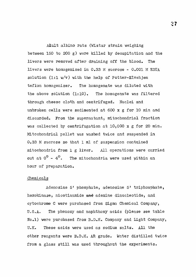
Adult albino rats (Wistar strain weighing
bet-ween 160 to 200 g ) were k i l l ed by decapitation and the
l i v e rs were removed a f t e r draining o f f the blood. The
l i v e rs were homogenized in 0.33 M sucrose - 0.001 M EDTA
solution (1:1 w/v) with the help of Potter-Elvehjem
te f l on homogenizer. The homogenate was diluted with
the above solution (1:10). The homogenate was f i l t e r e d
through cheese cloth and centri fuged. Nuclei and
unbroken ce l l s were sedimented at 600 x g f o r 10 min and
discarded. From the supernatant, mitochondrial f rac t ion
was col lected by centri fugation at 10,000 x g for 20 min.
Mitochondrial pe l l e t was washed twice and suspended in
0.33 M sucrose so that 1 ml of suspension contained
mitochondria from 1 g l i v e r . A l l operations were carried
out at 0® - 4° . The mitochondria were used within an
hour of preparation.
Chemicals
Adenosine 5' phosphate, adenosine 5' triphosphate,
hexokinase, nicotinamide a id- adenine dinucleotide, and
cytochrome C were purchased from Sigma Chemical Company,
U.S.A. The phenoxy and naphthoxy acids (please see table
No. l ) were purchased from B.D.H. Company and Light Company,
U.K. These acids were used as sodium sa l ts . A l l the
other reagents were B.D.H. AR grade. Water d i s t i l l e d twice
from a glass s t i l l was used throughout the experiments.

Table 1; Acids used as sodium sal ts in the present studies
Formula Abbrevi-Gorapound of the ated Molecular
compound as weight
/S-Naphthoxy /Q-NOA 202.21
p.Chloro-phenoxy p-GlA 186.5 acetic acid
2,4-Dichloro-phenoxy 2,4 D 221,05 acetic acid
2,4,5-Tr ichloro-phenoay C l - f Y o - C H ^ C O O H 2,4,5 TA 255.5 acetic acid CI
2 ,4 ,6-Tr i - C V ^ chloro-phenoxy c i v Vo-CH^CHgCOOH 2,4,5 TF 269.5 propionic acid CI
Methods
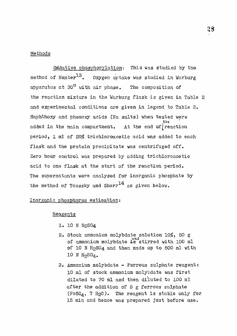
Q}ddative phosphorylation: This was studied by the 1 o .
method of Hunter-^*^. Oxygen uptake was studied in Warburg
apparatus at 30° with a i r phase. The composition of
the react ion mixture in the Warburg f l a sk i s given in Table 2
and experimental conditions are given in legend to Table 3,
Naphthoxy and phenoxy acids (Na sa l t s ) when tested were tWe.
added in the main compartment. At the end of j lreact ion
period, 1 ml of 20% t r i ch loroace t i c acid was added to each
f l a sk and the prote in prec ip i ta te was centri fuged o f f .
Zero hour control was prepared by adding t r i ch lo roace t i c
acid to one f l a sk at the start of the react ion period.
The supernatants were analysed f o r inorganic phosphate by 14
the method of Taussky and Shorr as given below.
Inorganic phosphorus estimation; Reagents
1. 10 N H2SO4 2. Stock ammonium molybdate solution 10$ , 50 g
wo-S
of ammonium molybdate ts s t i r r ed with 100 ml of 10 N H2SO4 and then made up to 500 ml with 10 W H2SO4.
3. Ammonium molybdate - Ferrous sulphate reagent: 10 ml of stock ammonium molybdate was f i r s t di luted to 70 ml and then di luted to 100 ml a f t e r the addition of 5 g ferrous sulphate (FeS04, 7 H2O). The reagent i s stable only f o r 15 min and hence was prepared just before use.
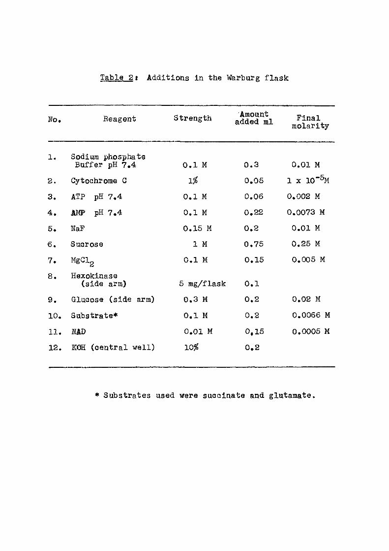
Table 2t Additions in the Warburg f l ask
No. Reagent Strength Amount added ml Final
molarity
1. Sodium phosphate Buffer pH 7.4 0.1 M 0.3 0.01 M
2. CytcQhrome G 1% 0.05 1 X 10"%
3. ATP pH 7.4 0.1 M 0.06 0.002 M
4. JIMP pH 7.4 0.1 M 0.22 0.0073 M
5. NaF 0.15 M 0.2 0.01 M
6. Sucrose 1 M 0.75 0.25 M
7. MgClg 0.1 M 0.15 0.005 M
8. Hexokinase (side arm) 6 mg/flask 0.1
9. Glucose (side arm) 0.3 M 0.2 0.02 M
10. Substrate* 0.1 M 0.2 0.0066 M
11. MD 0.01 M 0,15 0.0005 M
12. KOH (central we l l ) 10^ 0.2
• Substrates used were succinate and glutamate.

29
standard cal ibrat ion graph was prepared by using a standard solution of dipotassium hydrogen phosphate ( i X - 16/range) .
Procedure; 0.5 ml of the TCA supernatant from
the Warburg f lask -was made upto 2 ml with d i s t i l l e d
water. 2 ml of ammonium'molybdate-ferrous sulphate
reagent was added. The colour was read a f t e r 30 min at
room temperature in the Klet t Summerson colorimeter using
f i l t e r No.66, against the reagent blank. The colour i s
stable f o r 2 hours.
P/O was calculated as the ra t io of ^moles
inorganic phosphate disappeared to ^atom oxygen consumed
over a period of 20 min.
+ l'^ Protein estimation; Fol in CioCaltoau^ ^
Reagents;
1. Solution A (a ) Na2C03 : 15 g 5 5
(b) NaOH : 3 g 5 Dissolved 5 in 750 ml
(c ) Sodium potassium J of water tartrate : 0.74 g 5
2. Solution B GUSO4. 5 H2O : 2.5 g ifi-WAS dissolved in 500 ml of water.
3. Alkaline Cu solution : 50 ml of A • 1 ml of B.
4. Folin Ciocalteau reagent.
30
Into a 2 l i t r e f l ask were introdaced 100 g sodium
tungstate (Na2W04. 2 H2O), 25 g sodium molybdate 2 H2O,
700 ml water, 50 ml 85% phosphoric acid, 100 ml conc. HGl
and refluxed for 10 hrs. 150 g lithium sulphate and
50 ml v/ater along with a few drops of bromine were then
added. The mixture was boiled fo r 15 min to remove
excess bromine. A f ter cooling i t was made upto 1 l i t r e
with water and f i l t e r e d . Reagent should not have any
greenish t inge. This was diluted 1:1 with water before
use.
Method was
An aliquot/made up to 1 ml with water. To this
was added 5 ml of reagent 3 and after half an hour 0,5 ml
of diluted Fol in Ciocalteau reagent. The color developed
was measured a f te r half an hour in Klet t Summerson co lor i -
meter with F i l t e r No.66,
A standard curve was prepared with albumin in the
range (1 /ug - 100 ^ g ) .
Results
In this chapter are presented the results of the
e f f e c t of substituted phenoxy acids on oxidative phosphory-
lat ion property of rat l i v e r mitochondria. Table 3 gives
the P/O rat ios when succinate and glutamate were used as
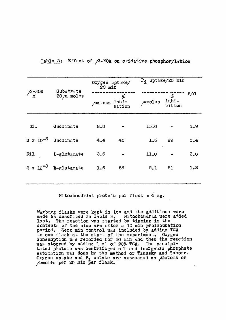
Table 3 ; E f f e c t of /3-NOA on oxidat ive phosphorylation
/ '3-lJOA M
Sabstrate 20/a moles
Oxygen aptake/ 20 min
Pjl uptak:e/20 min
/aatoms inh i - /imoles Jnhi-/ b i t ion ^ bit io i
P/0
b i t i on
Ni l Succinate
3 X 10"^ Succinate
Nil L-glutaraate
8.0
4 .4 45
3.6
3 X 10"^ B-glutamate 1.6 56
16.0
1.6
11.0
2.1
89
81
1.9
0.4
3.0
1.3
Mitochondrial protein per f l a sk s 4 mg.
Warburg f lasks were kept in ice and the additions were made as described in Table 2. Mitochondria were added l a s t . The reaction was started by t ipping in the contents of the side arm a f te r a 10 min preincubation period. 2ero min control was included by adding TGA to one f l ask at the s tar t of the experiment. Oxygen consumption was recorded f o r 20 min and then the react ion was stopped by adding 1 ml of 20^ TGA. The prec ip i -tated protein was centri fuged o f f and inorganic phosphate estimation was done by the method of Taussky and Schorr. Oxygen uptake and uptake are expressed as /iatoms or /umoles per 20 min per f l a s k .
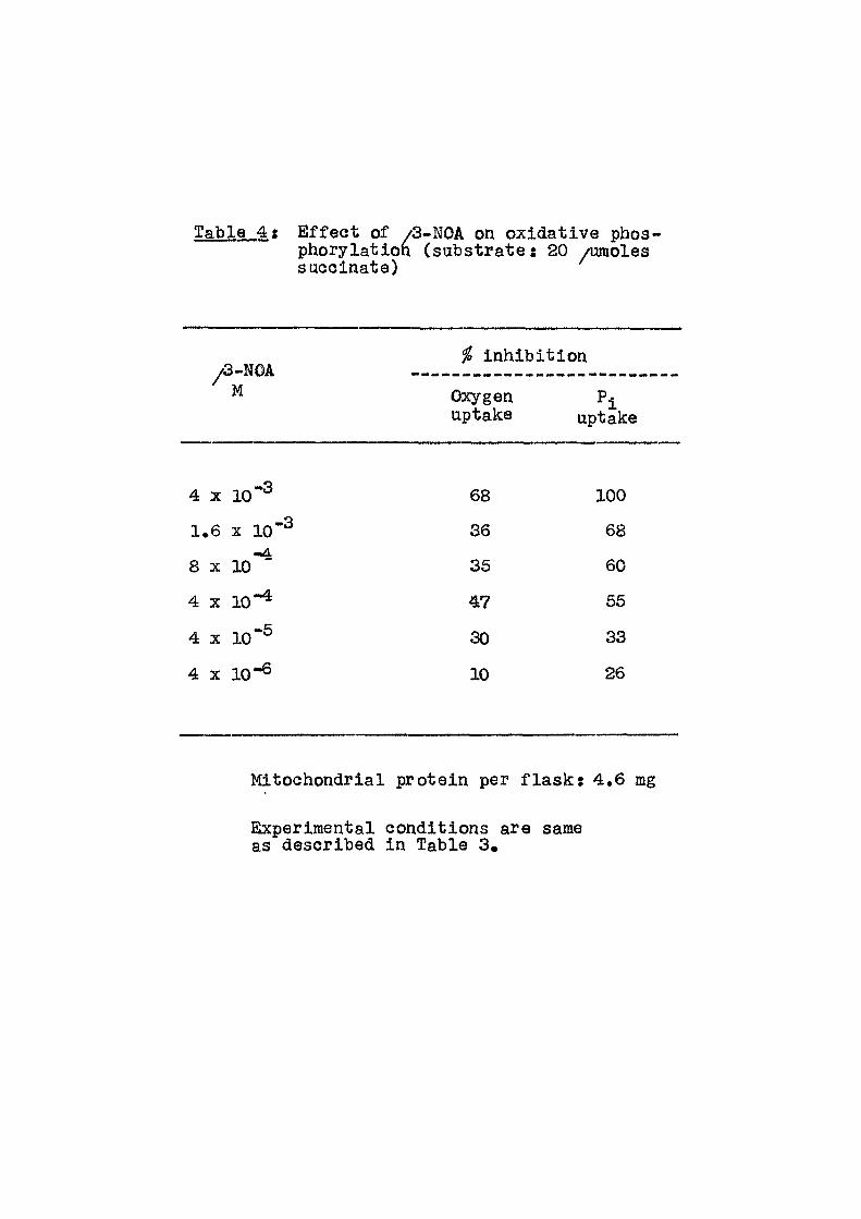
Table 4 ; E f fec t of E f fec t of /3-NOA on oxidative phos-phorylation (substrate: 20 /umoles suGoinate)
i inhibit ion /3-NOA
^ Oxygen P^ uptake uptake
4 X 10"^ 68 100
1.6 X 10"^ 36 68
8 X 10*^ 35 60
4 X 10"^ 47 55
4 X 10"^ 30 33
4 X 10"® 10 26
Mitochondrial protein per f lasks 4.6 mg
Experimental conditions are same as described in Table 3.
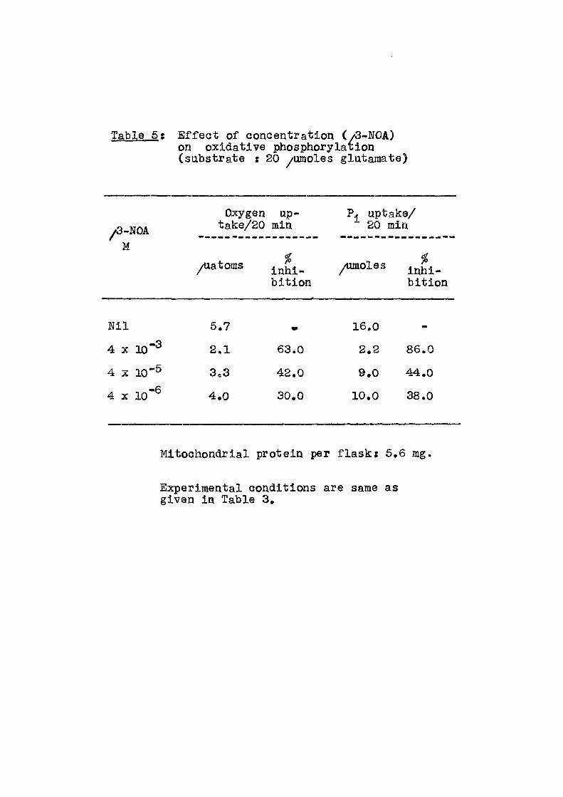
Table 5 : E f f e c t of concentration (/3-NOA) on oxidative phosphorylaiion (substrate : 20 /umoXes glutamate)
/3-NOA M
Oxygen up-take/20 min
P. uptake/ 20 min
/latoms bi t ion
inhi-bi t ion
N i l 6.7 w 16.0 -
4 X 10"^ 2.1 63.0 2.2 86.0
4 X 10"® 3,3 42.0 9.0 44.0
4 X 10"^ 4.0 30.0 10.0 38.0
Mitochondrial protein per f l a s k : 6,6 mg,
Experimental conditions are same as given in Table 3.

Table 6 ; E f f ec t of p-GlA concentration on oxidative phosphorylation
•Oxygen uptake/ p uutake/ P-CIA 20 min •^2o'min
% r - -
Ni l 7 . 1 1 6 . 0 - 2 . 2
4 X l o " ^ 3 . 7 4 8 6 . 4 60 1 . 7
- 3 1 . 6 X 10 4 . 2 4 1 1 0 . 8 33 2 . 5
8 X 1 0 " ^ 5 . 5 23 1 2 . 0 2 5 2 . 2
4 X 10 5 . 6 2 1 1 3 . 5 16 2 . 4
1 . 6 X 1 0 " ^ 5 . 7 20 1 0 , 5 3 4 1 . 8
4 X 10 5 . 6 2 1 1 2 . 0 2 5 2 . 1
4 X 1 0 " ® 5 . 6 2 1 1 4 . 0 13 2 . 5
• 20/umoIes succinate as substrate
Mitochondrial protein per f l a s k : 5.2 mg
Experimental conditions are the same as described in Table 3.

Table 7 ; E f f e c t of 2.4 D on ox idat ive phosphorylation
• Oxygen uptake P^ uptake/ 20 min 20 min
2 , 4 D M
/uatoms %
inh i -b i t i on
/umoles %
inh i -b i t ion
P / 0
Ni l 7 . 0 1 4 . 6 2 . 1
4 X 1 0 " ^ 2 . 7 6 1 M» 100 -
1 . 6 X 1 0 " ^ 4 . 2 4 0 2 . 0 8 6 0 . 6
8 X 1 0 " ^ 4 . 1 4 1 7 . 0 52 1 . 7
4 X I D " ^ 3 . 6 4 8 8 . 2 4 4 2 . 3
1 . 6 X 1 0 " ^ 3 . 8 4 6 1 0 . 5 28 2 . 7
4 X 1 0 " ^ 4 . 2 4 0 1 1 . 4 22 2 . 7
- 6 4 X 10 3 . 8 4 6 1 1 . 7 20 3 . 0
» 20/umoles suocinate as substrate
Mitochondrial protein per f l a sk s 5 mg
Experimental conditions are same as described i^i Table 3,
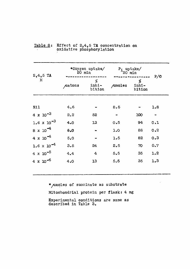
Table 8 : E f f e c t of 2,4,5 TA concentration on oxidative phosphorylation
2,4,5 TA M
•Oxygen uptake/ 20 min
yiiatoms inhi -b i t ion
Pi uptake/ 20 min
P/0 i
Aunoles inhi-b i t ion
N i l 4.6 - 8.5 - 1.8
4 X 20-3 2.2 52 mm 100 -
1.6 X 1 0 " ^ 4.0 13 0.5 94 0 .1
8 X 10-^ €.0 1.0 88 0.2
4 X 10-^ 5.0 «w 1.5 82 0.3
1.6 X 1 0 " ^ 3.5 24 2.5 70 0.7
4 X 10-S 4.4 4 5.5 35 1.2
4 X 10⁻⁶ 4.0 13 5.5 35 1.3
•/umoles of succinate as substrate
Mitochondrial protein per f l a s k : 4 mg
Experimental conditions are same as described In Table 3.
Table 9 ; E f f ec t of 2,4,5 TA coneentration on oxidative phosphorylation (substrate : 20/umoles glatamatfl)
Oxygen uptake/ P< uptake/ 20 min 20 min
•Cf'ifO J-A M
/Uatoms %
inhi-bi t ion
/umoles %
inhi -b i t ion
t/
Ni l 7.2 19 2.6 -3
4 X 10 5.2 28 6 68 1.2 -4.
4 X 10 6.3 13 12 36 1.9
4 X 1 0 " ® 7.4 15 21 2.0
4 X l o " ® 7.3 18 5 2.6
Mitochondrial protein per f l a s k ; 6,2 mg
Experimental conditions are same as described in Table 3,

illMbilicw of Table 10: E f f ec t of protein concentration on^oxidative
phosphorylation (Substrate s 20 /Umoles succinate).
I I f P/0 ii
iMito- I *Oxygen u p t a k e / i P T u p U k e 2,4,6 Jchon- J 20 min i TA 4 X ^ _ _ _
10-^M I mg
|drial^ I /latoms % | /Um^l^i Jprotein | A ^nhl- f ' |
I b i t ion I I i £ i.
- 1 2.6 - 6.0 - 2.3
- 2 3.4 - 8.8 - 2.6
- 4 3.5 - 10.0 - 2.8
- 6 3.5 - 10.0 - 2.8
- 8 4.0 - 10.0 - 2.6
• 1 0.9 66 1.8 70 2.0
• 2 1.4 60 5.0 43 3.6
4 2.6 26 6.4 36 2.6
* 6 4.0 - 7.0 30 1.8
* 8 3.5 13 7.0 30 2.0
Experimental conditions are the same as given in
Table 3.

Table 11; E f f e c t of 2,4,5 TP concentration on oxidative phosphorylation (Substrate : 20 yomoles glutamate). '
2,4. ,6 TP
Oxygen uptake/ 20 min
Pi uptake/ "20 min P/0 2,4. ,6 TP ytiatoms
inhi-bi t ion
yumoles i inhi -b i t ion
P/0
N i l 5.2 - 14.0 -
«
2.7
4 X 10 2.6 52 1.0 93 0.4
4 X 10-^ 3.5 33 3.8 73 1.0
4 X 10-^ 3.4 35 7.0 50 2.0
4 X 10 "6 5.0 4 9.9 29 2.0
Mitochondrial protein per f l ask : 5.2 mg.
Experimental conditions are the same as given in
Table 3.
Table 12; E f f e c t of 2,4,6 TP ooncentration on oxidative phosphorylation (Substrate : 20 yumoles Succinate).
Oxygen uptake/ 20 min
Pi uptake 20 min
2,4,5 M
TP yuatoms % inh i -b i t ion
yumoles % inh i -b i t ion
P/0
N i l 6.2 - 11.0 - 1.8
4.0 X 10⁻⁴ 6.0 3 1.2 89 0.2
1.6 X 10⁻⁴ 5.8 6 8.0 2.1 1.4
4.0 X 10⁻⁶ 6.0 3 7.0 36 1.2
4.0 X 10"® 5.0 19 9.6 13 1.9
Mitochondrial protein per f lask : 4.6 mg.
Experimental conditions are the same as given
in Table 3.

Table 13: E f f ec t of 2,4 DNP on oxidative phosphorylation in presence of 2 ,4 ,6 TP, (20 /imoles Succinate).
Oxygen uptake 20 min
? ! uptake/ 20 min
DEP M
2 , 4 , 6 TP M
^uatoms
% inhi-b i t ion /Umoles
i inhi -b i t ion
P/0
DH 6 . 0
N i l N i l 3 . 9 - 9 . 4 - 2 . 4
1 X Ni l 3 . 8 - 2 . 0 80 0 . 6
Ni l 4 X 1 0 - 4 2 . 8 27 - 100 -
1 X 1 0 " ^ 4 X 1 0 - 4 3 . 6 - - 100 -
pH 7 , 0
N i l N i l 5 . 0 - 1 0 . 1 - 2 . 0
1 X 1 0 " ^ Ni l 3 . 4 28 1 . 3 68 0 . 4
Ni l 4 X 1 0 - 4 4 . 2 16 1 . 0 90 0 . 2
1 X 1 0 " ® 4 X 1 0 - 4 5 . 7 - - 100 -
Mitochondrial protein per f lask : 3 mg.
Experimental conditions are same as given in Table 3,
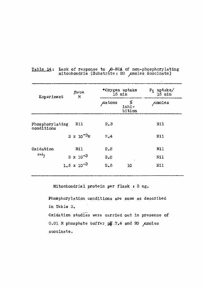
: Lack of response to yfi-NOA of non-phosphorylating mitochondria (Substrate : 20 ^omoles Succinate)
Experiment (ihJOf\
M •Oxygen
15 ] uptake
siin Pi uptake/
15 min Experiment (ihJOf\
M yuatoms %
inhi-b i t ion
/umoles
Phosphorylating N i l 2 . 3 Ni l conditions
3 X 10"^M 2 , 4 N i l
Oxidation N i l 2 . 8 Ni l
3 X 1 0 - 3 3 . 0 1 N i l
1 . 5 X 10 2 . 5 10 Ni l
Mitochondrial protein per f lask : 3 mg.
Phosphorylation conditions are same as described
in Table 3,
Oxidation studies were carried out in presence of
0,01 M phosphate buffer p|[.7.4 and 20 ^ o l e s
succinate.
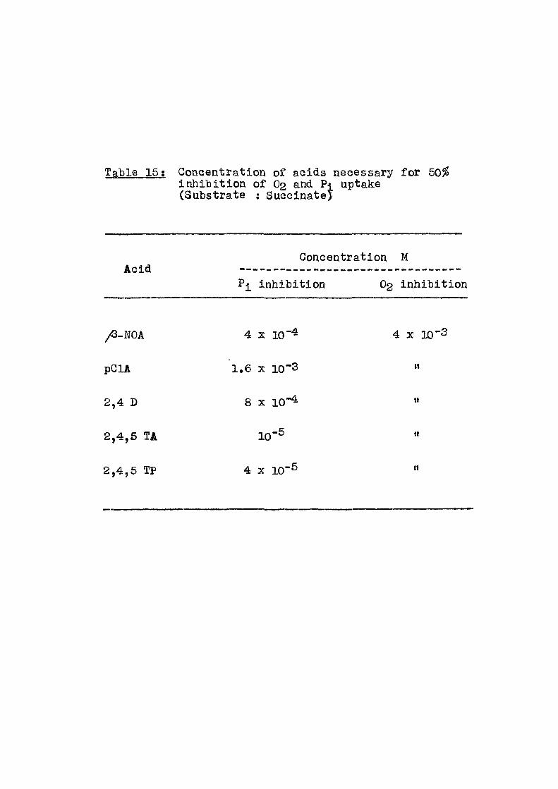
Table 15; Concentration of acids necessary for 50% inhibit ion of O2 and Pj, uptake (Substrate : Succinate)
Acid Concentration M
Pi inhibit ion O2 inhibit ion
/G-FOA 4 X 10"^ 4 X 10"3
pGlA 1.6 X 10~3 «
2,4 D 8 X 10⁻⁴ ti
2,4,6 TA 10"^ "
2,4,5 TP 4 X 1 0 ' ^

31
substrates. With succinate as the substrate, P/O rat io
i s 1.9 and v/ith glutamate 3.0, In the presence of
^-NOA at 3 X 10"^ M concentration, the phosphorylation
capacity was almost completely inhibited -whereas oxygen
consumption was reduced by about 50%. Table 4 gives the
results of the e f f e c t of ^-NOA concentration on both
inorganic phosphate uptake and oxygen consumption. Table 5
includes the e f f e c t of ^-NOA concentration when glutamate u;<ts ts used as the substrate. In Tables 6-12 are given the
results of varying the concentrations of d i f f e r en t acids
from 10"% to 10 '% . The compounds studied are p-t
chlorophenoxy acetic acid (pClA); 2,4 dichlorophenoxy
acetic acid (2,4 D); 2,4,5 trichlorophenoxy acet ic acid
(2,4,5 TA) and 2,4,5 trichlorophenoxy propionic acid
(2,4,5 TP) as sodium sal ts with succinate as the
substrate.
For experiments in Table 10, the concentration of
2,4,5 TA was kept constant at 4 x 10"% whereas the protein
per f lask was varied from 1 mg to 8 mg.
itie
Tables9 and 11 g i v e [ e f f e c t of the last two
compounds with glutamate as substrate. In Table 13 are fiven
the results of 2,4,5 TP action in the presence of 2,4 DNP.
Results of action of ^-NOA on phosphorylating and non-
phosphorylating mitochondria are given in Table 14.
32
The mitochondria were made non-phosphorylating
by exposure to d i s t i l l ed v/ater f o r half an hour. The O-t^joaratus
experiment in Warburg[was carried out both under phospho-
ry lat ing and non-phosphorylating conditions. I t i s seen
that under phosphorylating condition, no j-norsanic
phosphate uptake was noticed, ascertaining that the
mitochondrial preparation was completely uncoupled.
Table 16 indicates the concentration of the
various compounds studied to e f f e c t 50^ inhibit ion of
oxidation and phosphorylation capacity of rat l i v e r
mitochondria.
Discussion
The results presented in this Chapter have given
an indication that substituted phenoxy acids (sodium
sa l t ) have the property of inhibit ing the oxidative
phosphorylation in rat l i v e r mitochondria. The mito-
chondrial preparation used in the present study was iso lated
in an optimum stage, a f ac t borne out by the normal P/0
rat ios obtained when both succinate and glutamate were
used as substrates. The range of P/O with succinate
as substrate was between 1.8 to 2.6 and f o r glutamate
2.6 to 3.0. The concentrations of the phenoxy acids
employed has been varied over the range 10"% to 10~%.

33
Taking into consideration the e f f e c t of these compounds
over this range i t can be seen that t-hoao as summarised
in Table 16, the e f f e c t i v e concentrotion f o r 50% inhibi-
tion of oxygen uptake in the phosphorylating mitochondria
i s of the order of 4 x 10"%, whereas that f o r inhibit ion
of inorganic phosphate incorporation ranged from 10"3 f o r
PCIA to 10~®M fo r 2,4,6 TA. In f a c t , when mitochondria
were made non-phosphorylating by exposure to d i s t i l l e d
water, i t i s seen from results in Table 14 that th© /3-NOA
had no e f f e c t on oxygen consumption even at 3 x 10⁻³m
concentration. This indicated a poss ib i l i t y that the
above compounds may be having their action on the
phosphorylating machinery of the coupled mitochondria
than on the electron transport chain. In this manner,
these types of acids may have an action similar to that
exhibited by the -well-known phosphorylation inhibitors
l ike oligomycin and atracty los ide. The concentration at
which oligomycin exerts i t s e f f e c t i s 0,2 - 0.4 /umole/g
protein as shown by Lardy et a l and f o r atractyloside
0,25 /umole/g protein as shown by Bruni. Thus i t
appears that the naphthoxy and phenoxy acids that have
been employed in the present invest igat ion are not as
potent as oligomycin or atracty los ide. Having obtained
a preliminary indication that the compounds under study
a f f e c t the phosphorylation s i te of mitochondrial oxidative

56
phosphorylation, further experiments ^ere planned to study
their action on other properties of mitochondria related
to energy conservation mechanism. These included,
swelling-contraction cycle, energy dependent ion uptake,
Pi-ATP exchange, stimulated ATP ases and respiratory
control as studied by polarograph.
The results of these studies are presented in the
fol lowing pages.
Section B
Polorographic studies of oxygen uptake
Experimental
Respiratory control ; This was studied with the
help of a vibrating platinum electrode f i t t e d to a
Tinsley recording polarograph. The oxygen consumption
measurements were carried out at 20°. Measurements
were carried out at -0.6 vo l t s . The reaction was carried
out in a 6 ml capacity cuvette. The suspending medium
f o r mitochondria as described by Chance and Williams^®
contained 3mM KEl2P04> ISmM K2HPO4; 6mM MgCl2> 26 mM NaCl;
12mM NaF and 58mM KCl. The mitochondrial suspension was
added to 4 ml of the above medium (5-7 mg mitochondrial
protein per cuvette ) . Succinate, ADP and other compounds
were added as indicated in the accompanying f i gures . The
35
Polarographlc trace of oxygen consumption by rat l i ver
mitochondria
F ig , l a t Respiratory control in presence of added
ADP. The composition of the reaction medium was as
described on page . Oxygen consumption was started
by the addition of 10 mM succinate as indicated ( s l ) and
additions of ADP are indicated by arrows. The oxygen
concentration trace was recorded with the help of Tinsley
recording polarograph. Figures in parenthesis indicate the
rate of oxygen consumption. S.G, (Respiratory Control) is
the ra t i o of the rate of oxygen consumption during the
phosphorylation of ADP to the rate a f te r the ADP is
phosphorylated.
F i g . l b : E f f ec t of DNP on oxygen consumption.
Experimental conditions were same as in F ig . l a . Additions
were made as indicated by the arrow.
F i g . ic : e f f e c t of beta-NOA on phosphorylation of MDP
" Id : " PGIA " "
" le : " 2,4-D " "
I f : " 2,4,5-TA " "
" Ig : " 2,4,6-TP "
The above compounds (as sodium sa l t s ) were added
along with mitochondria at 0 min at a conc. 4 x 10"^ M.
240
^ c oi
/ 0
ADV A40 1 A D P FLSO
ADP S80 wyuwoles
At.P/0 = 1-9 R.C.= 8 0
_L J. 2 4
Time min ) f i a i a .
240
s
c w
o
ADP 440 -m/J moles
•J' PNP 3Xto'^M i
± a 4
T i m e (^mln) P IQ- lL .
6
^40
s
4)
o
ADP
ADP
2 4 Ti-me ( mxa")
F IQ. lc .
240
a 01
o
AT>P
At>P
± 2. 4
Time Cmin) f i 6 . i.a.
240 1-
c
0
AT>P
t ADP
i
_L ±
240
c 4» tli>
0
ADP
2 4 . T ime Cn T )
P I G - l e . TiTrie (^Tnxr\)
FIG.l i^.
240
2 c m tw ^ O
AtP
T i m e (^ruxn") F i a l ^ .

36
oxygen concentration of the solution was tak^n as 240 « ^ M .
The value O2 » 0 for the polarograph -was obtained by
passing % gas through the reaction mixture and thus
making i t anaerobic.
Results and Discussion
Oxygen consumption in mitochondria iso lated from
rat l i v e r was studied with the help of a vibrating
platinum electrode. When succinate was used as
substrate and I IP was added in 440 m/umole and
880 m^mole quantities as shown in F ig . l a , the ADP/0
value was about 1.9. The respiratory control was about 17 8.0. Chance^ has defined the respiratory control
ra t i o as the respira-tory rate in the presence of added
ADP to the rate obtained fo l lowing i t s (ADP) expenditure.
This compares well with the value of 5.9 reported by
Estabrook^^.
These studies were also carried out in presence-
of beta-NOA and the other phenoxy acids. These acids
were added at the start of the experiment along with -4
the mitochondria^at 4 x 10 M Con. The results are
given in f igures 1 c to 1 g. I t i s observed that in
the case of beta-NOA, pClA and 2,4 D a f t e r the addition
of AEP the pattern of oxygen consumption obtained i s

37
d i f f e r en t from that of the normal mitochondrial prepa-ration (Fig . l a ) . Studies employing Warburg technique have
has already indicated that in the presence of these
compounds phosphorylation capacity i s impaired. Hence
the amount of ADP phosphorylated cannot be obtained in
this series of experiments. When 2,4,6 TR and 2,4,5 T4P
were added (Fig . I f and I g ) , there was no increase in
the rate of oxygen consumption a f ter the addition of ADP
indicating that the added ADP was not being phosphorylated
as in F ig . l a : 2,4,5 TP and 2,4,5 TA seem to be mdre
e f f e c t i v e inhibitors than beta-ITOA, pClA and 2,4 D. A
similar observation has been made in the Warburg studies.
Stimulation of oxygen consuijption in the
presence of DKP (3 x 10 "%) i s recorded in F ig . l b .
Summary
1. /3-naphthoxy acet ic acid and some analogues of
phenoxy acids are found to inhibi t the oxidative phosphory-
la t ion of rat l i v e r mitochondria. Phosphorylation capacity
i s a f fected more than the oxidation capacity.
2. The various acids were employed as sodium salts
in the range 10~% to 10-%. The extent of inhibi t ion
Varied between 100$ to 20^ depending on the concentration
and nature of the acid.
62
3. For inhibi t ion of oxygen uptake 10"^
concentration was necessary whereas inhibi t ion of P j
uptake the concentration varied between - 10*"%.
4 . Oxyg&n uptake in non-phosphorylating mitochondria
iras not at a l l a f f e c t ed .
6. In studies with oxygen electrode i t was
noticed that the naphthoxy and various phenoxy acids
used w ere able to reduce or completely inhib i t the
phosphorylation of added ATP. This resulted in altered
polarographic trace of oxygen consumption.
39
References
1. Hollunger, G. (1956), Acta Pharm.Toxicol., Suppl. 1.
2. Lardy, H.A., Connelly, J.E. & Johnson, D. (1964),
Biochemistry, 3, 1961.
3. Connelly, J.L. & Lardy, H.A. (1964), Biochemistry, 3, 1969,
4. Graven, S.N., Lardy, H.A. & Rutter, A. (19GG),
Biochemistry, 5, 1735.
5. Bruni, A . , Contessa, A.R. & Luciani, S. (1962), Biochim. Biophys. Acta, 60, 301i
6. Chappell, J.B. & Crofts, A.R. (1965), Biochem. J. , 707.
7. Huijing, F. & Slater, E.G. (1961), J.Biochemistry (Japan), 493.
8. Lee, C.P. & Brnster, L. (1965), Biochem.Biophys. Res. Comm., 18, 523.
9. Lam, K.W., Warshaw, J.B. & Sanadi, D.R. (1966), Arch.Biochem.Biophys., 117, 594.
10. Van Buskirk, J.J. & F r i s e l l , W.R. (1967), Biochim. Biophys. Acta, 143, 292.
11. Wikstrom, M.K.F. & Saris, N.E.L. (1969), European J. Biochem., 9, 160.
e 12. Schneider, W.C. & Hodboom, G.H. (1950),
J.Bio3j.Chem., 183, 123.
13. Hunter, E.F. (1955), Methods in Enzymology, Vol.11,
p.610, Ed.by New York Acad. Press.
14. Taussky, H.H. & Shorr, E. (1953), J.Biol.Chem., 202^ 675.
15. Lowry, O.H., Rosebrough, N.R., Farr, A .L . & Randall, R.J.
(1951), J.Biol.Chem., l ^ , 265.
16. Chance, B. & Williams, G»R. (1955), J.Biol.Chem., 217, 383,
17. Chance, B. (1959), Ciba Pound. Symp. Regulation of Cel l Metabolism, L i t t l e Brovn and Company, Boston, p.91, 18. Estabrook, R.W. (1967), Methods in Enzymology, Vol.X,
page 41, Ed. by Estabrook, R.W. & Pullmann, M.S. Acad.Press Inc. , New York.
CHAPTER I I I
SWELLING-CONTRACT ION CIGIE OF MITOCHONDRIA
0
CHAPTER I I I
SWELEING-COETBACTION CYCIE OF MITOCHONDRIA
Introduction
Af ter the i n i t i a l studies by Tedeschi and Harris-2
and Raaflaub indicating the passive and act ive volume
chan|es in mitochondria in in v i t ro systems, considerable
amount of -work has been carried out on the swell ing-
contraction cycle of mitochondria. A number of compounds
including a fevj ant ib iot ics have been shown to considerably
a f f e c t these changes. Some of these compounds are known
to influence the mitochondrial energy transfer
reactions.
Connelly and Lardy showed the inhibi t ion of ATP
reversal of swollen mitochondria by oligomycin and 3
aurovertin . I t was also reported that other ant ib iot ics
l ike nonactin, monactin, dionactin and tr inact in are able
to induce swelling of mitochondria in presence of cations.
I t was proposed that the ant ib iot ics act by inducing
alterations in ion translocation system. Showdomycin®,
an ant ib io t ic , w i l l also cause swelling of mitochondria
at high pH.
Besides ATP and Krebs cycle substrates as energy
donors recent work indicates that even f a t t y acid oxidation
u
may provide the energy f o r the contraction process of
mitochondria . I t has been shown that the ant ib io t i c
Dio-g"^ i s able to i n i t i a t e large amplitude s-welling of
rat l i v e r mitochondria with absolute requirement of
energy supply.
The ro l e of ATP ase during swell ing phenomena ia o
reported by several workers. Chappell and Perry
observed that the mitochondrial swell ing was i n i t i a t ed
by the la tent ATP ase. Lehminger and his coworkers®
observed that the contraction of the mitochondria was
fol lowed by a low ATP ase l e v e l . Recently Zimmer et al^*^
reported the part played by the ATP ase a c t i v i t y and ATP
l eve l s of mitochondria during swel l ing . N- (N-acety l -4
sulphamoylphenyl) raaleimide, a new der ivat i ve of
maleimide i s able to a l ternate or retard the swelling
of mitochondria^^. This may be re lated to the a lky lat ion
of ATP ase. Green and his coworkers^^ demonstrated that
the mitochondrial swell ing i s due to a ser ies of confor-
mational changes in the cr is tae and the inner membrane.
This requires some driving fo rce which i s supplied by
electron transfer or hydrolysis of ATP. -lo
Recently Green and coworkers have carr ied out
detai led study of conformational changes produced during
energized swell ing of beef heart mitochondria. They also

42
have shown that there are two forms of energised swelling
of beef heart mitochondria^"^. The nature of energised
swelling of beef heart mitochondria are described and
compared with those of pseudoenergised swel l ing.
Studies with phenoxy acids reported in the
previous chapter have indicated their e f f ec t i veness in
inhibit ing the phosphorylation mechanism of ac t i ve ly
respiring mitochondria. In the present chapter are
reported the e f f e c t s of these compounds on the act ive
swelling-contraction cycle of the mitochondria.
Experimental
The mitochondria were iso lated as described in
Chapter 11, page 2,6 . The swell ing-contraction studies
were carried out by the method of Lehninger^^ as modified
below. To each 5 ml of 0.125^ KCl-0.02 M tris-HCl
pH 7.4 in a test tube was added an aliquot of mitochondrial
suspension such t.hat the i n i t i a l turbidi^metric reading
reading was about 400 (or E520 "^alue of 0.8) in a Klet t
Summerson colorimeter with f i l t e r No.52. The tubes were
maintained at 22° during experimental period and readings
were taken every two minutes fo r a period of 20 min. The
swelling was induced by the addition of either inorganic
phosphate, calcium or thyroxine at 0 rain. Contraction
of these swollen mitochondria was in i t i a t ed by the
43
addition of 0,005 M ATP, along with 0.003 M MgClg,
O.OOIM EDTA and 200/ug. bovine serum albumin. The
phenoxyacid addition -was made as mentioned in each f igure
either at 0 min or sometime a f ter swelling was in i t i a t ed .
A l l the reagents used were BDH, AR Grade. ATP
was purchased from Sigma Chemical Co., U.S.A.
Results
The results presented in the fo l lowing f igures
indicate the swelling patterns in presence of various
established swelling agents l ike Ca"*"*", inorganic phosphate
and thyroxine. The reversal of the swelling phenomenon
in the above cases has been studied in presence of ATP. v/<tr/'oas
The e f f e c t of various naphthoxy and[phenoxy acids (Na sa l t s )
on the ATP induced reversal of the swelling i s also presented
here. el
As shoxm in F ig . 1, mitochondria maintain^their
in tegr i ty in the medium employed during the experimental
period which in this case 3rS 20 minutes. I t i s also seen
that the acids which were added at 0 min. by themselves
did not induce any appreciable swelling (5-20^) of the
mitochondria over the experimental period (Fig . 1 ) .
In the next experiment, the mitochondria were swollen
fcr 12 min. in presence of inorganic phosphate (5 mM).

a
Fig... 1; E f f e c t of phenoxy acids on mitochondrial suspension.
An aliquot of mitochondria (equivalent of 6 mg protein) was suspended in 5 ml of 0.126 M KGl- 0.02 M tris-HGl buf fer pH 7.4 at 22°. Various phenoxy acids as sodium salts at 4 X 10"^ M concentration were added at 0 min. Readings were taken at an interval of 2 min. fo r a period of 20 min Klet t Summerson Colorimeter with f i l t e r No.62.
Curve No.l is control and No.2 to 6 represent in presence of /G-NOA; PCIA; 2,4,0; 2,4,5-TA; 2,4,6-TP respect ive ly .
F ig . 2; E f f e c t of phenoxy acids on mitochondrial swelling induced t)y inorganic phosphate.
The experimental conditions are same as given in F ig . 1.
Swelling was induced by the addition of 6mM inorganic phosphate and the acids
'4 X 10"^ as sodium salts were added at the end of 12th min as indicated by the arrow.
Curves 2-6 represent swelling pattern in presence of /-NOA; PGM; 2,4-0; 2,4,6-TA; 2,4,6-TP respect ive ly .
45
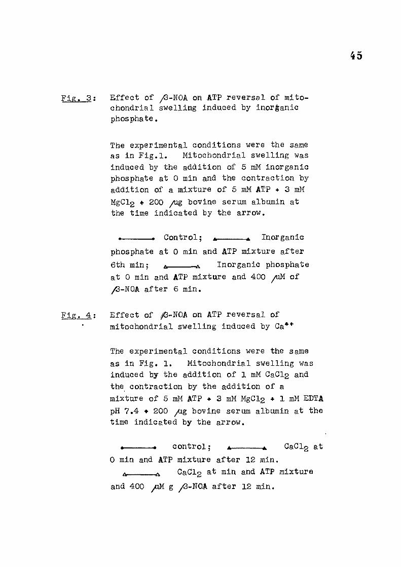
F ig . 3: E f f ec t of /S-NOA on ATP reversal of mito-chondrial swelling induced by inorganic phosphate,
The experimental conditions were the same as in F i g . l . Mitochondrial swelling was induced by the addition of 5 mM inorganic phosphate at 0 min and the contraction by addition of a mixture of 6 mM ATP + 3 mM MgGl2 • 200 /ug bovine serum albumin at the time indicated by the arrow.
. > Control; m ^ Inorganic
phosphate at 0 min and ATP mixture a f te r
6th min; a Inorganic phosphate at 0 min and ATP mixture and 400 /uM of /S-NOA after 6 min.
F i g . 4 ; E f f ec t of /3-NOA on ATP reversal of mitochondrial swelling induced by Ga**"
The experimental conditions were the same as in F ig . 1. Mitochondrial swelling was induced by the addition of 1 mM GaGl2 and the contraction by the addition of a mixture of 5 mM ATP + 3 mM MgGl2 • 1 mM EDTA pH 7.4 • 200 /ig bovine serum albumin at the time indicated by the arrow.
contro l ; A A. GaGl2 at
0 min and ATP mixture a f ter 12 min. ^ GaCl2 at min and ATP mixture
and 400 /iM g y<3-N0A a f t e r 12 min.

6 12 T i m e ^ mxn )
^ m . 1 T ime rniri)
F IQ. a
8 la Tinrie ( int-a )
F IQ. 3.
8 la Ti-m^ ini-n )
P I Q . 4

46
Fig . 5-8; E f f ec t of phenoxy acids on ATP reversal of mitochondrial swelling induced by inorganic phosphate.
The experimental conditions are same as
described in Fig . 1. Swelling of mito-
chondria Was induced by the addition of
5 mM inorganic phosphate at 0 min and
the mitochondrial contraction by addition
of ATP mixture a f ter 6 min as indicated
by arrow.
• Control; A 4. Inorganic
phosphate at 0 min and ATP mixture at 6th min.
zw A inorganic phosphate at 0 min. and
ATP mixture along with phenoxy acid at 6th min,
For expt. in Fig. 5 2 x 1 0 P G l A; ti n ti
Fig. 6 4 X 10-^M 2,4,D;
F ig . 7 4 X lO-^M 2,4,5,TA
Fig. 8 4 X 10-^M 2,4,5-TP.

OS
0-6
O OJ iTi t£r
0-4
0-2
6 iZ 16 T i m e CTnin)
F I G . 5.
20 O 8 12. 16 Tirne C^i"!^")
F I Q . 6
Ti-me ^ rialn.) FIG- T.
6 12 16 T ime C ^ ^ ^ )
F I Q . 8
20

47
F ig . 9; E f fec t of ^-NDA on ATP reversal of mito-chondrial swelling Induced by inorganic phosphate.
The experimental conditions were same as described in F ig . 1. Swelling of mitochondria was induced by the addition of 6 mM inorganic phosphate and the mitochondrial contraction by ATP miicbure as given in Fig. 3.
« • Control.
A • Inorganic phosphate at 0 min and ATP mixture a f ter 16 min.
A A Inorganic phosphate and ATP mixture at 0 min and 400 /iM /Q-NOA a f t e r 16 min.
o o Inorganic phosphate and 400 aM ^-NOA are at 0 min.
F ig . 10; E f fec t of /S-NOA on ATP reversal of mitochondrial swelling induced by L-thyroxine.
The experimental conditions were same as given in F ig . 1. Swelling of mitochondria was induced by the addition of 10 ^M L-thyroxine and the mitochondrial contraction by a mixture of 5 mM ATP and 200 /ug bovine serum albumin.
Control.
L-thyroxine at 0 min and ATP mixture a f ter 12 min.
L-thyroxine and ATP mixture at 0 min and 400 /uM /3-NOA a f t e r 12 min. ^
Inorganic phosphate + ATP mixture at 0 min. along with 400 /uM /3-NOA
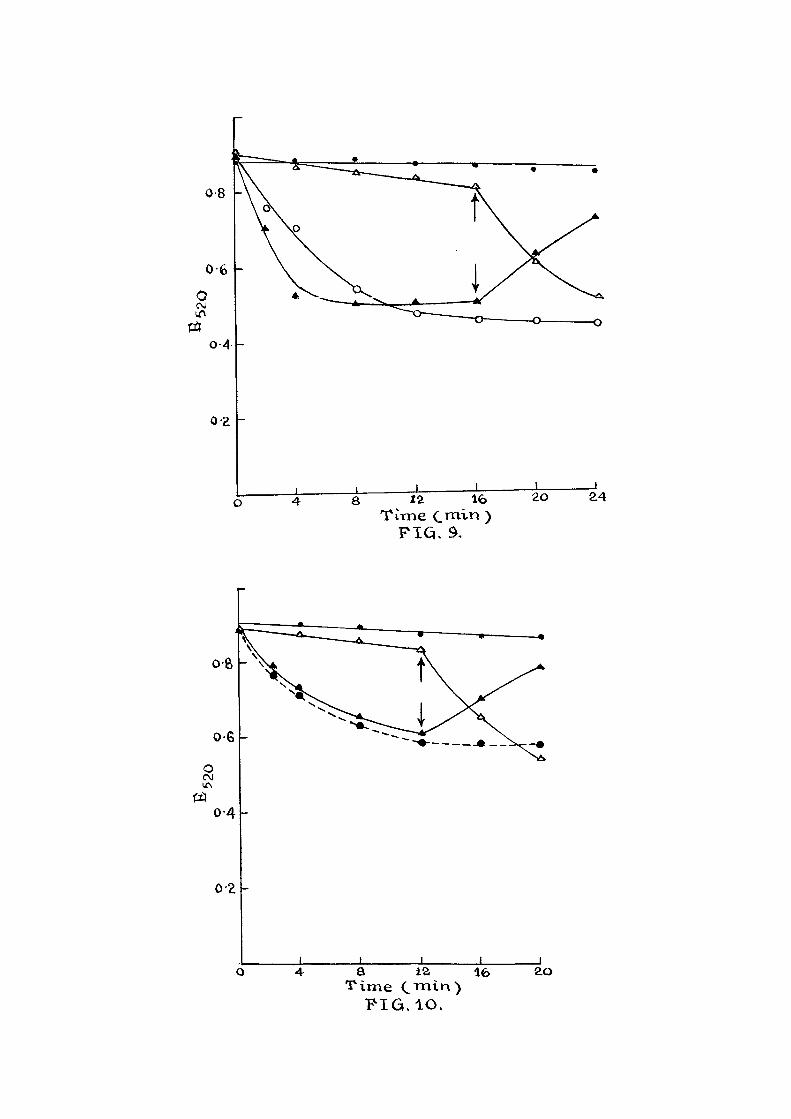
o s 12 16 T ime CiT i-i >
F I Q . 9,
20
a 12, T ime
F I G . I O .
ao
49
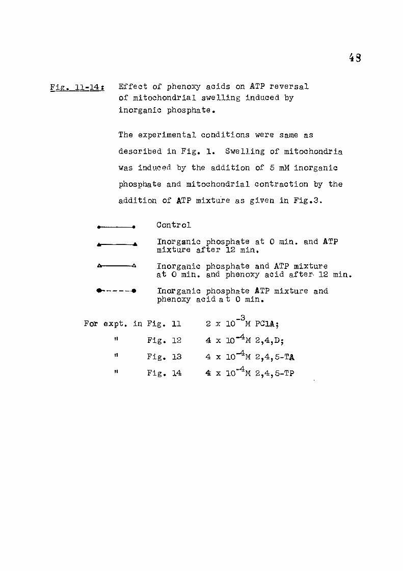
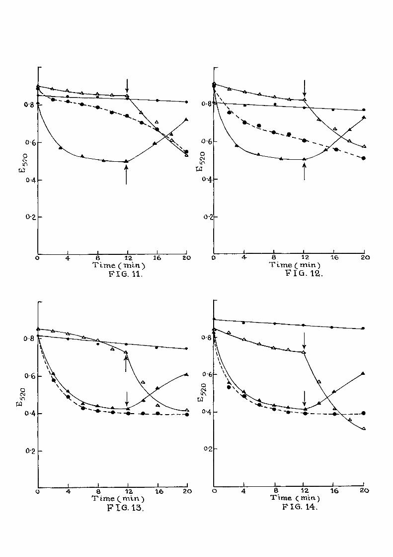
Ef fec t of phenoxy acids on ATP reversal of mitochondrial swelling induced by inorganic phosphate.
The experimental conditions were same as
described in F ig , 1. Swelling of mitochondria
was induced by the addition of 5 mM inorganic
phosphate and mitochondrial contraction by the
addition of ATP mixture as given in F ig .3 .
• • Oontrol
^ Inorganic phosphate at 0 min. and ATP mixture a f t e r 12 min,
^ ^ Inorganic phosphate and ATP mixture at 0 min. and phenoxy acid after^ 12 min,
Inorganic phosphate ATP mixture and phenoxy acid a t 0 min.
For expt, in F ig . 11 2 x PGIA;
" F ig . 12 4 X 10"% 2,4,D;
" F ig . 13 4 X 10"^M 2,4,5-TA
" F i g . 14 4 X lO' ^M 2,4,6-TP
6 12 36 T ime ( tn in )
FIG. 11."
8 12 16 T ime (^min)
FIG. 12.
8 12 16 T ime (^mln )
F i a 13.
8 12 16 Time (^min)
PIG. 14.
20
Then various phenoxy acids (4 x 10 "%) were tested indi-
v idual ly . I t i s noticed from Fig . 2 that they did not
have any additional swelling e f f e c t even in presence of
inorganic phosphate. In Figs. 3-8 are given results of
e f f e c t of various acids on the ATP reversal of swollen
mitochondria. Mitochondria were swollen in the presence
of inorganic phosphate (5 raM) or (F ig . 4 ) , for
a period of 6 or 10 min and then ATP ( 5"inM ) was added
which induced contraction. This contraction was par t i a l l y
inhibited when various phenoxy acids (4 x iQ-'^M) -were added
along with ATP as shown in these f i gures . In Figs. 9 and
10, the mode of addition of ATP and ^-NOA was a l tered.
I t i s seen that when ATP i s added along with the swelling
agent^<either inorganic phosphate, (F ig .9 ) or thyroxine
(Fig.10) at 0 min, the swelling phenomenon was inhibited.
At the end of 16 min (F i g .9 ) , or 12 min (F ig .10) , when
^-NOA was added ATP e f f e c t was to some extent nu l l i f i e d
and the mitochondria showed swelling due to the action
of the swelling agent. When inorganic phosphate, ATP and
/3-NOA were added at the start of the experiment, the
ATP e f f e c t was overcome from the beginning and the swelling
was noticed from the start of the experiment. The ATP
reversal of the swollen mitochondria i s also included f o r
comparison. Similar results with phenoxy acids are
observed in Figs.11-14. These acids were able to overcome
the ATP e f f e c t pa r t i a l l y .

30 Discussion
Observations in the preceding chapter have indi -
cated that the naphthoxy and phenoxy acids inhib i t the
phosphorylating capacity of the mitochondria whereas the
electron transport property is not a f f ec ted in the non-
phosphor yla ting par t i c l e s . In the present chapter a
property of mitochondria that i s connected with energy
transfer r eactions i s shown to be a f f ec ted by the above
acids*
Swelling of the mitochondria in presence of
agents l i k e calciumj inorganic phosphate and thyroxine
and i t s reversal by addition of ATP under proper condi-
tions i s we l l established by the studies of Lohninger and
his coworkers . The results presented in the present
chapter have indicated that this property of ATP could be
par t i a l l y reversed by compounds l i k e /3-NOA, 2,4 dichloro-
phenoxy acet ic acid and 2,4,6-trichlorophenoxy propionic
acid. When the mitochondria were allowed to swell f o r
periods of 8-12 minutes in the presence of either calcium,
inorganic phosphate or thyroxine and then 0»005 M ATP
(mixture) was added, about B0% of the swelling was
reversed in a further period of about 12 minutes, li^en,
however, along with ATP the inhibitors were added at a
concentration of 4 x 10*"%, the ATP e f f e c t was abolished
51
to varying extents. Similar lowering of ATP e f f e c t
was obtained as seen from F ig . 3, when ATP was present
along with the swelling agent from the beginning of the
incubation period and the inhibitors were added a f t e r a
lapse of 12 minutes. In these cases, the protect ive e f f e c t
of ATP was immediately removed to a considerable extent.
When inorganic phosphate, ATP and the inhibitor were
present simultaneously from the start of the experiment
the e f f e c t of ATP could not be seen at a l l . Similar
observations have been made when the swelling was
induccd either by calcium or thyroxine.
Thus i t appears that the above acids which are
already shown to inhibit oxidative phosphorylation
are also capable of reversing another energy transfer
dependent property of the mitochondria, i . e . ATP
induced reversal of the high amplitude swelling of
the mitochondria.
SUMMARY
1. ATP is knoxm to have the property of
inducing contraction in mitochondria that have undergone
swelling in presence of agents l i ke inorganic phosphate,
Ca " or thyroxine.
52
Present work shows that this ATP e f f e c t Is
pa r t i a l l y reduced in presence of 10*"% concentration
of the various phenoxy acids.
2. This e f f e c t on ATP property could be
demonstrated even when the order of additions of ATP
and the acids was changed in d i f f e r en t experiments.
3. Mitochondria suspended in the Tris-HGl-KGl
medium swell only about 5 to 15^ in presence of these
acids ( 1 0 " % ) .

33 REFERENCES
1. Tedeschi, H. & Harris, D.L. (1955), Arch.Biochem.Biophys,, 58, 52,
2. Raaflaub, J. (1953), HeXv, Physiol , et Pharmacol.Acta, 11, 142 (quoted from the above paper).
3. Connelly, J.L, & Lardy, H.A. (1964), Biochemistry (USA), 3, 1969.
4. Graven, S.N., Lardy, H.A. & Rutter, A. (1966), Biochemistry (USA), 5, 1735.
5. Hadler, H . I . , Claybourn, B.E. & Tschang, T. (1968), Biochem.Biophys.Res.eomm., 25.
6. Kuttis, J . , Nakatani, M, & Mcmurray, W.C. (1968), Arch.Biochem.Biophys., 126, 634.
7. Fisher, R.R. & Guil lory, R.J. (1968), Biochim. Biophys. Acta. , 162, 182.
8= Chappell, -JcB. & Perry, S.V. (1954), Mature, 173, 1094.
9. Vignais, P.V., Vignais, P.M. & Lehninger, A .L . , (1964), J.Biol.Chem., 2 ^ , 2002.
10. Zimmer, G., Ette, H. & Geek, P. (1969),
Nature, 2 ^ , 1160.
11. Zimmer, G. & Krieger, K, (1968), FE BSL 1, 142.
12. Blondin, G.A., Va i l , ¥.J, and Green, D.E. (1969), Arch.Biochem.Biophys., 129, 158.
13. Asai, J . , Blondin, G.A., Va i l , ¥ .J . & Green, D.E., (1969), Arch.Biochem.Biophys., 1^2, 524.
14. Blondin, G.A. & Green, D.E. (1969), Arch.Biochem.Biophys., 132, 509.
15. Lehninger, A .L . (1959), J.Biol.Chem., 2465.

CHAPTER IV"
ACTIVE ION TRANSPORT
54
CHAPTER I V
ACTIVE ION TRANSPORT
Introduction
Letininger^ has c l a s s i f i ed the act ive ion
accumulation by mitochondria under t-wo categor ies ,
massive loading and l imited loading. For studies in
the f i r s t category mitochondria are exposed to re la -
t i v e l y high concentrations of Ca^* 3MM, T jhereas
l imited loading i s studied at about lOO ciM calcium
concentration. In the former category there are
chances of damage to the oxidative phosphorylation
machinery, hence the dynamics of ion uptake are best
studied in l imited loading experiments. Ac t i ve ion
uptake i s supported ei ther by electron transport-or by
ATP hydrolysis as energy source. When the uptake i s
supported by oxidation of respiratory substrate ATP i s
also required in the medium, in ca ta ly t i c amount,
A proper explanation fo r ATP requirement during
succinate as energy source f o r Ca^* accumulation i s not
yet knovirx although Christiansen et a l^ using sub-
mitochondrial par t i c l e s , suggest}^ that the nucleotide ATP
may be required to induce an appropriate conformation in
85
the protein. During Ca translocation or energy
production in intact mitochondria the nucleotide may
be the primary translocated ion.
Mitochondria isolated from rat l i v e r and beef
heart have been extensively used in ion uptake studies.
Relat ive ly l i t t l e spec i f i c i t y to-wards cations i s en-3 4 countered in these ion uptake studies. Brier ley '
2+
using beef heart mitochondria has shown that Zn
and other heavy metal cations accelerate the energy
linked uptake of Mg^* and K*. He has compared this
e f f e c t with that of gramicidin®. Chelators l ike
EDTA are also shown to act ivate the accumulation of Na*
and Li*^. Azzone and Azzl^ also noted that EDTA with
rat l i v e r mitochondria gives results similar to those
induced by valinomycin and gramicidin. This EDTA
interaction with the membrane appears to represent s t i l l
another chemically d ist inct reaction which can bring
about the act ivat ion of the mitochondrial ion transport.
Thus chelators, heavy metal ions, th io l group reagents,
ant ib iot ics and parathyroid hormone a l l seem to a f f e c t
the ion transport in mitochondria, Q
Mela and Chance with the help of dual wave-length
spectrophotometer and employing indicator murexide have

56
2+ P+
studied the kinet ics of Ca and Mn' uptake and the
concomittant pH changes in mitochondria.
Mitchell and Moyle® have recently studied the
translocation of anions, cations and certain acids in
rat l i v e r mitochondria. As the act ive ion uptake i s
carried out in the presence of substrates or ATP as
energy source, i t i s but natural that inhibitors of either
electron transport or phosphorylation reactions are able
to a f f e c t this ion uptake. Compounds l i ke oligomycin
have already been sho-wn to inhibi t ATP dependent ion
up take CN7 antimycin and DNP inhibi t succinate
dependent ion uptake.
As results presented in the previous two
chapters have indicated the poss ib i l i t y that the phenoxy
acids under investigation a f f e c t the energy transfer
reactions, their influence on the act ive ion uptake
•was studied. The results of these studies are presented
in this chapter.
Experimental
Materials and Chemicals
Rat l i v e r mitochondrial preparation employed f o r
experiments described in this chapter was prepared as
57
mentioned in Chapter I I (page 2L6 ) . Radioactive " ^Ca
was purchased from Bhabha Atomic Research Centre,
Trombay, Bombay. A l l the other reagents used vere BDH
(AR grade).
Methods
Calcium uptake was studied by the method of
Lehninger^*^ as given belovi. The incubation medium
contained 8.3 mM tris-HCl buffer pH 7.4, 66 mM NaCl,
6 mM phosphate pH 7.4, 2 mM MgClg, 2 mM CaClg C^^CaCl2 oy li'i^n fijf c^ken f{TP Wdi He e>\tr-p Scurce
50,000 cpm); 8 mM Na succinate and 4j8 mM ATi{. The
f i n a l volume was 3 ml. To this incubation mixture was
added 0.5 ml of mitochondria. The incubation was
continued for a period of 10 min at 30°. At the end
of this period, the tubes were immediately chi l led in
i ce and centrifuged. The mitochondrial pe l l e t was
suspended in 2 ml d i s t i l l e d water fo r a period of one
hour, at the end of which protein was precipitated by
1 ml of 20% TCA and the prec ip i tate centrifuged o f f .
The supernatant was f reed of TCA by two successive
extractions with 5 ml diethyl ether. The solution was
analysed f o r calcium and phosphorus. Radioactive calcium
was counted with the help of an end window gas f l ow,
GM counter connected to a tracerlab scaler. No
correction f o r sel f absorption was applied. Inorganic
59
phosphorus was estimated by the method of Tj/'aussky and
Shorr as described in Chapter I I (page ) . Protein
was determined by the method of Lorry et a l (Chapter I I ,
page 2,9 ) .
Preparation of sub-mltochondrial par t i c l es :
Method of Devlin and Lehninger^^ (modi f ied) : Rat
l i v e r mitochondria prepared by the method described
in Chapter I I , page , were suspended in 0,05 M
sucrose such that mitochondria of 10 gm l i v e r were in
1 ml of suspension. To this was added 0,66 vo l of
recrysta l l i zed digitonin in 0.05 M sucrose with conti-
nuous s t i r r ing . A f ter 20 min at 0°, 0.26 M sucrose
i s added to bring the digitonin concentration to 0.8^.
The suspension i s then maintained at 0° f o r 20 min. Then
0.25 M sucrose i s added to reduce the dig i tonin concen-
trat ion to 0,25^. The suspension was centrifuged
20,000 X g f o r 20 min. The precipitate discorded
and a pe l l e t sedimenting at 80,000 x g for 20 min
In rotor S.W. 39 in Spinco preparative centri fuge was
co l lected. This p e l l e t suspended in d i s t i l l e d water
was used for the experiment.

Table 16; E f f e c t of ^-NOA on active ion uptake by whole mitochondria.
5 Without NOa| With NOA 10""% g ^ Experiment
Endogenous 270 0.00
Succinate • Cat ATP
ATP alone
1220
1560
300
210
67
87
(Mitochondria iso-lated from Phaseolus vulgar is )
Succinate 49 49 N i l
Act ive calcium uptake studies -were carried out by the method described under Methods and Materials.
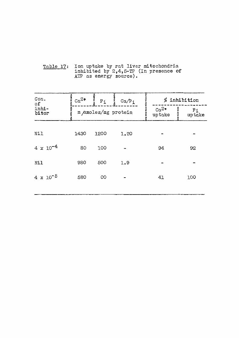
Table 17; Ion uptake by rat l i v e r mitochondria inhibited by 2,4,5-TP ( In presence of ATP as energy source).
Con. of inhi-bitor
Ca 2+ P i j Ca/^i inhibit ion
5 Ca2-» S I m/imoles/mg protein I J
N i l
4 X 10"^
Ni l
4 X 10"^
1430 1200 1.20
80 100
980 500 1.9
580 00
94 92
41 100
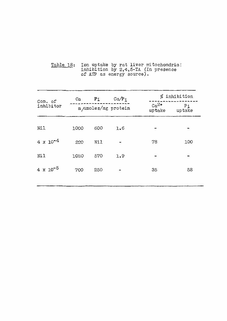
Table 18; Ion uptake by rat l i v e r mitochondria: inhibit ion by 2,4,5-TA (In presence of ATP as energy source).
Oa P i Oa^ i
inhibitor X ^ e up/aL
N i l 1000 600 1.6
4 X 10"^ 220 TJil - 78 100
N i l 1080 570 1.9
4 X 10"® 700 250 - 35 58
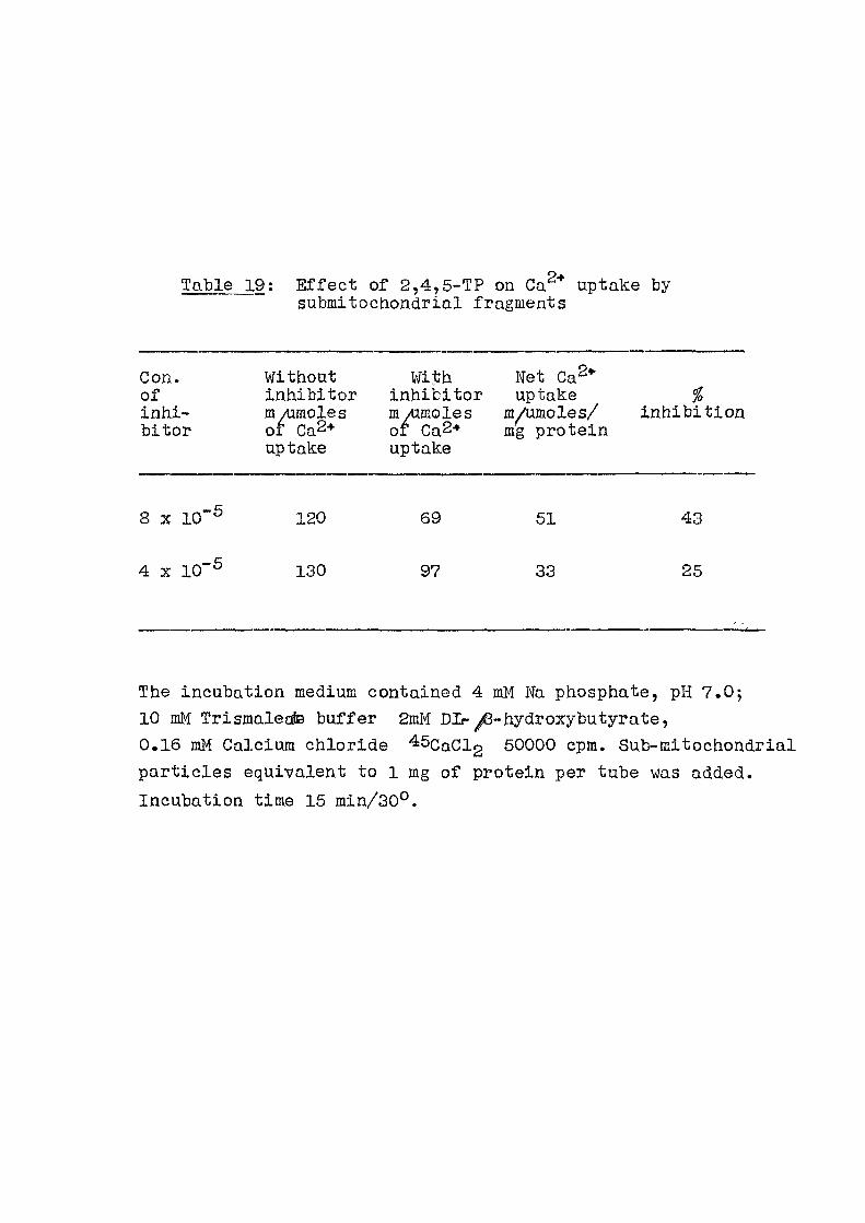
Table 19: Ef fec t of 2,4,5-TP on Ca "* uptake by submitochondrial fragments
Con. of inhi-bitor
Without inhibitor m /timoles of Ca2+ uptake
With inhibi tor m /imoles of Ca2+ uptake
Net Ca^* uptake
m/umoles/ mg protein
inhibi t ion
8 X 10""^ 120 69 51 43
4 X 10"^ 130 97 33 25
The incubation medium contained 4 mM Fa phosphate, pH 7.0; 10 mM Trismalecte buffer 2mM DL- ^-hydroxybutyrate, 0.16 mM Calcium chloride 45caCl2 50000 cpm. Sub-mitochondrial part ic les equivalent to 1 mg of protein per tube was added. Incubation time 15 min/30°.
59
Results
Studies with ion transport by rat l i v e r mitocfiondria
and the e f f e c t of naphthoxy and phenoxy acids are described
in this chapter. As can be seen from Table 18 in
presence of ATP as energy source, net calcium uptake was
1.0 ^moles/mg protein and net P i uptake was 0.6 /umoles/
mg protein. Thus Ca/Pi uptake rat io was 1.6. The
value for this ra t io in d i f f e r en t experiments under
our conditions ranged from 1.1 to 1,9.
When act ive ion uptake was studied in the presence
of ^-NOA (10~%) with succinate and cata lyt ic amount
of ATP, the calcium uptake was reduced by <61% whereas
when ATP was the energy source act ive calcium uptake
was inhibited to the extent of 87^ (Table 1 ) . Studies
employing mitochondria isolated from the plant source,
cotyledons of Phaseolus vulgaris indicate that when
succinate i s used as the energy source there i s no inhi-
bit ion of calcium uptake in presence of /Q-NOA^^.
Reason for this w i l l be given under 'Discussion'. 0
In Tables 17 and 18 are given the e f f e c t of
2,4,5-TP and 2,4,5-TA on calcium uptake in presence of
ATP as the energy source. At 4 x 10"%, there i s
about complete inhibit ion of calcium uptake whereas eveno-t
60
4 X 10"% concentration the inhibitory effect i s s t i l l
seen.
The active calcium uptake was studied with sub-
mitochondrial fragments prepared by digitonin treatment.
With this preparation, i t was not possible to show ion
uptake when succinate was used as the substrate. With
^-hydroxy butyrate i t was observed (Table l 9 ) that
calcium uptake was inhibited from 25 to 50^, in presence
of 4 - 8 X lO'^M 2,4,5-TP.
Discussion
One of the ways in which the energy l iberated
during electron transport by the mitochondria, i s ut i l i sed
i s active transport of ions across the mitochondrial
membrane. Besides this i t i s mainly u t i l i sed fo r the
formation of ATP and to maintain the mitochondrial
in t eg r i t y . Uptake of calcium in presence of energy
source l ike Krebs cycle substrates or ATP i s wel l docu-1 3
mentedin l i t erature by the work of Lehninger , Brierley
and others. Ca/Pi ra t i o in the range of 1,6 i s reported
f o r rat l i v e r mitochondria by Lehninger and f o r heart
mitochondria by Br ier ley .
Ca/Pi rat ios reported in the present invest igat ion
agree f a i r l y wel l with those reported in l i t e ra ture .
61
I t i s noticed ttiat in the presence of ^-NOA both
calcium and phosphate uptake were reduced when either
succinate or ATP were used as energy source. This
succinate dependent uptake i s known to have an absolute
requirement for cata lyt ic amounts of ATP in the reaction
mixture. When, however, ion uptake is studied with
plant mitochondria under our conditions there i s no
ATP requirement when succinate was the energy
source^^. This ion uptake was not inhibited even
in the presence of /3-NOA at 10~%. Thus i t appears
that /3-NOA in ter f e rs with reactions u t i l i s ing ATP.
Studies with swelling contraction phenomena of rat
l i v e r mitochondria have already indicated that s-tiefe tl esc-
compounds are able to overcome the ATP e f f e c t .
When active ion uptake was studied in the
presence of other halogen substituted phenoxy acids,
a similar inhibitory e f f e c t on ion uptake was noticed.
2,4,5-TA and 2,4,5-TP were at least 50% e f f e c t i v e
at 10"% concentration. Similar inhibit ion of ion
uptake has been reported by other workers in the
presence of various inhibi tors of energy transfer
reactions.
62 SUMMARY
1. E f f e c t of various phenoxy acidic on the
act ive ion uptake by rat l i v e r mitochondria has been
studied.
2. I t was noticed that when succinate was the
energy source, ion uptake w«s inhibited whereas
with ATP as the energy source, the inhibit ion was about
87^ at the same concentration of the inhib i tor .
3. In presence of 2,4,6-TA or 2,4,5-TP at
4 X 10*"% concentration, the inhibi t ion of act ive
Ga2+ Uptake was about 90 - 1%'3% and at 4 x
about 50%*
4. Ca "'" uptake by sub-mitochondrial fragments
prepared by digi tonin treatment was also inhibited
between 25-43^ when inhibitor concentration was about
In this experiment, /3-hydroxybutyrate served
as energy source. With these par t i c l es , succinate or
ATP did not support any ac t i ve ion uptake.
63
REFERENCES
1. Lehninger, A.L. (1967), Adv.Enz., 259.
2. Christiansen, R.O., Loyter, A . , Steensland, H., Saltzgaber, J. & Racker, E. (1969), J.Biol.Ghem., 244, 4428.
3. Br ier ley, G.P. & Settlemire, C.T. (1967),
J.Biol.Chem., ^ 2 , 432A-.
4. Br ier ley , G.P. (1967), J.Biol.Ghem., 1115.
5. Brier ley. G.P., Settlemire, C.T. & Knight, V.A. , (1968), Arch.Biochem.Biophys., 126, 276.
6. Settlemire, C.T., Hunter, G.R. & Br ier ley , G.P. (1968), Biochim.Biophys.Acta, 162, 487.
7. Azzone, G.F., Azz i , A. in 'Regulation of metabolic processes in mitochondria', Ed.by Tager, J.M., Papa, S. , Quagl iar ie l lo , E. & S la ter , E.G., Elsevier Pub,Co., Amsterdam, p.332.
8. Mela, L. & B.Chance, (1968), Biochemistry (USA), 7, 4.05S.
9. Mitchel l , P. & Moyle, J. (1969), European J.Biochem,, 9, 149.
10. Lehminger, A.L. , Rossi, C.S. & Greena-walt, J.¥. (1963), Biochem.Biophys.Res.Comm., n^, 444.
11. Devlin, T.M. and Lehninger, A.L. (1958), J.BioSi.Chem., 233, 1586.
12. Venugopal, T.D. & Patwardhan, M.V. (1967), Phytochem., 6, 351.
-o-
CHAPTER V
MITOGHONDEIAIi ATPase, Pi-ATP EXCHANGE AID OTHER EMYMS SYSTEMS
64 CHAPTER V
MITOCHONDRIAL ATP-ASE, Pi-ATP EXCHANGE AND OTHER ENZYME SYSTEMS
INTRODUCTION
During the studies of oxidative phosphorylation,
a number of ATP dependent mitochondrial properties have
been studied. A number of par t ia l reactions which are
2,4 DNP sensit ive l i k e ATP ase a c t i v i t y and exchange
reactions taken as r e f l e c t i ons of the occurrence of
the revers ible intermediate steps in oxidat ive phos-
phorylation^. The best studied among these i s the ATP ase
ac t i v i t y both in intact mitochondria and phosphorylating
submitochondrial pa r t i c l e s .
Pj^-ATP exchange reaction
This reaction was f i r s t described by Boyer and
his coworkers^. I t was inhibited by 2,4 DNP, oligomycin,
a tracty late and atracty los ide and did not require
net electron transport. Although exchange reactions 18
l i k e Pi-ATP, ADP-ATP and H2 0 - ATP have been studied
in connection with mechanism of oxidat ive phosphorylation,
Backer has drawn attention to some of the important
res t f i c t ions^ in their interpretat ion.
Groot^ has recent ly shown that substrates l i k e
succinate, ^-hydroxybutyrate and malonate inhibited the
65
exchange react ion. This inhib i t ion according to the
author i s due to the competition between the substrate ion
and ATP f o r entry into the mitochondrion.
The mitochondrial ATP ase i s knovn to be oligomycin
sens i t i ve . On soiubi l isat ion of the enzyme, however,
oligomycin has no e f f e c t . Backer's work^ has shown that
in the whole mitochondria there i s a protein f rac t i on which
confers oligomycin sens i t i v i t y on the ATP ase system.
Apparently this factor is not solubil ised along with ATP ase.
Eecently Tzagoloff et al^ in their detai led studies on the
mitochondrial ATP ase system have pur i f ied the oligomycin
sensi t ive ATP ase complex from bovine heart mitochondria
and studied i t s propert ies.
Lardy et a l have shown the e f f e c t of homologues
of nonactin series on induced ATP ase a c t i v i t y at in
the presence of monovalent cat ions. Veldsma-Currie and 7
Slater found that succinate and other organic acid anions
were able to inhibit DNP induced ATP asee in ra t l i v e r
mitochondria. This i s explained on the basis of competi-
t ion with 2,4 DNP for penetration of the mitochondrial
membrane.
o
MacLennan and Asai have mentioned the locat ion of
oligomycin insensit ive ATP ase complex on the head piece
portion of the t r i pa r t i t e repeating unit of the
cr is tae membrane. Wernier and Hokney® tr ied to corre la te
66 mitochondrial ATP ase a c t i v i t y with ion movements,
part icular ly a comparison is made between Na and K in
inducing l i gh t scattering changes and ATP ase a c t i v i t y .
In section A e f f e c t of phenoxy acids on various induced
ATP ases and Pi-ATP exchange reaction i s described. In
section B are given results of e f f e c t of these acids
on a few other mitochondrial and extra mitochondrial
enzyme systems*
Experimental
Mitochondrial f rac t ion was prepared as described
in Chapter I I page 2.6 • A l l the chemicals were BDH
AR grade. Dlcumarol was purchased from Nutrit ional Biochemicals Corporation, Cleveland, U.S.A.
Methods
I * AlPr lse i The incubation mediiam contained
8 mM tris-HGl buffer pH 7.4, 192 mM NaCl, 6.6 mM ATP and
mitochondria (1.8 mg prote in ) . The various stimulators
were added at a concentration of 10**% or as mentioned
in Table 20. Final volume was 1.6 ml. The incubation
was carried out for 30 minutes at 30°. At the end of
this period, the reaction was stopped by the addition of
0.06 ml of 20^ TGA. The prec ip i ta te was centrifuged o f f
and the inorganic phosphate released was estimated in the
supernatant by the method described in Chapter I I , page
67
2. P i - A T P exobaneereaction This was studied by
the method of Cooper and Lehninger^^, The incubation
mixture contained 0,006 M ATP, O.OIM radioactive inorganic
phosphate ( t o t a l counts put in 550,000 cpm). The f i na l
volume was 3 ml. The incubation was carried out for
20 min at 20°,and 0.6 ml of 20^ perchloric acid was te stajo iU veatfi'fiv,.
added[. The protein precipitated was removed by cent r i -
fugation and the ATP in the supernatant was adsorbed on
activated charcoal (60 mg charcoal per tube). The charcoal
was washed twice with d i s t i l l e d water and suspended in 3 ml
of 60^ ethanol containing 0.3M ammonia. Aliquot of this
suspension was dried and counted for rad ioact iv i ty in
end window gas f low G.M, counter. No correction for
se l f absorption was applied.
Results
E f f ec t of the inhibitors on the fol lowing
systems has been reported in this chapter, ( l ) ATP ases
stimulated by various compounds and (2) Pi-ATP exchange
reaction.
In Table 20, are given the results of inhibit ion
by /3-MOA of ATP ase ac t i v i t y observed in the presence of
Mg " , Ca^^, MP, arsenate, dicumarol and sodium palmitate.
In the presence of these compounds, ATP ase ac t i v i t y of
the whole mitochondria was stimulated between 80-240^,
In the presence of y^3-N0A, most of these a c t i v i t i e s were
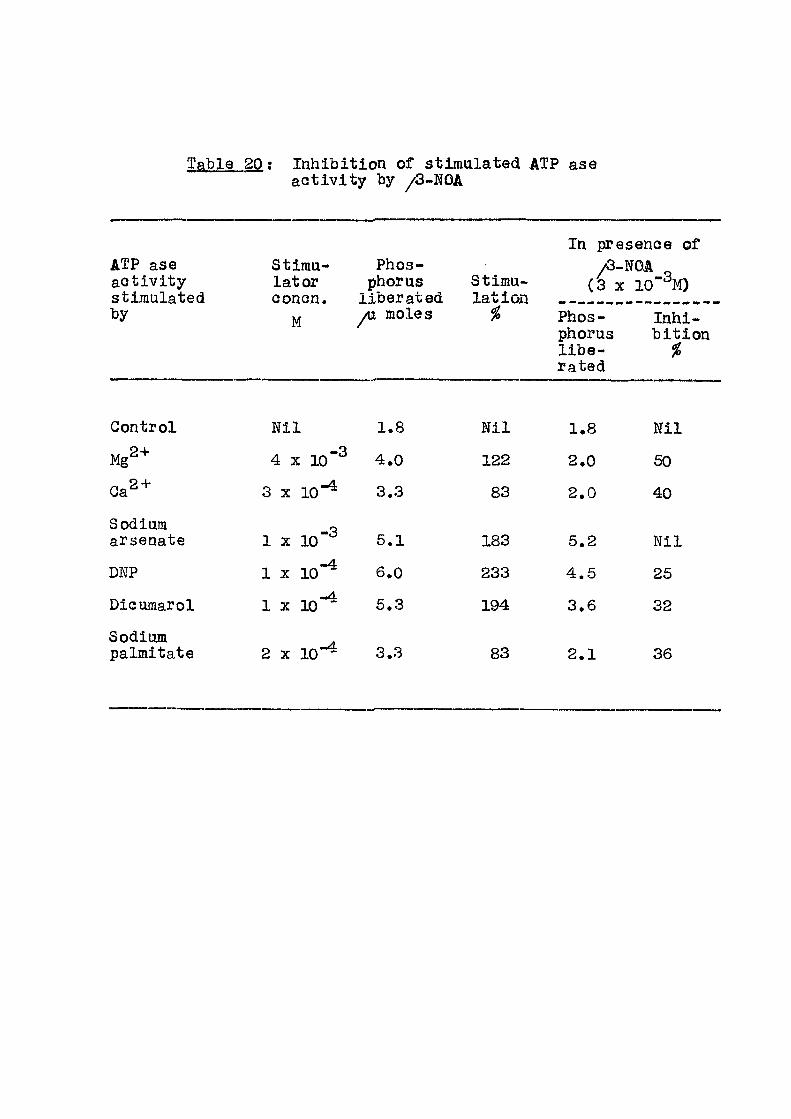
Inhibit ion of stimulated ATP ase ac t i v i t y by /3-NOA
In presence of ATP ase stimu- Phos- /3-NOA ^
(3 X lO' ^M) ac t i v i t y lator phorus Stimu-/3-NOA ^
(3 X lO' ^M) stimulated conon. l iberated lat ion by M yu moles % Phos- inhi-M yu moles
phorus b i t ion l i b e - % rated
Control N i l 1.8 N i l 1.8 N i l
M g 2 + 4 X 10"^ 4.0 122 2.0 50
0 a 2 + 3 X 1 0 " ^ 3.3 83 2.0 40
Sodinm arsenate 1 X 10"^ 5.1 183 5.2 N i l
DNP 1 X 10"^ 6.0 233 4.5 25
Dicumarol - A
1 X 10 5.3 194 3.6 32
Sodium palmitate 2 X 10"^ 3.3 83 2.1 36

s E f f ec t of inhibitors on stimulated ATP ases
Stimu- Inhi -lator b i tor
GonG.
Inhibit ion % in presenGe of
/3- p-GlA 2,4,D 2,4,5- 2,4,5-y w - t , , - ^
NOA TA
Ga " lO^^M 48 32 21 18 19
10 "^M 23 8 22 30 50
DNP 10 "^M 20 17 32 24 33
" 10"% 13 15 17 16 24
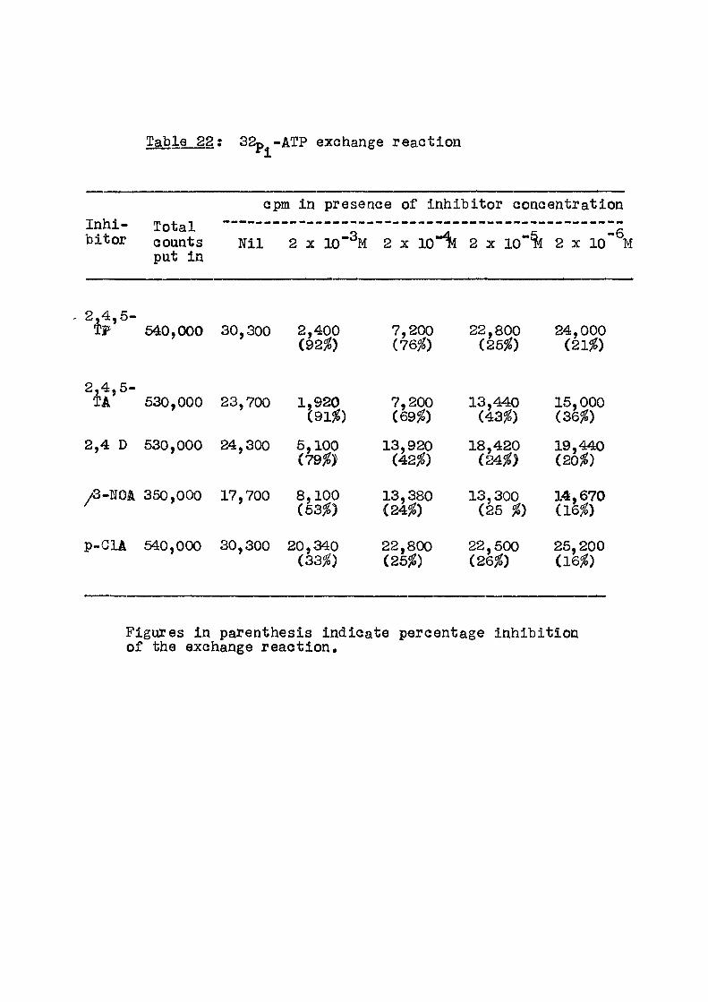
Table 22; 32p^-ATP exchange reaction
cpm in presence of inhibitor concentration Inhi- Total : 22 ^ I s bitor counts N i l 2 x 10"" M 2 x 1 0 2 x 10**% 2 x 10 M
put in
2,4,5-TP 640,000 30,300 2,400 7,200 22,800 24,000
(92^) (76fo) (25%) (21%)
2 4 5-TA* 530,000 23,700 1,920 7,200 13,440 15,000
(91jg) (69^) (43^) (36^)
2,4 D 530,000 24,300 5,100 13,920 18,420 19,440 (79^) (42^) (24^) (20%)
/3-mik 350,000 17,700 8,100 13,380 13,300 14,670 (53^) (24^) (25 %) (16%)
p-GlA 540,000 30,300 20,340 22,800 22,500 25,200 (33^) (25%) (26%) (16%)
Figures in parenthesis indicate percentage inhibit ion of the exchange react ion.
68
inhibited by 25-50^. Unstimulated ATP ase and the one
stimulated by arsenate were however not e f f e c t ed . Table 21
indicates that other phenoxy acids used in the present
investigation inhibited the ATP ase ac t i v i t y stimulated
by Ga "*" or DNP by 25-50% when used at a concentration
of 10"% or lO'^M.
Another mitochondrial property connected with
oxidative phosphorylation studied was Pi-ATP exchange ax
reaction. In Table 89 are given the results of inhi -
b i t ion of the exchange reaction when the inhibitor con-
centration was varied from 2 x lo'^M to 2 x lo"^M. At the
lower concentration, there was about 20% Inhibit ion which
increased upto 90% in some eases when the inhibitor —2
concentration was raised to 10 M, The control phosphate
incorporation in the exchange reaction was about 6% of the
t o ta l counts put into the reaction mixture.
SUMMABY
1. ATP ase ac t i v i t y of mitochondria stimulated
by addition of Ga ' or DNP was inhibited between 20 to
50% when the various acids were added (as sodium sa l t ) -3 -4
over concentration of 10 m or 10 m. Unstimmlated
ATP ase ac t i v i t y was not a f f e c t ed .
2. 32p^-ATP exchange reaction of the whole mito-
chondria was also inhibited between 20^ to 90% depending
on the concentration and the nature of the acid employed.
69
EFFECT OF /3-NOA ON OTHER ENZYME REACTIONS
Introduction
Af ter studying the e f f e c t s of ^ N O A and
phenoxy acids on mitochondrial oxidative phosphory-
lat ion propert ies! i t was decided to study the e f f e c t
of these compounds on other enzymes. For th is , the
fol lowing enzyme systems were selected: ( i ) amino
transferase and ( i i ) myokinase, ( i i i ) eatalase,
Civ) acid phosphatase, a l l from mitochondrial f rac t i on ,
( v ) acid phosphatase, ( v i ) alkal ine phosphatase both
from rat serum, ( v i i ) microsomal glucose 6-phosphatase
and ( v i i i ) ATP creatine transphosphorylase from
rabbit muscle. During this work i t was necessary to
pur i fy enzymes ( i i ) and ( v i i i ) upto a certain stage of
puri ty . The methods of pur i f i cat ion employed are des-
cribed under ^Experimental'. /3-KOA was employed in
the present experiments as a representative of the
compounds employed in the other studies. The mito-
chondrial f ract ion was col lected at 10,000 g fo r
20 minutes and washed twice with 0.26M sucrose.

70
Exp er linen ta l
12
i ) Mitocliondrlal (Aspartate - 2-oxoglutarste) aminotransferase
Beag;ents
Buffered aspartate: DL-aspartic acid (1.33 g )
and 0.86 g K2HPO4 are dissolved in double d i s t i l l ed
water, adjusted with KOH to pH 7.4 and diluted to 50 nsl.
o( -ketoglutarate: 0.1 M adjusted to pH 7.4.
Ani l ine c i t r a t e : C i t r i c acid solution made by
dissolving 60 g of c i t r i c acid in 50 ml water. Equal
parts of c i t r i c acid and ani l ine are mixed Just before
use.
Buffers O.IM potassium phosphate pH 7.4.
Trichloroacetic acid i 100 %
2)4 Dinitrophenyi hydrazine reagent: 100^ in 2N HGl.
Toulene: Water saturated
Alcohol ic KOH: 2.5 g/,K0H in 95% ethanol.
Procedure
The reaction mixture consisting of 0.1 ml of
mitochondria (about 1.6 mg prote in ) , 0.5 ml of buffered
aspartate, 0.2 ml of o(-ketoglutarate and lO^^M ^3-NOA
when necessary in a f i n a l volume of 2 ml,was incubated
71
at 37° fo r 10 min a f t e r which 0.1 ml of TCA was added
to stop the react ion. 0.1 ml of ani l ine c i t r a t e was then
added and incubated f o r 10 min to convert oxalo»cetate
formed in the reaction to pyruvate.
To each tube was added 1 ml of 2,4 dinitrophenyl
hydrazine reagent and the reaction was allowed to go at
37® for 5 min. 2 ml of water saturated toluene was then
added to extract the hydra zone of pyruvate. 0.6 ml of
toluene layer was added to 6 ml of a lcohol ic KOH and
the colour developed a f t e r 10 min was measxared in a K le t t
Summerson Photoelectric Colorimeter with f i l t e r No. 62.
i i . Myokinese from rat l i v e r mitochondri^a
Puri f icat ion^^;
Rat l i v e r mitochondria were treated with 10 ml
of 0.01 M KCl f o r 16 min. The supernatant was co l lected
at 10,000 g for 20 min. To the supernatant was added
2Iil HCl and st irred to decrease the pH from 6 to 2.
After 20 min of s t i r r ing the pH was increased to 7 by the
addition of 2N HaOH. The prec ip i tate formed was centr i -
fuged o f f and discarded. To the supernatant 1 M zinc
acetate was added with constant s t i r r ing to a f i n a l
concentration of 0.0185 M during which the pH was maintained
at 7 with 2 N HaOH. The prec ip i tate was co l lected and
washed with 0.05 M acetate pH 5.0, resuspended in the same
buffer and dissolved by decreasing the pH to 3.6 with
2N HGl. This was used as the source of enzyme.

u
The ac t i v i t y of the enzyme was determined by
coupling the myokinase catalysed reaction with the ATP-GE
transphosphorylase 2 ADP ATP + AMP (1)
ATP + Cr ADP + Cr~P (2)
ATP-creatine transphosphorylase was pur i f i ed as
described on page 75
A stock reaction mixture consisting of 0.002M
MgSO^, 0.08 M creatine and 0.08 M glycine adjusted to
pH 8.0
0.008 M ADP
0 . 1 M cysteine
5% aaimonium molybdate
8% perchloric acid
Reducing agent: 0.2 g of l amino-2 naphthol-4-
sulphonic acid, 12 g NaHSOg and 2.4 g of Na^SO^ in
d i s t i l l e d water to make 100 ml.
grocedure
In 10 ml graduated pyrex test tubes the fo l lowing
al iquots were pipetted. 1 ml of stock reaction mixture,
0.6 ml of 0.008 M ADP, 0.2 ml of 0.1 M f r esh l y neutralized
cysteine and 0.2 ml of ATP-creatine transphosphorylase
(0.6 mg/ffil). The reaction mixture was pre-incubated
73
at 30° f o r 10 min during which time any trace of ATP
contaminant in the ADP was dephosphorylated and measxired
as Gr ~ P at zero time. The reaction was in i t ia ted by
the addition of 0.1 ml properly di luted myokinase and then
reaction was stopped a f ter 16 min with l ml of ammonium
molylDdate and 6 ml of perchloric ac id .
A f ter a 30 min Cr P hydrolysis period at room
temperature 0.4 ml of reducing agent was added, made upto
10 ml with d i s t i l l e d water and colour read at 660 m/u.
14
i i i . Catala^e
Reagents
Buffered pubgtrat^; 5 ml of 0 .1 M phosphate
buffer pH 7 and 3 ml of 3^ HgOg are made upto 100 ml.
TiOcz 1 g TiOg and 10 g KgSO^ were digested with
150 ml conc. HgSO^ for 6 hours in a Kje ldahl f l a sk . The
mixture was cooled and diluted ten times with d i s t i l l e d
water and f i l t e r e d . A 6 ml a l iquot was tested c o l o r i -
metr ical ly with 2 ml of 0.01 N HgOg (excess) and the
stock solution was diluted with H^SO^ such that 6 ml of
this solution with 2 ml of 0»01 N H O gave a K le t t ^ a
reading of over 150 units with f i l t e r Wo. 42.
Proc edure
In a set of 3 tubes was pipetted 5 ml of the
titanium reagent and in another tube 10 ml of i ce cold
74
buffered HgOg solution. Fresh rat l i v e r mitochondrial
f ract ion was diluted 1:160, and 2 ml were pipetted in the
buffered H^Og solution in an ice bath. Af ter a brisk
mixing, 2 ml were withdrawn from the mixture and the
enzyme reaction was stopped by pipett ing i t into a
tube containing the titanium reagent (This should be
over within lo seconds). The time was noted. Exactly
a f te r 2 and 4 min 2 ml of each of the reaction mixture
was withdrawn similar ly and pipetted into the other two
tubes. The intensity of the yellow colour of peroxytitanic
acid thus developed by the interaction of residual H^Og
with the titanium reagent was read in a K le t t Summerson
Photoelectr ic Colorimeter using f i l t e r Wo. 42.
A suitable blank for the above set of tubes
was obtained by using 10 ml of d i s t i l l e d water instead
of the buffered substrate in the same manner,
i v and v) Acid phosphatase
The reaction medium contained 1 ml of 0.1 M
sodium acetate buffer pH 5.0, 0.2 ml 0.06 M sodium
beta-glycerophosphate, 0.6 ml rat serum (or mito-
chondrial f rac t ion ) and 6 x 10"% beta-NOA when added.
The f i n a l volume was 3 ml. This was incubated f o r 20 min
at 37° and the reaction stopped by l ml 10^ t r ich loroacet ic acid.

75
v i ) Alkal ine phosphatase
The reaction mixture contained 1 ml 0.1 M
sodium bicarbonate biaffer pH 9, 0.2 ml 0.06 M sodium
beta-glyeerophosphate, 0.1 ml rat serum and when added
6 X 1 0 " ^ beta-NOA. The f i n a l volume was S ml. The
reaction mixture was incubated at 37° for i hr and
the reaction was stopped by l ml 20^ t r ich loroacet ic
acid.
v i i ) Glucos e-S-Phospha ta s e^^
The reaction mixture consisting of 30 /umoles
of Tris-maleate buffer pH 6,6. 20 Amioles of glucose-
6-phosphate pH 7$ and microsomal protein f rac t i on
( f i n a l volume 1.5 ml) was incubated at 30° f o r 20 min.
At the end of this period, the react ion was stopped
by 1 ml 10^ TGA.
In the case of enzyme systems ( i v ) , ( v ) ,
( t i ) and ( v i i ) inorganic phosphate released at the end
of the reaction was measured by the method of Taussky
and Shorr/as described on page ZS
v i i i ) ATP-Greatine transphosphorylase
Purification^*^
A l l the operations were done at 0 - 4 ° . Rabbit
muscle was homogenized in a waring blendor using 2 ;i.itres
of cold 0.01 M KGl per kg of ground muscle, and the
76
iiomogenate was squeezed through cheese cloth and f i l t r a t e
co l l ec ted . Solid NH^Cl was added to the f i l t r a t e to a
concentration of 0*1 M and pH adjusted to 9.0 with 6 M NH^OH,
A f ter half an hoiar 1.6 vo l of cold ethanol was added and
st irred f o r 2.6 hours at 20°. The formed prec ip i ta te was
discarded by centrifi:igation, 2 M MgaO^ (pH 8.5) was
added to the supernatant with s t i r r ing to a f i n a l concentra-
tion of 0.03 M and cold ethanol (1.5 times the volume
,of 2 M MgSO^ used) was added. A f te r s t i r r ing f o r 1/2 hour
at 20° the prec ip i tate was co l l ec ted , twice resuspended o
and extracted at 0 with 0.7 M Mg acetate pH 9.0. Each
time the insoluble protein w a s separated by centr i fugat ion.
From the supernatant alcohol f r a c t i on between
36-60J^ was co l lected, dissolved in 30 ml of 0.05 M
ammonium c i t ra t e pH 9 and dialysed against 1 l i t r e
of 0.05 M ammoniiam c i t ra t e f o r 6 hours and then against
two changes of 1.7 x 10**^ NH^OH.
The enzyme at this stage was used for the assay
and also ut i l i sed in the assay of mybkinase.
Reagents : 605 HGIO4
(]ffl^)gM0^0244H20
Reducing agent; 0.2 g of 1 amino 2 naphthol 4-sulphonic acid, 12 g MaHsO and 2.4 g of Na ao^ in distil led'^water to 100 ml. 0.08 M creatine 0.1 MgSO^ 0.005 M ATP (Na"^) pH 7 0.4 M glycine (Na"*") buf fer pH 9.
Table 23: E f f ec t of beta-Naphthoxy acetic acid (sodium sa l t ) on enzyme systems.
Eazyme Source Units of enzyme ac t i v i t y
Goneen- -/3-N04 +/S-NOA inhi -trat ion b i t ion /Q-NOA M
i ) Amino-trans-
Rat l i v e r 1 X 10
ferase®- mitochondrial f rac t ion*
i i ) Myoki-naseb
i i i ) Gata-lase
i v ) Acid phos tase phosDho-
v ) Acid
-3 1700 1700
• ti
Rat phospho- serum ase'
v i ) Alkaline phospha- " tase°
v i i ) Glucose- Rat l i v e r 6-phos- micro-phatase^ somes
v i i i ) ITP-crea-tine trans- Rabbit phospho- muscle rylases
1 X 10"^ 32 30
Please see table No.2^4-...
6 X 10~2
6 X 1 0 ~ 2
6 X 1 0 " 2
8 X 10 -3
2 X 1 0 ~ 3
7.5
320
170
420
680
5.2
342
106
323
440
N i l
7'
30
N i l
40
23
24
Units : a ) b) c ) d) e )
y^g pyruvate formed/mg protein. yag Pj[ liberated/mg protein yug Pj_ liberated/ml serum /ug Pi liberated/30 min/mg protein yag Pi liber^ted/15 min//ig protein,
* This f ract ion was col lected at 10,000 x g for 20 min and washed twice with 0.26 M sucrose.
Table 24; E f f e c t of /Q^Naphtohxyacetate, and sodium acetate on G<atalase,
% inhib i t ion in presence of Concentration
M /3-WOA Acetate
2.5 X lO'^M 40 30
1.5 X 10-% 37 27
3 X 10 "^M 61 22
6 X 10"% 66 25
Conditions of assay for catalase are given
in 'experimental ' .
77 ProceduJe
Each tube contained 0.024 M creat ine, 0.006 M MgSO^,
0.096 glycine buffer pH 9, 0.001 M ATP, when added 0.001 M
^3-NOA as sodium salt and 0.6 ml of (1:100 diluted in g lyc ine
bu f f e r ) enzyme. The tubes were incubated at 30° for 16 min
and reaction was stopped by addition of 0.8 ml of
A zero hour control was also run to correct f o r any inorganic
phosphate present in the AIEP sample. To each tube was
added 1 ml of 6% ammonium molybdate and a f t e r 30 min
0.4 ml reducing agent. A f te r waiting f o r 10 min tlae in-
organic phosphate was determined in K le t t Summerson Co lo r ! ,
meter with f i l t e r Wo. 66.
Besults and Discussion
Results in Table Nos. 23 and 24 g ive the
e f f e c t of beta-NOA on various enzyme systems studied. Even
at a very high concentration of l o " *^ , the acid (as sodium
sa l t ) was able to inhibit the enzymes only between 7-40/2.
Thus this group of compounds does not appear l i k e l y to
a f f e c t in v ivo the various enzyme systems studied here.
I t is seen from Table No. 24 that at similar concentration,
beta-WOA i s more inhibitory than acetate towards catalyse
a c t i v i t y . This enzyme i s known to be inhibited by various
anions l i k e sul fa te , phosphate, chloride and acetate .
Thus e f f e c t of beta-NOA can be attr ibuted to i t s anionic nature
as the acid i s used in the form of i t s sodium sa l t .
Summary
The other enzyme systems studied were not a f f ec ted
appreciably by /S-NOA even at 10"% concentration.
79 REFERENCES
1. RaGker, E. (1965), Mechanisms of Bioenergetics, Academic Press, p 117.
2. Boyer, P.D., Luchsinger, W.W & Falcone, A.B, (1956), J. Biol.Chem., 223, 405.
3. Groot, G.S.P. (1969), Biochim.Biophys.Acta, 1 8 0 > 4 3 9 .
4. Racker, E. , Blochem.Biophys«Res.Coimn. (1963), 10, 435.
5. Tzago lo f f , A. , Byington, K.H. & Maclennan, D.H. (1968), J.Biol.Chem., 2 ^ , 2405,
6. Graven, S.N., Lardy. H.A., Johnson, D.& Rutter, A. (1966), Biochemistry (USA), 5, 1729.
7. Veldsema-Gurrie, R.D. & Slater , E.G. (1968), Biochim.Biophys .Ac ta ,162 , . ^lOo
8. MacLennan, D.H. & Asai, J. (1968), BiochemoBiophys.ReSoGomman., 441.
9 o Wenner, G.E. & Hackney, J„H. ( 1 9 6 9 ) ,
Biochemistry (USA), 8 , 9 3 0 o
10. Recknagel, R.O. & Anthony, D.O. (1959), J.Biol.Ghem., 234, 1052o
11. Gooper, G. & Lehninger, A.L. (1957), J.Biol.Ghem., 224, 561.
12. Tonhazy, N.E., White, N.G. and Umbreit, V / . W . (1950), Irch.Biochem., 28, 36.
13. Noda, L. and Kuby, S.A. (1963), Methods in Enzymol.,
y l , p.223, Academic Press.
14. Manjrekar, S.P. (Unpublished).
15. Gianetto, R. and De Duve, C. (1955), Biochem.J., 59, 433.
16. Recknagel, R.O. and Lombardi, B. (1961), J.Biol.Ghem,, 236, 564.
17. Noda, L,5 Kuby, S. and Lardy, H. (1965), Methods in Enzymol., i i , 605.
18. Nicholls, P. (1961), Biochem.J,, 81, 365,
119
GBNERAIi DISCUSSIONS ON CHAPTERS I I -V

80
GEMBRAL DISCUSSION ON CHAPTERS I I
E f f ec t of /3-na|)hthoxy acetic a d d and substituted
phenoxy adds on rat l i ve r mitochondrial properties
The inhibitors of mitochondrial oxidative
phosphorylation reaction can be c l ass i f i ed into three
categories as already mentioned in the general introduction,
The three categories are ( i ) inhibitors of electron
transport, ( i i ) inhibitors of phosphorylation mechanisms
and ( i i i ) uncouplers. The studies carried out with some
phenoxy acids and /3-naphthoxy acetic acid in the present
investigation have given an indication that these
compounds belong to the second category i . e . they inhibit
phosphorylation and other related reactions coupled to
electron transport.
Other compounds belonging to this category
and already described in l i t e ra ture are azide^, o l i { o 3 4
mydn , atractyloside , potassium atractylate ,
dicycloh<
lamine'^.
dicyclohexyl-carbodimide^, formaldehyde® and hydroxy-
p
Lardy and his colleagues have screened a number
of ant ib iot ics for their action on mitochondrial reactions,
They found oligomycin to be a very potent inhibitor of
oxidative phosphorylation, 2 yug oligomycin (4 m/umole)
9t
per atout 9 mg mitochondrial protein completely inhibited
phosphorylation with L-glutamate, succinate and 3
hydroxybutyrate as substrates. I t rac ty lcs ide is also
e f f e c t i v e at a concentration of 2yv.g (2.5 m^umole) per
10 mg mitochondrial protein, whereas the phenoxy acids
employed in the present studies were e f f e c t i v e at about
200-300 /1^(1 - 1.3/amole) per 10 mg mitochondrial
protein. These compared favourably with the concen-
trations of formaldehyde and hydroxylamina required fo r
inhibit ion of phosphorylation. According to Van Buskirk
and F r i s e l l 0,4 to 1.26 mg of formaldehyde (15-37/Umbles)
per 10 mg mitochondrial protein was necessary for inhib i -
tion of phosphorylating respirat ion. In these studies
the respiratory control r a t i o was lowered from a" value
of 4 to 1 in presence of the above concentration of
formaldehyde.
Ifikstrom and Saris'^ employing rat l i v e r mito-
chondria observed that fo r half maximal inhibit ion of
state 3 respiration and oxidative phosphorylation, the
hydroxyIsmine concentration required was about 280
(8.4/umoles) per 10 mg protein.
Beechy and his coworkers^ have shown that
dicyclohexyl-carbodiimide inhibited the state 3 respirat ion
at a concentration of about 66 yug (0.33 ^mo l e ) per 10 mg
protein.

82
I t Is reported in literature that oligomycin
inhibit ion of Goupled respirat ion was overcome by d in i t r o -
phenol, A similar e f f e c t of dinitrophenol was noticed
in the present studies with 2,4,6-triGhlorophenoxy
propionic acid. Results of oxidative phosphorylation
studies gave an indication that the phenoxy acids may
be acting on the mitochondria in a manner similar to
oligomycin and a t rady l o s ide . Further experiments were
therefore planned to get more e:^idence for this s imi lar i ty
between the action of phenoxy acids and oligomycin.
A number of mitochondrial properties connected with the
energy transfer reactions were studied towards this end.
8 Lehninger has shown inhibit ion of ITP dependent
act ive ion uptakes by oligomycin. 4 , 5 ^ g of oligomycin
per 3 mg mitochondrial protein just about completely
inhibited ATP dependent Ga " uptake. Another ant ib iot ic
aurovertin^ inhibited by 50%, ATP driven Gb.^'* uptake at
a concentration of 10 /xg per mg protein.
Another mitochondrial property influenced by ATP
is the swelling contraction cycle of the mitochondria.
ATP is known to reverse the swelling of mitochondria, 2*
induced by agents l ike inorganic phosphate, Ga or
L-thryoxine. Between 4 to 30/ug of either oligomycin^^
or atractylate'^ can bring about complete inhibit ion of
this ATP e f f e c t . Formaldehyde® also at a concentration
B3
of 2 mg per 10 mg mitochondrial protein diminished
the ATP reversal . In the present investigation, i t
Was noticed that the various phenoxy-acids and
naphthoxy acetic acid at 4 x 10~^M concentration reduced
the ATP reversal of mitochondrial swell ing. This
indicated that ut i l i sa t ion of ATP energy for mitochondrial
contraction was diminished in the presence of these acids,
ATPase ac t i v i t y stimulated by the presence of
e i ther Ga " or 2,4,-dinitrophenol is inhibited to
various extents by the established inhibitors of energy
transfer reactions. About 4 to 10/ag of oligomycin or
atractyloside per 10 mg mitochondrial protein are e f f e c -
t i v e^ '^ . Hydroxylamine*^ was necessary at a concentration
of 1 mg per 10 mg mitochondrial protein. A similar
observation employing phenoxy acids as sodium sal ts is
reported in the present invest igat ion.
Exchange reactions l ike P^-ATP and ADP-ATP
which are connected with the mitochondrial phosphorylation 3 4
reactions are suppressed in the presence of atractylate '
and oligomycin. About 0.3/ug of oligomycin^ is e f f e c t i v e .
In the present study, when the phenoxy acids were employed
over a range of lO"^ to lO'^M, the ^^^-ATP exchange
reaction was inhibited between 20-90^ depending on the
nature and the amount of acid employed.

84
The mitochondrial properties examined in the
present investigation include coupled phosphorylation,
ion transport, swelling contsaetion cycle, various
stimulated ATPases and P^ - ITP exchange reaction. A
few other mitochondrial and extramitochondrial enzymes
were also studied. A l l these are energy dependent
reactions. Literature survey indicates that these are
inhibited by compounds like oligomycin and atrac ly los ide .
Since the phenoxy acids employed in the present inves t i -
gation also e f f ec ted these reactions, i t is concluded
on the basis of the data obtained that the la t ter compounds
behave in a manner similar to oligomycin. Other mito-
chondrial and serum enzymes investigated did not appear
to be a f fected to an appreciable extent.
Taking into consideration the data co l lected in
the present invest igat ion, i t i s possible to conclude
that the action of the phenoxy acids is on the energy
transfer reactions of the mitochondria. The concen-
trations required of these acids were, however, far in
excess of either oligomycin or atractyloside. The
later compounds are e f f e c t i v e at 1/200 the concentration
of phenoxy acids.
These phenoxy acids and ^^-naphthoxy acetic
acid are used in horticulture as growth regulators at a
CHAPTER VI
POLYPHENOLOXIDASE FROM PEEL OF THE MANGO FRUIT (MANGIFERA INDIGA)

96
CHAPTER Vi :
POLYPHENOL OXIDASE FKOM PEEL CF HIE MANGO FBUIT
Introduction
Polyphenol oxidase catalyses the oxidation of
polyphenollQ compounds to give quinones. The quinones
then undergo polymerization to give complex coloured
products. This enzyme is widely distributed in a number
of sources l ike mushroom^ 'banana^ , potato^, sweet f) v
potato , apple ' , e tc . I t s action leads to enzymatic
discoloration of various f ru i t s and vegetables, when they
are s l iced or damaged. Most of the polyphenol oxidases
have a pH optimum round 5-6, The enzyme is known to be
a copper protein and the copper is present both in the
reduced and the oxidised form.
Tyrosinase and laccase also belong to the category
of enzymes acting on phenolic compounds. Tyrosinase is
found in number of animals and plants. This enzyme
catalyses oxidation of tyrosine to orthoquinone leading
to the formation of dark pigment melanin, Laccase is
distinguished from the polyphenol oxidase as the la t ter
is sensit ive to carbon monoxide and is not endowed with
the blue colour of laccase.
87
Recently, polyphenol oxidase from potato has
been studied by Pa t i l and ZuGker^, They have pur i f ied
the enzyme about 170 f o l d and studied i t s ac t i v i t y
towards various substrates l ike diphenols and mono-
phenols. Mushroom polyphenol oxidase has also been
puri f ied by Smith and Krueger^.
In the present invest igat ion, polyphenol oxidase
has been puri f ied from the peel of the mature mango
f r u i t (Mangifera indica) and some of i t s properties studied.
Experimental
Materials
Fully mature mango f ru i t s were harvested from
the local gardens and kept at room temperature (28° ) .
These were taken for enzyme iso lat ion on 9th or 10th day.
By this time, the f ru i t s reach cl imacteric maximum stage.
Immediately af ter harvest, f ru i t s are in pre-climacteric
stage.
Enzyme puri f icat ion
Acetone dried powder; 50 g peel of the f ru i t
was homogenized in Waring blendor with 400 ml ch i l l ed
acetone. The suspension was immediately f i l t e r e d on
buchner funnel. The powder was washed further with
acetone, dried and stored at 0°.
6S
2. Extraction of the enzyme ; 4 g of the
powder was extracted with 32 ml of 0.1 M phosphate
buf fer (pH 7,2) by constant s t i r r ing for 30 min.
The extract was f i l t e r e d through cheese cloth and centr i -
fuged.
3. Calcium acetate treatment ; To 30 ml
of clean supernatant was added 150 mg of calcium
acetate to remove the contaminating polyphenoles that
were l e f t in the powder a f ter acetone treatment. After
a period of 15 min, clear solution was co l lected by
centri fugation and the precipitate discarded. The
enzyme solution was dialysed against 0,001 M phosphate
buffer (pH 7,2) f o r 3 hours,
4. Alcohol f ract ionat ion: From the above
solution, the enzyme was precipitated between 30-70^
alcohol concentration. The temperature during the
operation was maintained between 0° to-40. The protein
was col lected by centri fugation, dissolved in 0,01M
phosphate buffer (pH 7,2) and dialysed against 0,001 M
phosphate buffer f o r four hours.
5. PEAS column chromatography; The above enzyme
Was placed on a DEAB-cellulose column (1,5 x 7 cm).
Af ter stepwise gradient elution with phosphate buffer of
pH 7,2 (0.01 M - 0,025 M - 0.05 M - 0,1 M), the enzyme

69
f ract ion was oUAJid in 0.05 M phosphate eluate.
The enzyme at this stage of parity has been
used in a l l the studies described in this chapter.
Protein nitrogen of acetone dried powder was
estimated by Kjeldahl method and on subsequent fract ions
by absorption reading at 260 m^ and 280 in Beckman
spectrophotometer model DK-2A.
Enzyme assay: Method of Mayer, Hard and Ben-9
Shaul was followed for assay of the enzyme. The cuvette
contained 0.01 M c i t ra te - 0.025 M phosphate buffer pH
5.5, 0.0033 M catechol (substrate) , 0.5 ml of enzyme
(protein range 10 to 2250 / depending on the fract ion
u t i l i z ed ) . The f i n a l volume was 3 ml. The reaction was
started by the addition of the substrate. The enzyme
blank cuvette contained a l l reagents except substrate.
The increase in absorption due to oxidation of
the substrate was recorded in Beckman ra t i o recording
spectrophotometer model DK-21 at 395 m/U for a period of
two min at 30 sec in terva l . The spec i f i c a c t i v i t i e s of
the enzyme at various stages of pur i f icat ion were calculated
from the d i f ference in the i-eadings of 1 min to 2 min. The
absorption maximum of the oxidised product was determined by
recording i t s spectrum in the i n i t i a l experiments.
90
Results and Discussion
In Table 25 are given results of pur i f icat ion
procedure adopted. I t is possible to puri fy the enzyme
about f i f t y f o l d with the use of alcohol f ract ionat ion
and DEAS cel lulose column chromatography. Use of
calcium acetate in the i n i t i a l stages has been
advocated by Pa t i l and ^ucker^. According to them,
use of calcium acetate removed some colored pigments
which otherwise inter fere in pur i f icat ion at a later
stage. Attempts at ammonium sul fate f ract ionat ion were
not very successful as no compact pe l l e t could be obtained
and the precipitate had a tendency to f l o a t during centr i -
fugation. This d i f f i c u l t y was overcome in alcohol
f ract ionat ion. This may Indicate association of the
enzyme with l ip id constituents in s i tu .
In Table 26 are given substrate spec i f i c i t y
values. I t is observed that by taking catechol ac t i v i ty
as hundred units only 4 methyl catechol is s l i gh t l y more
react ive . Gall ic acid which has previously been
observed in this laboratory to be the major substrate
in situ for enzyme from this source is about half act ive .
Same pattern is obtained when the enzyme is isolated from
either climacteric or pre-climacteric f r u i t .
Km values fo r the enzyme puri f ied upto DEAE
stage, with catechol as substrate are 2 x 10"^ M in

Table 26: ^ Pur i f i cat ion of polyphenol 03cidase from mango f r u i t peel (PrecliniacteriG s tage ) .
Treatment Total Protein Speci f ic volume mg/ml ac t i v i ty
1. Original extract 33 4.5 0.09
2. Galcinm acetate treated extract 30 1.5 0.08
3. 30^ - 70^ alcohol 4.5 0.9 0.15
4. DEAE cel lulose column 5.0 0.02 4.6
The enzyme assay was carried out in Beckman recording spectrophotometer model DK:-2A at 395 mya.
The cuvette contained 0.01 M c i t ra te -025 M phosphate buf fer pH 5,5, .0033 M catechol (substrate) 0.5 ml of enzyme (10 y to 2250 / protein range depending upon the f ract ion used). The f i n a l volume was 3 ml. The reaction was started by the addition of substrate. The enzyme blank contained a l l reagents except substrate.
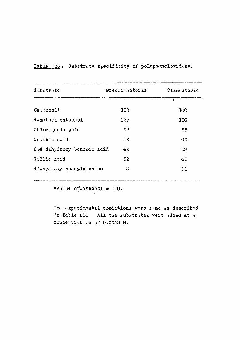
Table 26; Substrate specif l o i t y of polyphenoloxidase.
Substrate Precllmaoterlo Climacteric
Catechol* 100 100
4-methyl catechol 137 100
Chlorogenic acid 62 55
Caf fe ic acid 52 40
3:4 dihydroxy benzoic acid 42 38
Gal l ic acid 52 45
di-hydroxy phen]?^lalanine 8 11
•Value oi^batechol = 100.
The experimental conditions were same as described in Table 25. A l l the substrates were added at a concentration of 0.0033 M.
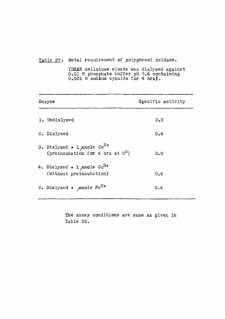
Table 27; Metal requirement of p.olyphenol oxidase.
(DEAS cel lulose eluate was dialysed against 0.01 M phosphate buf fer pH 7.4 containing 0.001 M sodium cyanide for 4 hrs ) .
Enzyme Spec i f i c a c t i v i t y
1. Undialysed 3.2
2. Dialysed 0.4
3. Dialysed • 1 yomole Gu^* (preincubation for 4 hrs at 0°) 3.0
4 . Dialysed • 1 yamole Cu "* (without preincubation) 0.4
5. Dialysed * yumole Fe^"* 0.4
The assay conditions are same as given in Table 25.
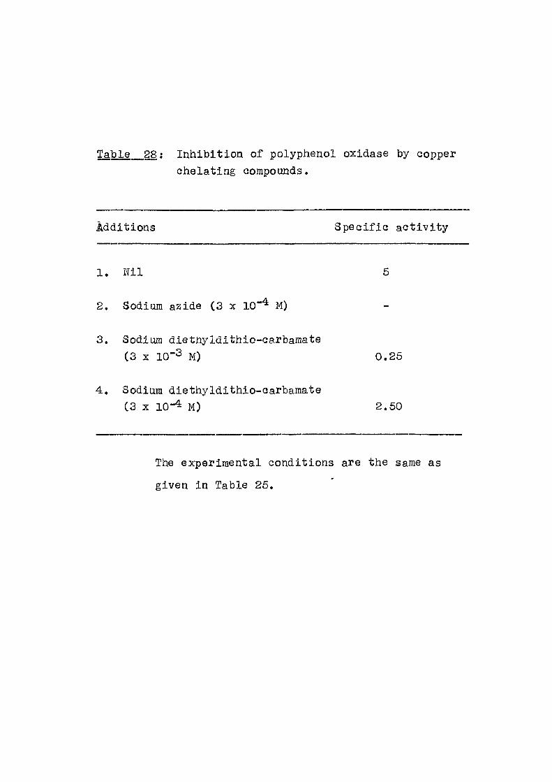
Table 28: Inhibit ion of polyphenol oxidase by copper ohelating oorapoands.
Additions Spec i f i c ac t i v i t y
1. N i l 5
2. Sodium azide (3 x 10"'^ M)
3. Sodium dietnyldithio-carbamate ( 3 X 10~3 M) 0 . 2 6
4. Sodium diethyldithio-carbamate ( 3 X 1 0 - 4 2 . 5 0
The experimental conditions are the same as
given in Table 25.
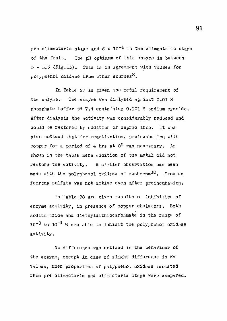
91
pre-ellmacteriG stage and 5 x 10*" in the Glimacteric stage
of the f r u i t . The pH optimum of this enzyme is between
5 - 5,5 ( F i g .15 ) , This is in agreeraeat with values for /
polyphenol oxidase from other sources^.
In Table 27 is given the metal requirement of
the enzyme. The enzyme was dialysed against 0.01 M
phosphate buf fer pH 7.4 containing 0.001 M sodium cyanide.
A f t e r d ia lys is the ac t i v i t y was considerably reduced and
could be restored by addition of cupric i ron. I t was
also noticed that f o r react ivat ion, preincubation with
copper f o r a period of 4 hrs at 0° was necessary. As
shown in the table mere addition of the metal did not
restore the a c t i v i t y , A similar observation has been
made with the polyphenol oxidase of mashroom^'^. Iron, as
ferrous su l fa te was not act ive even a f t e r preincubation.
In Table 28 are given resul ts of inhib i t ion of
enzyme a c t i v i t y , in presence of copper che lators . Both
sodium azide and diethyldithiocarbamate in the range of
to 10"^ M are able to inhib i t the polyphenol oxidase
a c t i v i t y .
No d i f f e rence was noticed in the behaviour of
the ensjyme, except in case of s l i gh t d i f f e rence in Km
values, when propert ies of polyphenol oxidase iso la ted
from pre-cl imacter ic and cl imacter ic stage were compared.
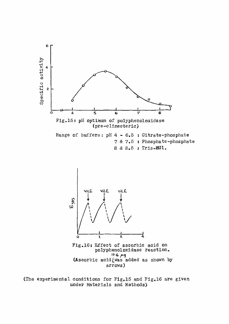
6 r
F ig ,15 : pH optimum of polyphenoloxidase (pre-c l imacter ic )
Range of buffers pH 4 - 6,6 : Citrate-phosphate 7 & 7.5 : Phosphate-phosphate 8 & 8.5 : Tr is-H6i .
F ig .16 : E f f e c t of ascorbic acid on polyphenoloxidase react ion,
17-G /Ag CAsaorbic aGid/_was added as shown by
arrows)
(The experimental conditions f o r Fig,15 and Fig,16 are given under Materials and Methods)
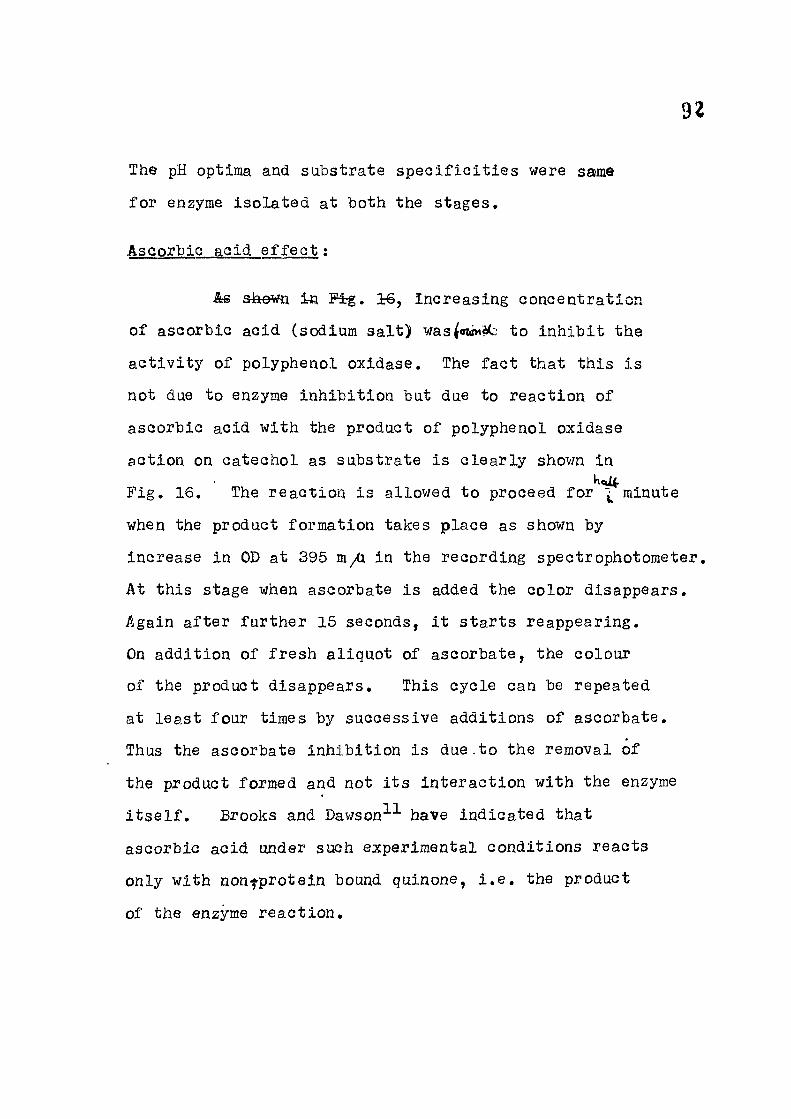
92
The pH optima and substrate spec i f i c i t i e s were same
for enzyme isolated at both the stages.
Ascorbic acid e f f e c t :
te Fig . 16, increasing concentration
of ascorbic acid (sodium sa l t ) MJas mnU:: to inhibit the
ac t i v i t y of polyphenol oxidase. The fact that this i s
not due to enzyme inhibit ion but due to reaction of
ascorbic acid with the product of polyphenol oxidase
action on catechol as substrate is c lear ly shown in
F ig . 16. The reaction is allowed to proceed for ^ minute
when the product formation takes place as shown by
increase in OD at 395 m/i in the recording spectrophotometer,
At this stage when ascorbate is added the color disappears.
Again a f te r further 15 seconds, i t starts reappearing.
On addition of fresh aliquot of ascorbate, the colour
of the product disappears. This cycle can be repeated
at least four times by successive additions of ascorbate.
Thus the ascorbate inhibition is due.to the removal of
the product formed and not i t s interaction with the enzyme
i t s e l f . Brooks and Dav/son^^ have indicated that
ascorbic acid under such experimental conditions reacts
only with non-j-protein bound quinone, i . e . the product
of the enzyme react ion.
03
Summary
1. Polyphenol oxidase from peel of the f r u i t
(Mangifera indic^) was puri f ied about f i f t y - f o l d .
2. The pH optimum was between 5 - 5,5.
3. Substrate spec i f i c i t y was determined with
enzyme isolated from both preclimacteric and cliraacteric
stage.
4. I t was possible to remove copper by dia lys is
against cyanide when ac t i v i t y was lost and could be
restored by addition of copper. Copper chelators
inhibited enzyme a c t i v i t y .
5. E f f e c t of ascorbic acid was studied.

94
1. Ponting, J.D. (1954), J.Am.Chem.Soc., 76, 662.
2. Frieden, E. and Ottesen, M. (1959), Bioohem.Biophys.Aota, M j 248.
3. Smith, J.L. and Krueger, E.G. (1962), J.Biol.Ghem.,
237y 1121.
4. Palmer, J.K. (1963), Plant Physio l . , 38, .508.
6. Baruah, P. and Swain, T. (1953), Biochem.J., 392.
6. Eiger, I . Z . and Dawson, G.R. (1949), Arch.Biochem., 21, 194.
7. Harel, E. , Mayer, A.M. and Shain, Y. (1966), J .Sc i .Fd . l g r i o . , 17, 389.
8. Pa t i l , S.S. and Sucker, M. (1965), J.Biol.Ghem., 240, 3938.
9. Mayer, A.M., Harel, E. and Ben-3haul, R. (1966), Phytoohem., 5, 783.
10. Kertesz, D. and Zito, E. (1965), Biochim. Biophys. 96, 447.
11. Brooks, D.W. and Dawson, G.R. (1966), From the Biochemistry of Copper, p.343, Proceedings of the Symposium. Ed. by Peisach, J. , Aisen, P . , Blumberg, W.E., AP.

GENERAL SUMMARY

95
GENERAL SUMMARY
A. The e f f e c t s of ^-nephtiioxy acet ic acid,
p-chlorophenoxy acet ic acid, 2,4-dichlorophenoxy
acet ic acid, 2,4,6-trichlorophenoxy acet ic scid and
2,4,6-trichlorophenoxy propionic acid ( in the form of
sodium sa l ts ) have been studied on oxidat ive phosphory-
lat ion and related properties of rat l i v e r mitochondria.
1. In the phosphorylating mitochondria
respirat ion and the coupled phosphorylation in presence
of succinate or glutamate was inhibited when these
aciots were used over a concentration range of lO^^M -
10"%. Waen the mitochondria were made non-phosphory-
lat ing by exposure to d i s t i l l e d water the respirat ion
was not a f f e c t ed .
2. The ^TP reversal of mitochondrial swelling n
induced by either 5 mM inorganic phosphate, Ca "*" or
L-thyroxine was par t ia l l y prevented by the phenoxy acids
at 10"%.
2+
3. ATP dependent act ive Ga uptake was
reduced between 40-80/^ in the presence of the above
acids.
4; ATP ase ac t i v i t y estimated in the presence
of Ga or 2,4-dinitrophenol was also inhibited between

96
20-60^ in the presence of or phenoxy acids.
The ^^Pj^-ATP exchange reaction was also inhibi ted.
A l l the reactions of the mitochondrial studies
above are connected with the phosphorylation mechanism
of the mitochondria. The data col lected indicates that
the phenoxy acids and /3-naphthoxy acet ic acid inhibited
the energy transfer reactions of the mitochondria. These
compounds did not a f f e c t appreciably other enzyme systems
studied.
These compounds thus resemble the ant ib io t i c
oligomycin or the glueoside atracty los ide in their
propert ies. These phenoxy acids are however about
200 times less potent than the established inhibitors
mentioned above. Formaldehyde and hydroxylamine the
other inhibitors of energy transfer reactions reported
in l i t e ra ture are e f f e c t i v e over the range of 10**^ -
10 M which is comparable to phenoxy acids employed
in the present study.
B. Polyphenoloxidase has been pur i f i ed about f i f t y -
f o l d from the peel of the mango f r u i t Mangifera indica.
I t s pH optimum, substrate s p e c i f i c i t y , K^ and copper
involvement have been studied.