effect of stressors on haematological and hormonal ... sharma, et al.pdf ·...
TRANSCRIPT
Int.J.Curr.Microbiol.App.Sci (2017) 6(5): 357-369
357
Original Research Article https://doi.org/10.20546/ijcmas.2017.605.041
Effect of Stressors on Haematological and
Hormonal Parameters of Garra gotyla gotyla
Jyoti Sharma1*
, Shabir Ahmed Dar2, A.N. Sayani
3 and Seema Langer
4
1Government Degree College, Kathua, J&K, India
2Government Degree College, Baramulla, Jammu and Kashmir- 193 103, India
3College of Fisheries, Junagadh Agricultural University, Veraval, Gujarat- 362 265, India
4Department of Zoology, University of Jammu-181 101, India
*Corresponding author
A B S T R A C T
Introduction
Stress can be described as the physiological
response to a stressor. In other words, stress is
an internal physiological state that is caused
by external conditions. Stress can also be
described as an internal hormonal response of
a living organism caused by environmental or
other external factors that moves that
organism out of its normal physiological
resting state, or homeostasis (Selye, 1973).
Stress can disturb the normal physiological
equilibrium or homeostasis of fish by forcing
a reallocation of energy within its system.
Stress in fish, a key member of aquatic
environment and which also form a valuable
commodity for human consumption (proteins,
16-23%) may be induced by various abiotic
International Journal of Current Microbiology and Applied Sciences ISSN: 2319-7706 Volume 6 Number 5 (2017) pp. 357-369 Journal homepage: http://www.ijcmas.com
Studies on stress in different species of fish has been widely made but not much has been
done in hill stream fishes especially, Garra gotyla gotyla. Haematological and hormonal
parameters are the most common stress indicators. In the present study, an attempt has
been made to study the effect of stressors natural (Starvation) and anthropogenic
(Manganese) on haematological [Total erythrocyte count (TEC), Haemoglobin (Hb),
Haematocrit (Hct), Total leucocyte count (TLC) and Differential leucocyte count (DLC)]
and hormonal (Cortisol and Glucose) parameters of fish, Garra gotyla gotyla for an
experimental period of 9 weeks. Under the effect of natural stressor, starved fishes were
found to exhibit significant decline in TEC, Hb, Hct and TLC. DLC when viewed revealed
a decrease in lymphocytes, monocytes, eosinophils and basophils whereas neutrophils and
thrombocytes rather exhibited an appreciable increase. A significant increase (P<0.01) in
cortisol and glucose levels were observed up to 5th
week and here after a significant
decline was noticed during rest period of experimental duration. Garra gotyla gotyla also
depicted significant decline in TEC, Hb and Hct under the effect of manganese toxicity
(MnSO4-1.96mg/l). Contrary to RBC dependent parameters (TEC, Hb and Hct) TLC
depicted significant increase and among Differential leucocyte count (DLC) lymphocytes,
monocytes and eosinophils register an increase but neutrophils, basophils and thrombocyte
population exhibit a decline in their number. Cortisol and glucose levels were noticed to
increase up to 4th
week and after that exhibit a declining trend in their values during the
rest period (5th
-9th
week) of experimental duration.
K e y w o r d s
Stress, Natural,
Anthropogenic,
Haematology,
Hormones,
Garra gotyla gotyla.
Accepted:
04 April 2017
Available Online: 10 May 2017
Article Info

Int.J.Curr.Microbiol.App.Sci (2017) 6(5): 357-369
358
environmental factors (change in water
temperature, pH, O2 concentrations, starvation
etc) (Gupta, 2009; Raina, 2011). Besides
these natural stressors, heavy metals and
xenobiotics (anthropogenic stressors) are
regarded as the serious pollutants which act as
major source of stress to fishes (Tavares-Dias
and Barcellos, 2005) which find their entry
into waterbodies through industrial, domestic
and agricultural discharge system. All these
natural and anthropogenic stressors disturb
the homeostatic mechanism of fishes besides
creating considerable stress to fishes
(Vosyliene and Kazlauskiene, 1999).
Fish respond to chemicals and other stressors
at intensity levels that are often far below
those that can be detected by terrestrial
animals (Wendelaar Bonga, 1997). Fish are
more sensitive to stressors than many other
vertebrates because their physiological
homeostasis is intimately bound to and
dependent upon the water in the surrounding
environment. Disturbance of water and ion
homeostasis during stress is due to the very
intimate relationship between body fluids in
the gills and the ambient water.
Physiological responses of fish to
environmental stressors have been grouped
broadly as primary and secondary. Primary
responses, which involve the initial
neuroendocrines, include the release of
catecholamines from chromaffin tissue
(Randall and Perry, 1992; Reid et al., 1998)
and the stimulation of the hypothalamic-
pituitary-interrenal (HPI) axis culminating in
the release of corticosteroid hormones into
circulation (Donaldson, 1981; Wendelaar
Bonga, 1997; Mommsen et al., 1999 and
Martinez-Porchas et al., 2009). Secondary
responses include changes in plasma and
tissue ions and metabolite levels,
haematological features, and heat-shock or
stress proteins (HSPs), all of which relate to
physiological adjustments such as
metabolism, respiration, acid-base status,
hydromineral balance, immune function and
cellular responses (Pickering, 1981; Iwama et
al., 1997 and Gupta et al., 2012).
Additionally, tertiary responses occur which
refer to aspects of whole-animal performance
such as changes in growth condition, overall
resistance to disease, metabolic scope for
activity, behaviour, and ultimately survival
(Wedemeyer et al., 1990; Martinez-Porchas et
al., 2009 and Gupta et al., 2012). Depending
on its magnitude and duration, stress may
affect fish at all levels of organization, from
molecular and biochemical to population and
community (Adams, 1990).
Haematological evaluation of fish provides
valuable facts concerning the physiological
response of fish to changes in the external
environment. Study of haematological
parameters on one hand help in establishing
the health status of fish and on other is the
cheapest, trusted and well known tool to
monitor the ambient aquatic environment of
the fish (Allen, 1994; Buthelezi et al., 2000
and Raina, 2011). Blood is a sensitive
indicator of stress and any physiological
dysfunctioning in fish’s body get reflected as
alterations in its blood constituents.
Blood being the medium of intercellular and
intracellular transport, comes in contact with
various organs and tissues of the body and
thus can pose a direct threat to physiological
functions of the fish. Xenobiotics (like heavy
metals/ pesticides) rapidly bind to the blood
proteins and thus may induce haematological
changes on one hand and histopathological on
the other.
In fishes like mammals, the glucocorticoids
are important in regulating a number of
functions that enable them to respond to stress
and to resist stressors (Munch et al., 1984).
Glucocorticoid steroid hormones regulate the
production and functioning of a great many

Int.J.Curr.Microbiol.App.Sci (2017) 6(5): 357-369
359
proteins and are important not only in
regulation of homeostatic functions like
metabolism and osmoregulation but also in
their capacity to affect immune functions.
Stress has been reported to elevate plasma
cortisol which is one of important
glucocorticoid (Pottinger and Mosuwe, 1994;
Wendelaar Bonga, 1997; Pottinger et al.,
2003 and Haukenes et al., 2008) and many
researchers consider it as a “rule of thumb”
that fishes undergoing stressful situations
exhibit plasmatic increase in cortisol levels.
Cortisol not only activates glycogenolysis and
gluconeogenesis in fish but also activates the
chromaffin cells to increase the release of
catecholamines which further increase
glycogenolysis and modulate cardiovascular
and respiratory function (Reid et al., 1992,
1998). This whole process increases the
substrate levels (glucose) to produce enough
energy as per the demand and thus prepare the
fish for an emergency situation (Rottmann et
al., 1992 and Gupta et al., 2012).
Presently, therefore a study has been
undertaken to evaluate the effect of stressors
both natural (Starvation) and anthropogenic
(Manganese) on haematological and
hormonal parameters of fish Garra gotyla
gotyla for a period of 9 weeks.
Materials and Methods
Garra gotyla gotyla were collected with the
help of cast net from the Jhajjar stream of
Jhajjar Kotli region of Jammu, J&K, India.
After acclimatization, the 96hours LC50 value
of MnSO4 was determined as 3.2mg/l. One
group of fish was exposed to 60% sublethal
concentration of MnSO4 (1.96 mg/l) and other
group was starved for a period of 9 weeks.
The haematological parameters viz. TEC, Hb,
Hct, TLC and DLC and cortisol and glucose
levels of control and stressed (starved and
metal treated) fishes were studied by
collecting blood samples with the help of
disposable insulin syringes by making an
incision through the heart of fish. TEC and
TLC were counted with the help of improved
Neubauer haemocytometer (Maule and
Schreck, 1990). DLC was counted by
methodology adopted by Anderson (2003).
Hb was estimated by using Sahlis
haemoglobinometer (Dethloff et al., 1999).
Hct was determined by centrifugation method
(Wintrobe, 1967). For the estimation of
cortisol and glucose blood was collected in
plastic Eppendrof tubes. After centrifugation,
blood plasma was removed and the samples
were then analyzed for measuring the levels
of cortisol by Radioimmunoassay following
the methodology adopted by Tort et al.,
(1998). Glucose was estimated following the
methodology followed by Correl and Langley
(1956).The results obtained were analyzed
statistically by one way analysis of variance
(ANOVA) by SPSS software for determining
the significance of change from control.
Results and Discussion
Compared to control groups, starved fishes
were found to exhibit significant decline
(P<0.01) in TEC, Hb, Hct and TLC. DLC
depicted decrease in lymphocytes, monocytes,
eosinophils and basophils whereas neutrophils
and thrombocytes rather exhibited an
increment in their number (Table 1and Figure
1e–f). A significant increase (P<0.01) in
cortisol and glucose levels were observed
upto 5th
week and after that a decline was
observed in their values from 6th
to 9th
week
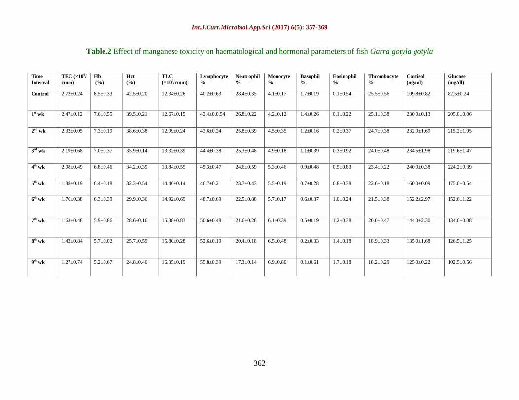
(Table 1). Manganese treated fishes showed
significant decline (P<0.01) in TEC, Hb and
Hct while TLC depicted significant increase.
Lymphocytes, monocytes and eosinophils
register an increase in their population but
neutrophils, basophils and thrombocyte
population depicted decrease in their
population (Table 2 and Figure 2c-f). Cortisol
and glucose levels were noticed to increase
upto 4th
week and after that exhibit a
declining trend in their values during the rest
period (5th
-9th
week) of experimental duration

Int.J.Curr.Microbiol.App.Sci (2017) 6(5): 357-369
360
(Table 2). Comparison of data of controls
with that of starved and manganese treated
groups very clearly indicates that there is a
marked decline in TEC, Hb and Hct at the end
of experimental period in both starved and
manganese treated fishes (Tables 1 and 2).
Similar to present findings, Jenkin and Smith
et al., (2003), Tyagi and Srivastva (2005),
Gupta (2008), Gupta et al., (2009), Raina
(2011), Sachar (2011) and Gupta (2012) have
also reported declining trend in TEC, Hb and
Hct of fishes following subjection to
starvation and different metals.
Present authors propose that starvation and
metal toxicity results in decreased rate of
erythropoises in haemopoietic organs and
senescence in pre-existent cells of blood
stream. Moreover there was no or null
replacement of these cells by new ones for
want of availability of nutrients under
prevailing condition of starvation and due to
toxic effects of metal (Figures 1c-f and 2c-f).
Present viewpoint get an added support from
the work of Santhakumar et al., (2000), Gupta
et al., (2009) and Gupta (2012) who also have
observed similar observations/results under
the prevailing condition of starvation and
metal toxicity.
Presently, besides affecting erythrocyte
number (Tables 1 and 2) starvation and metal
toxicity has also been found to result in
marked anomalies in shape of RBCs as well
as nucleus compared to that of control
(Figures 1(a-f) and 2(a-f)). The distorted
RBCs which make their appearance during
the 1st week (metal treated) and 2
nd week
(starvation) of experimental period in very
few number register an increase with the
advancement of experiment indicating clearly
that TEC not only decline quantitatively but
qualitatively also (Figures 1c, 1d, 2d and e).
Distorted/ abnormal shape of RBCs can lead
to tissue hypoxia by reducing the oxygen
carrying capacity of RBCs and same has also
been earlier reported by workers viz. Das
(1998), Yang and Chen (2003) and Verma
(2007). These morphological changes in
erythrocytes initiate the process of RBC
destruction and ultimately lead to their
complete degeneration.
In tune with TEC, Hb and Hct also exhibited
a significant decrement (P<0.01) in their
values following an exposure to starvation
and metal toxicity. The possible reason for
decline in Hb and Hct, according to present
author, seemingly appears to be because of
decline in the number of normal RBCs and
the null replacement of deformed cells by
normal ones. Similar to present findings Rios
et al., (2005), Gupta (2009) and Raina (2011)
also reported decline in normal RBCs as a
major factor contributing in declining of Hb
and Hct in starved and metal treated fishes.
White blood cells or leucocytes are the cells
of immune system which defend the body of
organism against infectious as well as foreign
materials. Review of literature reveals that
there are two schools of thought regarding the
response of leucocytes to various stressors
and xenobiotics. According to workers of first
school (Iwama et al., 1976; Mishra and
Srivastava, 1979; Ellis, 1981; Sharma and
Gupta, 1984 and Adeymo, 2007) there is a
decrease in TLC whereas workers of second
school viz. Torres et al., (1984), Garg et al.,
(1989), Singh and Tandon (2009) and
Buthelizi et al., (2000) advocated increase in
their number in response to stress of any kind.
Presently our results are in accordance with
first group of workers for starved group of
fishes and to second group of workers for
metal treated groups. The increase in TLC, as
observed metal treated groups can be
attributed to a stimulation of the immune
system in response to tissue damage caused
by manganese whereas in starved fishes stress
of starvation result by deficient nourishment
leads to weakening of immune system and
hence in decrement in number of leucocytes.

Int.J.Curr.Microbiol.App.Sci (2017) 6(5): 357-369
361
Table.1 Effect of starvation on haematological and hormonal parameters of Garra gotyla gotyla
Time
Interval
TEC
(×106/cmm)
Hb
(%)
Hct
(%) TLC
(×103/cmm)
Lymphocyte
%
Neutrophil
%
Monocyte
%
Basophil
%
Eosinophil
%
Thrombocyte
%
Cortisol
(ng/ml)
Glucose
(mg/dl)
Control 2.68±0.25 8.4±0.21 41.7±0.10 13.96±0.24 40.2±0.02 24.2±0.41 4.3±0.22 1.5±0.25 1.2±0.29 28.6±0.14 115.0±0.84 78.6±0.25
1st wk 2.62±0.14 8.2±0.52 41.2±0.43 13.42±0.53 38.8±0.16 25.6±0.30 4.1±0.16 1.4±0.39 1.1±0.36 29.0±0.35 125.5±0.25 90.5±0.19
2nd wk 2.48±0.64 7.9±0.18 39.7±0.58 13.05±0.19 35.2±0.38 27.4±0.26 3.8±0.38 1.2±0.07 1.0±0.02 31.4±0.25 138.0±1.20 106.3±0.34
3rd wk 2.23±0.71 7.5±0.57 38.0±0.62 12.69±0.47 34.3±0.12 27.6±0.04 3.7±0.14 1.0±0.18 0.9±0.18 32.5±0.17 146.5±0.38 125.4±0.16
4th wk 2.02±0.26 7.0±0.83 36.5±0.41 12.34±0.33 31.7±0.37 29.8±0.50 3.5±0.69 0.9±0.69 0.7±0.33 33.4±0.19 175.2±1.54 155.2±1.24
5th wk 1.85±0.05 6.5±0.23 34.0±0.86 12.08±0.64 29.4±0.62 31.4±0.19 3.1±0.17 0.7±0.18 0.5±0.12 34.9±0.14 210.5±0.86 180.0±1.05
6th wk 1.62±0.63 6.2±0.43 33.5±0.36 11.72±0.99 26.9±0.14 33.8±0.18 2.9±0.37 0.6±0.55 0.4±0.43 35.4±0.36 172.0±0.46 158.0±1.42
7th wk 1.55±0.19 5.8±0.38 32.4±0.14 11.37±0.12 24.5±0.29 34.9±0.34 2.5±0.19 0.6±0.59 0.3±0.73 37.2±0.25 158.3±1.24 135.3±0.36
8th wk 1.48±0.47 5.5±0.61 31.2±0.57 11.11±0.34 23.3±0.81 35.7±0.17 2.2±0.34 0.3±0.85 0.2±0.15 38.3±0.10 140.0±0.78 120.5±1.15
9th wk 1.40±0.35 5.3±0.49 30.0±0.19 10.68±0.27 21.6±0.16 35.8±0.44 1.9±0.12 0.1±0.47 0.2±0.66 40.4±0.38 124.5±0.69 114.8±0.64
Int.J.Curr.Microbiol.App.Sci (2017) 6(5): 357-369
362
Table.2 Effect of manganese toxicity on haematological and hormonal parameters of fish Garra gotyla gotyla
Time
Interval
TEC (×106/
cmm)
Hb
(%)
Hct
(%)
TLC
(×103/cmm)
Lymphocyte
%
Neutrophil
%
Monocyte
%
Basophil
%
Eosinophil
%
Thrombocyte
%
Cortisol
(ng/ml)
Glucose
(mg/dl)
Control 2.72±0.24 8.5±0.33 42.5±0.20 12.34±0.26 40.2±0.63 28.4±0.35 4.1±0.17 1.7±0.19 0.1±0.54 25.5±0.56 109.8±0.82 82.5±0.24
1st wk 2.47±0.12 7.6±0.55 39.5±0.21 12.67±0.15 42.4±0.0.54 26.8±0.22 4.2±0.12 1.4±0.26 0.1±0.22 25.1±0.38 230.0±0.13 205.0±0.06
2nd wk 2.32±0.05 7.3±0.19 38.6±0.38 12.99±0.24 43.6±0.24 25.8±0.39 4.5±0.35 1.2±0.16 0.2±0.37 24.7±0.38 232.0±1.69 215.2±1.95
3rd wk 2.19±0.68 7.0±0.37 35.9±0.14 13.32±0.39 44.4±0.38 25.3±0.48 4.9±0.18 1.1±0.39 0.3±0.92 24.0±0.48 234.5±1.98 219.6±1.47
4th wk 2.08±0.49 6.8±0.46 34.2±0.39 13.84±0.55 45.3±0.47 24.6±0.59 5.3±0.46 0.9±0.48 0.5±0.83 23.4±0.22 240.0±0.38 224.2±0.39
5th wk 1.88±0.19 6.4±0.18 32.3±0.54 14.46±0.14 46.7±0.21 23.7±0.43 5.5±0.19 0.7±0.28 0.8±0.38 22.6±0.18 160.0±0.09 175.0±0.54
6th wk 1.76±0.38 6.3±0.39 29.9±0.36 14.92±0.69 48.7±0.69 22.5±0.88 5.7±0.17 0.6±0.37 1.0±0.24 21.5±0.38 152.2±2.97 152.6±1.22
7th wk 1.63±0.48 5.9±0.86 28.6±0.16 15.38±0.83 50.6±0.48 21.6±0.28 6.1±0.39 0.5±0.19 1.2±0.38 20.0±0.47 144.0±2.30 134.0±0.08
8th wk 1.42±0.84 5.7±0.02 25.7±0.59 15.80±0.28 52.6±0.19 20.4±0.18 6.5±0.48 0.2±0.33 1.4±0.18 18.9±0.33 135.0±1.68 126.5±1.25
9th wk 1.27±0.74 5.2±0.67 24.8±0.46 16.35±0.19 55.8±0.39 17.3±0.14 6.9±0.80 0.1±0.61 1.7±0.18 18.2±0.29 125.0±0.22 102.5±0.56
Int.J.Curr.Microbiol.App.Sci (2017) 6(5): 357-369
363
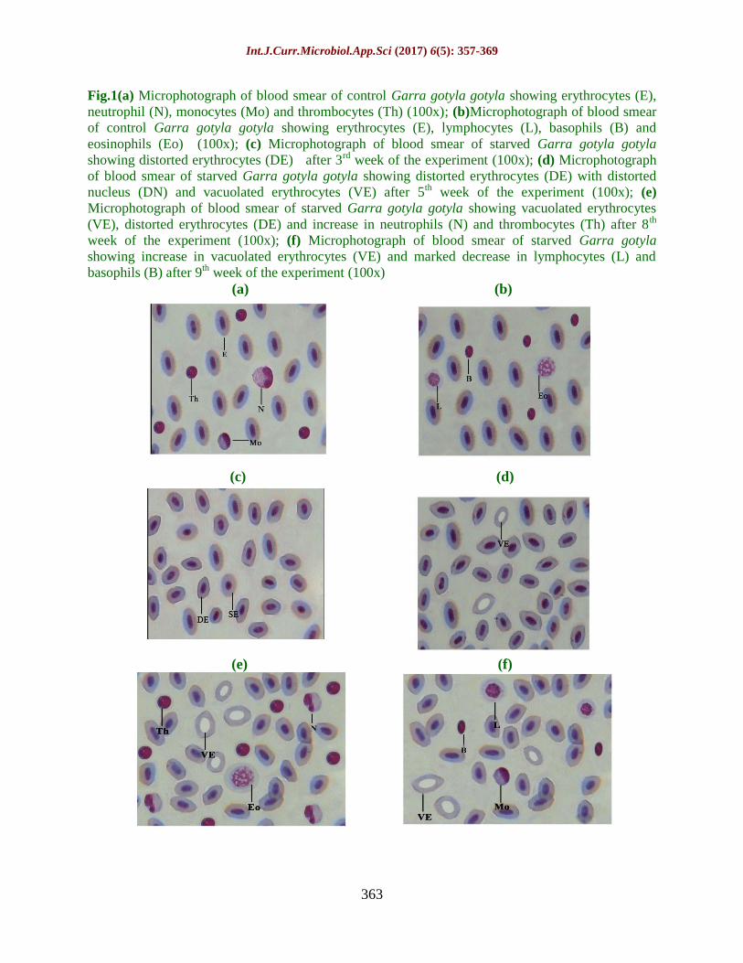
Fig.1(a) Microphotograph of blood smear of control Garra gotyla gotyla showing erythrocytes (E),
neutrophil (N), monocytes (Mo) and thrombocytes (Th) (100x); (b)Microphotograph of blood smear
of control Garra gotyla gotyla showing erythrocytes (E), lymphocytes (L), basophils (B) and
eosinophils (Eo) (100x); (c) Microphotograph of blood smear of starved Garra gotyla gotyla
showing distorted erythrocytes (DE) after 3rd week of the experiment (100x); (d) Microphotograph
of blood smear of starved Garra gotyla gotyla showing distorted erythrocytes (DE) with distorted
nucleus (DN) and vacuolated erythrocytes (VE) after 5th week of the experiment (100x); (e)
Microphotograph of blood smear of starved Garra gotyla gotyla showing vacuolated erythrocytes
(VE), distorted erythrocytes (DE) and increase in neutrophils (N) and thrombocytes (Th) after 8th
week of the experiment (100x); (f) Microphotograph of blood smear of starved Garra gotyla
showing increase in vacuolated erythrocytes (VE) and marked decrease in lymphocytes (L) and
basophils (B) after 9th week of the experiment (100x)
(a) (b)
(c) (d)
(e) (f)
Int.J.Curr.Microbiol.App.Sci (2017) 6(5): 357-369
364
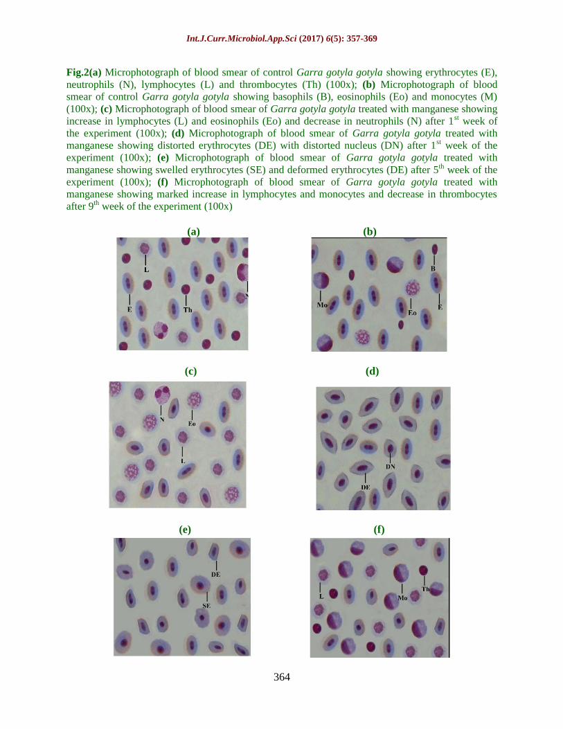
Fig.2(a) Microphotograph of blood smear of control Garra gotyla gotyla showing erythrocytes (E),
neutrophils (N), lymphocytes (L) and thrombocytes (Th) (100x); (b) Microphotograph of blood
smear of control Garra gotyla gotyla showing basophils (B), eosinophils (Eo) and monocytes (M)
(100x); (c) Microphotograph of blood smear of Garra gotyla gotyla treated with manganese showing
increase in lymphocytes (L) and eosinophils (Eo) and decrease in neutrophils (N) after 1st week of
the experiment (100x); (d) Microphotograph of blood smear of Garra gotyla gotyla treated with
manganese showing distorted erythrocytes (DE) with distorted nucleus (DN) after 1st week of the
experiment (100x); (e) Microphotograph of blood smear of Garra gotyla gotyla treated with
manganese showing swelled erythrocytes (SE) and deformed erythrocytes (DE) after 5th week of the
experiment (100x); (f) Microphotograph of blood smear of Garra gotyla gotyla treated with
manganese showing marked increase in lymphocytes and monocytes and decrease in thrombocytes
after 9th week of the experiment (100x)
(a) (b)
(c) (d)
(e) (f)

Int.J.Curr.Microbiol.App.Sci (2017) 6(5): 357-369
365
In depth study of the DLC (Table 1) further
reveals that decrease in TLC in starved fishes
can be an outcome of increase in neutrophils
and thrombocytes and decrease in monocytes,
eosinophils and basophils whereas increase in
TLC upon manganese exposure can be very
safely attributed to an increase in
lymphocytes, monocytes and eosinophils.
Neutrophils, basophils and thrombocytes
however, have been observed to witness a
decline in their number (Table 2).
Lymphocytes being important component of
DLC help the fish to fight against infection by
producing antibodies (Klesius et al., 1999).
Decrease observed in lymphocyte number in
starved fishes may result in decreased
antibody production due to inhibitory
response while increased availability of
lymphocytes under metal intoxication
possibly results in increased antibody
production due to stimulatory response of
lymphocytes. Such inhibitory and stimulatory
responses of lymphocytes have also been
reported by Gill and Pant (1985), Adewoye
(2010) and Gupta (2012) against number of
natural and anthropogenic stressors. Further
increase observed in neutrophils and
thrombocytes (starved fishes) and in
monocytes and eosinophils (metal treated)
may be in view of the fact that monocytes,
eosinophils and basophils (starved fishes) and
neutrophils, basophils and thrombocytes
(metal treated) which too are the other
members of phagocytic machinery in starved
and metal treated groups show a dip under the
stress of starvation and metal toxicity.
Mahajan and Dheer (1979), Ishikawa et al.,
(2007) and Devi et al., (2008) also reported
such changes in number of agranulocytes and
granulocytes under the stress of starvation and
metal toxicity.
It is on record (Barton, 1997 and Martinez-
Porchas et al., 2009) that cortisol hormone
secreted, as primary response under the stress
in fishes activates the process of
glycogenolysis and gluconeogenesis to
release more and more glucose in general
circulation. This has been held as a secondary
response in fish under the stress by Barton
(1997) and Begg and Pankhrust (2004).
Glucose so produced by making available
greater supply of energy to fish help them to
tide over the stress induced by starvation and
metal toxicity (Rottmann et al., 1992).
Presently too, in line with above, elevated
levels of cortisol has been observed
simultaneously to result in the increased
levels of serum glucose (Tables 1 and 2) in
Garra gotyla gotyla. Thus observed increase
in serum cortisol level may plausibly be
ascribed to starvation and metal toxicity
related hyperglycemic condition in all
stressed fishes.
Review of literature further reveals that there
is limit up to which cortisol can be secreted
by hypothalamus pituitary interrenal axis in
fishes during acute response (Dickhoff, 1989
and Martinez-Porchas et al., 2009). This limit
varies in different fishes depending on their
age, maturity, species, duration of stress etc.
After attaining this limit, the cortisol level of
fishes, all of the above workers held return to
basal levels to avoid tissue damage. Such
damage has also been observed by Dickhoff
(1989) and Stein-Behrun and Sapolsky (1992)
in salmons, where high levels of cortisol was
observed to cause death in Pacific salmon
(Onchorhynchus sps.) by tissue degeneration
and damage of homeostatic mechanism.
Interestingly although no fish mortality has
been observed during the entire experimental
period of nine weeks but increase in cortisol
reached peak/ highest level only up to 5th
week in starved and 4th
week in metal treated
fishes and thereafter though cortisol still was
higher than controls but could never cross the
peak level. Rather, the extent of increase now
revealed a declining trend.
Int.J.Curr.Microbiol.App.Sci (2017) 6(5): 357-369
366
The possible reason for decline observed in
the values of cortisol in both starved and
metal treated groups is that hypothalamus
pituitary interrenal axis (HPI), a system
responsible for secretion of cortisol get
exhausted due to stress of starvation and
metal toxicity by causing down regulation of
this system through negative feedback in fish
Garra gotyla gotyla. In consonance with
present viewpoint, Barton et al., (2005) and
Fast et al., (2008) also reported exhaustion of
endocrine system in stressed fishes to be the
possibly causative of decline in titre of
cortisol after exhibiting an initial peak.
On the basis of foregoing discussion it can be
safely deduced that stressor of any kind
natural or anthropogenic affect the
haematological and hormonal balance in fish
and by affecting these systems result in
deterioration of fish quality which can have
detrimental effects on human health.
Such studies therefore, all the more become
important as these may help by making us
knowledgeable as to how different fish
species (presently Garra gotyla gotyla)
become highly resistant/ tolerant to survive
under stressful conditions of starvation and
metal toxicity. Such conditions exist
frequently in natural environment of plains, in
general and hilly area (presently) in particular.
Such studies have far reaching effects, not
only on the quality fish production but also on
its progeny, and moreover appear to be of
great help, particularly to the fish farmers in
working out the appropriate food regimes and
physic-chemical properties in the
establishment of culture practice for different
fish species of hilly region.
References
Adewoye, S.O. 2010. Haematological and
biochemical changes in Clarias
gariepinus exposed to Trephosita vogelii
extract. Adv. Appl. Sci. Res., 1(1): 74-79.
Adeyemo, O.K. 2007. Haematological profile
of Clarias gariepinus (Burchell, 1822)
exposed to lead. Turkish J. Fisheries and
Aquatic Sci., 7: 163-169.
Allen, P. 1994. Changes in the haematological
profile of cichlid, Oreochromis auratus
during acute inorganic mercury
intoxication. Comp. Biochem. Physiol.,
108C: 117-121.
Anderson, D.P. 2003. Text book of fish
immunology, Narendra Publishing House,
1-177.
Barton, B.A, Ribas, L., Acerete, L. and Tort, L.
2005. Effects of chronic confinement on
physiological responses of the juvenile
gilthead sea bream, Sparus aurata L. to
acute handling. Aquaculture Res., 36:
172-179.
Barton, B.A. 1997. Stress in finfish: past,
present and future – a historical
perspective. In: Iwama, G. K., Pickering,
A. D., Sumpter, J. P. and Schreck, C. B.
eds. Fish Stress and Health in
Aquaculture, pp. 1-33. Soc. Exp. Biol.
Sem. Ser. 62, Cambridge Univ. Press,
Cambridge, U.K.
Begg, K. and Pankhurst, N.W. 2004. Endocrine
and metabolic responses to stress in a
laboratory population of the tropical
damselfish Acanthochromis
polyacanthus. J. Fish Biol., 64: 133–145.
Buthelezi, P.P., Wepener, V. and Cyrus, D.P.
2000. The sublethal effect of zinc at
different water temperatures on selected
haematological variables in Oreochromis
mossambicus. American J. Aquatic Sci.,
25: 146-151.
Correl, N.V. and Langley, R.W. 1956.
Glycogen determination in liver and
muscle by use of anthrone reagent. J.
Biol. Chem., 26: 583-593.
Das, B.K. 1998. Studies on the effect of some
pesticide and commonly used chemicals
on Indian major carps and their
ecosystem. M.Phil. Thesis, Orissa
University of Agriculture and
Technology, Bhubaneshwar (India), 139-
162.

Int.J.Curr.Microbiol.App.Sci (2017) 6(5): 357-369
367
Dethloff, G.M., Schlenk, D., Khan, S. and
Bailey, H.C. 1999. The effects of copper
on blood and biochemical parameters of
rainbow trout (Oncorhynchus mykiss.
Arch. Environ. Contam. Toxicol., 36: 415-
423.
Devi, P., Baruah, D., Baruh, B.K. and
Borkotok, A. 2008. Impact of
endosulphan on some haematological
parameters of Channa punctatus (Bloch.
Poll Res., 27(3): 485-488.
Dickhoff, W.W. 1989. Salmonids and annual
fishes: Death after sex. In M. P.
Schreibman and C. G. Scanes (eds.),
Development, maturation, and senescence
of neuroendocrine systems: A
comparative approach, pp. 253–268.
Academic Press, New York.
Donaldson, E.M. 1981. The pituitary-interrenal
axis as an indicator of stress in fish. In:
Stress and Fish (Pickering A. D., ed..
Academic Press, London and New York.
Ellis, A.E. 1981. Stress and the modulation of
defense mechanism in fish. In: Stress and
Fish Pickering AD (Ed). Academic Press,
London, 3: 147-171.
Fast, M.D., Hosoya, S., Johnson, S.C. and
Afonso, L.O.B. 2008. Cortisol response
and immune-related effects of Atlantic
salmon (Salmo salar Linnaeus) subjected
to short- and long-term stress. Fish
Shellfish, 24: 194-204.
Garg, V.K., Garg, S.K. and Tyagi, S.K. 1989.
Manganese induced haematological and
biochemical anomalies in Heteropneustes
fossilis. J. Environ. Biol., 10(4): 349-353.
Gill, T.S. and Pant, J.C. 1985. Erythrocytic and
leucocytic response to cadmium in fresh
water fish Puntius conchonius. Environ.
Res., 36: 327-335.
Gupta, K. 2008. Copper induced toxicity on
haematological and haemopoietic profile
of Puntius sophore (Ham). M. Phil.
Thesis, University of Jammu, Jammu.
Gupta, K. 2012. Studies on effect of heavy
metal toxicity on the histophysiology of
blood and haemopoietic tissues of some
fish species, Ph. D Thesis. University of
Jammu.
Gupta, K., Langer, S., Sharma, J. and Sharma,
S. 2012. Effect of different sublethal
concentrations of Manganese on the
levels of cortisol in Garra gotyla gotyla.
Int. J. Scientific Res. Publications, 2(10):
1-3. ISSN 2250-3153.
Gupta, K., Sachar, A., Gupta, K. and Raina, S.
2009. Effect of lindane on haematological
parameters of minor carp, Labeo boga
(Ham). Biosci. Biotechnol. Res. Asia.,
6(2): 695-702.
Gupta, R. 2009. Stress related haematological
changes in Cyprinus carpio (Ham).
M.Phil. Thesis, University of Jammu,
Jammu.
Haukenes, A.H., Barton, B.A. and Bolligs, H.
2008. Cortisol responses of pallid
sturgeon and yellow perch following
challenge with lipopolysaccharide. J. Fish
Biol., 72: 780-784.
Ishikawa, N.M., Ranzani-Paiva, M.J.T.,
Lombardi, J.V. and Ferreira, C.M. 2007.
Haematological parameters in Nile
Tilapia, Oreochromis niloticus exposed to
sublethal concentrations of mercury.
Braz. J. Zool., 50(4): 619-626.
Iwama, G.K., Greer, G.L. and Arkin, P.A. 1976.
Changes in some haematological
characteristics of Coho Salmon
(Onchorhynchus kisutch) in response to
acute exposure to dehydroabieticacid
(DHAA) at different exposure levels. J.
Fish Res. Board Can., 33: 285-289.
Iwama, G.K., Pickering, A.D., Sumpter, J.P.
and Schreck, C.B. 1997. Fish stress and
health in aquaculture. Cambridge Univ.
Press, Cambridge, U.K. Soc. Exp. Biol.
Sem. Ser., 62.
Jenkins, F., Smith, et al. 2003. Effect of
sublethal concentration of endosulphan on
haematological and serum biochemical
parameters in the carp, C. carpio. Bull.
Environ. Contam. Toxicol., 70: 993-947.
Klesius, P.H., Lim, C. and Shoemaker, C. 1999.
Effect of feed deprivation on innate
resistance and antibody response to
Flavobacterium columnare in channel
catfish, Ictalurus punctatus. Bull.
European Association of Fish

Int.J.Curr.Microbiol.App.Sci (2017) 6(5): 357-369
368
Pathologist, 19: 156-158.
Mahajan, C.L. and Dheer, J.M.S. 1979b.
Seasonal variations in the blood
constituents of an air breathing fish,
Channa punctatus Bloch. J. Fish Biol.,
14: 413-417.
Martinez-Porchas, M., Martinez-Cordova, L.R.
and Ramos-Enriquez, R. 2009. Cortisol
and Glucose: Reliable indicators of Fish
Stress. Pan-American J. Aquatic Sci.,
4(2): 158-178.
Martinez-Porchas, M., Martinez-Cordova, L.R.
and Ramos-Enriquez, R. 2009. Cortisol
and Glucose: Reliable indicators of Fish
Stress. Pan-American J. Aquatic Sci.,
4(2): 158-178.
Maule, A.G. and Schreck, C.B. 1990. Changes
in the number of leucocytes in immune
organs of juvenile coho after acute stress
or cortisol treatment. J. Aquatic Animal
Health, 2: 298-304.
Mishra, S. and Srivastava, A.K. 1979.
Haematology as index of sublethal
toxicity of zinc in a freshwater teleost.
Bull. Environm. Contam. Toxicol., 22:
695-698.
Mommsen, T.P., Vijayan, M.M. and Moon,
T.W. 1999. Cortisol in teleost: dynamics,
mechanism of action, and metabolic
regulation. Reviews on Fish Biol.
Fisheries, 9: 211-268.
Munch, A., Guryre, P.M. and Holbrook, N.J.
1984. Physiological functions of
glucocorticoids in stress and their relation
to pharmacological actions. Endocr. Rev.,
5(1): 25-44.
Pickering, A.D. 1981. Stress and fish.
Academic Press, New York.
http://icb.oxfordjournals.org/content/42/3/
517.
Pottinger, T.G. 2003. The selection of trout for
high and low responsiveness to stress:
progress and prospects. Trout News,
CEFAS, 36: 14–16.
Pottinger, T.G. and Mosuwe, E. 1994. The
corticosteroidogenic response of brown
and rainbow trout alevins and fry to
environmental stress during a critical
period. General and Comparative
Endocrinol., 95: 350-362.
Raina, S. 2011. Effect of environmental stress
on haematology and immune organs of
Labeo species. Ph. D thesis. University of
Jammu, Jammu.
Randall, D.J. and Perry, S.F. 1992.
Catecholamines. In: Hoar, W.S., Randall,
D.J., Farrell, A.P. Eds., Fish Physiology-
The Cardiovascular System, Vol. XIIB.
Academic Press, New York. pp. 255–300.
Reid, S.D., Moon, T.W. and Perry, S.F. 1992.
Rainbow trout hepatocyte beta-
adrenoceptors, catecholamine
responsiveness, and effects of cortisol.
American J. Physiol., 262: 794-799.
Reid, S.G., Bernier, N.J. and Perry, S.F. 1998.
The adrenergic stress response in fish:
control of catecholamine storage and
release. Comparative Biochemi. Physiol.,
Part C, 120: 1-27.
Rios, F.S., Oba, E.T., Fernandes, M.N., Kalinin,
A.L. and Ramtin, F.T. 2005. Erytrocytes
senescence and haematoloical changes
induced by starvation in the Neotropical
fish traira, Hoplias malabaricus
(Caraciformes, Erythrinidae. Comp.
Biochem. Physiol., 140: 281-287.
Rottmann, R.W., Francis-Floyd, R. and
Durborow, R. 1992. The role of stress in
fish disease. SRAC Publication, 4: 474.
Sachar, A. 2011. Studies on effect of organic
and inorganic pollutants on haematology,
blood biochemistry and immune organs in
some fishes of Jammu region. University
of Jammu. Ph. D Thesis.
Santhakumara, M., Balaji, M. and Amudu, K.
2000. Adaptive changes in respiratory
movements of an air breathing fish,
Anabas testudineus exposed to
organophosphate pesticide,
monocroptophos. Eco. Env. Conserv.,
6(1): 67-69.
Selye, H. 1973. The evolution of stress concept.
Am. Sci., 61692-699.
Sharma, R.C. and Gupta, N. 1984.
Carbontetrachloride induces
haematological alterations in Clarias
batrachus (L). J. Environ. Biol., 3: 127-
131.

Int.J.Curr.Microbiol.App.Sci (2017) 6(5): 357-369
369
Singh, B.P. and Tandon, P.K. 2009. Effect of
river pollution on haematological
parameters of fish, Wallago attu. Res.
Environ. Life Sci., 2(4): 211-214.
Stein-Behrens, B.A. and Sapolsky, R.M. 1992.
Stress, glucocorticoids, and aging. Aging,
4: 197-210.
Tavares-Dias, M. and Barcellos, J.F.M. 2005.
Peripheral blood cells of the
armoredcatfish Hoplosternum littorale
Hancock, 1828: a morphological and
cytochemical study. Braz. J. Morphol.
Sci., 22: 215 - 220.
Torres, P., Tort, L., Planas, J. and Flos, R. 1984.
Effects of confinement stress and
additional zinc treatment on some blood
parameters in the dogdish (Scyliorhinus
conicula. Comp. Biochem. Physiol., 83C:
89-92.
Tort, L., Padros, F., Rotllant, J. and Crespo, S.
1998. Winter syndrome in the gilthead
sea bream Sparus aurata. Immunological
and Histopathological features. Fish and
Shellfish immunol., 8: 37-47.
Tyagi, A. and Srivastava, N. 2005.
Haematological changes of fish Channa
punctatus (Bloch) to chronic zinc
exposure. J. Environ. Biol., 26(2): 429-
432.
Verma, G. 2007. Lindane (an insecticide)
induced haematological changes in a
minor carp, Puntius sophore (Ham). M.
Phil. Thesis, University of Jammu,
Jammu.
Vosyliene, M.Z. and Kazlauskiene, N. 1999.
Alterations in fish health state parameters
after exposure to different stressors. Acta
Zoologica Hydrobiologica, 9(2): 1392-
1657.
Vosyliene, M.Z., Kazlauskiene, N. and
Svecevicius, G. 2003. Effect of heavy
metal model mixture on biological
parameters of rainbow trout
Oncorhynchus mykiss. Environ. Sci.
Pollut. Res., 10: 103–107.
Wedemeyer, G.A., Barton, B.A. and McLeay,
D.J. 1990. Stress and acclimation In:
Schreck, C.B., Moyle, P.B. Eds. Methods
for Fish Biology. American Fisheries
Society, Bathesda, MD., 451-489.
Wendelaar Bonga, S.E. 1997. The stress
response in fish. Physiol. Rev., 77: 591-
625.
Wintrobe, M.M. 1967. “Clinical Haematology”
VI ED., Philadelphia, Lea and Febiger.
Yang, J.L. and Chen, H.C. 2003. Effects of
gallium on common carp (Cyprinus
carpio): acute test, serum biochemistry
and erythrocyte morphology.
Chemosphere, 53: 877-882.
How to cite this article:
Jyoti Sharma, Shabir Ahmed Dar, A.N. Sayani and Seema Langer. 2017. Utilization of Mango Peel
Powder (MPP) in Mango Nectar Formulation. Int.J.Curr.Microbiol.App.Sci. 6(5): 357-369.
doi: http://dx.doi.org/10.20546/ijcmas.2017.605.041