effect of the environment on gene transfer in clostridium
TRANSCRIPT

Page 1 of 285
Effect of the environment on gene transfer in
Clostridium difficile
Thesis submitted by
Ladan Khodadoost
For the degree of
DOCTOR OF PHYLOSOPHY
University College London
Department of Microbial Diseases
UCL Eastman Dental Institute
256 Gray’s Inn Road
London WC1X 8LD
UK
-2019-

Page 2 of 285
Declaration
I, Ladan Khodadoost, confirm that the work presented in this thesis is my own. Where
information has been derived from other sources, I confirm that this has been indicated
in the thesis.

Page 3 of 285
Abstract
Clostridium difficile is a pathogenic bacterium that can colonise both humans and
various animals. Toxin production leads to clinical symptoms ranging from mild to
severe diarrhoea and can result in potentially fatal pseudomembranous colitis. These
symptoms are caused by the disruption of the cytoskeleton and tight junctions of gut
epithelial cells by the toxins. C. difficile responds to several biological compounds
found in the human intestinal environment such as bile salts leading to spore
germination and colonization. In this study we investigated the response of C. difficile
to mammalian pancreatic α-amylase with production of a mucoid colony phenotype
that results from increased secretion of extracellular proteins and carbohydrates.
Furthermore, the effect of amylase on horizontal gene transfer in C. difficile was
investigated using conjugative transposon Tn5397 and non-conjugative mobilisable
plasmid pMTL9301. A significant increase in the frequency of Tn5397 transfer was
observed when amylase was present. pMTL9301 transfer was not affected by amylase
but significantly decreased when DNase was added to eliminate transformation.
Further investigations into the molecular basis of the DNase-sensitive plasmid transfer
into C. difficile showed that the oriT region of pMTL9301 (derived from RK2) is not
required for transfer between E. coli and C. difficile strains 630Δerm and CD37 and
that this oriT-independent transfer is abolished in the presence of DNase when CD37
is the recipient. Transfer to the 630Δerm strain is DNase resistant even without an
obvious oriT, when E. coli CA434 is used as a donor and is sensitive to DNase when
E. coli HB101 is the donor, suggesting that a ‘novel cell-to-cell transformation-like
mechanism’ occurs in C. difficile.

Page 4 of 285
Impact statement
A fundamental understanding of the evolutionary pressures which select for resistance
is a prerequisite to design strategies to stop the spreading of antibiotic resistance
genes (ARGs). The work in this study has shown that C. difficile has a remarkable
ability to obtain new DNA. The unexpected observation that it can take up plasmid
DNA encoding antibiotic resistance from an unrelated microorganism without a
complete conjugation system or a cis acting oriT suggests that the organism has the
potential to acquire almost any DNA sequence. Our study also has implications for the
containment of genetically modified organisms, as we have shown that non-
conjugative non-mobilisable plasmids can still be taken up by an organism that was
previously thought not to be naturally competent, and it is important to determine how
common this phenomenon is in nature.
The ‘novel cell-to-cell transformation-like’ mechanism shown here for the first time in
C. difficile, may occur in environments outside the laboratory at non-negligible
frequencies if several conditions are met. In this respect, further experiments using
plasmid-free strains of E. coli and other C. difficile strains will be required. Furthermore,
other results suggest that non-conjugative plasmids are more mobile than was
previously believed (Zechner et al., 2012). Our finding not only can provide insights
into evolution of acquired resistance genes but also can offer a strategy for combatting
the antibiotic-resistance crisis.

Page 5 of 285
Acknowledgment
I would like to begin by thanking Professor Peter Mullany for providing me with the
opportunity to conduct a PhD at UCL and for all his support with the lab work, the
writing and all the helpful discussion. I would like to thank Dr. Sean Nair for his input
and helpful comments on my thesis.
I thank Dr. Haitham Hussain who has been absolutely fantastic during my PhD, and
even through the endless questions he maintained a friendly and helpful attitude. I am
so grateful for your help and support.
I would like to thank Dr. Rachel Leeson for all her support throughout my studies.
Thank you.
I would also like to thank Professor Nigel Minton for pMTL9301 and CA434. I thank
Mike Young for helpful discussions.
I thank Dr. Luisa De Sordi for her advice and encouragement, and for her continued
moral support during the first year of my PhD.
None of this work could have been performed without the support I have had from so
many friends I have made at the Eastman; Asyura, Supanan, Deena, Ajijur, Zeina,
Enas, Supathep, Marika.
I would like to thank Janet and other staff at Kingston hospital, Diabetic day unit who
listened patiently when I had diabetes burnout. Thanks for all the care and support.
Finally, I am forever indebted to my family for their understanding, endless patience
and encouragement when it was most required.

Page 6 of 285
Table of Contents
List of abbreviations .............................................................................................................................. 16
1. Chapter 1 General Introduction .................................................................................................... 19
1.1. General characteristics of Clostridium difficile ..................................................................... 20
1.2. C. difficile infection, treatment and prevention ................................................................... 22
1.2.1. Disease and risk factors................................................................................................. 22
1.2.2. Treatment and prevention ............................................................................................ 24
1.3. Molecular pathogenesis and virulence ................................................................................. 25
1.3.1. Toxins ............................................................................................................................ 25
1.3.1.1. C. difficile TcdA and TcdB ...................................................................................... 25
1.3.1.2. C. difficile binary toxins ......................................................................................... 31
1.3.2. Surface layer and cell wall proteins .............................................................................. 32
1.3.3. Polysaccharides ............................................................................................................. 35
1.3.4. Flagella .......................................................................................................................... 35
1.3.5. Fibronectin-binding proteins ........................................................................................ 36
1.4. C. difficile epidemiology ........................................................................................................ 37
1.4.1. Typing method .............................................................................................................. 37
1.4.2. Epidemiology of CDI and hypervirulence ...................................................................... 38
1.5. Horizontal gene transfer via mobile genetic elements ......................................................... 41
1.5.1. Transformation ............................................................................................................. 41
1.5.2. Transduction ................................................................................................................. 44
1.5.3. Conjugation ................................................................................................................... 46
1.5.3.1. Conjugative plasmids ............................................................................................ 47
1.5.3.2. Conjugative transposons ....................................................................................... 51
1.5.3.2.1. Tn916 ................................................................................................................. 52
1.5.3.2.2. Tn1549 ............................................................................................................... 53
1.5.3.2.3. Excision and integration by site-specific recombination ................................... 53
1.5.3.3. Mobile genetic elements in C. difficile .................................................................. 55
1.5.3.3.1. Tn5397 ............................................................................................................... 55
1.5.3.3.2. Other conjugative transposons in C. difficile 630 .............................................. 58
1.5.3.4. Mobilisable elements in C. difficile ....................................................................... 60
1.5.3.4.1. Tn4453 ............................................................................................................... 60
1.5.3.4.2. Tn5398 ............................................................................................................... 61
1.5.4. Additional C. difficile MGEs ........................................................................................... 63

Page 7 of 285
1.5.4.1. skinCD ..................................................................................................................... 63
1.5.4.2. IStron ..................................................................................................................... 64
1.6. Genetic manipulation tools ................................................................................................... 65
1.6.1. Shuttle conjugative Transposon: Tn916 ........................................................................ 65
1.6.2. Other transposon-based mutagenesis systems ............................................................ 66
1.6.3. Transfer of replication-proficient plasmids into C. difficile from E. coli donor ............. 69
1.6.4. Use of ClosTron system: a group II intron derivative for targeted mutagenesis:
directed gene inactivation ............................................................................................................ 71
1.6.5. CRISPR-Cas system ........................................................................................................ 73
1.6.5.1. CRISPR-Cas system in C. difficile ............................................................................ 73
1.6.5.2. Using CRISPR-Cas system to generate C. difficile mutants .................................... 74
1.7. Aim of the study .................................................................................................................... 77
2. Chapter 2 Materials and methods ................................................................................................ 78
2.1. Bacterial strains and culture methods .................................................................................. 79
2.2. Molecular techniques ........................................................................................................... 81
2.2.1. Genomic DNA extraction .............................................................................................. 81
2.2.2. Plasmid extraction ......................................................................................................... 82
2.2.3. Oligonucleotide synthesis ............................................................................................. 83
2.2.4. Standard Polymerase chain reaction (PCR) ................................................................... 83
2.2.5. Agarose gel electrophoresis .......................................................................................... 84
2.2.6. PCR product purification ............................................................................................... 84
2.2.7. DNA extraction from agarose gel .................................................................................. 85
2.2.8. Restriction endonuclease reaction ............................................................................... 86
2.2.9. DNA ligation reaction .................................................................................................... 86
2.3. Transformation into competent E. coli CA434 ...................................................................... 86
2.4. Filter mating .......................................................................................................................... 87
2.5. DNA sequencing reactions .................................................................................................... 88
2.6. DNA sequence analysis ......................................................................................................... 88
2.7. Statistical Analysis ................................................................................................................. 89
3. Chapter 3 Exposure to pancreatic α-amylase promotes exopolymer secretions in C. difficile..... 90
3.1. Introduction .......................................................................................................................... 91
3.1.1. C. difficile extracellular polymeric substance (EPS) ....................................................... 91
3.1.1.1. Definition of EPS .................................................................................................... 91
3.1.1.1.1. C. difficile extracellular carbohydrate ................................................................ 92
3.1.1.1.2. C. difficile extracellular protein .......................................................................... 93
3.1.1.2. Mucoid bacteria .................................................................................................... 94

Page 8 of 285
3.2. Aims and objectives .............................................................................................................. 96
3.3. Materials and methods ......................................................................................................... 97
3.3.1. Stains and culture media ............................................................................................... 97
3.3.2. Preparation of basal defined medium .......................................................................... 98
3.3.3. Growth curves ............................................................................................................... 98
3.3.4. EPS purification ............................................................................................................. 98
3.3.4.1. Carbohydrate assay ............................................................................................... 99
3.3.4.2. Protein assay ......................................................................................................... 99
3.4. Results ................................................................................................................................. 100
3.4.1. Pancreatic α-amylase induces a colony morphology change in C. difficile ................. 100
3.4.2. Starch is not required for amylase-induced mucoidy ................................................. 102
3.4.3. Growth promotion by pancreatic α-amylase .............................................................. 102
3.4.4. Determining the extracellular carbohydrate content of C. difficile exposed to
pancreatic α-amylase .................................................................................................................. 104
3.4.5. Determining the extracellular protein content of C. difficile exposed to pancreatic α-
amylase………………………………………………………………………………………………………………………………105
3.5. Discussion ............................................................................................................................ 107
4. Chapter 4 Pancreatic α-amylase disrupts/ inhibits C. difficile biofilm formation ....................... 113
4.1. Introduction ........................................................................................................................ 114
4.1.1. Biofilms ........................................................................................................................ 114
4.1.1.1. Definition of biofilm ............................................................................................ 114
4.1.1.2. Biofilm structure ................................................................................................. 115
4.1.1.3. Main roles of EPS in the biofilm .......................................................................... 116
4.1.1.3.1. EPS and antibiotic resistance ........................................................................... 116
4.1.1.3.2. EPS and horizontal gene transfer (HGT) .......................................................... 117
4.1.1.4. Biofilm formation and development ................................................................... 118
4.1.2. Biofilm formation by C. difficile ................................................................................... 118
4.1.3. Environmental stresses and biofilm formation ........................................................... 120
4.2. Aims and objectives ............................................................................................................ 122
4.3. Materials and methods ....................................................................................................... 123
4.3.1. Stains and culture media ............................................................................................. 123
4.3.2. Biofilm assay................................................................................................................ 124
4.4. Results ................................................................................................................................. 125
4.4.1. C. difficile R20291 exhibits greater biomass (number of adhered cells) production than
630Δerm and 630ΔermΔspo0A ................................................................................................... 125
4.4.2. C. difficile biomass formation is significantly decreased in the presence of pancreatic
α-amylase. ................................................................................................................................... 127

Page 9 of 285
4.4.2.1. Treated tissue culture plates ............................................................................... 127
4.4.2.2. Non-treated tissue culture plates ....................................................................... 129
4.5. Discussion ............................................................................................................................ 131
5. Chapter 5 Investigations into the effects of pancreatic α-amylase and deoxyribonuclease I on
horizontal gene transfer in C. difficile ................................................................................................. 140
5.1. Introduction ........................................................................................................................ 141
5.2. Aims and objectives ............................................................................................................ 143
5.3. Methods and materials ....................................................................................................... 144
5.3.1. Strains, plasmids and culture media ........................................................................... 144
5.3.2. Chemical transformation ............................................................................................ 146
5.3.3. Confirmation of transformation .................................................................................. 146
5.3.4. Mating experiments .................................................................................................... 148
5.3.5. Confirmation of transfer ............................................................................................. 150
5.4. Results ................................................................................................................................. 154
5.4.1. Tn5397 conjugation occurs at a significantly higher frequency in the presence of
pancreatic α-amylase than when amylase is not present. ......................................................... 154
5.4.2. Tn5397 transfer into C. difficile is not affected by deoxyribonuclease I (DNase I) ..... 156
5.4.3. Conjugative transposon 023_Tn3 transfers from toxigenic strain CD305 into CD37, and
the transfer is not affected by pancreatic α-amylase and/ or DNase ......................................... 158
5.4.4. Transfer of pMTL9301 is not affected by pancreatic α-amylase ................................. 160
5.4.5. DNase drastically reduces pMTL9301 frequency of transfer from E. coli strain CA434
into C. difficile CD37 .................................................................................................................... 162
5.5. Discussion ............................................................................................................................ 164
6. Chapter 6 Investigation of DNase-sensitive plasmid transfer into Clostridium difficile .............. 169
6.1. Introduction ........................................................................................................................ 170
6.2. Aims and objectives ............................................................................................................ 176
6.3. Methods and materials ....................................................................................................... 178
6.3.1. Strains, plasmids and culture media ........................................................................... 178
6.3.2. Direct transformation of C. difficile CD37 with pMTL9301 ......................................... 181
6.3.2.1. Transformation assay with purified plasmid DNA in liquid culture .................... 181
6.3.2.2. Transformation assay with purified plasmid in the presence of CA434 ............. 181
6.3.2.3. Transformation using cell-free filtrate (supernatant) ......................................... 182
6.3.2.4. Transformation using heat-killed donor cells ..................................................... 183
6.3.3. Construction of pMTL9301∆oriT ................................................................................. 184
6.3.3.1. Restriction endonuclease reaction and ligation ................................................. 184
6.3.3.2. Chemical transformation .................................................................................... 187
6.3.4. Confirmation of construct ........................................................................................... 188

Page 10 of 285
6.3.5. Mating experiments .................................................................................................... 190
6.3.6. Confirmation of transfer ............................................................................................. 193
6.3.7. Plasmid pMTL9301ΔoriT fate in C. difficile .................................................................. 196
6.3.8. Bioinformatics Search for putative competence genes in C. difficile .......................... 197
6.3.8.1. Search for competence genes in C. difficile 630 ................................................. 197
6.3.8.2. Search for the com genes in CD37 ...................................................................... 198
6.3.9. ClosTron mutagenesis ................................................................................................. 200
6.3.10. Investigation of the potential influence of the transfer genes on plasmid RK2 on
DNase sensitive oriT-independent plasmid transfer into C. difficile ........................................... 204
6.4. Results ................................................................................................................................. 208
6.4.1. DNase sensitive plasmid transfer into C. difficile ........................................................ 208
6.4.1.1. Using purified plasmid DNA in liquid culture of C. difficile did not result in
transformation ........................................................................................................................ 208
6.4.1.2. Transfer of free plasmid DNA into CD37 did not occur in the presence of plasmid-
free CA434……………………………………………………………………………………………………………………….. 208
6.4.1.3. Using heat-killed donor or donor culture supernatant/ filtrate did not result in
plasmid transfer into CD37 ..................................................................................................... 209
6.4.1.4. Plasmid transfer into C. difficile requires close contact between live donor cells
and recipients, and DNase treatment only reduces the transfer frequency of pMTL9301 into
CD37 but has no effect on the transfer frequency to C. difficile 630Δerm ............................. 211
6.4.2. A complete oriT is not required for plasmid transfer into C. difficile .......................... 212
6.4.2.1. pMTL9301∆oriT construction and validation ...................................................... 212
6.4.2.2. Deletion of the oriT from pMTL9301 does not abolish transfer from E. coli to C.
difficile but does abolish transfer to strain CD37 in the presence of DNase .......................... 214
6.4.2.3. Deletion of the oriT from pMTL9301 does not abolish transfer from E. coli strain
HB101 to C. difficile 630∆erm but does abolish transfer to CD37 in the absence of DNase. 215
6.4.3. Analysis of transconjugants/ transformants ............................................................... 218
6.4.4. Transfer of Tn5397 and Tn916 from Bacillus subtilis is not affected by DNase
treatment .................................................................................................................................... 220
6.4.5. Investigation of the potential influence of the com genes on DNase-sensitive plasmid
transfer into C. difficile ................................................................................................................ 221
6.4.5.1. Bioinformatic Search for putative competence genes in C. difficile ................... 221
6.4.5.2. Mating experiment using C. difficile ClosTron-based mutants as recipients in the
absence and presence of DNase ............................................................................................. 225
6.4.6. Investigation of the potential influence of plasmid RK2 transfer genes on
pMTL9301ΔoriT transfer into C. difficile ..................................................................................... 230
6.4.6.1. Bioinformatic search for the presence of tra and trb genes in E. coli strain RR1
(HB101 RecA+) genome ........................................................................................................... 230
6.4.6.2. PCR amplification of the transfer genes in E. coli RR1 (HB101 RecA+) ................ 234

Page 11 of 285
6.5. Discussion ............................................................................................................................ 237
7. Chapter 7 General discussion and future work .......................................................................... 243
References .......................................................................................................................................... 251
Appendices .......................................................................................................................................... 281
Appendix 1. Composition of basal defined medium (BDM) ........................................................... 282
Appendix 2. Purity of hog pancreatic α-amylase preparation ........................................................ 283
Appendix 3. Publication resulting from this study .......................................................................... 285

Page 12 of 285
List of tables
Table 1.1 General features of the genome of C. difficile ....................................................................... 22
Table 1.2 Major risk factors for C. difficile infection (CDI) ..................................................................... 23
Table 2.1 Bacterial strains used in this study ........................................................................................ 80
Table 3.1 Bacterial strains used in this study ........................................................................................ 97
Table 3.2 Appearance of mucoidy of C. difficile in response to medium supplemented with pancreatic enzymes .............................................................................................................................................. 101
Table 4.1 Bacterial strains used in this study ...................................................................................... 123
Table 5.1 Bacterial strains and plasmids used in this study ............................................................... 145
Table 5.2 The list of primers used in this study to confirm the presence of pMTL9301 in E. coli CA434 transformants ...................................................................................................................................... 147
Table 5.3 The list of primers used in this study to confirm the transfer of MGEs into C. difficile in the absence and presence of pancreatic α-amylase and/ or DNase. ....................................................... 151
Table 5.4 Mating experiment between B. subtilis BS6A and C. difficile CD37 in the absence and presence of pancreatic α-amylase. ..................................................................................................... 155
Table 5.5 The effect of DNase alone and in combination with pancreatic α-amylase on Tn5397 frequency of transfer. .......................................................................................................................... 157
Table 5.6 Frequency of 023_Tn3 transfer per donor or recipient. ...................................................... 161
Table 5.7 Mating experiment between E. coli CA434::pMTL9301 and C. difficile CD37 in the absence and presence of pancreatic α-amylase ............................................................................................... 163
Table 5.8 The effect of DNase alone and in combination with pancreatic α-amylase on pMTL9301 transfer into C. difficile ......................................................................................................................... 165
Table 6.1 Bacterial strains and plasmids used in this study ............................................................... 190
Table 6.2 The list of primers used for analysis of the construction of pMTL9301∆oriT ....................... 193
Table 6.3 The list of primers used for the analysis of transconjugants/ transformants ....................... 196
Table 6.4 The list of primers used in this study to detect competence genes in C. difficile ................ 203
Table 6.5 The list of primers used for ClosTron mutagenesis ............................................................ 207
Table 6.6 The list of primers used for detecting the tra and trb genes in E. coli strain HB101 (parent strain of CA434). ................................................................................................................................. 209
Table 6.7 Direct transformation of C. difficile CD37 with free plasmid pMTL9301. ............................ 214
Table 6.8 Frequency of pMTL9301 and pMTL9301ΔoriT transfer into C. difficile (±SD) .................... 221
Table 6.9 Bioinformatic search for putative competence genes in strain 630 ..................................... 227
Table 6.10 Frequency of pRPF185 and pMTL9301 (control) transfer into C. difficile. ........................ 231
Table 6.11 Bioinformatics search for transfer genes in E. coli strain HB101 ...................................... 235
Table 6.12 Summary of the results………………………………………………………………………….240
Table A.2 Identity of protein present in hog pancreatic α-amylase preparation .................................. 278

Page 13 of 285
List of Figures
Figure 1.1 Gram staining of C. difficile strain 630. ................................................................................ 20
Figure 1.2 Schematic representation of PaLoc in toxigenic 630 and non-toxigenic C. difficile CD37 .. 26
Figure 1.3 Schematic representation of the uncommon PaLoc integration sites in a small number of non-toxigenic C. difficile strains ............................................................................................................. 27
Figure 1.4 Proposed ABCD domain structure model of toxin B ............................................................ 28
Figure 1.5 Mechanisms of action of TcdA and TcdB ............................................................................ 30
Figure 1.6 Schematic representation of CDT locus in C. difficile .......................................................... 31
Figure 1.7 S-layer protein complex in C. difficile. .................................................................................. 33
Figure 1.8 The S-layer locus ................................................................................................................. 33
Figure 1.9 Number of death certificates with C. difficile mentioned, by sex, in England and Wales, deaths registered between 1999 and 2016 ........................................................................................... 40
Figure 1.10 The processes of transformation in Gram-negative and Gram-positive bacteria .............. 43
Figure 1.11 Types of transduction ......................................................................................................... 45
Figure 1.12 Schematic representation of T4SS in conjugative plasmid pIP501 isolated from S. agalactiae as a model for T4SS in Gram-positive bacteria. .................................................................. 50
Figure 1.13 Schematic diagram of CTn fate ......................................................................................... 52
Figure 1.15 Tn916 excision and integration. ......................................................................................... 54
Figure 1.16 Schematic representation of Tn5397 recombination ......................................................... 56
Figure 1.14 Schematic representation of Tn916, Tn1549 and Tn5397 ................................................ 57
Figure 1.17 Comparison of Tn916 to CTn3 (Tn5397), CTn1, CTn6 and CTn7. ................................... 59
Figure 1.18 Comparison of Tn1549 to CTn2, CTn4 and CTn5. ............................................................ 59
Figure 1.19 Schematic representation of Tn4453 ................................................................................. 61
Figure 1.20 Schematic representation of Tn5398 ................................................................................. 62
Figure 1.21 Schematic representation of skinCd in strain 630. .............................................................. 64
Figure 1.22 Schematic diagram of the cut-and-paste mechanism of transposition performed by the mariner elements .................................................................................................................................. 67
Figure 1.23 Map of plasmid pMTL-SC1. ............................................................................................... 68
Figure 1.24 Schematic diagram of construction of replication-proficient shuttle vectors to be used in C. difficile. .................................................................................................................................................. 70
Figure 1.25 Group II intron structure and splicing mechanism; Retrohoming ....................................... 72
Figure 1.26 Schematic representation of Plasmid pMTL007 ................................................................ 72
Figure 1.27 Comparison of Type I and Type II CRISPR-Cas systems ................................................. 74
Figure 1.28 The first CRISPR-Cas9 vector used to introduce mutagenesis in the C. difficile genome. .............................................................................................................................................................. 76
Figure 1.29 Schematic diagram of Cas9-mediated C. difficile mutant construction ............................. 76
Figure 3.1 Organization of the C. difficile cell envelope. ....................................................................... 95
Figure 3.2 C. difficile colony morphology change in the presence of 10 µM pancreatic α-amylase ... 101
Figure 3.3 Growth of C. difficile strains A) R20291 and B) 630∆erm at 37 °C in BHI broth with varying concentrations of pancreatic α-amylase. ............................................................................................ 103

Page 14 of 285
Figure 3.4 Total amounts of water-soluble extracellular carbohydrate secreted by R20291 and 630Δerm was measured using phenol-sulphuric acid assay .............................................................. 104
Figure 3.5 Total amounts of water-soluble extracellular carbohydrate secreted by R20291 and 630Δerm was measured by phenol-sulphuric acid assay ................................................................... 105
Figure 3.6 Total amounts of extracellular protein secreted by R20291 and 630Δerm was measured. ............................................................................................................................................................ 106
Figure 3.7 Total amounts of extracellular protein secreted by R20291 and 630Δerm ........................ 106
Figure 4.1 Biomass formation by C. difficile strains (1) R20291, (2) 630Δerm and (3) 630ΔermΔspo0A on glass coverslips, treated tissue culture plates and non-treated tissue culture plates .................... 126
Figure 4.2 Effect of pancreatic α-amylase on biomass formation by C. difficile strains A) R20291 B) 630Δerm C) 630ΔermΔspo0A on treated tissue culture plates .......................................................... 128
Figure 4.3 Effect of pancreatic α-amylase on biomass formation by C. difficile strains A) R20291 B) 630Δerm C) 630ΔermΔspo0A on non-treated tissue culture plates ................................................... 130
Figure 5.1 Schematic representation of Tn5397 and plasmid pMTL9301 and the locations of primers used in this study to confirm the transfer. ........................................................................................... 153
Figure 5.2 The location of CD305 novel transposons. ........................................................................ 160
Figure 6.1 Schematic representation of the oriT region on plasmid RK2 ........................................... 175
Figure 6.2 Schematic representation of the membrane-spanning and membrane-associated DNA transfer apparatus in plasmid RK2. ..................................................................................................... 176
Figure 6.3 Schematic representation of E. coli-C. difficile shuttle plasmid pMTL9301 showing the protocol for deleting oriT ..................................................................................................................... 189
Figure 6.4 Schematic cartoon of the generation of pMTL9301∆oriT. .................................................. 190
Figure 6.5 Location of primers on pMTL9301. .................................................................................... 198
Figure 6.6 Location of primers on pathogenicity locus (PaLoc) .......................................................... 198
Figure 6.7 Location of primer pairs onTn5397 and Tn916 .................................................................. 199
Figure 6.8 Schematic representation of Clostridium-E. coli modular plasmid pMTL007 .................... 204
Figure 6.9 PCR screening for intron integration .................................................................................. 205
Figure 6.10 PCR amplification to confirm the oriT deletion ................................................................. 216
Figure 6.11 Sequencing results of the 800 bp region deleted from pMTL9301 using EcoRI restriction enzyme in order to construct pMTL9301∆oriT .................................................................................... 217
Figure 6.12 PCR analysis of transconjugants/ transformant. .............................................................. 222
Figure 6.13 HindIII and XbaI digestion of the plasmids pMTL9301 and pMTL9301∆oriT extracted from putative transconjugants/ transformants ............................................................................................. 223
Figure 6.14 EcoRI digestions of pMTL9301 and pMTL9301ΔoriT shows that plasmid remained structurally stable before and after transfer. ........................................................................................ 223
Figure 6.15 PCR amplification of the comEA, cinA and ftsK genes in C. difficile ............................... 226
Figure 6.16 Genetic organisations of the target genes and intron targeting site. ............................... 230
Figure 6.17 Schematic representation of plasmid pRPF185 .............................................................. 233

Page 15 of 285
Figure 6.18 PCR amplification of the tra genes in E. coli strain HB101 ............................................. 239
Figure 6.19 PCR amplification of the traK and trbA in E. coli strain HB101. ...................................... 239
Figure A.2 SDS-PAGE analysis to confirm the purity of pancreatic α-amylase.................................. 277

Page 16 of 285
List of abbreviations
µg microgram
μl microlitre
°C degrees Celsius
AAD antibiotic associated diarrhoea
ARG antibiotic resistant gene
BHI brain heart infusion
BHIB brain heart infusion containing 5% defibrinated horse blood
BDM basal defined medium
bp basepair
CDI C. difficile infection
CDS coding sequence
CDT C. difficile binary toxin
CPE cytopathic effect
CTn Conjugative transposon
DNA deoxyribonucleic acid
dNTP deoxynucleotide-triphosphate
EBS exon binding site EPS extracellular polymeric substance
g gram
h hours
Hfr high frequency recombination
HGT horizontal gene transfer
IBS intron binding site

Page 17 of 285
ICE integrative and conjugative element
INDEL insertion/ deletion
IPTG Isopropyl-β-D-thio-galactoside
IS-element insertion sequence element
kb kilobase
l litre
LCT large clostridial toxin
M molar concentration
mg milligram
MGE mobile genetic element
min minutes
ml millilitre
mM milli molar molH2O molecular biology grade water mpf mating pair formation
mRNA messenger RNA
ng nanogram
nm nanometre
OD600 optical density at 600 nm
ORF open reading frame
oriT origin of transfer
PaLoc pathogenicity locus
PBS phosphate buffered saline
PCR polymerase chain reaction
RAM Retrotranspositional-activated marker

Page 18 of 285
RNA ribonucleic acid RNA-seq RNA sequencing rpm revolutions per minute
rRNA ribosomal RNA
RT room temperature
RT-PCR reverse transcriptase PCR
S-layer surface layer
sec seconds
SD standard deviation
skin sigK intervening sequence
SNP single nucleotide polymorphism
TcdA C. difficile toxin A
TcdB C. difficile toxin B
T4SS type IV secretion system
w/v weight per volume

Page 19 of 285
Chapter 1 General Introduction

Page 20 of 285
1.1. General characteristics of Clostridium difficile
Clostridium difficile (now also referred to as Clostridioides difficile) was originally
isolated from the faeces of healthy human newborns and designated Bacillus difficile
by Hall and O’Toole in 1935 (Hall & O'Toole, 1935). This report as well as a publication
by Snyder (1937) characterized the organism as a Gram-positive rod-shaped
bacterium (Figure 1.1). It was also revealed that the isolated Gram-positive bacterium
is an obligate anaerobe, produces spores and secretes toxins. The toxin was shown
to cause convulsions when administrated subcutaneously into animal models in a
similar manner as tetanus toxin produced by Clostridium tetani (Snyder, 1937). The
organism was later designated Clostridium difficile and it was shown that it usually
infects human intestinal tract (Smith & King, 1962). The toxin of C. difficile was later
found in patients suffering from antibiotic associated pseudomembranous colitis
(Viscidi et al., 1981). Furthermore, clindamycin-resistant toxin-producing C. difficile
was found to be responsible for most incidents of clindamycin associated enterocolitis
in hamsters (Bartlett et al., 1977, Bartlett et al., 1978).
Figure 1.1 Gram staining of C. difficile strain 630.
C. difficile is Gram-positive, rod-shaped
and spore-forming. This image was made
using an Olympus BX51 microscope
equipped with a Qlmaginig,
MicroPublisher 5.0RTV camera
(Retrieved from (Brouwer, 2013))

Page 21 of 285
The first C. difficile strain to be fully sequenced was 630 which was first isolated from
a patient with severe pseudomembranous colitis in a hospital outbreak in Switzerland
(Wust et al., 1982, Sebaihia et al., 2006). C. difficile 630 genome consists of a 4.29
Mb chromosome and a 7.8 kb plasmid pCD630 (Sebaihia et al., 2006). The average
GC content of the chromosome is low (29%); however, a large number of mobile
genetic elements (MGEs) (11%) were found within the chromosome with a relatively
high GC content (up to 47%) (Sebaihia et al., 2006). These include seven conjugative
transposons (CTn1, CTn2, CTn3 (Tn5397), CTn4, CTn5, CTn6 and CTn7), one non-
conjugative transposon (Tn5398), two prophages, a prophage-like sigK intervening
sequence element and IStrons (Table 1.1) (Braun et al., 2000, Farrow et al., 2001,
Haraldsen & Sonenshein, 2003, Sebaihia et al., 2006).
Further analysis of the genomes of fifteen C. difficile strains showed that the organism
has very low genome conservation (23-26%) and the core genome consists of 947 to
1033 coding sequences (CDS) (Scaria et al., 2010). Other organisms with low genome
conservations between their strains are Streptococcus pneumoniae (46.5%) and
Campylobacter jejuni (59.2%) (Champion et al., 2005, Hiller et al., 2007). It is
estimated that the shared core genome of C. difficile strains is as low as 16% which is
lower than any bacterial species described so far (Knight et al., 2015).
The pan genome is the entire gene set of all strains of a species and includes core
genome (genes present in all strains) and variable genome (present only in some
strains of a species) (Guimarães et al., 2015). The pan genome of Streptococcus
agalactiae and S. pneumoniae are 1806 CDS and 5100 CDS, respectively (Tettelin et
al., 2005, Hiller et al., 2007), whereas the pan genome of C. difficile is predicted to be
9640 CDS (Scaria et al., 2010).

Page 22 of 285
The reason C. difficile has the largest predicted pan genome might be due to the
presence of many MGEs in this organism that confers diversity among the species
(Lewis et al., 2017).
Table 1.1 General features of the genome of C. difficile.
(Retrieved from (Sebaihia et al.,
2006)). The table shows the
features of the chromosome and
plasmid pCD630 in strain 630
1.2. C. difficile infection, treatment and prevention
1.2.1. Disease and risk factors
Antibiotic associated diarrhea (AAD) is mediated by an unbalanced gut microbiome or
dysbiosis resulting from numerous causes such as extensive antibiotic therapies,
anticancer treatments and antiretroviral drugs for the treatment of HIV-infected
patients (Dudek-Wicher et al., 2018, Pinto-Cardoso et al., 2018, Pouncey et al., 2018).
It is a mild and self-limiting condition affecting 5-39% of people who have a disrupted
microbiota (Barbut & Meynard, 2002). C. difficile infection (CDI) is a form of AAD which
causes gastrointestinal diseases ranging from mild diarrhea and fever to severe
pseudomembranous colitis, toxic megacolon, multiorgan failure or even death
(Antonara & Leber, 2016). One of the reasons that C. difficile causes serious problems
within healthcare units is that spores produced by this organism survive for long

Page 23 of 285
periods on abiotic objects and they are resistant to heat, acids and antibiotics (Barra-
Carrasco & Paredes-Sabja, 2014). C. difficile spreads through the oral-faecal route
and causes disease in humans by producing two protein exotoxins (toxin A and toxin
B) with cytotoxic activities against intestinal epithelial cells (Rupnik et al., 2009, Mullish
& Williams, 2018). The host adaptive immune system plays an important part in
determining the severity of the CDI in that high IgG production following exposure to
C. difficile leads to a better protection (Rupnik et al., 2009). The major risk factor for
CDI is prolonged antibiotic consumption that disrupts diversity of the gut microbiota
which in turn causes spore germination and vegetative growth of the organism in
vulnerable people including elderly and immunosuppressed patients (Eze et al., 2017).
Risk factors for CDI are summarized in table 1.2.
Table 1.2 Major risk factors for C. difficile infection (CDI). (Adapted from (Mullish & Williams, 2018)).
Risk factors Details
Antibiotics Almost all antibiotics (e.g., clindamycin and certain penicillin)
Acid suppressant
medications
proton-pump inhibitors (PPI) and H2 -receptor antagonists
Age CDI infection rate is 10-fold enhanced in people aged >65
Hospitalization Recent hospitalisation, prolonged hospitalisation (>7 days), and/ or
prolonged antibiotic courses, being admitted to a room where the
previous patient had CDI
Immunosuppression E.g., those receiving cancer chemotherapies (Cózar-Llistó et al.,
2016).

Page 24 of 285
1.2.2. Treatment and prevention
Until recently, the antibiotics metronidazole and vancomycin were the only therapeutic
options to treat CDI. Intravenous immunoglobulin (IVIg) and/ or surgical intervention
(colectomy) have also been used in severe cases (Mullish & Williams, 2018). However,
CDI has become more difficult to treat with conventional antibiotics for several
reasons. First, the rate of CDI recurrence has increased (Nair et al., 1998, Noren et
al., 2004). Second, the rate of CDI treatment failure with metronidazole has increased
(>20%) (Musher et al., 2005, Kelly & LaMont, 2008). Third, hypervirulent strains such
as NAP1/ 027 with poor response to conventional antibiotics have emerged (Brazier
et al., 2008, Martin et al., 2016). Novel approaches to treat CDI are now being taken
including the use of new antibiotics such as fidaxomicin. It has been shown that
fidaxomicin is an efficient therapeutic option to treat recurrent CDI (Lee et al., 2016).
However, its poor effectiveness against the epidemic NAP1/ 027 strain and severe
colitis cases as well as the high cost of production have caused concerns regarding
its use (Penziner et al., 2015, Mullish & Williams, 2018).
Other therapeutic approaches to treat CDI are manipulation of the gut microbiota with
probiotics and/ or faecal microbiota transplantation (FMT). There are very few studies
regarding the effectiveness of probiotics in CDI treatment with all being uncertain about
the type and dose of specific organism to be administrated. Therefore, probiotics are not
recommended at present (Mullish & Williams, 2018). In contrast, it has been shown
that FMT is more effective than vancomycin for treating recurrent CDI when delivered
by colonoscopy (Cammarota et al., 2015). FMT is now being administered in the UK
(Mullish & Williams, 2015), but there is an interest to refine the route of delivery into a
pill or drink to reduce the potential drawbacks (Kao et al., 2017).

Page 25 of 285
Another alternative therapeutic option is the development of vaccines to prevent the
disease in adults at risk of CDI. Studies are being conducted to design a vaccine
against the C. difficile TcdA toxin for which phase II trials are in progress (Foglia et al.,
2012).
At present, prevention of spread is one of the most important strategies to battle C.
difficile in the hospital settings by isolation of infected patients, disinfection of the
patient environment and good hand hygiene for health care workers to reduce the
number of C. difficile outbreaks (Gerding et al., 2008).
1.3. Molecular pathogenesis and virulence
1.3.1. Toxins
1.3.1.1. C. difficile TcdA and TcdB
The main virulence factors of C. difficile are cytotoxins TcdA and TcdB encoded on
the pathogenicity locus (PaLoc ~ 19.6 kb). The PaLoc is a genetic locus that is only
found in toxigenic strains of C. difficile and harbours five genes, tcdA encoding toxin
A, tcdB encoding toxin B, two regulatory genes tcdC (anti-sigma factor), tcdR (sigma
factor) and tcdE encoding a holin-like protein with lytic activity to facilitate the release
of the TcdA and TcdB into the environment (Figure 1.2) (Rupnik et al., 2005). TcdC is
an acidic membrane associated protein acting as a negative regulator of toxin
production. It has a unique mechanism of action and shares no homology with other
regulatory proteins (Matamouros et al., 2007). TcdR acts as a positive regulator of toxin
expression and shares homologies with TetR and BotR regulators of tetanus and
botulinum toxins, respectively (Rupnik et al., 2005).

Page 26 of 285
TcdA and TcdB are the main virulence factors responsible for symptomatic cases of
CDI (Di Bella et al., 2016). They belong to a family of large toxins present in different
members of the genus Clostridium; including lethal toxin (TcsL) and haemorrhagic
toxin (TcsH) from Clostridium sordellii, alpha toxin (TcnA) from Clostridium novyi, and
TpeL from Clostridium perfringens (Busch et al., 2000, Voth et al., 2006, Amimoto et
al., 2007).
The entire PaLoc is absent from the majority of non-toxigenic strains and is replaced
by a 115 bp sequence that is highly conserved among non-toxigenic C. difficile strains
such as strain CD37 (Figure 1.2) (Braun et al., 1996). Non-toxigenic C. difficile,
however, can acquire the PaLoc from toxigenic organisms through horizontal gene
transfer, resulting in the conversion of non-toxigenic strains to toxin producers
(Brouwer et al., 2013).
Figure 1.2 Schematic representation of PaLoc in toxigenic 630 and non-toxigenic C. difficile CD37.
The 5′ flanking gene cdu1, the 3′ flanking gene cdd1, positive regulator gene tcdR, negative
regulator gene tcdC, holing-like pore-forming protein encoding gene tcdE, and toxin genes tcdA
and tcdB are shown. In many non-toxigenic strains, PaLoc is replaced by a 115 bp non-coding
region.

Page 27 of 285
There are some exceptions regarding the PaLoc integration site in non-toxigenic
strains (Elliott et al., 2014). For example, a PCR ribotype (033) isolate contains only
the tcdC gene and 2456 bp of the tcdA gene containing a 681 bp deletion, together
representing 6521 bp of the PaLoc (Geric Stare & Rupnik, 2010). Immediately
upstream of this region, a variant of conjugative transposon Tn6218 is located (Figure
1.3) (Dingle et al., 2014). Another exception is strain WA12 which was found to be
responsible for a rare case of C. difficile bacteraemia in Australia. This isolate contains
neither toxin A nor toxin B, but it has binary toxin genes (section 1.3.1.2). Besides, a
7.2 kb region unrelated to the PaLoc is present within the PaLoc integration site in this
strain. The functions of the putative genes within this region have not yet been
identified (Figure 1.3) (Elliott et al., 2009).
Figure 1.3 Schematic representation of the uncommon PaLoc integration sites in a small number of non-toxigenic C. difficile strains.
PaLoc is replaced by a 115 bp non-coding region in the majority of non-toxigenic C. difficile strains
(Figure 1.2). However, there are strains that are exceptions to this including a PCR ribotype (033)
isolate and C. difficile strain WA12.

Page 28 of 285
The TcdA and TcdB toxins comprise different functional domains including a C-
terminal binding domain for recognition of unknown receptors on the surface of
intestinal epithelial cells, a translocation domain for entering the target cell and an N-
terminal domain with catalytic activity to glycosylate the target molecule (Figure 1.4)
(von Eichel-Streiber et al., 1996, Rupnik et al., 2009). Due to similarities between C.
difficile toxins and other AB-type toxins such as diphtheria toxin, a structure- function
relationship that consists of a biologically active domain and a binding domain was first
proposed for the TcdA and TcdB (Collier, 2001). However, an ABCD domain structure
model (multi-modular structure) was later suggested to better describe the structure-
function relationship of these toxins (Figure 1.4) (Jank & Aktories, 2008).
Figure 1.4 Proposed ABCD domain structure model of toxin B.
(Retrieved from (Jank & Aktories, 2008)). The biologically active glucosyltransferase A-domain is
located at the N terminus and includes the DXD motif involved in Mn2+ coordination. The C terminus
(domain B) consists of polypeptide repeats and is involved in receptor binding. It is hypothesized
that the DXG motif is involved in processing the toxin and that the hydrophobic region may be
involved in pore formation and delivery of the cysteine catalytic domain into the target cell.
Page 29 of 285
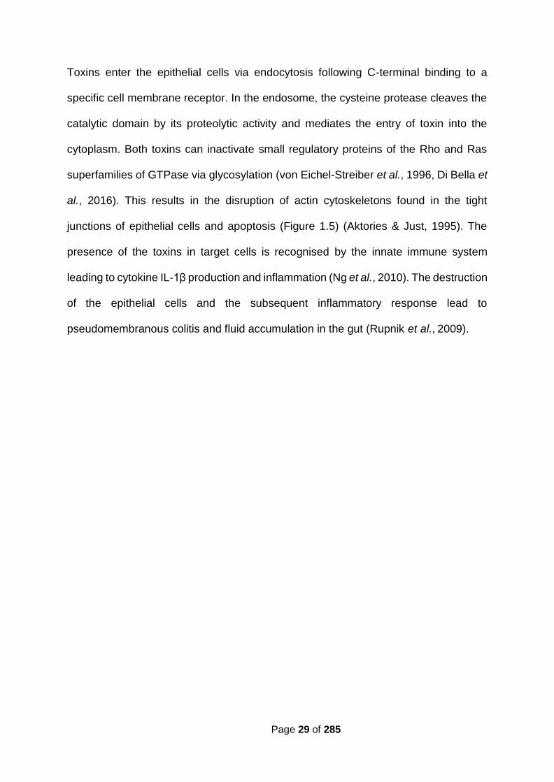
Toxins enter the epithelial cells via endocytosis following C-terminal binding to a
specific cell membrane receptor. In the endosome, the cysteine protease cleaves the
catalytic domain by its proteolytic activity and mediates the entry of toxin into the
cytoplasm. Both toxins can inactivate small regulatory proteins of the Rho and Ras
superfamilies of GTPase via glycosylation (von Eichel-Streiber et al., 1996, Di Bella et
al., 2016). This results in the disruption of actin cytoskeletons found in the tight
junctions of epithelial cells and apoptosis (Figure 1.5) (Aktories & Just, 1995). The
presence of the toxins in target cells is recognised by the innate immune system
leading to cytokine IL-1β production and inflammation (Ng et al., 2010). The destruction
of the epithelial cells and the subsequent inflammatory response lead to
pseudomembranous colitis and fluid accumulation in the gut (Rupnik et al., 2009).

Page 30 of 285
Figure 1.5 Mechanisms of action of TcdA and TcdB.
(Retrieved from (Di Bella et al., 2016)). Seven main mechanisms of toxin delivery into epithelial
cells. 1) toxin binding to the host cell surface receptor; 2) toxins internalization via receptor-mediated
endocytosis; 3) endosome acidification via cysteine protease activity; 4) pore formation; 5) GTD
release from the endosome to the host cell cytoplasm; 6) Rho GTPases inactivation by
glycosylation; 7) downstream effects within the host cell such as toxins‐induced cytokine IL-1β
production. The colour codes are as follows: N-terminal glycosyltransferase domain (red), cysteine
protease domain (blue), delivery domain (yellow).

Page 31 of 285
1.3.1.2. C. difficile binary toxins
In addition to TcdA and TcdB, some C. difficile strains also produce a third toxin, binary
toxin (Popoff et al., 1988). This is a member of the family of clostridial binary toxins,
which have ADP-ribosyltransferase activity, and include the C. perfringens iota toxin,
Clostridium spiroforme toxin and Clostridium botulinum C2 toxins C and D (Popoff et
al., 1988, Perelle et al., 1997). C. difficile binary toxin is encoded on the binary toxin
(CDT) locus which consists of two toxin genes cdtA and cdtB as well as a positive
transcriptional regulator cdtR (Figure 1.6) (Carter et al., 2007). The toxin consists of
two peptides, CdtB which binds an unknown surface receptor and translocates CdtA,
the catalytic domain. CdtA ribosylates actin molecules and disrupts the cytoskeletons
of the epithelial cells (Popoff et al., 1988). PaLoc negative, CDT positive C. difficile
strains can cause cytopathic effects (CPE) on cell lines in vitro and colonize hamster
models in vivo (Geric et al., 2006). It is possible that CDT increases C. difficile
adherence to the epithelial cells by forming microtubule-based membrane structures
(Schwan et al., 2009).
Figure 1.6 Schematic representation of CDT locus in C. difficile.
ORFs are represented by blue
arrows. The complete locus is 6.2 kb
long. The binary toxin consists of a
binding component (CDTB) and an
enzymatic component (CDTA).

Page 32 of 285
1.3.2. Surface layer and cell wall proteins
The surface of many prokaryotic cells is covered by a proteinaceous layer, known as
surface layer (S-layer) forming a two-dimensional structure which is visible by electron
microscopy. S-layer is found on both Gram-positive and Gram-negative bacteria and
is comprised of one or more types of S-layer proteins (SLP). SLPs play important roles
in bacterial growth, survival, immune system and interactions with the host (Calabi et
al., 2001, Mori & Takahashi, 2018).
The crystalline or paracrystalline S-layer of C. difficile is composed of two distinct SLPs:
high- molecular-weight SLP (HMW-SLP ~ 40 kDa) and low-molecular-weight SLP
(LMW- SLP ~ 35 kDa) (Figure 1.7) (Sarker & Paredes-Sabja, 2012). A single gene,
slpA that is conserved amongst all C. difficile strains, encodes a precursor protein SlpA
which has three domains: (i) N-terminal signalling domain, (ii) highly variable LMW
region, (iii) highly conserved HMW region (up to 97% sequence identity between the
strains) (Fagan & Fairweather, 2011). The signalling domain guides the translocation
of the SlpA across the cell membrane via an accessory Sec system (Fagan &
Fairweather, 2011). The precursor protein is then cleaved by cysteine protease Cwp84
present in the cell wall (Kirby et al., 2009) to generate two SLPs which later self-
assemble to form the mature S-layer (Figure 1.7) (Kirk et al., 2016).

Page 33 of 285
Figure 1.7 S-layer protein complex in C. difficile.
(Retrieved from (Awad et al., 2014). Figure
shows the maturation steps of the SlpA
protein. Three stages are shown: A) the
removal of the signal peptide, B) cleavage
of SLP by the protease Cwp84 to generate
HMW SLP and LMW SLP, C) the formation
of the S-layer matrix by re-association of the
LMW and HMW SLPs.
The slpA locus (36.6 kb in strain 630) contains 11 slpA paralogs and there are 17 more
paralogs throughout the genome (Sebaihia et al., 2006, Fagan et al., 2011, Monot et
al., 2011). In total, the C. difficile genome encodes 28 paralogues of SlpA that make
up the clostridial cell wall protein (CWP) family (Kirk et al., 2016). All these genes
encode proteins with an N-terminal signal peptide and three putative cell wall binding
domains with significant similarity to HMW SLP (Calabi et al., 2001, Karjalainen et al.,
2001, Bradshaw et al., 2018). Sequencing results of 57 C. difficile strains revealed that
a 10 kb cassette within the S-layer locus has higher inter-strain variability compared to
the rest of the locus (Figure 1.8) (Dingle et al., 2013).
Figure 1.8 The S-layer locus.
(Retrieved from (Kirk et al., 2016). C. difficile strain 630 encodes 29 cell wall proteins. Twelve of
these, including the S-layer precursor SlpA, are encoded within a single genomic locus (green
arrows) that also encodes the S-layer secretion ATPase SecA2 (red arrow) and five unrelated
proteins (black arrows). The variable S-layer cassette region is highlighted.

Page 34 of 285
Cwp84, a cysteine protease involved in SlpA maturation, is so far the best
characterized cell wall protein in C. difficile (Bradshaw et al., 2015). This protein plays
a significant role in SlpA processing since a cwp84 insertional knockout strain was
shown to be unable to cleave the SlpA precursor (Kirby et al., 2009). Cwp84 is also
able to degrade extracellular matrix proteins in vitro, indicating that it may be involved
in tissue degradation and bacterial dissemination during infection (de la Riva et al.,
2011). However, a cwp84 mutant was shown to be fully virulent in a hamster model
and complete protection was never achieved when Cwp84 was used as an
immunization agent (Kirby et al., 2009, Sandolo et al., 2011). A second cysteine
protease, Cwp13 is a paralogue of Cwp84, sharing 63% amino acid identity (de la Riva
et al., 2011). Cwp13 plays a role in the processing of Cwp84 but is not essential in
Cwp84 activity or SlpA processing (de la Riva et al., 2011). It is possible that Cwp13 is
involved in the removal of damaged or misfolded proteins on the cell surface (de la Riva
et al., 2011).
Another cell wall protein involved in adherence is Cwp66 (66 kDa) containing a domain
homologous to Bacillus subtilis autolysin, CwlB. Cwp66 was detected with antibodies
raised against the surface proteins of heat-shocked bacteria, indicating that it plays a
role as a surface-associated heat-shock protein (Waligora et al., 2001).
The largest characterized member of the cell wall protein family that is encoded
outside the S- layer cassette is CwpV (167 kDa). This protein has a phase variable
expression, with only 5% of cells in a population expressing the protein in vitro
(Emerson et al., 2009). CwpV plays a potential role in bacterial interaction and biofilm
formation as well as protection against bacteriophage infection by preventing phage
DNA replication (Emerson et al., 2009, Reynolds et al., 2011, Sekulovic et al., 2015).

Page 35 of 285
1.3.3. Polysaccharides
Three cell surface polysaccharides with immunogenic properties, PSI, PSII and PSIII
are present in C. difficile strains, with ribotype 027 expressing the PSII most
abundantly (Monteiro et al., 2013). Investigations into vaccine production are mainly
focused on these polysaccharides, particularly PSII. Glycan microarrays carrying
synthetic PSI, PSII and PSIII detect IgG in the sera of CDI patients, and anti-PSII IgA
has been detected in the faeces of patients (Martin et al., 2013, Awad et al., 2014). A
vaccine candidate based on the PSII glycoprotein conjugated to recombinant TcdA
and TcdB fragments has been shown to raise antibodies against PSII and C. difficile
toxins in animal models. Therefore, it is suggested that vaccines containing a
combination of polysaccharides and toxin antigens could be successfully
manufactured in the future (Romano et al., 2014).
1.3.4. Flagella
Flagella play an important role in host invasion, colonization and biofilm formation. The
two best characterized C. difficile flagellar proteins are FliC, the major flagellin
structural monomer, and FliD, the cap protein (Tasteyre et al., 2000, Tasteyre et al.,
2001). The role of flagella in CDI is strain dependent. For example, flagella are
important in the epidemic PCR ribotype 027 strain R20291 adhesion; however, the
absence of flagella in strain 630Δerm does not reduce the adherence (Dingle et al.,
2011, Baban et al., 2013).

Page 36 of 285
1.3.5. Fibronectin-binding proteins
Fibronectin is a large molecular weight glycoprotein involved in bacterial adhesion. C.
difficile has a 68 kDa fibronectin-binding protein (Fbp68) which binds to extracellular
matrix components such as fibronectin, fibrinogen and vitronectin (Cerquetti et al.,
2002). The precise role of Fbp68 in C. difficile pathogenesis is not completely
understood. A C. difficile strain 630 fbp68 mutant was unexpectedly shown to adhere
more effectively in vitro than the wild-type strain. In vivo analysis demonstrated that an
fbpA mutant was able to be shed in faeces at the same rate as wild-type strain in
monoxenic mice but its colonization of caecal cells was reduced. The same result was
observed when the experiment was performed on human-microbiota associated mice,
suggesting that Fbp68 is primarily involved in C. difficile colonization (Barketi-Klai et
al., 2011).
Another Fibronectin-binding protein, ZmpI with sequence similarity to Bacillus
anthracis lethal factor was shown to play a role in invasiveness and spread of C.
difficile during infection (Cafardi et al., 2013).
A collagen binding protein, CbpA was also found in C. difficile and shown to bind to
collagens I and V, the most common collagen components present in many tissues
including the gut (Péchiné et al., 2018).

Page 37 of 285
1.4. C. difficile epidemiology
1.4.1. Typing method
Restriction endonuclease analysis (REA), pulsed field gel electrophoresis (PFGE),
PCR ribotyping, toxinotyping and multilocus sequence typing (MLST) are the most
common typing methods to study C. difficile. REA and PFGE methods are based on
the DNA patterns after restriction digestion (Clabots et al., 1993, Gal et al., 2005). PCR
ribotyping is based on the fact that the intergenic spacer region between the 16S and
23S rRNA is different between the multiple pairs of alleles in an individual cell. The
presence of multiple copies of these genes in most bacteria results in PCR products
of various sizes when PCR is performed by using universal primers. Closely related
isolates show identical patterns; however, distant isolates show variations which help
to categorize the isolates (Cartwright et al., 1995). Toxinotyping is a method in which
PCR amplification of the PaLoc fragments restricted with one to four endonucleases
is performed to identify restriction fragment length polymorphisms of the products
(Rupnik et al., 1998). In MLST, six to seven housekeeping genes are sequenced to
reveal the genetic relationship between strains (Lemee et al., 2004). Although PCR
ribotyping is the preferred method for genotyping C. difficile (Janezic, 2016), whole
genome sequencing (WGS) has been proved to be more efficient and accurate to
distinguish between strains that have been categorized as indistinguishable by
conventional typing methods (Yuan Kong et al., 2016).

Page 38 of 285
1.4.2. Epidemiology of CDI and hypervirulence
The occurrence, clinical presentation, severity and epidemiology of CDI have changed
significantly over the last decade and C. difficile has become a major clinical problem
(Legenza et al., 2018). Healthcare associated infections (HCAIs) are a significant
financial and social burden (Plowman et al., 2001). Because of the rising incidence of
CDI and associated mortality, a mandatory surveillance was announced in 2004 in the
UK to facilitate epidemiological monitoring of C. difficile strains particularly those
isolated from the healthcare units (Goldenberg et al., 2011). C. difficile has historically
been known as a nosocomial pathogen associated with prolonged antibiotic exposure.
However, community-acquired C. difficile infections have been observed in
populations previously considered low risk, such as healthy peripartum women,
children, antibiotic naïve patients, and those with minimal or no recent healthcare
exposure (Wilcox et al., 2008, Depestel & Aronoff, 2013). As CDI initially emerged as
a nosocomial infection in 1978 and became more prevalent in the community, creative
strategies to reduce the risk factors are urgently required (Al-Jashaami & DuPont,
2016).
Reports of epidemic C. difficile outbreaks in the UK and other western countries have
dramatically increased since 2000 (McDonald et al., 2005, Warny et al., 2005). The
isolates from these outbreaks were all shown to be more virulent, secreting higher
levels of toxins, more resistant to fluoroquinolones and more involved in CDI
recurrence (Kuijper et al., 2006). Most of these isolates were categorized as ribotype
027 and reported to be hypervirulent (Razavi et al., 2007). As described earlier, the
PaLoc region harbours genes such as tcdC and tcdR to regulate the expression of the
tcdA and tcdB (Mani & Dupuy, 2001). Ribotype 027 strains have an 18 bp deletion in

Page 39 of 285
the tcdC region which leads to the production of a truncated protein and disruption in
the regulation of toxin genes (Warny et al., 2005). In 2010 in the UK, hypervirulent
strains isolated from patients all belonged to the ribotype 027 (Cartman et al., 2010);
however, in Netherlands, an emergence of isolates belonging to the PCR ribotype 078
was reported. Ribotype 078 contains a 39 bp deletion in the tcdC gene as well as an
early stop codon (Goorhuis et al., 2008). Although both ribotypes, 027 and 078 have
deletions in the tcdC gene, the role of this gene in the emergence of hypervirulent
strains is not clear since complementation of the tcdC mutation has given inconclusive
results (Carter et al., 2011, Cartman et al., 2012). The emergence of other
hypervirulent isolates was reported in China (ribotype 002), Germany (ribotype 001)
and Italy (ribotype 018) (Borgmann et al., 2008, Spigaglia et al., 2010, Cheng et al.,
2011). Therefore, hypervirulence in C. difficile is no longer considered to be limited to
ribotype 027. It has been revealed that some isolates which had previously been
designated ribotype 027 were in fact ribotypes 176, 198 and 244. These ribotypes are
highly similar to the ribotype 027; however, deletions in the tcdC regions of these
ribotypes have never been reported (Valiente et al., 2012).
The elevated production of the TcdA and TcdB toxins has been reported in vitro for
ribotype 027 (Warny et al., 2005). However, a study using a human gut model showed
that the production of toxins by ribotype 027 strains is not higher per unit of time, but
the duration of toxin production is prolonged, which may lead to the increased severity
of symptoms for patients infected with a ribotype 027 isolate (Freeman et al., 2007). It
was also reported that ribotype 027 isolates have a higher level of sporulation
(Merrigan et al., 2010). However, another study showed that the rate of sporulation
varies between strains within a ribotype, and increased or decreased sporulation has
been observed for both epidemic and non-epidemic ribotypes.

Page 40 of 285
Therefore, sporulation is no longer considered as a specific characteristic of
hypervirulent isolates (Burns et al., 2011). Although a significant difference in the
epidemiology of CDI has been noticed over the past two decades, no solid criteria
have yet described the hypervirulence. Therefore, most reports simply refer to them
as epidemic strains (Robinson et al., 2014).
Between 2008 and 2016 the number of C. difficile related deaths in England and Wales
has decreased gradually in both men and women, most likely due to the improvements
in hospital procedures and better management (Figure 1.9).
Figure 1.9 Number of death certificates with C. difficile mentioned, by sex, in England and Wales, deaths registered between 1999 and 2016.
Graph drawn based on the data provided by Office for National Statistics website. Release date:
30 October 2017. Colour codes: blue represents men and red represents women (Adapted from
(Windsor-Shellard, 2017)).
)

Page 41 of 285
1.5. Horizontal gene transfer via mobile genetic
elements
Horizontal gene transfer (HGT) involves DNA transfer from donor cells to recipient
cells, followed by replication (in the case of plasmids) and integration (in the case of
transposons and bacteriophages). Therefore, it differs from vertical transmission of the
genes from parent to offspring (Burmeister, 2015). It was first described in
microorganisms in the late 1940s (Tatum & Lederberg, 1947), and has long been
recognized as an important factor in the evolution of bacteria and archaea. Mobile
genetic elements (MGEs), which include cytoplasmically located replicating plasmids
and chromosomally located transposons and genomic islands, are important
mediators of HGT. The three main mechanisms of HGT are transformation,
transduction and conjugation.
1.5.1. Transformation
The first identified HGT mechanism was transformation, which is a direct uptake of
exogenous DNA from the environment. It was first observed in S. pneumoniae that
became virulent when exposed to heat-killed virulent cells (Griffith, 1928). This was
confirmed later by demonstrating that nonvirulent strains acquired virulence when
inoculated together with the extracted DNA from heat-killed virulent strains (Avery et
al., 1944). DNA uptake occurs during the competence state of bacteria, which can
occur naturally or can be artificially induced. A total of 82 species are known to be
naturally transformable, with an approximately equal number between Gram-negative
and Gram-positive bacteria (Johnston et al., 2014).

Page 42 of 285
The competence stage of naturally transformable bacteria is usually triggered by
specific conditions such as pH and nutrient availability or at a specific phase of
bacterial growth (Wilson & Bott, 1968, Solomon & Grossman, 1996). The mechanisms
of bacterial transformation are similar between transformable bacteria. Conserved
proteins encoded by genes that form the com regulon are expressed during the
competence state. Double-stranded DNA (dsDNA), as a substrate for transformation,
is bound and captured by a transformation pilus (Johnston et al., 2014). The binding
sites on competent cells vary among bacteria (20- 50 sites in B. subtilis, 33-75 sites in
S. pneumoniae and 4-8 sites in Haemophilus influenzae) (Singh, 1972, Deich & Smith,
1980, Dubnau, 1999). The captured dsDNA is then internalised as single-stranded
DNA (ssDNA) through a transmembrane channel. As Gram-negative bacteria contain
two layers of membrane, there are two transmembrane channels, one for each layer,
while Gram-positive bacteria use only one transmembrane channel (Fig 1.10).
Once the DNA enters the cell, it could be degraded to nucleotides that can be used in
other metabolic functions such as DNA replication. The internalised ssDNA could be
recombined into the bacterial genome through homologous recombination (Johnston
et al., 2014). The internalised ssDNA is bound by DNA processing protein A (DprA),
which then recruits RecA recombinase to the DNA and promotes recombination
(Mortier-Barriere et al., 2007). In the case of plasmids, if it contains an origin of
replication that can be recognised by the host, recircularisation and second strand
synthesis will occur and result in plasmid maintenance (Thomas & Nielsen, 2005).

Page 43 of 285
Figure 1.10 The processes of transformation in Gram-negative and Gram-positive bacteria.
(Retrieved from (Johnston et al., 2014). The exogenous double-stranded DNA (dsDNA) is
captured by a transformation pilus (Tfp), which consists mainly of ComGC and PilE subunits in
Gram-positive and Gram-negative bacteria, respectively. dsDNA is transported into cytosol via
transmembrane channels (ComEC for Gram-positive bacteria, and PilQ and ComA for Gram-
negative bacteria). The captured dsDNA is internalised as single-stranded DNA (ssDNA) into
the cytosol, which is then bound by DNA processing protein A (DprA). The recombinase RecA
multimers assemble on ssDNA and promotes homologous recombination.

Page 44 of 285
1.5.2. Transduction
Transduction is a mechanism of HGT during which DNA from a phage-infected
bacterium is transferred into a recipient cell via bacteriophage. It was discovered by
Lederberg and Zinder (1952), who showed the transfer of chromosomal DNA from one
strain of Salmonella typhimurium to another mediated by phage P22. By having phage
as a vector, this mechanism does not require a cell-to-cell contact, and the donor DNA
can be protected from physical and chemical agents (Calero-Caceres & Muniesa,
2016). There are two types of transduction: generalized and specialized. In
generalized, bacteriophages can pick up any portion of the host DNA, whereas in
specialized, they only pick up specific parts of the host DNA (Figure 1.11) (Wagner &
Waldor, 2002). The transferable DNA sequences range from chromosomal DNA to
MGEs such as plasmids, transposons and genomic islands (Brown-Jaque et al.,
2015).
The first report of the bacteriophage-mediated antibiotic resistance gene transfer
between C. difficile strains was published in 2013. In this work, an erythromycin
resistance gene, erm(B) located on a mobile element, Tn6215 was transferred from
strain CD80 into CD062. The transfer was confirmed to be mediated by a
bacteriophage called ΦC2 since a control mixture containing CD062 and CD80 filtrate
without ΦC2 did not give rise to erythromycin-resistant derivatives of strain CD062
(Goh et al., 2013).

Page 45 of 285
Figure 1.11 Types of transduction.
(Retrieved from (Salmond & Fineran, 2015). A) Generalised transduction is the transfer of
random DNA fragments of the host (shown in blue) to the neighbouring bacteria. B) Specialised
transduction is the transfer of prophage DNA (shown in orange) and the bacterial DNA, located
next to the prophage DNA (shown in blue) to a new recipient cell.

Page 46 of 285
1.5.3. Conjugation
Conjugation is a form of HGT between bacterial cells that occurs via a close contact
between donor and recipient (cell-to-cell contact). The process was first discovered in
Escherichia coli for the fertility plasmid (F-plasmid) (Hayes, 1953). In 1947, it was
shown that when two auxotrophic mutant strains of E. coli with specific nutritional
requirements were grown together in a mixed culture, recombination of the mutant
strains could occur which resulted in prototrophic isolates (Tatum & Lederberg, 1947).
The recombination was mediated by a fertility factor (F-factor) present in at least one
of the bacteria to facilitate the formation of mating pair (Lederberg et al., 1952, Hayes,
1953). The F-factor was believed to be an autonomously replicating element capable
of converting an F- recipient strain into an F+ donor strain and encoding all the proteins
required for the process (Hayes, 1953, Adelberg & Pittard, 1965). The F-plasmid was
also able to integrate into the chromosomal DNA leading to high frequency
recombination (Hfr). This process leads to a transfer of part or even the entire donor
chromosome which can recombine with the recipient chromosome after mating
(Adelberg & Pittard, 1965). Further analysis on the recombinant cells revealed that
linear DNA was transferred and integrated into the recipient genome. Some genes on
the chromosome were transferred with higher frequencies than others depending on
their distance from the origin of transfer (oriT) (Adelberg & Pittard, 1965). Hfr has been
found in several Gram-negative bacteria such as E. coli (Lloyd & Buckman, 1995),
Rhodobacter sphaeroides (Suwanto & Kaplan, 1992) and Yersinia pseudotuberculosis
(Lesic et al., 2012).
In Gram-positive organism Lactococcus lactis, a plasmid encoded lactose fermenting
enzyme is present which turns lactose fermentation negative strains to strains with the

Page 47 of 285
ability to ferment lactose (Gasson & Davies, 1980). It was demonstrated that
conjugation occurred with higher frequencies in L. lactis cells with a clumping
phenotype associated with CluA, a membrane bound protein encoded by a
chromosomally located sex factor. The lactose fermentation plasmid itself was able to
integrate into the chromosomal DNA within the sex factor region through
recombination by the IS- element carried by the plasmid (Gasson et al., 1992). The
sex factor was responsible for the unidirectional transfer of the chromosomal DNA in
an Hfr-like manner as genes located close to the sex factor in a clockwise orientation
transferred with a higher frequency than those located further away from the oriT and
in an anticlockwise orientation (van Kranenburg & de Vos, 1998).
1.5.3.1. Conjugative plasmids
Conjugative plasmids are self-replicating, double-stranded and generally circular units
of DNA; however, examples of linear plasmids are also known including those found
in Gram-positive filamentous bacteria of the genus Streptomyces (Lee et al., 2011).
For many years, conjugative plasmids have been associated with the dissemination of
antibiotic resistance genes amongst bacterial species (Palmer et al., 2010).
A sophisticated multiprotein secretion apparatus called a type IV secretion system
(T4SS) is used by most conjugative plasmids to transfer DNA from donor to recipient
(Smillie et al., 2010). There are similarities among bacterial species at the initial steps
of conjugation in which relaxase is required for preparing ssDNA and beginning the
process of transfer (Zechner et al., 2012). However, the actual translocation process
including DNA transfer through the donor cell envelope and insertion into the recipient
is different between Gram-positive and Gram-negative bacteria due to differences in
Page 48 of 285
the structure of their cell envelopes (Goessweiner-Mohr et al., 2014). The mechanism
of T4SS in many Gram-positive bacteria involves the passage of ssDNA, except for
the Actinomycetales, in which dsDNA is transferred (Vogelmann et al., 2011).
Conjugation is initiated by close contact between a suitable donor and a recipient
(Lawley et al., 2003). Some Gram-positive bacteria can stimulate mating by producing
a pheromone-like peptide as observed in many enterococcal strains (Hirt et al., 2002,
Chandler & Dunny, 2004). Once contact is established, a mating signal is generated,
and the relaxase binds to the dsDNA and forms a relaxosome which is a complex of
proteins that facilitates transfer by binding to the DNA (Smith & Thomas, 2004). This
nucleoprotein complex nicks the plasmid DNA at a specific site known as origin of
transfer (oriT) to give a single-stranded substrate suitable for transfer via mating pair
formation (mpf) (Grohmann et al., 2003).
Relaxase is the key enzyme to initiate conjugation and functions by catalysing a break
in a specific phosphodiester bond in the nic site within the transfer origin, oriT
(Grohmann et al., 2003). Comparisons among a wide range of oriT regions have
revealed that they all carry inverted repeats (IRs) that flank the nic site. These IR
sequences form a hairpin structure to be recognized and cleaved by the relaxosome.
The sequence of the IRs varies among the conjugative or mobilisable elements but
their location relative to the nic site is similar (Guiney & Yakobson, 1983, Goessweiner-
Mohr et al., 2014). The most widely distributed relaxase belongs to the broad host
range plasmid RP4/ RK2, first isolated in connection with an outbreak of antibiotic
resistance in Pseudomonas aeruginosa and Klebsiella pneumoniae strains in
Birmingham in 1969 (Ingram et al., 1973, Zatyka & Thomas, 1998).

Page 49 of 285
Proteins involved in T4SS are all encoded by one or more transfer (tra) operons. Two
major Tra proteins, TraE and TraJ have ATPase activities essential for fuelling the
DNA translocation process and assembling the mating pair formation. TraJ binds to
TraI and together they position TraJ to the cell membrane through protein-protein
interaction. Therefore, TraI and TraJ form a two-partner T4SS coupling protein which
is crucial in all T4SSs. TraJ also interacts with the relaxase, further confirming its
important role as a coupling protein to link the relaxosome to the transfer channel
(Goessweiner- Mohr et al., 2014). Another Tra protein for which an absolute
requirement in T4SS has been confirmed is peptidoglycan hydrolysing protein, TraG
which is the first PG- hydrolysing protein characterized (Arends et al., 2013). Other
important Tra proteins such as TraM, TraL and TraO are involved in channel formation
and adhesion (Figure 1.12) (Goessweiner-Mohr et al., 2014).

Page 50 of 285
Figure 1.12 Schematic representation of T4SS in conjugative plasmid pIP501 isolated from S. agalactiae as a model for T4SS in Gram-positive bacteria.
(Retrieved from (Goessweiner-Mohr et al., 2014). First, TraA relaxase binds to the oriT. After being
cleaved at the nic site, the single-stranded plasmid DNA is engaged with the transfer channel via
TraJ-TraI coupling proteins. TraG hydrolyses the peptidoglycan layer to form punches (faded grey
colour). The N-terminus of the T4SS proteins is marked (N). Arrows indicate protein-protein
interactions. The thickness of the arrows shows the strength of the interactions. PG, peptidoglycan;
CM, cytoplasmic membrane; CP, cytoplasm.

Page 51 of 285
1.5.3.2. Conjugative transposons
Conjugative transposons (CTns), also known as integrative and conjugative elements
(ICEs), can transfer into a recipient via conjugation and integrate into the host genome
through site-specific recombination (Burrus et al., 2002). They excise from the host
genome and form a circular DNA molecule during transposition and conjugation (Scott
et al., 1988). The circular intermediate can then insert back into the site from which it
has excised, transpose into a different site within the same chromosome, transfer to a
recipient via conjugation or be degraded and lost from the host cell (Figure 1.13)
(Clewell et al., 1995). Like conjugative plasmids, CTns contain an oriT (Jaworski &
Clewell, 1995). Nevertheless, unlike the conjugative plasmids, their replication occurs
while integrated in the chromosome using the replication machinery of the host.
However, a study by (Lee et al., 2010) demonstrated that ICEBs1, a CTn originally
found in B. subtilis, replicates in a plasmid-like manner when it is in the circular state.
Moreover, conjugative transposon Tn916 can replicate autonomously using the rolling-
circle mechanism. The oriT in Tn916 functions as the origin of replication where the
Tn916-encoded relaxase initiates the replication (Wright & Grossman, 2016).

Page 52 of 285
Figure 1.13 Schematic diagram of CTn fate.
(Retrieved from (Brouwer, 2013)). The chromosome is represented by a green circular molecule.
The linear CTn integrated in the chromosome is represented by a red line. The circular intermediate
of the CTn is represented by a red circle. A single strand of the CTn, nicked at the oriT, is transferred
from donor into recipient. Blue cells represent a resistant phenotype, conferred by an antibiotic
resistance gene on the CTn and orange cells represent a susceptible phenotype.
1.5.3.2.1. Tn916
Tn916 is the first CTn discovered and was originally isolated from Enterococcus
faecalis in 1981 by Franke and Clewell who characterized it as a transposon with the
ability to transfer from plasmid-free donor strains into tetracycline-susceptible strains,
and therefore it was hypothesized that this element was self-transmissible (Franke &
Clewell, 1981). Since then, many Tn916-like elements have been discovered in
several bacterial species and all have a similar backbone. Most of these elements
confer resistance to tetracycline by encoding proteins responsible for protecting 30S
ribosomal subunits in the mRNA translation complex (Roberts & Mullany, 2009).
Tn916-like elements have a modular structure including accessory genes which are
not involved in transfer and often encode functional proteins which contribute to the
Page 53 of 285
environmental adaptability of the host (Roberts & Mullany, 2009). They can carry
genes encoding antimicrobial peptides (Rauch & De Vos, 1992), adhesins, virulence
associated genes (Seth-Smith et al., 2012), chlorocatechol degradation (Ravatn et al.,
1998), and metabolism of sucrose, biphenyl and salicylate (Hochhut et al., 1997, Nishi
et al., 2000). The accessory module is often flanked by genes for transcriptional
regulation. Another functional module of the CTns is the integration module encoding
the recombinase and excisionase proteins responsible for transposition of the
element. The conjugation module encodes all the proteins required for conjugation
(Figure 1.14, refer forward to page 57) (Roberts & Mullany, 2011).
1.5.3.2.2. Tn1549
Another family of CTns are Tn1549-like elements in which the conjugation module
differs from Tn916 (Figure 1.14, refer forward to page 57). Tn1549 was originally
discovered in E. faecalis strain 268-10 where it is integrated in the plasmid pAD1. The
element carries the vanB operon conferring vancomycin resistance (Garnier et al.,
2000). Transfer of Tn1549- like elements from Clostridium symbiosum into
Enterococcus faecium and E. faecalis has been shown in animal models (Launay et
al., 2006).
1.5.3.2.3. Excision and integration by site-specific recombination
In CTns, the process of site-specific recombination including excision and integration
is mainly mediated by two protein families: serine or tyrosine recombinases (Grindley
et al., 2006). The latter mediates the integration and excision of Tn916 (Taylor &
Churchward, 1997, Garnier et al., 2000). Excision begins with endonucleolytic cuts at

Page 54 of 285
each end of the element generating single-stranded non-complementary
hexanucleotides at each end called coupling sequences (Abbani et al., 2005). The
coupling sequences then join by covalent bonds forming a circular intermediate
molecule with a heteroduplex at the joints. These heteroduplexes are then resolved
by DNA repair or replication (Figure 1.15) (Caparon & Scott, 1989).
Figure 1.15 Tn916 excision and integration.
(Adapted from (Caparon & Scott, 1989)). The thick lines are Tn916 and the thin lines are the flanking
regions. Nucleotides in yellow are from Tn916 and those in green are from the target site.
Nucleotides in red are the product of DNA repair through replication or mismatch repair. a) Excision:
a staggered cut at the coupling sequence results in the excision of Tn916 with non-complementary
heteroduplex which is later resolved by DNA replication. b) Integration: staggered cleavage at the
3’ end of the new target site and the heteroduplex at the joint of the circular intermediate molecule
generates molecules that are subsequently ligated together, resulting in an inserted Tn916 with
heteroduplexes at each end. These are resolved by DNA replication resulting in two copies, each
flanked by different DNA sequences.

Page 55 of 285
1.5.3.3. Mobile genetic elements in C. difficile
1.5.3.3.1. Tn5397
Transfer of tetracycline resistance between C. difficile strains was first reported by
Ionesco (1980) and Smith et al., (1981). These workers showed that the transfer was
resistant to DNase, could not be mediated by donor culture filtrate, and cell-to-cell
contact was required, and therefore suggested that it was mediated by a conjugation-
like mechanism (Ionesco, 1980, Smith et al., 1981). When the first report of Tn916
transfer in E. faecalis was published, it was hypothesized that the tetracycline
resistance determinant could be encoded by a Tn916-like element (Franke & Clewell,
1981, Smith et al., 1981). Transfer of the element from C. difficile strain 630 at a low
frequency (5x 10⁻⁷ transconjugants per donor) was detected in the absence of a
detectable plasmid (Wüst & Hardegger, 1983). Furthermore, a homology between this
element and Tn916 was confirmed via DNA hybridization in which at least six of seven
HincII fragments of Tn916 from E. faecalis hybridised with tetracycline resistant C.
difficile strains. Therefore, a close relationship between Tn916 and the tetracycline
resistance determinant in C. difficile was suggested. Despite the great (95%)
homology between two elements, differences were also observed such as the number
of HindIII cleavage sites within the transposons (Hächler et al., 1987). Finally, the
element was shown to be a conjugative transposon and designated Tn5397 (Mullany
et al., 1995). The sequencing also showed that Tn5397 contained a group II intron;
the first report of the presence of an intron in a conjugative transposon and the first
report of a group II intron in Gram- positive bacteria (Mullany et al., 1996). Despite
bearing great structural similarities, the differences between Tn916 and Tn5397 are
noticeable (Figure 1.14, refer forward to page 57).

Page 56 of 285
A single serine recombinase, TndX is responsible for the entire recombination process
of Tn5397 (Wang et al., 2000). When this element is integrated into the genome, it is
flanked by a GA dinucleotide at each end. TndX recombinase mediates staggered cuts
at these sites and results in the formation of dinucleotide overhangs at each end which
then join after strand exchange with the target site to form the circular intermediate of
Tn5397 (Wang & Mullany, 2000).
Figure 1.16 Schematic representation of Tn5397 recombination.
(Adapted from (Wang & Mullany, 2000)) The blue lines represent Tn5397 and the red lines
represent the chromosomal DNA. The coupling sequences of the element and the specific target
site are shown by the nucleotide pairs G-C and A-T, respectively. a) Tn5397 is in the circular
intermediate form and a suitable empty target site is present in the chromosome. b) Staggered
cleavage of Tn5397 and the chromosome by the serine recombinase TndX. c) Tn5397 is ligated
with the chromosomal DNA generating homoduplexes.
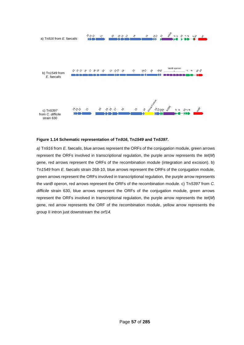
Page 57 of 285
Figure 1.14 Schematic representation of Tn916, Tn1549 and Tn5397.
a) Tn916 from E. faecalis, blue arrows represent the ORFs of the conjugation module, green arrows
represent the ORFs involved in transcriptional regulation, the purple arrow represents the tet(M)
gene, red arrows represent the ORFs of the recombination module (integration and excision). b)
Tn1549 from E. faecalis strain 268-10, blue arrows represent the ORFs of the conjugation module,
green arrows represent the ORFs involved in transcriptional regulation, the purple arrow represents
the vanB operon, red arrows represent the ORFs of the recombination module. c) Tn5397 from C.
difficile strain 630, blue arrows represent the ORFs of the conjugation module, green arrows
represent the ORFs involved in transcriptional regulation, the purple arrow represents the tet(M)
gene, red arrow represents the ORF of the recombination module, yellow arrow represents the
group II intron just downstream the orf14.
a) Tn916 from E. faecalis
b) Tn1549 from E. faecalis
c) Tn5397 from C. difficile
strain 630

Page 58 of 285
1.5.3.3.2. Other conjugative transposons in C. difficile 630
An investigation into the genome of C. difficile 630 revealed the presence of six other
putative CTns (in addition to Tn5397) in this strain. The elements are named CTn1,
CTn2, CTn4, CTn5, CTn6 and CTn7 based on their location within the genome.
Tn5397 was named CTn3 even though mostly the initial designation is used in the
literature and throughout this thesis (Sebaihia et al., 2006). Like Tn5397, CTn1, CTn6
and CTn7 are related to Tn916 (Figure 1.17). CTn1 and CTn7 both encode predicted
ABC transporters on their accessory module; however, no substrates for these protein
products have yet been identified. The accessory module of CTn6 encodes several
hypothetical proteins for which no clear roles have been shown. Both CTn1 and CTn6
elements encode for a tyrosine recombinase and excisionase which are predicted to
mediate the recombination of their elements, whereas CTn7 encodes a serine
recombinase which presumably facilitates its recombination (Sebaihia et al., 2006).
CTn2, CTn4 and CTn5 all encoding ABC transporters on their accessory modules are
part of the Tn1549 family (Figure 1.18). CTn4 encodes a tyrosine recombinase and
excisionase for excision into the circular intermediate CTn and insertion into a suitable
target site. CTn2 and CTn5 both encode serine recombinases for the recombination
(Sebaihia et al., 2006). Five of these six putative elements are predicted to encode
ABC transporters; however, the substrate of these transporters have not yet been
identified. It has been shown that the ABC transporter of CTn7 has homology to
magnesium ABC transporters (Sebaihia et al., 2006).

Page 59 of 285
Figure 1.17 Comparison of Tn916 to CTn3 (Tn5397), CTn1, CTn6 and CTn7.
(Retrieved from (Sebaihia et al., 2006)). Each module on the CTns is color coded: pink:
recombination, purple: accessory function, green: conjugation, light blue: transcriptional regulation.
The dark blue region in Tn5397 is the group II intron. SR is serine recombinase, TR is tyrosine
recombinase. Red lines between the elements represent protein similarity.
Figure 1.18 Comparison of Tn1549 to CTn2, CTn4 and CTn5.
(Retrieved from (Sebaihia et al., 2006)). Each module on the CTns is colour coded: pink:
recombination, purple: accessory function, green: conjugation, light blue: transcriptional regulation.
SR is serine recombinase, TR is tyrosine recombinase. Red lines between the elements represent
protein similarity.

Page 60 of 285
In order to determine whether these putative transposons are capable of excision,
PCR reactions were performed using specific primer pairs to amplify junctions between
the element and host genome, the joints of the element in a circular form and the
repaired target site after excision. PCR products indicating the excision were produced
with CTn1, CTn2, CTn4, CTn7 (Sebaihia et al., 2006, Brouwer et al., 2011). CTn6 is
the only putative conjugative transposon in strain 630 for which no PCR product could
be detected (Brouwer et al., 2011). Furthermore, to investigate the transfer of these
putative conjugative transposons, the ClosTron system (section 1.6.4) was used to
mark the accessory modules of the transposons and create selectable markers. All
marked elements (except for CTn6) transferred from strain 630 into the recipient strain
CD37 at frequencies between 10-4 to 10-9 transconjugants per donor (Brouwer et al.,
2011).
1.5.3.4. Mobilisable elements in C. difficile
1.5.3.4.1. Tn4453
Mobilisable transposons are transposable genetic elements that also encode
mobilisation functions but are not in themselves conjugative. They rely on co-resident
conjugative elements to facilitate their transfer to recipient cells (Crellin & Rood, 1998).
Chloramphenicol resistance in C. perfringens is conferred by a chloramphenicol
acetyltransferase (CatP) protein that is encoded by the accessory module on a
mobilisable transposon Tn4451 (Lyras et al., 1998). An investigation into the cat gene
in C. difficile strains revealed that this gene is located on a mobilisable transposon
very similar to Tn4451 and designated as catD (Wren et al., 1988). The catD gene
harbouring transposon in C. difficile has an 89% identity with the one in C. perfringens

Page 61 of 285
and is designated Tn4453 (Figure 1.19) (Wren et al., 1988, Lyras & Rood, 2000).
Insertion and excision of this element is mediated by a serine recombinase, TnpX.
There are other genes located on the element including tnpZ, tnpV, tnpY and tnpW.
The tnpZ gene is involved in mobilisation of the element and encodes the functional
mobility (Mob) protein, but the function of the others is unknown (Adams et al., 2002).
Figure 1.19 Schematic representation of Tn4453.
The red arrow represents tnpX, the purple arrow represents catD. Other ORFs are represented by blue arrows.
1.5.3.4.2. Tn5398
In addition to the transfer of tetracycline resistance from C. difficile 630 into C. difficile
CD37, transfer of erythromycin resistance has also been observed at low frequencies
(~ 10-8 per donor) (Wüst & Hardegger, 1983). It was later shown that the transfer of
erythromycin resistance could also occur from C. difficile 630 into other bacterial
species such as Staphylococcus aureus (Hächler et al., 1987) and B. subtilis (Mullany
et al., 1995) in the absence of any detectable plasmid. Analysis of target sites of the
element showed that it has a preferred insertion site in C. difficile but multiple sites in
B. subtilis (Mullany et al., 1995). Thus, it was assumed that the element could be a
conjugative transposon (Mullany et al., 1995). Further studies revealed that C. difficile
630 contains two copies of adenine N-6- methyltransferase erm(B) genes designated

Page 62 of 285
erm1(B) and erm2(B) separated by a 1.34 kb direct repeats (Figure 1.20) (Farrow et
al., 2000). Erm(B) confers erythromycin resistance through ribosomal protection but
only erm2(B) is functional. An analysis to fully characterise the genomic arrangement
of Tn5398 revealed that this element does not encode genes for excision, integration
or conjugation, and therefore does not fit into the standard category of conjugative,
mobilisable or transposable elements (Farrow et al., 2001). In addition to the two
erm(B) genes, the element carries homologs of Tn916 genes including orf13, orf9 and
orf7 which are assumed to be involved in the regulation of the element. To be
mobilised, Tn5398 needs an oriT and a recognizable nic site. The presence of two
potential oriT sites, which are located within the coding sequence of orf298 has been
confirmed in this element (Farrow et al., 2001). The nic sites (TGGTGT) of these two
potential oriT sites are identical to the nic sites found on Tn916 and Tn5397 (Farrow
et al., 2001).
Figure 1.20 Schematic representation of Tn5398.
The purple arrows represent erm1(B) and erm2(B), the latter is functional in Tn5398 (highlighted by
red line). Other ORFs are represented by blue arrows.
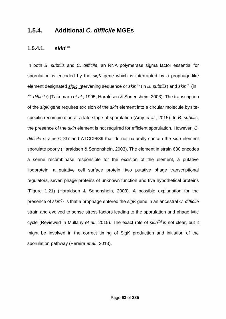
Page 63 of 285
1.5.4. Additional C. difficile MGEs
1.5.4.1. skinCD
In both B. subtilis and C. difficile, an RNA polymerase sigma factor essential for
sporulation is encoded by the sigK gene which is interrupted by a prophage-like
element designated sigK intervening sequence or skinBs (in B. subtilis) and skinCd (in
C. difficile) (Takemaru et al., 1995, Haraldsen & Sonenshein, 2003). The transcription
of the sigK gene requires excision of the skin element into a circular molecule by site-
specific recombination at a late stage of sporulation (Amy et al., 2015). In B. subtilis,
the presence of the skin element is not required for efficient sporulation. However, C.
difficile strains CD37 and ATCC9689 that do not naturally contain the skin element
sporulate poorly (Haraldsen & Sonenshein, 2003). The element in strain 630 encodes
a serine recombinase responsible for the excision of the element, a putative
lipoprotein, a putative cell surface protein, two putative phage transcriptional
regulators, seven phage proteins of unknown function and five hypothetical proteins
(Figure 1.21) (Haraldsen & Sonenshein, 2003). A possible explanation for the
presence of skinCd is that a prophage entered the sigK gene in an ancestral C. difficile
strain and evolved to sense stress factors leading to the sporulation and phage lytic
cycle (Reviewed in Mullany et al., 2015). The exact role of skinCd is not clear, but it
might be involved in the correct timing of SigK production and initiation of the
sporulation pathway (Pereira et al., 2013).

Page 64 of 285
Figure 1.21 Schematic representation of skinCd in strain 630.
The green arrow represents the serine recombinase, other ORFs are represented by blue arrows.
The interrupted sigK is represented by an interrupted red arrow.
1.5.4.2. IStron
Insertion sequence elements (IS-elements) are small genetic elements capable of
mediating their own transposition into different target sites by encoding a transposase
enzyme (Mahillon & Chandler, 1998). Group I introns are mobile genetic elements
which encode ribozymes to catalyse their own splicing from a precursor mRNA (self-
splicing), allowing exons to ligate and form mature mRNA (Hausner et al., 2014). In
2000, a chimeric genetic element was found within the tcdA gene in C. difficile strain
C34. This element had characteristics of both IS-elements and group I introns;
therefore, it was designated IStron, CdIst1 (Braun et al., 2000, Hasselmayer et al.,
2004). The CdIst1 element splices from the pre-mRNA molecule leading to the normal
expression of the tcdA gene (Braun et al., 2000). The element carries two CDS
encoding a putative transposase (IS-element feature) and a ribozyme (group I intron
feature). The presence of CdIst1 in C. difficile was shown by southern blot and PCR
analysis and confirmed by the whole genome sequencing of strain 630 (Braun et al.,
2000, Sebaihia et al., 2006). A homolog with 63% sequence identity to CdIst1 is
present in Bacillus cereus (Tourasse et al., 2006); however, similar IStron elements
have not been reported in any other organism (Amy et al., 2015).
5’ 3’

Page 65 of 285
1.6. Genetic manipulation tools
Historically, genetic manipulation of C. difficile has been proved to be extremely
difficult since the transfer of foreign DNA by electroporation or chemical transformation
has never been reported. However, remarkable progresses have been made through
whole genome sequencing (WGC) projects and applying different genetic tools such
as shuttle vectors, ClosTron and CRISPR-cas9, which all have resulted in significant
advances in C. difficile researches (Mullany et al., 1994, Purdy et al., 2002, O'Connor
et al., 2006, Heap et al., 2007, McAllister et al., 2017, Wang et al., 2018). These
genetic manipulation tools have provided important information about the structure
and organization of the C. difficile genome, genetic diversity, epidemiology,
pathogenicity and evolution of this organism (Knight et al., 2015, Muñoz et al., 2018).
1.6.1. Shuttle conjugative Transposon: Tn916
The first study of gene cloning in C. difficile was performed by using Tn916 as a shuttle
conjugative transposon (Mullany et al., 1994). In this work, a pBR322-based vector
designated pCI195 (replicable in E. coli, non-replicable in C. difficile) carrying the catP
gene and a 4.2 kb region of conjugative transposon Tn919 (Casey et al., 1991) was
used to transform B. subtilis strain BS17 (CU2189:: Tn916ΔE (tet(M) gene is replaced
by an erm(B) gene)). Chloramphenicol (encoded by pCI195) and erythromycin
(encoded by Tn916ΔE) resistant transformants appeared on selective plates.
Southern blot analysis showed that a single copy of pCI195 was integrated into
Tn916ΔE via homologous recombination (transposon:: integrative plasmid). Filter
mating was performed between B. subtilis transformant donor and C. difficile strain

Page 66 of 285
CD37 recipient. Chloramphenicol and erythromycin resistant transconjugants
appeared on selective plates. Southern blot analysis confirmed that Tn916ΔE::pCI195
was excised from B. subtilis genome and inserted into the C. difficile chromosome at
a specific target site. The possibility of using conjugative transposons for gene cloning
in C. difficile was further investigated by cloning a fragment of the tcdB gene into
plasmid pCI195. The plasmid was then transferred into CD37 from B. subtilis strain
BS17. DNA isolated from transconjugants was digested and probed with the cloned
fragment of the tcdB gene from pCI195. Results confirmed the successful transfer
since one hybridising fragment appeared with each transconjugant (Mullany et al.,
1994).
1.6.2. Other transposon-based mutagenesis systems
The elements from the superfamily mariner transposons are widely distributed in
nature and found in rotifers, insects, fungi, plants and mammals (Robertson, 1993,
Arkhipova & Meselson, 2005, Muñoz-López & García-Pérez, 2010). These elements
are between 1 and 5 kb and encode a transposase which is flanked by two inverted
repeats (Plasterk et al., 1999, Muñoz-López & García-Pérez, 2010). The transposition
of mariner elements is a non-replicative process and involves a cut-and-paste
mechanism in which two transposase monomers bind to the inverted repeats at each
end of the transposon, forming a structure called single-end complex. Then, the
transposon ends are brought together by the transposase monomers and generate a
second structure called paired-end complex which leads to the transposon excision.
The transposase dimer recognizes a random TA dinucleotide sequence to carry out
the insertion (Figure 1.22) (Bryan et al., 1990).

Page 67 of 285
Figure 1.22 Schematic diagram of the cut-and- paste mechanism of transposition performed by the mariner elements.
Three stages of the cut-and -
paste mechanism: Single-End
complex, Paired-End complex
and Target-Capture complex
are shown in this diagram.
A mariner-based transposon system using Himar1 transposase for in vitro random
mutagenesis has been developed in C. difficile. The facts that (i) Himar1 transposase
is the only requirement for the cut-and-paste transposition mechanism and (ii) the
element inserts into a TA rich target site make this element suitable for an organism
with low GC content such as C. difficile (Cartman & Minton, 2010).
Pseudo-suicidal plasmids pMTL-SC0 and pMTL-SC1 were constructed to deliver
mariner-based transposon into C. difficile R20291. These two mariner plasmids are
identical except that the expression of Himar1 in pMTL-SC1 is controlled by the tcdB
gene promotor, but pMTL-SC0 lacks the inducible promotor (negative control). Both
plasmids are pBP1-based (from C. botulinum), chloramphenicol (Cm)/ thiamphenicol
(Tm) resistant (the resistance markers are located on the transposon), replicable in E.
coli and carry the conjugative transfer gene traJ (Figure 1.23). Plasmids were
transferred into R20291 and TmR colonies appeared on selective agar at the frequency
of 4.5 x 10⁻⁴ for the pMTL-SC1 transconjugant culture.

Page 68 of 285
PCR showed that (i) the transposon-based catP gene conferring CmR was present in
the R20291 chromosome (confirming the transposition), (ii) plasmid-based Himar1
transposase gene was not present in the genome (showing that the transposon was
mobilised, and the plasmid was lost from the cell) (iii) plasmid-based PtcdB was not
found in the genomic DNA (showing that plasmid did not integrate into the
chromosome by homologous recombination). The randomness of the insertion was
verified by sequencing and southern blot analysis (Cartman & Minton, 2010).
Figure 1.23 Map of plasmid pMTL-SC1.
(Retrieved from (Cartman & Minton, 2010)).
Expression of the mariner transposase gene
Himar1 was controlled by the tcdB promotor,
PtcdB. The plasmid backbone consists of the
pBP1 replicon of C. botulinum (repA and orf2), the
erm(B) gene, the Gram-negative replicon ColE1,
and the conjugal transfer gene traJ. This vector is
adapted from pMTL80000 modular system for
Clostridium shuttle plasmids (Heap et al., 2010).
Transposon mutagenesis approach has also been used in the hypervirulent strain
R20291 to generate a comprehensive transposon library containing more than 70,000
mutants. Using transposon-directed insertion site sequencing (TraDIS), a set of 404
essential genes, required for growth in vitro has been identified. Furthermore, 798
genes essential for C. difficile sporulation and transmission have also been determined
by using the same technique. All of these genes could be attractive targets for
developing new drugs to treat CDI (Dembek et al., 2015).

Page 69 of 285
1.6.3. Transfer of replication-proficient plasmids into C.
difficile from E. coli donor
The use of autonomously replicating (replication-proficient) plasmids has been
investigated in C. difficile. The first mobilisable oriT-based shuttle vector to be used
was developed by Purdy et al., (2002). A non-conjugative plasmid containing an oriT
from a broad-host-range conjugative plasmid RK2, an origin of replication derived from
naturally occurring C. difficile plasmid pCD6 and an origin of replication derived from
plasmid ColE1 was constructed (Figure 1.24). This construct was designated
pMTL9301 and subjected to transfer from E. coli CA434 (HB101 carrying conjugative
plasmid R702) donor at a frequency of 5.7 x 10⁻⁵ transconjugants per donor into C.
difficile strain CD37. pMTL9301 was found to be relatively stable after transfer as only
8% of the cells lost the plasmid after 32 subcultures in the absence of antibiotic.

Page 70 of 285
Figure 1.24 Schematic diagram of construction of replication-proficient shuttle vectors to be used in C. difficile.
(Retrieved from (Purdy et al., 2002)). To generate the initial shuttle vectors a 2 kb fragment
isolated from pCD6 was cloned into pMTL23E to generate pCD35E. A derivative of the pCD35E
was also constructed containing the catP gene of the C. perfringens plasmid pJIR418 to yield
pCD35EC. Mobilizable plasmid pMTL35ECoriT was then constructed through the insertion of a
fragment harbouring oriT derived from the conjugative plasmid RK2. To construct pMTL9301,
pMTL23E was reduced in size through the indicated sequential deletions to evade restriction
modification system.

Page 71 of 285
1.6.4. Use of ClosTron system: a group II intron
derivative for targeted mutagenesis: directed gene
inactivation
The ClosTron system is an efficient tool to produce targeted gene knock-outs in C.
difficile and other clostridia (Heap et al., 2007). This genetic approach is based on the
TargeTronTM system which directs inactivation of specific genes (Zhong et al., 2003).
This system exploits mobile group II introns from the ltrB gene in L. lactis that can
recognize and insert into the target site by base pairing (Figure 1.25). This system can
be reprogrammed to insert into any desired target sequence (Karberg et al., 2001,
Zhong et al., 2003). To enable screening of the mutants, the group II intron contains
the erm(B) gene which itself is interrupted by a group I intron. The three elements are
arranged in such a way so that only after successful integration of the group II intron,
the group I intron splices out and the erm(B) gene is activated. This marker is called a
Retrotransposition-Activated Marker (RAM) and is an important feature of the
ClosTron (Mohr et al., 2000).
Heap et al., (2007) adapted the TargeTron system originally developed by (Zhong et
al., 2003) to introduce targeted mutagenesis in clostridia. The group II intron derived
from L. lactis (Ll.ItrB), the erm(B) gene from the E. faecalis plasmid pAMβ1 and the
group I intron from the td gene of phage T4 were all cloned into a pCD6-based E. coli-
Clostridium shuttle vector pMTL007 (Figure 1.26) (Heap et al., 2007). Moreover, an
IPTG inducible promotor was cloned in; however, it was later replaced by the tcdB
promotor in a revised version of the system (Heap et al., 2010).

Page 72 of 285
Figure 1.25 Group II intron structure and splicing mechanism; Retrohoming
(Retrieved from (Enyeart et al., 2014)). A) Group II intron RNA secondary structure. The example
shown is the L. lactis Ll.LtrB group II intron. Intron RNA domains are shown in different colours,
and the 5’ and 3’ exons (E1 and E2, respectively) are thicker dark and light blue lines, respectively.
The large ‘loop’ region of double helical domain (DIV), which encodes the group II intron reverse
transcriptase (RT), is shown as a dashed line. B) Crystal structure of the group II intron (Toor et
al., 2010). Different domains are coloured as figure A. C) Group II intron RNA splicing and reverse
splicing. Double-stranded DNA is indicated by double lines and RNA as a single line. E1 and E2
are shown in dark and light blue, respectively; the intron and intron RNA are shown in red; and
the intron-encoded RT is shown in green.
Figure 1.26 Schematic representation of Plasmid pMTL007
(Retrieved from (Heap et al., 2007)).
Group II intron derived from L. lactis
(Ll.ItrB), oriT/ traJ required for the
transfer, ColE1 required for replication
in E. coli are shown. Plasmid
pMTL007 is constructed based on
pCD6; naturally occurring plasmid in
C. difficile strain CD6.

Page 73 of 285
1.6.5. CRISPR-Cas system
1.6.5.1. CRISPR-Cas system in C. difficile
Bacteria rely on diverse defence mechanisms that allow them to fight against viral
infections and exposures to invading nucleic acids. In many bacteria and most archaea
clustered regularly interspaced palindromic repeat (CRISPR) provides acquired
immunity against viruses and plasmids by targeting nucleic acid in a sequence-specific
manner (Jansen et al., 2002). Generally, CRISPR-mediated immunity functions in
three basic stages: Adaptation: acquisition of invading genetic elements referred to as
protospacers and their incorporation into the host chromosome where they are
subsequently referred to as spacers; Expression: CRISPR array transcription into
CRISPR RNA (cr RNA) that together with specific CRISPR associated proteins (Cas)
including endonucleases form a complex to attack foreign DNA; Target interference:
crRNA containing one spacer flanked with repeat sequences serves as guide RNA to
recognize foreign DNA by complementary base pairing. The recognition leads to the
degradation of targeted nucleic acid (Marraffini, 2015).
CRISPR-Cas systems are grouped into six types, designated type I to type VI
(Makarova et al., 2015). The type I CRISPR-Cas system is the most diverse type and
is divided into subtypes A to F and U. The subtype I-B CRISPR-Cas system is present
in clostridia and mainly characterized by a subtype specific protein Cas8b that is part
of the Cascade complex (S. Makarova et al., 2011). The function of CRISPR array is
summarized in figure 1.27.

Page 74 of 285
Figure 1.27 Comparison of Type I and Type II CRISPR-Cas systems.
(Retrieved from (Pyne et al., 2016)). CRISPR arrays composed of palindromic direct repeats (dark
blue and dark green) and spacers (light blue and light green) are first transcribed into a precursor
crRNA. The resulting transcript is cleaved and processed into individual mature crRNA by Cas6
endonuclease (Type I) or RNase III (Type II). A single guide RNA (gRNA) is the final mature
transcript to invade nucleic acids via base pairing between the crRNA and the invader protospacer
sequence. Type I interference requires a multiprotein Cascade complex (cas6, cas8b, cas7, cas5
in C. difficile). Type I and Type II interference mechanisms require recognition of one of multiple
protospacer adjacent motifs (PAM) sequences (red).
1.6.5.2. Using CRISPR-Cas system to generate C. difficile
mutants
During the last decade, genome editing by CRISPR-Cas9 technology, the most
popular biotechnological application of CRISPR, has been rapidly progressing (Hsu et
al., 2014, Barrangou & Horvath, 2017). Numerous CRISPR-based tools have been
developed which proved to be remarkably efficient in prokaryotes and eukaryotes (Hsu
et al., 2014, Barrangou & Horvath, 2017). In prokaryotes, the presence of endogenous
CRISPR-Cas systems simplifies the genome editing process since it does not require
the introduction of additional factors to the system (Barrangou & Horvath 2017).

Page 75 of 285
Several studies on the application of type I-B system in genome editing have recently
been published. The first application of CRISPR-based genome editing in Clostridium
sp. was reported by Pyne et al., (2016). In this work, a plasmid vector harbouring an
artificial CRISPR array composed of a specific protospacer to target the gene of
interest in Clostridium pasteurianum was used. This approach resulted in a fast and
markerless gene deletion (Pyne et al., 2016). Further studies confirmed the efficiency
of this method in other Clostridium species (Zhang et al., 2018).
In order to investigate the role of selenoproteins in C. difficile physiology and growth,
the CRISPR-Cas9 mutagenesis system was used in strain JIR8094 to delete the selD
gene encoding selenophosphate synthetase (essential for incorporation of selenium
into selenoproteins). In this work, a wild type cas9 gene from Streptococcus pyogenes
was cloned into pJK02 Cas9-producing plasmid (Figure 1.28). The expression of cas9
was under the control of tetracycline-inducible tetR promotor. The Cas9
endoribonuclease was directed towards the gene of interest with the aid of guide RNA
which was also cloned into the CRISPR vector and expressed under the control of
native glutamate dehydrogenase (gdh) promoter. The complete CRISPR-Cas9
plasmid was transformed into E. coli HB101 containing conjugative plasmid, RK25 to
be used as a donor for conjugation with C. difficile R20291. Transconjugants appeared
on BHI medium were tested for mutation by PCR. A frequency of ~1 selD deletion in
every 5 colonies was observed (~20% mutation frequency). Moreover, C. difficile
R20291ΔselD showed a growth defect compared to the wild-type strain when tested
in a protein rich medium. This work was the first successful application of the CRISPR-
Cas9 system in C. difficile (Figure 1.29) (McAllister et al., 2017).

Page 76 of 285
Figure 1.28 The first CRISPR-Cas9 vector used to introduce mutagenesis in the C. difficile genome.
(Retrieved from (McAllister et al., 2017)).
Major features are 1) targeting region for
homologous recombination in purple and
orange 2) gRNA under the expression of the
gdh promoter, 3) The tetR promoter that
regulates the expression of the S. pyogenes
cas9 gene
Figure 1.29 Schematic diagram of Cas9-mediated C. difficile mutant construction.
Deletion of the chromosomally-encoded selD gene was made by homologous recombination
from a donor region located on the CRISPR-Cas9 plasmid (Figure 1.28) during repair of a Cas-
9 mediated double- stranded DNA break. The location of the crRNA target region in selD is
indicated by the cut DNA (Adapted from (McAllister et al., 2017)).

Page 77 of 285
1.7. Aim of the study
The aim of this study is to determine the effect of environmental changes on horizontal
gene transfer in C. difficile.
Biochemical approach has initially been undertaken to describe the effect of different
concentrations of pancreatic α-amylase on C. difficile. This approach was taken to
characterise the bacterial response by assessing changes in colony morphology
through secretion of exopolymers, subsequent biofilm formation and resistance to
stresses. The effect of pancreatic α-amylase on horizontal gene transfer in C. difficile
has also been determined following changes observed in the exopolymer secretion.
Experiments were set up to test the hypothesis that any structural change in bacterial
cell surface due to exposure to environmental stress could affect the frequency of
MGEs transfer. Tn5397 and pMTL9301 were chosen to test this and the frequencies
of transfer were measured in the presence of pancreatic α-amylase. Subsequently, it
was tested if the addition of deoxyribonuclease (DNase) could affect the transfer
frequencies of these elements. The frequency of pMTL9301 transfer into C. difficile
strain CD37 was significantly decreased when DNase was present. The molecular
basis of this observation has extensively been studied in this thesis.

Page 78 of 285
Chapter 2 Materials and methods

Page 79 of 285
2.1. Bacterial strains and culture methods
Bacterial strains used in this study are listed in Table 2.1. C. difficile and B. subtilis
strains were grown on Brain Heart Infusion (BHI) agar plates (Oxoid Ltd, Basingstoke,
UK) supplemented with 5% defibrinated horse blood (BHIB) (E & O laboratories,
Bonnybridge, UK) or in BHI broth. Broth and agar plates were pre-reduced before
culturing C. difficile. All C. difficile strains were grown at 37 °C in anaerobic conditions
(80% N2, 10% H2 and 10% CO2) in Macs-MG-1000-Anaerobic work station (Don
Whitley scientific).
E. coli was grown on LB agar plates or in LB broth (Sigma-Aldrich Company Ltd,
Dorset, UK). E. coli and B. subtilis strains were grown in aerobic conditions unless co-
cultured with C. difficile.
When specified, deoxyribonuclease I (DNase) from bovine pancreas and/ or α-
amylase from porcine pancreas was added to the mating mix to a final concentration
of 50 μg/ml and 10 µM (700 µg/ ml), respectively.
All antibiotics, DNase and α-amylase were obtained from Sigma-Aldrich Company Ltd
(Dorset, UK).
The storage of all bacterial isolates was made by adding an equal volume of 20% (v/v)
of sterile glycerol to the broth overnight culture, resulting in 10% (v/v) glycerol stock.
One ml aliquots of the glycerol stocks were kept at -80 °C.

Page 80 of 285
Table 2.1 Bacterial strains used in this study. Tc = tetracycline, Erm = erythromycin, Rif = rifampicin, R = resistant, S = sensitive
Bacterium Ribotype Properties Source
C. difficile
630 012 TcR ErmR RifS Wust et al., (1982),
Wüst & Hardegger,
(1983)
630Δerm 012 TcR ErmS RifS Hussain et al., (2005)
R20291 027 TcS ErmS RifS Stabler et al., (2006)
CD305 023 Isolated from a 74-year-
old patient with severe
symptoms in 2008
Dr. H. A. Shaw
CD37 009 TcS ErmS RifR Smith et al., (1981)
E. coli
DH5α F- recA1 endA1 hsdR17 (rk-, mk+) phoA supE44
λ-thi-1 gyrA96 relA
Gibco, BRL
HB101 Hybrid of E. coli K12 and E. coli B, parent of
CA434
Boyer & Roulland-
Dussoix, (1969)
CA434 HB101 carrying the IncP conjugative plasmid
R702
Williams et al.,
(1990), Purdy et al.,
(2002)
B. subtilis
BS6A B. subtilis CU2189:: Tn5397, TcR Roberts et al., (1999)
BS34A B. subtilis CU2189:: Tn916,containing a single
copy of Tn916
Roberts et al., (2003)

Page 81 of 285
2.2. Molecular techniques
2.2.1. Genomic DNA extraction
Genomic DNA was isolated using the Gentra Puregene Yeast/Bact. Kit (Qiagen,
Crawley, UK) according to the manufacturer’s instructions; however, changes were
made to the amount of the solutions that were used. Ten millilitres of overnight culture
(16 h) was centrifuged for 5 min at 4,500 x g, and the cell pellet was resuspended in
1 ml PBS. The cell suspension was transferred to a microcentrifuge tube and
centrifuged for 1 min at 16,000 x g. The cell pellet was resuspended in 600 μl cell
suspension solution. Five microliters of lytic enzyme solution were added, and the
tubes were inverted several times for mixing before they were incubated at 37 °C for
30 min. Tubes were centrifuged at 16,000 x g for 1 min and the supernatant was
removed with a pipette. Cells were resuspended in 600 μl cell lysis solution and
incubated at 80 °C for 5 min. Next, 5 μl of RNase A solution was added and the tubes
were inverted 10 times to mix and incubated at 37 °C for 30 min. Samples were
transferred to ice and 200 μl protein precipitation solution was added and vortexed for
20 sec after which the samples were incubated on ice for 30 min. The tubes were
centrifuged for 3 min at 16,000 x g and the supernatant was poured into a
microcentrifuge tube containing 600 μl isopropanol. The tubes were inverted
approximately 50 times and centrifuged for 1 min at 16,000 x g. The supernatant was
discarded, and the tubes were drained on tissue paper. The DNA pellet was washed
by adding 600 μl 70% ethanol and inverting several times. The tubes were centrifuged
for 1 min at 16,000 x g and the supernatant was discarded and the tubes drained on
tissue paper. The samples were air dried for 15 minutes and 100 μl sterile distilled H2O

Page 82 of 285
was added. The DNA was incubated for 1 h at 65 °C and overnight at RT. Samples
were analysed using a Nanodrop 1000 for quality and quantity and stored at -20 °C.
2.2.2. Plasmid extraction
Plasmid DNA purification was carried out using QIAprep Spin Miniprep Kit (Qiagen,
Crawley, UK). This protocol was designed for purification of up to 20 μg of high copy
plasmid DNA from 1-5 ml overnight cultures of E. coli in LB medium. All centrifugation
steps were carried out at 17,900 x g (13,000 rpm) in a table-top microcentrifuge. The
bacterial cells were pelleted, and the supernatant discarded. The pelleted cells were
resuspended in 250 μl of Buffer P1 and transferred to a microcentrifuge tube. Buffer
P2 (250 μl) was then added and the tube was inverted gently for 4-6 times. 350 μl of
Buffer N3 was added to the mixture, and again the tube was immediately inverted 4-6
times. The tube was centrifuged for 10 min and the supernatants were applied to the
QIAprep spin column by decanting or pipetting. The spin column was centrifuged for
1 min and the flow-through discarded. The spin column was washed by adding 750 μl
of Buffer PE and centrifuging for 1 min. The flow-through was discarded and the spin
column was centrifuged for an additional minute to remove residual wash buffer. The
QIAprep spin column was then placed in a clean 1.5 ml microcentrifuge tube. The DNA
was eluted by adding 30-50 μl of Buffer EB (10 mM Tris-Cl, pH 8.5) or distilled water
to the centre of the spin column membrane, left to stand for 1 min at room temperature
(25 °C) and centrifuged for 1 min. The plasmid DNA was ready for further applications.
Page 83 of 285
2.2.3. Oligonucleotide synthesis
All the oligonucleotides used in this study were synthesised by Sigma-Aldrich (Dorset,
UK). The primers were designed either manually or by using DNAMAN and Primer3
web-based software (http://biotools.umassmed. edu/bioapps/primer3_www.cgi). The
lists and sequences of primers in this study are shown in each chapter.
2.2.4. Standard Polymerase chain reaction (PCR)
PCR amplification was performed by using Biometra T3000 Thermocycler (Biometra,
Glasgow, UK). The standard PCR reactions were prepared with a total volume of 30
μl, composing of 15 μl 2X Biomix Red (Bioline, London, UK), 2 μl of each primer (10
pmol/μl), 10 μl molecular biology grade water (molH2O) and 1 μl DNA template (50-
100 ng). Biomix Red contains the Taq DNA polymerase that can amplify 1 kb in 30
sec and generate ‘A’ overhang for TA cloning. The standard conditions were carried
out as followings; (i) initial denaturation at 94 °C for 3 min (ii) denaturation at 94 °C for
1 min (iii) annealing at 50-65 °C (depending on primers) for 30 sec (iv) extension at 72
°C for 1-3 min (depending on the expected size of amplicons), repeated step (ii)-(iv)
for 35 cycles (v) final elongation at 72 °C for 5 min and stored samples at 4 °C. The
annealing temperature employed was 5 °C lower than the Tm° of the primer with the
lower melting temperature.

Page 84 of 285
2.2.5. Agarose gel electrophoresis
Agarose gel electrophoresis was performed to visualise the DNA products (genomic
DNA, plasmids, PCR products, digestion products and ligation products). A standard
concentration for the gel was 1-1.5% (w/v) agarose (Bioline, London, UK) prepared
with 1X TAE (tris-acetate-EDTA) buffer. Gels were stained with GelRed (1:10,000
dilution) (Biotium, Cambridge, United Kingdom). The DNA products (except the PCR
products amplified by Biomix Red) were mixed with 5X loading buffer (Bioline, London,
UK) and loaded into the gel wells. One microliter of either HyperLadder 1 kb (Bioline,
London, UK) or 1 kb extension ladder (Life Technologies, Paisley, UK) was also added
as a size reference. The electrophoresis was run at 50-100 V for 60-90 min. Gels were
visualised under UV excitation using an Alpha Imager (Alpha InnoTech, Exeter, UK)
and the image was captured by AlphaView software (Alpha InnoTech, Exeter, UK).
2.2.6. PCR product purification
Purification of PCR products was performed by using the QIAquick PCR Purification
Kit (Qiagen, Crawley, UK). This protocol was conducted to clean the PCR products by
removing primers, enzymes, salts and other impurities. All the centrifugation steps
were done at 14680 x g (13000 rpm, Eppendorf centrifuge 5415 D). Five volumes of
Buffer PB was added to 1 volume of the PCR products. After mixing the solution, the
mixture was transferred to the QIAquick spin column and centrifuged for 1 min. The
flow-through was discarded and 700 μl of Buffer PE was then added to the column.
After 1 min centrifugation, the flow-through was discarded from the collection tube, and
additional centrifugation was done for 1 min.

Page 85 of 285
The spin column was then transferred to a new sterile 1.5 ml microcentrifuge tube. The
DNA was eluted by adding 30 μl of molecular biology grade water to the centre of the
membrane, left to stand for 3 min and centrifuged for 1 min. The purified DNA was kept
in -20 °C freezer.
2.2.7. DNA extraction from agarose gels
This was performed with QIAquick Gel Extraction Kit (Qiagen, Crawley, UK). This
protocol was used to purify and retrieve only specific DNA bands of interest on an
agarose gel. All the centrifugation steps were done at 14680 x g (13000 rpm,
Eppendorf centrifuge 5415 D). The DNA was subjected to the agarose gel
electrophoresis (section 2.2.5). The DNA with the size of interest was then excised
from the gel by visualising under UV light and cut with a clean scalpel. The gel slice
was transferred to a 1.5 ml microcentrifuge tube and weighed. Three volumes of Buffer
QG was then added to 1 volume of gel and incubated in a heat block at 50 °C. The
tube was occasionally vortexed to help dissolve the gel. After the gel was completely
dissolved, 1 volume of isopropanol was added and mixed. The mixture was then
transferred to the QIAquick spin column and centrifuged for 1 min. After discarding the
flow through, 500 μl of buffer QG was added and centrifuged for 1 min. The flow-
through was then discarded and 700 μl of buffer PE was added to the column. After 1
min centrifugation, the flow through was discarded and the column centrifuged again for
1 min. The spin column was then transferred to a new sterile 1.5 ml microcentrifuge
tube. The DNA was eluted by adding 30 μl of molecular biology grade water to the
centre of the membrane, left to stand for 3 min and centrifuged for 1 min. The extracted
DNA was kept in -20 °C freezer.

Page 86 of 285
2.2.8. Restriction endonuclease reaction
DNA digestion was performed by using restriction enzymes (NEB, Hitchin, UK). The
standard digestion reactions were prepared in a 10 μl total volume, containing 1 μl
restriction enzymes (20 U), 1 μl 10X digestion buffer, 1-5 μl DNA samples and topped
up with molecular biology grade water. The reactions were incubated at 37 °C for at
least 1h for complete digestion unless stated otherwise. The digestion products were
then either purified by QIAquick PCR Purification Kit (Qiagen, Manchester, UK) with
the same protocol as in section 2.2.6 or visualised on an agarose gel as described in
section 2.2.5.
2.2.9. DNA ligation reaction
Ligation of DNA was carried out using T4 DNA ligase (NEB, Hertfordshire, UK)
according to the manufacturer’s instructions. Ligation was carried out using a molar
ratio of 1:3 vector to insert. The ligation mixture was incubated overnight at 4 °C with
regular T4 DNA ligase or incubated for 10 min at room temperature (25 °C) with Quick
T4 DNA ligase, dependent on the requirement of the experiment.
2.3. Transformation into competent E. coli CA434
Competent cells were prepared by growing an overnight culture in LB broth and using
2 ml to inoculate a further 100 ml of LB broth. Cells were grown to OD600 ~ 0.3. The
broth culture was centrifuged at 4,500 x g for 5 min at 4 °C. The cells were
resuspended in 10 ml ice cold 100 mM CaCl2 and incubated on ice for 20 min. The

Page 87 of 285
cells were centrifuged again, as above, and resuspended in 0.5 ml CaCl2. After 15 min
incubation on ice, 70 µl DMSO was added, and the cells were incubated on ice for 15
min. Following this, another 70 µl DMSO was added. Cells were subjected to snap
freezing using an ethanol bath with dry ice. The cells were stored at -80 °C. The
protocol is based on the papers by (Mandel & Higa, 1970) and (Cohen et al., 1972).
Transformation was performed as described for the p-GEM-T vector system
(Promega, Southampton, UK). Competent cells were thawed on ice and 50 µl of cells
were added to a tube containing the target DNA. The cells were incubated on ice for
30 min after which the cells were heat-shocked at 42 °C for 45 sec after which the tube
was placed on ice again for 2 min. 950 µl of SOC medium (Sigma Aldrich Ltd,
Gillingham, UK) was added to the tube and the cells were incubated at 37 °C for one
hour with shaking at 200 rpm. After this, the cell suspension was transferred onto
selective LB agar (Sigma Aldrich Ltd, Gillingham, UK.) plates and incubated over night
at 37 °C.
2.4. Filter mating
This method is based on that described by Mullany et al., (1990). Cultures of both
donor and recipient strains were grown overnight for 16 h. These were used to start a
10 ml culture of the donor strain and a 50 ml culture of the recipient strain, both at an
OD600 ~ 0.1. These were grown shaking at 50 rpm. After 4-6 h, when the OD600 was
between 0.5 and 0.6, the cells were centrifuged for 10 min at 4,500 x g and the pellets
were resuspended in 500 µl BHI broth. The two cultures were mixed and 200 µl of the
mixture was spread onto each of four 0.45 µm pore size cellulose nitrate filters
(Sartorius, Epsom, UK), on BHIB plates. After 24 h the filters were placed into 25 ml

Page 88 of 285
tubes and 1 ml BHI broth was added. The tubes were vortexed, and the resulting cell
suspension was spread on plates selective for transconjugants. In filter- mating using
B. subtilis as recipient, these plates are incubated aerobically in order to select against
the C. difficile donor. Serial dilutions were spread on selective plates to count the
number of donor and recipient cells harvested from the filters. After 72-96 h the
putative transconjugants were counted and sub-cultured onto fresh selective plates.
Identity of the transconjugants was confirmed by PCR using a primer pair that is
specific for the recipient strain and a primer pair that is specific for the transferring
element; these are specified in the relevant chapters.
2.5. DNA sequencing reactions
The sequencing of plasmids and PCR products were performing by Genewiz, formerly
Beckman Coulter Genomics (Essex, UK). The concentration of DNA products was
determined by NanoDropTM 1000 Spectrophotometer (Thermo Scientific, Surrey, UK).
Ten microliters of plasmids (100 ng/μl) and 5 μl of PCR products (50 ng/μl) were
prepared for sequencing with appropriate primers.
2.6. DNA sequence analysis
The sequencing results were analysed by using BioEdit version 7.2.0
(http://www.mbio.ncsu.edu/bioedit/bioedit.html) and DNAMAN. For the samples
sequenced with more than one reaction, the sequences were combined by using the
CAP contig function in the BioEdit software (Huang, 1992). The comparison of DNA
sequences to the nucleotide and protein database by using the National Centre for

Page 89 of 285
Biotechnology Information (NCBI) tools, BlastN and BlastX, respectively (Altschul et
al., 1990). The sequence alignment was performed by using Clustal Omega
(http://www.ebi.ac.uk/Tools/msa/clustalo/) (Sievers et al., 2011).
2.7. Statistical Analysis
Statistical analysis was conducted on data using Microsoft Excel 2016. The statistical
test used was the 2 paired students T-test, equal variance was assumed. For
environmental stress analysis, one-way or two-way ANOVA were used, and again
equal variance was assumed. In both cases, P values < 0.05 were considered
significant and P values < 0.001 were considered highly significant.

Page 90 of 285
Chapter 3 Exposure to pancreatic α-amylase
promotes exopolymer secretions in C.
difficile

Page 91 of 285
3.1. Introduction
3.1.1. C. difficile extracellular polymeric substance
(EPS)
3.1.1.1. Definition of EPS
The abbreviation “EPS” was first introduced by Wingender et al., (1999) as a general
term to refer to various macromolecules such as polysaccharides, proteins, lipids,
phospholipids, nucleic acids (DNA and/ or RNA) and other compounds such as metals
secreted by bacterial cells (Wingender et al., 1999). EPS is a complex high-molecular-
weight polymer mixture which protects bacterial cells from dehydration, desiccation
and toxic materials (Li et al., 2016). In addition, it can be consumed by starved cells
as a carbon or energy source in conditions of nutrient deficiency (Zhang & Bishop,
2003). It also has a significant role as a strong adhesive to bind bacterial cells together
to form microbial aggregates and biofilms (Bales et al., 2013). The major components
of EPS are carbohydrates, proteins and DNA which can vary in quantity and
composition based on different factors such as culture media, growth phase,
environmental and host signals (Nielsen et al., 1996).
The key component of C. difficile biofilm is the EPS matrix (Dawson et al., 2012). As
well as protecting vegetative cells and spores from environmental stresses, the EPS
facilitates cell-to-cell communication and interaction by immobilising cells and keeping
them very close to each other (reviewed in Flemming et al., 2007). The presence of
extracellular immunogenic polymers on bacterial cell surfaces and their accessibility
to the adaptive mucosal immune system makes them favourable vaccine candidates;
therefore, the polysaccharide and protein contents of C. difficile EPS are being

Page 92 of 285
investigated to design vaccines targeting the human immune response against C.
difficile infections (CDI) (Laverde et al., 2014, Leuzzi et al., 2014).
3.1.1.1.1. C. difficile extracellular carbohydrate
In addition to peptidoglycan (PG) which is a largely conserved cell wall structure
composed of glycan molecules cross-linked by short peptide chains, three anionic
polysaccharides (PSs), specific to C. difficile have also been identified (Kirk et al.,
2016). The presence of PS-I, a pentaglycosyl phosphate and PS-II, a hexaglycosyl
phosphate were first reported in C. difficile strains MOH900 and MOH718
(Ganeshapillai et al., 2008). By using nuclear magnetic resonance (NMR), the third C.
difficile specific polysaccharide that is a novel lipoteichoic acid (LTA) was also
discovered in clinical and environmental isolates of C. difficile (Reid et al., 2012).
Investigations into C. difficile polysaccharides revealed that unlike PS-I which is only
associated with hypervirulent ribotype 027 isolates, PS-II and PS-III are more widely
distributed across C. difficile strains with PS-III having a highly conserved structure
among all strains studied (Figure 3.1) (Ganeshapillai et al., 2008, Reid et al., 2012).
In recent years, C. difficile surface polymers have been investigated extensively to be
used as vaccine candidates due to their antigenic properties and potential to control
infection and colonization (Ghose & Kelly, 2015). It has been shown that anti-PS-I IgG
is elevated in horse sera following exposure to both native and synthetic PS-I (Jiao et
al., 2013). The first dual vaccine containing synthetic PS-I conjugated with a subunit
of C. difficile toxin B was designed as a promising therapeutic agent against CDI (Jiao
et al., 2013). However, since PS-I is not widespread amongst C. difficile strains, PS-II
and PS-III are more attractive options to develop polysaccharide-based vaccines.

Page 93 of 285
Anti-PS-II antibodies can be detected in humans and pigs following exposure to C.
difficile (Oberli et al., 2011). Therefore, PS-II-based vaccines conjugated with carriers
such as CRM197 (non-toxic mutants of diphtheria toxin) have been designed that have
the ability to confer immunity in mice (Oberli et al., 2011, Monteiro et al., 2013).
3.1.1.1.2. C. difficile extracellular protein
The bacterial cell surface layer (S-layer) is a crystalline or para-crystalline array
covering the entire cell (Fagan & Fairweather, 2014). It is found in a broad range of
bacteria, including environmental species and pathogens as well as many Archaea
(de la Fuente-Núñez et al., 2012). The best characterised examples of C. difficile
extracellular proteins are Cwp84, a cysteine protease which cleaves the SlpA
precursor (Janoir et al., 2007), Cwp66, a putative adhesin (Waligora et al., 2001) and
CwpV, a phase variable protein (Emerson et al., 2009). Cwp84 and Cwp66 have
proteolytic and adhesive activities, respectively crucial for bacterial attachment and
dissemination during infection (discussed further in chapter 1) (Figure 3.1) (Waligora
et al., 2001).
SLPs appear to be crucial in bacterial attachment to the host cells as chemical removal
or treatment with anti- SLP antibodies disrupts C. difficile attachment to human HeLa
cells (Oatley et al., 2018). In addition to playing an important role in bacterial adhesion,
SLPs have immunogenic properties as anti-SLP antibody has been detected in
patients with C. difficile associated diarrhoea (CDAD) (Wright et al., 2008, Bruxelle et
al., 2016). SLPs induce maturation of dendritic cells (DCs) and secretion of interleukin-
12 (IL-12) and tumour necrosis factor α (TNFα) in mice, followed by the subsequent
phagocytosis and bacterial cell death (Ryan et al., 2011).

Page 94 of 285
3.1.1.2. Mucoid bacteria
The components of culture medium play a key role in determining the character and
morphology of bacterial colonies. Some bacterial species (e.g., Klebsiella spp.)
produce mucoid colonies which are shiny, flowing and slimy with irregular edges (Kandi,
2015). Typically, overexpression of the bacterial EPS due to variations in growth
conditions including environmental and host signals, temperature, stress, pH, DNA
damage, and nutrient limitations can result in mucoid colony formation both in vivo and
in vitro (Boucher et al., 1997).
It has been observed that when hog pancreatic α-amylase was incorporated into
charcoal Muller Hinton agar (MHA), a change in colony morphology occurred in
Campylobacter jejuni strains. The colonies appeared larger and more mucoid than
those grown on MHA without amylase. This response was specific to hog pancreatic
α-amylase and was not evident when amylase from Aspergillus oryzae or human
salivary amylase was used. Moreover, the mucoid colonies were shown to contain
increased quantity of carbohydrate and form significantly higher amounts of biofilm
compared to nonmucoid ones. Exposure to α-amylase protected C. jejuni from
environmental stresses, indicating that increased carbohydrate secretion possibly
plays an important role in persistent C. jejuni infection by increased biofilm formation,
increased interaction with human cell lines and increased ability to colonize chickens
(Jowiya et al., 2015).
Another example of mucoid colony formation in association with increased secretion
of the EPS has been observed in Pseudomonas aeruginosa isolated from patients with
cystic fibrosis. Phenotypic switching in P. aeruginosa acts as a strong virulence factor,
allowing the bacteria to survive despite the chronic inflammatory response.

Page 95 of 285
Mucoidy in this strain is caused by a mutation in the mucA gene. Because of this
mutation, P. aeruginosa produces increased amounts of EPS to protect itself from the
innate clearance mechanism in the lung (Boucher et al., 1997).
Mucoidy, overexpression of EPS and biofilm formation have been shown to be linked
in numerous gut pathogens (Jowiya et al., 2015). Dapa et al., 2013 showed that C.
difficile can produce biofilms comprised primarily of extracellular proteins,
polysaccharides and DNA. This suggests that EPS overexpression and the resultant
mucoid colony formation by this anaerobic gut pathogen may play a role in CDI
recurrence.
Figure 3.1 Organization of the C. difficile cell envelope.
(Retrieved from (Kirk et al., 2016)). C. difficile has a proteinaceous surface layer (S-layer). The
S-layer is covered and functionalized by members of the CWP family; shown are the phase
variable anti-phage CwpV and cysteine protease Cwp84. Secretion of the SlpA precursor and
CwpV is mediated by the accessory Sec system; ATPase SecA2. Following the SlpA precursor
translocation through the cell membrane and secretion, Cwp84 (green arrow) cleaves the pre-
SlpA into HMW and LMW SLPs. These subunits form a heterodimer which is the basis of the S-
layer.

Page 96 of 285
3.2. Aims and objectives
The hypothesis examined in this chapter is that the intestinal pathogen C. difficile can
detect and respond to mammalian pancreatic α-amylase by overexpression of EPS.
Objectives
• To determine the colony morphology of C. difficile in the presence of pancreatic
α-amylase
• To determine the amounts of carbohydrate secreted by C. difficile in the
presence of pancreatic α-amylase
• To determine the amounts of protein secreted by C. difficile in the presence of
pancreatic α-amylase

Page 97 of 285
3.3. Materials and methods
3.3.1. Strains and culture media
The list of bacterial strains used in this chapter are shown in table 3.1. C. difficile strains
were grown in brain heart infusion (BHI) agar or broth from Oxoid Ltd (Basingstoke,
UK), supplemented with 5% defibrinated horse blood (E and O Laboratories,
Bonnybridge, Scotland, UK) and incubated in an anaerobic atmosphere (80% nitrogen,
10% hydrogen and 10% carbon dioxide) in Macs-MG-1000-Anaerobic work station
(Don Whitley scientific). Media were supplemented when required with pancreatic α-
amylase from porcine pancreas obtained from Sigma-Aldrich (Dorset, UK) to final
concentrations of 0.1 µM, 1 µM and 10 µM. C. difficile selective supplement obtained
from Oxoid Ltd (Basingstoke, UK) was added to the plates supplemented with α-
amylase.
Table 3.1 Bacterial strains used in this study. Tc = tetracycline, Erm = erythromycin, Rif = rifampicin, R = resistant, S = sensitive
Bacterium Ribotype Properties Source
630Δerm 012 TcR ErmS RifS Hussain et al., (2005)
R20291 027 TcS ErmS RifR Stabler et al., (2006)

Page 98 of 285
3.3.2. Preparation of basal defined medium
Composition of basal defined medium (BDM) was described by (Karasawa et al.,
1995). A slight amendment was made to the protocol by replacing individual amino
acids with essential and non-essential amino acid mixtures obtained from Sigma-
Aldrich (Dorset, UK) (Appendix 1). BDM agar plates were prepared by adding
pancreatic α-amylase and C. difficile selective supplement. They were then inoculated
with C. difficile and incubated at 37 °C in an anaerobic condition.
3.3.3. Growth curves
C. difficile strains R20291 and 630Δerm were revived from frozen stock onto BHI agar
and grown for 48 h. A single colony was inoculated into 10 ml BHI broth (Oxoid) and
grown overnight at 37 °C in an anaerobic condition with shaking at 50 rpm. The
overnight culture (OD600 ~ 0.8) was diluted into 50 ml BHI broth with or without
pancreatic α-amylase (0.1, 1 and 10 µM) to OD600 ~ 0.1 and incubated at 37 °C under
anaerobic conditions with shaking at 50 rpm. The optical density at 600 nM (OD600)
was recorded hourly.
3.3.4. EPS purification
The growth was removed from BHI agar plates supplemented with or without
pancreatic α-amylase and suspended in 5 ml phosphate buffered saline (PBS) (Sigma-
Aldrich) or 1 M sodium chloride (NaCl). For proteomics analysis, the preparation was
done in the presence and absence of 1 μl protease inhibitor cocktail powder (catalogue
number: P2714, Sigma, UK) prepared according to the supplier’s instruction.

Page 99 of 285
The bacterial suspension was washed at 200 rpm on a rotary platform for 1.5 h at 30
ºC then vortexed for 15 min and washed at 200 rpm for a further 1.5 h at room
temperature. The bacteria were removed by centrifugation at 1,800 x g for 15 min and
the material in the supernatant was precipitated by addition of 4 volumes of ice-cold
acetone and left at 4 °C overnight. The precipitate was recovered by centrifugation at
450 x g for 5 min, washed in water and dried in a Speedvac SPD1010 (Thermo
Scientific). Finally, the pellet was dissolved in 1 ml of distilled water and, if not used
immediately, was stored at -70 °C.
3.3.4.1. Carbohydrate assay
Total carbohydrate was measured using the phenol-sulphuric acid assay (DuBois et
al., 1956) with glucose as the standard.
3.3.4.2. Protein assay
The acetone-precipitated material was assayed for protein using a Micro BCA
Protein Assay Kit (Pierce, Thermo Scientific) according to the manufacturer’s
instruction.

Page 100 of 285
3.4. Results
3.4.1. Pancreatic α-amylase induces a colony
morphology change in C. difficile
When hog pancreatic α-amylase was incorporated into BHI agar, a change in colony
morphology was observed (Figure 3.2). C. difficile strains R20291 and 630∆erm
colonies grown on BHI agar supplemented with α-amylase were mucoid and larger
than those grown on BHI agar without α-amylase. This response was specific to hog
pancreatic α-amylase and was not evident in the presence of amylase from Aspergillus
oryzae or human salivary amylase. Furthermore, porcine mucin, porcine bile, porcine
pancreatic trypsin and bovine serum albumin (BSA) were also tested at a range of
different concentrations and no change in colony morphology was observed (Table
3.2). The fact that mucoidy was only observed in the presence of hog pancreatic α-
amylase shows that C. difficile can detect and respond to this specific amylase. No
difference was observed between strains in the degree of mucoidy in response to
enzyme supplementation (no visual difference). The lowest concentration at which
mucoidy was visible was 10 µM (Figure 3.2). The hog pancreatic α-amylase
preparation contained only pancreatic α-amylase and no other proteins (Appendix 2).

Page 101 of 285
Table 3.2 Appearance of mucoidy of C. difficile in response to medium supplemented with pancreatic enzymes. Mucoidy and non-mucoidy are marked with (+) and (-), respectively. The experiment is repeated three
times with similar results.
Bacterium Pancreatic
α-amylase
α-
amylase
from A.
oryzae
Human
salivary
α-
amylase
BSA Trypsin Mucin Porcine
bile
R20291 + - - - - - -
630Δerm + - - - - - -
No amylase 10 µM amylase
A)
B)
Figure 3.2 C. difficile colony morphology change in the presence of 10 µM pancreatic α- amylase.
A) R20291, B) 630Δerm.
Colonies are mucoid and larger in the presence of amylase than those grown in the absence of
amylase. The difference in size is more evident in strain 630Δerm, but the degree of mucoidy is
similar between the strains.

Page 102 of 285
3.4.2. Starch is not required for amylase-induced
mucoidy
To investigate whether the mucoid phenotype is due to α-amylase hydrolysing starch
molecules as its substrate, C. difficile was grown on BDM (for method see section
3.3.2). BDM lacks starch and contains glucose and amino acids as energy sources.
C. difficile strains R20291 and 630Δerm formed mucoid colonies when cultured on α-
amylase-supplemented BDM. We observed that the mucoidy was lost when the
mucoid colonies were re-streaked on BDM without α-amylase. These results suggest
that C. difficile detects and responds to pancreatic α-amylase as an environmental
signalling factor and the change in colony morphology is not due to amylase enzymatic
activity on the starch.
3.4.3. Growth promotion by pancreatic α-amylase
Since the colonies produced by C. difficile on BHI plates supplemented with pancreatic
α-amylase were larger as well as being more mucoid, the growth of C. difficile R20291
and 630∆erm in the presence and absence of varying concentrations of hog pancreatic
α-amylase was compared in BHI broth. C. difficile R20291 culture supplemented with
α-amylase had a higher optical density at every time point; however, this was not
statistically significant (P > 0.05) (Figure 3.3.A). C. difficile 630Δerm culture
supplemented with 10 µM pancreatic α-amylase showed statistically significant
increased rate of growth (P < 0.05) (Figure 3.3.B).

Page 103 of 285
Figure 3.3 Growth of C. difficile strains A) R20291 and B) 630∆erm at 37 °C in BHI broth with varying concentrations of pancreatic α-amylase.
The optical density (OD600) was measured at hourly intervals. Statistical significance was analysed
using one-way ANOVA. P value < 0.05 was considered significant. Data shown is the mean and
standard deviation from three independent experiments. A = amylase
A)
B)

Page 104 of 285
3.4.4. Determining the extracellular carbohydrate
content of C. difficile exposed to pancreatic α-amylase
To determine if the mucoid phenotype observed in the presence of α-amylase is a
result of increased carbohydrate secretion, the EPS was prepared from C. difficile
strains R20291 and 630Δerm (section 3.3.4) grown on increasing concentrations of α-
amylase and the carbohydrate content was measured (section 3.3.5). Extraction of
extracellular carbohydrate was compared using PBS (Figure 3.4) or 1 M NaCl (Figure
3.5) as solvents. Using the latter, carbohydrate extraction was significantly higher in
the presence of pancreatic α-amylase (Figure 3.5).
Figure 3.4 Total amounts of water-soluble extracellular carbohydrate secreted by R20291 and 630Δerm was measured using phenol-sulphuric acid assay.
No significant difference was observed when EPS was purified with PBS. The statistical test
used was the two-way ANOVA. P value < 0.05 was considered significant. Data shown in the
mean and standard deviation from three independent experiments.

Page 105 of 285
Figure 3.5 Total amounts of water-soluble extracellular carbohydrate secreted by R20291 and 630Δerm was measured by phenol-sulphuric acid assay.
1 M NaCl was used to extract EPS. Results showed a significant increase in the amounts of
carbohydrate in the presence of 10 µM amylase. The statistical test used was the two-way ANOVA. P
value < 0.05 was considered significant. The data shown are representative of three independent
experiments performed in triplicate.
3.4.5. Determining the extracellular protein content of
C. difficile exposed to pancreatic α-amylase
To determine if the mucoid phenotype observed in the presence of α-amylase is a
result of increased protein secretion, the EPS was prepared from C. difficile strains
R20291 and 630Δerm (section 3.3.4) grown on increasing concentrations of α-
amylase and the protein content was determined (section 3.3.6). Extraction of
extracellular protein was compared using PBS (Figure 3.6) or 1 M NaCl (Figure 3.7)
as solvents. Using the latter, protein extraction was significantly higher in the presence
of pancreatic α-amylase (Figure 3.7).

Page 106 of 285
Figure 3.6 Total amounts of extracellular protein secreted by R20291 and 630Δerm was measured.
No significant difference was observed when EPS purified with PBS. The statistical test used was
the two-way ANOVA. P value < 0.05 was considered significant. Data shown in the mean and
standard deviation from three independent experiments.
Figure 3.7 Total amounts of extracellular protein secreted by R20291 and 630Δerm.
1 M NaCl was used to extract EPS. Results showed a significant increase in the amounts of protein
in the presence of 0.1 and 10 µM amylase. The statistical test used was the two-way ANOVA. P
value < 0.05 was considered significant. The data shown are representative of three independent
experiments performed in triplicates.

Page 107 of 285
3.5. Discussion
Overexpression of EPS and the subsequent conversion to the mucoid phenotype is
well-described in numerous bacterial species particularly those involved in cystic
fibrosis (Zlosnik et al., 2011). Furthermore, infection with a non-mucoid phenotype can
be more rapidly eliminated following antibiotic treatment than infection with mucoid
isolates (Zlosnik et al., 2011).
Several Gram-negative bacteria including Campylobacter jejuni, Burkholderia cepacia,
Pseudomonas aeruginosa, Vibrio cholerae and Vibrio parahaemolyticus are able to
express mucoidy as a result of increased EPS secretion (Enos-Berlage & McCarter,
2000, Lee et al., 2005, Bartholdson et al., 2008, Jowiya et al., 2015). In the case of B.
cepacia, increased EPS production is due to interaction with onion skin which the
organism encounters within its life cycle (Bartholdson et al., 2008).
The mucoid phenotype in Gram-positive bacteria is best characterised in some strains
of Staphylococcus aureus (e.g., strains Smith diffuse and M which produce abundant
polysaccharide intercellular adhesion (PIA) and poly-N-acetylglucosamine (PNAG))
(Lister & Horswill, 2014). These highly mucoid, heavily encapsulated S. aureus strains
are more resistant to phagocytosis than non-mucoid strains in animal models (Koenig,
1962). In a separate study (Lee et al., 1987), a correlation between the amounts of
surface associated polysaccharide and staphylococcal virulence was found. It was
shown that strains with lower amounts of extracellular polysaccharide were cleared
more easily from the bloodstream in animal models than those that were highly mucoid
and encapsulated.

Page 108 of 285
To link mucoid colony formation by C. difficile strains to increased secretion of EPS in
response to pancreatic α-amylase, we undertook a biochemical approach to quantify
the change in carbohydrate and protein production following supplementation of the
culture media with varying concentrations of pancreatic α-amylase.
Owing to the facts that pancreatic α-amylase induced mucoidy in C. difficile strains,
we hypothesized that the increased secretion of carbohydrates and proteins in EPS
was responsible for the morphology change in R20291 and 630Δerm. However, it
would be advantageous to further extend this analysis to look for the presence of lipids
and nucleic acids and their response to pancreatic α-amylase as a host signal. Our
results indicated that mucoidy was not caused by amylase enzymatic activity on starch
molecules since it also appeared on amylase-supplemented basal defined medium
(BDM) which lacks starch. Mucoidy was lost after sub-culturing mucoid colonies on
amylase-free BDM, indicating that signals received from the presence of amylase are
required for mucoid colony formation and the presence of starch, the substrate of
amylase, is not essential. By ruling out the role of digestive activity of α-amylase in
mucoid colony formation, we suggest that detection of the amylase signal by C. difficile
is a regulatory pathway which contributes to the phenotypic change.
C. difficile is able to recognize and respond to a number of biological compounds that
it encounters in the human GI tract including bile salts (Sorg & Sonenshein, 2008,
Francis et al., 2013), short chain fatty acids (Antharam et al., 2013), organic acids
(Ferreyra et al., 2014), mucin constituent MUC1 (Linden et al., 2008) and sugar
compounds (Ng et al., 2013) . The results of this chapter show that C. difficile is also
able to recognise mammalian pancreatic α-amylase specifically and respond with the
increased production of exopolymers.

Page 109 of 285
A well-studied example of C. difficile strains detecting and responding to the host
signals is the recognition of bile salts such as taurocholate and glycocholate (primary
bile salts conjugated with taurine and glycine) by their spores in the human gut. Spore
germination requires the release of Ca++-dipicolinic acid (DPA) from the spore core
and the subsequent hydrolysis of spore cortex (Wilson, 1983). It has been determined
that a germination-specific protein, CspC which is a bile acid germinant receptor
mediates bile acid recognition and the subsequent spore germination in C. difficile.
Mutation in the cspC disrupts the initiation of spore germination (Sorg & Sonenshein,
2008). Based on our results, it is possible that a similar recognition mechanism is
involved in detecting the presence of pancreatic α-amylase signal by C. difficile.
In addition, strain R20291, but not 630∆erm, showed a significant increase in protein
secretion (P < 0.05) when cultured with 0.1 µM amylase. Although 10 µM is the
concentration at which mucoidy was evident, we assume that both C. difficile strains
can detect the presence of 0.1 µM amylase; however, the response may not be
presented as overexpression of exopolymers or morphological changes. Further
studies are needed to determine C. difficile alternative behavioural responses to the
varying concentrations of signalling molecules. Data obtained from numerous studies
show that not only can C. difficile detect the presence of biological compounds in the
gut, but also any slight changes in their concentration could trigger the response and
facilitate bacterial colonization. For instance, the effects of varying concentrations of
bile salts on C. difficile spore germination have been investigated by exposing spores
to taurocholate at concentrations ranging from 0.001% to 10%. 0.001% was the lowest
concentration which resulted in colony formation by approximately 0.0002% of the total
number of spores indicating that C. difficile can still detect the environmental signals,
albeit at a low level (Sorg & Sonenshein, 2008).

Page 110 of 285
C. difficile encounters both bile salts and pancreatic α-amylase at the same point in
the small intestine. The pancreatic duct connects to the common bile duct before the
hepatopancreatic ampulla, after which both ducts open into the intestine (Hopkins &
Wilson, 2018). It has been suggested that encountering bile salts would benefit C.
difficile to invade epithelial cells within the human GI tract. (Kochan et al., 2017). The
idea of the bacterium using biological compounds as environmental signals is
supported by Lindén et al., (2008), who suggested that C. difficile uses mucin (MUC2)
to regulate expression of genes associated with pathogenicity. The results presented
in this chapter indicate a similar role for pancreatic α-amylase in that α-amylase-
induced EPS expression may promote C. difficile aggregation, adherence to epithelial
cells, biofilm formation and the subsequent colonization as the initial steps of infection
(discussed further in chapter 4). Interestingly, C. difficile is amongst the most
frequently isolated microorganism associated with hyperamylasemia (high blood
amylase level) during acute gastroenteritis (Tositti et al., 2001). We assume that the
overexpression of C. difficile surface associated antigenic polysaccharides and
proteins in the presence of pancreatic α-amylase could be linked to survival in the gut
and pathogenicity.
In addition to bile acids and α-amylase, C. difficile can also detect signals from short
chain fatty acids (SCFAs) in the human gut. Antharam et al., (2013) showed that
several species of bacteria with the ability to produce SCFAs particularly butyric
producing anaerobic bacteria (e.g. Ruminococcaceae and Lachnospiraceae) were
absent or reduced in number during C. difficile infection. Butyric acid is known to have
anti-inflammatory effects by increasing antimicrobial peptide level and mucin
production.

Page 111 of 285
Furthermore, it decreases the permeability of the epithelial cells by inducing the
expression of tight junction proteins (Cook & Sellin, 1998, Wong et al., 2006).
Therefore, depleted butyrogenic bacteria and subsequent butyric acid level in the
human gut can be detected by opportunistic pathogens such as C. difficile and lead
to successful colonization and infection. Hence, replenishing reduced number of
butyrate-producing bacteria can aid the design of novel therapeutic approaches to
tackle C. difficile infection (Antharam et al., 2013).
Another finding of this study is the ability of pancreatic α-amylase to promote C. difficile
growth. The results presented here have shown that supplementation of the pancreatic
α-amylase into C. difficile culture media resulted in increased growth rate in liquid
culture. Although the difference in growth rates with and without amylase is not
statistically significant in strain R20291 (P > 0.05), it is likely that it is biologically
relevant. Increase in bacterial growth rate in response to intestinal secretions has been
documented. For instance, norepinephrine; a neurotransmitter hormone in the body,
has been shown to significantly increase the rate of growth in C. jejuni (Cogan et al.,
2007). Moreover, Helicobacter pylori growth rate is stimulated by gastrin; a
gastrointestinal hormone produced by g cells in the duodenum and in the pyloric antrum
of the stomach (Chowers et al., 1999). We hypothesize that a similar response
mechanism to the GI tract growth stimulating components is present in C. difficile
strains to promote the growth.

Page 112 of 285
In general, results obtained from this chapter show that C. difficile can respond
specifically to mammalian pancreatic α-amylase by producing a mucoid phenotype
through secretion of exopolymers. These results highlight the importance of looking
beyond well-documented virulence factors of C. difficile (including toxins and
colonization factors such as fibronectin-binding proteins, SLPs, CWPs and flagella)
and measuring the bacterial response to the host factors when determining the
relationship between C. difficile pathogenicity and human health.
Furthermore, it is crucial to highlight the potential of our observations in this chapter
because of their importance in the understanding of the adaptation and subsequent
evolution of C. difficile. It has been revealed that horizontal gene transfer is a driving
force in the evolution of microorganisms. Horizontal transfer rates are typically higher
in biofilm communities compared with those in planktonic states (Madsen et al., 2012).
If the increased secretion of extracellular polymers in C. difficile in response to
environmental stresses (in this case, pancreatic α-amylase) leads to more bacterial
attachment and biofilm formation, it is possible that the rate of antibiotic resistance
and/ or virulence genes transfer is also increased within the cells encapsulated in EPS.

Page 113 of 285
Chapter 4 Pancreatic α-amylase disrupts/
inhibits C. difficile biofilm formation

Page 114 of 285
4.1. Introduction
4.1.1. Biofilms
4.1.1.1. Definition of biofilm
Biofilms are heterogenous microbial communities that can form on solid surfaces, at
air-liquid interfaces (i.e., pellicle biofilms), or in liquid as cellular clusters (i.e., flocs)
(Hall-Stoodley et al., 2004, Marti et al., 2011, Nadell et al., 2016). The ability to form
biofilms is widespread across many bacterial genera; both Gram-positive and Gram-
negative and on many surfaces in natural and industrial environments (Vlamakis et al.,
2013). Depending on the environmental conditions at which the biofilms form, they
contain cells of one (mono-species), two (dual-species) or many bacterial species
(multi-species). A typical biofilm can be composed of 10-25% cells and 75-90%
extracellular polymeric substances (EPSs) (Zhao et al., 2017).
Biofilm formation enhances the ability of several pathogens to colonize and establish
an infection by providing them with an enclosed environment to escape host immune
response and resist antimicrobial agents (Mah & O'Toole, 2001). They also have the
potential to be problematic in many man-made materials such as medical devices and
industrial settings, leading to rising costs in health care and industrial sectors (Garrett
et al., 2008).
Biofilms also harbour some beneficial properties which could be applied in many areas
including biochemistry, bioremediation and electrochemistry (Erable et al., 2010). For
example, biofilms produced by Geobacter sulfurreducens and Rhodoferax
ferrireducens act as an electron transporter (electro-active biofilm) and can be used to

Page 115 of 285
reduce excessive levels of heavy metals (e.g., chromium and uranium) in
contaminated soil and water (Reimers et al., 2001, Mitra & Mukhopadhyay, 2016).
C. difficile produces biofilms in vitro (Dawson et al., 2012, Ðapa et al., 2013) and in
vivo (Soavelomandroso et al., 2017), an ability which could be significant for
colonization, adherence to the epithelial cells and persistence in the host
gastrointestinal (GI) tract and may be involved in the recurrence of C. difficile infection
(CDI) in some patients.
4.1.1.2. Biofilm structure
The architecture and structure of the biofilm is provided by EPS which in many cases
is highly hydrated and strongly attached to the underlying surface (Limoli et al., 2015).
The composition of the EPS matrix is heterogenous within both mono and multi-
species biofilms and provides biofilms with 50% to 90% of the total organic carbon
(Reviewed in Madsen et al., 2018). The liquid phase of the EPS is the viscous section
of the biofilm, providing it with resilience against physical stresses and environmental
pressures (Renner & Weibel, 2011). Furthermore, spaces between bacterial cells are
filled with the EPS molecules which shape the structure of the biofilm and determine
the living conditions of the cells (Persat et al., 2015).
Many bacterial species (e.g., Klebsiella pneumoniae) living in the deep parts of the
biofilm phenotypically differ from planktonic cells since they have limited access to
nutrient sources, oxygen and other environmental growth factors. Consequently, they
are slow or non-growing and have reduced susceptibility to antimicrobial agents
(Wentland et al., 1996, Becker et al., 2001).

Page 116 of 285
The chemical and physical properties of the EPS vary significantly among biofilms
(Jahn & Nielsen, 1995). All major macromolecules including polysaccharides, proteins,
nucleic acids, peptidoglycans, and lipids can be found in a biofilm (reviewed in
Flemming et al., 2007). Polysaccharide is among the predominant components of the
EPS matrix mediating essential biofilm properties including adhesion, aggregation of
the bacterial cells, water retention, nutrient delivery and protection against
environmental stresses (Limoli et al., 2015).
Extracellular DNA (eDNA) plays a key role during early stages of biofilm formation by
some bacterial species such as Listeria monocytogenes (Harmsen et al., 2010,
Harmsen et al., 2010). Different mechanisms such as autolysis and active secretion
as well as membrane vesicle-mediated DNA release are associated with the presence
of eDNA in the biofilm matrix (Ibáñez de Aldecoa et al., 2017). It has been
hypothesized that the presence of eDNA provides mature biofilms with stability and
integrity since the addition of DNase to some bacterial biofilms (e.g., P. aeruginosa)
disrupts the structure (Whitchurch et al., 2002).
4.1.1.3. Main roles of EPS in the biofilm
4.1.1.3.1. EPS and antibiotic resistance
Microorganisms inside the biofilm are more resistant to antimicrobial agents than their
planktonic counterparts (Jefferson, 2004, Qi et al., 2016). The significant increase in
antimicrobial resistance in sessile bacteria results from the EPS acting as a diffusion
barrier. This feature has been seen in the case of tobramycin, a polycationic
aminoglycoside that interacts with polyanionic P. aeruginosa EPS matrix and gets
trapped before reaching the core (Taylor et al., 2014).
Page 117 of 285
However, antibiotics that do not react with the EPS due to their electric charges diffuse
easily through the matrix (Mah, 2012). For example, fluorescence-labelled vancomycin
diffuses through the biofilm matrix of Staphylococcus aureus human isolates and
reaches the bacterial cells within the biofilm. Therefore, EPS acting as a physical
diffusion barrier alone is not enough for antibiotic resistance in bacterial biofilms (Daddi
Oubekka et al., 2012).
Additionally, the availability of oxygen may also affect susceptibility to the
antimicrobials as agar-enclosed E. coli biofilm shows a reduced susceptibility to
aminoglycoside antibiotics because of decreased uptake by the oxygen-deprived cells
(Tresse et al., 1995).
4.1.1.3.2. EPS and horizontal gene transfer (HGT)
Enhanced resistance of biofilms to antibiotics can also be conferred by an increased
rate of horizontal gene transfer. The EPS matrix provides a stable optimum condition
for conjugation which is one of the common mechanisms of HGT and requires cell-to-
cell contacts between donors and recipients (Madsen et al., 2018). For example, the
transfer ratio of plasmid pB10 harbouring antibiotic resistance genes from
Pseudomonas putida to E. coli increases from 2/1000 (transconjugants/ cfu) in
planktonic culture to 8/100 within the biofilm (Van Meervenne et al., 2014).

Page 118 of 285
4.1.1.4. Biofilm formation and development
Biofilm formation is divided into three main stages: initial attachment (adherence),
maturation and dispersal (Watnick & Kolter, 2000). The initiation of biofilm formation
requires changes in gene expression which lead to a transition of the bacterium from
a planktonic form (free swimming) to a sessile form (O'Toole et al., 2000).
To understand the process of biofilm formation, it is essential to investigate the
properties of both bacterial cell surfaces and abiotic or biotic surfaces to which the
bacterial cells attach. The characteristics of the surface have a significant impact on
biofilm formation that is normally enhanced on rougher and more hydrophobic surfaces
(Donlan, 2002). Moreover, the topography and characteristics of the bacterial cell
surface play a significant role in biofilm formation. For example, the presence of
flagella, pili or fimbria impact the amount of biofilm, as bacterial cells must overcome
the repulsive forces of the surface materials and these extracellular structures help the
cells to remain attached to the surface until the permanent attachment mechanism is
involved (Koczan et al., 2011, Gu et al., 2016).
4.1.2. Biofilm formation by C. difficile
Biofilm formation by a few clostridial species, such as mono-species biofilm of
Clostridium perfringens and multi-species biofilm of C. difficile alongside other
anaerobic gut bacteria, has been reported (Varga et al., 2008, Donelli et al., 2012).
Type IV pilus (T4P) plays an important role in C. perfringens gliding, adhesion and
biofilm formation (Craig et al., 2004, Varga et al., 2008). The role of T4P in C. difficile
early biofilm formation has been determined in C. difficile strains R20291 and 630Δerm
(Maldarelli et al., 2016).

Page 119 of 285
Using field emission scanning electron microscopy to examine the biofilm formed on a
biliary stent in vitro, a multi-species biofilm of C. difficile, Clostridium baratii, Clostridium
fallax, Clostridium bifermentans, Finegoldia magna, and Fusobacterium necrophorum
was reported (Donelli et al., 2012). This was the first study showing biofilm formation
by C. difficile although in combination with other anaerobic species. Dawson et al.,
(2012) described the first mono-species biofilm formed by C. difficile strains 630 and
R20291 in vitro. R20291 produced significantly higher amounts of biofilms as
measured by crystal violet (CV) assay and higher numbers of bacteria within the biofilm
than those produced by 630.
Biofilm formation in vitro was further investigated and shown to contribute to C. difficile
tolerance to oxygen and antibiotics, including metronidazole and vancomycin used to
treat infected patients (Semenyuk et al., 2015, Mathur et al., 2016, James et al., 2018).
Since then, the composition of the C. difficile biofilm has also been analysed by
labelling with antibodies against whole bacteria and treating the biofilm with proteinase
and DNase. Results showed that the C. difficile biofilm matrix is composed of proteins,
surface polysaccharides (primarily PSII) and eDNA (Ðapa et al., 2013).
In addition to the biofilm components, different factors regulating biofilm formation in
C. difficile have also been characterised. For example, surface-associated Cwp84
which is involved in S-layer maturation, negatively regulates biofilm formation by a
mechanism which is still unknown (Pantaléon et al., 2015). In contrast, an autoinducer
(AI-2) signalling molecule of quorum sensing, LuxS (Carter et al., 2005) positively
regulates C. difficile biofilm formation (Dawson et al., 2012, Ðapa et al., 2013).

Page 120 of 285
The first in vivo mono-species biofilm formed by C. difficile has recently been identified
by (Soavelomandroso et al., 2017) in a mouse model. These workers showed that
distribution of bacterial cells on the epithelial cells varies between C. difficile strains in
which 630Δerm distributes mostly as single cells, whereas R20291 tends to form
numerous bacterial aggregates. More research is required to correlate this observation
with R20291 hypervirulence and the recurrence of CDI.
A genome-wide study to compare gene expression between 630Δerm planktonic cells
and biofilm-associated cells has been conducted by (Poquet et al., 2018). 751 genes
(20% of C. difficile genome) are differently expressed between biofilm and planktonic
cells and almost half of them are up-regulated (338 genes). Many up-regulated genes
are involved in translation and metabolic pathways including central metabolism,
whereas few down-regulated genes are involved in sporulation indicating that C.
difficile biofilm cells are metabolically active.
4.1.3. Environmental stresses and biofilm formation
The stresses encountered by C. difficile from its natural reservoir in the animal and
human GI tract to hospital environment, soil, food and water are varied involving
changes in gut metabolites, temperature, pH and oxygen concentrations (Janezic et
al., 2016). Examples of proteins which have an integral role in the ability of C. difficile
to adapt to environmental changes include Spo0A, the master regulator for
sporulation. This protein also plays a significant role in C. difficile biofilm formation
(Dawson et al., 2012). Sporulation and biofilm formation are linked in other bacterial
species such as B. subtilis in which the switch between biofilm formation and
sporulation depends on the concentration of phosphorylated Spo0A (Spo0A-P).

Page 121 of 285
Intermediate levels of Spo0A-P induce biofilm formation, while high levels induce
sporulation in the cell (Vlamakis et al., 2013). It has been shown that mutation in the
spo0A gene results in decreased adhesion to the abiotic surfaces in early stages of C.
difficile biofilm formation (Dawson et al., 2012, Ðapa et al., 2013). Therefore, it is
suggested that the Spo0A regulator acts as a switch between different pathways
including toxin production (Deakin et al., 2012), biofilm formation and sporulation in C.
difficile depending on the environmental stress factors (Ðapa et al., 2013).
C. difficile biofilm formation in the presence and absence of 0.1 M glucose and 0.3 M
sodium chloride (NaCl) was investigated by (Ðapa et al., 2013). Both strains R20291
and 630 formed higher amounts of biofilm in BHI supplemented with 0.1 M glucose. In
the absence of glucose, strain 630 formed significantly lower amounts of biofilm
compared to what was seen in the presence of glucose. However, the presence of
glucose did not significantly affect biofilm formation in strain R20291. The presence of
NaCl inhibited biofilm formation in both strains. It has therefore been suggested that
C. difficile biofilm formation responds to the presence of stress factors by over or under
expression of biofilm-related EPS molecules.
In addition, mucoid growth of bacteria (discussed in chapter 3) which can be defined
as ‘biofilms’ has been shown to increase survival in the presence of environmental
stresses. Mucoid strains of P. aeruginosa which are characterised by excessive
amounts of exo-polysaccharide alginate showed a significant increase in viability
within chlorinated water in comparison to non-mucoid cells. When alginate was
removed, survival was reduced (Dingemans et al., 2016).

Page 122 of 285
4.2. Aims and objectives
The hypothesis examined in this chapter is that biomass formation by C. difficile can
be induced in response to pancreatic α-amylase (owing to the fact that without a
microscopic examination or image analysis techniques, ‘biofilm’ would not be a precise
terminology to describe sessile bacterial cells, in this work, we use the word ‘biomass’
to define C. difficile cells adhered to the surface)
Objectives
• To determine the effect of pancreatic α-amylase on C. difficile strains R20291
and 630Δerm biomass formation
• To determine the role of Spo0A in biomass formation by including
630ΔermΔspo0A in our experiments
• To determine the role of hydrophobicity of different abiotic surfaces and surface
roughness on C. difficile biomass formation

Page 123 of 285
4.3. Materials and methods
4.3.1. Strains and culture media
The list of bacterial strains used in this chapter are shown in table 4.1. C. difficile strains
were grown in brain heart infusion (BHI) agar or broth from Oxoid Ltd (Basingstoke,
UK), supplemented with 5% defibrinated horse blood (E and O Laboratories,
Bonnybridge, Scotland, UK) and incubated in an anaerobic atmosphere (80% nitrogen,
10% hydrogen and 10% carbon dioxide) in a Macs-MG-1000- Anaerobic work station
(Don Whitley scientific). Media were supplemented when required with pancreatic α-
amylase from porcine pancreas obtained from Sigma- Aldrich (Dorset, UK) to final
concentrations of 0.1 µM, 1 µM and 10 µM. C. difficile selective supplement obtained
from Oxoid Ltd (Basingstoke, UK) was added to the plates supplemented with α-
amylase.
Table 4.1 Bacterial strains used in this study. Tc = tetracycline, Erm = erythromycin, Rif = rifampicin, R = resistant, S = sensitive
Bacterium Ribotype Properties Source
630Δerm 012 TcR ErmS RifS Hussain et al., (2005)
R20291 027 TcS ErmS RifR Stabler et al., (2006)
630ΔermΔspo0A 012 630Δerm containing spo0A::erm(B)
Heap et al., (2007); Deakin et al., (2012)

Page 124 of 285
4.3.2. Biofilm assay
Biofilms were grown using a method adapted from (Dawson et al., 2012). Briefly, a
single colony of each C. difficile strain (Table 4.1) was inoculated into 10 ml pre-
reduced BHI broth and grown for 18 h at 37 °C under anaerobic conditions with
shaking at 50 rpm. The overnight cultures were diluted to an OD600 ~ 0.1 and 100 µl of
each culture was added to 1 ml pre-reduced BHI broth inside each well of 24-well
tissue culture plates (treated and/ or non-treated, Sarstedt, Germany) and 24-well
tissue culture plates containing glass coverslips. To test biofilm growth in the presence
of amylase, 0.1, 1 and 10 µM hog pancreatic α-amylase (Sigma-Aldrich) was
supplemented into one set of wells and incubated for 6 days at 37 °C under anaerobic
conditions.
For the crystal violet assay, BHI broth was removed by pipetting and the wells were
washed twice with 1 ml pre-reduced PBS. Crystal violet (CV, Sigma. UK, 0.1% w/v) was
added to the wells and incubated for 30 min at room temperature. The excess crystal
violet was removed from the wells by three washes with 1 ml PBS. Crystal violet was
extracted by adding methanol to the wells and incubation for 15 min at room
temperature. The OD595 was measured using a spectrophotometer (Pharmacia
biotech ultrospec 2000, Italy).

Page 125 of 285
4.4. Results
4.4.1. C. difficile R20291 exhibits greater biomass
(number of adhered cells) production than 630Δerm
and 630ΔermΔspo0A
The ability of C. difficile strains (Table 4.1) to form biomass on glass coverslips, treated
tissue culture plates and non-treated tissue culture plates was assessed.
Quantification of biomass formation using the crystal violet assay showed that the total
biomass produced by the spo0A mutant derivative of 630Δerm was significantly (P <
0.001) lower than that of 630Δerm on all abiotic surfaces tested. Moreover, strain
R20291 produced higher amounts of biomass compared to strain 630Δerm after 6
days of growth at 37 °C (P < 0.05). In general, our results show that R20291 produced
higher amounts of biomass than 630Δerm and mutation of the spo0A gene significantly
decreased the amounts of biomass formation (Figure 4.1). These results are in
agreement with those reported by (Ðapa et al., 2013) in that R20291 produced
significantly higher amounts of biofilms compared to strain 630 and the spo0A mutant
derivative of R20291 demonstrated significantly decreased amounts of biofilm
compared to the wild-type strain. Furthermore, our results indicate that C. difficile
biomass formation on different abiotic surfaces vary depending on the hydrophobicity
and surface roughness, with more hydrophilic treated tissue culture plates providing
an optimal condition to grow biomass than hydrophobic non-treated polystyrene plates
(Figure 4.1).

Page 126 of 285
Figure 4.1 Biomass formation by C. difficile strains (1) R20291, (2) 630Δerm and (3) 630ΔermΔspo0A on glass coverslips, treated tissue culture plates and non-treated tissue culture plates.
Biomass formation by R20291 is significantly (P < 0.05) higher than 630Δerm on all abiotic
surfaces. Biomass formation by spo0A mutant derivative of strain 630Δerm is significantly (P <
0.001) lower than the wild-type strain on all abiotic surfaces. C. difficile biomass grows better on
treated tissue culture plates compared to glass coverslips and non-treated tissue culture plates.
Results are mean counts of three independent experiments. Error bars represent standard
deviation. Statistical significance is denoted by * (P < 0.05) and *** (P < 0.001) as determined by
two-way ANOVA.
Treated Tissue culture plates Non-treated tissue culture plates Glass coverslips
630ΔermΔspo0A
630Δerm R20291
2
1.5
1
0.5
0
***
2.5
*
3
OD
59
5

Page 127 of 285
4.4.2. C. difficile biomass formation is significantly
decreased in the presence of pancreatic α-amylase.
4.4.2.1. Treated tissue culture plates
The ability of C. difficile strains (Table 4.1) to form biomass on treated tissue culture
plates was assessed in the presence of pancreatic α-amylase. The results showed
that all C. difficile strains produced highly significantly decreased amounts of biomass
(P < 0.001, ~ 4-fold) in the presence of all concentrations of pancreatic α-amylase
(Figure 4.2).

Page 128 of 285
Figure 4.2 Effect of pancreatic α-amylase on biomass formation by C. difficile strains A) R20291 B) 630Δerm C) 630ΔermΔspo0A on treated tissue culture plates.
A highly significant decrease in biomass formation was observed in the presence of all different
concentrations of amylase. Results are mean counts of three independent experiments. Error bars
represent standard deviation. Statistical significance is denoted by *** (P < 0.001) as determined by
one-way ANOVA.
A)
B)
C)

Page 129 of 285
4.4.2.2. Non-treated tissue culture plates
Biomass formation on non-treated tissue culture plates in the presence of pancreatic
α-amylase resulted in significantly decreased biomass quantities (P < 0.001). Strain
R20291 showed ~ 5-fold decrease in biomass formation in the presence of amylase.
Strain 630Δerm showed a ~ 4-fold reduction and strain 630ΔermΔspo0A presented a
~ 2.5-fold reduction when pancreatic α-amylase was added to the culture media. Like
α-amylase-supplemented biomass growth on treated tissue culture plates, the
significant decrease in biomass formation was observed in the presence of all
concentrations of α-amylase tested (Figure 4.3).

Page 130 of 285
Figure 4.3 Effect of pancreatic α-amylase on biomass formation by C. difficile strains A) R20291 B) 630Δerm C) 630ΔermΔspo0A on non-treated tissue culture plates.
A highly significant decrease in biomass formation was observed in the presence of all different
concentrations of pancreatic α-amylase. Results are mean counts of three independent
experiments. Error bars represent standard deviation. Statistical significance is denoted by * (P
< 0.05) and *** (P < 0.001) as determined by one-way ANOVA.
A)
B)
C)

Page 131 of 285
4.5. Discussion
C. difficile’s life cycle is complex (Smits et al., 2016). It begins when a host is
contaminated with environmental or hospital spores. Dysbiosis in the gut microbiota
leads to spore germination and vegetative cell proliferation (Abt et al., 2016). Adhesion
to the epithelial cells and stress-adaptation factors play important roles in colonization
and pathogenicity (Poquet et al., 2018). One of the challenges in understanding
diseases caused by C. difficile is how the bacterium can cause recurrent CDI despite
successful treatment of initial infection (Shields et al., 2015). Recurrent CDI can be
treated successfully by the second course of the same antibiotics (vancomycin,
metronidazole) which was initially applied to treat the first episode of infection.
However, up to 65% of patients may have multiple recurrence throughout their lives
(Cole & Stahl, 2015).
Cells within biofilms are known to be resistant to antimicrobial agents compared to
their planktonic form of growth (Stewart, 2015). It has been hypothesized by
Soavelomandroso et al., (2017) and Ðapa et al., (2013) that C. difficile biofilm
formation may play a part in the recurrence of C. difficile infection. However, there is
little known about the factors triggering C. difficile biofilm formation in the human gut
(Ðapa et al., 2013, Hammond et al., 2014). Determining these factors is important for
developing appropriate control measures and a deeper understanding of recurrent gut
colonization by C. difficile.
Our data show that C. difficile cells do not attach particularly well to the glass
coverslips. Moreover, epidemic hypervirulent strain R20291 adheres better to the
treated and/ or non-treated tissue culture plates and forms more biomass compared
to strain 630Δerm and its spo0A mutant derivative 630ΔermΔspo0A. It has been

Page 132 of 285
determined by Warny et al., (2005) that strain R20291 produces higher amounts of
toxins A and B in vitro compared to strain 630. This observation might explain the
hypervirulence of strain R20291 and its involvement in the increase and severity of the
CDI in North America and Europe between 2000 and 2008 (Lanis et al., 2010).
Although more investigations need to be done to explain the colonization pathways of
these strains in vivo, higher biomass formation by R20291 observed in our study may
indicate its better colonization and persistence during the infection and the consequent
hypervirulence. In agreement with our results, (Dawson et al., 2012) demonstrated
that R20291 can form higher amounts of biofilm compared to 630 (P < 0.05) on tissue
culture plates, suggesting that there might be a link between severity of the disease
caused by this particular strain and its increased biofilm/ biomass formation.
Sporulation and biofilm formation are closely linked in B. subtilis by Spo0A, sporulation
transcription factor (Fujita & Losick, 2005). In B. subtilis, Spo0A regulates one of these
three physiological pathways depending on the environmental signals: (i) sporulation,
(ii) matrix production (iii) cannibalism, all controlled by different states of Spo0A
phosphorylation (Aguilar et al., 2010). In case of low levels of Spo0A phosphorylation,
the expression of genes such as exopolysaccharide biosynthesis genes (epsA-O) are
induced which lead to exopolymeric matrix production and biofilm formation (Hamon
& Lazazzera, 2001). High levels of Spo0A phosphorylation lead the bacterium into the
irreversible sporulation pathway; however, sporulation itself is not required for biofilm
formation (Kearns et al., 2005). The role for Spo0A in C. difficile biomass/ biofilm
formation was assessed in this chapter and the results showed that the quantity of
biomass formed on treated and/ or non-treated tissue culture plates by
630ΔermΔspo0A is significantly reduced compared to the parent strain 630Δerm (P <
0.001). Dawson et al., 2012 showed that inactivation of the master regulator Spo0A

Page 133 of 285
using the ClosTron system in strain R20291 visibly decreased the amounts of biofilm
quantified by CV assay compared to R20291 wild-type strain and the spo0A
complement. Besides, the thickness of R20291Δspo0A biofilm measured by confocal
microscopy showed 36% reduction after 3 days and 69% reduction after 6 days. We
hypothesize that C. difficile Spo0A has a similar regulatory function as B. subtilis
Spo0A that is to switch between numerous pathways such as sporulation, biofilm
formation and toxin production (Mackin et al., 2013) as a response to different
environmental signals. It has been determined that C. difficile sporulation occurs in
planktonic growth, but not in biofilms under the same growth conditions which provides
further evidence for the Spo0A role in C. difficile biofilm formation (Ðapa et al., 2013).
Furthermore, the C. difficile spo0A mutant has been shown to cause infection in mice,
but it does not persist in murine gut and does not transfer to other mice efficiently due
to lack of sporulation ability, indicating that Spo0A plays an essential role in CDI
including persistence (reoccurrence) and transmission (Deakin et al., 2012).
The data presented in this chapter show that C. difficile produces significantly
decreased amounts of biomass on tissue culture plates in the presence of pancreatic
α-amylase. The rate of reduction in biomass formation was similar in all C. difficile
strains tested (P < 0.001) and using different abiotic surfaces with various
hydrophobicities did not have any significant impact on the results. Furthermore, the
decrease in biomass formation was observed in response to all concentrations of
amylase (0.1, 1 and 10 µM) to the same extent, suggesting an inhibitory or perhaps a
disruptive effect of pancreatic α-amylase at any concentration on C. difficile biomass
formation.
The data presented in chapter 3 revealed that exposure to pancreatic α-amylase
induced mucoid colony formation in C. difficile which was associated with increased

Page 134 of 285
secretion of EPS. Although overexpression of the EPS is usually linked to increased
levels of biofilm formation in numerous bacterial species such as P. aeruginosa (Jones
& Wozniak, 2017), C. jejuni (Jowiya et al., 2015) and Legionella pneumophila (Wang
et al., 2016), the finding from our study indicates that α-amylase-induced EPS
overexpression in C. difficile strains R20291 and 630Δerm results in a significant
reduction and/ or disruption of the biomass.
Antibiofilm activity of α-amylase has been investigated in strains of S. aureus (Watters
et al., 2016) and up to 97% decrease in biofilm formation by all S. aureus strains was
found. Scanning electron microscopy confirmed the detachment of EPS and bacterial
cells from the surface following the incorporation of α-amylase into the culture medium.
A dose-dependent response from S. aureus strains to amylase was found; however,
increasing the time duration of treatment led to a reduction of concentration required
to degrade the biofilm. Therefore, it has been suggested that anti-polysaccharide
enzymes such as α-amylase could be a therapeutic agent to be used in clinical settings
against biofilm producing pathogens (Watters et al., 2016).
In another study, the antibiofilm activity of α-amylase from B. subtilis strain S8-18,
against MRSA, P. aeruginosa ATCC10145 and Vibrio cholera was investigated using
CV assay and scanning electron microscopy. The results revealed the inhibitory
impact of α-amylase on the initiation of biofilm formation by 73%. In addition to the
inhibition, a mature robust biofilm was degraded when exposed to α-amylase. The
inhibitory and/or disruptive effect of amylase on biofilms could lead to antibiofilm
therapeutic approaches (Kalpana et al., 2012).
Antibiofilm activity of α-amylase is not exclusive to mono-species biofilms. Treatment
of dual-species biofilms composed of S. aureus and P. aeruginosa with α-amylase
resulted in more than 90% disruption of the biofilm structure formed on plastic cell

Page 135 of 285
culture coverslips after 48 h. Amylase-dependent biofilm disruption was also noticed
when a murine model coinfected with S. aureus and P. aeruginosa was tested. Similar
to our findings in which the lowest concentration of pancreatic α-amylase (0.1 µM) is
adequate to disrupt C. difficile biomass, the concentration as low as 0.0025% of α-
amylase significantly reduced S. aureus and P. aeruginosa dual-species biofilm
formation (Algburi et al., 2017).
One of the predominant components of biofilm EPS matrix is polysaccharide which
has various roles including adhesion to the surface, protection against stress and
environmental factors, cell-to-cell interactions, and structure. The presence of
polysaccharides in the intercellular regions of C. difficile biofilms has been confirmed
using fluorescently labelled concanavalin A (ConA) and analysis with confocal laser-
scanning microscopy (Semenyuk et al., 2015). As a glycoside hydrolase, α-amylase
cleaves α-1,4 glycosidic linkage between two or more monosaccharides (Naumoff,
2011). Owing to the fact that polysaccharides are the most abundant macromolecules
constituting biofilms, we hypothesize that cleavage of glycosidic bonds that link them
together may give rise to EPS degradation, detachment of the bacterial cells from the
surfaces and the consequent substantial reduction in the amounts of C. difficile
biofilms.
Together with the antibiofilm activity, α-amylase may exhibit some surfactant
properties to inhibit biofilm formation by preventing initial attachment of cells onto
surfaces. It is possible that amylase displays an anti-adhesive activity by reducing
surface tension between hydrophobic surface of bacterial cells and the abiotic surface
and allowing them to spread across the surface but not aggregate to form a biomass.
Surfactant/ biosurfactant compounds have been shown to have the ability to inhibit
biofilm formation in pathogens such as P. putida 852 and Rhodococcus erythropolis

Page 136 of 285
via influencing bacteria/ surface interaction (Feng et al., 2013). In a study of antibiofilm
activity of biosurfactants, purified Lactobacillus biosurfactant composed of
carbohydrate, lipid and protein disrupts the biofilm of E. coli and Staphylococcus
saprophyticus by modifying the cell surface hydrophobicity and preventing bacterial
adhesion to the abiotic surface (Morais et al., 2017). Furthermore, a significant
disruption or removal of S. aureus biofilms developed on polystyrene tissue culture
plates using nutrient broth and milk as the growth media has been achieved by
Rhamnolipid, a biosurfactant compound produced by P. aeruginosa strains (SS et al.,
2017).
Physicochemical properties and hydrophobicity of a solid surface as well as
interactions between bacterial cells and abiotic surfaces determine the nature of initial
adherence at the early stages of biofilm development. In order to optimise biomass
formation by C. difficile strains, we grew biomass on a variety of abiotic surfaces
including polystyrene plates and glass coverslips using the same culture medium. Six-
day-old biomass was subjected to the crystal violet staining assay and measurements
of absorbance at 595 nm of the dye solution were taken to determine the amount of
biomass formed. The data obtained here show that the absorbance values of
resolubilized CV stain indicating biomass formation on glass coverslips were notably
lower in R20291, 630Δerm and 630ΔermΔspo0A after 6 days by approximately 30-
fold, 20-fold and 10-fold respectively, compared to values which were obtained from
biofilm formation on polystyrene plates (Figure 4.1). It is possible that the significant
reduction in attachment to the glass coverslips could result from surface properties
such as roughness and gloss.

Page 137 of 285
The effect of surface roughness on biofilm formation has been determined by Mei et
al., (2011) who analysed the role of composite resins with varying surface roughness
on streptococcal adhesion force. Streptococcus sanguinis and Streptococcus mutans
adhesion to three orthodontic composites, smooth (roughness 20 nm), moderately
rough (150 nm) and rough (350 nm) were measured using atomic force microscopy.
The results showed that adhesion was increased with increasing surface roughness.
Therefore, it has been determined that bacterial adhesion to the surfaces and biofilm
formation are directly influenced by surface roughness since gaps, irregularities and
holes on the rough surface provide a favourable niche for biofilm producing bacteria
(Teughels et al., 2006). Moreover, surface roughness aids bacterial cells to aggregate
efficiently and adhere firmly due to increased contacts and prolonged interactions as
well as providing protection against environmental stresses. For example, the
incidence of dental implant site infections is higher in porous materials compared with
glossy and robust ones (Shah et al., 2013).
A modification to the tissue culture surface to make the typically hydrophobic
polystyrene more hydrophilic maximises animal cell attachment and makes treated
tissue culture plates favourable for eukaryotic cell line growth in vitro (Kwasny &
Opperman, 2010). Despite the animal cells which have a primarily hydrophilic outer
surface, different types of bacteria have hydrophilic or hydrophobic surfaces, but the
degree of hydrophobicity can vary based on bacterial species, age, surface structure,
growth medium and pH (Katsikogianni & Missirlis, 2004). There is a correlation
between bacterial surface hydrophobicity and adhesion, i.e. the more hydrophilic cells
adhere strongly to the hydrophilic surfaces, whereas hydrophobic cells tend to attach
to hydrophobic abiotic surfaces (Giaouris et al., 2015). However, bacterial
communities within the biofilm are heterogenous and it is possible to see both groups

Page 138 of 285
of hydrophilic and hydrophobic cells. Furthermore, bacteria can switch between a
hydrophobic and hydrophilic phenotype in response to changes in temperature,
molecular component of the surrounding media and growth phase (Rühs et al., 2014,
Oliveira et al., 2001). Therefore, due to the hydrophobicity of bacterial cell surface and
possible fluctuations between hydrophobic and hydrophilic nature of the biofilm, more
biomass formation was observed when treated tissue culture plates were used.
Antibiofilm activity of α-amylase derived from multiple sources such as microbes and
the mammalian pancreas has been shown in V. cholera (Kalpana et al., 2012) , S.
aureus (Craigen et al., 2011, Watters et al., 2016, Fleming et al., 2017) and P.
aeruginosa (Stiefel et al., 2016) where the glycoside hydrolase activity of α-amylase
hydrolyses α(1,4) glycosidic linkages of polysaccharides present within the biofilm.
The results in this chapter showed a similar role for pancreatic α-amylase in inhibiting
and/ or disrupting biofilm formation by C. difficile.
Our findings together with other studies suggest that the use of enzymes may be an
effective way of eradicating biofilms and a promising strategy to improve treatment of
bacterial infection. By targeting macromolecules present in the EPS which encloses
and protects the microbes, degrading enzymes facilitate detachment of the cells from
the biofilm and their release into the environment as planktonic bacteria. By isolating
and purifying these enzymes, clinicians can add them to pre-formed biofilms at high
concentrations in order to disrupt the biofilm structure and make the biofilm-associated
bacteria more susceptible to the host immune response and antimicrobials (Fleming
& Rumbaugh, 2017). Targeted delivery of enzyme therapeutics has been tried for
cancer therapy using nanocarriers (NCs) (Dean et al., 2017). A similar approach could
be used to deliver antibiofilm enzymes to patients suffering from CDI reoccurrence.

Page 139 of 285
Moreover, the frequency of horizontal gene transfer mechanisms such as conjugation
is considerably higher between sessile microorganisms than their planktonic
counterparts since donors and recipients are in close contacts within biofilms.
Targeting biofilms with degrading enzymes can lead to reduced frequencies of
conjugation and antibiotic resistance or virulence genes transfer.

Page 140 of 285
Chapter 5 Investigations into the effects of
pancreatic α-amylase and deoxyribonuclease
I on horizontal gene transfer in C. difficile
Page 141 of 285
5.1. Introduction
Horizontal gene transfer (HGT) plays an important role in microbial evolution and
enables microorganisms to adapt and survive in a changing environment (Oliveira et
al., 2017). Evidence suggests that HGT can be influenced by changes in bacterial
lifestyle, particularly growth temperature, pH, oxygen utilization and nutrient availability
(Zeng & Lin, 2017). Moreover, host factors such as metabolites (e.g., bile salts), host
defence factors (e.g., antimicrobial peptides), enzymes and chemicals found in the
mammalian gastrointestinal (GI) tract (e.g., stress hormones) also influence the HGT
between gut bacteria (Fuchsman et al., 2017). The impact of stress factors on HGT
between gut bacteria is not limited to the GI tract hormones and/ or metabolites. Other
factors such as bacterial SOS response and quorum sensing (QS) system can also
influence the HGT frequencies. Incorporation of SOS-inducing agents (e.g.,
fluoroquinolones and β-lactams) in culture media stimulates the transfer of antibiotic
resistance and virulence genes via conjugation (Beaber et al., 2004), natural
transformation (Charpentier et al., 2012) and transduction (Kim et al., 2016).
It has been shown that the frequency of transformation in B. subtilis is significantly
enhanced by stress factors such as starvation and/ or antibiotic exposure. When B.
subtilis strains BG2036 or BR151 were cultured on a basal defined medium (BDM),
transformation occurred at frequencies ranging from 9 x 10⁻² to 5.3 x 10⁻³
transformants/ µg DNA. Addition of tryptophan to the BDM decreased the frequency
from 8 x 10⁻² to 7 x 10⁻⁵ transformants/ µg DNA (Zhang et al., 2018). However, there
are conflicting reports showing that the addition of nutrient sources to culture media
does not always decrease the transformation frequency.

Page 142 of 285
In other words, the effect of nutrient sources on HGT varies depending on the
transferring element, microbial habitat and other physiological parameters such as
bacterial population (Hirkala & Germida, 2004). Furthermore, higher transformation
frequencies (1000- fold) were obtained when kanamycin, erythromycin and
chloramphenicol were added to the BDM agar inoculated with B. subtilis, indicating that
exposure to antibiotics influences the frequency of HGT (Zhang et al., 2018).
In order to adapt to the constantly changing intestinal environment, C. difficile must
react to many stresses including exposure to antibiotics and environmental shocks such
as heat, pH and oxidative shock. Stress-induced enhanced horizontal gene transfer
has only been reported in C. difficile strain R20291 (Kirk & Fagan, 2016). The
conjugation frequency from E. coli to C. difficile was increased from 10-7 to 10-4
transconjugants/ per recipient in response to 15 minutes of 50 °C heat shock before
transferring the mating mixture onto selective plates (Kirk & Fagan, 2016).
In general, our understanding of HGT processes is mainly obtained from in vitro
experiments. There is still a significant knowledge gap regarding in vivo HGT
occurrence and the role of environmental stress factors affecting the HGT frequencies
in GI tract and/ or different ecological niches. A better understanding of these factors
and the way they stimulate antibiotic resistance (AR) or virulence gene transfer can be
used to develop effective strategies to combat rising AR in pathogenic bacteria.

Page 143 of 285
5.2. Aims and objectives
We hypothesize that the EPS overexpression and its accumulation on the cell surface
in response to pancreatic α-amylase affects the frequency of HGT in C. difficile since
transfer of genetic elements from one bacterium to another via conjugation requires
the element to pass through the cell membranes of both donor and recipient, and
therefore be influenced by any structural alterations of the cell surface.
Objectives
• To determine the effect of pancreatic α-amylase and DNase I on the conjugal
transfer of Tn5397 from B. subtilis to C. difficile CD37
• To determine the effect of pancreatic α-amylase and DNase I on the conjugal
transfer of conjugative transposon, 023_Tn3 from C. difficile strain CD305
(ribotype 023) into C. difficile CD37
• To determine the effect of pancreatic α-amylase and DNase I on the transfer of
mobilisable plasmid pMTL9301 from E. coli strain CA434 into C. difficile CD37

Page 144 of 285
5.3. Methods and materials
5.3.1. Strains, plasmids and culture media
The list of bacterial strains and plasmids are shown in table 5.1. E. coli strains carrying
plasmids were grown in Luria-Bertani (LB) broth and agar from Sigma-Aldrich (Dorset,
UK) and Life Technologies (Paisley, UK), respectively at 37 °C. C. difficile and B.
subtilis strains were grown in brain heart infusion (BHI) agar or broth from Oxoid Ltd
(Basingstoke, UK), the former supplemented with 5% defibrinated horse blood (E and
O Laboratories, Bonnybridge, Scotland, UK) and incubated under anaerobic
conditions (80% nitrogen, 10% hydrogen and 10% carbon dioxide) in a Macs-MG-
1000-Anaerobic work station (Don Whitley scientific) or aerobically. Media were
supplemented when required with antibiotics at the following concentrations:
erythromycin 400 μg/ml or 10 μg/ml, rifampicin 25 μg/ml and tetracycline 10 μg/ml.
When specified, deoxyribonuclease I (DNase) from bovine pancreas and/ or α-
amylase from porcine pancreas was added to the mating mix to a final concentration
of 50 μg/ml and 10 µM, respectively. All the antibiotics, pancreatic α-amylase and
DNase were obtained from Sigma-Aldrich (Dorset, UK). The storage of all bacterial
isolates was made by adding an equal volume of 20% (v/v) of sterile glycerol to the
broth overnight culture, resulting in 10% (v/v) glycerol stock. One ml aliquots of the
glycerol stocks were kept at -80 °C.

Page 145 of 285
Table 5.1 Bacterial strains and plasmids used in this study. Tc = Tetracycline, Erm = Erythromycin, Rif = Rifampicin. R = Resistant, S = Sensitive
Bacterium, Plasmid Properties Reference, source
C. difficile CD37 (ribotype
009)
TcS ErmS RifR Smith et al., (1981)
C. difficile 630∆erm
(ribotype 012)
TcR ErmS RifS Hussain et al., (2005)
C. difficile CD305 (ribotype
023)
Isolated from a 74-year-old
patient with severe symptoms
in 2008
Dr Trevor Lawley,
Unpublished, Welcome
Sanger Institute
DH5α F- recA1 endA1 hsdR17 (rk-,
mk+) phoA supE44 λ-thi-
1 gyrA96 relA
Gibco BRL
E. coli CA434 HB101 carrying the IncP
conjugative plasmid R702
Purdy et al., (2002)
Williams et al., (1990)
B. subtilis strain BS6A B. subtilis CU2189:: Tn5397,
TcR
Roberts et al., (1999)
pMTL9301 Erythromycin resistance
encoding E. coli-C. difficile
shuttle vector
Purdy et al., (2002)

Page 146 of 285
5.3.2. Chemical transformation
E. coli strain CA434 competent cells preparation and transformation were carried out
based on the protocol described in chapter 2 (Section 2.3).
5.3.3. Confirmation of transformation
Twenty transformants were picked from 400 µg/ml erythromycin-supplemented
selective LB plates and inoculated into 10 ml LB broth containing 200 µg/ml
erythromycin. Cultures were incubated with shaking (200 rpm) for 16 hours at 37 ᵒC.
Plasmid was extracted from 3 ml of culture using a Mini-prep kit (QIAprep Spin
Miniprep Kit, Cat No. 27106) based on the manufacturer’s instructions and eluted in
molecular biology grade water. Plasmid was assessed by restriction digest and PCR
before the correct transformant was selected as a donor and stored at -20 °C or -80
°C for conjugation experiments.
Plasmid pMTL9301 was digested with EcoRI. Approximately, 1 µg of plasmid DNA
was digested with 1 µl restriction enzyme (10 U) with appropriate 1 X reaction buffer
in a final volume of 25 µl for 16 h at 37 °C water bath. Digested DNA was separated
on a 1% agarose gel with an appropriate marker. Fragments were visualized on a
transilluminator after staining with GelRed Nucleic acid gel stain (Bioline, United
Kingdom). Plasmid DNA was amplified using primer pairs (Table 5.2) in a standard
PCR reaction containing 2 µl template DNA, 22 µl BioMixTM Red (Bioline, London, UK),
0.5 µl of each primer in a final volume of 50 µl.

Page 147 of 285
PCR conditions are as follows: [Denaturation: 94 °C 4 min] x 1 cycle, [Denaturation:
94 °C 30 sec, Annealing: 50 °C-58 °C 1.5 min, Elongation: 72 °C 1min ~ 1 Kb product]
x 25 cycles, [Elongation: 72 °C 5 min] x 1 cycle, Pause, 4 °C. Samples were separated
on a 1% agarose gel with an appropriate marker. Bands were visualised on a
transilluminator after staining with GelRed. Desired constructs were stored at -20 °C
or -80 °C.
Table 5.2 The list of primers used in this study to confirm the presence of pMTL9301 in E. coli CA434 transformants.
Primer pair erm(B) F and erm(B) R amplifies 800 bp erythromycin resistance gene carried by
pMTL9301 as its selective marker. Primer pair ORIT-1 and ORIT-2 amplifies a sequence of 800 bp
within the origin of transfer oriT, successful amplification of the oriT indicates that the plasmid carries
this region which is essential for conjugation. All primers synthesised by Sigma- Aldrich, UK.
Primer Sequence (5’-3’) Annealing
Tm°
Product size
erm(B)
F
GTCCCGGGCCTCTTGCGGGATCAAAAG 81.2 800 bp
erm(B)
R
GATCCGGACTCATAGAATTATTTCCTC 65.5
ORIT-
1
GTGCCTTGCTCGTATC 54.7 800 bp
ORIT-
2
CCTGCTTCGGGGTCATTATAG 64.4

Page 148 of 285
5.3.4. Mating experiments
C. difficile recipient strain CD37 (Table 5.1) was grown in 20 ml BHI broth using
Coring® polystyrene tissue culture flasks with vented caps (VWR, USA) for 18-20
hours under anaerobic conditions at 37 °C. This was used to start a 10 ml culture at
an OD600 ~ 0.1. This was grown shaking at 50 rpm for 4-6 hours until the OD600 was
between 0.6 and 0.8 (mid exponential phase). E. coli donor strain CA434 (HB101
carrying the conjugative plasmid R702) (Table 5.1) was grown in 10 ml LB broth
supplemented with 200 µg/ml erythromycin for 16-18 hours with shaking (200 rpm) at
37 °C. A one-ml aliquot was centrifuged at 14680 x g (13000 rpm, Eppendorf centrifuge
5415 D) for 1 minute in an Eppendorf tube and washed twice by repeating the
centrifugation step with sterile PBS to remove antibiotics.
B. subtilis donor strain BS6A (Table 5.1) was grown shaking at 200 rpm in 10 ml BHI
broth supplemented with tetracycline (5 µg/ml) for 18 hours. Next day, the culture was
grown for 3-4 hours in 100 ml BHI broth supplemented with 5 µg/ml tetracycline in a
500 ml flask at 37 °C until mid- exponential phase (OD600 ~ 0.45-0.6). Cells were
centrifuged at 4000 x g for 15 min and washed twice with PBS.
The harvested E. coli cells were resuspended in 0.5 ml BHI broth and mixed with 0.5
ml overnight culture of C. difficile inside the anaerobic chamber. In some cases, 50
µg/ml deoxyribonuclease I from bovine pancreas (DNase I) (Sigma-Aldrich, UK) and/
or 10 µM pancreatic α-amylase (Sigma-Aldrich, UK) were added to the mating mix.
This mating mix was then spotted onto antibiotic free BHI plates and incubated
anaerobically for 18 hours. In case of using B. subtilis as a donor, 100 µl of mating mix
was spread onto 0.45 µm pore size nitrocellulose filters (Merck, UK), placed on BHI
plates and incubated anaerobically for 18 hours.

Page 149 of 285
The bacterial growth was harvested by flooding the agar surface with 1 ml pre-reduced
BHI broth twice to ensure good recovery of transconjugants. When B. subtilis was a
donor, the filters were removed from the agar plates and placed in 20 ml tubes
containing 1 ml pre-reduced BHI broth and vortexed for 10-20 seconds. 100 µl of the
cell suspension was plated onto BHI agar supplemented with erythromycin (10 µg/ml)
to select for plasmid uptake, tetracycline (10 µg/ml) to select for Tn5397 uptake, D-
cycloserine (250 µg/ml) and cefoxitin (8 µg/ml) to select against E. coli or B. subtilis.
Plates were incubated anaerobically for 48-72 hours and examined regularly for
transconjugants.
Putative transconjugants were picked and re-streaked onto fresh selective plates
containing appropriate antibiotics. Control plates were also included in initial
experiments containing E. coli, B. subtilis or C. difficile alone and incubated alongside
experimental plates to confirm there were no surviving donor cells or spontaneous
mutants.
In case of using C. difficile as a donor, cultures of both donor (CD305; RifR, ErmR)
(Table 5.1) and recipient (CD37; RifR, ErmS) were grown for 16 h in pre-reduced BHI
broth. These were used to start a 10 ml culture of the donor strain and a 50 ml culture
of the recipient, both at an OD600 ~ 0.1. These were grown shaking at 50 rpm
anaerobically. After 4-6 h, when the OD600 was between 0.6 and 0.8, the cultures were
centrifuged for 10 min at 4,500 x g and the pellet was resuspended in 500 ml pre-
reduced BHI broth. The two cultures were mixed, DNase (50 µg/ml) was added and
200 µl was spread onto each of four 0.45 µm pore size cellulose nitrate filters
(Sartorius, Epsom, UK), on antibiotic free BHI agar. After 24 h the filters were placed
into 25 ml tubes and 1 ml BHI broth was added. The tubes were vortexed, and the
resulting cell suspension was spread onto selective plates containing rifampicin 25

Page 150 of 285
µg/ml (to select for CD37) and erythromycin 10 µg/ml (to select for RAM). After 72-96
h, the putative transconjugants were counted and sub-cultured onto fresh selective
plates. In some cases, 10 µM pancreatic α-amylase was added to the mating mix to
investigate the role of amylase on HGT in C. difficile.
5.3.5. Confirmation of transfer
PCR was used to confirm the transfer using the appropriate primers (Table 5.3). In
some cases, the tcdB gene or the region flanking the PaLoc (these confirm that the
transconjugants are C. difficile strain 630∆erm or CD37, respectively), was amplified
and sequenced. Furthermore, the stpK gene was also amplified and sent for
sequencing to confirm the genuineness of the transconjugants using primers listed in
table 5.3. PCR reaction was carried out using MyTaqTM Red Mix (Bioline, UK) as
described previously in chapter 2. PCR conditions are as follows: [Denaturation: 94 °C
4 min] x 1 cycle, [Denaturation: 94 °C 30 sec, Annealing: 50 °C-58 °C 1.5 min,
Elongation: 72 °C 1 min ~ 1 Kb product] x 25 cycles, [Elongation: 72 °C 5 min] x 1
cycle, Pause, 4 °C. Primers were designed with the aid of DNAMAN (Lynnon, USA)
and checked using Sigma oligonucleotides design/ ordering online software (Sigma-
Aldrich, UK). Locations of the primers are shown in figure 5.1.

Page 151 of 285
Table 5.3 The list of primers used in this study to confirm the transfer of MGEs into C. difficile in the absence and presence of pancreatic α-amylase and/ or DNase.
Primer Sequence (5’-3’) Annealing
Tm°
Product
size
tndX F30 CTTACAATGTTAAAACAGCAAG 59.4 1.6 kb
tndX R1100 GTATGAAATTGCGTGAGTAGTGC 62.4
erm(B) F GTCCCGGGCCTCTTGCGGGATCAAAAG 81.2 800 bp
erm(B) R GATCCGGACTCATAGAATTATTTCCTC 65.5
Lok 3F GAAAGAAGAACATAATTTACCAG 55.4 750 bp
Lok 1R CTGCACATCTGTATACATATAACTG 57.0
tcdB-F CAATGAACTTGTACTTCGAGATAAG 59.8 400 bp
tcdB-R CTCACCTCCATAGTTATATCTTATACGG 62.1
stpK F1900 CATTAATGATGATATAGACTTTACATCAGAAG 62 420 bp
stpK R1120 CAGCCGCAATTACTTTTAATC 60.2

Page 152 of 285
CD305_02409
F
(AS246)
CAGGAAGTACAGGCAATCCTAAAG 61 ~ 130 bp
CD305_02409
R
(AS247)
CCACTCTGATACATTTTTATTATCACTTGG 61.3
CD305_02451
F
(AS218)
GCGAGGGCGAGGACATTC 60.5 ~ 130 bp
CD305_02451
R
(AS219)
GTCGCTTGCGGCAAGGTC 60.5
CD305_0248
F
(AS224)
GGACAACATATTTTTGGAACGGTAAAAG 60.7 ~ 130 bp
CD305_0248
R
(AS225)
CATCATTAAACCCACCACTCAAAC 59.3

Page 153 of 285
Figure 5.1 Schematic representation of Tn5397 and plasmid pMTL9301 and the locations of primers used in this study to confirm the transfer.
A) Grey arrows represent the ORFs of the conjugation module. Blue arrow represents the
interrupted orf14. The pink arrow represents the Group-II intron. The red arrow represents the tet(M)
gene. The green arrows represent the ORFs involved in transcriptional regulation. The yellow arrow
represents the serine recombinase (tndX). The purple arrows represent the binding site of the
primers to amplify the tndX gene. B) Schematic representation of pCD6- derived E. coli- C. difficile
shuttle vector containing erythromycin resistance selective marker (erm(B)). Origin of transfer (oriT)
derived from broad host range IncP conjugative plasmid RK2. ColE1 origin of replication to replicate
in E. coli. Green arrows represent primer pair to amplify the oriT region. Yellow arrows represent
primer pair to amplify the erm(B) gene (Adapted from (Purdy et al., 2002)).

Page 154 of 285
5.4. Results
5.4.1. Tn5397 conjugation occurs at a significantly
higher frequency in the presence of pancreatic α-
amylase than when amylase is not present.
Conjugal transfer of Tn5397 was performed using B. subtilis strain BS6A (Table 5.1)
donor and C. difficile CD37 recipient. Transfer of Tn5397 into CD37 occurred at a
frequency of 2.06 x 10⁻⁶ transconjugants per donor (standard deviation is 1.07 x 10⁻⁷)
or 4.72 x 10⁻⁵ transconjugants per recipient (standard deviation is 3.99 x 10⁻⁶) in the
presence of pancreatic α-amylase, whereas a transfer frequency of 5.02 x 10⁻⁸
transconjugants per donor (standard deviation is 1.55 X 10⁻⁸) or 5.93 x 10⁻⁷
transconjugants per recipient (standard deviation is 9.68 x 10⁻⁸) was detected in the
absence of pancreatic α-amylase. Therefore, the frequency of Tn5397 transfer was
significantly increased (P < 0.05) when 10 µM pancreatic α-amylase was incorporated
into the mating mix (Table 5.4).

Page 155 of 285
Table 5.4 Mating experiment between B. subtilis BS6A and C. difficile CD37 in the absence and presence of pancreatic α-amylase. a SD Standard deviation
Experiment.
#
1 2 3
Treatment Amylase No
amylase
Amylase No
amylase
Amylase No
amylase
Total
number of
BS6A cells
(CFU/ml)
3 x 10⁸
5 x 10⁸
2 x 10⁸
2 x 10⁸
2 x 10⁸
8 x 10⁸
Total
number of
CD37 cells
(CFU/ml)
1 x 10⁷
2 x 10⁷
1 x 10⁷
4 x 10⁷
4 x 10⁷
5 x 10⁷
Conjugation
frequency (per
recipient, SD)
a
5.51 ± 2.2 x
10-5
7 ± 1.04 x 10⁻⁷
4.25 ± 2.4 x
10-5
4.25 ± 2 x
10-7
4.3 ± 2.5 x
10-5
6.8 ± 1.6 x
10-7
Conjugation
frequency
(per donor, ±
SD) a
1.84 ± 0.9 x
10-6
2.83 ± 0.52 x
10-8
2.12 ± 0.6 x
10-6
8 ± 1.3 x
10-8
2.1 ± 0.43 x
10-6
4.25 ± 1.4 x
10-8

Page 156 of 285
5.4.2. Tn5397 transfer into C. difficile is not affected by
deoxyribonuclease I (DNase I)
The effect of DNase (50 µg/ml) alone and in combination with pancreatic α-amylase
on Tn5397 transfer was investigated using B. subtilis strain BS6A as donor (Table 5.1)
and C. difficile CD37 recipient. DNase resistance is an indicator of conjugation and no
change in the frequency of conjugation was expected when DNase was added to the
mating mix.
A significant increase (P < 0.05) in Tn5397 transfer has already been demonstrated
when pancreatic α-amylase was incorporated into the mating mix (Table 5.4).
Similarly, a significant increase was observed in the presence of DNase and α-
amylase combination (Table 5.5). The frequency was not affected by DNase alone
proving that Tn5397 transfer is by conjugation and DNA is always protected from the
degrading activity of DNase (this is the first formal proof that Tn5397 transfers via
conjugation).

Page 157 of 285
Table 5.5 The effect of DNase alone and in combination with pancreatic α-amylase on Tn5397 frequency of transfer. aSD, standard deviation
Treatments
None Amylase (10
µM)
DNase (50
µg/ml)
Amylase +
DNase
Number of
donors (CFU/
ml)
2 x 10⁸
2 x 10⁸
2 x 10⁸
2 x 10⁸
Number of
recipients
(CFU/ml)
4 x 10⁷
3 x 10⁷
6 x 10⁷
3 x 10⁷
Frequency of
conjugation/
(donor, ± SD) a
1.5 ± 0.23 x 10⁻⁷
1.7 ± 0.7 x 10⁻⁶
1.5 ± 0.55 x 10⁻⁷
1.4 ± 0.92 x10⁻⁶
Frequency of
conjugation/
(recipient, ± SD) a
7.2 ±1.2 x 10⁻⁷
1.1 ± 0.4 x 10⁻⁵
4.6 ± 2.6 x 10⁻⁷
1.2 ± 0.8 x 10⁻⁵

Page 158 of 285
5.4.3. Conjugative transposon 023_Tn3 transfers from
toxigenic strain CD305 into CD37, and the transfer is
not affected by pancreatic α-amylase and/ or DNase
Unpublished work by Shaw et al., (2018) has shown that the CD305 (Table 5.1)
genome harbours a 136.4 kb insertion consisting of 103 predicted coding sequences
(CD305_02397-02499) inserted within the CD630_CTn2 locus (Figure 5.2). Three
serine recombinases are distributed along this region (CD305_02395, CD305_02439,
CD305_02469), suggesting that this island is potentially composed of three
transposable elements named as 023_Tn1, 023_Tn2 and 023_Tn3.
Figure 5.2 The location of CD305 novel transposons.
(Retrieved from (Shaw et al., 2018)). Whole genome analysis of C. difficile CD305 revealed a 136.4
kb region of putative transposable elements harbouring 103 ORFs (CD305_02397-02499) inserted
within the 630_CTn2 locus. This insertion is partially matched to half of 630_CTn7 and part of CTn3.
The presence of three serine recombinases suggests that this region is composed of at least three
transposable elements.

Page 159 of 285
These putative transposons were marked with a ClosTron to provide a selectable
marker, so that transfer from CD305 to other strains of C. difficile can be detected
(Shaw et al., 2018). In this study, filter mating was performed between CD305 donor
and CD37 recipient in the absence and presence of pancreatic α-amylase and DNase.
Results showed that only 023_T3 was transferred at a frequency of 10-7
transconjugants per donor or recipient and the presence of DNase or pancreatic α-
amylase had no effect on the transfer. PCR was performed using primers listed in table
5.3 based on the protocol described in chapter 2.
Table 5.6 Frequency of 023_Tn3 transfer per donor or recipient. aSD, standard deviation. bAmy, pancreatic α- amylase
Donor Recipient Frequency of conjugation/ donor, SDa Frequency of conjugation/ recipient,
SDa
+
DNase
No
DNase
+
bAmy
No
bAmy
+
DNase
No
DNase
+
bAmy
No
bAmy
CD305
(clone 1)
CD37
1.42 ± 0.8
x 10-7
4.53 ± 0.9
x 10-7
5 ± 2.3
x 10-7
5.6 ± 2.3
x 10-7
5.4 ± 0.5
x 10-7
6.6 ± 1.8
x 10-7
4 ± 0.7
x 10-7
4.8 ± 0.7
x 10-7
CD305
(clone 2)
CD37
5.5 ± 1.2
x 10-7
2.4 ± 0.8
x 10-7
3.5 ± 1.6
x 10-7
1.4 ± 0.8
x 10-7
6 ± 0.3
x 10-7
2.8 ± 0.4
x 10-7
3 ± 1.2
x10-7
3.7 ± 1.2
x 10-7

Page 160 of 285
5.4.4. Transfer of pMTL9301 is not affected by
pancreatic α-amylase
E. coli strain CA434 (Table 5.1) was mated with C. difficile CD37 in the absence and
presence of pancreatic α-amylase. The frequency of conjugation was 9.75 x 10⁻⁷
transconjugants per recipient (standard deviation is 1.41 x 10⁻⁷) and 3 x 10⁻⁸
transconjugants per donor (standard deviation is 3.75 x 10⁻⁹) in the absence of
amylase. When pancreatic α-amylase was present in the mating mixture, the
conjugation frequency was 8.6 x 10⁻⁷ transconjugants per recipient (standard deviation
is 5.73 x 10⁻⁸) and 4.12 x 10⁻⁸ transconjugants per donor (standard deviation is 2.28 x
10-9). Results showed that the transfer of pMTL9301 was not affected by pancreatic α-
amylase (Table 5.7). PCR was performed to confirm the transfer using primers listed
in table 5.3.

Page 161 of 285
Table 5.7 Mating experiment between E. coli CA434 containing pMTL9301 and C. difficile CD37 in the absence and presence of pancreatic α-amylase. a SD, Standard deviation
Experiment.
#
1 2 3
Treatment Amylase No
amylase
Amylase No
amylase
Amylase No
amylase
Total number
of CA434
(CFU/ml)
3.8 x 10⁹
4.78 x 10⁹
3.2 x 10⁹
4.12 x 10⁹
3.88 x 10⁹
4.18 x 10⁹
Total number
of CD37
(CFU/ml)
1.8 x 10⁸
1.52 x 10⁸
1.63 x 10⁸
1.71 x 10⁸
1.85 x 10⁸
1.78 x 10⁸
Conjugation
frequency
(per recipient,
± SD) a
9.7 ± 1.4 x 10⁻⁷
1 ± 0.2 x 10⁻⁶
8.2 ± 1.2 x
10-7
8.8 ± 1.3 x
10-7
7.8 ± 0.8 x
10-7
7.8 ± 1.4 x
10-7
Conjugation
frequency
(per donor, ±
SD) a
4.5 ± 1.2 x 10⁻⁸
3.9 ± 1.1 x
10-8
4.12 ± 0.7 x
10-8
4.6 ± 1.1 x
10-8
3.7 ± 0.6 x
10-8
3.3 ± 0.8 x
10-8

Page 162 of 285
5.4.5. DNase drastically reduces pMTL9301 frequency
of transfer from E. coli strain CA434 into C. difficile
CD37
E. coli strain CA434 (Table 5.1) and C. difficile CD37 were mated under three different
conditions: (i) pancreatic α-amylase, (ii) DNase, (iii) pancreatic α-amylase and DNase.
It has already been shown that unlike Tn5397, pMTL9301 frequency of transfer is not
affected by pancreatic α-amylase (Table 5.7). Here, the effect of DNase and DNase/
α-amylase combination on pMTL9301 transfer was investigated. Results showed that
the addition of 50 µg/ml DNase to the mating mixture significantly decreased the
frequency of transfer from 10-7 to 10-9 transconjugants per donor, but it did not
completely abolish it (P < 0.001) (Table 5.8). The decrease in the frequency of transfer
was also observed in the presence of DNase and α-amylase combination. PCR was
performed to confirm the transfer using primers listed in table 5.3.

Page 163 of 285
Table 5.8 The effect of DNase alone and in combination with pancreatic α-amylase on
pMTL9301 transfer into C. difficile. aSD, standard deviation
Treatments
None Amylase DNase Amylase +
DNase
Number of
donors (CFU/
ml)
3.3 x 10⁸
2.8 x 10⁸
4 x 10⁸
4.3 x 10⁸
Number of
recipients
(CFU/ ml)
5.3 x 10⁷
6 x 10⁷
4.8 x 10⁷
4.5 x 10⁷
Frequency of
transfer/ (donor,
SD) a
5.75 ± 1.03 x 10 -7
6.56 ± 1.1 x 10 -7
5 ± 2.2 x 10⁻⁹
5 ± 2.2 x 10⁻⁹
Frequency of
transfer /
(recipient, SD) a
3.58 ±1.1 x 10 -6
3 ± 0.9 x 10⁻⁶
4 ± 1.06 x 10⁻⁸
4 ± 1.06 x 10⁻⁸

Page 164 of 285
5.5. Discussion
The effect of 10 µM pancreatic α-amylase and 50 µg/ml deoxyribonuclease I (DNase)
on the conjugal transfer of Tn5397 and pMTL9301 from B. subtilis and E. coli donors,
respectively into C. difficile CD37 recipient was investigated in this chapter. Tn5397
transferred following filter mating at a frequency of 5 ± 1.22 x 10⁻⁸ transconjugants per
donor. The frequency of transfer was significantly (P < 0.05) increased to 2.06 ± 1.07
x 10-6 transconjugants per donor when filter mating was performed in the presence of
pancreatic α-amylase, but no difference was observed when 50 µg/ ml DNase I was
added to the mating mix.
Subinhibitory concentrations of antibiotics induce various stress responses such as
increased frequency of antibiotic resistance and/ or virulence genes transfers amongst
bacterial species (Ubeda et al., 2005). Furthermore, subinhibitory concentrations of
biocides induce the conjugal transfer of Tn916-like elements and any antibiotic
resistance gene they carry (Seier-Petersen et al., 2014). The effect of ethanol,
hydrogen peroxide, chlorhexidine digluconate and sodium hypochlorite on the
conjugative transfer of Tn916 between B. subtilis strains were analysed and results
showed that ethanol significantly (P < 0.05) increased the transfer frequency of Tn916
by 5 orders of magnitude. In contrast, hydrogen peroxide, chlorhexidine digluconate
and sodium hypochlorite did not have any effect on the transfer frequency (Seier-
Petersen et al., 2014). It has been hypothesized that ethanol does not only stimulate
the transfer of Tn916, but it can also affect the conjugal transfer of many other members
of theTn916-like family including Tn5397 and subsequently the dissemination of their
antibiotic resistance genes (Seier-Petersen et al., 2014).

Page 165 of 285
Transcription of the tet(M) gene in Tn916 leads to the transcription of genes involved
in the conjugation and recombination of the element. Transcription of tet(M) i s
regulated by a tetracycline-dependent transcriptional attenuation mechanism
controlled by the levels of charged tRNA molecules inside the cells (Su et al., 1992).
Therefore, it is suggested that any stress that the cell encounters which results in the
accumulation of charged tRNA is also likely to cause an increase in the transcription
of tet(M) and downstream genes and possibly an increase in transfer (Roberts &
Mullany, 2009).
It is possible that a similar interaction is taking place when pancreatic α-amylase is
added to the mating mix. In the mixture of B. subtilis BS6A and C. difficile CD37, an
amylase-induced stress response occurs to promote Tn5397 transfer since pancreatic
α-amylase treatment significantly increased the conjugation frequency of Tn5397
(Table 5.4). Amylase-induced stress response in C. difficile has already been reported
in chapter 3 where mucoid colony formation and EPS overexpression was observed
in the presence of 10 µM pancreatic α-amylase.
However, amylase-induced stress response was not observed when pMTL9301 was
transferred from E. coli owing to fact that this shuttle vector is a lab construct, designed
to carry foreign genes into C. difficile and possibly does not contain regulatory genes
required to sense the presence of an environmental stress factor.
Overall, the data obtained from this chapter clearly highlights that the incorporation of
pancreatic α-amylase into the mating mixture significantly increases the frequency of
Tn5397 transfer into C. difficile by two orders of magnitude. Even though there are
differences between the regulatory regions of Tn5397 and Tn916 and the knowledge
about the regulation of the Tn916/Tn1545 family is almost completely limited to Tn916,

Page 166 of 285
the expression of tetracycline resistance gene is still inducible in Tn5397, as
determined by comparative growth curves analysis and reverse transcriptase PCR
(Roberts, 2001). Thus, the stress-induced increase in the horizontal transfer of MGEs
is a possible concern which can lead to the dissemination of conjugative elements
harbouring antibiotic resistance and/ or virulence genes amongst different bacterial
species and give rise to antibiotic resistance crisis.
In this chapter, we also showed the occurrence of putative novel transposon 023_Tn3
transfer from toxigenic strain CD305 into C. difficile CD37 at a frequency of 10-7
transconjugants per donor confirming that this region is a transferable element. The
transfer was shown to be DNase-resistant indicating that this novel transposon
transfers via conjugation. Results also showed that the frequency of transfer was not
influenced by the presence of pancreatic α-amylase. Further investigation is required
to explain why Tn5397 frequency of transfer was influenced by the α-amylase,
whereas 023_Tn3, another CTn present in C. difficile strain CD305 was not.
In contrast to pancreatic α-amylase, the addition of DNase significantly (P < 0.001)
reduced the transfer frequency of pMTL9301 by two orders of magnitude which was
an unexpected observation since conjugation is expected to be DNase resistant. The
effect of DNase on HGT in C. difficile was first reported by (Wasels et al., 2015). These
workers performed filter mating experiments to evaluate the role of DNase on transfer
frequencies of Tn5398 from C. difficile 630 and Tn6215 from C. difficile F17 into CD37
and CD13. Addition of DNase to the mating mixture lowered the frequency of Tn5398
transfer into CD13 but not into CD37. Moreover, transfer of Tn6215 from F17 was not
detected in the presence of DNase when only CD13 was used as a recipient.

Page 167 of 285
In this experiment, cell-to-cell contact was required, and free DNA did not result in the
transfer of Tn6215. These workers suggested that a possible transformation-like
mechanism occurs in C. difficile which is not like conventional transformation since the
transfer was not completely abolished by DNase and more importantly, it required the
presence of live donor cells (Wasels et al., 2015).
A donor-dependent transformation-like mechanism has been reported in B. subtilis. In
this process, plasmid pAPR8-1, an E. coli- B. subtilis non-conjugative non-mobilisable
shuttle vector harbouring selective markers for both organisms has been used to
transform B. subtilis. Antibiotic resistant transformants appeared on selective plates
and plasmid encoded protease activity was only expressible in B. subtilis. The results
showed that DNase-sensitive transformation did occur in this experiment but did not
behave like conventional transformation. Unlike well-known transformation
mechanisms, the presence of donor was crucial as no transfer occurred when naked
plasmid was used. Furthermore, the addition of DNase lowered the transformation
frequency but did not stop it. This finding is similar to our observation when pMTL9301
transfer frequency was noticeably decreased but not completely abolished in the
presence of DNase. This will be discussed at greater length in the next chapter.
A DNase-sensitive cell-to-cell transformation-like mechanism has also been shown by
(Etchuuya et al., 2011) whose results seem to be partly related to the results of this
chapter. They reported that non-conjugative plasmid pHSG299 (a pUC-like high-copy
cloning vector that lacks the tra, mob, and nic regions) transfers from plasmid
harbouring E. coli donor strains including DH5α, MG1655, CAG18439, HB101 and
MC4100 into plasmid-free E. coli recipient at frequencies between 10-4 and 10-6 per
donor (depending on the donor and recipient combinations). Addition of DNase
significantly decreased (P < 0.05) plasmid transfer frequency but did not completely

Page 168 of 285
stop it. A DNase-sensitive transformation-like mechanism of plasmid transfer was also
observed when heat-killed donors were used indicating that the mechanism of transfer
was a type of transformation. However, when purified plasmid DNA was used in the
same culture system, no transformants appeared on the selective plates with any
combinations of strains tested indicating that the presence of donor was required for
transfer which was then termed as cell-to-cell transformation. This observation did not
fit into the conventional definition of transformation since the absence of donor strains
did not result in any transfer. Further experiments by the same group showed that E.
coli strain CAG18439 was able to release a peptide signalling molecule to promote
cell-to-cell transformation and keep the donor and recipient in proximity (Etchuuya et
al., 2011). It is possible that E. coli strain CA434 which was used as a donor in our
study secretes a signalling molecule to stimulate the CD37 recipient to take up plasmid
DNA in a DNase-sensitive transformation-like manner. Cell-free CA434 filtrate will be
subjected to gene transfer experiments to further investigate the role of donor cells
(Chapter 6).
Based on our observations, the response of HGT to the external stress factors varies
depending on the mobile element itself and the nature of stress-inducing molecule.
More investigations are needed to understand these variations and the molecular
basis behind the increased frequency of Tn5397 transfer from B. subtilis and/ or
increased excision from the chromosome in the presence of pancreatic α-amylase.
Moreover, more investigations are required to find out why the transfer of pMTL9301
was sensitive to DNase even though conjugation is known to be resistant to DNase.

Page 169 of 285
Chapter 6 Investigation of DNase-sensitive
plasmid transfer into Clostridium difficile

Page 170 of 285
6.1. Introduction
The remarkable genome plasticity in prokaryotes is mediated by horizontal gene
transfer (HGT) which is one of the key factors in the success of microorganisms to
evolve and adapt to the changing environment. HGT is an extremely advantageous
feature of microbial life; however, it contributes to one of the biggest threats to global
health that is the development of antimicrobial resistance (AMR). There are three main
mechanisms of HGT, transformation in which naked DNA is taken up by the recipient
organism, transduction in which bacteriophages are responsible for the transfer of
genetic materials from a donor to a recipient and conjugation in which the conjugation
apparatus (encoded by specialized genetic elements) mediates the transfer of
conjugative or mobilisable elements from a donor to a recipient (Oliveira et al., 2017).
Conjugative transfer of mobile DNA elements is usually mediated by Type IV secretion
systems (T4SSs) which transport single-stranded DNA and protein substrates across
prokaryotic cell envelopes generally by a mechanism requiring direct contact with a
recipient cell. Conjugative T4SSs are encoded on conjugative plasmids, integrative
and conjugative elements also known as ICEs or conjugative transposons (Reviewed
in Grohmann et al., 2018). One of the best-studied T4SSs is encoded by the broad-
host-range plasmid RK2, a 60 kb self-transmissible plasmid belonging to the
incompatibility group IncP. Conjugative transfer of the plasmid requires a cis-acting
origin of transfer (oriT) and several trans-acting mating pair formation (Mpf) complex
proteins encoded by genes located on regions Tra1 and Tra2 (Motallebi-Veshareh et
al., 1992). DNA sequence analysis showed that the RK2 oriT is composed of a 112 bp
region containing a 19 bp imperfect inverted repeat sequence and a 20 bp nick region
(Figure 6.1) (Guiney & Yakobson, 1983).

Page 171 of 285
A)
112 bp core region of oriT
5’CCGGCCAGCCTCGCAGAGCAGGATTCCCGTTGAGCACCGCCAGGTGCGAATAAGGGACA
GTGAAGAAGGAACACCCGCTCGCGGGTGGGCCTACTTCACCTATCCTGCCCGG3’
3’GGCCGGTCGGAGCGTCTCGTCCTAAGGGCAACTCGTGGCGGTCCACGCTTATTCCCTGT
CACTTCTTCCTTGTGGGGCAGCGCCCACCCGGATGAAGTGGATAGGACGGGCC5’
Figure 6.1 Schematic representation of the oriT region on plasmid RK2
(information for this figure retrieved from (Guiney & Yakobson, 1983))
A) Schematic representation of multidrug resistant plasmid RK2 (60 Kb) encoding conjugative
T4SS, B) 112 bp core oriT region containing the 19 bp imperfect inverted repeat (IR) (sequence in
green) required for hairpin structure formation to be recognized and nicked by the relaxase. The
sequence also contains an imperfect direct repeat (sequence in pink).
Origin of transfer region (oriT) (on
plasmid RK2
B)

Page 172 of 285
Briefly, DNA relaxase (one of the key enzymes to initiate the conjugation) catalyses
cleavage at the unique nic site within the oriT in a strand and site-specific manner
leading to the formation of a relaxosome (Furste et al., 1989). The genes involved in
the relaxosome formation are from the Tra1 region and named as follows: traH, traI,
traJ and traK (Furste et al., 1989). Genes traL, traM, traF are also located on the Tra1
region and involved in the translocation of the DNA across the cell membrane (Grahn
et al., 2000). The traG gene also located on the Tra1 region connects the relaxosome
and the mating pair formation (mpf) complex. Ten genes from the Tra2 region encode
essential components of the mpf complex are trbB, trbC, trbD, trbE, trbF, trbG, trbH,
trbI, trbJ, and trbL which have all been determined by constructing knockout mutations
in each gene of the Tra2 region (Figure 6.2) (Lessl et al., 1993, Haase et al., 1995).
Figure 6.2 Schematic representation of the membrane-spanning and membrane- associated DNA transfer apparatus in plasmid RK2.
(Retrieved from (Grahn et al., 2000)). TraI (relaxase), TraH, TraJ and TraK form the relaxosome.
TraG connects the ralaxosome and the Mpf complex. TrbB, TrbC, TrbE, TrbF, TrbG, TrbH,
TrbL, TrbJ and TrbI from the Tra2 region and TraF from the Tra1 region are involved in the mating
pair formation.

Page 173 of 285
There are a number of other conjugation systems that do not fit the conventional well-
studied T4SS-mediated transfer from donor to recipient. In the genus Streptomyces,
(hyphae-producing, Gram-positive filamentous bacteria) a unique conjugation system
exists in which only a single plasmid-encoded protein, TraB, is sufficient to translocate
a double stranded DNA into a recipient (Thoma & Muth, 2012). There is a resemblance
between the pore-forming TraB and the chromosome segregator protein FtsK,
suggesting a similar but not identical function for the TraB during conjugation
(Vogelmann et al., 2011). During chromosomal segregation, the DNA is already
aligned at the closing septum. FtsK can easily translocate it to the daughter cell without
the need for a pore in the septum (Gordon & Wright, 2000). However, TraB has to
translocate the DNA through intact cell envelopes of the donor and the recipient, and
therefore it needs a fusion protein which is normally found in the hyphal tips of
mycelium and responsible for fusion of peptidoglycan layers of donor and recipient
(Reuther et al., 2006). Sequence analysis of the Streptomyces lividans plasmid pIJ101
(The first conjugative plasmid from a Gram-positive bacterium to be fully sequenced
(Kendall & Cohen, 1988)) revealed that beside the TraB, specific 8 bp repeats present
within the plasmid clt locus (cis-acting-locus of transfer ~ 50 bp) is also required to
initiate conjugation by forming a recognizable loop structure (Pettis & Cohen, 1994).
Despite the existence of unconventional conjugation systems which do not require a
conjugative relaxase and a T4SS complex, in all conjugation systems DNA is protected
within the cells, and therefore the process is resistant to deoxyribonuclease (DNase).
In contrast to conjugation in transformation, the recipient takes up free DNA from the
environment and incorporates it into its genome via homologous recombination. In the
case of plasmids, if it contains a recognizable origin of replication, it initiates replicating
by using the host cell’s DNA replication machinery resulting in plasmid maintenance.

Page 174 of 285
Bacteria that are capable of natural transformation encode proteins mediating this
process which is typically sensitive to DNase (Blokesch, 2016).
C. difficile has been shown to have a highly mobile and mosaic genome. A large
proportion (11%) of the genome is composed of mobile genetic elements mainly in the
form of integrative conjugative elements (ICEs) (discussed further in chapter 1)
(Sebaihia et al., 2006). These elements (CTn1, CTn2, Tn5397, CTn4, CTn5, CTn7
and non-conjugative transposon Tn5398) are capable of transfer between C. difficile
strains and in some cases (Tn5397, Tn5398) to other bacterial species such as B.
subtilis and Enterococcus spp. (Mullany et al., 1990, Mullany et al., 1995, Jasni et al.,
2010, Brouwer et al., 2011). In addition to the ICEs, the PaLoc is also capable of
transfer from toxigenic to nontoxigenic strains. The transfer is DNase resistant, and
therefore it is presumably mediated by conjugation (Brouwer et al., 2013).
One of the reasons that gene transfer in C. difficile was investigated was to develop
genetic tools to study this important pathogen. Conjugative transposons were the first
genetic elements used to genetically manipulate the C. difficile genome. Initially, the
conjugative transposon Tn916 was used for gene cloning. Genes were introduced into
Tn916 in B. subtilis and transferred from this host to C. difficile (Mullany et al., 1994).
Subsequently, the use of replication-proficient plasmids was investigated to introduce
small shuttle plasmids such as pMTL9301 from E. coli donor to C. difficile (Purdy et
al., 2002). pMTL9301 contains E. coli and C. difficile origins of replication and an oriT
derived from the broad host range plasmid R702 (Tra+, Mob+, conjugative plasmid
from RK2/ RP4 family). The means of transfer had been assumed to be by conjugation
since the mobilisation of pMTL9301 resulted from the oriT recognition by the T4SS
encoded by R702, but it was never proven by mutagenesis. Therefore, a DNase
sensitivity test had never been performed to formally prove the occurrence of

Page 175 of 285
conjugation as the means of pMTL9301 transfer into C. difficile. Only one research
article has been published demonstrating the effect of DNase on HGT in C. difficile
(Wasels et al., 2015). Filter mating experiments were performed to evaluate the role
of DNase on transfer frequencies of Tn5398 from C. difficile 630 and Tn6215 from C.
difficile F17 into CD37 and CD13. Addition of DNase to the mating mixture lowered
the frequency of Tn5398 transfer into CD13 but not into CD37. Moreover, transfer of
Tn6215 from F17 was not detected in the presence of DNase when only CD13 was
used as a recipient. In this experiment, cell-to-cell contact was required, and free DNA
did not result in the transfer of Tn6215. These workers suggested that a possible
transformation-like mechanism occurs in C. difficile which is not like conventional
transformation since the transfer was not completely abolished by DNase. In contrast
to Tn6215 and Tn5398 conjugation-like transfer into C. difficile, the transfer of Tn5397
and Tn916 from B. subtilis donors has never been tested for DNase sensitivity.

Page 176 of 285
6.2. Aims and objectives
In chapter 5, we observed a notable decrease in the frequency of conjugation (from
10-7 to 10-9 per donor) when pMTL9301 was transferred from E. coli strain CA434 into
C. difficile CD37 in the presence of 50 µg/ ml DNase. This chapter focuses on
exploring the reason behind this significant reduction (P < 0.001) and finding out if a
‘novel’ mechanism of DNA transfer exists in C. difficile. We hypothesize that
pMTL9301 is capable of transfer both by a DNase sensitive transformation-like
mechanism and a DNase resistant conventional conjugation.
Objectives
• To investigate direct transformation of C. difficile strain CD37 by purified non-
conjugative, mobilisable plasmid pMTL9301
• To investigate the effect of plasmid-free CA434 donor cells, CA434 donor filtrate
(supernatant) and heat-killed plasmid-free CA434 on DNA transfer into CD37.
• To determine the effect of oriT deletion on plasmid transfer into C. difficile
strains in the absence and presence of DNase
• To determine the presence of competence genes in CD37 and 630Δerm and
their role in DNase-sensitive plasmid transfer into C. difficile
• To inactivate potential competence genes in the C. difficile genome using
ClosTron mutagenesis and perform mating experiments between C. difficile
mutant recipients and E. coli donors

Page 177 of 285
• To investigate the presence of T4SS in E. coli strain HB101 (CA434 parental
strain)
• To determine the effect of DNase onTn5397 and Tn916 transfer from B. subtilis
to C. difficile

Page 178 of 285
6.3. Methods and materials
6.3.1. Strains, plasmids and culture media
The list of bacterial strains and plasmids are shown in table 6.1. E. coli strains carrying
plasmids were grown in Luria-Bertani (LB) broth and agar from Sigma-Aldrich (Dorset,
UK) and Life Technologies (Paisley, UK), respectively at 37°C. C. difficile and B.
subtilis strains were grown in brain heart infusion (BHI) agar or broth from Oxoid Ltd
(Basingstoke, UK), the former supplemented with 5% defibrinated horse blood (E and
O Laboratories, Bonnybridge, Scotland, UK) and incubated in an anaerobic
atmosphere (80% nitrogen, 10% hydrogen and 10% carbon dioxide) in Macs-MG-
1000-Anaerobic work station (Don Whitley scientific) or aerobically. Media were
supplemented when required with antibiotics at the following concentrations:
erythromycin 400 μg/ml or 10 μg/ml, rifampicin 25 μg/ml and tetracycline 10 μg/ml.
When specified, deoxyribonuclease I (DNase) from bovine pancreas was added to the
mating mix to a final concentration of 50 μg/ml. All antibiotics and DNase enzyme were
obtained from Sigma-Aldrich (Dorset, UK). All restriction enzymes were obtained from
New England Bio (Hitchin, UK). The storage of all bacterial isolates was made by
adding an equal volume of 20% (v/v) of sterile glycerol to the broth overnight culture,
resulting in 10% (v/v) glycerol stock. One ml aliquots of the glycerol stocks were kept
at -80°C.

Page 179 of 285
Table 6.1 Bacterial strains and plasmids used in this study.
Organisms, Plasmids Characteristics, Resistance Markers Reference, source
C. difficile 630∆erm (ribotype
012)
Tcr Erms Rifs Hussain et al., (2005)
C. difficile CD37 (ribotype
009)
Tcs Erms Rifr Smith et al., (1981)
DH5α F- recA1 endA1 hsdR17 (rk-,
mk+) phoA supE44 λ-thi-1 gyrA96 relA
Gibco BRL
E. coli HB101 Hybrid of E. coli K12 and E. coli B,
parent of CA434
Boyer & Roulland-
Dussoix, (1969),
Obtained from Promega,
USA
E. coli CA434 HB101 carrying the IncP conjugative
plasmid R702
Williams et al.,
(1990), Purdy et al.,
(2002)
B. subtilis strain BS6A B. subtilis CU2189:: Tn5397, Tcr Roberts et al., (1999)
B. subtilis strain BS34A B. subtilis CU2189:: Tn916,containing
a single copy of Tn916
Roberts et al., (2003)
pRPF185 E. coli-C. difficile shuttle vector with
tetracycline-inducible
promoter; Ptet::gusA cat CD6oriV
RP4oriT-traJ pMB1oriV
Fagan & Fairweather,
(2014)
pMTL9301 Erythromycin resistance encoding E.
coli-C. difficile shuttle vector
Purdy et al., (2002)

Page 180 of 285
pMTL9301∆oriT pMTL9301 with 800 bp EcoRI
fragment containing oriT deleted
This study

Page 181 of 285
6.3.2. Direct transformation of C. difficile CD37 with
pMTL9301
6.3.2.1. Transformation assay with purified plasmid DNA in
liquid culture
Plasmid DNA purification was carried out based on the protocol discussed in chapter
2. DNA concentration and purity were determined by ultraviolet (UV) absorbance and
an A260/A280 ratio of 1.9 was obtained, indicating a high degree of purity for DNA.
Restriction digest and gel electrophoresis were performed for assessing DNA quality.
200 µl of exponential phase (OD600 ~ 0.8) BHI culture of C. difficile strain CD37 overnight
culture was mixed with four different concentrations of purified plasmid DNA (5, 10, 15
and 20 µg/ml) inside the anaerobic chamber. Strain CD37 and plasmid mixture were
spotted on pre-reduced antibiotic free BHI plates and incubated anaerobically for 18
h. The overnight growth was harvested by 1 ml pre-reduced BHI broth and used to
inoculate selective plates containing 10 µg/ml erythromycin. Viable colony counts were
also determined before and after adding the plasmid DNA to check the viability of the
recipient cells throughout the experiment.
6.3.2.2. Transformation assay with purified plasmid in the
presence of CA434
2 x 10 ml LB broth containing 200 µg/ml erythromycin were inoculated with a single
colony of CA434 (test) and CA434 (pMTL9301) (positive control). The inocula were
grown at 37 °C with shaking (200 rpm) for 16 h and centrifuged at 4000 x g for 15 min.
The pellet was mixed with 200 µl of exponential phase (OD600 ~ 0.8) CD37 overnight
culture inside the anaerobic chamber. Four different concentrations of purified plasmid

Page 182 of 285
DNA (5, 10, 15 and 20 µg/ml) were added to the CD37 and CA434 (test) mixture. All
reactions were mixed gently by pipetting, spotted onto antibiotic free BHI plates and
incubated anaerobically for 18 h. The growth was harvested in 1 ml pre-reduced BHI
broth and used to inoculate selective plates containing 10 µg/ml erythromycin and C.
difficile selective supplement. The number of C. difficile and E. coli cells was
determined before and after adding the plasmid DNA.
6.3.2.3. Transformation using cell-free filtrate (supernatant)
The overnight culture of CA434 in exponential phase was centrifuged at 4000 x g for
15 min, and the liquid of the supernatant was collected for further study. The cell-free
filtrate was obtained by filtering the LB culture through a sterile 0.22 µM- pore
membrane filters (Merck, UK). The cells were harvested by centrifugation at 14680 x
g (13000 rpm, Eppendorf centrifuge 5415 D) in an Eppendorf tube. The cell-free
supernatant was then mixed with 1 ml C. difficile CD37 overnight culture (OD600 ~ 0.8).
Four microgram purified plasmid DNA was added to the CD37 and donor filtrate
mixture and mixed gently by pipetting. The reaction was then spotted onto antibiotic-
free BHI plates for 18 h and then transferred onto erythromycin-supplemented BHI
agar plates. Viable colony count of recipient cells was determined before and after
adding the plasmid DNA.

Page 183 of 285
6.3.2.4. Transformation using heat-killed donor cells
CA434 (pMTL9301) overnight culture was washed twice by 1 ml phosphate buffered
saline (PBS) to remove erythromycin prior to killing the cells by application of heat at
70 °C for 30 min. This procedure resulted in no viable cells as determined by plate
counts. Heat-killed cells were used in liquid transformation experiments as described
above. Briefly, 1 ml of heat-killed donor was mixed with 1 ml C. difficile strain CD37
(OD600 ~ 0.8). Four microgram plasmid DNA was added to the heat-killed donor and
recipient mixture and spotted onto antibiotic free plates. The 18 h-growth was then
harvested by 1 ml BHI broth and spread onto selective plates supplemented with
appropriate antibiotics.

Page 184 of 285
6.3.3. Construction of pMTL9301∆oriT
6.3.3.1. Restriction endonuclease reaction and ligation
Plasmid pMTL9301 (Figure 6.3) was digested with EcoRI (NEB, Hitchin, UK) to delete
the oriT region (800 bp EcoRI fragment containing oriT). The standard digestion
reaction was prepared in a 10 µl total volume, containing 1 μl restriction enzymes (20
U), 1 μl 10 X digestion buffer, 1 µg plasmid DNA sample and topped up with molecular
biology grade water. The reaction was incubated at 37 °C for at least 1 h for complete
digestion. The digest was separated on a 1% agarose gel and the appropriate band
(800 bp) extracted using a gel extraction kit (QIAquick Gel Extraction KIT, cat No.
28704) and eluted in 50 μl elution buffer (EB). The reaction was then purified using the
PCR clean- up kit (Qiagen, Manchester, UK) and eluted in 30 μl molecular biology
grade water. The protocols used for the gel extraction and PCR purification were
mentioned in chapter 2.
For ligation, 2 µl of T4 DNA ligase, 1X T4 DNA ligase buffer (in a total volume of 40
µl) and 4 µl molecular biology grade water were added to the 30 µl purified DNA (the
whole content from the gel extraction and DNA purification) and incubated
approximately for 18 h at 15 °C-20 °C to generate pMTL9301∆oriT (DNA ligation was
discussed in chapter 2). The ligation reaction was gently mixed and kept on ice to be
transformed and stored (Figure 6.4).

Page 185 of 285
Digestion with EcoRI
Uncut
vector
~ 7 Kb
EcoRI
digestion
Figure 6.3 Schematic representation of E. coli-C. difficile shuttle plasmid pMTL9301 showing the protocol for deleting oriT.
A) Schematic representation of pMTL9301 (Purdy et al., 2002). The oriT region between the
EcoRI sites is shown in red. B) The EcoRI digestion products from pMTL9301 were separated
on 1% agarose gel. Lane M, 1 Kb molecular ladder (NEB)
1 kb
0.5 kb
M B)
A)
oriT ~ 800 bp
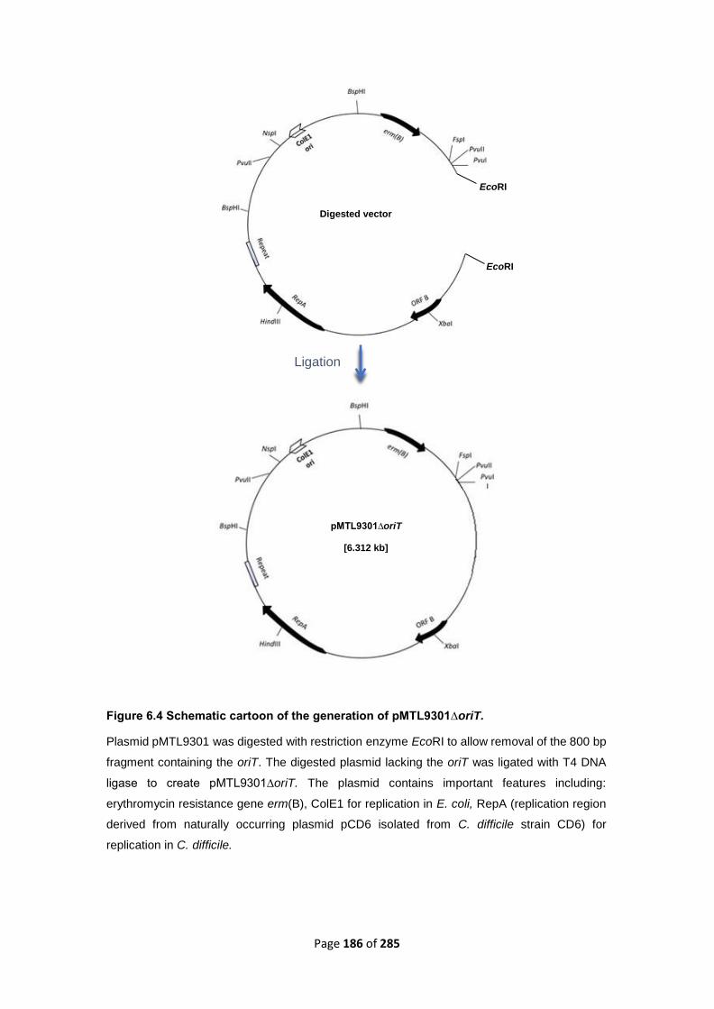
Page 186 of 285
Figure 6.4 Schematic cartoon of the generation of pMTL9301∆oriT.
Plasmid pMTL9301 was digested with restriction enzyme EcoRI to allow removal of the 800 bp
fragment containing the oriT. The digested plasmid lacking the oriT was ligated with T4 DNA
ligase to create pMTL9301∆oriT. The plasmid contains important features including:
erythromycin resistance gene erm(B), ColE1 for replication in E. coli, RepA (replication region
derived from naturally occurring plasmid pCD6 isolated from C. difficile strain CD6) for
replication in C. difficile.
Digested vector
Ligation
pMTL9301∆oriT
[6.312 kb]
EcoRI
EcoRI

Page 187 of 285
6.3.3.2. Chemical transformation
Generation of CA434 competent cells: Bacteria were grown in 100 ml LB broth in 250-
500 ml conical flasks at 37 °C with shaking to reach the OD600 ~ 0.6-1.0. Cultures were
chilled on ice for 30 minutes and centrifuged at 4000 x g for 15 minutes. Bacterial
pellets were gently resuspended in pre-chilled sterilized water and centrifuged at 4000
x g for 15 minutes. Pellets were resuspended in pre-chilled sterilized 10% glycerol
solution and centrifuged at 4000 x g for 15 minutes and then resuspended in pre-
chilled sterilized 10% glycerol solution. Bacteria were divided into 50 µl aliquots and
immediately frozen on a dry ice and ethanol bath prior to storage at -80 °C.
Competent cells of E. coli strain CA434 together with commercially prepared
competent cells of E. coli strain HB101 (Promega, USA) were transformed with wild
type plasmid pMTL9301 or ligation mixtures pMTL9301∆oriT. Briefly plasmid or
ligation mixtures were added to 50 μl of E. coli competent cells which were thawed on
ice. Bacteria were incubated on ice for 45-60 minutes before being subjected to a heat
shock at 42 °C for 45 seconds. Cells were returned to ice for 2 minutes before adding
800 µl pre-warmed super optimal broth with catabolite repression (SOC) medium.
Cultures were incubated at 37 °C with shaking (200 rpm) for 1 h in 1.5 ml tubes. Bacteria
were spread onto pre-warmed LB agar plates containing appropriate antibiotic (400
µg/ml erythromycin) before incubation at 37 °C for 16-18 h. Overnight cultures
supplemented with 200 µg/ml erythromycin were prepared from a single colony and
used to prepare glycerol stocks. Stocks were kept at -80 °C to be used for mating
experiments.

Page 188 of 285
6.3.4. Confirmation of construct
Twenty E. coli strain CA434 transformants were picked from 400 µg/ml erythromycin-
supplemented selective LB plates and inoculated into 10 ml LB broth containing 200
µg/ml erythromycin. Cultures were incubated with shaking (200 rpm) for 16 h at 37 °C.
Plasmid was extracted from 3 ml of culture using a Mini-prep kit (QIAprep Spin
Miniprep Kit, cat No. 27106) based on manufacturers instruction and eluted in molH2O.
Plasmids were assessed by restriction digest and PCR before the correct construct
was selected and stored at -20 °C or -80 °C. Plasmids pMTL9301 and pMTL9301∆oriT
were digested with EcoRI. Approximately, 1 µg of wild type or oriT-deleted plasmids
were digested with 1 µl restriction enzyme (10 U) with appropriate 1 X reaction buffer
in a final volume of 25 µl for 16 h at 37 °C water bath. Digested DNA was separated
on a 1% agarose gel with an appropriate marker; fragments were visualized on a
transilluminator after staining with GelRed Nucleic acid gel stain (Bioline, United
Kingdom).
Plasmid DNA was amplified using primer pairs (Table 6.2) in a standard PCR reaction
containing 2 µl template, 22 µl BioMixTM Red (Bioline, London, UK), 0.5 µl of each
primer in a final volume of 50 µl. PCR conditions [Denaturation: 94 °C 4 min] x 1 cycle,
[Denaturation: 94 °C 30 sec, Annealing: 50 °C-58 °C 1.5 min, Elongation: 72 °C 1
min~1 Kb product] x 25 cycles, [Elongation: 72 °C 5 min] x 1 cycle, Pause, 4 °C.
Samples were separated on a 1% agarose gel with an appropriate marker; bands were
visualised on a transilluminator after staining with GelRed. Desired constructs were
stored at -20 °C or -80 °C.

Page 189 of 285
Table 6.2 The list of primers used for analysis of the construction of pMTL9301∆oriT.
Primer pair erm(B) F and erm(B) R amplify erythromycin resistance gene carrying by pMTL9301
plasmid. ORIT-1 and ORIT-2 primer pair amplifies a sequence of 800 bp within oriT. oriT (F1050)
and oriT (R2210) primer pair amplify the region flanking the oriT and in pMTL9301∆oriT will
produce a PCR product of 300 bp. All primers synthesised by Sigma-Aldrich, UK.
Primer Sequence (5’-3’) Annealing
Tm°
Product size
erm(B) F GTCCCGGGCCTCTTGCGGGATCAAAAG 81.2 800 bp
erm(B) R GATCCGGACTCATAGAATTATTTCCTC 65.5
ORIT-1 GTGCCTTGCTCGTATC 54.7 800 bp
ORIT-2 CCTGCTTCGGGGTCATTATAG 64.4
oriT
(F1050)
GGAAAGTTACACGTTACTAAAGGG 60.7 300 bp
oriT
(R2210)
GTACTCTCTCTCTTATATATACAAATGG
55.9

Page 190 of 285
6.3.5. Mating experiments
C. difficile recipient strains CD37 and 630∆erm were grown in 20 ml BHI broth using
Coring® polystyrene tissue culture flasks with vented caps (VWR, USA) for 18-20 h in
anaerobic condition at 37 °C. These were used to start a 10 ml culture at an OD600 ~
0.1. These were grown shaking at 50 rpm for 4-6 h until the OD600 was between 0.6
and 0.8 (mid exponential phase). E. coli donor strains CA434 (HB101 carrying the
conjugative plasmid R702) and HB101 (Parent strain of CA434) were grown in 10 ml
LB broth supplemented with 200 µg/ml erythromycin for 16-18 h with shaking (200
rpm) at 37 °C. A one ml aliquot was centrifuged at 14680 x g (13000 rpm, Eppendorf
centrifuge 5415 D) for 1 minute in an Eppendorf tube and washed twice by repeating
the centrifugation step with sterile PBS to remove antibiotics.
When using B. subtilis as a donor, strains BS6A and BS34A (Table 6.1) were grown
shaking at 200 rpm in 10 ml BHI broth supplemented with tetracycline (5 µg/ml) for 18
h. Next day, the culture was grown for 3-4 h in 100 ml BHI broth supplemented with 5
µg/ml tetracycline in 500 ml flask at 37 °C until mid- exponential phase (OD600 ~ 0.45-
0.6). Cells were centrifuged at 4000 x g for 15 min and washed twice with PBS.
The harvested E. coli cells were resuspended in 0.5 ml BHI broth and mixed with 0.5
ml overnight culture of C. difficile inside the anaerobic chamber. In some cases, 50
µg/ml deoxyribonuclease I from bovine pancreas (DNase) (Sigma-Aldrich, UK) was
added to the mating mix. This was then spotted onto antibiotic free BHI plates and
incubated anaerobically for 18 h. When using B. subtilis as a donor, 100 µl of mating
mix was spread on 0.45 µm pore size nitrocellulose filters (Merck, UK) on BHI plates
and incubated anaerobically for 18 h.

Page 191 of 285
Bacteria was harvested by flooding the agar surface with 1 ml pre-reduced BHI broth
twice to ensure good recovery of transconjugants.
When B. subtilis was donor, the filters were removed from the agar plates and placed
in 20 ml tubes containing 1 ml pre-reduced BHI broth and vortexed for 10-20 seconds.
100 µl of the cell suspension was plated onto BHI agar supplemented with
erythromycin (10 µg/ml) to select for plasmids, tetracycline (10 µg/ml) to select for
Tn5397 or Tn916, D-cycloserine (250 µg/ml) and cefoxitin (8 µg/ml) to select against
E. coli or B. subtilis. Plates were incubated anaerobically for 48-72 h and examined
regularly for transconjugants. Putative transconjugants were picked and re-streaked
onto fresh selective plates containing appropriate antibiotics. Control plates were also
included in initial experiments containing E. coli, B. subtilis or C. difficile alone and
incubated alongside experimental plates to confirm there were no surviving donor cells
or spontaneous C. difficile mutants.

Page 192 of 285
Table 6.3 The list of primers used for the analysis of transconjugants/ transformants.
Locations of the primers are shown in figures 6.5, 6.6 and 6.7. Primers synthesized by Sigma-
Aldrich, UK.
Primer Sequence (5’-3’) Annealing
Tm° Product size
oriT (F1050) GGAAAGTTACACGTTACTAAAGGG 81.2 300 bp
oriT (R2210) GTACTCTCTCTCTTATATATACAAATGG 65.5
Lok 3F GAAAGAAGAACATAATTTACCAG 55.4 750 bp
Lok 1R CTGCACATCTGTATACATATAACTG 57.0
tcdB-F CAATGAACTTGTACTTCGAGATAAG 59.8 400 bp
tcdB-R CTCACCTCCATAGTTATATCTTATACGG 62.1
tet M (R smaI)
GACCCGGGACTAAGTTATTTTATTGAAC 65.9 1.2 Kb
tet M (F smaI)
GTCCCGGGAGTATATCGACCAGCAGACC 76.2
tndX F30 CTTACAATGTTAAAACAGCAAG 59.4 800 bp
tndX R1100 GTATGAAATTGCGTGAGTAGTGC 62.4
stpK F1900 CATTAATGATGATATAGACTTTACATCAGAAG 62 420 bp
stpK R1120 CAGCCGCAATTACTTTTAATC 60.2

Page 193 of 285
6.3.6. Confirmation of transfer
PCR was used to confirm the presence of plasmids or transconjugants using the
appropriate primers. In some cases, the tcdB gene or the region flanking the PaLoc
(these confirm that the transconjugants are C. difficile strain 630∆erm or strain CD37,
respectively) was amplified and sequenced. Furthermore, the stpK gene was also
amplified and sequenced to confirm the genuineness of the transconjugants using
primers listed in table 6.3. Both strains contain the stpK gene; however, there are SNPs
and INDELs that differ in this gene between CD37 and 630∆erm allowing spontaneous
mutants of the donor to be distinguished from genuine transconjugants. PCR reaction
was carried out using MyTaqTM Red Mix (Bioline, UK) as described previously in
chapter 2. PCR conditions are as follows: [Denaturation: 94 °C 4 min] x 1 cycle,
[Denaturation: 94 °C 30 sec, Annealing: 50 °C-58 °C 1.5 min, Elongation: 72 °C 1
min~1 Kb product] x 25 cycles, [Elongation: 72 °C 5 min] x 1 cycle, Pause, 4 °C.
Primers were designed with the aid of DNAMAN (Lynnon, USA) and checked using
Sigma oligonucleotides design/ordering online software (Sigma-Aldrich, UK). Location
of primers is shown in figures 6.5, 6.6 and 6.7.

Page 194 of 285
Figure 6.5 Location of primers on pMTL9301.
oriT (F1050) and oriT (R2210) primer pair
amplifies a 300 bp region flanking the oriT
in pMTL9301∆oriT and <1 Kb region in
pMTL9301. Primer pair ORIT-1 and
ORIT-2 amplifies an 800 bp region within
the oriT in pMTL9301 but does not have a
binding site in pMTL9301∆oriT.
Figure 6.6 Location of primers on pathogenicity locus (PaLoc).
A) Lok 3F and Lok 1R primer pair amplifies region flanking pathogenicity locus in toxigenic strain
630Δerm. No amplification was expected in toxigenic strains as the amplicon is over 20 Kb, too large
to be amplified under current PCR conditions. tcdB-F and tcdB-R primer pair amplifies the tcdB gene
specific to toxigenic strains. B) In non-toxigenic C. difficile strain CD37, a product of 750 bp is
amplified as the PaLoc is replaced by a 115 bp non-coding region in this strain (Braun et al., 1996).
A) PaLoc region in toxigenic strain 630
B) PaLoc region in non-toxigenic strain CD37

Page 195 of 285
Figure 6.7 Location of primer pairs onTn5397 and Tn916.
A) tndX F and tndX R primer pair amplifies a region of 700 bp within the tndX gene. B) tetM (FsmaI)
and tetM (R smaI) amplify a region of 1.8 Kb within the tet(M) gene.

Page 196 of 285
6.3.7. Plasmid pMTL9301ΔoriT fate in C. difficile
In order to make sure that plasmids pMTL9301 and pMTL9301ΔoriT remained
structurally stable during transfer into C. difficile, two millilitres of CD37 (pMTL9301) or
CD37 (pMTL9301ΔoriT) overnight cultures grown in 10 ml pre-reduced BHI broth with
5 µg/ml erythromycin for 16-18 h at 37 °C were subjected to centrifugation at 14689 x
g (13000 rpm Eppendorf centrifuge 5415 D) for 2 minutes. Bacteria were digested with
5 µg/ml lytic enzyme solution and 20 µg/ ml lysozyme for 30 minutes at 37 °C.
Plasmids were extracted using a Mini-prep kit and eluted in 30 µl molecular biology
grade water. Ten microliters of the eluates were transformed into DH5α competent
cells (Table 1) (see chapter 2 for the protocol). Transformants were plated onto LB
agar with 400 µg/ml erythromycin and incubated at 37 °C for 16 h. Five to ten colonies
were selected and inoculated into 5 ml LB broth containing 200 µl/ml erythromycin,
followed by an overnight incubation at 37 °C with shaking at 200 rpm. Plasmid was
extracted from 3 ml of culture using the Mini-prep kit and eluted in 30 µl molecular
biology grade water. Plasmid DNA from CD37 (pMTL9301) and CD37
(pMTL9301ΔoriT) was also isolated by making whole genome DNA preparation
(Puregene Yeast/Bact.KitB, Qiagen, UK) (see chapter 2 for the protocol), then using
this to transform E. coli strain DH5α competent cells. Plasmids were subsequently
isolated from this strain as described above. Isolated plasmids were subjected to
digestion with EcoRI for 18 h at 37 °C water bath. The digest was separated on a 1%
agarose gel with an appropriate marker. PCR was performed on pMTL9301,
pMTL9301ΔoriT before transfer and pMTL9301ΔoriT after transfer. Four microliters of
plasmid templates were mixed with 1 µl primers oriT (F1050) and oriT (R2210) (Table
6.3), 50 µl MyTaqTM Red Mix and 44 µl water to reach the total volume of 100 µl
reaction.

Page 197 of 285
PCR was carried out under the following conditions: [Denaturation: 94 °C 4 min] x 1
cycle, [Denaturation: 94 °C 30 sec, Annealing: 50 °C 1.5 min, Elongation: 72 °C 1 min]
x 25 cycles [Elongation: 72 °C 5 min] x1, pause 4 °C. Two microliters of the PCR
reaction was used for gel electrophoresis and a 300 bp band representing region
flanking the oriT in pMTL9301ΔoriT was observed. A second PCR was performed on
the whole content of PCR reaction (100 µl) to amplify the oriT flanking region in
pMTL9301ΔoriT (before and after transfer). PCR product was extracted and
sequenced at (GENEWIZ, Essex, UK) to determine whether the oriT flanking region
remained intact before and after pMTL9301ΔoriT transfer.
6.3.8. Bioinformatics Search for putative competence
genes in C. difficile
6.3.8.1. Search for competence genes in C. difficile 630
Whole genome sequencing of B. subtilis reveals a number of putative competence
(com) genes involved in transformation pathways for which the biological activities
have been shown (Hamoen et al., 2003). The sequences of the known functional com
genes deposited in GenBank were used here to search for competence genes in C.
difficile. The presence of the com genes in C. difficile was predicted based on DNA
sequence homology alignment algorithms such as BLASTx and BLASTn (Altschul et
al., 1990) using C. difficile 630 whole genome annotated sequence as a reference
(query) (Sebaihia et al., 2006). To predict the biological function of a com gene more
accurately another approach was to search with the amino acid sequence of the
predicted protein using protein prediction tool BLASTp (Altschul et al., 1990).

Page 198 of 285
However, sequences that are identified with the highest E-values do not necessarily
have the most relevant predicted biological function. High E-values in BLAST searches
may be caused by a common ancestry of the proteins or structural similarities rather
than similar biological function (Tian & Skolnick, 2003). The hits of the search
determining the chance that a homologue with a similar predicted function as the query
were analysed using NCBI Open Reading Frame finder (Wheeler et al., 2005) and the
SMART tool that can recognise conserved protein domains from various databases.
The presence of conserved domains in an amino acid sequence can help to support
the predicted function of the protein.
6.3.8.2. Search for the com genes in CD37
PCRs were carried out using primers to amplify regions within the comEA, cinA, ftsK
and CDftsK genes (Wasels et al., 2015) using B. subtilis and 630Δerm as positive
controls. Primers were designed manually, checked for specificity in NCBI/ Primer-
BLAST online software and synthesized by Sigma-Aldrich, UK. Primers are listed in
table 6.4. PCR reactions were carried out using MyTaqTM Red Mix (Bioline, UK) as
described previously under the following condition: [94 °C 4 min] x 1 cycle, [94 °C 30
sec, 55 °C 1.5 min, 72 °C 1 min] x 25 cycles [72 °C 5 min] x1, hold 4 °C. Annealing
temperatures and extension times were adjusted according to the primer set and
expected product length (Table 6.4). PCR products containing amplified regions of the
comEA, cinA and ftsK genes were purified using QIAquick PCR Purification Kit
(QIAGEN, UK) and sequenced at GENEWIZ (Essex, UK).

Page 199 of 285
Table 6.4 The list of primers used in this study to detect competence genes in C. difficile.
Primer pairs to detect (i) the comEA, cinA, ftsK and CDftsK genes in C. difficile strains CD37 and
630∆erm, (ii) the cat (P) gene harboring by pRPF185 in E. coli donors are shown in this table.
Primers were designed by Sigma-Aldrich.
Primers Sequence (5’-3’) Annealing Tmᵒ
Product size
comEA F GTGGAGCAGTAAATAGACCTGGA 63.2 328 bp
comEA R CCAACCCCAGGTAGAGAATCC 65.6
cinA F AAGTCCTTTAGCAACGCCCT 63.8 842 bp
cinA R TTGGGCCAACTGGTGATGAT 67.6
ftsK F ATTGAAGCCCCAATTCCTGGT 67.5 1 kb
ftsK R GAGGCAGATGCTTGACCAC 63.6
CD ftsK F GCTGGTTGTCGGACTGTTTC 64.7 770 bp
CD ftsk R AACGGTCAATGCAGGAAAGC 66.2
Cat P (F) CCTGCCACTCATCGCAGT 58.5 190 bp
Cat P (R) CCACCGTTGATATATCCC 60
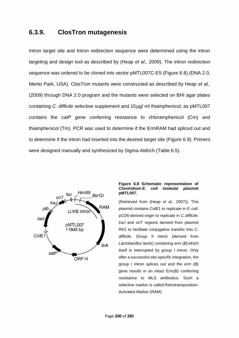
Page 200 of 285
6.3.9. ClosTron mutagenesis
Intron target site and Intron redirection sequence were determined using the intron
targeting and design tool as described by (Heap et al., 2009). The intron redirection
sequence was ordered to be cloned into vector pMTL007C-E5 (Figure 6.8) (DNA 2.0,
Menlo Park, USA). ClosTron mutants were constructed as described by Heap et al.,
(2009) through DNA 2.0 program and the mutants were selected on BHI agar plates
containing C. difficile selective supplement and 15 µg/ ml thiamphenicol, as pMTL007
contains the catP gene conferring resistance to chloramphenicol (Cm) and
thiamphenicol (Tm). PCR was used to determine if the ErmRAM had spliced out and
to determine if the intron had inserted into the desired target site (Figure 6.9). Primers
were designed manually and synthesized by Sigma-Aldrich (Table 6.5).
Figure 6.8 Schematic representation of Clostridium-E. coli modular plasmid pMTL007.
(Retrieved from (Heap et al., 2007)). This
plasmid contains ColE1 to replicate in E. coli.
pCD6-derived origin to replicate in C. difficile.
traJ and oriT regions derived from plasmid
RK2 to facilitate conjugative transfer into C.
difficile. Group II intron (derived from
Lactobacillus lactis) containing erm (B) which
itself is interrupted by group I intron. Only
after a successful site-specific integration, the
group I intron splices out and the erm (B)
gene results in an intact Erm(B) conferring
resistance to MLS antibiotics. Such a
selective marker is called Retrotransposition-
Activated Marker (RAM).

Page 201 of 285
Figure 6.9 PCR screening for intron integration.
The intron on the plasmid is represented by the upper yellow arrow. The intron integrated in the
target site is shown by the lower yellow arrow. The red arrows show the interrupted target site. The
blue arrows represent the ErmRAM, which in the upper schematic is interrupted by the group I
intron, shown as a grey arrow. PCR primers are represented by black half-arrows (Table 6.5).
[Reproduced from the protocol for Clostridial Gene Knockout using pMTL007 (The Minton
Laboratory UoN, 2006)].
Plasmids harbouring ClosTron-based mutation in the comEA, cinA and ftsK genes
were recovered from GFC filters according the manufacturer’s instruction (DNA 2.0,
USA). Recovered plasmids were run on 1% agarose gel to make sure they were not
degraded. The concentration of plasmids was measured using a NanoDrop 1000
spectrophotometer (Thermo Fisher Scientific, UK). Plasmids were transformed into E.
coli strain DH5α commercially prepared competent cells and stored at -80 °C.
Gene transfer experiments were performed as described by Heap et al., (2009).
Briefly, ClosTron mutants were transformed into chemically competent CA434 donor
cells. Transformants appeared on LB plates supplemented with 25 µg/ml
chloramphenicol. PCR was performed to ensure successful transformation.
Transformants were grown for 18 h in LB broth containing 12.5 µg/ ml
chloramphenicol, washed with sterile PBS and the harvested pellet was mixed with
CD37 or 630Δerm recipient (0D600 ~ 0.8).

Page 202 of 285
Serial dilutions of the growth harvested from BHI mating plates were set up to determine
the cell counts of donor and recipient cells. Donor cells containing the ClosTron
insertion were selected on chloramphenicol (25 μg/ml) supplemented LB plates. C.
difficile CD37 recipient cells were selected on rifampicin (25 μg/ml) supplemented BHI
plates. C. difficile 630Δerm recipient cells were selected on tetracycline (10 µg/ml)
supplemented BHI plates. CD37 transconjugants were selected on thiamphenicol (15
μg/ml) and rifampicin (25 μg/ml). 630Δerm transconjugants were selected on
thiamphenicol (15 μg/ml) and tetracycline (10 µg/ml). Identification of the
transconjugants was confirmed by two sets of PCRs, one using a primer pair F + R to
amplify the wild type gene with the RAM insert to give a band of 4 Kb and the other
using a primer pair F + (exon binding) EBS universal to amplify the wild type gene just
upstream of the ermRAM to give a band of 0.4 Kb (Table 6.5).
To investigate the role of the com genes in DNase-sensitive conjugation-like
mechanism in C. difficile, pRPF185 (Table 6.1,) was transformed into chemically
competent CA434 cells and commercially prepared competent HB101 cells (Table
6.1), alongside two control transformations using pMTL9301 and pUC19. Plasmids
were extracted using QIAprep SpinMiniprep Kit (QIAGEN, UK) and digested with SmaI
and BamHI restriction enzymes to allow assessment of the plasmid quality. Two bands
were expected in both cases, pMTL9301 produced fragments of the expected size
(1700 bp and 5412 bp), and pRPF185 produced fragments of the expected size (1866
bp and 7185 bp). Conjugation was performed as described by Heap et al., 2009. Briefly,
E. coli strains CA434 and HB101 were used as donor strains for pRPF185. CD37,
CD37∆comEA::ErmRAM, CD37∆cinA::ErmRAM, 630∆erm,
630∆erm∆comEA::ErmRAM and 630∆erm∆cinA::ErmRAM were used as recipients.
DNase (50 µg/ml) was added to the mating mix when required.

Page 203 of 285
Table 6.5 The list of primers used for ClosTron mutagenesis.
Target (F) and (R) primer pairs were designed manually specific for each target gene, checked by
DNAMAN and synthesized by Sigma-Aldrich, UK. EBS universal primer is common to all PCRs for
intron targeting. The EBS primer was designed by Heap et al., (2009) and synthesized by Sigma-
Aldrich. Locations of the primers are shown in figure 6.9.
Primers Sequence (5’-3’) Annealing Tmᵒ
cinA closT (F) CAGGTGGTCTTGGGCCAACTGGTG 76.1
cinA closT (R) GCAACTGTTGGATTAGTCTGCTC 63.9
comEA closT (F) CGTTTATAAATCGCTTATTTTCAAAGATG 64.9
comEA closT (R) GATAGATTGACTCCATTTAAATCTGC 61.8
ftsk closT (F) GGTATAGCAGGTGGATGGTTAATAAG 63.3
ftsk closT (R) CCTCATTAACTGGCTCTGCTAC 61.7
EBS universal CGAAATTAGAAACTTGCGTTCAGTAAAC 60

Page 204 of 285
6.3.10. Investigation of the potential influence of the
transfer genes on plasmid RK2 on DNase sensitive
oriT-independent plasmid transfer into C. difficile
Bioinformatic search for transfer genes in E. coli strain HB101
The sequencing of the Birmingham IncP-alpha plasmid RK2 (GeneBank Accession
number: BN000925) revealed a number of putative tra and trb genes involved in
conjugation (Pansegrau et al., 1994). All these annotated transfer genes in the
sequence of RK2 were studied in E. coli strain RR1 (HB101 RecA+) (Jeong et al.,
2017) at the nucleotide level using BLASTn and amino acid level using BLASTp
(Altschul et al., 1990).
Another approach to verify the presence of the tra and trb genes in the HB101 genome
is to perform standard PCR with gene specific primers. All the tra and trb genes were
subjected to PCR using primers listed in table 6.6. Primers were designed manually
specifically to amplify regions inside the target genes, checked with NCBI/ Primer-
BLAST and synthesized by Sigma-Aldrich, UK. PCR conditions were as follows: [ 94
°C 4 min] x 1 cycle, [94 °C 30 sec, 55 °C 1.5 min, 72 °C 1 min] x 25 cycles [72 °C 5
min] x 1 cycle, hold 4 °C. Samples were separated on a 1% agarose gel with an
appropriate marker. Bands were visualized on a transilluminator after staining with
GelRed.

Page 205 of 285
Table 6.6 The list of primers used for detecting the tra and trb genes in E. coli strain HB101 (parent strain of CA434).
Primers Sequence (5’-3’) Annealing Tmᵒ Product size
traL F GCGCAGTACAAGATGGACAA 63.9 237 bp
traL R GCTGATGAGGTAATGCGACA 63.9
traK F CTCGTCACCATTTGGGAGCA 68.5 405 bp
traK R CCTTTTTGTCCGGTGTTGGGG 68.4
traF F CACCAAAAGCATTCCGGTCG 69.6 388 bp
traF R ATTGACAGGCCCGAAGTAGC 65.3
traG F CAGAACCTTGCCACCCTGAT 66.3 799 bp
traG R TCGTGGTGCCTGTCAGTTTT 65.7
traM F TGAAGAGCTGATCCGGGAGA 67.4 265 bp
traM R GGCGCTGTCCTTCATTACCT 65.3
traI F GCGGAAACGGCTTCATCTTC 68 917 bp
traI R TCCGTCATTTCGTTCGGGTT 68.6
traJ F GGTGTACTGCCTTCCAGACG 64.7 133 bp
traJ R GACGTGCTCATAGTCCACGA 64
trbA F CAACATCCTTCGTCTGCTCG 65.9 274 bp
trbA R CGGCAAAACCACACTGATGC 68.8
trbB F CAGGCGATTATAGAAACGGT 60.5 762 bp

Page 206 of 285
trbB R TTACAGGGTTTTGGTGATGT 59.3
trbC F GAATCGCGGCATTTTGTTC 65.4 240 bp
trbC R ATCAGGGTTCGGAAGAAGG 63
trbD F AACCGAGAAAACCTGTTCAT 60 270 bp
trbD R GTATTGCTTCCCTTGGCTAT 59.6
trbE F CTCATCAACACGATGAACCT 60.2 689 bp
trbE R TTCATCAGCTCTTCGATCTC 59.7
trbF F GGTGACGCCGGACGTAGCTT 70.6 247 bp
trbF R GCCTTTCACCACGCCTTGCC 73.1
trbG F CAAGTCAACTCGATCAACCT 59.3 531 bp
trbG R AATCGTATCGACGATGTAGC 59.2
trbH F CCGCCTACAACCAGACCATT 66 308 bp
trbH R CATGACGGTCAGGCGATACA 67.1
trbI F GCGAAGATCAAATGGCACCG 69.8 455 bp
trbI R CTTGATCGCCTCTTCCAGCA 67.8
trbJ F GAGGAATACCGGACGCAGTT 65.2 298 bp
trbJ R CGTTGGCCTTTTTCTGCGAT 68.2
trbL F TGGAGTGATGCTGATCGTCG 67.6 679 bp
trbL R CCTGTTGTTGCTTGCGGAAA 68.2
trbM F AGCTGGGACGACCAGTTCTA 63.8 215 bp

Page 207 of 285
trbM R CCATAGCGACCGTACTGCTG 65.6
trbN F TCGCCATTCGGGAAAAGGAG 70.3 249 bp
trbN R TGCGCGAGTGATAGTTAGCG 66.3
trbP F TTCATTGCGTTGGGTGGAGT 67.7 386 bp
trbP R CAAAGAGCGGCAAGATGCAG 68.2

Page 208 of 285
6.4. Results
6.4.1. DNase sensitive plasmid transfer into C. difficile
Results shown in the previous chapter revealed that the frequency of pMTL9301
transfer from E. coli strain CA434 into C. difficile CD37 was significantly decreased
when DNase was present in the mating mixture (from 10-7 to 10-9 per donor). To gain
a deeper insight into these observations the following experiments were performed, all
under the same conditions.
6.4.1.1. Using purified plasmid DNA in liquid culture of C.
difficile did not result in transformation
Results obtained from chapter 5 implied that pMTL9301 might be transferring from E.
coli to C. difficile by transformation. To investigate this further, 5, 10, 15 and 20 µg/ml
of pMTL9301 was added to CD37 overnight culture (OD600 ~ 0.8). No erythromycin
resistant (ErmR) transformants were obtained when DNA was used to treat 108 CFU/
ml CD37 cells (Table 6.7).
6.4.1.2. Transfer of free plasmid DNA into CD37 did not occur in
the presence of plasmid- free CA434
Purified plasmid DNA in various concentrations (5, 10, 15 and 20 µg/ml) was added to
the mixture of CD37 recipient and plasmid-free CA434. As shown in table 6.7, no ErmR
transformants was detected after 24-48 h. This indicates that the presence of donor
cells harbouring the plasmid is required for its transfer into CD37 and the combination

Page 209 of 285
of plasmid-free CA434 and free plasmid is not sufficient for transfer. This experiment
was performed under the same conditions as when pMTL9301-harbouring CA434 was
used.
6.4.1.3. Using heat-killed donor or donor culture supernatant/
filtrate did not result in plasmid transfer into CD37
To examine whether a donor-secreted component in LB broth stimulates the plasmid
transfer into CD37, we performed the following two experiments: (i) Exposure of the
donor culture to heat (70 °C) for 30 minutes, (ii) Passing the donor overnight culture
through 0.22 µm-pore membrane filters to have a cell-free supernatant/ filtrate. These
experiments did not result in the formation of ErmR transformants on selective plates
(Table 6.7). Therefore, the presence of live donors harbouring the plasmid is required
for the DNase sensitive plasmid transfer into C. difficile.

Page 210 of 285
Table 6.7 Direct transformation of C. difficile CD37 with free plasmid pMTL9301. ErmR: erythromycin resistant.
Effect of media
conditioned with E.
coli and/or plasmid
Recipient Viable count of
recipient (CFU/ ml)
Number of
transformants (ErmR
colonies on selective
plates)
Purified plasmid CD37 1.67 ± 1.44 x 10⁸ None
Plasmid-free CA434
+ Plasmid
CD37 1.15 ± 1.17 x 10⁸ None
Heat-killed donor + 4
µg purified plasmid
CD37 1.2 ± 1.28 x 10⁸ None
Donor filtrate + 4 µg
purified plasmid
CD37 1.64 ± 1.58 x 10⁸ None

Page 211 of 285
6.4.1.4. Plasmid transfer into C. difficile requires close contact
between live donor cells and recipients, and DNase treatment
only reduces the transfer frequency of pMTL9301 into CD37
but has no effect on the transfer frequency to C. difficile
630Δerm
The effect of DNase on plasmid transfer into C. difficile was investigated using strain
630Δerm as well as CD37 to determine whether the DNase-sensitive transformation-
like mechanism is a recipient-dependent phenomenon. Plasmid pMTL9301 was
transferred from E. coli strain CA434 to C. difficile CD37 at a frequency of around 3.26
× 10-5 transconjugants per donor and to C. difficile 630Δerm at a frequency of around
3 × 10⁻⁵ transconjugants per donor. Incorporation of DNase into the mating mix prior
to plating onto selective agar resulted in the frequency of transfer decreasing by 5
orders of magnitude to around 10-10 transconjugants per donor for CD37 but no change
in transfer frequency was observed when 630Δerm was used as the recipient (Table
6.8) (Figure 6.12).

Page 212 of 285
6.4.2. A complete oriT is not required for plasmid
transfer into C. difficile
6.4.2.1. pMTL9301∆oriT construction and validation
In order to determine if oriT is required for the transfer of plasmid pMTL9301 DNA into
C. difficile, the oriT and nic sites from pMTL9301 were deleted (for methods see
section 6.3.3). Transformants were detected by PCR (Figures 6.10) and sequencing
(Figure 6.11).
Figure 6.10 PCR amplification to confirm the oriT deletion.
Lanes 1, 2 and 3 are PCR amplification of the oriT using primer pairs ORIT-1 and ORIT-2, no band
suggests the successful deletion of the oriT. Lanes 4, 5 and 6 are PCR amplification of the region
flanking the oriT using primers oriT(F1050) and oriT(R2210), in pMTL9301ΔoriT it shows a product
approximately 300 bp. Lane M is 1 Kb molecular ladder.

Page 213 of 285
901 TACTAAAAAT CAGTTTCATC AAGCAATGAA ACACGCCAAA GTAAACAATT TAAGTACCGT
961 TACTTATGAG CAAGTATTGT CTATTTTTAA TAGTTATCTA TTATTTAACG GGAGGAAATA
1021 ATTCTATGAG TCGCTTTTGT AAATTTGGAA AGTTACACGT TACTAAAGGG AATGTAGATA
1081 AATTATTAGG TATACTACTG ACAGCTTCCA AGGAGCTAAA GAGGTCAGGC TGCGCAACTG
1141 TTGGGAAGGG CGATCGGTGC GGGCCTCTTC GCTATTACGC CAGCTGATAA GGCCTTATTG
1201 TGGAATATTG AAGGGGAATT CGCCCTTCCT GCTTCGGGGT CATTATAGCG ATTTTTTCGG
1261 TATATCCATC CTTTTTCGCA CGATATACAG GATTTTGCCA AAGGGTTCGT GTAGACTTTC
1321 CTTGGTGTAT CCAACGGCGT CAGCCGGGCA GGATAGGTGA AGTAGGCCCA CCCGCGAGCG
1381 GGTGTTCCTT CTTCACTGTC CCTTATTCGC ACCTGGCGGT GCTCAACGGG AATCCTGCTC
1441 TGCGAGGCTG GCCGGCTACC GCCGGCGTAA CAGATGAGGG CAAGCGGATG GCTGATGAAA
1501
CCAAGCCAAC
CAGGAAGGGC
AGCCCACCTA
TCAAGGTGTA
CTGCCTTCCA
GACGAACGAA
1561 GAGCGATTGA GGAAAAGGCG GCGGCGGCCG GCATGAGCCT GTCGGCCTAC CTGCTGGCCG
1621 TCGGCCAGGG CTACAAAATC ACGGGCGTCG TGGACTATGA GCACGTCCGC GAGCTGGCCC
1681 GCATCAATGG CGACCTGGGC CGCCTGGGCG GCCTGCTGAA ACTCTGGCTC ACCGACGACC
1741 CGCGCACGGC GCGGTTCGGT GATGCCACGA TCCTCGCCCT GCTGGCGAAG ATCGAAGAGA
Figure 6.11 Sequencing results of the 800 bp region deleted from pMTL9301 using EcoRI restriction enzyme in order to construct pMTL9301∆oriT.
The sequence of the oriT is highlighted in green (~112 bp). The inverted repeat sequence
(IR~19bp) within the nic site (recognized by the conjugative apparatus of plasmid R702) is in
purple and underlined. The sequencing result shows that the oriT is successfully removed from
the plasmid. No obvious homologs to the oriT or the IR sequence were found elsewhere on the
plasmid backbone.

Page 214 of 285
6.4.2.2. Deletion of the oriT from pMTL9301 does not abolish
transfer from E. coli to C. difficile but does abolish transfer to
strain CD37 in the presence of DNase
The fact that pMTL9301 transfer to CD37 is drastically reduced in the presence of
DNase (chapter 5) indicates that as well as transferring by conjugation, the plasmid
may also be entering C. difficile by a transformation-like mechanism. To investigate
this further, we deleted the oriT region from pMTL9301 to generate pMTL9301ΔoriT
(Figure 6.4). This plasmid transferred from E. coli strain CA434 in the absence of
DNase to CD37 and 630Δerm at frequency of approximately 10-10 per donor, a much
lower frequency than observed with pMTL9301 (Table 6.8). No transfer (the detection
limit was <10-10 transconjugants per donor or recipient) was observed to CD37 when
DNase was included in the medium, although transfer to 630Δerm was still observed
(Table 6.8). That pMTL9301 and pMTL9301ΔoriT were transferred to C. difficile CD37
and 630Δerm was confirmed by preparing plasmids from representative
transconjugants and demonstrating that they contain either intact oriT (pMTL9301) or
had this region deleted in the case of transconjugants containing pMTL9301ΔoriT
(Figure 6.12). Transfer of pMTL9301ΔoriT into C. difficile was verified by DNA
sequencing and restriction mapping (Figure 6.13). That the transconjugants were
genuine C. difficile strains and not E. coli spontaneous mutants were confirmed by
PCR and sequencing (as described in the methods). Furthermore, the transconjugants
could not grow aerobically indicating that they are not E. coli spontaneous mutants.
Plasmid fate was also investigated by extracting pMTL9301ΔoriT from C. difficile
transconjugants/ transformants and PCR amplification (Figure 6.14).

Page 215 of 285
6.4.2.3. Deletion of the oriT from pMTL9301 does not abolish
transfer from E. coli strain HB101 to C. difficile 630∆erm but
does abolish transfer to CD37 in the absence of DNase.
According to Purdy et al., (2002), CA434 was made by transferring the conjugative
plasmid R702 into HB101 (Table 6.1). Therefore, in order to investigate the role of
R702 in the transfer of pMTL9301 and pMTL9301ΔoriT, HB101 was used as a donor
strain. Plasmid pMTL9301 transferred from this host to 630Δerm at a frequency of
around 10-9 per recipient and 10-10 per donor and at a frequency of 10-9 per recipient
and 10-10 per donor to CD37 (Figure 6.12) (Table 6.8). When HB101 containing
pMTL9301ΔoriT was used as the donor and 630Δerm the recipient, transconjugants
appeared at a similar frequency, but no transconjugants were observed when CD37
was the recipient (Figure 6.12). When DNase was incorporated in the media, no
transconjugants were obtained in any of the above combinations of recipients and
donors (Table 6.8). The detection limit in these experiments was <10-10
transconjugants per donor or recipient. That transconjugants/ transformants were
genuine was confirmed as described above and in the methods section.

Page 216 of 285
Strains Frequency of transfer/ donor Frequency of transfer/
recipient
Donor Recipient No DNase DNase (50
µg/ml)
No DNase DNase (50
µg/ml)
CA434
(pMTL9301)
CD37 3.26 ± 1.24
ˣ 10-5
3.67 ± 1.41 ˣ
10-10 (a)
3.32 ± 4.71 ˣ 10-6
7.53 ± 4.71 ˣ
10-10 (a)
CA434
(pMTL9301
ΔoriT)
CD37 4.68 ± 1.24
ˣ 10-10 (b)
ND 6.65 ± 3.48 ˣ
10-10 (b)
ND
CA434
(pMTL9301)
630Δerm 3 ± 0.47 ˣ
10-5
4.5 ± 0.47 ˣ
10-5
1.62 ± 0.04 ˣ
10-5
1.8 ± 0.04 ˣ
10-5
CA434
(pMTL9301
ΔoriT)
630Δerm 1.98 ± 1.13
ˣ 10-10 (c)
1.67 ± 1.13 ˣ
10-10 (c)
4.6 ± 1.5 ˣ 10-9
(c)
6.2 ± 1.5 ˣ
10-9 (c)
HB101
(pMTL9301)
CD37 4.3 ± 2 ˣ
10-10 (d)
ND 5.67 ± 2.85 ˣ
10-9 (d)
ND

Page 217 of 285
HB101
(pMTL9301
ΔoriT)
CD37 ND ND ND ND
HB101
(pMTL9301)
630Δerm 4.67 ± 1.1 ˣ
10-10 (e)
ND 4.09 ± 1.01 ˣ
10-9 (e)
ND
HB101
(pMTL9301
ΔoriT)
630Δerm 2.08 ± (0) ˣ
10-10 (f)
ND 2.3 ± (0) ˣ 10-9
(f)
ND
Table 6.8 Frequency of pMTL9301 and pMTL9301ΔoriT transfer into C. difficile (±SD).
SD, data were expressed as standard deviation (SD) based on at least three independent
experiments. ND, no transconjugants/ transformants detected after mating, i.e. a conjugation
frequency below the detection limit (<10-10).
Each experiment was repeated at least three times on separate occasions. All colonies obtained
were verified using PCR (as described in the Methods and Results section) to confirm that they
were indeed the correct strain harbouring the expected plasmid. a) C. difficile strain CD37
(pMTL9301) transconjugants/ transformants arose at a frequency of 10–10 had 9–16 colonies per
mating. b) C. difficile strain CD37 (pMTL9301ΔoriT) transconjugants/ transformants arose at a
frequency of 10–10 had 1–3 colonies per mating. c) C. difficile strain 630Δerm (pMTL9301ΔoriT)
transconjugants/ transformants arose at a frequency of 10–10 to 10−9 had 3–12 colonies per mating.
d) C. difficile strain CD37 (pMTL9301ΔoriT) transconjugants/ transformants arose at a frequency of
10–10 to 10−9 had 1–3 colonies per mating. e) C. difficile strain 630Δerm (pMTL9301)
transconjugants/ transformants arose at a frequency of 10–10 to 10−9 had 1–3 colonies per mating.
f) C. difficile strain 630Δerm (pMTL9301ΔoriT) transconjugants/ transformants arose at a frequency
of 10–10 to 10−9 had 1–3 colonies per mating.
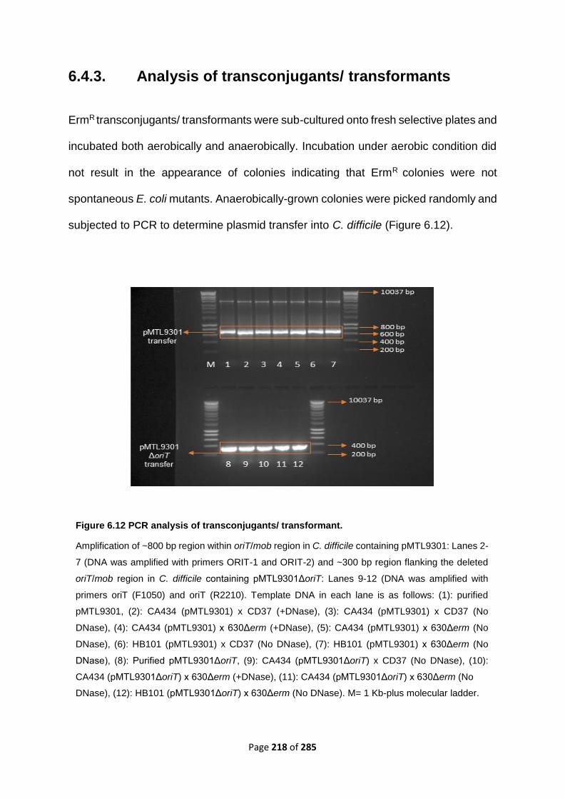
Page 218 of 285
6.4.3. Analysis of transconjugants/ transformants
ErmR transconjugants/ transformants were sub-cultured onto fresh selective plates and
incubated both aerobically and anaerobically. Incubation under aerobic condition did
not result in the appearance of colonies indicating that ErmR colonies were not
spontaneous E. coli mutants. Anaerobically-grown colonies were picked randomly and
subjected to PCR to determine plasmid transfer into C. difficile (Figure 6.12).
Figure 6.12 PCR analysis of transconjugants/ transformant.
Amplification of ~800 bp region within oriT/mob region in C. difficile containing pMTL9301: Lanes 2-
7 (DNA was amplified with primers ORIT-1 and ORIT-2) and ~300 bp region flanking the deleted
oriT/mob region in C. difficile containing pMTL9301ΔoriT: Lanes 9-12 (DNA was amplified with
primers oriT (F1050) and oriT (R2210). Template DNA in each lane is as follows: (1): purified
pMTL9301, (2): CA434 (pMTL9301) x CD37 (+DNase), (3): CA434 (pMTL9301) x CD37 (No
DNase), (4): CA434 (pMTL9301) x 630Δerm (+DNase), (5): CA434 (pMTL9301) x 630Δerm (No
DNase), (6): HB101 (pMTL9301) x CD37 (No DNase), (7): HB101 (pMTL9301) x 630Δerm (No
DNase), (8): Purified pMTL9301ΔoriT, (9): CA434 (pMTL9301ΔoriT) x CD37 (No DNase), (10):
CA434 (pMTL9301ΔoriT) x 630Δerm (+DNase), (11): CA434 (pMTL9301ΔoriT) x 630Δerm (No
DNase), (12): HB101 (pMTL9301ΔoriT) x 630Δerm (No DNase). M= 1 Kb-plus molecular ladder.

Page 219 of 285
Restriction digestion of
pMTL9301∆oriT extracted
from C. difficile
transconjugant/
transformant ~ 800 bp
Figure 6.13 HindIII and XbaI digestion of the plasmids pMTL9301 and pMTL9301∆oriT extracted from putative transconjugants/ transformants.
Lane 1: Uncut pMTL9301. Lane 2: pMTL9301 digested with HindIII and XbaI resulted in two bands
(~ 4000 bp and ~ 3000 bp). Lane 3: Uncut pMTL9301∆oriT, Lane 4: pMTL9301∆oriT digested
with HindIII and XbaI resulted in two bands (~ 3000 bp and ~ 2500 bp)
Uncut pMTL9301
(+ control)
Figure 6.14 EcoRI digestions of pMTL9301 and pMTL9301ΔoriT shows that plasmid remained structurally stable before and after transfer.
Lane 3: uncut pMTL9301 before transfer. Lane 4: cut pMTL9301 before transfer. Group 1:
digested pMTL9301 extracted from C. difficile transconjugants/ transformants (800 bp oriT is
isolated from pMTL9301). Lane 5: uncut pMTL9301ΔoriT before transfer. Lane 6: cut
pMTL9301ΔoriT before transfer. Group 2: digested pMTL9301ΔoriT extracted from C. difficile
transconjugants/ transformants (the 800 bp EcoRI fragment bearing oriT was removed during
plasmid construction (see section 6.4.2.1)). M= 1 Kb ladder. Digestion patterns did not show any
difference before and after transfer.
Group 1 Group 2
molH2O
oriT ~ 0.8 Kb

Page 220 of 285
6.4.4. Transfer of Tn5397 and Tn916 from Bacillus
subtilis is not affected by DNase treatment
In order to test if the DNase sensitive transfer is a more general phenomenon, we
examined the transfer of Tn5397 and Tn916 both carrying tetracycline resistance gene
from B. subtilis to C. difficile CD37. Tn916 and Tn5397 containing transconjugants
were obtained at a frequency of around 1 × 10⁻⁷ and 1 × 10⁻⁸ transconjugants per donor,
respectively very similar to previously reported transfer frequencies for these elements
(Mullany et al., 1990, Mullany et al., 1994). This was the same in both the presence
and absence of DNase (data not shown). DNase-resistant conjugative transfer of
Tn5397 and Tn916 was confirmed by PCR using primers summarised in table 6.3. The
location of primers is illustrated in figure 6.7.

Page 221 of 285
6.4.5. Investigation of the potential influence of the com
genes on DNase-sensitive plasmid transfer into C.
difficile
6.4.5.1. Bioinformatic Search for putative competence genes in
C. difficile
In order to further investigate the DNase sensitive oriT-independent plasmid transfer
into C. difficile, we decided to look for possible competence (com) genes, potentially
involved in the transformation-like mechanism observed. Analysing the whole genome
sequence of C. difficile 630 (Genebank AM180355) (Sebaihia et al., 2006) using DNA
sequence homology alignment algorithms such as BLASTx and BLASTn showed that
three putative competence genes including comEA, cinA and ftsK in B. subtilis genome
are present in CD630 with 99%, 100% and 99% similarity, respectively. BLASTp was
also used with translated coding sequences of these genes against B. subtilis
reference sequence for searching protein databases. The comEA-encoded ComEA
competence protein, cinA-encoded competence damage-inducible protein A and ftsK-
encoded DNA Ftsk/SpoIII translocase proteins are all candidate proteins for DNA
translocation in C. difficile. A search for the presence of other com genes (Hamoen et
al., 2003) in C. difficile was also carried out using BLASTn and BLASTp; however, no
homologues were found for these (Table 6.9).
Therefore, the comEA, cinA, and ftsk genes with the following locus tags:
CD630_24970, CD630_14090 and CD630_13240, respectively were chosen for
further investigations to look for potential competence proteins mediating the DNase
sensitive oriT-independent plasmid transfer into C. difficile.

Page 222 of 285
Coding sequences (CDS) of these putative genes were predicted using NCBI Open
Reading Frame (ORF) finder and the length of each gene was as follows: comEA (705
bp), cinA (1257 bp) and ftsK (2412 bp). Primers were designed to amplify each gene
(Table 6.4) and PCR was carried out to confirm their presence in C. difficile strains
CD37 and 630Δerm (Figure 6.15). Sequencing of the PCR products confirmed that
these genes are present.
Figure 6.15 PCR amplification of the comEA, cinA and ftsK genes in C. difficile.
Lane 1: molecular biology grade water. Lane 2: comEA in 630Δerm. Lane 3 and 4: comEA in CD37.
Lane 5: cinA in 630Δerm. Lane 6 and 7: cinA in CD37. Lane 8: ftsK in 630Δerm. Lane 9 and 10: ftsK
in CD37. Lane 11: CDftsK in 630Δerm. Lane 12 and 13: CDftsK not found in CD37. Lane M: 1 Kb
molecular ladder.

Page 223 of 285
Table 6.9 Bioinformatic search for putative competence genes in strain 630.
Presence of B. subtilis competence related genes was investigated in CD630 using DNA
sequence homology alignment algorithms such as BLASTp and BLASTn (the comEA, cinA and
ftsK genes, presumably involved in DNA translocation processes in C. difficile are not shown in
this table as they have been discussed above)
Gene BLASTn BLASTp Description
comK No similarity No similarity Competence transcription
factor, ComK activated
proteins that directly
function in DNA uptake.
comS No similarity No similarity Transporter protein,
Regulation of competence
comG No similarity No similarity Regulation of competence
comC No similarity type 4 prepilin peptide:
11% identity
type 4 prepilin-like
proteins leader peptide-
processing enzyme
comEB No similarity No similarity Required for protein
binding
comER No similarity No similarity Transmembrane protein
comP No similarity 1) 26% identity to
sensor histidine
kinase
Clostridiales,
2) 28% identity to
sensor histidine
kinase in C.
difficile
3) 46% identity to
2-iminoacetate
synthase ThiH
[Clostridiales]
Sensor histidine kinase
comA No similarity 33% identity to
aminopeptidase P family
protein [Clostridiales]
transcriptional regulatory
protein

Page 224 of 285
comQ No similarity No similarity Competence regulatory
protein
comB/yitC No similarity No similarity 2-phosphosulfolactate
phosphatase
secA No similarity 1) 74% identity to
an
uncharacterised
protein in CD630
YccF
2) 61% identity to
type I methionyl
aminopeptidase
[Clostridiales]
3) 51% identity to
protein
translocase
subunit SecA 1
[Clostridiales]
4) 57% identity to
ATP-dependent
helicase
[Clostridiales]
Cell membrane
associated subunit of
type II secretion
system, protein
translocase
comI No similarity 100% identity to putative
type IV prepilin
peptidase [Clostridioides
difficile 630]
Late competence inhibitor
gene
comJ Not Found No similarity Competence specific
protein
yjbF Not Found No similarity Competence-induced
protein CoiA
comEC No similarity 1) 22% identity to
a membrane
protein in C.
difficile
2) 28% identity to
MBL fold protein
3) 32% identity to
MBL fold
hydrolase in
Clostridia
Competence protein

Page 225 of 285
6.4.5.2. Mating experiment using C. difficile ClosTron-based
mutants as recipients in the absence and presence of DNase
Successful retargeting of the intron was only achieved in the comEA and cinA genes.
The ClosTron-based inactivation of the ftsK gene was unsuccessful despite four
independent experiments. Genetic organisations of the target genes and intron
targeting site are shown in figure 6.16. Since the Group II intron (used for ClosTron
mutagenesis) and pMTL9301 both contain the erm(B) gene as a selective marker,
chloramphenicol-resistant plasmid pRPF185 (Figure 6.17) (Table 6.1) was chosen as
the shuttle vector to assess the role of C. difficile competence genes on plasmid
transfer. E. coli strains CA434 and HB101 were both used as donor strains as using
HB101 allows the role of R702 on pRPF185 transfer to be studied. Transconjugants
were screened by PCR to confirm the transfer. Our results show that inactivation of
the comEA or cinA has no effects on plasmid DNA transfer into C. difficile. Plasmid
pRPF185 transfers from CA434 donor into C. difficile mutant strains at approximately
the same frequency as into wild-type strains (10-4 transconjugants per donor).
Moreover, incorporation of DNase into the mating mix prior to plating onto selective
agar, did not impact the frequency of transfer. When E. coli strain HB101 was used as
the donor, the plasmid transfer was not detectable (detection limit < 10-10) (Table 6.10).

Page 226 of 285
A) 5’
B)
C)
Figure 6.16 Genetic organisations of the target genes and intron targeting site.
ORFs that were ClosTron insertion targets are shown in orange, the integration site of the ClosTron
constructs are indicated by red arrows. ORFs upstream and downstream of the target sites are
shown in grey. A) ClosTron mutagenesis in the comEA gene. B) ClosTron mutagenesis in the cinA
gene. C) ClosTron mutagenesis in the ftsK gene
3’
5’ 3’
5’ 3’

Page 227 of 285
Table 6.10 Frequency of pRPF185 and pMTL9301 (control) transfer into C. difficile. ±SD, standard deviation. ND, no transconjugants/ transformants
Donor recipient Frequency of transfer per donor
DNase (50 µg/ml) No DNase
CA434 (pRPF185) CD37 6.25 ± 1.1 x 10⁻⁴ 7.5 ± 1.1 x 10⁻⁴
CA434 (pRPF185) CD37::∆comEA::ErmRAM 7.3 ± 2.5 x 10⁻⁴ 6 ± 2.3 x 10⁻⁴
CA434 (pRPF185) CD37::∆cinA::ErmRAM 5.8 ± 1.8 ± x 10-4 6.1 ± 1.8 x 10⁻⁴
CA434 (pRPF185) 630∆erm 3.7 ± 1.2 x 10⁻⁴ 4.3 ± 1.4 x 10⁻⁴
CA434 (pRPF185) 630∆erm::∆comEA::ErmRAM 6.6 ± 1.5 x 10⁻⁴ 6.35 ± 1.8 x 10⁻⁴
CA434 (pRPF185) 630∆erm:: ∆cinA::ErmRAM 4.6 ± 2.2 x 10⁻⁴ 5.1 ± 2.1 x 10⁻⁴
HB101 (pRPF185) CD37 ND ND
HB101 (pRPF185) CD37::∆comEA::ErmRAM ND ND
HB101 (pRPF185) CD37::∆cinA::ErmRAM ND ND

Page 228 of 285
HB101 (pRPF185) 630∆erm ND ND
HB101 (pRPF185) 630∆erm::∆comEA::ErmR AM ND ND
HB101 (pRPF185) 630∆erm:: ∆cinA::ErmRAM ND ND
CA434 (pMTL9301) CD37 2.64 ± 2.5 x 10⁻¹⁰ 3.3 ± 4.2 x 10⁻⁶
CA434 (pMTL9301) 630∆erm 3.8 ± 2.8 x 10⁻⁵ 1.7 ± 0.8 x 10⁻⁵
HB101
(pMTL9301)
CD37 4 ± 3 x 10⁻¹⁰ 2.8 ± 3.1 x 10⁻¹⁰
HB101
(pMTL9301)
630∆erm 4.65 ± 1.2 x 10⁻¹⁰ 4.08 ± 1.01 x 10⁻¹⁰

Page 229 of 285
Figure 6.17 Schematic representation of plasmid pRPF185
(Adapted from (Fagan & Fairweather, 2011). E. coli- C. difficile shuttle vector with tetracycline-
inducible promoter; Ptet ::gusA, Chloramphenicol resistance gene catP, C. difficile pCD6-derived
origin of replication oriV, plasmid RP4-derived origin of transfer oriT, plasmid pMB1-derived oriV to
replicate in E. coli.

Page 230 of 285
6.4.6. Investigation of the potential influence of plasmid
RK2 transfer genes on pMTL9301ΔoriT transfer into C.
difficile
6.4.6.1. Bioinformatic search for the presence of tra and trb
genes in E. coli strain RR1 (HB101 RecA+) genome
An essential feature for conjugation is the presence of conjugation apparatus typically
encoded by Type IV secretion system (described in chapter 1) in the donor cell to
mediate the DNA transfer. In order to determine if the tra and trb genes of plasmid
RK2 are present in HB101 the published sequence (Jeong et al., 2017) was searched
(Table 6.11). HB101 lacks almost the whole transfer (tra) operon of the conjugative
plasmid RK2, but contains the traX, traL, traK, traF, traH, traG, traM, traI and traJ
genes with 100% homology (except for the traF which has 18% homology) both at
nucleotide and amino acid level. TraJ and TraI are responsible for the oriT region
recognition and are crucial for the initiation of conjugation and relaxosome formation.
TraM is also required for nicking double stranded DNA and initiating conjugation by
signalling the relaxase that a stable mating pair is formed, and transfer can begin (Table
6.11).
None of the trb genes (except for the trbA) which is required for the mpf complex was
present in strain RR1 (HB101 RecA+). Therefore, according to the NCBI BLAST search
results presented in table 6.11, there might be parts of the type IV secretion system
present in E. coli RR1 (HB101 RecA+) to facilitate the transfer of plasmid pMTL9301
into C. difficile strains, but some genes such as the trb genes (except for the trbA) and
traA gene which have previously been shown to be essential for conjugation are
missing.

Page 231 of 285
Table 6.11 Bioinformatics search for transfer genes in E. coli strain HB101.
Transfer
gene
BLASTn BLASTp Function
traA No significant similarity No significant similarity Encodes the precursor
of the pilus subunit
prepilin
traQ No significant similarity No significant similarity TraA maturation
traX -Suicide vector pTEC (100%)
-E. coli plasmid RP4 (100%)
Unnamed protein
product (plasmid)
(100%)
-TraA maturation
-translational coupling
protein
(Very short product: 13
aa)
traL E. coli plasmid RP4 (100%) TraL (plasmid) (100%) Pilus assembly
trak -Suicide vector pTEC (100%)
-E. coli plasmid (100%)
No significant similarity -Putative helicase
-DNA binding protein
-oriT binding protein
-Pilus assembly
traB No significant similarity No significant similarity Pilus assembly
traV Not found in database Not found in database Pilus assembly
traC No significant similarity No significant similarity -DNA primase and
ssDNA binding protein
-Pilus assembly
traF E. coli plasmid RP4 (18%) No significant similarity Mating pair formation
traH E. coli plasmid RP4 (100%) TraH (plasmid) (100%) Relaxosome
stabilisation

Page 232 of 285
traG E. coli plasmid RP4 (100%) TraG (plasmid) (100%) Putative DNA
transporter during the
conjugation
traM E. coli plasmid RP4 (100%) TraM (plasmid) (100%) - Directly required for
nicking dsDNA
- Signals that a stable
mating pair has
formed, and transfer
can begin
traD No significant similarity No significant similarity Unknown
traI E. coli plasmid RP4 (100%) TraI (plasmid) (100%) -TraI DNA relaxase
-Has a central role in
conjugation= Relaxase
-Remains covalently
linked to the 5’ end of
the nicked DNA during
transfer and involved in
relaxosome formation
traJ Suicide vector pTEC (100%) TraJ (plasmid) oriT recognising protein
trbA E. coli plasmid RP4 (100%),
Query cover: 11%
No significant similarity Global regulator
encoded in the Tra2
region, it represses
expression of transfer
genes (traJ, traK, traG,
trfA, trbB). TrbA is a
repressor protein.
trbB No significant similarity No significant similarity Mating pair formation,
TrbB belongs to a
family of bacterial
surface/ membrane
associated structures
such as pili and
transport system.

Page 233 of 285
trbC No significant similarity No significant similarity Mating pair formation
trbD No significant similarity No significant similarity Mating pair formation
trbE No significant similarity No significant similarity Mating pair formation
trbF No significant similarity No significant similarity Mating pair formation,
essential for transfer
between E. coli strains
trbG No significant similarity No significant similarity Mating pair formation
trbH No significant similarity No significant similarity Mating pair formation
trbI No significant similarity No significant similarity Mating pair formation
trbJ No significant similarity No significant similarity Mating pair formation
and entry exclusion
trbK No significant similarity No significant similarity TrbK entry exclusion
protein
trbL No significant similarity No significant similarity -Mating pair formation
-Putative
topoisomerase
trbM No significant similarity No significant similarity Mating pair formation
trbN No significant similarity No significant similarity Mating pair formation
trbO No significant similarity No significant similarity Not known
trbP No significant similarity No significant similarity possible role in pilus
assembly

Page 234 of 285
6.4.6.2. PCR amplification of the transfer genes in E. coli RR1
(HB101 RecA+)
To confirm that the HB101 strain used in thesis experiment contained a partial tra
operon, PCR was performed using primers summarised in table 6.6. E. coli strain
CA434 (HB101::RK2) and DH5α (F-) were used as positive and negative controls,
respectively. PCR was carried out on the genes which showed similarity to the
reference sequence, RK2 (Table 6.11). These were traG, traI, traJ, traF, traL, traM, traK
and trbA. Despite the bioinformatic results showing a partial presence of the tra operon
in HB101, no tra (Figure 6.18 and 6.19) and trb genes (Figure 6.19) was amplified in
HB101. Multiple PCR reactions were carried out using amended annealing
temperatures and/or number of cycles to improve the PCR reaction. It is possible that
these genes are still present but there was a PCR failure.

Page 235 of 285
Figure 6.18 PCR amplification of the tra genes in E. coli strain HB101.
Lane 1: molecular biology grade water. Lanes 2, 3 and 4 are nonspecific amplification of the traK
gene. PCR for this gene was repeated and results are shown in figure 6.21. Lane 5: traG in DH5α (-
control), Lane 6: traG in CA434 (+control), Lane7: traG in HB101. Lane 8: traI in DH5α (-control),
Lane 9: traI in CA434 (+control), Lane 10: traI in HB101. Lane 11: traJ in DH5α (-control), Lane 12:
traJ in CA434 (+control), Lane 13: traJ in HB101. Lane 14: traF in DH5α (-control), Lane 15: traF in
CA434 (+control), Lane 16: traF in HB101. Lane 17: traL in DH5α (-control), Lane 18: traL in CA434
(+control), Lane 19: traL in HB101. Lane 20: traM in DH5α (-control), Lane 21: traM in CA434
(+control), Lane 22: traM in HB101. Lane M: hyper molecular ladder. Sizes of the fragments are
shown in the picture.
Figure 6.19 PCR amplification of the traK and trbA in E. coli strain HB101.
Lane 1: molecular biology grade water. Lane 2: traK in DH5α (-control), Lane 3, traK in CA434
(+control), Lane 4, traK in HB101. Lane 5: trbA in DH5α (-control), Lane 6: trbA in CA434
(+control), Lane 7: trbA in HB101. Lane M: hyper molecular ladder. Sizes of the fragments are
shown in the picture.

Page 236 of 285
Donor Recipient Plasmid Variation in plasmid transfer
frequency per donor under different
conditions
With DNase Without DNase
CA434 CD37 pMTL9301 + +++
CA434 CD37 pMTL9301ΔoriT - +
CA434 630Δerm pMTL9301 +++ +++
CA434 630Δerm pMTL9301ΔoriT + +
HB101 CD37 pMTL9301 - +
HB101 CD37 pMTL9301ΔoriT - -
HB101 630Δerm pMTL9301 - +
HB101 630Δerm pMTL9301ΔoriT - +
CA434 CD37 pRPF185 +++ +++
CA434 CD37::ΔcomEA::ErmRAM pRPF185 +++ +++
CA434 CD37::ΔcinA::ErmRAM pRPF185 +++ +++
CA434 630Δerm pRPF185 +++ +++
CA434 630Δerm::ΔcomEA::ErmRAM pRPF185 +++ +++
CA434 630Δerm::ΔcinA::ErmRAM pRPF185 +++ +++
HB101 CD37 pRPF185 - -
HB101 CD37::ΔcomEA::ErmRAM pRPF185 - -
HB101 CD37::ΔcinA::ErmRAM pRPF185 - -
HB101 630Δerm pRPF185 - -
HB101 630Δerm::ΔcomEA::ErmRAM pRPF185 - -
HB101 630Δerm::ΔcomEA::ErmRAM pRPF185 - -
Table 6.12 Summary of the results. (-): No detectable transfer (below the detection limit= 10-10).
(+++): transfer (10-4, 10-5). (+): 10-times decrease in tramsfer frequency (10-10)

Page 237 of 285
6.5. Discussion
So far, introduction of plasmid DNA into C. difficile has only been possible through
conjugative transfer from a suitable donor bacterium. E. coli strain CA434 can be used
to deliver plasmids to C. difficile by classical mating experiments (Purdy et al., 2002).
However, formal proof that the primary means of transfer is conjugation, i.e. DNase
resistant transfer, has never been reported. Several key features are required for a
successful plasmid transfer including a replicon for replication in E. coli, a replicon for
replication in C. difficile, a selective marker, transfer proteins encoded by plasmid
R702 and an origin of transfer (oriT) in the shuttle vector to be recognized and
mobilised by R702 conjugative apparatus (Purdy et al., 2002).
The results presented in this chapter and the observations from the previous chapter
indicate that C. difficile can take up plasmid DNA by at least two mechanisms, a DNase
resistant conjugation-like mechanism and a DNase sensitive mechanism. However,
the DNase sensitive mechanism differs from most previously described transformation
mechanisms since naked DNA alone is not sufficient to transform C. difficile and the
presence of live donor cells containing the plasmid DNA is necessary.
Based on our results, it seems that in plate mixtures of E. coli CA434 and C. difficile
CD37, a DNase sensitive mechanism and a conventional DNase resistant conjugation-
like mechanism occur simultaneously. However, the frequencies of transfer are
considerably different, as DNase treatment significantly reduces the frequency from
10-5/ donor to 10-10/ donor but does not completely abolish plasmid transfer.
Furthermore, deletion of oriT reduces plasmid transfer frequencies in the absence of
DNase but does not stop it. Low frequency transfer of pMTL9301ΔoriT to CD37 was
completely sensitive to DNase as no transconjugant/ transformant appeared on

Page 238 of 285
selective plates after 72 hours. A similar DNase sensitive mechanism has been
observed when C. difficile donors containing Tn6194 were mixed with C. difficile
recipient strains CD37 and CD13 (Wasels et al., 2015). In these experiments, the
presence of live donor cells and cell-to-cell contacts between donors and recipients
were essential for the transfer (Wasels et al., 2015). Whether or not the transfer into
C. difficile recipient still occurs without a recognisable oriT was not investigated in this
study.
The results presented in this chapter show that the oriT/ mob region is not required for
transfer of pMTL9301ΔoriT from E. coli CA434 into C. difficile 630Δerm and the
transfer occurs at the frequency of 10-10 per donor both in the absence and presence
of DNase. It has previously been shown that plasmids pC194, pBS42 and pHP13, all
lacking an obvious oriT/ mob region can still be mobilised by an integrative and
conjugative element ICEBs1 at high frequencies (~ 0.07%-3% plasmid containing
transconjugants/ donor) from B. subtilis donor (Lee et al., 2012). These workers have
hypothesized that the plasmid replicative relaxase is required for plasmid transfer and
it may function both in replication and transfer. In this process, the coupling protein
encoded by ICEBs1 binds to the replicative relaxosome encoded by the element and
guides it towards the ICE conjugation machinery (Lee et al., 2012). It is possible that
the conjugative plasmid R702 present in strain CA434 recruits the replicative
relaxosome encoded by pMTL9301ΔoriT and mediates a similar interaction to guide
this plasmid towards the conjugation apparatus. This finding shows that the possible
interaction between conjugative elements with replicative relaxase as well as
conjugative relaxase might lead to the dissemination of antibiotic resistance and other
genes.

Page 239 of 285
In this chapter, it has also been confirmed that transfer of Tn5397 and Tn916 from B.
subtilis into C. difficile is completely resistant to DNase since the addition of DNase
did not affect the frequencies of conjugation. What’s more, it has previously been
shown that transfer of the PaLoc from C. difficile toxigenic strains into nontoxigenic
strains is also DNase resistant suggesting that PaLoc transfers via a conjugation-like
mechanism (Brouwer et al., 2013).
In order to study the effects of putative competence proteins encoded by C. difficile,
ClosTron mutants were made in the cinA and comEA genes. Although several
attempts were made to inactivate the ftsK gene, no mutant was generated. The mutant
strains were used as recipients of pRPF185 (Cmr, Tmr) mobilisable plasmid from E.
coli donors CA434 and HB101. Experiments were carried out in the absence and
presence of DNase. It has been observed that the competence genes in C. difficile may
be employed in the transformation-like mechanism observed in this work, as the
transfer of pRPF185 into C. difficile mutants was not stopped. The frequency of
pRPF185 transfer into C. difficile was higher than pMTL9301 (Table 6.10), further
demonstrating that acquisition of genetic elements by C. difficile can be influenced by
the genetic element itself as well as donor and recipient. Furthermore, It is possible
that unlike pMTL9301, pRPF185 transfer was resistant to DNase. Plasmid pRPF185
contains the traJ gene encoding an oriT-recognizing protein essential for initiation of
conjugation (Figure 6.17). It is likely that the presence of this gene led to the higher
frequency of pRPF185 transfer compared to pMTL9301 (Table 6.10).
Work by (Wang et al., 2007) has shown that a non-conjugative plasmid can be
transferred from E. coli strain HB101 into B. subtilis via a DNase sensitive mechanism,
but the transfer was not abolished by DNase. They hypothesized that DNA was
protected from degrading activity of DNase by solid agar. A similar observation was

Page 240 of 285
obtained throughout our study, in which plasmid transfer occurred from HB101 donor
into C. difficile recipient. In addition to the hypothesis proposed by Wang et al., (2007),
it is possible that the partial tra operon in HB101 (Table 6.11) mediates the formation
of a mating bridge that is accessible by DNase.
In order to further investigate the DNase sensitive oriT-independent plasmid transfer
into C. difficile, E. coli strain HB101 was used as a donor strain to determine if R702
is required for the transfer process. Our results show that both pMTL9301and
pMTL9301ΔoriT transfer from E. coli HB101 donor into CD37 and 630Δerm at a lower
frequency (~ 10-10 per donor) compared with when strain CA434 was used as a donor
(~ 10-5 per donor). Owing to the fact that plasmid transfer from HB101 is completely
DNase sensitive and does not require an obvious oriT (there is no difference in the
frequency of transfer of pMTL9301 and pMTL9301ΔoriT), it indicates that a
transformation-like mechanism may being employed by C. difficile to take up plasmids
from HB101 donor cells. Moreover, the fact that the transfer frequency is much lower
from HB101 than from CA434 indicates that there might be more than one mechanism
in C. difficile to acquire plasmid DNA from the donor cells, but one is functioning more
efficiently than the other.
A BLAST search of the HB101 genome sequence
(https://www.ncbi.nlm.nih.gov/nuccore/CP011113) (Jeong et al., 2017) against RK2
sequence (https://www.ncbi.nlm.nih.gov/nuccore/BN000925.1) revealed that this
strain contains some tra genes; however, PCR results revealed that none of these
genes including TraF-encoding gene (responsible for mating pair formation), TraM-
encoding gene (responsible for sending signals to the relaxase to initiate the
conjugation) and TraJ-encoding gene (responsible for dsDNA relaxation), all essential
for conjugation are present in this strain (Figure 6.17).

Page 241 of 285
Moreover, none of the trb genes was found in the HB101 genome (Figure 6.18). The
trb genes are known to be involved in mating pair formation and mating bridge
construction (Zatyka & Thomas, 1998). It is possible that the tra genes in HB101
mediate the formation of a mating pair which is accessible by DNase and not effectively
protecting the transferring plasmid DNA. Thus, the mating bridge formed by HB101 is
more porous than that made by RK2 since HB101 lacks part of the tra operon.
An alternative is that the DNase sensitive oriT-independent plasmid transfer into C.
difficile is via a transformation-like mechanism which requires close contact between
donor and recipient. For example, horizontal gene transfer in Thermus thermophilus
does not fit into the conventional definition of transformation and/or conjugation. In this
species, a large DNA fragment can be transferred through a novel mechanism which
requires the presence of both donor and recipient and needs genes from the natural
transformation machinery since mutation in some of these genes stops DNA transfer
in a DNase resistant oriT independent manner. It has been suggested that donor cell
pushes the DNA from the donor in a conjugation-like mechanism and recipient pulls
the DNA inside the cell using components of the natural transformation system (Blesa
et al., 2015). It is possible that a similar system is present in C. difficile in which the E.
coli donor is secreting DNA or the lysed sub-population of E. coli releases DNA into
the environment. The secreted DNA is subsequently pulled by the C. difficile
competence system in a DNase sensitive manner.
Etchuuya et al., (2011) have reported a similar system of gene transfer between E. coli
strains in which free DNA was not sufficient for transformation and the presence of live
donor cells was required. This system was shown to be DNase sensitive and termed
‘cell-to-cell’ transformation. These workers determined that a polypeptide was
released from the donor cells to promote cell-to-cell transformation by acting as a
Page 242 of 285
pheromone-like component of the transformation mechanism. Another similar system
has been observed in the transfer of pAPR8-1 (Apr, Kmr); a non-conjugative shuttle
vector from E. coli to B. subtilis on solid agar. This system was shown to be DNase
sensitive and required close contact between donor and recipient. It was also shown
that adding E. coli donor without plasmid, E. coli supernatant liquid and cell-free filtrate
into a mixture of recipient and free plasmid led to a 10-fold increase in the
transformation frequency. However, when diluted donor culture was used,
transformation was reduced or abolished. Therefore, it was hypothesized that the E.
coli donor had some stimulating effect on enhancing the transformation ability of B.
subtilis (Wang et al., 2007).
The results presented in this chapter highlight the extraordinary ability of C. difficile to
receive new DNA from the environment. The surprising observation that it can take up
plasmid DNA from an unrelated donor such as E. coli strain HB101 with an incomplete
conjugation apparatus and only some of the tra genes or without a cis-acting oriT
(although oriV-encoded relaxase may be able to act as a conjugative relaxase)
indicate that C. difficile has a remarkable capability to acquire almost any DNA
sequence. Factors such as the genetic element itself and the type of donor and/or
recipient strain influence the gene transfer process in C. difficile; presumably the only
restricting factors are the ability of incoming DNA to replicate or be incorporated into
the C. difficile genome.

Page 243 of 285
Chapter 7 General discussion and
future work

Page 244 of 285
A ‘novel cell-to-cell transformation-like’ mechanism in C. difficile has been discovered
in this study. The results generated here have a considerable impact on the
understanding of C. difficile biology, both at the biochemical and molecular levels.
The data presented in this thesis show that C. difficile can detect the presence of
pancreatic α-amylase and respond by secretion of EPS. This response resulted in a
change in colony morphology to a mucoid phenotype through the secretion of EPS
which includes the secretion of proteins and carbohydrates. Bacteria such as
Burkholderia cepacia (Bartholdson et al., 2008), Enterococcus faecalis (Bottone et al.,
1998), Streptococcus pneumoniae (Allegrucci & Sauer, 2007) and Pseudomonas
aeruginosa (Friedl et al., 1992), all have the ability to express mucoidy through
secretion of an EPS. These EPS can consist of polysaccharides, extracellular proteins,
DNA or lipids and in some cases, they can be a combination of some or even all of
these molecules (Costerton et al., 1999).
With the discovery that pancreatic α-amylase-induced EPS in C. difficile consists of
proteins and carbohydrates, it would be advantageous to assess whether other
macromolecules such as lipids and DNA are also increased in quantity. This, in turn,
would help to characterise the link between major metabolic pathways in this organism
and the presence of environmental stress factors.
Furthermore, the molecular mechanism behind the recognition of pancreatic α-
amylase and elucidating the signal transduction pathway need to be investigated. This
could be done through screening a C. difficile transposon mutant library on BHI agar
supplemented with and without pancreatic α-amylase to check for colonies not
showing mucoidy in response to α-amylase. A library could be constructed using a
mariner transposon-based system which has a recognition sequence of TA and would

Page 245 of 285
provide a good degree of genome coverage; it has previously been demonstrated to
produce random in vivo transposon mutagenesis of C. difficile R20291 (Cartman &
Minton, 2010, Dembek et al., 2015).
Secretion of EPS is a prerequisite to biofilm formation and has been observed in a
number of enteric bacteria such as Helicobacter pylori (Stark et al., 1999), Salmonella
typhimurium (Ledeboer & Jones, 2005) and Campylobacter jejuni (Jowiya et al., 2015).
However, our results demonstrate that C. difficile pre-exposed to α-amylase and thus
expressing EPS exhibits significantly decreased biofilm formation on abiotic surfaces.
These results could be explained by enzymatic activity of α-amylase to hydrolyse
glycosidic linkages of the monosaccharides present in the EPS. Anti-biofilm activity of
amylase has been reported in a number of bacteria such as Bacillus subtilis (Kalpana
et al., 2012), Bacillus cereus (Vaikundamoorthy et al., 2018) and Klebsiella pneumoniae
(Mohamed et al., 2018).
An alternative possibility is that α-amylase acts as a biosurfactant that reduces surface
tension between individual molecules of the biofilm at the surface and inhibits the initial
attachment stage of C. difficile biofilm formation. The latter hypothesis could be tested
by drop collapse test (Jain et al., 1991), oil spreading assay (Morikawa et al., 2000),
emulsification assay (Rosenberg et al., 1979) and surface tension measurement using
a tensiometer. Since the bacteria within the biofilms are less susceptible to
antimicrobials, the anti-biofilm feature of α-amylase (either as a biosurfactant or
polysaccharide degrading enzyme) would lead to the possibility of incorporating such
enzymes into treatment of C. difficile infection to prevent the biofilm formation on the
surface of epithelial cells.

Page 246 of 285
Transfer of genetic elements from one bacterium to another via conjugation requires
the element to pass through the cell membranes of both donor and recipient, and
therefore it will be influenced by any structural changes of the cell surface. It was
hypothesised that α-amylase-induced overexpression of exopolymers may have
effects on horizontal gene transfer in C. difficile. This was tested by a set of mating
experiments to introduce Tn5397 or pMTL9301 into C. difficile CD37 in the absence
and presence of pancreatic α-amylase. By also adding DNase I to the mating mixtures,
we could eliminate transformation.
The frequency of Tn5397 transfer was shown to be increased by two orders of
magnitude in the presence of α-amylase. Stress-induced increase in conjugal transfer
of Tn916 has been observed in the presence of ethanol, tetracycline and non-
tetracycline ribosome targeting antibiotics (Seier-Petersen et al., 2014, Scornec et al.,
2017). It is possible that any stress that the cell encounters which results in the
accumulation of charged tRNA is also likely to cause an increase in the transcription
of tet(M) and downstream genes and possibly an increase in transfer.
Unlike Tn5397, pMTL9301 transfer was not affected by α-amylase, but it was
significantly decreased by DNase. It is likely that this artificial plasmid does not carry
regulatory regions to detect and respond to the stress factors. The results of this study
have shown that the response of horizontal gene transfer to the external stress factors
varies depending on the mobile element itself and the nature of the stress-inducing
molecule. More investigations are needed to understand these variations and the
molecular basis behind the increased frequency of Tn5397 transfer from B. subtilis.
A deep knowledge of stressed-induced horizontal gene transfer would be beneficial in
the development of future control measures for CDI by lowering the likelihood that

Page 247 of 285
horizontally transferred antibiotic resistance or virulence genes end up in the genome
of C. difficile. Additionally, determining signal transduction pathways in C. difficile
responsible for the detection and response to HGT-inducing stress factors would
improve our knowledge of C. difficile biology, in particular with respect to the molecular
basis of such responses.
The significant reduction in plasmid transfer frequency into C. difficile CD37 in the
presence of DNase was further investigated by construction of pMTL9301ΔoriT.
Deletion of the oriT from pMTL9301 did not abolish transfer from E. coli strain CA434
to C. difficile 630Δerm but did abolish transfer to CD37 in the presence of DNase. In
the absence of DNase, the transfer of pMTL9301 and pMTL9301ΔoriT still occurred
from E. coli strain HB101 donor that lacks the helper plasmid.
The data presented in this thesis, for the first time, show that plasmids transfer into
C. difficile both by a DNase resistant conjugation-like mechanism and a DNase
sensitive transformation-like mechanism. However, the latter is not like most
previously described transformation mechanisms in that naked DNA is not sufficient
for transfer and live donor cells are required. More work is required to determine why
transfer of some genetic elements is sensitive to DNase, whereas the transfer of other
genetic elements is not, but this is likely to depend on both the elements themselves
and the donor and recipient strains. A detailed molecular analysis is required to
analyse the ‘novel cell-to-cell transformation-like mechanism’ observed in C. difficile.
This could be done using efficient genetic tools such as CRISPR/ cas9 technology to
completely remove the conjugative transfer (tra) genes from the helper plasmid RK2.
The bioinformatics study here of the sequenced genome of HB101 resulted in the
discovery of a partial T4SS in this strain. It was expected that these genes would
Page 248 of 285
encode an incomplete T4SS enough to mediate transfer although at a low frequency.
However, none of these genes were amplified by standard PCR from the HB101 strain
genomic DNA which we used for our experiments. Using the same reference strain
subjected to whole genome sequencing as a donor and inactivating the tra genes,
would help to determine whether an incomplete T4SS mediates the transfer of wild-
type and mutant plasmids into C. difficile.
The data from this PhD also establish that non-conjugative, non-mobilisable plasmids
can still be taken up by an organism that was previously thought not to be naturally
competent, and it is important to determine how common this phenomenon is in
nature. Furthermore, an intact oriT is not required for transfer. As MGEs are
responsible for the spread of antibiotic resistance genes, the continued research on
the molecular basis of their mechanism of transfer is essential, as it will provide an
insight into the events, that lead to their dissemination. If we fully understand the
mechanisms by which C. difficile acquires resistance, then we may be able to come up
with strategies to halt the antibiotic resistance crisis.
To conclude, we have identified a novel transformation-like mechanism in C. difficile
which unlike the conventional transformation mechanism, requires the presence of live
donors. A limitation of this work is that we don’t know the prevalence of this
phenomenon in the mammalian gut. Further works should be undertaken to survey its
prevalence in various niches, to determine how common this mechanism of gene
transfer is and its possible clinical relevance for treating CDI.

Page 249 of 285
Future work
To explain the DNase-sensitive oriT-independent plasmid transfer into C. difficile, two
hypotheses have been developed. Hypothesis 1. To explain the observation that some
ICE (Wasels et al., 2015) and a shuttle plasmid (this study) sometimes transfer via a
DNase sensitive transformation- like mechanism, we propose that only a partial tra
operon is required in the donor and that the recipient requires a partial competence
system. C. difficile encodes homologues of some of the proteins required for
transformation, namely ComEA (79% at the amino acid level), CinA part of the type IV
pilus assembly, FtsK and RecA (section 6.4.5). We shall delete the comEA homologue
and each of the genes that is predicted to interact with it. In addition, the CinA encoding
homologue, FtsK and RecA will be deleted. In-frame deletions will be made so that the
expression of neighbouring genes is not affected. This has recently been shown to be
possible using CRISPR editing technology (McAllister 2017). The ability of the mutants
to act as recipients with donors containing ICEs (both ones whose transfer is DNase
sensitive and resistant) or shuttle plasmids will be tested. Selected transconjugants will
be subjected to whole genome sequencing (WGS) to determine if just the mobile
element under investigation is transferred or if other parts of the chromosome are also
transferred. Moreover, we shall delete the remaining tra genes from HB101 and then
test the ability of this mutant strain to transfer the plasmid into C. difficile. If a partial tra
system is required, this will abolish transfer from this strain into C. difficile.
Hypothesis 2. To explain the observation that an origin of transfer is not required for
shuttle plasmid transfer we propose that the origin of replication can be used as a
substitute transfer origin or that double stranded DNA is being transferred and an origin
is not required. If the oriV is being used as an oriT, then a tra system would be required
in the donor and there should be no requirement for com genes in the recipient. This will

Page 250 of 285
be tested by deleting the oriT and determining if the plasmids can still transfer. If the
alternative hypothesis is correct (i.e., double stranded DNA is transferred) we would
expect that there would be no transfer in the com mutants (as presumably the com
homologue would be required to take up the plasmid) but that transfer would still be
possible from a donor lacking all the tra genes particularly the mob gene encoding the
protein required for nicking, as there would be no requirement for nicking the plasmid
prior to transfer. To test if double stranded DNA is transferred, we will make use of the
differential sensitivity to the SalI restriction-modification system of double and single
stranded DNA. That is if single stranded DNA is transferred to a strain expressing SalI,
it will be resistant to the enzyme, whereas double stranded DNA will be digested.

Page 251 of 285
References

Page 252 of 285
Abbani M, Iwahara M & Clubb RT (2005) The structure of the excisionase (Xis) protein from conjugative transposon Tn916 provides insights into the regulation of heterobivalent tyrosine recombinases. Journal of molecular biology 347: 11-25.
Abt MC, McKenney PT & Pamer EG (2016) Clostridium difficile colitis: pathogenesis and host defence. Nature reviews Microbiology 14: 609-620. Adams V, Lyras D, Farrow KA & Rood JI (2002) The clostridial mobilisable transposons. Cellular and molecular life sciences: CMLS 59: 2033-2043. Adelberg EA & Pittard J (1965) Chromosome Transfer in Bacterial Conjugation. Bacteriological Reviews 29: 161-172. Aguilar C, Vlamakis H, Guzman A, Losick R & Kolter R (2010) KinD is a checkpoint protein linking spore formation to extracellular-matrix production in Bacillus subtilis biofilms. MBio 1. Aktories K & Just I (1995) Monoglucosylation of low-molecular-mass GTP-binding Rho proteins by clostridial cytotoxins. Trends in cell biology 5: 441-443. Al-Jashaami LS & DuPont HL (2016) Management of Clostridium difficile Infection. Gastroenterology & hepatology 12: 609-616. Algburi A, Comito N, Kashtanov D, Dicks LMT & Chikindas ML (2017) Control of Biofilm Formation: Antibiotics and Beyond. Applied and Environmental Microbiology 83: e02508-02516. Allegrucci M & Sauer K (2007) Characterization of Colony Morphology Variants Isolated from Streptococcus pneumoniae biofilms. Journal of Bacteriology 189: 2030. Altschul SF, Gish W, Miller W, Myers EW & Lipman DJ (1990) Basic local alignment search tool. Journal of molecular biology 215: 403-410. Amimoto K, Noro T, Oishi E & Shimizu M (2007) A novel toxin homologous to large clostridial cytotoxins found in culture supernatant of Clostridium perfringens type C. Microbiology 153: 1198- 1206. Amy J, Johanesen P & Lyras D (2015) Extrachromosomal and integrated genetic elements in Clostridium difficile. Plasmid 80: 97-110. Antharam VC, Li EC, Ishmael A, Sharma A, Mai V, Rand KH & Wang GP (2013) Intestinal dysbiosis and depletion of butyrogenic bacteria in Clostridium difficile infection and nosocomial diarrhea. Journal of clinical microbiology 51: 2884-2892. Antonara S & Leber AL (2016) Diagnosis of Clostridium difficile infections in children. Journal of clinical microbiology 54: 1425. Arends K, Celik E-K, Probst I, et al. (2013) TraG Encoded by the pIP501 Type IV Secretion System Is a Two-Domain Peptidoglycan-Degrading Enzyme Essential for Conjugative Transfer. Journal of Bacteriology 195: 4436.

Page 253 of 285
Arkhipova IR & Meselson M (2005) Diverse DNA transposons in rotifers of the class Bdelloidea. Proceedings of the National Academy of Sciences of the United States of America 102: 11781-11786. Avery OT, Macleod CM & McCarty M (1944) studies on the chemical nature of the substance inducing transformation of pneumococcal types: induction of transformation by a desoxyribonucleic acid fraction isolated from pneumococcus type iii. The Journal of experimental medicine 79: 137-158. Awad MM, Johanesen PA, Carter GP, Rose E & Lyras D (2014) Clostridium difficile virulence factors: Insights into an anaerobic spore-forming pathogen. Gut microbes 5: 579-593. Baban ST, Kuehne SA, Barketi-Klai A, Cartman ST, Kelly ML, Hardie KR, Kansau I, Collignon A & Minton NP (2013) The Role of Flagella in Clostridium difficile Pathogenesis: Comparison between a Non-Epidemic and an Epidemic Strain. PLOS ONE 8: e73026. Bales PM, Renke EM, May SL, Shen Y & Nelson DC (2013) Purification and Characterization of Biofilm-Associated EPS Exopolysaccharides from ESKAPE Organisms and Other Pathogens. PLOS ONE 8: e67950.
Barbut F & Meynard JL (2002) Managing antibiotic associated diarrhoea. Probiotics may help in prevention 324: 1345-1346.
Barketi-Klai A, Hoys S, Lambert-Bordes S, Collignon A & Kansau I (2011) Role of fibronectin-binding protein A in Clostridium difficile intestinal colonization. Journal of medical microbiology 60: 1155- 1161. Barra-Carrasco J & Paredes-Sabja D (2014) Clostridium difficile spores: a major threat to the hospital environment. Future microbiology 9: 475-486. Barrangou R & Horvath P (2017) A decade of discovery: CRISPR functions and applications. Nature microbiology 2: 17092. Bartholdson SJ, Brown AR, Mewburn BR, Clarke DJ, Fry SC, Campopiano DJ & Govan JR (2008) Plant host and sugar alcohol induced exopolysaccharide biosynthesis in the Burkholderia cepacia complex. Microbiology 154: 2513-2521. Bartlett JG, Onderdonk AB, Cisneros RL & Kasper DL (1977) Clindamycin-associated colitis due to a toxin-producing species of Clostridium in hamsters. J Infect Dis 136: 701-705. Bartlett JG, Moon N, Chang TW, Taylor N & Onderdonk AB (1978) Role of Clostridium difficile in antibiotic-associated pseudomembranous colitis. Gastroenterology 75: 778-782. Beaber JW, Hochhut B & Waldor MK (2004) SOS response promotes horizontal dissemination of antibiotic resistance genes. Nature 427: 72-74. Becker P, Hufnagle W, Peters G & Herrmann M (2001) Detection of differential gene expression in biofilm-forming versus planktonic populations of Staphylococcus aureus using micro representational-difference analysis. Appl Environ Microbiol 67: 2958-2965. Blesa A, César CE, Averhoff B & Berenguer J (2015) Noncanonical cell-to-cell DNA transfer in Thermus spp. is insensitive to Argonaute-mediated interference. Journal of Bacteriology 197: 138.

Page 254 of 285
Blokesch M (2016) Natural competence for transformation. Current biology: CB 26: R1126-r1130. Borgmann S, Kist M, Jakobiak T, Reil M, Scholz E, von Eichel-Streiber C, Gruber H, Brazier JS & Schulte B (2008) Increased number of Clostridium difficile infections and prevalence of Clostridium difficile PCR ribotype 001 in southern Germany. Euro surveillance: bulletin Europeen sur les maladies transmissibles = European communicable disease bulletin 13. Bottone EJ, Patel L, Patel P & Robin T (1998) Mucoid encapsulated Enterococcus faecalis: an emerging morphotype isolated from patients with urinary tract infections. Diagnostic microbiology and infectious disease 31: 429-430. Boucher JC, Yu H, Mudd MH & Deretic V (1997) Mucoid Pseudomonas aeruginosa in cystic fibrosis: characterization of muc mutations in clinical isolates and analysis of clearance in a mouse model of respiratory infection. Infect Immun 65: 3838-3846. Boyer HW & Roulland-Dussoix D (1969) A complementation analysis of the restriction and modification of DNA in Escherichia coli. Journal of molecular biology 41: 459-472. Bradshaw WJ, Roberts AK, Shone CC & Acharya KR (2015) Cwp84, a Clostridium difficile cysteine protease, exhibits conformational flexibility in the absence of its propeptide. Acta crystallographica Section F, Structural biology communications 71: 295-303. Bradshaw WJ, Roberts AK, Shone CC & Acharya KR (2018) The structure of the S-layer of Clostridium difficile. Journal of cell communication and signalling 12: 319-331. Braun V, Hundsberger T, Leukel P, Sauerborn M & von Eichel-Streiber C (1996) Definition of the single integration site of the pathogenicity locus in Clostridium difficile. Gene 181: 29-38. Braun V, Mehlig M, Moos M, Rupnik M, Kalt B, Mahony DE & von Eichel-Streiber C (2000) A chimeric ribozyme in Clostridium difficile combines features of group I introns and insertion elements. Molecular microbiology 36: 1447-1459. Brazier JS, Raybould R, Patel B, Duckworth G, Pearson A, Charlett A & Duerden BI (2008) Distribution and antimicrobial susceptibility patterns of Clostridium difficile PCR ribotypes in English hospitals, 2007-08. Euro surveillance: bulletin Europeen sur les maladies transmissibles = European communicable disease bulletin 13. Brouwer MSM (2013) Investigation into the mobile genetic elements of Clostridium difficile Thesis, UCL Eastman Dental Institute. Brouwer MSM, Warburton PJ, Roberts AP, Mullany P & Allan E (2011) Genetic Organisation, Mobility and Predicted Functions of Genes on Integrated, Mobile Genetic Elements in Sequenced Strains of Clostridium difficile. PLOS ONE 6: e23014. Brouwer MSM, Roberts AP, Hussain H, Williams RJ, Allan E & Mullany P (2013) Horizontal gene transfer converts non-toxigenic Clostridium difficile strains into toxin producers. Nature Communications 4: 2601. Brown-Jaque M, Calero-Caceres W & Muniesa M (2015) Transfer of antibiotic-resistance genes via phage-related mobile elements. Plasmid 79: 1-7.

Page 255 of 285
Bruxelle JF, Mizrahi A, Hoys S, Collignon A, Janoir C & Pechine S (2016) Immunogenic properties of the surface layer precursor of Clostridium difficile and vaccination assays in animal models. Anaerobe 37: 78-84. Bryan G, Garza D & Hartl D (1990) Insertion and excision of the transposable element mariner in Drosophila. Genetics 125: 103-114. Burmeister AR (2015) Horizontal Gene Transfer. Evolution, Medicine, and Public Health 2015: 193- 194. Burns DA, Heeg D, Cartman ST & Minton NP (2011) Reconsidering the sporulation characteristics of hypervirulent Clostridium difficile BI/NAP1/027. PLoS One 6: e24894. Burrus V, Pavlovic G, Decaris B & Guedon G (2002) Conjugative transposons: the tip of the iceberg. Molecular microbiology 46: 601-610. Busch C, Schömig K, Hofmann F & Aktories K (2000) Characterization of the Catalytic Domain of Clostridium novyi Alpha-Toxin. Infection and Immunity 68: 6378-6383. Cafardi V, Biagini M, Martinelli M, Leuzzi R, Rubino JT, Cantini F, Norais N, Scarselli M, Serruto D & Unnikrishnan M (2013) Identification of a Novel Zinc Metalloprotease through a Global Analysis of Clostridium difficile Extracellular Proteins. PLOS ONE 8: e81306. Calabi E, Ward S, Wren B, Paxton T, Panico M, Morris H, Dell A, Dougan G & Fairweather N (2001) Molecular characterization of the surface layer proteins from Clostridium difficile. Molecular microbiology 40: 1187-1199. Calero-Caceres W & Muniesa M (2016) Persistence of naturally occurring antibiotic resistance genes in the bacteria and bacteriophage fractions of wastewater. Water research 95: 11-18. Cammarota G, Masucci L, Ianiro G, Bibbo S, Dinoi G, Costamagna G, Sanguinetti M & Gasbarrini A (2015) Randomised clinical trial: faecal microbiota transplantation by colonoscopy vs. vancomycin for the treatment of recurrent Clostridium difficile infection. Alimentary pharmacology & therapeutics 41: 835-843. Caparon MG & Scott JR (1989) Excision and insertion of the conjugative transposon Tn916 involves a novel recombination mechanism. Cell 59: 1027-1034. Carter GP, Purdy D, Williams P & Minton NP (2005) Quorum sensing in Clostridium difficile: analysis of a luxS-type signalling system. Journal of medical microbiology 54: 119-127. Carter GP, Lyras D, Allen DL, Mackin KE, Howarth PM, O'Connor JR & Rood JI (2007) Binary toxin production in Clostridium difficile is regulated by CdtR, a LytTR family response regulator. J Bacteriol 189: 7290-7301. Carter GP, Douce GR, Govind R, et al. (2011) The Anti-Sigma Factor TcdC Modulates Hypervirulence in an Epidemic BI/NAP1/027 Clinical Isolate of Clostridium difficile. PLOS Pathogens 7: e1002317. Cartman ST & Minton NP (2010) A mariner-Based Transposon System for In Vivo Random Mutagenesis of Clostridium difficile. Applied and Environmental Microbiology 76: 1103-1109.

Page 256 of 285
Cartman ST, Heap JT, Kuehne SA, Cockayne A & Minton NP (2010) The emergence of 'hypervirulence' in Clostridium difficile. International journal of medical microbiology: IJMM 300: 387-395.
Cartman ST, Kelly ML, Heeg D, Heap JT & Minton NP (2012) Precise manipulation of the Clostridium difficile chromosome reveals a lack of association between the tcdC genotype and toxin production. Appl Environ Microbiol 78: 4683-4690.
Cartwright CP, Stock F, Beekmann SE, Williams EC & Gill VJ (1995) PCR amplification of rRNA intergenic spacer regions as a method for epidemiologic typing of Clostridium difficile. Journal of clinical microbiology 33: 184-187. Casey J, Daly C & Fitzgerald GF (1991) Chromosomal integration of plasmid DNA by homologous recombination in Enterococcus faecalis and Lactococcus lactis subsp. lactis hosts harbouring Tn919. Appl Environ Microbiol 57: 2677-2682. Cerquetti M, Serafino A, Sebastianelli A & Mastrantonio P (2002) Binding of Clostridium difficile to Caco-2 epithelial cell line and to extracellular matrix proteins. FEMS immunology and medical microbiology 32: 211-218. Champion OL, Gaunt MW, Gundogdu O, Elmi A, Witney AA, Hinds J, Dorrell N & Wren BW (2005) Comparative phylogenomic of the food-borne pathogen Campylobacter jejuni reveals genetic markers predictive of infection source. Proceedings of the National Academy of Sciences of the United States of America 102: 16043-16048. Chandler JR & Dunny GM (2004) Enterococcal peptide sex pheromones: synthesis and control of biological activity. Peptides 25: 1377-1388. Charpentier X, Polard P & Claverys JP (2012) Induction of competence for genetic transformation by antibiotics: convergent evolution of stress responses in distant bacterial species lacking SOS? Current opinion in microbiology 15: 570-576. Cheng VC, Yam WC, Lam OT, et al. (2011) Clostridium difficile isolates with increased sporulation: emergence of PCR ribotype 002 in Hong Kong. European journal of clinical microbiology & infectious disease: official publication of the European Society of Clinical Microbiology 30: 1371-1381. Chowers MY, Keller N, Tal R, Barshack I, Lang R, Bar-Meir S & Chowers Y (1999) Human gastrin: A Helicobacter pylori specific growth factor. Gastroenterology 117: 1113-1118. Clabots CR, Johnson S, Bettin KM, Mathie PA, Mulligan ME, Schaberg DR, Peterson LR & Gerding DN (1993) Development of a rapid and efficient restriction endonuclease analysis typing system for Clostridium difficile and correlation with other typing systems. Journal of clinical microbiology 31: 1870-1875. Clewell DB, Flannagan SE & Jaworski DD (1995) Unconstrained bacterial promiscuity: the Tn916- Tn1545 family of conjugative transposons. Trends in microbiology 3: 229-236. Cogan TA, Thomas AO, Rees LE, Taylor AH, Jepson MA, Williams PH, Ketley J & Humphrey TJ (2007) Norepinephrine increases the pathogenic potential of Campylobacter jejuni. Gut 56: 1060-1065. Cohen SN, Chang AC & Hsu L (1972) Nonchromosomal antibiotic resistance in bacteria: genetic transformation of Escherichia coli by R-factor DNA. Proceedings of the National Academy of Sciences of the United States of America 69: 2110-2114.

Page 257 of 285
Cole SA & Stahl TJ (2015) Persistent and Recurrent Clostridium difficile Colitis. Clinics in colon and rectal surgery 28: 65-69. Collier RJ (2001) Understanding the mode of action of diphtheria toxin: a perspective on progress during the 20th century. Toxicon: official journal of the International Society on Toxinology 39: 1793- 1803. Cook SI & Sellin JH (1998) Review article: short chain fatty acids in health and disease. Alimentary pharmacology & therapeutics 12: 499-507. Costerton JW, Stewart PS & Greenberg EP (1999) Bacterial biofilms: a common cause of persistent infections. Science (New York, NY) 284: 1318-1322. Cózar-Llistó A, Ramos-Martinez A & Cobo J (2016) Clostridium difficile Infection in Special High-Risk Populations. Infectious Diseases and Therapy 5: 253-269. Craig L, Pique ME & Tainer JA (2004) Type IV pilus structure and bacterial pathogenicity. Nature reviews Microbiology 2: 363-378. Craigen B, Dashiff A & Kadouri DE (2011) The use of commercially available alpha-amylase compounds to inhibit and remove Staphylococcus aureus biofilms. The open microbiology journal 5: 21-31.
Crellin PK & Rood JI (1998) Tn4451 from Clostridium perfringens is a mobilisable transposon that encodes the functional Mob protein, TnpZ. Molecular microbiology 27: 631-642. Daddi Oubekka S, Briandet R, Fontaine-Aupart MP & Steenkeste K (2012) Correlative time-resolved fluorescence microscopy to assess antibiotic diffusion-reaction in Biofilms. Antimicrobial Agents and Chemotherapy 56: 3349. Ðapa T, Leuzzi R, Ng YK, Baban ST, Adamo R, Kuehne SA, Scarselli M, Minton NP, Serruto D & Unnikrishnan M (2013) Multiple factors modulate biofilm formation by the anaerobic pathogen Clostridium difficile. Journal of Bacteriology 195: 545. Dawson LF, Valiente E, Faulds-Pain A, Donahue EH & Wren BW (2012) Characterisation of Clostridium difficile biofilm formation, a role for Spo0A. PLoS One 7: e50527. De la Fuente-Núñez C, Mertens J, Smit J & Hancock REW (2012) The bacterial surface layer provides protection against antimicrobial peptides. Applied and environmental microbiology 78: 5452-5456. De la Riva L, Willing SE, Tate EW & Fairweather NF (2011) Roles of cysteine proteases Cwp84 and Cwp13 in biogenesis of the cell wall of Clostridium difficile. J Bacteriol 193: 3276-3285. Deakin LJ, Clare S, Fagan RP, Dawson LF, Pickard DJ, West MR, Wren BW, Fairweather NF, Dougan G & Lawley TD (2012) The Clostridium difficile spo0A gene is a persistence and transmission factor. Infect Immun 80: 2704-2711. Dean SN, Turner KB, Medintz IL & Walper SA (2017) Targeting and delivery of therapeutic enzymes. Therapeutic delivery 8: 577-595. Deich RA & Smith HO (1980) Mechanism of homospecific DNA uptake in Haemophilus influenzae transformation. Molecular & general genetics: MGG 177: 369-374.

Page 258 of 285
Dembek M, Barquist L, Boinett CJ, Cain AK, Mayho M, Lawley TD, Fairweather NF & Fagan RP (2015) High-throughput analysis of gene essentiality and sporulation in Clostridium difficile. mBio 6: e02383-02314. Depestel DD & Aronoff DM (2013) Epidemiology of Clostridium difficile infection. Journal of pharmacy practice 26: 464-475. Di Bella S, Ascenzi P, Siarakas S, Petrosillo N & di Masi A (2016) Clostridium difficile Toxins A and B: Insights into pathogenic properties and extraintestinal effects. Toxins 8: 134. Dingemans J, Monsieurs P, Yu S-H, Crabbé A, Förstner KU, Malfroot A, Cornelis P & Van Houdt R (2016) Effect of shear stress on Pseudomonas aeruginosa Isolated from the cystic fibrosis lung. mBio 7: e00813-00816. Dingle KE, Elliott B, Robinson E, et al. (2014) Evolutionary history of the Clostridium difficile pathogenicity locus. Genome biology and evolution 6: 36-52. Dingle KE, Didelot X, Ansari MA, et al. (2013) Recombinational switching of the Clostridium difficile S- layer and a novel glycosylation gene cluster revealed by large-scale whole-genome sequencing. J Infect Dis 207: 675-686. Dingle TC, Mulvey GL & Armstrong GD (2011) Mutagenic analysis of the Clostridium difficile flagellar proteins, FliC and FliD, and their contribution to virulence in hamsters. Infect Immun 79: 4061-4067. Donelli G, Vuotto C, Cardines R & Mastrantonio P (2012) Biofilm-growing intestinal anaerobic bacteria. FEMS immunology and medical microbiology 65: 318-325.
Donlan RM (2002) Biofilms: microbial life on surfaces. Emerging infectious diseases 8: 881-890.
Dubnau D (1999) DNA Uptake in Bacteria. Annual review of microbiology 53: 217-244.
DuBois M, Gilles KA, Hamilton JK, Rebers PA & Smith F (1956) Colorimetric method for determination of sugars and related substances. Analytical Chemistry 28: 350-356. Dudek-Wicher RK, Junka A & Bartoszewicz M (2018) The influence of antibiotics and dietary components on gut microbiota. Przeglad gastroenterologiczny 13: 85-92. Elliott B, Reed R, Chang BJ & Riley TV (2009) Bacteraemia with a large clostridial toxin-negative, binary toxin-positive strain of Clostridium difficile. Anaerobe 15: 249-251. Elliott B, Dingle KE, Didelot X, Crook DW & Riley TV (2014) The complexity and diversity of the Pathogenicity Locus in Clostridium difficile clade 5. Genome biology and evolution 6: 3159-3170. Emerson JE, Reynolds CB, Fagan RP, Shaw HA, Goulding D & Fairweather NF (2009) A novel genetic switch controls phase variable expression of CwpV, a Clostridium difficile cell wall protein. Molecular microbiology 74: 541-556. Enos-Berlage JL & McCarter LL (2000) Relation of capsular polysaccharide production and colonial cell organization to colony morphology in Vibrio parahaemolyticus. Journal of bacteriology 182: 5513-5520.

Page 259 of 285
Enyeart PJ, Mohr G, Ellington AD & Lambowitz AM (2014) Biotechnological applications of mobile group II introns and their reverse transcriptases: gene targeting, RNA-seq, and non-coding RNA analysis. Mobile DNA 5: 2. Erable B, Duteanu NM, Ghangrekar MM, Dumas C & Scott K (2010) Application of electro-active biofilms. Biofouling 26: 57-71. Etchuuya R, Ito M, Kitano S, Shigi F, Sobue R & Maeda S (2011) Cell-to-cell transformation in Escherichia coli: a novel type of natural transformation involving cell-derived DNA and a putative promoting pheromone. PloS one 6: e16355. Eze P, Balsells E, Kyaw MH & Nair H (2017) Risk factors for Clostridium difficile infections – an overview of the evidence base and challenges in data synthesis. Journal of Global Health 7: 010417. Fagan RP & Fairweather NF (2011) Clostridium difficile has two parallel and essential Sec secretion systems. The Journal of biological chemistry 286: 27483-27493. Fagan RP & Fairweather NF (2014) Biogenesis and functions of bacterial S-layers. Nature reviews Microbiology 12: 211-222. Fagan RP, Janoir C, Collignon A, Mastrantonio P, Poxton IR & Fairweather NF (2011) A proposed nomenclature for cell wall proteins of Clostridium difficile. Journal of medical microbiology 60: 1225- 1228. Farrow KA, Lyras D & Rood JI (2000) The Macrolide-Lincosamide-Streptogramin B resistance determinant from Clostridium difficile 630 contains two erm(B) Genes. Antimicrobial Agents and Chemotherapy 44: 411-413. Farrow KA, Lyras D & Rood JI (2001) Genomic analysis of the erythromycin resistance element Tn5398 from Clostridium difficile. Microbiology 147: 2717-2728. Feng W, Swift S & Singhal N (2013) Effects of surfactants on cell surface tension parameters and hydrophobicity of Pseudomonas putida 852 and Rhodococcus erythropolis 3586. Colloids and surfaces B, Biointerfaces 105: 43-50. Ferreyra JA, Wu KJ, Hryckowian AJ, Bouley DM, Weimer BC & Sonnenburg JL (2014) Gut microbiota- produced succinate promotes C. difficile infection after antibiotic treatment or motility disturbance. Cell host & microbe 16: 770-777. Fleming D & Rumbaugh K (2017) Approaches to dispersing medical biofilms. Microorganisms 5: 15. Fleming D, Chahin L & Rumbaugh K (2017) Glycoside hydrolases degrade polymicrobial bacterial biofilms in wounds. Antimicrobial agents and chemotherapy 61: e01998-01916. Foglia G, Shah S, Luxemburger C & Pietrobon PJ (2012) Clostridium difficile: development of a novel candidate vaccine. Vaccine 30: 4307-4309. Francis MB, Allen CA, Shrestha R & Sorg JA (2013) Bile acid recognition by the Clostridium difficile germinant receptor, CspC, is important for establishing infection. PLOS Pathogens 9: e1003356.

Page 260 of 285
Franke AE & Clewell DB (1981) Evidence for a chromosome-borne resistance transposon (Tn916) in Streptococcus faecalis that is capable of "conjugal" transfer in the absence of a conjugative plasmid. Journal of Bacteriology 145: 494-502.
Freeman J, Baines SD, Saxton K & Wilcox MH (2007) Effect of metronidazole on growth and toxin production by epidemic Clostridium difficile PCR ribotypes 001 and 027 in a human gut model. Journal of Antimicrobial Chemotherapy 60: 83-91. Friedl P, Konig B & Konig W (1992) Effects of mucoid and non-mucoid Pseudomonas aeruginosa isolates from cystic fibrosis patients on inflammatory mediator release from human polymorphonuclear granulocytes and rat mast cells. Immunology 76: 86-94. Fuchsman CA, Collins RE, Rocap G & Brazelton WJ (2017) Effect of the environment on horizontal gene transfer between bacteria and archaea. PeerJ 5: e3865. Fujita M & Losick R (2005) Evidence that entry into sporulation in Bacillus subtilis is governed by a gradual increase in the level and activity of the master regulator Spo0A. Genes & development 19: 2236-2244. Furste JP, Pansegrau W, Ziegelin G, Kroger M & Lanka E (1989) Conjugative transfer of promiscuous IncP plasmids: Interaction of plasmid-encoded products with the transfer origin. Proc Natl Acad Sci USA 86: 1771-1775. Gal M, Northey G & Brazier JS (2005) A modified pulsed-field gel electrophoresis (PFGE) protocol for subtyping previously non-PFGE typeable isolates of Clostridium difficile polymerase chain reaction ribotype 001. The Journal of hospital infection 61: 231-236. Ganeshapillai J, Vinogradov E, Rousseau J, Weese JS & Monteiro MA (2008) Clostridium difficile cell- surface polysaccharides composed of pentaglycosyl and hexaglycosyl phosphate repeating units. Carbohydrate research 343: 703-710. Garnier F, Taourit S, Glaser P, Courvalin P & Galimand M (2000) Characterization of transposon Tn1549, conferring VanB-type resistance in Enterococcus spp. Microbiology 146 (Pt 6): 1481-1489. Garrett TR, Bhakoo M & Zhang Z (2008) Bacterial adhesion and biofilms on surfaces. Progress in Natural Science 18: 1049-1056. Gasson MJ & Davies FL (1980) High-frequency conjugation associated with Streptococcus lactis donor cell aggregation. Journal of Bacteriology 143: 1260-1264. Gasson MJ, Swindell S, Maeda S & Dodd HM (1992) Molecular rearrangement of lactose plasmid DNA associated with high-frequency transfer and cell aggregation in Lactococcus lactis 712. Molecular microbiology 6: 3213-3223. Gerding DN, Muto CA & Owens RC, Jr. (2008) Measures to control and prevent Clostridium difficile infection. Clinical infectious diseases: an official publication of the Infectious Diseases Society of America 46 supplement 1: S43-49. Geric B, Carman RJ, Rupnik M, Genheimer CW, Sambol SP, Lyerly DM, Gerding DN & Johnson S (2006) Binary toxin-producing, large clostridial toxin-negative Clostridium difficile strains are enterotoxin but do not cause disease in hamsters. J Infect Dis 193: 1143-1150.

Page 261 of 285
Geric Stare B & Rupnik M (2010) Clostridium difficile toxinotype XI (A-B-) exhibits unique arrangement of PaLoc and its upstream region. Anaerobe 16: 393-395. Ghose C & Kelly CP (2015) The prospect for vaccines to prevent Clostridium difficile infection. Infectious disease clinics of North America 29: 145-162. Giaouris E, Heir E, Desvaux M, et al. (2015) Intra- and inter-species interactions within biofilms of important foodborne bacterial pathogens. Frontiers in microbiology 6: 841-841. Goessweiner-Mohr N, Arends K, Keller W & Grohmann E (2014) Conjugation in Gram-Positive Bacteria. Microbiol Spectr 2: Plas-0004-2013. Goh S, Hussain H, Chang BJ, Emmett W, Riley TV & Mullany P (2013) Phage ϕC2 mediates transduction of Tn6215, encoding erythromycin resistance, between Clostridium difficile strains. mBio 4: e00840.
Goldenberg SD, Price NM, Tucker D, Wade P & French GL (2011) Mandatory reporting and improvements in diagnosing Clostridium difficile infection: an incompatible dichotomy? The Journal of infection 62: 363-370.
Goorhuis A, Bakker D, Corver J, Debast SB, Harmanus C, Notermans DW, Bergwerff AA, Dekker FW & Kuijper EJ (2008) Emergence of Clostridium difficile infection due to a new hypervirulent strain, polymerase chain reaction ribotype 078. Clinical infectious diseases: an official publication of the Infectious Diseases Society of America 47: 1162-1170. Gordon GS & Wright A (2000) DNA segregation in bacteria. Annual review of microbiology 54: 681- 708. Grahn AM, Haase J, Bamford DH & Lanka E (2000) Components of the RP4 conjugative transfer apparatus form an envelope structure bridging inner and outer membranes of donor cells: implications for related macromolecule transport systems. Journal of bacteriology 182: 1564-1574. Griffith F (1928) The Significance of Pneumococcal Types. The Journal of Hygiene 27: 113-159. Grindley ND, Whiteson KL & Rice PA (2006) Mechanisms of site-specific recombination. Annu Rev Biochem 75: 567-605. Grohmann E, Muth G & Espinosa M (2003) Conjugative plasmid transfer in gram-positive bacteria. Microbiol Mol Biol Rev 67: 277-301. Gu H, Chen A, Song X, Brasch ME, Henderson JH & Ren D (2016) How Escherichia coli lands and forms cell clusters on a surface: a new role of surface topography. Scientific reports 6: 29516. Guimarães LC, Florczak-Wyspianska J, de Jesus LB, Viana MVC, Silva A, Ramos RTJ, Soares SdC & Soares SdC (2015) Inside the Pan-genome - Methods and Software Overview. Current Genomics 16: 245-252. Guiney DG & Yakobson E (1983) Location and nucleotide sequence of the transfer origin of the broad host range plasmid RK2. Proceedings of the National Academy of Sciences of the United States of America 80: 3595-3598.

Page 262 of 285
Haase J, Lurz R, Grahn AM, Bamford DH & Lanka E (1995) Bacterial conjugation mediated by plasmid RP4: RSF1010 mobilization, donor-specific phage propagation, and pilus production require the same Tra2 core components of a proposed DNA transport complex. Journal of Bacteriology 177: 4779. Hächler H, Berger-Bächi B & Kayser FH (1987) Genetic characterization of a Clostridium difficile erythromycin-clindamycin resistance determinant that is transferable to Staphylococcus aureus. Antimicrobial Agents and Chemotherapy 31: 1039-1045. Hächler H, Kayser FH & Berger-Bächi B (1987) Homology of a transferable tetracycline resistance determinant of Clostridium difficile with Streptococcus (Enterococcus) faecalis transposon Tn916. Antimicrobial Agents and Chemotherapy 31: 1033-1038. Hall-Stoodley L, Costerton JW & Stoodley P (2004) Bacterial biofilms: from the natural environment to infectious diseases. Nature reviews Microbiology 2: 95-108. Hall IC & O'Toole E (1935) Intestinal flora in new-born infants: With a description of a new pathogenic anaerobe, Bacillus difficilis. American Journal of Diseases of Children 49: 390-402. Hammond EN, Donkor ES & Brown CA (2014) Biofilm formation of Clostridium difficile and susceptibility to Manuka honey. BMC complementary and alternative medicine 14: 329-329. Hamoen LW, Venema G & Kuipers OP (2003) Controlling competence in Bacillus subtilis: shared use of regulators. Microbiology 149: 9-17. Hamon MA & Lazazzera BA (2001) The sporulation transcription factor Spo0A is required for biofilm development in Bacillus subtilis. Molecular microbiology 42: 1199-1209. Haraldsen JD & Sonenshein AL (2003) Efficient sporulation in Clostridium difficile requires disruption of the sigmaK gene. Molecular microbiology 48: 811-821. Harmsen M, Lappann M, Knochel S & Molin S (2010) Role of extracellular DNA during biofilm formation by Listeria monocytogenes. Appl Environ Microbiol 76: 2271-2279.
Hasselmayer O, Braun V, Nitsche C, Moos M, Rupnik M & von Eichel-Streiber C (2004) Clostridium difficile IStron CdISt1: Discovery of a Variant Encoding Two Complete Transposase-Like Proteins. Journal of Bacteriology 186: 2508-2510. Hausner G, Hafez M & Edgell DR (2014) Bacterial group I introns: mobile RNA catalysts. Mobile DNA 5: 8. Hayes W (1953) The mechanism of genetic recombination in Escherichia coli. Cold Spring Harbor symposia on quantitative biology 18: 75-93. Hayes W (1953) Observations on a transmissible agent determining sexual differentiation in Bacterium coli. Journal of general microbiology 8: 72-88. Heap JT, Pennington OJ, Cartman ST & Minton NP (2009) A modular system for Clostridium shuttle plasmids. Journal of microbiological methods 78: 79-85. Heap JT, Pennington OJ, Cartman ST, Carter GP & Minton NP (2007) The ClosTron: a universal gene knock-out system for the genus Clostridium. Journal of microbiological methods 70: 452-464.

Page 263 of 285
Heap JT, Kuehne SA, Ehsaan M, Cartman ST, Cooksley CM, Scott JC & Minton NP (2010) The ClosTron: Mutagenesis in Clostridium refined and streamlined. Journal of microbiological methods 80: 49-55. Hiller NL, Janto B, Hogg JS, et al. (2007) Comparative genomic analyses of seventeen Streptococcus pneumoniae strains: insights into the pneumococcal supragenome. J Bacteriol 189: 8186-8195. Hirkala DL & Germida JJ (2004) Field and soil microcosm studies on the survival and conjugation of a Pseudomonas putida strain bearing a recombinant plasmid, pADPTel. Canadian journal of microbiology 50: 595-604. Hirt H, Schlievert PM & Dunny GM (2002) In vivo induction of virulence and antibiotic resistance transfer in Enterococcus faecalis mediated by the sex pheromone-sensing system of pCF10. Infection and Immunity 70: 716-723. Hochhut B, Jahreis K, Lengeler JW & Schmid K (1997) CTnscr94, a conjugative transposon found in enterobacteria. Journal of Bacteriology 179: 2097-2102. Hopkins RJ & Wilson RB (2018) Treatment of recurrent Clostridium difficile colitis: a narrative review. Gastroenterology report 6: 21-28. Hsu PD, Lander ES & Zhang F (2014) Development and applications of CRISPR-Cas9 for genome engineering. Cell 157: 1262-1278. Huang X (1992) A contig assembly program based on sensitive detection of fragment overlaps. Genomics 14: 18-25. Hussain HA, Roberts AP & Mullany P (2005) Generation of an erythromycin-sensitive derivative of Clostridium difficile strain 630 (630Deltaerm) and demonstration that the conjugative transposon Tn916DeltaE enters the genome of this strain at multiple sites. Journal of medical microbiology 54: 137-141. Ibáñez de Aldecoa AL, Zafra O & González-Pastor JE (2017) Mechanisms and regulation of extracellular DNA release and its biological roles in microbial communities. Frontiers in microbiology 8: 1390-1390. Ingram LC, Richmond MH & Sykes RB (1973) Molecular characterization of the R factors implicated in the carbenicillin resistance of a sequence of Pseudomonas aeruginosa strains isolated from burns. Antimicrobial agents and chemotherapy 3: 279-288. Ionesco H (1980) Transferable tetracycline resistance in "Clostridium difficile" (author's transl). Annales de microbiologie 131a: 171-179. Jahn A & Nielsen PH (1995) Extraction of extracellular polymeric substances (EPS) from biofilms using a cation exchange resin. Water Science and Technology 32: 157-164. Jain DK, Collins-Thompson DL, Lee H & Trevors JT (1991) A drop-collapsing test for screening surfactant-producing microorganisms. Journal of microbiological methods 13: 271-279.

Page 264 of 285
James GA, Chesnel L, Boegli L, deLancey Pulcini E, Fisher S & Stewart PS (2018) Analysis of Clostridium difficile biofilms: imaging and antimicrobial treatment. The Journal of antimicrobial chemotherapy 73: 102-108.
Janezic S (2016) Direct PCR-Ribotyping of Clostridium difficile. Methods in molecular biology (Clifton, NJ) 1476: 15-21. Janezic S, Potocnik M, Zidaric V & Rupnik M (2016) Highly Divergent Clostridium difficile Strains Isolated from the Environment. PLOS ONE 11: e0167101. Jank T & Aktories K (2008) Structure and mode of action of clostridial glycosylating toxins: the ABCD model. Trends in microbiology 16: 222-229. Janoir C, Pechine S, Grosdidier C & Collignon A (2007) Cwp84, a surface-associated protein of Clostridium difficile, is a cysteine protease with degrading activity on extracellular matrix proteins. J Bacteriol 189: 7174-7180. Jansen R, Embden JD, Gaastra W & Schouls LM (2002) Identification of genes that are associated with DNA repeats in prokaryotes. Molecular microbiology 43: 1565-1575. Jasni AS, Mullany P, Hussain H & Roberts AP (2010) Demonstration of conjugative transposon (Tn5397)-mediated horizontal gene transfer between Clostridium difficile and Enterococcus faecalis. Antimicrobial Agents and Chemotherapy 54: 4924-4926. Jaworski DD & Clewell DB (1995) A functional origin of transfer (oriT) on the conjugative transposon Tn916. Journal of Bacteriology 177: 6644-6651. Jefferson KK (2004) What drives bacteria to produce a biofilm? FEMS Microbiol Lett 236: 163-173. Jeong H, Sim YM, Kim HJ & Lee SJ (2017) Unveiling the hybrid genome structure of Escherichia coli RR1 (HB101 RecA (+)). Front Microbiol 8: 585. Jiao Y, Ma Z, Hodgins D, Pequegnat B, Bertolo L, Arroyo L & Monteiro MA (2013) Clostridium difficile PSI polysaccharide: synthesis of pentasaccharide repeating block, conjugation to exotoxin B subunit, and detection of natural anti-PSI IgG antibodies in horse serum. Carbohydrate research 378: 15-25. Johnston C, Martin B, Fichant G, Polard P & Claverys J-P (2014) Bacterial transformation: distribution, shared mechanisms and divergent control. Nature Reviews Microbiology 12: 181-196.
Jones CJ & Wozniak DJ (2017) Psl produced by mucoid Pseudomonas aeruginosa contributes to the establishment of biofilms and immune evasion. mBio 8: e00864-00817. Jowiya W, Brunner K, Abouelhadid S, et al. (2015) Pancreatic amylase is an environmental signal for regulation of biofilm formation and host interaction in Campylobacter jejuni. Infect Immun 83: 4884- 4895. Kalpana BJ, Aarthy S & Pandian SK (2012) Antibiofilm activity of alpha-amylase from Bacillus subtilis S8-18 against biofilm forming human bacterial pathogens. Applied biochemistry and biotechnology 167: 1778-1794.

Page 265 of 285
Kandi V (2015) Bacterial capsule, colony morphology, functions, and its relation to virulence and diagnosis. Annals of Tropical Medicine and Public Health 8: 151-153. Kao D, Roach B, Silva M, et al. (2017) Effect of oral capsule- vs colonoscopy-delivered Faecal microbiota transplantation on recurrent Clostridium difficile infection: A randomized clinical trial. Jama 318: 1985-1993. Karasawa T, Ikoma S, Yamakawa K & Nakamura S (1995) A defined growth medium for Clostridium difficile. Microbiology 141 (Pt 2): 371-375. Karberg M, Guo H, Zhong J, Coon R, Perutka J & Lambowitz AM (2001) Group II introns as controllable gene targeting vectors for genetic manipulation of bacteria. Nature biotechnology 19: 1162-1167. Karjalainen T, Waligora-Dupriet AJ, Cerquetti M, Spigaglia P, Maggioni A, Mauri P & Mastrantonio P (2001) Molecular and genomic analysis of genes encoding surface-anchored proteins from Clostridium difficile. Infection and immunity 69: 3442-3446.
Katsikogianni M & Missirlis YF (2004) Concise review of mechanisms of bacterial adhesion to biomaterials and of techniques used in estimating bacteria-material interactions. European cells & materials 8: 37-57.
Kearns DB, Chu F, Branda SS, Kolter R & Losick R (2005) A master regulator for biofilm formation by Bacillus subtilis. Molecular microbiology 55: 739-749. Kelly CP & LaMont JT (2008) Clostridium difficile-more difficult than ever. The New England journal of medicine 359: 1932-1940. Kendall KJ & Cohen SN (1988) Complete nucleotide sequence of the Streptomyces lividans plasmid pIJ101 and correlation of the sequence with genetic properties. Journal of bacteriology 170: 4634- 4651. Kim JC, Chui L, Wang Y, Shen J & Jeon B (2016) Expansion of Shiga toxin-producing Escherichia coli by use of bovine antibiotic growth promoters. Emerging infectious diseases 22: 802-809. Kirby JM, Ahern H, Roberts AK, Kumar V, Freeman Z, Acharya KR & Shone CC (2009) Cwp84, a surface-associated cysteine protease, plays a role in the maturation of the surface layer of Clostridium difficile. The Journal of biological chemistry 284: 34666-34673. Kirk JA & Fagan RP (2016) Heat shock increases conjugation efficiency in Clostridium difficile. Anaerobe 42: 1-5. Kirk JA, Banerji O & Fagan RP (2016) Characteristics of the Clostridium difficile cell envelope and its importance in therapeutics. Microbial biotechnology 10: 76-90. Knight DR, Elliott B, Chang BJ, Perkins TT & Riley TV (2015) Diversity and Evolution in the Genome of Clostridium difficile. Clinical Microbiology Reviews 28: 721. Kochan TJ, Somers MJ, Kaiser AM, et al. (2017) Intestinal calcium and bile salts facilitate germination of Clostridium difficile spores. PLoS Pathog 13: e1006443.

Page 266 of 285
Koczan JM, Lenneman BR, McGrath MJ & Sundin GW (2011) Cell surface attachment structures contribute to biofilm formation and xylem colonization by Erwinia amylovora. Applied and environmental microbiology 77: 7031-7039. Koenig MG (1962) Factors relating to the virulence of staphylococci. I. Comparative studies on two colonial variants. The Yale journal of biology and medicine 34: 537-559. Kuijper EJ, Coignard B & Tull P (2006) Emergence of Clostridium difficile-associated disease in North America and Europe. Clinical microbiology and infection: the official publication of the European Society of Clinical Microbiology and Infectious Diseases 12 supplement 6: 2-18. Kwasny SM & Opperman TJ (2010) Static biofilm cultures of Gram-positive pathogens grown in a microtiter format used for anti-biofilm drug discovery. Current protocols in pharmacology Chapter 13: 13A.18.11-13A.18. Lanis JM, Barua S & Ballard JD (2010) Variations in TcdB activity and the hypervirulence of emerging strains of Clostridium difficile. PLOS Pathogens 6: e1001061. Launay A, Ballard SA, Johnson PD, Grayson ML & Lambert T (2006) Transfer of vancomycin resistance transposon Tn1549 from Clostridium symbiosum to Enterococcus spp. in the gut of gnotobiotic mice. Antimicrob Agents Chemother 50: 1054-1062. Laverde D, Wobser D, Romero Saavedra LF, Hogendorf W, Marel G, Berthold M, Kropec A, Codée J & Huebner J (2014) Synthetic teichoic acid conjugate vaccine against nosocomial Gram-positive bacteria. PLoS One. 9(10): e110953 Lawley TD, Klimke WA, Gubbins MJ & Frost LS (2003) F factor conjugation is a true type IV secretion system. FEMS Microbiol Lett 224: 1-15. Ledeboer NA & Jones BD (2005) Exopolysaccharide sugars contribute to biofilm formation by Salmonella enterica serovar typhimurium on HEp-2 Cells and Chicken Intestinal Epithelium. Journal of Bacteriology 187: 3214. Lederberg J, Cavalli LL & Lederberg EM (1952) Sex compatibility in Escherichia Coli. Genetics 37: 720- 730.
Lee B, Haagensen JAJ, Ciofu O, Andersen JB, Høiby N & Molin S (2005) Heterogeneity of biofilms formed by nonmucoid Pseudomonas aeruginosa isolates from patients with cystic fibrosis. Journal of clinical microbiology 43: 5247-5255.
Lee C, Louie TJ, Weiss K, Valiquette L, Gerson M, Arnott W & Gorbach SL (2016) Fidaxomicin versus vancomycin in the treatment of Clostridium difficile infection: Canadian outcomes. The Canadian Journal of Infectious Diseases & Medical Microbiology = Journal Canadien des Maladies Infectieuses et de la Microbiologie Médicale 2016: 8048757. Lee CA, Babic A & Grossman AD (2010) Autonomous plasmid-like replication of a conjugative transposon. Molecular microbiology 75: 268-279. Lee CA, Thomas J & Grossman AD (2012) The Bacillus subtilis conjugative transposon ICEBs1 mobilizes plasmids lacking dedicated mobilization functions. J Bacteriol 194: 3165-3172.

Page 267 of 285
Lee HH, Hsu CC, Lin YL & Chen CW (2011) Linear plasmids mobilize linear but not circular chromosomes in Streptomyces: support for the 'end first' model of conjugal transfer. Microbiology 157: 2556-2568. Lee JC, Betley MJ, Hopkins CA, Perez NE & Pier GB (1987) Virulence studies, in mice, of transposon- induced mutants of Staphylococcus aureus differing in capsule size. J Infect Dis 156: 741-750. Legenza L, Barnett S, Rose W, Bianchini M, Safdar N & Coetzee R (2018) Epidemiology and outcomes of Clostridium difficile infection among hospitalised patients: results of a multicentre retrospective study in South Africa. BMJ Global Health 3. Lemee L, Dhalluin A, Pestel-Caron M, Lemeland JF & Pons JL (2004) Multilocus sequence typing analysis of human and animal Clostridium difficile isolates of various toxigenic types. Journal of clinical microbiology 42: 2609-2617. Lesic B, Zouine M, Ducos-Galand M, Huon C, Rosso M-L, Prévost M-C, Mazel D & Carniel E (2012) A Natural System of Chromosome Transfer in Yersinia pseudotuberculosis. PLoS genetics 8: e1002529. Lessl M, Balzer D, Weyrauch K & Lanka E (1993) The mating pair formation system of plasmid RP4 defined by RSF1010 mobilization and donor-specific phage propagation. Journal of bacteriology 175: 6415-6425. Leuzzi R, Adamo R & Scarselli M (2014) Vaccines against Clostridium difficile. Human vaccines & immunotherapeutics 10: 1466-1477. Lewis BB, Carter RA, Ling L, Leiner I, Taur Y, Kamboj M, Dubberke ER, Xavier J & Pamer EG (2017) Pathogenicity Locus, Core Genome, and Accessory Gene Contributions to Clostridium difficile virulence. mBio 8. Li SW, Sheng GP, Cheng YY & Yu HQ (2016) Redox properties of extracellular polymeric substances (EPS) from electroactive bacteria. Scientific reports 6: 39098. Limoli DH, Jones CJ & Wozniak DJ (2015) Bacterial extracellular polysaccharides in biofilm formation and function. Microbiology spectrum 3: 10.1128/microbiolspec. MB-0011-2014. Linden SK, Florin TH & McGuckin MA (2008) Mucin dynamics in intestinal bacterial infection. PLoS One 3: e3952.
Lister JL & Horswill AR (2014) Staphylococcus aureus biofilms: recent developments in biofilm dispersal. Frontiers in cellular and infection microbiology 4: 178-178. Lloyd RG & Buckman C (1995) Conjugational recombination in Escherichia coli: genetic analysis of recombinant formation in Hfr x F- crosses. Genetics 139: 1123-1148. Lyras D & Rood JI (2000) Transposition of Tn4451 and Tn4453 involves a circular intermediate that forms a promoter for the large resolvase, TnpX. Molecular microbiology 38: 588-601. Lyras D, Storie C, Huggins AS, Crellin PK, Bannam TL & Rood JI (1998) Chloramphenicol resistance in Clostridium difficile is encoded on Tn4453 transposons that are closely related to Tn4451 from Clostridium perfringens. Antimicrobial agents and chemotherapy 42: 1563-1567.

Page 268 of 285
Mackin KE, Carter GP, Howarth P, Rood JI & Lyras D (2013) Spo0A differentially regulates toxin production in evolutionarily diverse strains of Clostridium difficile. PloS One 8: e79666-e79666.
Madsen JS, Sorensen SJ & Burmolle M (2018) Bacterial social interactions and the emergence of community-intrinsic properties. Current opinion in microbiology 42: 104-109.
Madsen JS, Hansen LH, Burmølle M & Sørensen SJ (2012) The interconnection between biofilm formation and horizontal gene transfer. FEMS Immunology & Medical Microbiology 65: 183-195. Mah TF (2012) Biofilm-specific antibiotic resistance. Future microbiology 7: 1061-1072. Mah TF & O'Toole GA (2001) Mechanisms of biofilm resistance to antimicrobial agents. Trends in microbiology 9: 34-39. Mahillon J & Chandler M (1998) Insertion sequences. Microbiol Mol Biol Rev 62: 725-774. Makarova KS, Wolf YI, Alkhnbashi OS, et al. (2015) An updated evolutionary classification of CRISPR- Cas systems. Nature reviews Microbiology 13: 722-736. Maldarelli GA, Piepenbrink KH, Scott AJ, et al. (2016) Type IV pili promote early biofilm formation by Clostridium difficile. Pathogens and disease 74. Mandel M & Higa A (1970) Calcium-dependent bacteriophage DNA infection. Journal of molecular biology 53: 159-162. Mani N & Dupuy B (2001) Regulation of toxin synthesis in Clostridium difficile by an alternative RNA polymerase sigma factor. Proceedings of the National Academy of Sciences of the United States of America 98: 5844-5849. Marraffini LA (2015) CRISPR-Cas immunity in prokaryotes. Nature 526: 55-61. Marti S, Nait Chabane Y, Alexandre S, Coquet L, Vila J, Jouenne T & Dé E (2011) Growth of Acinetobacter baumannii in pellicle enhanced the expression of potential virulence factors. PLOS One 6: e26030. Martin CE, Broecker F, Eller S, Oberli MA, Anish C, Pereira CL & Seeberger PH (2013) Glycan arrays containing synthetic Clostridium difficile lipoteichoic acid oligomers as tools toward a carbohydrate vaccine. Chemical communications (Cambridge, England) 49: 7159-7161. Martin JS, Monaghan TM & Wilcox MH (2016) Clostridium difficile infection: epidemiology, diagnosis and understanding transmission. Nature reviews Gastroenterology & hepatology 13: 206-216. Matamouros S, England P & Dupuy B (2007) Clostridium difficile toxin expression is inhibited by the novel regulator TcdC. Molecular microbiology 64: 1274-1288. Mathur H, Rea MC, Cotter PD, Hill C & Ross RP (2016) The efficacy of thuricin CD, tigecycline, vancomycin, teicoplanin, rifampicin and nitazoxanide, independently and in paired combinations against Clostridium difficile biofilms and planktonic cells. Gut pathogens 8: 20. McAllister KN, Bouillaut L, Kahn JN, Self WT & Sorg JA (2017) Using CRISPR-Cas9-mediated genome editing to generate C. difficile mutants defective in selenoproteins synthesis. Scientific reports 7: 14672.

Page 269 of 285
McDonald LC, Killgore GE, Thompson A, Owens RC, Jr., Kazakova SV, Sambol SP, Johnson S & Gerding DN (2005) An epidemic, toxin gene-variant strain of Clostridium difficile. The New England journal of medicine 353: 2433-2441. Mei L, Busscher HJ, van der Mei HC & Ren Y (2011) Influence of surface roughness on streptococcal adhesion forces to composite resins. Dental materials: official publication of the Academy of Dental Materials 27: 770-778. Merrigan M, Venugopal A, Mallozzi M, Roxas B, Viswanathan VK, Johnson S, Gerding DN & Vedantam G (2010) Human hypervirulent Clostridium difficile strains exhibit increased sporulation as well as robust toxin production. J Bacteriol 192: 4904-4911. Mitra A & Mukhopadhyay S (2016) Biofilm mediated decontamination of pollutants from the environment. AIMS Bioengineering 3 (1): 44-59. Mohamed SH, Mohamed MSM, Khalil MS, Azmy M & Mabrouk MI (2018) Combination of essential oil and ciprofloxacin to inhibit/eradicate biofilms in multidrug-resistant Klebsiella pneumoniae. Journal of applied microbiology 125: 84-95.
Mohr G, Smith D, Belfort M & Lambowitz AM (2000) Rules for DNA target-site recognition by a lactococcal group II intron enable retargeting of the intron to specific DNA sequences. Genes & development 14: 559-573.
Monot M, Boursaux-Eude C, Thibonnier M, Vallenet D, Moszer I, Medigue C, Martin-Verstraete I & Dupuy B (2011) Reannotation of the genome sequence of Clostridium difficile strain 630. Journal of medical microbiology 60: 1193-1199. Monteiro MA, Ma Z, Bertolo L, et al. (2013) Carbohydrate-based Clostridium difficile vaccines. Expert review of vaccines 12: 421-431. Morais IMC, Cordeiro AL, Teixeira GS, Domingues VS, Nardi RMD, Monteiro AS, Alves RJ, Siqueira EP & Santos VL (2017) Biological and physicochemical properties of biosurfactants produced by Lactobacillus jensenii P6A and Lactobacillus gasseri P65. Microbial cell factories 16: 155. Mori N & Takahashi T (2018) Characteristics and immunological roles of surface layer proteins in Clostridium difficile. Annals of laboratory medicine 38: 189-195. Morikawa M, Hirata Y & Imanaka T (2000) A study on the structure-function relationship of lipopeptide biosurfactants. Biochimica et biophysica acta 1488: 211-218. Mortier-Barriere I, Velten M, Dupaigne P, et al. (2007) A key presynaptic role in transformation for a widespread bacterial protein: DprA conveys incoming ssDNA to RecA. Cell 130: 824-836. Motallebi-Veshareh M, Balzer D, Lanka E, Jagura-Burdzy G & Thomas CM (1992) Conjugative transfer functions of broad-host-range plasmid RK2 are coregulated with vegetative replication. Molecular microbiology 6: 907-920. Mullany P, Wilks M & Tabaqchali S (1995) Transfer of macrolide-lincosamide-streptogramin B (MLS) resistance in Clostridium difficile is linked to a gene homologous with toxin A and is mediated by a conjugative transposon, Tn5398. The Journal of antimicrobial chemotherapy 35: 305-315.

Page 270 of 285
Mullany P, Wilks M, Puckey L & Tabaqchali S (1994) Gene cloning in Clostridium difficile using Tn916 as a shuttle conjugative transposon. Plasmid 31: 320-323. Mullany P, Pallen M, Wilks M, Stephen JR & Tabaqchali S (1996) A group II intron in a conjugative transposon from the gram-positive bacterium, Clostridium difficile. Gene 174: 145-150. Mullany P, Wilks M, Lamb I, Clayton C, Wren B & Tabaqchali S (1990) Genetic analysis of a tetracycline resistance element from Clostridium difficile and its conjugal transfer to and from Bacillus subtilis. Journal of general microbiology 136: 1343-1349. Mullish BH & Williams HRT (2015) Obstacles to establishing an NHS faecal transplant programme. BMJ 351. Mullish BH & Williams HR (2018) Clostridium difficile infection and antibiotic-associated diarrhoea. Clinical medicine (London, England) 18: 237-241. Muñoz-López M & García-Pérez JL (2010) DNA Transposons: Nature and Applications in Genomics. Current Genomics 11: 115-128. Muñoz M, Ríos-Chaparro DI, Herrera G, Soto-De Leon SC, Birchenall C, Pinilla D, Pardo-Oviedo JM, Josa DF, Patarroyo MA & Ramírez JD (2018) New Insights into Clostridium difficile (CD) infection in Latin America: Novel description of toxigenic profiles of diarrhoea-associated to CD in Bogotá, Colombia. Frontiers in Microbiology 9: 74. Musher DM, Aslam S, Logan N, Nallacheru S, Bhaila I, Borchert F & Hamill RJ (2005) Relatively poor outcome after treatment of Clostridium difficile colitis with metronidazole. Clinical infectious diseases: an official publication of the Infectious Diseases Society of America 40: 1586-1590.
Nadell CD, Drescher K & Foster KR (2016) Spatial structure, cooperation and competition in biofilms. Nature reviews Microbiology 14: 589-600. Nair S, Yadav D, Corpuz M & Pitchumoni CS (1998) Clostridium difficile colitis: factors influencing treatment failure and relapse-a prospective evaluation. The American journal of gastroenterology 93: 1873-1876. Naumoff DG (2011) Hierarchical classification of glycoside hydrolases. Biochemistry Biokhimiia 76: 622-635.
Ng J, Hirota SA, Gross O, et al. (2010) Clostridium difficile toxin-induced inflammation and intestinal injury are mediated by the inflammasome. Gastroenterology 139: 542-552, 552.e541-543.
Ng KM, Ferreyra JA, Higginbottom SK, et al. (2013) Microbiota-liberated host sugars facilitate post- antibiotic expansion of enteric pathogens. Nature 502: 96-99. Nielsen PH, Frolund B & Keiding K (1996) Changes in the composition of extracellular polymeric substances in activated sludge during anaerobic storage. Applied microbiology and biotechnology 44: 823-830. Nishi A, Tominaga K & Furukawa K (2000) A 90-Kilobase conjugative chromosomal element coding for biphenyl and salicylate catabolism in Pseudomonas putida KF715. Journal of Bacteriology 182: 1949-1955.

Page 271 of 285
Noren T, Akerlund T, Back E, Sjoberg L, Persson I, Alriksson I & Burman LG (2004) Molecular epidemiology of hospital-associated and community-acquired Clostridium difficile infection in a Swedish county. Journal of clinical microbiology 42: 3635-3643. O'Connor JR, Lyras D, Farrow KA, Adams V, Powell DR, Hinds J, Cheung JK & Rood JI (2006) Construction and analysis of chromosomal Clostridium difficile mutants. Molecular microbiology 61: 1335-1351. O'Toole G, Kaplan HB & Kolter R (2000) Biofilm formation as microbial development. Annual review of microbiology 54: 49-79. Oatley P, Kirk J, Jones S & Fagan R (2018) Spatial organization of Clostridium difficile S-layer biogenesis. bioRxiv_micrbio doi: https://doi.org/10.1101/405993 Oberli MA, Hecht ML, Bindschadler P, Adibekian A, Adam T & Seeberger PH (2011) A possible oligosaccharide-conjugate vaccine candidate for Clostridium difficile is antigenic and immunogenic. Chemistry & biology 18: 580-588. Oliveira PH, Touchon M, Cury J & Rocha EPC (2017) The chromosomal organization of horizontal gene transfer in bacteria. Nature Communications 8: 841.
Oliveira R, Azeredo J, Teixeira P & P. Fonseca A (2001) the role of hydrophobicity in bacterial adhesion. Bioline 11-22. Palmer KL, Kos VN & Gilmore MS (2010) Horizontal gene transfer and the genomics of enterococcal antibiotic resistance. Current opinion in microbiology 13: 632-639. Pansegrau W, Lanka E, Barth PT, Figurski DH, Guiney DG, Haas D, Helinski DR, Schwab H, Stanisich VA & Thomas CM (1994) Complete nucleotide sequence of Birmingham IncP alpha plasmids.
Compilation and comparative analysis. Journal of molecular biology 239: 623-663. Pantaléon V, Soavelomandroso AP, Bouttier S, Briandet R, Roxas B, Chu M, Collignon A, Janoir C, Vedantam G & Candela T (2015) The Clostridium difficile protease Cwp84 modulates both biofilm formation and cell-surface properties. PLOS One 10: e0124971. Péchiné S, Bruxelle JF, Janoir C & Collignon A (2018) Targeting Clostridium difficile surface components to develop immunotherapeutic strategies against Clostridium difficile infection. Frontiers in microbiology 9: 1009-1009. Penziner S, Dubrovskaya Y, Press R & Safdar A (2015) Fidaxomicin therapy in critically ill patients with Clostridium difficile infection. Antimicrobial Agents and Chemotherapy 59: 1776. Pereira FC, Saujet L, Tome AR, Serrano M, Monot M, Couture-Tosi E, Martin-Verstraete I, Dupuy B & Henriques AO (2013) The spore differentiation pathway in the enteric pathogen Clostridium difficile. PLoS genetics 9: e1003782. Perelle S, Gibert M, Bourlioux P, Corthier G & Popoff MR (1997) Production of a complete binary toxin (actin-specific ADP-ribosyltransferase) by Clostridium difficile CD196. Infect Immun 65: 1402- 1407.

Page 272 of 285
Persat A, Nadell CD, Kim MK, Ingremeau F, Siryaporn A, Drescher K, Wingreen NS, Bassler BL, Gitai Z & Stone HA (2015) The mechanical world of bacteria. Cell 161: 988-997.
Pettis GS & Cohen SN (1994) Transfer of the plJ101 plasmid in Streptomyces lividans requires a cis- acting function dispensable for chromosomal gene transfer. Molecular microbiology 13: 955-964. Pinto-Cardoso S, Klatt NR & Reyes-Terán G (2018) Impact of antiretroviral drugs on the microbiome: unknown answers to important questions. Current opinion in HIV and AIDS 13: 53-60. Plasterk RH, Izsvak Z & Ivics Z (1999) Resident aliens: the Tc1/mariner superfamily of transposable elements. Trends in genetics: TIG 15: 326-332. Plowman R, Graves N, Griffin MA, Roberts JA, Swan AV, Cookson B & Taylor L (2001) The rate and cost of hospital-acquired infections occurring in patients admitted to selected specialties of a district general hospital in England and the national burden imposed. The Journal of hospital infection 47: 198-209. Popoff MR, Rubin EJ, Gill DM & Boquet P (1988) Actin-specific ADP-ribosyltransferase produced by a Clostridium difficile strain. Infect Immun 56: 2299-2306. Poquet I, Saujet L, Canette A, Monot M, Mihajlovic J, Ghigo J-M, Soutourina O, Briandet R, Martin- Verstraete I & Dupuy B (2018) Clostridium difficile Biofilm: Remodeling metabolism and cell surface to build a sparse and heterogeneously aggregated architecture. Frontiers in Microbiology 9. Pouncey AL, Scott AJ, Alexander JL, Marchesi J & Kinross J (2018) Gut microbiota, chemotherapy and the host: the influence of the gut microbiota on cancer treatment. Ecancermedicalscience 12: 868- 868. Purdy D, O'Keeffe TA, Elmore M, Herbert M, McLeod A, Bokori-Brown M, Ostrowski A & Minton NP (2002) Conjugative transfer of clostridial shuttle vectors from Escherichia coli to Clostridium difficile through circumvention of the restriction barrier. Molecular microbiology 46: 439-452. Pyne ME, Bruder MR, Moo-Young M, Chung DA & Chou CP (2016) Harnessing heterologous and endogenous CRISPR-Cas machineries for efficient markerless genome editing in Clostridium. Scientific reports 6: 25666. Qi L, Li H, Zhang C, Liang B, Li J, Wang L, Du X, Liu X, Qiu S & Song H (2016) Relationship between antibiotic resistance, biofilm formation, and biofilm-specific resistance in Acinetobacter baumannii. Frontiers in microbiology 7: 483-483. Rauch PJ & De Vos WM (1992) Characterization of the novel nisin-sucrose conjugative transposon Tn5276 and its insertion in Lactococcus lactis. Journal of Bacteriology 174: 1280-1287. Ravatn R, Studer S, Zehnder AJB & Roelof van der Meer J (1998) Int-B13, an unusual site-specific recombinase of the bacteriophage P4 integrase family is responsible for chromosomal insertion of the 105-kilobase clc Element of Pseudomonas sp. Strain B13. Journal of Bacteriology 180: 5505- 5514. Razavi B, Apisarnthanarak A & Mundy LM (2007) Clostridium difficile: emergence of hypervirulence and fluoroquinolone resistance. Infection 35: 300-307. Reid CW, Vinogradov E, Li J, Jarrell HC, Logan SM & Brisson JR (2012) Structural characterization of surface glycans from Clostridium difficile. Carbohydrate research 354: 65-73.

Page 273 of 285
Reimers CE, Tender LM, Fertig S & Wang W (2001) Harvesting energy from the marine sediment-- water interface. Environmental science & technology 35: 192-195. Renner LD & Weibel DB (2011) Physicochemical regulation of biofilm formation. MRS bulletin 36: 347-355. Reuther J, Gekeler C, Tiffert Y, Wohlleben W & Muth G (2006) Unique conjugation mechanism in mycelial Streptomycetes: a DNA-binding ATPase translocates unprocessed plasmid DNA at the hyphal tip. Molecular microbiology 61: 436-446. Reviewed in Flemming H-C, Neu TR & Wozniak DJ (2007) The EPS matrix: the "house of biofilm cells". Journal of bacteriology 189: 7945-7947. Reviewed in Grohmann E, Christie PJ, Waksman G & Backert S (2018) Type IV secretion in Gram- negative and Gram-positive bacteria. Molecular microbiology 107: 455-471.
Reviewed in Madsen JS, Sorensen SJ & Burmolle M (2018) Bacterial social interactions and the emergence of community-intrinsic properties. Current opinion in microbiology 42: 104-109.
Reviewed in Mullany P, Allan E & Roberts AP (2015) Mobile genetic elements in Clostridium difficile and their role in genome function. Research in microbiology 166: 361-367. Reynolds CB, Emerson JE, de la Riva L, Fagan RP & Fairweather NF (2011) The Clostridium difficile cell wall protein CwpV is antigenically variable between strains but exhibits conserved aggregation-promoting function. PLOS Pathogens 7: e1002024. Roberts AP (2001) Investigation into the molecular genetics of the conjugative transposon Tn5397. Thesis, University College London. Roberts AP & Mullany P (2009) A modular master on the move: the Tn916 family of mobile genetic elements. Trends in microbiology 17: 251-258. Roberts AP & Mullany P (2011) Tn916-like genetic elements: a diverse group of modular mobile elements conferring antibiotic resistance. FEMS microbiology reviews 35: 856-871. Roberts AP, Pratten J, Wilson M & Mullany P (1999) Transfer of a conjugative transposon, Tn5397 in a model oral biofilm. FEMS Microbiol Lett 177: 63-66. Roberts AP, Hennequin C, Elmore M, Collignon A, Karjalainen T, Minton N & Mullany P (2003) Development of an integrative vector for the expression of antisense RNA in Clostridium difficile. Journal of microbiological methods 55: 617-624. Robertson HM (1993) The mariner transposable element is widespread in insects. Nature 362: 241- 245. Robinson CD, Auchtung JM, Collins J & Britton RA (2014) Epidemic Clostridium difficile strains demonstrate increased competitive fitness compared to nonepidemic isolates. Infection and Immunity 82: 2815. Romano MR, Leuzzi R, Cappelletti E, Tontini M, Nilo A, Proietti D, Berti F, Costantino P, Adamo R & Scarselli M (2014) Recombinant Clostridium difficile toxin fragments as carrier protein for PSII surface polysaccharide preserve their neutralizing activity. Toxins (Basel) 6: 1385-1396.

Page 274 of 285
Rosenberg E, Zuckerberg A, Rubinovitz C & Gutnick DL (1979) Emulsifier of Arthrobacter RAG-1: isolation and emulsifying properties. Appl Environ Microbiol 37: 402-408. Rühs PA, Böcker L, Inglis RF & Fischer P (2014) Studying bacterial hydrophobicity and biofilm formation at liquid–liquid interfaces through interfacial rheology and pendant drop tensiometry. Colloids and Surfaces B: Biointerfaces 117: 174-184. Rupnik M, Wilcox MH & Gerding DN (2009) Clostridium difficile infection: new developments in epidemiology and pathogenesis. Nature reviews Microbiology 7: 526-536. Rupnik M, Avesani V, Janc M, von Eichel-Streiber C & Delmee M (1998) A novel toxinotyping scheme and correlation of toxinotypes with serogroups of Clostridium difficile isolates. Journal of clinical microbiology 36: 2240-2247. Rupnik M, Dupuy B, Fairweather NF, et al. (2005) Revised nomenclature of Clostridium difficile toxins and associated genes. Journal of medical microbiology 54: 113-117. Ryan A, Lynch M, Smith SM, et al. (2011) A Role for TLR4 in Clostridium difficile infection and the recognition of surface layer proteins. PLOS Pathogens 7: e1002076. S. Makarova K, H. Haft D, Barrangou R, et al. (2011) Evolution and classification of the CRISPR-Cas systems. Nature reviews Microbiology 9: 467-477. Salmond GP & Fineran PC (2015) A century of the phage: past, present and future. Nature reviews Microbiology 13: 777-786. Sandolo C, Pechine S, Le Monnier A, Hoys S, Janoir C, Coviello T, Alhaique F, Collignon A, Fattal E & Tsapis N (2011) Encapsulation of Cwp84 into pectin beads for oral vaccination against Clostridium difficile. European journal of pharmaceutics and biopharmaceutics: official journal of Arbeitsgemeinschaft fur Pharmazeutische Verfahrenstechnik eV 79: 566-573. Sarker MR & Paredes-Sabja D (2012) Molecular basis of early stages of Clostridium difficile infection: germination and colonization. Future microbiology 7: 933-943.
Scaria J, Ponnala L, Janvilisri T, Yan W, Mueller LA & Chang YF (2010) Analysis of ultra-low genome conservation in Clostridium difficile. PLoS One 5: e15147.
Schwan C, Stecher B, Tzivelekidis T, van Ham M, Rohde M, Hardt WD, Wehland J & Aktories K (2009) Clostridium difficile toxin CDT induces formation of microtubule-based protrusions and increases adherence of bacteria. PLoS Pathog 5: e1000626. Scornec H, Bellanger X, Guilloteau H, Groshenry G & Merlin C (2017) Inducibility of Tn916 conjugative transfer in Enterococcus faecalis by subinhibitory concentrations of ribosome-targeting antibiotics. The Journal of antimicrobial chemotherapy 72: 2722-2728. Scott JR, Kirchman PA & Caparon MG (1988) An intermediate in transposition of the conjugative transposon Tn916. Proceedings of the National Academy of Sciences of the United States of America 85: 4809-4813. Sebaihia M, Wren BW, Mullany P, et al. (2006) The multidrug-resistant human pathogen Clostridium difficile has a highly mobile, mosaic genome. Nature genetics 38: 779-786.

Page 275 of 285
Seier-Petersen MA, Jasni A, Aarestrup FM, Vigre H, Mullany P, Roberts AP & Agersø Y (2014) Effect of subinhibitory concentrations of four commonly used biocides on the conjugative transfer of Tn916 in Bacillus subtilis. The Journal of antimicrobial chemotherapy 69: 343-348. Sekulovic O, Ospina Bedoya M, Fivian-Hughes AS, Fairweather NF & Fortier LC (2015) The Clostridium difficile cell wall protein CwpV confers phase-variable phage resistance. Molecular microbiology 98: 329-342. Semenyuk E, Poroyko V, F Johnston P, Jones S, L Knight K, Gerding D & Driks A (2015) Analysis of bacterial communities during C. difficile infection in the mouse. Infection and immunity 83: 4383-4391. Seth-Smith HMB, Fookes MC, Okoro CK, et al. (2012) Structure, diversity, and mobility of the Salmonella pathogenicity island 7 family of integrative and conjugative elements within Enterobacteriaceae. Journal of Bacteriology 194: 1494. Shah SR, Tatara AM, D'Souza RN, Mikos AG & Kasper FK (2013) Evolving strategies for preventing biofilm on implantable materials. Materials Today 16: 177-182. Shaw HA, Khodadoost L, Preston MD, Mullany P & Wren B (2018) Transposable elements in RT023 contain novel cargo and are mobile (unpublished). Shields K, Araujo-Castillo RV, Theethira TG, Alonso CD & Kelly CP (2015) Recurrent Clostridium difficile infection: From colonization to cure. Anaerobe 34: 59-73. Sievers F, Wilm A, Dineen D, et al. (2011) Fast, scalable generation of high-quality protein multiple sequence alignments using Clustal Omega. Molecular systems biology 7: 539-539. Singh RN (1972) Number of deoxyribonucleic acid uptake sites in competent cells of Bacillus subtilis. Journal of Bacteriology 110: 266. Smillie C, Garcillan-Barcia MP, Francia MV, Rocha EP & de la Cruz F (2010) Mobility of plasmids. Microbiol Mol Biol Rev 74: 434-452. Smith CJ, Markowitz SM & Macrina FL (1981) Transferable tetracycline resistance in Clostridium difficile. Antimicrob Agents Chemother 19: 997-1003. Smith LD & King EO (1962) Occurrence of Clostridium difficile in infections of man. J Bacteriol 84: 65- 67. Smith MCA & Thomas CD (2004) An accessory protein is required for relaxosome formation by small Staphylococcal plasmids. Journal of Bacteriology 186: 3363. Smits WK, Lyras D, Lacy DB, Wilcox MH & Kuijper EJ (2016) Clostridium difficile infection. Nature reviews Disease primers 2: 16020. Snyder ML (1937) Further studies on Bacillus difficilis (Hall and O'Toole). The Journal of Infectious Diseases 60: 223-231. Soavelomandroso AP, Gaudin F, Hoys S, Nicolas V, Vedantam G, Janoir C & Bouttier S (2017) Biofilm Structures in a Mono-Associated Mouse Model of Clostridium difficile Infection. Front Microbiol 8: 2086.

Page 276 of 285
Solomon JM & Grossman AD (1996) Who's competent and when: regulation of natural genetic competence in bacteria. Trends in genetics: TIG 12: 150-155.
Sorg JA & Sonenshein AL (2008) Bile salts and glycine as cogerminants for Clostridium difficile spores. Journal of Bacteriology 190: 2505-2512.
Spigaglia P, Barbanti F, Dionisi AM & Mastrantonio P (2010) Clostridium difficile isolates resistant to fluoroquinolones in Italy: Emergence of PCR Ribotype 018. Journal of clinical microbiology 48: 2892. SS ES, Carvalho JWP, Aires CP & Nitschke M (2017) Disruption of Staphylococcus aureus biofilms using rhamnolipid biosurfactants. Journal of dairy science 100: 7864-7873. Stabler RA, Gerding DN, Songer JG, Drudy D, Brazier JS, Trinh HT, Witney AA, Hinds J & Wren BW (2006) Comparative phylogenomic of Clostridium difficile reveals clade specificity and microevolution of hypervirulent strains. J Bacteriol 188: 7297-7305. Stark RM, Gerwig GJ, Pitman RS, Potts LF, Williams NA, Greenman J, Weinzweig IP, Hirst TR & Millar MR (1999) Biofilm formation by Helicobacter pylori. Letters in applied microbiology 28: 121-126. Stewart PS (2015) Antimicrobial Tolerance in Biofilms. Microbiology spectrum 3: 10.1128/microbiolspec. MB-0010-2014. Stiefel P, Mauerhofer S, Schneider J, Maniura-Weber K, Rosenberg U & Ren Q (2016) Enzymes enhance biofilm removal efficiency of cleaners. Antimicrobial Agents and Chemotherapy 60: 3647. Su YA, He P & Clewell DB (1992) Characterization of the tet(M) determinant of Tn916: evidence for regulation by transcription attenuation. Antimicrobial agents and chemotherapy 36: 769-778. Suwanto A & Kaplan S (1992) Chromosome transfer in Rhodobacter sphaeroides: Hfr formation and genetic evidence for two unique circular chromosomes. J Bacteriol 174: 1135-1145. Takemaru K, Mizuno M, Sato T, Takeuchi M & Kobayashi Y (1995) Complete nucleotide sequence of a skin element excised by DNA rearrangement during sporulation in Bacillus subtilis. Microbiology 141 (Pt 2): 323-327. Tasteyre A, Karjalainen T, Avesani V, Delmee M, Collignon A, Bourlioux P & Barc MC (2000) Phenotypic and genotypic diversity of the flagellin gene (fliC) among Clostridium difficile isolates from different serogroups. Journal of clinical microbiology 38: 3179-3186. Tasteyre A, Karjalainen T, Avesani V, Delmée M, Collignon A, Bourlioux P & Barc MC (2001) Molecular characterization of fliD gene encoding flagellar cap and its expression among Clostridium difficile isolates from different serogroups. Journal of clinical microbiology 39: 1178-1183. Tatum EL & Lederberg J (1947) Gene recombination in the bacterium Escherichia coli. J Bacteriol 53: 673-684. Taylor KL & Churchward G (1997) Specific DNA cleavage mediated by the integrase of conjugative transposon Tn916. Journal of Bacteriology 179: 1117-1125. Taylor PK, Yeung ATY & Hancock REW (2014) Antibiotic resistance in Pseudomonas aeruginosa biofilms: Towards the development of novel anti-biofilm therapies. Journal of Biotechnology 191: 121-130.

Page 277 of 285
Tettelin H, Masignani V, Cieslewicz MJ, et al. (2005) Genome analysis of multiple pathogenic isolates of Streptococcus agalactiae: Implications for the microbial “pan-genome”. Proceedings of the National Academy of Sciences of the United States of America 102: 13950-13955. Teughels W, Van Assche N, Sliepen I & Quirynen M (2006) Effect of material characteristics and/or surface topography on biofilm development. Clinical Oral Implants Research 17: 68-81. Thoma L & Muth G (2012) Conjugative DNA transfer in Streptomyces by TraB: is one protein enough? FEMS Microbiol Lett 337: 81-88. Thomas CM & Nielsen KM (2005) Mechanisms of, and barriers to, horizontal gene transfer between bacteria. Nature reviews Microbiology 3: 711-721.
Tian W & Skolnick J (2003) How well is enzyme function conserved as a function of pairwise sequence identity? Journal of molecular biology 333: 863-882.
Toor N, Keating KS, Fedorova O, Rajashankar K, Wang J & Pyle AM (2010) Tertiary architecture of the Oceanobacillus iheyensis group II intron. RNA (New York, NY) 16: 57-69. Tositti G, Fabris P, Barnes E, Furlan F, Franzetti M, Stecca C, Pignattari E, Pesavento V & de Lalla F (2001) Pancreatic hyperamylasemia during acute gastroenteritis: incidence and clinical relevance. BMC Infectious Diseases 1: 18. Tourasse NJ, Helgason E, Okstad OA, Hegna IK & Kolsto AB (2006) The Bacillus cereus group: novel aspects of population structure and genome dynamics. Journal of applied microbiology 101: 579- 593. Tresse O, Jouenne T & Junter GA (1995) The role of oxygen limitation in the resistance of agar- entrapped, sessile-like Escherichia coli to aminoglycoside and beta-lactam antibiotics. The Journal of antimicrobial chemotherapy 36: 521-526. Ubeda C, Maiques E, Knecht E, Lasa I, Novick RP & Penades JR (2005) Antibiotic-induced SOS response promotes horizontal dissemination of pathogenicity island-encoded virulence factors in staphylococci. Molecular microbiology 56: 836-844. Vaikundamoorthy R, Rajendran R, Selvaraju A, Moorthy K & Perumal S (2018) Development of thermostable amylase enzyme from Bacillus cereus for potential antibiofilm activity. Bioorganic chemistry 77: 494-506. Valiente E, Dawson LF, Cairns MD, Stabler RA & Wren BW (2012) Emergence of new PCR ribotypes from the hypervirulent Clostridium difficile 027 lineage. Journal of medical microbiology 61: 49-56. van Kranenburg R & de Vos WM (1998) Characterization of multiple regions involved in replication and mobilization of plasmid pNZ4000 coding for exopolysaccharide production in Lactococcus lactis. Journal of Bacteriology 180: 5285-5290. Van Meervenne E, De Weirdt R, Van Coillie E, Devlieghere F, Herman L & Boon N (2014) Biofilm models for the food industry: hot spots for plasmid transfer? Pathogens and disease 70: 332-338. Varga JJ, Therit B & Melville SB (2008) Type IV pili and the CcpA protein are needed for maximal biofilm formation by the Gram-positive anaerobic pathogen Clostridium perfringens. Infection and immunity 76: 4944-4951.
Page 278 of 285
Viscidi R, Willey S & Bartlett JG (1981) Isolation rates and toxigenic potential of Clostridium difficile isolates from various patient populations. Gastroenterology 81: 5-9. Vlamakis H, Chai Y, Beauregard P, Losick R & Kolter R (2013) Sticking together: building a biofilm the Bacillus subtilis way. Nature reviews Microbiology 11: 157-168.
Vogelmann J, Ammelburg M, Finger C, Guezguez J, Linke D, Flötenmeyer M, Stierhof Y-D, Wohlleben W & Muth G (2011) Conjugal plasmid transfer in Streptomyces resembles bacterial chromosome segregation by FtsK/SpoIIIE. The EMBO journal 30: 2246-2254. Von Eichel-Streiber C, Boquet P, Sauerborn M & Thelestam M (1996) Large clostridial cytotoxins--a family of glycosyltransferases modifying small GTP-binding proteins. Trends in microbiology 4: 375- 382. Voth DE, Martinez OV & Ballard JD (2006) Variations in lethal toxin and cholesterol-dependent cytolysin production correspond to differences in cytotoxicity among strains of Clostridium sordellii. FEMS Microbiol Lett 259: 295-302. Wagner PL & Waldor MK (2002) Bacteriophage Control of Bacterial Virulence. Infection and Immunity 70: 3985.
Waligora AJ, Hennequin C, Mullany P, Bourlioux P, Collignon A & Karjalainen T (2001) Characterization of a cell surface protein of Clostridium difficile with adhesive properties. Infect Immun 69: 2144-2153.
Wang C, Saito M, Ogawa M & Yoshida S-i (2016) Colony types and virulence traits of Legionella feeleii determined by exopolysaccharide materials. FEMS Microbiology Letters 363: fnw098-fnw098. Wang H & Mullany P (2000) The Large Resolvase TndX Is Required and Sufficient for Integration and Excision of Derivatives of the Novel Conjugative Transposon Tn5397. Journal of Bacteriology 182: 6577. Wang H, Roberts AP, Lyras D, Rood JI, Wilks M & Mullany P (2000) Characterization of the ends and target sites of the novel conjugative transposon Tn5397 from Clostridium difficile: excision and circularization is mediated by the large resolvase, TndX. J Bacteriol 182: 3775-3783. Wang S, Hong W, Dong S, Zhang ZT, Zhang J, Wang L & Wang Y (2018) Genome engineering of Clostridium difficile using the CRISPR-Cas9 system. Clinical microbiology and infection: the official publication of the European Society of Clinical Microbiology and Infectious Diseases 24(10): 1095-1099. Wang X, Li M, Yan Q, Chen X, Geng J, Xie Z & Shen P (2007) Across genus plasmid transformation between Bacillus subtilis and Escherichia coli and the effect of Escherichia coli on the Transforming Ability of Free Plasmid DNA. Current Microbiology 54: 450-456. Warny M, Pepin J, Fang A, Killgore G, Thompson A, Brazier J, Frost E & McDonald LC (2005) Toxin production by an emerging strain of Clostridium difficile associated with outbreaks of severe disease in North America and Europe. Lancet (London, England) 366: 1079-1084. Wasels F, Spigaglia P, Barbanti F, Monot M, Villa L, Dupuy B, Carattoli A & Mastrantonio P (2015) Integration of erm(B)-containing elements through large chromosome fragment exchange in Clostridium difficile. Mobile genetic elements 5: 12-16.

Page 279 of 285
Watnick P & Kolter R (2000) Biofilm, city of microbes. J Bacteriol 182: 2675-2679. Watters CM, Burton T, Kirui DK & Millenbaugh NJ (2016) Enzymatic degradation of in vitro Staphylococcus aureus biofilms supplemented with human plasma. Infection and drug resistance 9: 71-78. Wentland EJ, Stewart PS, Huang CT & McFeters GA (1996) Spatial variations in growth rate within Klebsiella pneumoniae colonies and biofilm. Biotechnology progress 12: 316-321. Wheeler DL, Barrett T, Benson DA, et al. (2005) Database resources of the National Centre for Biotechnology Information. Nucleic Acids Research 33: D39-D45. Whitchurch CB, Tolker-Nielsen T, Ragas PC & Mattick JS (2002) Extracellular DNA required for bacterial biofilm formation. Science (New York, NY) 295: 1487. Wilcox MH, Mooney L, Bendall R, Settle CD & Fawley WN (2008) A case-control study of community- associated Clostridium difficile infection. The Journal of antimicrobial chemotherapy 62: 388-396. Williams DR, Young DI & Young M (1990) Conjugative plasmid transfer from Escherichia coli to Clostridium acetobutylicum. Journal of general microbiology 136: 819-826. Wilson GA & Bott KF (1968) Nutritional factors influencing the development of competence in the Bacillus subtilis transformation system. J Bacteriol 95: 1439-1449. Wilson KH (1983) Efficiency of various bile salt preparations for stimulation of Clostridium difficile spore germination. Journal of clinical microbiology 18: 1017-1019. Windsor-Shellard B (2017) Number of deaths where Clostridium difficile is involved. Office for National statistics, the UK. Wingender J, Neu TR & Flemming HC (1999) characterization, structures and function. Microbial extracellular polymeric substances 1-19. SpringerLink, Switzerland. Wong JM, de Souza R, Kendall CW, Emam A & Jenkins DJ (2006) Colonic health: fermentation and short chain fatty acids. Journal of clinical gastroenterology 40: 235-243. Wren BW, Mullany P, Clayton C & Tabaqchali S (1988) Molecular cloning and genetic analysis of a chloramphenicol acetyltransferase determinant from Clostridium difficile. Antimicrobial Agents and Chemotherapy 32: 1213.
Wright A, Drudy D, Kyne L, Brown K & Fairweather NF (2008) Immunoreactive cell wall proteins of Clostridium difficile identified by human sera. Journal of medical microbiology 57: 750-756.
Wright LD & Grossman AD (2016) Autonomous Replication of the Conjugative Transposon Tn916. Journal of Bacteriology 198: 3355. Wust J, Sullivan NM, Hardegger U & Wilkins TD (1982) Investigation of an outbreak of antibiotic- associated colitis by various typing methods. Journal of clinical microbiology 16: 1096-1101. Wüst J & Hardegger U (1983) Transferable resistance to clindamycin, erythromycin, and tetracycline in Clostridium difficile. Antimicrobial Agents and Chemotherapy 23: 784-786.

Page 280 of 285
Yuan Kong L, Eyre D, Sarah Walker A, et al. (2016) Comparison of Pulsed-Field Gel Electrophoresis and Whole Genome Sequencing in Clostridium difficile Typing. J Pediatric Infect Dis Soc 5(3): 329-332. Zatyka M & Thomas CM (1998) Control of genes for conjugative transfer of plasmids and other mobile elements. FEMS microbiology reviews 21: 291-319. Zechner EL, Lang S & Schildbach JF (2012) Assembly and mechanisms of bacterial type IV secretion machines. Philosophical Transactions of the Royal Society B: Biological Sciences 367: 1073-1087. Zeng X & Lin J (2017) Factors influencing horizontal gene transfer in the intestine. 18: 153-159. Zhang J, Zong W, Hong W, Zhang ZT & Wang Y (2018) Exploiting endogenous CRISPR-Cas system for multiplex genome editing in Clostridium tyrobutyricum and engineer the strain for high-level butanol production. Metabolic engineering 47: 49-59. Zhang X & Bishop PL (2003) Biodegradability of biofilm extracellular polymeric substances. Chemosphere 50: 63-69. Zhang X, Jin T, Deng L, Wang C, Zhang Y & Chen X (2018) Stress-induced, highly efficient, donor Cell- dependent cell-to-cell natural transformation in Bacillus subtilis. Journal of Bacteriology 200: e00267-00218. Zhao X, Zhao F, Wang J & Zhong N (2017) Biofilm formation and control strategies of foodborne pathogens: food safety perspectives. RSC Advances 7: 36670-36683. Zhong J, Karberg M & Lambowitz AM (2003) Targeted and random bacterial gene disruption using a group II intron (targetron) vector containing a retrotransposition-activated selectable marker. Nucleic acids research 31: 1656-1664. Zinder ND & Lederberg J (1952) Genetic exchange in Salmonella. J Bacteriol 64: 679-699. Zlosnik JE, Costa PS, Brant R, Mori PY, Hird TJ, Fraenkel MC, Wilcox PG, Davidson AG & Speert DP (2011) Mucoid and nonmucoid Burkholderia cepacia complex bacteria in cystic fibrosis infections. American journal of respiratory and critical care medicine 183: 67-72.

Page 281 of 285
Appendices

Page 282 of 285
Appendix 1. Composition of basal defined medium
(BDM)

Page 283 of 285
Appendix 2. Purity of hog pancreatic α-
amylase preparation
Preparations of hog pancreatic α-amylase (Sigma cat no. 10080) dissolved in
dH2O at concentration of 1 mg/ml were analysed by Dr Karen Homer (Kings
College). Samples were run on SDS-PAGE gel (Figure A.2)., the gel profile
shown is indicative of those seen in three independent experiments. The bands
were cut and analysed using LC-MS/MS and the hits are listed according to the
UniprotKB/SwissProt database from EBI. The data showed only pancreatic α-
amylase as the sole protein present in the sample (Table A.2).
Figure A.2 SDS-PAGE analysis to
1 2 confirm the purity of pancreatic α-amylase
Lane 1: 10-250 kDa protein
ladder (NEB). Lane 2: hog
pancreatic α- amylase.
Bands were excised from
the SDS-PAGE gel shown,
digested in-gel with trypsin
and analysed by LC-
MS/MS.
(UniprotKB/SwissProt
database

Page 284 of 285
Band No.1
Accession number2
Protein identity3
Peptides matched4
Coverage5 MW (kDa)6
1 P00690 Pancreatic α-amylase
6 40 57.1
Table A.2 Identity of protein present in hog pancreatic α-amylase preparation (Sigma-Aldrich) (Jowiya et al., 2015).
1 Band number as shown on the SDS PAGE gel in Figure A 1.1.
2, 3 Accession number and protein identity as given in the the
UniprotKB/SwissProt database from EBI.4 Number of peptides observed in
mass spectra matching to the protein in the database.
5 Percentage amino acid coverage (peptides observed/theoretical from
sequence data).
6 Theoretical molecular weight (kDa)

Page 285 of 285
Appendix 3. Publication resulting from this study
Khodadoost L, Hussain H and Mullany P (2017) Plasmids can transfer to
Clostridium difficile CD37 and 630Δerm both by a DNase resistant conjugation-
like mechanism and a DNase sensitive mechanism. FEMS 364: fnx208