effective dose of radiation on the eye, thyroid and pelvic region
TRANSCRIPT

i
EFFECTIVE DOSE OF RADIATION ON THE EYE, THYROID AND PELVIC REGION
RESULTING FROM EXPOSURES TO THE GALILEOS COMFORT CONE BEAM
COMPUTERIZED TOMOGRAPHIC SCANNER
Bwanga Phanzu
Degree of Master of Science in Dentistry by coursework and dissertation
A research report submitted to the Faculty of Health Sciences, University of the Health
Sciences. University of the Witwatersrand, Johannesburg, in partial fulfilment of the
requirements for the degree of Master of Science in Dentistry
Johannesburg, 2014
ii
DECLARATION
I, Bwanga Phanzu declare that this research report is my own work. It is being
submitted for the degree of Master of Science in Dentistry.
Signature
……………………………day of……………………………………………2014

iii
Dedication
To Chrystelle, Kevin and Roger Phanzu,
Mommy loves you
iv
Abstract
Introduction: Dental Cone beam CT has encountered great success in diagnostics and
treatment planning in dentistry. However, it makes use of ionizing radiation. Lots of
concern on the effects of x-rays on vital organs of the head and neck region has been
raised. Clarity on the amount of radiation received on these specific organs will be a
contribution to a better use of the emergent technology.
Aim: The aim of this study is to determine the potential dose of radiation received on
the eye and thyroid and to quantify the amount of potential scatter on the gonads during
CBCT examinations.
Material and Methods: Calibrated Lithium- Fluoride thermoluminescent dosimeters
were inserted inside an anthropomorphic phantom, on sites of the eye, thyroid and the
gonads. After its submission to a CBCT examination, using the high and standard
resolution for a similar scanning protocol, the dose of radiation received on each organ
was calculated according to the ICRP guidelines.
Results: An equivalent dose of 0.059 mGy was calculated for the eye. Compared to the
threshold dose of 0.5 Gy fixed by the ICRP 2007, this can be considered as relatively
low. The thyroid with an effective dose of 23.5 µSv represented 20% of the full body
effective dose existing in literature. The gonads absorbed an effective dose of 0.05
µSv, which was considered as negligible.
Conclusion: The doses calculated were considered as relatively low. However, dentists
must be aware of risks of cumulative exposure. Therefore adherence to the ALARA
principle and consideration of clinical indication for CBCT remain a priority.

v
Acknowledgements
My gratitude and recognition go to Professor Brian Buch for his committement to the
realisation of this project.
He has not only been a mentor and inspiration, but also a great motivator and advisor.
I would like to thank him for seeing in me a potential that I was not aware of.
I would like to thank Professor Willy Vangu for his guidance and technical advice.
I would like to thank Mr Bronwin Van Wyk, Mr Motshelo Boroto and Mr Thulani
Mabhengu and of the Medical Physics department for allowing me to make use of their
laboratory. I am grateful for their technical input and availability.
I would like to thank Mr Cornelius Nattey for his wonderful lectures on statistics.
And finally, my special thanks to the whole General Dental Practice Department of the
Wits Dental School for their positive attitude and their encouragements.

1
Table of Contents
DECLARATION II
DEDICATION III
ABSTRACT IV
ACKNOWLEDGEMENTS V
TABLE OF CONTENTS 1
LIST OF FIGURES 3
LIST OF TABLES 4
LIST OF ACRONYMS 5
1. CHAPTER I: INTRODUCTION 6
1.1 Introduction 6
1.1.1 Definition 7
1.1.2 Advantages and limitations 7
1.1.3 Effects of ionizing radiation 9
1.1.4 Method of calculation of effective dose of radiation 11
1.2 Literature review 14
1.3 Aim of the study 19
1.4 Objectives 20
2. CHAPTER II: MATERIAL, METHODS AND RESULTS 22
2.1 Introduction 22
2.2 Materials and methods 26
2.3 Results 29

2
3. CHAPTER III : CONCLUSIONS 38
3.1 Discussion 34
3.2 Conclusions 38
APPENDICES 39
REFERENCES 50

3
LIST OF FIGURES
Figure
1. Column chart comparing values of doses of radiation on VO1 and VO2 28
2. Copper plate with LiF TLDs 41
3. Computer with softwares 41
4. Annealing oven 41
5. TLD reader 41
6. Vacuum pump 42
7. Phantom positioned inside CBCT 42
4
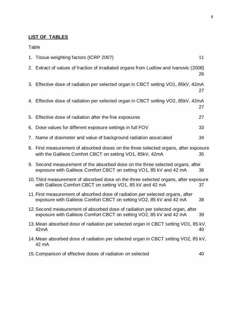
LIST OF TABLES
Table
1. Tissue weighting factors (ICRP 2007) 11
2. Extract of values of fraction of irradiated organs from Ludlow and Ivanovic (2008)
26
3. Effective dose of radiation per selected organ in CBCT setting VO1, 85kV, 42mA
27
4. Effective dose of radiation per selected organ in CBCT setting VO2, 85kV, 42mA
27
5. Effective dose of radiation after the five exposures 27
6. Dose values for different exposure settings in full FOV 33
7. Name of dosimeter and value of background radiation associated 34
8. First measurement of absorbed doses on the three selected organs, after exposure
with the Galileos Comfort CBCT on setting VO1, 85kV, 42mA 35
9. Second measurement of the absorbed dose on the three selected organs, after
exposure with Galileos Comfort CBCT on setting VO1, 85 kV and 42 mA 36
10. Third measurement of absorbed dose on the three selected organs, after exposure with Galileos Comfort CBCT on setting VO1, 85 kV and 42 mA 37
11. First measurement of absorbed dose of radiation per selected organs, after exposure with Galileos Comfort CBCT on setting VO2, 85 kV and 42 mA 38
12. Second measurement of absorbed dose of radiation per selected organ, after exposure with Galileos Comfort CBCT on setting VO2, 85 kV and 42 mA 39
13. Mean absorbed dose of radiation per selected organ in CBCT setting VO1, 85 kV, 42mA 40
14. Mean absorbed dose of radiation per selected organ in CBCT setting VO2, 85 kV,
42 mA
15. Comparison of effective doses of radiation on selected 40

5
LIST OF ACRONYMS
LiF: Lithium fluoride
RANDO®: Radiation Analogue Dosimeter
WITS: University of the Witwatersrand
FOV: Field OF View
ALARA: As Low As Reasonably Achievable
6
CHAPTER I: INTRODUCTION
I.1 Introduction
Technology has undergone profound changes during the past century. New equipment
is available in all sectors of life, from communications to dentistry.
In the dental field, Cone Beam Computed Tomography (CBCT) is one of the most
important technical innovations to this day1. This contemporary radiological imaging
modality is specifically designed for use on the maxillo-facial skeleton. 2, 3, 4
Prior to this new apparatus, oral and maxillofacial radiology mainly utilized two types of
imaging modalities in order to visualize hard tissue lesions. On one hand, there were
intra- oral surveys, panoramic radiographs and several extra oral views. Whether digital
or analogue, they were considered as Conventional Radiography (CR)
On the other hand, there was Computed Tomography (CT), which provided a
multiplanar accurate image of the exposed area. However, due to economic reasons,
lack of expertise and great amount of exposure to ionizing radiations, CT was reserved
for specialized imaging, depending on specific patient indications. This latter modali ty
was able to produce three-dimensional projections which in certain cases proved useful
in some aspects of dentistry.
These two technologies were considered as the standards of care in Oral and Maxillo -
facial imaging.5
7
1.1.1 Definition
When evaluating an emerging technology, the ideal approach is to compare it with the
existing gold standard, and make sure that its diagnostic accuracy is better, or at least,
as good as the one it can be expected to replace. 1, 5
Dental CBCT, can therefore be defined as an imaging modality that provides high
resolution cross-sectional images of an exposed area limited to the maxillo-facial
complex, analogous to CT, and which offers the capacity of a 3D reconstruction of that
same area. 5
1.1.2 Advantages and limitations
The first advantage of dental CBCT is that it overcomes the limitations of CR and
produces an image that is accurate, undistorted and reproducible.4 Indeed, the amount
of information gained from conventional or digitally captured plain radiographs was
limited by the fact that the three-dimensional anatomy of the area being exposed is
compressed into a two-dimensional image.
As a result of superimposition, two-dimensional radiographs reveal limited aspects of
three-dimensional anatomy, requiring, in most cases, a combination of different
conventional films taken in various planes.6Another benefit of CBCT is the production of
a multi planar image similar to CT for a less amount of radiation. Studies comparing
these two imaging techniques have shown that in terms of image quality, reproducibility
and validity CBCT produced superior images to the helical CT, with less radiation
exposure.5,7,8 It has been reported that the average effective radiation dose from CBCT
varies from 36, 9 to 50, 3 µSv. This is considered a 98% reduction, when compared to
8
established CT systems.5, 9 For this reason, CBCT has been recommended as a dose-
sparing technique for oral and maxillo-facial imaging. 5, 10 Relative affordability, x-ray
beam limitation with the possibility of different scan protocols and rapid scan time are
other reasons to make use of this impressive invention.11 The superiority of dental
CBCT compared to CT and CR is therefore well illustrated.
Unfortunately, like all excellent technologies, this machine has its limitations. One must
bear in mind that the effective dose from CBCT is still considerably higher than that from
CR.10, 13, 14Although better than CT from a radiation point of view, CBCT is just as much
affected by radiographic artifacts related to the x-ray beam. This reflects as a distortion
of images of metallic structures and the appearance of streaks and dark bands between
two dense structures. Furthermore, patient movement during the scan can affect the
sharpness of the final image.4, 11
A third disadvantage is that it can only demonstrate limited contrast resolution, mainly
due to relatively high scatter radiation during image acquisition. CBCT would not pose a
problem were the objective of the inquiry to visualize hard tissue only. However, it is
insufficient for soft tissue imaging.5
Difficulty of interpretation may be considered a limitation.10 Yet, the major inconvenience
of this emerging technology remains the use of ionizing radiation. Risks related to the
radiation doses generated by CBCT have been noted.1 According to the 2009 ICRP
reports, the risk of adult patient fatal malignancy related to CBCT is estimated to be
between 1/100000 and 1/350000 individuals. For children, it can be twice as much.5
9
1.1.3 Effects of ionizing radiation
Ionizing radiations, such as X rays, cause ionization of atoms, molecules, cells, tissues,
organs and eventually the whole body. This depends on the amount of radiation
received.
The response of organs to ionizing radiations depends on the sensitivity of each tissue.
It has been reported that reproductive cells as well as the intestinal mucosa have a high
sensitivity to ionizing radiation.
The salivary glands, the lens of the eye and the thyroid gland, on the other hand are
slightly less sensitive. Muscle and nerve tissues have been classified as relatively
insensitive.9 With regard to tissue responses to radiation exposure; two types of effects
have been described. Effects that depend on a certain threshold dose of exposure are
called non stochastic or deterministic effects, whereas the effects that are independent
of a minimal dose of exposure are known as stochastic effects.15
For the purpose of this study, our focus will be on the effects of ionizing radiations on
specific organs in the maxillo-facial region, as well as on the gonads situated in the
pelvic region. Indeed, although situated in the lower abdomen, the gonads may be
involuntary victims of scatter radiation during patient exposure to CBCT.
The lens of the eye contains a single layer of highly active dividing epithelial cells which
are sensitive to ionizing radiation. Some of these cells differentiate into mature lens
fibre cells. Lens transparency depends on the good condition of this layer. Ionizing
radiation may lead to mutation or death of these sensitive cells and cause disruption of

10
this layer. This may cause clouding of the lens and therefore cause the impairment of
vision known as cataract.16 The ICRP considers cataract as a non-stochastic effect of
ionizing radiation. They recommend an equivalent dose limit of 20mSv in a year,
averaged over a period of 5 years, with no single year exceeding 50mSv. The threshold
lens dose for radiation induced cataract is now at 0.5 Gy.16,17
The thyroid has been classified as an organ with a relatively low sensitivity to ionizing
radiation. This means that cell damage that may lead to cancer may occur at a minimal
dose, particularly before the age of 12.15 Thyroid cancer is classified as a stochastic
effect of radiation by the ICRP. 18
Generative cells are highly sensitive to ionizing radiation and there is no threshold dose
for cell injury. Exposure of the gonads may lead to damage of reproductive cells and
induce cell death or mutation. While cell death can lead to a reduction in the number of
gonads, mutation can lead to affected kindred cells that may harbor cancer or
malformations.19
In dental and maxillo-facial diagnostic imaging, the amount of exposure seldom reaches
the threshold doses for the eye. The chances of attainment of doses able to induce a
chain of cellular reactions that may lead to cancer in organs such as thyroid or gonads
are very low. However these doses are cumulative within a certain period of time.
Therefore, there is a risk of cell damage if the patient is submitted to repeated
exposures within a limited period. CBCT examinations are on the increase due to its
popularity. As a consequence thereof, patients face a greater risk of cumulative doses
of radiation. Dentists must therefore be aware of these consequences and take

11
necessary precautions in order to prevent future mutagenesis, carcinogenesis or
teratogenesis.
1.1.4 Method of calculation of effective dose of radiation
Determination of the dose or quantity of the radiation exposure is regulated by a part of
physics sciences called dosimetry. This science provides estimates of the biologic
effects of radiation and therefore permits its proper therapeutic and diagnostic usage.22
Dosimetry utilizes several concepts, but the most relevant to our study are absorbed
dose, equivalent dose, effective dose, and the personal dose equivalent.
Absorbed dose is expressed in Grays (Gy). It describes the energy absorbed from any
type of ionizing radiation per unit mass of any type of matter.22
Equivalent dose is more specific to the type of radiation concerned because it takes
into consideration the Radiation Weighting Factor (W R)
Equivalent dose (Hт) = Absorbed dose X Radiation weighting Factor (1)
Sieverts (Sv) = Gy X WR
HT may be expressed in Sv.
For the X-rays, the radiation weighting factor is 1. W R is provided by the ICRP.
Effective dose takes into consideration the biologic risks in humans exposed to
radiation. In other words it considers the absorbed dose of radiation, the type of
radiation, sensitivity and carcinogenic potential of the irradiated tissue, even without a
12
threshold dose. It therefore takes into consideration the possibilities of stochastic effects
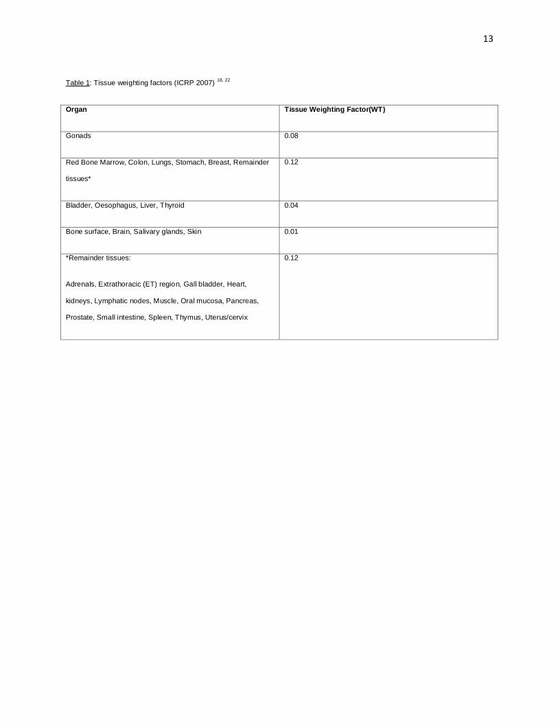
of radiation which are expressed by the Tissue Weighting Factor (W T). (Table 1)
Wт is provided and updated by the ICRP. The fraction of organ exposed (ƒ) is as well
taken into consideration. It is expressed in percentage. 18, 21, 22
E= Hт x ƒ x Wт (2)
Personal dose equivalent Hp(d) is an operational quantity defined by the International
Commission on Radiation Units and measurements (ICRU) and the ICRP. It is
recommended for monitoring low penetrating particles, such as β-particles as well as for
assessment of doses of radiation on external surfaces. The personal dose equivalent
utilized for the calculation of effective doses is Hp(10).The skin and the lens of the eye
are considered as external surfaces. The ICRP 2007 recommends a personal
equivalent dose of Hp (0.07) for the determination of an equivalent dose for these two
organs.26The previous ICRP recommendation of Hp (3) for the lens of the eye was
discontinued in 2007.26 The determination of Hp (0.07) requires a particular method of
calibration of dosimeters using specific phantoms according to the selected external
surface.8,10,27 In the equation of calculation of equivalent dose, the factor absorbed dose
is not a reading anymore, but a calculation using a specific conversion coefficient
relative to the method of calibration.16,18,23,24
13
Table 1: Tissue weighting factors (ICRP 2007) 18, 22
Organ Tissue Weighting Factor(WT)
Gonads 0.08
Red Bone Marrow, Colon, Lungs, Stomach, Breast, Remainder
tissues*
0.12
Bladder, Oesophagus, Liver, Thyroid 0.04
Bone surface, Brain, Salivary glands, Skin 0.01
*Remainder tissues:
Adrenals, Extrathoracic (ET) region, Gall bladder, Heart,
kidneys, Lymphatic nodes, Muscle, Oral mucosa, Pancreas,
Prostate, Small intestine, Spleen, Thymus, Uterus/cervix
0.12
14
1.2 Literature review
Several dosimetry studies on CBCT exist in literature and most of these studies rely on
the use of TLDs. In this regard, the reliability of LiF (Lithium-Fluoride) chips has been
proven. In 1987, Buch and Keddy conducted a study which demonstrated the reliability
of LiF chips. These TLDs have the characteristic of exhibiting relatively null
mechanically induced luminescence as well as the ability to store the information about
the irradiation received. As a result, these TLDs are indicated for experiments which
require displacements to sites remote from the measuring laboratory for purpose of
exposure.25
Unfortunately, LiF dosimeters have two disadvantages. They present non-linear
responses at high doses and are subject to background radiation at low doses.
Although relevant, these disadvantages have been cited in studies carried out on LiF in
the form of loose powder.25
LiF dosimeters historically existed in the form of rods and discs, but their major
shortcoming was that of orientation dependence. There was a significant difference in
readings in small surfaces compared to larger surfaces, with identical exposures.25 The
limitations of LiF in the form of loose powder were overcome by the use of LiF discs,
that were submitted to a calibration of 1Gy of ionizing radiation from a standard source
and submitted to the specific selection criteria of 5% below the mean dose calculated.25
In 2006, Ludlow et al. published a dosimetry study on three different oral and maxillo-
facial CBCT devices. For this purpose, they utilized LiF TLD chips that were selected as
15
advised in previous literature.25The exposures were effectuated on a Radiation
Analogue Dosimeter (RANDO®) phantom in which the TLDs were inserted. This study
determined the anatomical landmarks for insertion of TLDs and emphasized the use of
effective dose as well as the influence of, kV, mA settings different FOVS in dosimetry
studies. Although it did not make use of the Galileos Comfort ® CBCT, and calculated
average body effective doses, this study is often used as a reference for dosimetry, with
organ dose measurement as a research methodology.26
In 2008, Ludlow and Ivanovic compared the doses on CT with several CBCT devices,
including the Galileos® Sirona with the scan protocol of 85kV, 42 mA on full FOV.
This article focused on the doses in the maxillo-facial area and introduced the, ICRP
tissue weighting factors (2007) as well as different CBCT scan protocols. They
calculated equivalent doses of radiation for specific organs in the maxillo-facial region.
The thyroid absorbed an equivalent dose of 450µSv with the Galileos at maximum
exposure. The equivalent dose for the eye was not mentioned. The effective doses
calculated were average full body doses.10
In a 2012 publication, Pauwels et al. elaborated on the effective dose for dental CBCT
scanners. They calculated the doses on several machines, including the Galileos
Comfort® CBCT. Unfortunately, they used a CBCT scan protocol of full FOV, 85kV and
28mA. They calculated equivalent organ doses and recorded a dose of 380µSv for the
thyroid. The eye was not mentioned and the effective doses calculated were average
full body doses. 27

16
During the same year, Thorsten et al. compared the dosimetry of the CBCT with a
digital X-ray machine in orthodontic imaging. For this purpose, he used organ dose
measurement methodology, based on Ludlow’s approach. The machines used for
performing the exposures were i-CAT next generation® CBCT and an
orthopantomograph OP100/OC 100®. The existing scan protocols for i-CAT next
generation® and the Galileos Comfort® are different. They calculated equivalent doses
for the organs in the maxillo-facial area. The thyroid’s dose was of 167, 267, 150 and
350µSv according to the different scan protocols. The eye was excluded. The effective
doses calculated for the different machines were average full body doses. They proved
that the dose from CBCT was higher than the one from digital panoramic radiographs.
Their conclusion was that, while information gained from a CBCT examination was
benefic, it was the practitioner’s discretion to weigh between the risks encountered by
the patient and the benefit from the examination. 20
In 2013, surface skin doses were measured after exposure with four dental x-ray
imaging systems. For this purpose, three CBCT units and one combined conventional
panoramic-cephalometric unit were utilized. The latter unit was ProMax® pan/ceph x-ray
machine, while the CBCTs were Kodak 950® (Kodak Dental Systems, Care stream
Health, Rochester, NY, USA), i-CAT next generation® and Galileos Comfort®. The
selected FOVs were the large and medium ones only. This selection was justified by the
popularity of these FOVs in orthodontics for diagnostic and treatment planning. Several
scan protocols were included in the study. Amongst others, the full FOV, 85 kV. 42mA.
The dosimeters utilized for this purpose were optically stimulated luminescence (OSL)
dot dosimeters (nanoDOTS® dosimeters, Landauer Corp. Glenwood IL) and the
17
phantom was a head anthropomorphic phantom RS110® (Radiology Supported
Devices-RSD- Inc., Long beach, CA). The selected skin points for placement of the
dosimeters were on the lens of the eyes, the parotid glands, the submandibular gland
and the thyroid. After exposure of the phantom, according to the usual patient
positioning protocol in a CBCT machine, the TLDs were read and the figures reported
were converted into absorbed dose in mGys, using unit and scan specific calibration
factors. With the protocol scan of 85kV, 42mA and full FOV, for the Galileos Comfort®
CBCT the lens of the eye’s calculated absorbed dose was 0.94mGy.The highest dose
observed for the lens of the eye was of almost 4mGy with a scanning protocol of 120kV,
108mA and 20x18 FOV. The absorbed dose calculated at the skin surface on the
thyroid was of 0.46mGy, for the full FOV scan protocol 85kV, 42 mA. They justified this
negligible dose by considering the skin covering the anterior part of the thyroid gland as
out of the primary x-ray beam.28
In 2014, a study in Ontario calculated effective doses of different protocols using the
Sirona Galileos Comfort CBCT®. They used the organ dose approach in their
methodology, but the thermoluminescent chips were InLight® nanoDot TM OSL
dosimeters (Landauer, Glenwood, III) placed on Polymethyl methylacrylate (PMMA)
templates. These dosimeters were positioned on a RANDO phantom with referral to
Ludlow’s anatomic landmarks. Twelve scan protocols were used, including the Full FOV
HR (VO1) and the Full FOV standard (VO2) at 85kV, 42mA. The calculated effective
doses were maxillo-facial average dose. The scatter was considered negligible. Our
focus in this publication was on the calculated effective doses of protocol Full FOV HR

18
85kV, 42mA resulting in 142µSv compared to Protocol full FOV standard resolution
85kV, 42mA resulting in 140µSv.
This suggested that the changes in resolution settings had little or any impact on
effective dose.22
Finally, the manufacturer provided average full body effective doses on the different
settings, referred from a study by Ludlow JB on the dosimetry of the Galileos Dental®
CBCT provided settings. (Table 2) It was reported that in smaller FOVs (maxillary or
mandibular collimation) dose values could be reduced by approximately 15%.29
Various reports on doses from CBCT have been published in the literature yet many of
them refer to average full body doses or facial doses rather than to specific vital organs.
Studies are still needed in order to determine radiation safety for specific organs in the
maxillofacial region, as well as the effects of scatter radiation during CBCT
examinations. The question still remains, however, whether or not the use of CBCT as a
routine imaging modality for dental diagnosis induces overexposure to the patients.

19
1.3 Aim of the study
WITS Dental School has recently acquired the Galileos Comfort CBCT scanner. The
manufacturers refer to average full body doses rather than to specific vital organs.29
Most of the studies refer to average effective doses. It would appear that effective
doses to specific regions of the face have been ignored.
It is established that exposed dose is influenced by the parameters FOV, kVs and mAs
settings.26, 28, 30 Yet, comparisons of amount of exposure at the different resolution
settings, as well as the amount of scatter, particularly at the pelvic region, remain to be
investigated.
The specific aim of the current study is therefore to calculate the potential effective
doses of radiation to specific vital organs in the head and neck region emanating from
the CBCT scanner housed in the WITS Dental School at different resolution settings
(VO1 and VO2). It also aims to investigate the amount of scatter radiation to the pelvic
region for both scan protocols. The results of these observations are to be compared
with the average effective doses described in the literature as well as background
radiation.
It is hoped by means of this study to contribute to the elaboration of conclusions
relevant to the situation in South Africa.

20
1.4 Objectives
Primary objectives
The primary objectives of this study are:
-To calculate the potential effective dose of radiation to the lens of the Right eye after
exposure to the Galileos Comfort CBCT scanner on the setting VO1, 85kV, 42mA --
-To calculate the potential effective dose of radiation to the lens of the Left eye after
exposure to the Galileos Comfort CBCT scanner on the setting VO1, 85kV, 42 mA
-To calculate the potential effective dose of radiation to the lens of the Right eye after
exposure to the Galileos Comfort CBCT scanner on the setting VO2, 85kV, 42mA
-To calculate the potential effective dose of radiation to the lens of the Left eye after
exposure to the Galileos Comfort CBCT scanner on the setting VO2, 85kV, 42 mA
-To calculate the potential effective dose of radiation to the thyroid gland after exposure
to the Galileos Comfort CBCT scanner on the setting VO1, 85kV, 42mA
-To calculate the potential effective dose of radiation to the thyroid after exposure to the
Galileos Comfort CBCT scanner on the setting VO2, 85kV, 42mA
-To calculate the extent of scatter radiation to the gonads during maxillofacial
examinations using the Galileos Comfort CBCT scanner on the setting VO1, 85kV,
42mA
- To calculate the potential extent of scatter radiation to gonads during maxillofacial
examinations using the Galileos Comfort CBCT scanner on the setting VO2, 85kV,
42mA

21
Secondary objectives
The secondary objectives of this study are:
-To compare the calculated effective doses with the average effective doses as stated
in the literature
-To compare the calculated effective doses on the two different scan protocols
-To determine whether or not there is a need for additional protection of the patient
during such examinations
-To compare the equivalent absorbed dose on the eye with the threshold dose at the
eye fixed by the ICRP 2007.
-To compare the calculated effective doses with the background radiation.
22
CHAPTER II: MATERIALS, METHODS AND RESULTS
2.1 Introduction
A systematic review of the different methodologies employed in dosimetry studies for
CR, CT and CBCT has revealed that most of the studies encountered in literature have
utilized the method of organ dose measurement.21
This method relies on phantoms, implanted with dosimeters. 10, 14, 20, 21,26, 31, 32
Other methods such as computer tomography dose index by volume (CTDIvol), Monte
Carlo dose simulation programs, CT air-kerma length product (PKL, CT), Air kerma area
product (PKA), entrance skin surface dose and energy imparted have as well been
described.22
However, our study will rely on the organ dose measurement method. The calculations
of effective doses will be based on the absorbed dose measurements on the dosimeters
inserted inside a phantom, the radiation weighting factors, the tissue weighting factors
and the fraction of irradiated organ.
The phantom simulates human tissues with regard to tissue layers and radiation
absorption factors. It is therefore called an anthropomorphic phantom. Such phantoms
are fabricated with a natural human skeleton cast inside a material that has a radiologic
density equivalent to that of soft tissue. It is virtually indestructible, capable of
withstanding substantial impact and continuous handling without damage. These
23
phantoms are constructed in the form of detachable cross-sections with apertures
created for placement of dosimeters in the region of interest. 33
The specific phantom to be used is the RANDO® (Radiation Analogue Dosimeter, The
Phantom laboratory, Salem, NY)
The selected organs were the lens of the L eye, the lens of the R eye, the thyroid
gland and the gonads in the pelvic region. The first two organs were retained because
of their anatomical position in the head and neck region and their relatively high
sensitivity to ionizing radiation. Although they are not the primary indication for a CBCT
examination, these regions are most likely irradiated during CBCT exposures as they
are situated very close to the primary X-ray beam. As a result, they may receive
inadvertent exposure, because of their position. The gonads area may be affected by
scatter radiations. This region is very radiosensitive, as it harbors the reproductive cells.
The Sirona Galileos Comfort® CBCT scanner is housed in the Wits dental hospital.
The software installed in it is a GALAXIS, RECO® software which extends SIDEXIS to
include the processing of 3D data. Its functions are 3D reconstructions, storage, recall,
display and processing of 3D data.
The tube voltage is fixed at 85kV, with a current varying between 5-7mA.There are six
exposure settings where the tube voltage remains constant at 85kv, while the current
may vary between 10-42mA according to the size of the patient. The different exposure
settings available include 10, 14, 21, 28, 35 and 42mAs.
24
The Galileos Comfort® has two FOVs. The full FOV measures 15x15x15 spherically,
and the medium FOV (upper or lower jaw only) is 8.5 x15 x 15.
It is equipped with four viewing settings: VO1, VO1HC, VO2 and VO2HC. The
difference between these settings is simply the resolution and the contrast. In other
words, it is a question of image quality.
In this study two different protocols will be used. One using the VO1 setting and the
other using the VO2 setting. VO1 displays a high resolution and therefore has a smaller
pixel size. As a consequence, it occupies more space in the memory and requires a
bigger data volume (740MB).
VO2 displays a standard resolution and therefore has a bigger pixel size. As a result,
the data volume is smaller (approximately 390 MB).29
These viewing settings do not influence the amount of exposure received by the patient.
Yet, our study will compare the different amount of exposure received in the two
different scan protocols.
The dosimeters exist in several types. Newly optically stimulated luminescent
dosimeters (OSL) are the latest model in the market, but this study will make use of
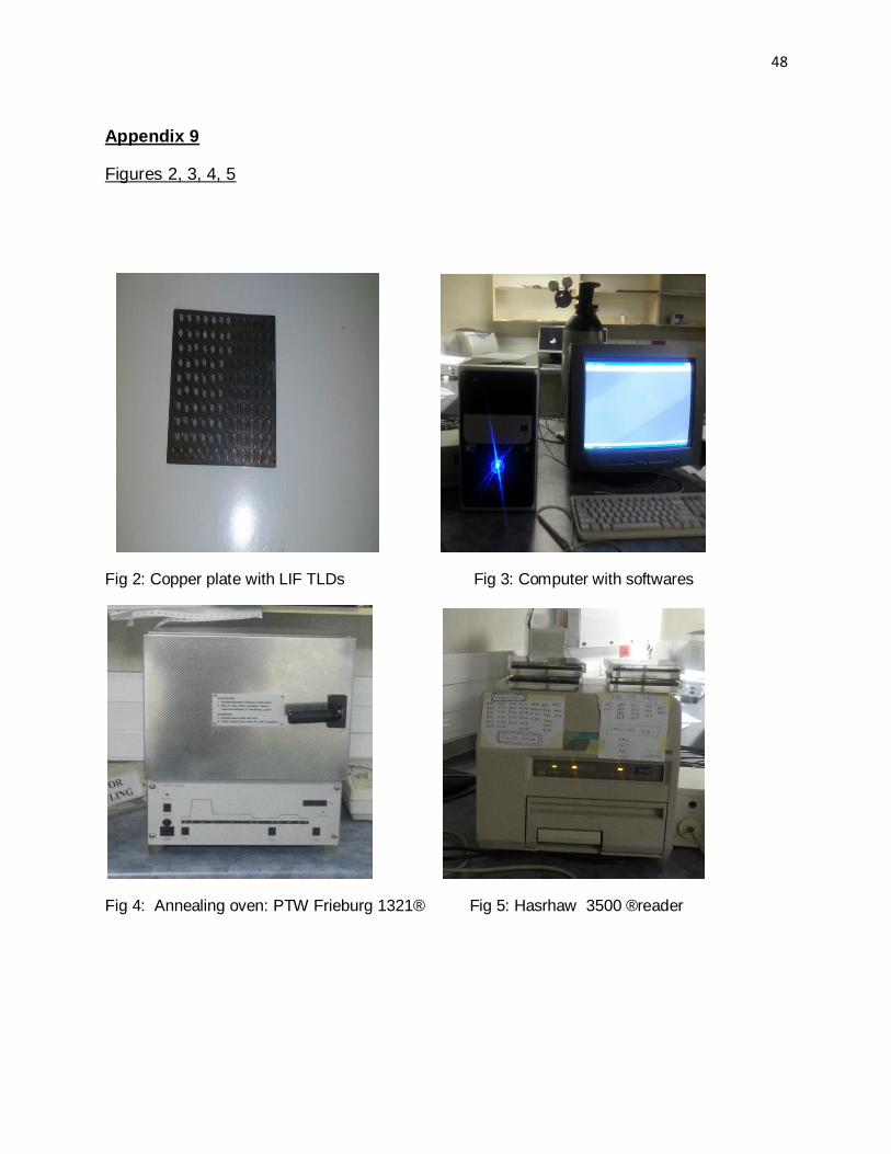
thermoluminescent dosimeters (TLDs), which are the most popular. (Fig. 2) TLDs rely
on the principle of thermoluminescence. Their role is to measure the absorbed dose of
radiation in the specific area, where they are placed.
Previous studies have elaborated on the reliability of lithium- fluoride TLDs. The
superiority of lithium fluoride (LiF) over other thermoluminescent materials has been

25
established. These discs when subjected to a standardized method of annealing and
selection may be relied upon to an accuracy of 90%. 25
However, with the introduction of personal dose equivalent Hp (0.07) by the ICRP in
1991, TLDs for external surfaces such as the skin and the lens of the eye require a
specific calibration.16, 24The dosimeters utilized, for the purpose of this research are
thermoluminescent TLDs (TLD100) discs.
The Medical-physics laboratory supplied the author with the facilities for annealing,
handling and reading of the dosimeters. The venue was equipped with a PTW-Frieburg
1321® oven. The reader was a Harshaw 825®, model 3500. Both equipments were
operated from a computer installed with the application softwares WINREMS for the
reader, and THELDO for the oven. (Fig. 3, 4, 5)
The dosimeters were handled with a Dymax 30 Charles Austen vacuum pump, in order
to prevent their contamination. (Fig. 6)

26
2.2 Materials and methods
The experiment took place in two different venues. The calibration and manipulation of
the dosimeters were performed in the laboratory whilst the RANDO® phantom, although
mounted and dismantled in the laboratory, was submitted to the exposures in the
hospital.
Inclusion and exclusion criteria
These criteria are based on the methods of selection of TLDs as discussed by Buch and
Keddy, 1987.25 The calibration of the 67 TLDs was performed using a photon energy of
6 MV on a Siemens linear accelerator. 1Gy of radiation was given to the batch of TLDs
for a field size of 14cm x14cm at the depth of 4.4cm in Perspex, which is equivalent to
5cm in water and the procedure was carried out in a black plastic container. The 49
TLDs that did not vary more than 5% from the mean value were considered in the
inclusion criteria. All the others were excluded. The gender of the phantom was of no
consequence as male and female gonads are situated in the pelvic region.
All of the 67 dosimeters responded positively to the selection criteria, in other words
they were all below 5% of the mean absorbed dose. According to the medical physicist
who performed the calibration, this could be justified by the fact that these dosimeters
were still relatively new and had not been submitted to a great number of exposures.
The forty-nine TLDs that had the readings nearest to the mean value were therefore
selected.

27
The first reading of the forty-nine dosimeters was done in order to measure the
background radiation. Afterwards, the TLDs were submitted to a second annealing in
order to exclude background radiation, prior to their insertion in the RANDO® phantom.
For this process the chips were placed inside a square annealing copper plate provided
with apertures to contain each dosimeter. The placement of the TLDs in the phantom
was realized according to anatomical positions used in literature with three in each of
the selected organs31, 32
For the thyroid, the TLDs were placed in position 9 of RANDO® phantom.
For the lens of the left and the right eye, they were placed in specific pouches, held in
place with tape on the anterior surface of the eye. As for the lower abdomen, the TLDs
were placed on position 33 of the RANDO® phantom.
In the three organs, considering the phantom in an anatomic position, the TLDs were
placed according to a linear pattern with one anterior, one medial and one posterior.
All the manipulations of the dosimeters were done in the medical physics laboratory,
where all the conditions for handling and reading of TLDs were met.
Once the TLDs in place, the phantom was conveyed from the medical-physics
laboratory to the dental hospital, where the Galileos Comfort® CBCT is housed.
In the hospital, the phantom was positioned in the machine with the midsagittal plane
centered in the image field and the occlusal plane parallel to the scan rotation
plane.10,14, 20, 26It was then subjected to the same type of x-ray examinations as those
that are usually conducted on patients. (Fig.7)

28
Two different CBCT scan protocols were used for this exposure. The first one was at
85kV, 42 mA on setting high resolution setting, and the second one at 85 kV, 42 mA on
standard setting.
After exposure, the phantom was returned to the medical physics laboratory, for
reasons of appropriate manipulation and reading of TLDs.
This procedure was repeated five times, three for the first protocol, and twice for the
second protocol.
Once the readings were processed, the background radiation was subtracted from the
radiation absorbed dose for each TLD. The mean absorbed dose per organ was then
calculated for both protocols and this led to the calculations of the different effective
doses per organ, for both protocols, according to the ICRP specifications.
29
2.3 Results
The data was collected in the form of tables where each selected TLD was attributed a
specific name symbolized from 1A1 to 9G9.These calibrated TLDs kept the same
position in the annealing copper plate during the entire experiment. The first table
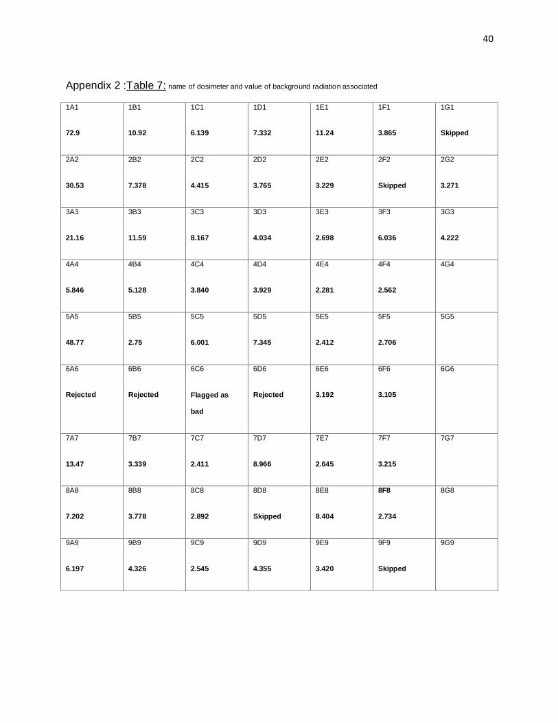
symbolizes the readings of the background radiation on each TLD. (Table 7)
The absorbed dose of each TLD was recorded for both protocols. These doses are
presented in tables 8, 9, 10, 11 and 12.
The shortcomings were encountered with the calculation of the effective dose on the
implementation of the equation: E= WT x HT x ƒ (2)
The equivalent dose (HT) could easily be calculated as the product of the absorbed
dose and the radiation weighting factor, based on the collected data. Limitations,
however, were encountered with the identification of the two other parameters in the
equation. Each organ had its own specifications for the tissue weighting factor and the
fraction of irradiated tissue.
The tissue weighting factor (WT) for the selected organs was obtained from the
literature. However, according the ICRP 2007 publication, the eye was not viewed as an
organ that would develop stochastic effects of ionizing radiation. It was therefore not
listed in the ICRP 2007 guidelines, among the organs that were attributed a tissue
weighting factor. (Table 1) However the threshold dose for cataract, considered a
deterministic effect of ionizing radiation on the lens of the eye was clearly stipulated. A
minimum exposure of 0.5 Sv, could induce radiation cataract. As a result, the calculated
dose of radiation for the lens of the eye was reduced to an equivalent dose with no
carcinogenic potential. The equivalent dose is always expressed in Sieverts.

30
The gonads and the thyroid are attributed tissue weighting factors by the ICRP. These
two organs are therefore considered by the ICRP, as having a carcinogenic potential
when exposed to a minimum dose of ionizing radiation. The dose of radiation to be
calculated for these two organs was therefore to remain an effective dose.
The determination of their respective fractions of irradiated tissues (ƒ) was a
challenge. There are estimations for this parameter in the literature for the thyroid gland
and other organs in the maxillo-facial region. Most of the dosimetry studies on dental
CBCT focus on the head and neck region. As a result, this study referred to Ludlow and
Ivanovic 2008 estimations of fraction of irradiated tissue values for the thyroid gland.
(Table 2) The gonads were given an estimated value of 1%. This was based on the
anatomical situation of the gonads, which are very far from the primary x-ray beam. An
estimation of 0% would have brought our results to null. This would have been contrary
to the observations on the data collection which recorded an absorbed dose on the
TLDs placed in this organ.
Table 2: Extract of values of fraction of irradiated organs from Ludlow and Ivanovic
Organ Fraction irradiated
Thyroid 100%
Eyes 100%
Salivary glands 100%
Skin 5%
Mandible 1.3%

31
The values of the equivalent and effective doses calculated for each organ are
presented in tables 3, 4and 5. The first two tables refer to the two different scan
protocols. (Table 3-4)
The third table summarizes the average dose values after the five exposures. (Table 5)
The average dose of exposure received on both eyes L and R is therefore: 58.995 µSv.
The comparison of the values of the effective doses on the two resolution settings is
illustrated on fig.1.

32
Table 3: Effective dose of radiation per selected organ in CBCT setting VO1, 85kV, 42mA
Selected Organ Mean Absorbed Dose
of Radiation (µGy(
Tissue Weighting
Factor ( ICRP 2007)/ Radiation weighting
factor
Estimated Fraction of
irradiated tissue in %
Dose of Radiation
(ICRP 2007) µSv
L eye 35.717 1 100 35.717 HT
R eye 68.569 1 100 68.569 HT
Thyroid 1001.755 0.04 100 40.072 E
Gonads 89.310 0.08 1 0.071 E
Table 4: Effective dose of radiation per selected organ in CBCT setting VO2, 85kV, 42m
Selected Organ
Mean Absorbed Dose
of Radiation µGy
Tissue Weighting
Factor (ICRP 2007)/ Radiation weighting
factor
Estimated Fraction of
irradiated tissue in %
Dose of
Radiation(ICRP 2007) µSv
L eye 30.378 1 100 30.378 HT
R eye 101.335 1 100 101.335 HT
Thyroid 173.46 0.04 100 6.938 E
Gonads 28.310 0.08 1 0.003 E
.
Table 5 : effective dose of radiation after the five exposures
Selected organ Mean absorbed dose of Radiation µGy
Tissue Weighting Factor (ICRP
2007)/Radiation weighting Factor
Fraction of irradiated tissue in %
Dose of Radiation (ICRP 2007) µSv
L eye 33.04 100 33.04 HT
R eye 84.95 100 84.95 HT
Thyroid 587.60 0.04 100 23.500 E
Gonads 58,81 0.08 1 0.047 E

33
Fig1: Column chart comparing values of doses of radiation on VO1 and VO2
0
20
40
60
80
100
120
L EYE R EYE THYROID GONADS
VO1
VO2
34
CHAPTER III: CONCLUSION
3.1 Discussion
This study examined doses of ionizing radiation emitted during a CBCT examination to
the eye, the thyroid and the gonads. Of all the organs exposed, the eye was the one
which seemed to show the highest dose of radiation. This could easily be explained by
the fact that the dose of exposure to the eye is an equivalent dose that is not influenced
by tissue weighting factors. One must therefore not compare the doses calculated for
the eye with those for the two other organs which may develop stochastic effects,
according to the ICRP 2007. The average equivalent dose for the lens of the eye was
59µSv. A dose of this nature, being almost 10 000 times less than the threshold, may be
considered negligible for producing a cataract. Literature has reported an estimated
dose of background radiation per annum received by every individual in normal
circumstances of approximately of 3mSv. However, there is a great discrepancy
between the equivalent dose of 0.059mSv to the eye as calculated in this study and the
absorbed dose of 0.94mGy calculated by Akyalcin et al. in 2013. Such a comparison
seems possible because the radiation weighting factor in the equation for calculating the
equivalent dose, based on absorbed dose, is 1. As a result, the dose calculated by
Akyalcin et al. seems to be 20 times greater. One must bear in mind that although both
these studies used the Sirona Galileos Comfort® CBCT scanner with similar scan
protocols and FOVs, as well as the organ dose measurement methodology, the
equipments used and the methods of calibration of dosimeters were totally different.
One study used the standard method of calibration used for deep tissues, while the
other used specific calibration of dosimeters and coefficients relative to external
35
surfaces. Another factor that could explain the discrepancy is the different types of
dosimeters used in both studies i.e. LiF versus OSL dosimeters. Nevertheless, both
results were relatively low.
The gonads showed results approximating the null figure attributed to the study effected
by Chambers D, 2014. The effective dose of 0.05µSv could be considered negligible.
This is consistent with Alkyacin et al.’s theory that justified the low dosage obtained at
the skin surface of the thyroid by considering it as being situated in the scatter region.
The thyroid was the organ where calculation of effective dose was the least challenging
as all the parameters to determine the effective dose were present. The specific
calibration of the dosimeters was that indicated for deep tissues. The organ is
anatomically situated in the proximity of the primary beam. However, Ludwig and
Ivanovic do not consider it as part of the scatter as they attributed it a fraction of
irradiated tissue similar to that of the salivary glands, which, anatomically are definitely
situated inside the primary beam. The figure of 23.5 µSv recorded in this study as an
effective dose for the thyroid is difficult to compare with doses in the literature because
most of the doses for this specific organ found in the literature are equivalent or
absorbed doses, the radiation weighting factor for X-rays being 1. However, the ICRP
classifies the thyroid as an organ that could develop cancer or mutation related to
ionizing radiation. As a result, the calculation of radiation exposure on this organ must
take into consideration the tissue weighting factor and the fraction of irradiated tissue.
This justifies the use of effective dose for the thyroid gland in this study.

36
These different types of dosages of the amount of radiation for one specific organ set a
limitation to our comparison between the doses in the study and the ones in literature.
Yet, the average full body doses recorded in literature are effective doses. They are
therefore comparable to the doses in the current study. Ludlow and Ivanovic calculated
128µSv for full FOV and maximum exposure, whereas Chambers recorded an exposure
of 140µSv for similar conditions. The manufacturer’s effective dose for a similar protocol
was shown to be the same as that of Ludlow and Ivanovic i.e. 128µSv.
Considering the author’s recording of 24µSv for the thyroid, one could estimate it as
approximately 19% of the full body average effective dose as compared to Ludlow and
17% compared to Chambers. In other words, the thyroid, according to the study under
consideration, would be absorbing approximately 20% of the full body irradiation
emanating from a CBCT scan.
It has been established that the amount of radiation to which a patient is exposed during
a CBCT examination is a function of the FOV, the kV of the machine and the amperage
setting. One of the objectives of this study was to compare the readings on the high
resolution and standard settings of the Galileos Comfort® CBCT without changing the
FOV, the kV or the mA. Chambers compared the two settings on the same machine
with a similar scan protocol and concluded that the there was no significant difference in
the effective doses. The study under consideration compared the two resolution settings
at an organ level. There was found to be a discrepancy between the two scan protocols
in general, although the greatest discrepancies were found for the right eye and the
thyroid. LiF dosimeters have been used in several organ dose measurement studies,

37
and their reliability has been shown in previous studies. Also the phantom used is
appropriate for the type of dosimeters used. It has, in fact, been established that the
majority of cases recorded in the literature were done on an anthropomorphic phantom
and LiF TLDs.21
According to literature studies, discrepancies have been explained by the position of
dosimeters in the apertures of the phantom.26Indeed, in the three organs studied, the
dosimeters were placed in sets of three, in a linear pattern: one anterior, one medial and
one posterior. This means that one dosimeter was always cranial to the field of radiation
while another was always caudal to the field. This fact might explain the discrepancies
on all three organs.
Another observation is that the method of calibration of dosimeters used in this study for
both the eyes and the other organs was similar. Literature studies consider the eye to
be a superficial structure and therefore require the calculation of a personal dose
equivalent for this organ.
Finally, the discrepancy might be the result of the number of exposures for each setting.
Indeed, three exposures were carried out for the high resolution, while the standard
resolution was only submitted to two exposures. The different population samples
considered in the calculation of the means for both scan protocols could have
influenced the results.

38
3.2 Conclusion
CBCT is a very useful tool in dentistry. However our study has demonstrated low doses
of radiation to the eye, a fairly insignificant amount of scatter to the gonads and a
contribution of approximately 20% of the full body dose to the thyroid. For a child of
about 12 years undergoing orthodontic treatment this could be considered a fairly
substantial dose. However, compared to our measurement of background radiation
these doses are still relatively low. As for the different resolution settings, further studies
at an organ level are still needed in order to justify these values. Meanwhile application
of the ALARA (As Low As Reasonably Achievable) principle by reduction of the FOV
and mA settings where applicable as well as a specific indication for a CBCT
examination is essential.

39
Appendices
Appendix 1:
Table 6: Dose values for different exposure settings in full FOV29
Deff: effective dose value -
Programs 1 2 3 4 5 6
Values 10 mAs 14mAs 21 mAs 28mAs 35mAs 42mAs
Deff ICRP 1991 14µSv 19 µSv 28 µSv 39 µSv 48 µSv 52 µSv
Deff ICRP 2007 30µSv 41 µSv 70 µSv 83 µSv 103 µSv 128µSv
40
Appendix 2 :Table 7: name of dosimeter and value of background radiation associated
1A1
72.9
1B1
10.92
1C1
6.139
1D1
7.332
1E1
11.24
1F1
3.865
1G1
Skipped
2A2
30.53
2B2
7.378
2C2
4.415
2D2
3.765
2E2
3.229
2F2
Skipped
2G2
3.271
3A3
21.16
3B3
11.59
3C3
8.167
3D3
4.034
3E3
2.698
3F3
6.036
3G3
4.222
4A4
5.846
4B4
5.128
4C4
3.840
4D4
3.929
4E4
2.281
4F4
2.562
4G4
5A5
48.77
5B5
2.75
5C5
6.001
5D5
7.345
5E5
2.412
5F5
2.706
5G5
6A6
Rejected
6B6
Rejected
6C6
Flagged as
bad
6D6
Rejected
6E6
3.192
6F6
3.105
6G6
7A7
13.47
7B7
3.339
7C7
2.411
7D7
8.966
7E7
2.645
7F7
3.215
7G7
8A8
7.202
8B8
3.778
8C8
2.892
8D8
Skipped
8E8
8.404
8F8
2.734
8G8
9A9
6.197
9B9
4.326
9C9
2.545
9D9
4.355
9E9
3.420
9F9
Skipped
9G9

41
Appendix 3
Scan protocol 1
Table 8: First measurement of absorbed doses on the three selected organs after exposure with the Galileos Comfort CBCT on
setting VO1, 85kV, 42mA
TLD name and selected
organ p
Absorbed dose of
radiation Reading (µGy)
Background Radiation (
µGy)
Absorbed dose of
radiation on Organ (µGy)
1A1 Gonads 114,4 72,9 41,5
2A2 Gonads 78,54 30,53 48,01
3A3 Gonads 48,57 21,16 27,41
4A4 R eye 31,01 5,84 25,17
5A5 R eye 50,34 48,77 1,57
6A6 Rejected _ Rejected _
7A7 R eye 36,63 13,47 23,16
8A8 L eye 35,52 7,202 28,318
9A9 L eye 38,55 6,197 32,353
1B1 L eye 38,79 10,92 27,87
2B2 Thyroid 796,1 7,378 788,722
3B3 Thyroid 962,9 11,59 951,31
4B4 Thyroid 2332 5,128 2326,872

42
Appendix 4
Table 9: Second measurement of the absorbed dose on the three selected organs, after exposure with Galileos Comfort CBCT
on setting VO1, 85 kV and 42 mA
TLD name and selected organ
Absorbed dose of radiation reading (µGy)
Background Radiation (µGy)
Absorbed dose of radiation on Organ (µGy)
1A1 R eye 181.4 72.9 108.5
2A2 R eye 103.4 30.53 72.87
3A3 R eye 75.48 21.16 54.32
4A4 L eye 50.67 5.846 44.824
5A5 L eye 37.04 48.77 Incoherent
6A6 Rejected _ _ _
7A7 L eye 35.77 13.47 22.3
8A8 Thyroid 276.4 7.202 269.198
9A9 Thyroid 208.3 6.197 202.103
1B1 Thyroid 264.7 10.92 253.78
2B2 Gonads 301.6 7.378 294.222
3B3 Gonads 44.7 11.59 33.11
4B4 Gonads Rejected 5.128 _

43
Appendix 5
Table 10: Third measurement of absorbed dose on the three selected organs, after exposure with Galileos Comfort CBCT on
setting VO1, 85 kV and 42 mA
TLD name and selected organ
Absorbed dose of radiation reading (µGy)
Background Radiation (µGy)
Absorbed dose of radiation on Organ (µGy)
5B5 Gonads 40.22 2.75 37.47
7B7 Gonads 100.6 3.339 97.261
8B8 Gonads 64.93 3.778 61.152
9B9 Thyroid 100.7 4.326 96.374
1C1 Thyroid 2667 6.139 2660.861
2C2 Thyroid 1471 4.415 1466.585
3C3 R eye 224.8 8.167 216.633
4C4 R eye 115.1 3.840 111.26
5C5 R eye 63.96 6.001 57.959
6C6 _ _ Flagged as bad
7C7 L eye 47.58 2.411 45.169
8C8 L eye 46.73 2.892 43.838
9C9 L eye 45.77 2.545 43.225

44
Appendix 6
Scan protocol 2
Table 11: First measurement of absorbed dose of radiation per selected organs, after exposure with Galileos Comfort CBCT on
setting VO2, 85 kV and 42 mA
TLD name and selected
organ
Absorbed dose of radiation
reading (µGy
Background Radiation (µGy)
Absorbed dose of radiation
on Organ (µGy)
1D1 Gonads 31.58 7.332 24.248
2D2 Gonads 21.20 3.765 17.435
3D3 Gonads 21.36 4.034 17.02
4D4 Thyroid 37.95 3.929 34.021
5D5 Thyroid 273.5 7.345 266.155
6D6 _ Rejected _
7D7 Thyroid 221.7 8.966 212.734
8D8 _ Skipped _
9D9 R eye 46.75 4.355 42.395
1E1 R eye 61.56 11.24 50.32
2E2 R eye 29.53 3.229 26.301
3E3 L eye 35.64 2.698 32.942
4E4 L eye 29.90 2.281 27.619
5E5 L eye 30.27 2.412 27.858
45
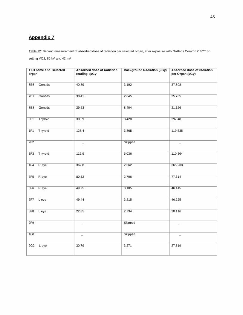
Appendix 7
Table 12: Second measurement of absorbed dose of radiation per selected organ, after exposure with Galileos Comfort CBCT on
setting VO2, 85 kV and 42 mA
TLD name and selected
organ
Absorbed dose of radiation
reading (µGy
Background Radiation (µGy) Absorbed dose of radiation
per Organ (µGy)
6E6 Gonads 40.89 3.192 37.698
7E7 Gonads 38.41 2.645 35.765
8E8 Gonads 29.53 8.404 21.126
9E9 Thyroid 300.9 3.420 297.48
1F1 Thyroid 123.4 3.865 119.535
2F2 _ Skipped _
3F3 Thyroid 116.9 6.036 110.864
4F4 R eye 367.8 2.562 365.238
5F5 R eye 80.32 2.706 77.614
6F6 R eye 49.25 3.105 46.145
7F7 L eye 49.44 3.215 46.225
8F8 L eye 22.85 2.734 20.116
9F9 _ Skipped _
1G1 _ Skipped _
2G2 L eye 30.79 3.271 27.519

46
Appendix 8
Tables 13,14 and 15
Table 13: Mean absorbed dose of radiation per selected organ in CBCT setting VO1, 85 kV, 42mA
Selected Organ Average exposed
dose on first
exposure (µGy)
Average exposed
dose on second
exposure (µGy)
Average exposed
dose on third
exposure (µGy)
Mean dose of
exposure (µGy)
L eye 29.514 33.56 44.077 35.717
R eye 16.633 60.458 128.617 68.569
Thyroid 1355.634 241.693 1407.94 1001.755
Gonads 38.973 163.666 65.29 89.310
Table 14: Mean absorbed dose of radiation per selected organ in CBCT setting VO2, 85 kV, 42 mA
Selected Organ
Average exposed dose on first exposure (µGy)
Average exposed dose on second exposure (µGy)
Mean dose of exposure (µGy)
L eye 29.47 31.287 30.378
R eye 39.672 162.999 101.335
Thyroid 170.97 175.95 173.46
Gonads 19.567 37.054 28.310
47
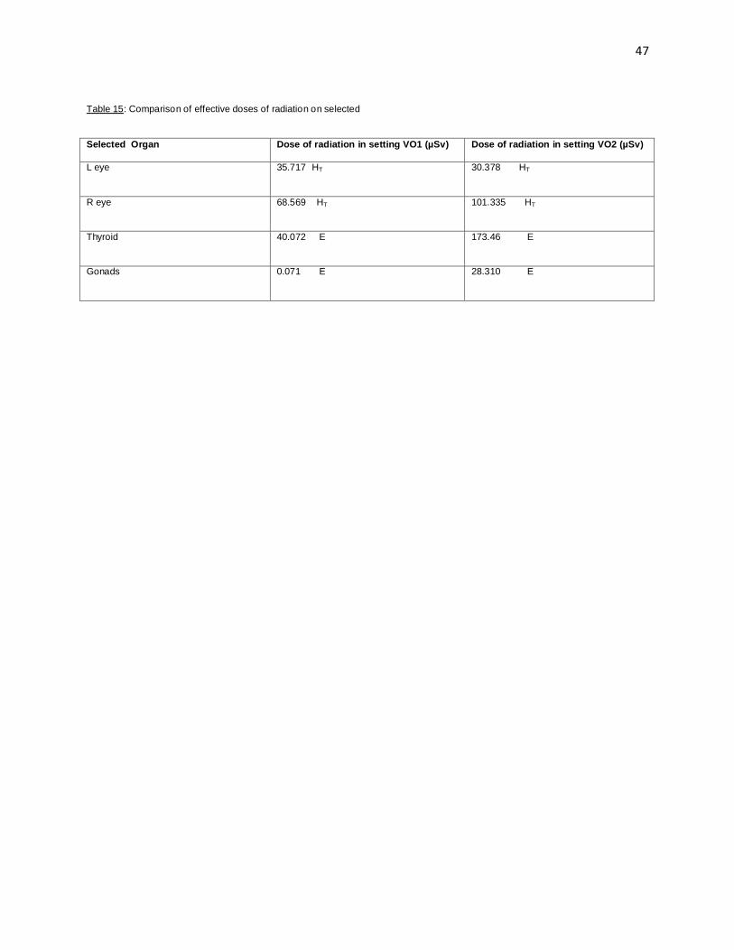
Table 15: Comparison of effective doses of radiation on selected
Selected Organ Dose of radiation in setting VO1 (µSv) Dose of radiation in setting VO2 (µSv)
L eye 35.717 HT 30.378 HT
R eye 68.569 HT 101.335 HT
Thyroid 40.072 E 173.46 E
Gonads 0.071 E 28.310 E
48
Appendix 9
Figures 2, 3, 4, 5
Fig 2: Copper plate with LIF TLDs Fig 3: Computer with softwares
Fig 4: Annealing oven: PTW Frieburg 1321® Fig 5: Hasrhaw 3500 ®reader

49
Appendix 10
Figures 6 and 7
Fig 6: Vacuum pump Fig 7: Rando® positioned inside the
CBCT

50
REFERENCES
1. Lofthag-Hansen S. Cone beam computed tomography radiation dose and image quality. Sweden Dental J Suppl. 2009. 209:4–55.
2. Arai Y, Tammisalo E, Iwai K, Hashimoto K, Shinoda K. Development of a compact computed tomographic apparatus for dental use. Dentomaxillofac Radiol.1999. 28(4):245–8.
3. Mozzo P, Procacci C, Tacconi A, Martini PT, Andreis IA. A new volumetric CT machine for dental imaging based on the cone-beam technique: preliminary results. Eur Radiol. 1998. 8:1558–64.
4. Durack C, Patel S. Cone beam computed tomography in endodontics. Brazilian dental journal. 2012. 179–91.
5. Adibi S, Zhang W, Servos T, O’Neill PN. Cone beam computed tomography in dentistry: what dental educators and learners should know. J Dent Educ. 2012. 76:1437–42.
6. Shintaku WH, Venturin JS, Azevedo B, Noujeim M. Applications of cone-beam computed tomography in fractures of the maxillofacial complex. Dent Traumatol. 2009;25:358–66.
7. Hashimoto K, Kawashima S, Araki M, Iwai K, Sawada K, Akiyama Y. Comparison of image performance between cone-beam computed tomography for dental use and four-row multidetector helical CT. J Oral Sci 2006. 48(1):27–34.
8. Hashimoto K, Kawashima S, Kameoka S, Akiyama Y, Honjoya T, Ejima K, et al. Comparison of image validity between cone beam computed tomography for dental use and multidetector row helical computed tomography. Dentomaxillofac Radiol. 2007. 36:465–71.
9. Schulze D, Heiland M, Thurmann H, Adam G. Radiation exposure during midfacial imaging using 4- and 16-slice computed tomography, cone beam computed tomography systems and conventional radiography. Dento maxillo facial radiology. 2004. 83–6.
10. Ludlow JB, Ivanovic M. Comparative dosimetry of dental CBCT devices and 64-slice CT for oral and maxillofacial radiology. Oral Surg Oral Med Oral Pathol Oral Radiol Endod 2008. 106(1):106–14.
11. Macleod I, Heath N. Cone-beam computed tomography (CBCT) in dental practice. Dent Update 2008. 35(9):590–2, 594–8.
12. Davies J, Johnson B, Drage N. Effective doses from cone beam CT investigation of the jaws. Dentomaxillofacial Radiology. 2012. 30–6.

51
13. Ludlow JB, Davies-Ludlow LE, Brooks SL. Dosimetry of two extraoral direct digital imaging devices: NewTom cone beam CT and Orthophos Plus DS panoramic unit. Dento maxillo facial radiology. 2003. 229–34.
14. Roberts JA, Drage NA, Davies J, Thomas DW. Effective dose from cone beam CT examinations in dentistry. Br J Radiol. 2009. 82:35–40.
15. Langland, Olaf E ; Langlais, Robert P: Preece JW. Principles of Dental Imaging.2nd ed. Lippincott Williams & Wilkins 2002.
16. Sandblom V. Evaluation of eye lens doses received by medical staff working in intervetional radilogy at sahlgrenska University Hospital.The Sahlgrenska Academy 2012.
17. Rehani MM, Vañó E, Ciraj-Bjelac O, Kleiman NJ. Radiation and cataract. Radiat Prot Dosim. 2011.147:300–4.
18. ICRP. The 2007 recommendations of the International Commission on Radiological Protection. Ann ICRP. 2007.37:1–332.
19. Mitchell RS, Kumar V, Abbas AK FN. Robbins Basic Pathology 8th Edition. 2007.
20. Thorsten G, Kolbeck Schieck JR, Pliska BT, Ahmad M, Larson BE. Dosimetry of a cone-beam computed tomography machine compared with a digital x-ray machine in orthodontic imaging. Am J Orthod Dentofac Orthop. 2012.141:436–43.
21. Lorenzoni DC, Bolognese AM, Garib DG, Guedes FR, Sant’anna EF. Cone-beam computed tomography and radiographs in dentistry: aspects related to radiation dose. Int J Dent. 2012. 2012:813768.
22. Chambers D.Effective dose of diierent scanning protocols using the Sirona Galileos comfort CBCT scanner.The School of graduate and Postdoctoral Studies, The University oe Westrn Ontario 2014
23. Wernli C. External Dosimetry. Operational Quantities and their Measurement. Health Protection Agency 2004.
24. Health Protection Agency. Application of the 2007 Recommendations of the ICRP to the UK. 2007
25. Buch B, Keddy RJ. Thermoluminescence dosimetry--how reliable is it? The Journal of the Dental Association of South Africa.1987.311–7.
26. Ludlow JB, Davies-Ludlow LE, Brooks SL, Howerton WB. Dosimetry of 3 CBCT devices for oral and maxillofacial radiology: CB Mercuray, NewTom 3G and i-CAT. Dentomaxillofac Radiol. 2006. 35(4):219–26.
52
27. Pauwels R, Beinsberger J, Collaert B, Theodorakou C, Rogers J, Walker A, et al. Effective dose range for dental cone beam computed tomography scanners. Eur J Radiol 2012. 81(2):267–71.
28. Akyalcin S, English DJ, Abramovitch KM RX. Measurement of skin dose from cone-beam computed tomography imaging. Head & Face Medicine. 2013. 9:28.
29. Sirona Dental systems. galileos operating instructions Galileos comfort. 2013.
30. Choo H, Mah JK. Three-Dimensional Cephalometrics in Clinical Practice: PCSO Bull. 2008. 25–9.
31. Buch B, Fensham R, Maritz MP. An assessment of the relative safety of dental x-ray equipment. SADJ : journal of the South African Dental Association. 2009. 348–50.
32. Buch B, Fensham R. Orthodontic radiographic procedures--how safe are they? SADJ : journal of the South African Dental Association. 2003. 6–10.
33. The Phantom laboratory. RAN100 The RANDO ® Phantom , RAN100 and RAN110 RAN100 phantom instruction booklet.1–3