effectiveness of 18f-fdg pet/ct in the diagnosis and
TRANSCRIPT

RESEARCH ARTICLE Open Access
Effectiveness of 18F-FDG PET/CT in thediagnosis and staging of osteosarcoma:a meta-analysis of 26 studiesFanxiao Liu1,2*† , Qingyu Zhang3†, Dongsheng Zhou1 and Jinlei Dong1
Abstract
Background: Multiple trials have attempted to assess the diagnostic value of 18F-fluorodeoxyglucose positron emissiontomography/computed tomography (18F-FDG PET/CT) in osteosarcoma with results remaining inconclusive. This studyaims to investigate the effectiveness of 18F-FDG PET and PET/CT in the diagnosis, staging, recurrence and metastasisformation observations of osteosarcoma through systematic review followed by meta-analysis.
Methods: Three electronic databases, Medline/PubMed, Embase and the Cochrane Library were utilized in this study.Eligible studies that assessed the performance of 18F-FDG PET/CT for the diagnosis, staging, restaging and recurrencemonitoring of osteosarcoma were retrieved utilizing specific search criteria. After screening and diluting out the non-conforming articles, all relevant articles and their data were identified and extracted to calculate the summary metricsinvolving sensitivity, specificity, diagnostic odd ratio (DOR), and area under the curve (AUC) to determine theeffectiveness of 18F-FDG PET in diagnosing osteosarcoma clinically.
Results: Out of 1976 articles searched, twenty-six studies were identified that were viable. All data from these articles,utilized in the quantitative analyses, showed after meta-analysis that when utilizing 18F-FDG PET or PET/CT it was betterwith a success rate of 90–100% for detecting primary lesions and distant metastases of patients with osteosarcoma.Similar results were also obtained for detecting lung and bone metastases in a subgroup analysis.
Conclusions: As such the investigation demonstrated that 18F-FDG PET and PET/CT are very accurate for the diagnosis,staging and recurrence monitoring of osteosarcoma. 18F-FDG-avid lesions should be further examined in osteosarcoma,especially for suspicious lung lesions.
Keywords: 18F-FDG PET, PET/CT, Metastases, Meta-analysis, Osteosarcoma, Diagnostic accuracy
BackgroundOsteosarcoma is the most frequent type of primary bonemalignancy in childhood and adolescence, which originatesfrom primitive mesenchymal stem cells that improperlyform osteoblasts that then deposit malignant osteoid [1].The combination of high-dose chemotherapy andlimb-salvage surgery has been shown to prolong the overall
survival of localized osteosarcoma to 65~70% [2]. However,the prognosis of patients with radiographically discernabledistant metastases is still unfavorable due to a large majo-rity of occult metastases present in lung, and the minorityin bone, lymph node and other parts of the body [3]. More-over, after limb-salvage operations, approximately 13.5% ofpatients have a local recurrence of the sarcoma [4]. Theoutcome of local recurrent osteosarcoma is even worsethan for patients with metastases alone [5]. Therefore, ac-curacy and early detection of local recurrence and distantmetastases formation have a crucial role in the risk stratifi-cation and the treatment of osteosarcoma.Several traditional imaging modalities have been used
for the diagnosis, staging and treatment monitoring ofosteosarcoma, such as plain radiographs, computed
© The Author(s). 2019 Open Access This article is distributed under the terms of the Creative Commons Attribution 4.0International License (http://creativecommons.org/licenses/by/4.0/), which permits unrestricted use, distribution, andreproduction in any medium, provided you give appropriate credit to the original author(s) and the source, provide a link tothe Creative Commons license, and indicate if changes were made. The Creative Commons Public Domain Dedication waiver(http://creativecommons.org/publicdomain/zero/1.0/) applies to the data made available in this article, unless otherwise stated.
* Correspondence: [email protected]; [email protected] Liu and Qingyu Zhang are first author’s: Contributed equally in theplanning, construction and writing of the manuscript as the first author.1Department of Orthopaedics, Shandong Provincial Hospital affiliated toShandong University, No.324, Road Jing Wu Wei Qi, Jinan 250021, Shandong,China2Department of Orthopaedic Surgery, Physical Medicine and Rehabilitation,University Hospital of Munich (LMU), Campus Grosshadern,Marchioninistrasse 23, 81377 Munich, GermanyFull list of author information is available at the end of the article
Liu et al. BMC Cancer (2019) 19:323 https://doi.org/10.1186/s12885-019-5488-5

tomography (CT) and magnetic resonance imaging(MRI). 18F-fluorodeoxyglucosepositron emission tomo-graphy (18F-FDG PET), through detecting the high up-take of 18F-FDG, a radioactive analogue of glucose,could identify sites with increased metabolic activity ofvarious malignant tumors. More recently, PET/CT,which combines metabolic data from PET and imagingdata from conventional CT, seems to be far more reli-able in diagnosing malignancies.A previous clinical study [6] had demonstrated that
osteosarcoma was 18F-FDG-avid. Uptake of 18F-FDG wasapplied for the diagnosis, chemotherapy response assess-ment, prognosis prediction and guidance of biopsies ofosteosarcoma [7]. Compared with bone scintigraphy,18F-FDG PET and PET/CT could identify bone, lung andother metastatic lesions. Subsequently, 18F-FDG PET andPET/CT have an advantage in assessing local recurrenceas they are not affected by imaging artifacts.Multiple studies have attempted to assess the diagnostic
accuracy of 18F-FDG PET and PET/CT in osteosarcoma.However, there seems to be considerable methodologicalvariability, including the methods for evaluating the FDGuptake and the standardized uptake value (SUV) at deter-mining whether the lesions are positive. Results remaininconclusive. Recently, two systematic review coupledmeta-analyses [8–10] tried to further clarify this issue, butnone of the included studies specifically aimed at osteosar-coma and did not statistically analyze the retrieveddata. 18F-FDG PET or PET/CT have not been consideredas the standardized components of the diagnostic algo-rithm for osteosarcoma. To reach a more precise result onthis topic, the present study sought to systematically col-lected previously published data from literatures and per-formed a statistically evaluation using meta-analysis to seeif 18F-FDG PET/CT is far more efficient at diagnosingosteosarcoma then present detection paradigms.
MethodsSearch strategy, inclusion and exclusion criteriaTwo investigators, blinded, independently and repeatedly,performed a systematic computerized article search usingthree databases (Medline/PubMed, Embase and theCochrane Library) with combinations of following keywords: “positron emission tomography” [all field] OR“PET” [all field] AND “osteosarcoma” [all field] OR “bonesarcoma” [all field]. No language limitations were imposed.This search process was completed on March 1, 2018 withno language and search limitations. Additionally, biblio-graphies of included studies were also searched by hand toexplore any potentially eligible trials.The targeted studies in the meta-analysis had to fulfill
all following criteria: a) clinical trials assessing the useful-ness of 18F-FDG PET or PET/CT in the diagnosis, staging,restaging and recurrence monitoring of osteosarcoma; b)
patients with osteosarcoma clinically diagnosed by histo-pathology, follow-up or other reference methods; c) suffi-cient data were provided to calculate the number oftrue-positive (TP), false-positive (FP), false-negative (FN)and true negative (TN) cases; d) if more than one articlecontained overlapping data, the most comprehensive orrecent article was included; and e) 18F-FDG was intraven-ously administered as an inducer.Exclusion criteria for this meta-analysis were: a) in vitro
or animal studies; b) trials with fewer than five participantswith osteosarcoma; c) posters presented at conferences/congresses (due to the lack of data and methodologydescription); d) not original research (reviews, editorials,meta-analyses, letters and comments).
Data extraction and quality assessmentThe following main information were extracted from ori-ginal articles: first author’s surname, year of publication,source of studies, basic characteristics of the participants(numbers, age and gender), study design, inclusion inter-val, technical details (image devices, injection dose andmethods of image analysis) and the time between injectionand image acquisition). Additionally, the cases of TP, FP,TN and FN were extracted directly or recalculatedthrough data presented in original articles based on differ-ent lesions such as primary, recurrence and metastases oflung and bone.The methodological quality of included studies was
appraised utilizing the QUADAS tool [11], which iscomposed of 14 items. Study following at least nineitems of these scores was deemed as high quality andwas included in this investigation.
Statistical analysisThis systematic review and meta-analysis confirmed thestandardized items described by “the Preferred ReportingItems for Systematic Reviews and Meta-Analyses(PRISMA)” statement [12]. To assess the accuracy of18F-FDG PET and PET/CT on the diagnosis, staging andrecurrence monitoring of osteosarcoma, the pooled esti-mates included the sensitivity, specificity, positive likelihoodratio (PLR), negative likelihood ratio (NLR), diagnostic oddratio (DOR) and the summary receiver operating characte-ristic curve (sROC) and AUC. Following recommendationsof Cochrance Handbook (www.cochrance.org/trainig/cochrance-handbook), study-heterogeneity was evaluatedusing Chi-squared and I-square statistic algorithms. Lowheterogeneity was defined as I-square < 50% and with P>0.1. To obtain a reliable result, all analyses were performedusing the random-effect model. The DOR is an indicator ofthe test accuracy, ranging from zero to infinity. A higherDOR indicates that the test is more accurate. The areasunder the curve (AUC) and Q*-index are two importantstatistics that reflect the diagnostic value. The AUC is
Liu et al. BMC Cancer (2019) 19:323 Page 2 of 12

defined as the area under sROC, and Q* is the pointwhere the sensitivity is equal to the specificity. The statis-tical analyses for the detection of primary lesions, recur-rence, lung metastases, bone metastases and all distantmetastases were performed separately.
ResultsStudy selectionAt primary retrieval, a total of 1952 articles were identi-fied, based on the search criteria, of which 1892 weresubsequently deemed non-viable after inclusion andexclusion criteria were applied. Out of the 84 articlesresulting from preliminary screening, 37 articles were re-ferred to irrelevant studies, 5 articles were duplicatepublications and 21 articles lacked sufficient data to cal-culate evaluation indicators and were excluded from themeta-analysis. The remaining 26 studies [13–38] deemedviable, published from 1998 to 2017, were included inthe meta-analysis. Although five of the trials [14–18]showed signs of having utilized the same patients, thesewere included in a different subgroup analyses. The se-lection process and reasons other articles were excludedare described in Fig. 1.
Characteristics of included studiesThe basic characteristics of the 26 included studies aresummarized in Table 1. The sizes of patients in the studiesranged from 5 to 206, and a total of 798 osteosarcoma pa-tients were included. A total of 17 studies were retrospect-ive design types whereas 8 studies were prospective. Basedon the QUADAS score, only one study [22] had a score of
9, nine studies [14, 16–18, 25, 30, 31, 34, 35] had a scoreof 11, five studies [15, 21, 23, 24, 32] a score of 12 with allremaining studies [13, 19, 20, 26–29, 33, 36–38] posses-sing a score of 13. The major information of PET or PET/CT imaging of each study was summarized in Table 2.Among them, sixteen studies presented that image acqui-sition were performed approximately 60min after FDG in-jection. The results of whole-body 18F-FDG PET orPET-CT were analyzed visually by experienced radiologistsin 7 studies [13, 20, 22, 30, 31, 34, 37] and by standardizeduptake values in 19 studies [14–19, 21, 23–29, 32, 33, 35,36, 38]. The diagnostic data (TPs, FPs, TNs and FNs) wereextracted directly or calculated through data provided inthe tables or bodies of each original article (Table 3).
Primary lesionThe diagnosis of the primary lesion was analyzed on apatient-based level. Fourteen studies [13, 15, 19, 21–27,30, 32, 33, 36] with 243 patients, with known osteosar-coma, were available. There were no threshold effects inthis data (p-value = 1.000). All primary lesions wereFDG-avid. The combined sensitivity of 18F-FDG PETand PET/CT in the detection of osteosarcoma was100%, while no specificity could be obtained. There wasno statistically significant heterogeneity in these esti-mates of sensitivity (I-square = 0.0%).
RecurrenceResults assessing the diagnostic performance of 18F-FDGPET and PET/CT for detecting recurrence of osteosarcomaas generated from the 7 studies [18, 24, 28, 29, 35, 37, 38],
Fig. 1 Selection process for studies included in the meta-analysis
Liu et al. BMC Cancer (2019) 19:323 Page 3 of 12

showed that the pooled sensitivity of 91% (95% CI of 81–96%), a specificity of 93% (95% CI of 87–97%), a PLR of7.36 (95% CI of 3.54–15.30), a NLR of 0.14 (95% CI of0.07–0.29), a DOR of 63.98 (95% CI of 19.29–212.18), theQ*-index of 0.8842 and AUC of 0.9452 (Fig. 2). No signifi-cance of heterogeneity between study for the pooled sensi-tivity (I-square = 0.0%) and the threshold effect (p-value =0.269) were presented in this analysis.
Lung metastasesIn total, 8 studies [14, 17, 28–30, 33, 34, 38] addressedthe diagnosis of lung metastases. The threshold effectswas no found in the data (p-value = 0.233). The com-bined sensitivity of 18F-FDG PET and PET/CT in detec-ting lung metastases of osteosarcoma was 81% (95% CIof 72–88%), specificity 94% (95% CI of 89–97%), PLR8.13 (95% CI of 4.19–15.77) and NLR 0.26 (95% of CI0.17–0.40). The pooled DOR was 48.85 (95% CI of
18.92–126.14), whereas the Q*-index was 0.8614 with anAUC of 0.9268 (Fig. 3).
Bone metastasesResults assessing the diagnostic performance of 18F-FDGPET and PET/CT for detecting bone metastasis of osteosar-coma as generated from the 6 studies [22, 29, 31, 33, 34,36], demonstrated that the pooled sensitivity of 93% (95%CI of 87–97%), a specificity of 97% (95% CI of 96–98%), aPLR of 9.81 (95% CI of 2.73–35.29), a NLR of 0.08 (95% CIof 0.04–0.18), a DOR of 174.19 (95% CI of 38.37–790.76),the Q*-index of 0.9397 and the AUC of 0.9813 (Fig. 4). Nosignificance of heterogeneity between study for the pooledsensitivity (I-square = 33.8%) and the threshold effect(p-value = 0.8279) were presented in this analysis.
All distant metastasesIn total, 11 studies [14, 17, 22, 28–31, 33, 34, 36, 38] ad-dressed the diagnosis of all distant metastases. There were
Table 1 Main characteristics of the included studies
Study, year Country No. ofSubjects
Gender(M/F)
Median/Meanage (years)
Age range(years)
Design Inclusion interval QUADASscores
Kole,1998 [13] Netherlands 5 3/2 20/20.2 17–24 Retrospective NR 13
Schulte,1999 [14] Germany 27 17/10 17/NR 5–36 Prospective Jan.1993-NR 11
Franzius,2000 [15] Germany 32 NR NR NR Retrospective Aug.1995-Jun.1999 11
Schulte,2000 [16] Germany 44 NR NR NR Retrospective Jan.1993-NR 12
Franzius,2001 [17] Germany 32 NR NR NR Retrospective Aug.1995-Jun.1999 11
Franzius,2002 [18] Germany 6 NR NR NR Retrospective NR 11
Yanagawa,2003 [19] Japan 5 4/1 14/14.4 11–20 Prospective Jun.1999-Mar.2000 13
Kneisl,2006 [20] USA 38 NR NR NR Retrospective Dec.1994-Nov.2004 13
Iagaru,2006 [21] USA 6 NR NR NR Retrospective Jan.2003-Dec.2005 12
Volker,2007 [22] Germany 11 NR NR 1–18 Prospective Dec.2003-Oct.2006 9
Shin,2008 [23] South Korea 7 NR NR 6–79 Retrospective May.2004-Jun.2007 12
Charest,2009 [24] Canada 24 NR NR NR Retrospective May.2004-Apr.2008 12
Hawkins,2009 [25] USA 40 NR 15.1/NR 7.1–31 Prospective Jul.1995-Aug.2004 11
Benz,2010 [26] USA 6 2/4 27/30.8 18–58 Prospective Feb.2005-Nov.2007 13
Lindholm,2011 [27] Finland 6 4/2 16.5/16.5 15–18 Prospective NR 13
Bandopadhyaya,2012 [30] India 22 14/8 21.55/NR 8–66 Prospective NR 13
Cistaro,2012 [28] Italy 11 NR NR/14 NR NR NR 13
Ozkan,2012 [29] Turkey 7 6/1 25/26.1 18–50 Retrospective 2007–2009 11
Bai,2013 [33] China 14 9/5 NR/14.9 8–22 Retrospective Jan.2009-Nov.2011 13
Byun,2013 [31] South Korea 206 127/79 15/NR 4–71 Retrospective Jan.2006-Nov.2011 11
Kong,2013 [32] South Korea 26 16/10 NR/21 9–55 Prospective May.2010-Aug.2011 12
Chang,2015 [35] South Korea 109 74/35 NR/17 NR Retrospective Feb.2002-Sep.2012 11
Quartuccio,2015 [34] Italy 20 10/10 15.5/NR NR Retrospective Jan.2006-Sep.2010 11
Hurley, 2016 [36] USA 39 19/20 Median 12 5–19 Retrospective 2003–2012 13
Angelini, 2017 [38] Italy 37 20/17 Mean 20 7–52 Retrospective 2008–2014 13
Sharp, 2017 [37] USA 8 5/3 Median 13 8–16 Retrospective Oct.2004-Feb. 2013 13
M male, F female, NR not reported
Liu et al. BMC Cancer (2019) 19:323 Page 4 of 12

no threshold effects in this data (p-value = 0.647). Thecombined sensitivity of 18F-FDG PET and PET/CT in de-tecting all distant metastases of osteosarcoma was 90%(95% CI of 86–93%), specificity 96% (95% CI of 95–97%),PLR 13.81 (95% CI of 5.77–33.06) and NLR 0.13 (95% CIof 0.07–0.23). Statistically heterogeneity was no found inthe estimates of the DOR (I-square = 49.8%). The pooledDOR was 125.67 (95% CI of 48.54–325.35). The Q*-indexwas 0.9103 with an AUC of 0.9639 (Fig. 5). To explorewhether the injected dose of 18F-FDG could affect thediagnostic accuracy of all distant metastases, we per-formed a subgroup analysis which demostrated that18F-FDG PET/CT, using a injected dose above 5MBq/kg,is similar to that using a lower dose (sensitivity of 88%[95% CI 76–95%] vs. 88% [95% CI 78–95%]; specificity of93% [95% CI 97–99%] vs. 98% [95% CI 97–99%]).
DiscussionOsteogenic sarcoma has an elevated rate of glycolysis and,consequently, a high uptake of 18F-FDG in malignant cells
[39] . A previous meta-analysis [40] revealed that the stan-dardized uptake values before (SUV1) and after (SUV2)chemotherapy in osteosarcoma were associated with thehistological response. SUV2:1< 0.5 or SUV2< 2.5 had pre-dictive significance for tumor necrosis. Multiple studiesinvestigated the diagnostic value of 18F-FDG PET andPET/CT for osteosarcoma, but no definitive results wereobtained. To settle these questions scientifically, in thepresent study, a rigorous inclusion criterion was prede-signed to collect published articles as comprehensive aspossible and subgroup analyses were conducted to pooloutcome estimates from individual studies only utilizingthe random-effect modeling. Importantly, this is the firstmeta-analysis evaluating the diagnostic utility of 18F-FDGPET/CT in osteosarcoma. By systemically analyzing theretrieved data, investigations demonstrated that 18F-FDGPET/CT had an excellent accuracy in the diagnosis, sta-ging and restaging of osteosarcoma.The value of PET in the differential diagnosis of bone
tumor and tumor-like lesions was first described by
Table 2 Technical aspects of the included studies
Study, year Image device Injected dose Time between injection and image acquisition PET image analysis
Kole, 1998 [13] PET 370 MBq 50 min Visual
Schulte, 1999 [14] PET 120–300 MBq 45–60 min Visual and semiquantitative
Franzius, 2000 [15] PET 3.7 MBq/kg 60 min Visual and semiquantitative
Schulte, 2000 [16] PET 120–300 MBq 45–60 min Visual and semiquantitative
Franzius, 2001 [17] PET 3.7 MBq/kg 60 min Visual and semiquantitative
Franzius, 2002 [18] PET 3.7 MBq/kg 60 min Visual and semiquantitative
Yanagawa, 2003 [19] PET 4.5 MBq/kg 50 min Visual and semiquantitative
Kneisl, 2006 [20] PET 444–740 MBq 60 min Visual
Iagaru, 2006 [21] PET/CT 151.7–721.5 MBq 60 min Visual and semiquantitative
Volker, 2007 [22] PET NR NR Visual
Shin, 2008 [23] PET/CT 8.14 MBq/kg 60 min Visual and semiquantitative
Charest, 2009 [24] PET/CT 379–500 MBq 60 min Visual and semiquantitative
Hawkins, 2009 [25] PET 259–370 MBq 45 min Visual and semiquantitative
Benz, 2010 [26] PET/CT 7.77 MBq/kg 60 min Visual and semiquantitative
Lindholm, 2011 [27] PET/CT 370 MBq 60 min Visual and semiquantitative
Bandopadhyaya, 2012 [30] PET/CT 370 MBq 60 min Visual
Cistaro, 2012 [28] PET/CT 120–277 MBq 60 min Visual and semiquantitative
Ozkan, 2012 [29] PET/CT 555 MBq 60 min Visual and semiquantitative
Bai, 2013 [33] PET/CT 3.5–5.7 MBq/kg 40–60 min Visual and semiquantitative
Byun, 2013 [31] PET/CT 7.4 MBq/kg or 370 MBq 60 min Visual
Kong, 2013 [32] PET/CT 8.14 MBq/kg NR Visual and semiquantitative
Chang, 2015 [35] PET/CT 7.4 MBq/kg or 370 MBq 60 min Visual and semiquantitative
Quartuccio, 2015 [34] PET/CT 113–596 MBq 72 min Visual
Hurley, 2016 [36] PET/CT 5.55 MBq/kg 60 min Visual and semiquantitative
Angelini, 2017 [38] PET/CT 5.55 MBq/kg NR Visual and semiquantitative
Sharp, 2017 [37] PET/CT 5.18 or 5.55 MBq/kg 60 min Visual
NR not reported
Liu et al. BMC Cancer (2019) 19:323 Page 5 of 12

Table 3 Diagnosis accuracy data on each examination– or lesion–based analysis
Study, year Total TP FP FN TN Lesions sites
Kole, 1998 [13] 5 5 0 0 0 Primary lesion
Schulte, 2000 [14] 44 44 0 0 0 Primary lesion
Yanagawa, 2003 [19] 5 5 0 0 0 Primary lesion
Iagaru, 2006 [21] 6 6 0 0 0 Primary lesion
Volker, 2007 [22] 11 11 0 0 0 Primary lesion
Shin, 2008 [23] 7 7 0 0 0 Primary lesion
Charest, 2009 [24] 12 12 0 0 0 Primary lesion
Hawkins, 2009 [25] 40 40 0 0 0 Primary lesion
Benz, 2010 [26] 6 6 0 0 0 Primary lesion
Lindholm, 2011 [27] 6 6 0 0 0 Primary lesion
Bandopadhyaya, 2012 [30] 22 22 0 0 0 Primary lesion
Bai, 2013 [33] 14 14 0 0 0 Primary lesion
Kong, 2013 [32] 26 26 0 0 0 Primary lesion
Hurley, 2016 [36] 39 39 0 0 0 Primary lesion
Franzius, 2002 [18] 6 6 0 0 0 Recurrence
Charest, 2009 [24] 12 6 0 1 5 Recurrence
Cistaro, 2012 [28] 11 10 0 0 1 Recurrence
Ozkan, 2012 [29] 8 3 0 0 5 Recurrence
Chang, 2015 [35] 109 7 6 2 94 Recurrence
Angelini, 2017 [38] 37 22 0 1 14 Recurrence
Sharp, 2017 [37] 10 10 0 0 0 Recurrence
Schulte, 1999 [14] 27 4 0 0 23 Lung
Franzius, 2001 [17] 49 4 0 4 41 Lung
Bandopadhyaya, 2012 [30] 22 10 1 0 11 Lung
Cistaro, 2012 [28] 23 16 0 3 4 Lung
Ozkan, 2012 [29] 8 1 0 0 7 Lung
Bai, 2013 [33] 14 2 0 0 12 Lung
Quartuccio, 2015 [34] 56 27 5 5 19 Lung
Angelini, 2017 [38] 37 12 0 3 22 Lung
Volker, 2007 [22] 31 28 0 3 0 Bone
Ozkan, 2012 [29] 8 1 0 0 7 Bone
Bai, 2013 [33] 14 7 0 0 7 Bone
Byun, 2013 [31] 833 52 15 3 763 Bone
Quartuccio, 2015 [34] 21 16 1 0 4 Bone
Hurley, 2016 [36] 40 5 3 0 32 Bone
Schulte,1999 [14] 27 4 0 0 23 All metastatic lesions
Franzius, 2001 [17] 49 4 0 4 41 All metastatic lesions
Volker, 2007 [22] 42 31 0 3 8 All metastatic lesions
Bandopadhyaya, 2012 [30] 22 10 1 0 11 All metastatic lesions
Cistaro, 2012 [28] 38 27 2 3 6 All metastatic lesions
Ozkan, 2012 [29] 8 3 0 0 5 All metastatic lesions
Bai, 2013 [33] 28 9 0 0 19 All metastatic lesions
Byun, 2013 [31] 833 52 15 3 763 All metastatic lesions
Quartuccio, 2015 [34] 101 59 9 5 28 All metastatic lesions
Liu et al. BMC Cancer (2019) 19:323 Page 6 of 12

Schulte et al.(2000) [15]. Using a tumor-to-backgroundratio of 3.0 as the cutoff for determining malignant bonelesions, the sensitivity, specificity and accuracy were0.93, 0.667 and 0.817, respectively. Although there wereseveral false negative cases, there were none for patientswith osteosarcoma (n = 44). A subsequent meta-analysis[41] also reported the outstanding ability of 18F-FDGPET to distinguish between benign and malignant boneand soft tissue tumors. In studies included in the presentmeta-analysis, all primary osteosarcoma lesions (n = 243)were 18F-FDG-avid with a good pooled sensitivity of100%. However, it must be noted, that as a nonspecificmanifestation on 18F-FDG PET, osteosarcoma is not dis-tinctly discernible from other highly malignant bone sar-comas, such as Ewing sarcoma [42].Detecting recurrent or residual osteosarcoma has been
a challenge for clinicians due to the post-therapeuticchanges and imaging artifacts caused by metallic endo-prothesis [43]. In 1996, Garcia and co-workers reportedthe diagnostic value of 18F-FDG PET in 48 suspected
recurrent musculoskeletal sarcomas (including 18 osteo-sarcoma patients), indicating a good sensitivity andspecificity (98 and 90%, respectively) [44]. 18F-FDG PEThas an advantage in assessing local recurrence because itis not affected by the imaging artifact. Nevertheless,elevated 18F-FDG uptake could be affected bypost-treatment changes, irrespective of local recurrence[45, 46]. In this investigation, a good accuracy was ob-served of 18F-FDG PET in detecting recurrent osteosar-coma (local relapses or distant metastases), which aresimilar to the conclusions for other recurrent malignanttumors [47, 48].Although osteosarcoma has a tendency to metastasize
early, which would modify the outcome of osteosarcomawith an unfavorable survival, the prognosis of patients withresectable metastatic lesions is relatively good [49–51].18F-FDG PET and PET/CT are useful tools for detectingpossible malignant lesions. Therefore, a subgroup analysiswas performed on the metastasis-based data in thepresented study.
Table 3 Diagnosis accuracy data on each examination– or lesion–based analysis (Continued)
Study, year Total TP FP FN TN Lesions sites
Hurley, 2016 [36] 105 20 8 4 73 All metastatic lesions
Angelini, 2017 [38] 74 29 1 3 41 All metastatic lesions
TP True positive, FP False positive, FN False negative, TN True negative
Fig. 2 Diagnostic performance of 18F-FDG PET and PET/CT for the recurrence of osteosarcoma: (1) Pooled sensitivity (2) Pooled specificity (3)Pooled diagnostic odds ratio (4) Summary receiver operating characteristic curve (sROC) with the Q*-index
Liu et al. BMC Cancer (2019) 19:323 Page 7 of 12

Fig. 3 Diagnostic performance of 18F-FDG PET and PET/CT for osteosarcoma lung metastasis: (1) Pooled sensitivity (2) Pooled specificity (3) Pooleddiagnostic odds ratio (4) Summary receiver operating characteristic curve (sROC) with the Q*-index
Fig. 4 Diagnostic performance of 18F-FDG PET and PET/CT for osteosarcoma bone metastasis: (1) Pooled sensitivity (2) Pooled specificity(3) Pooled diagnostic odds ratio (4) Summary receiver operating characteristic curve (sROC) with the Q*-index
Liu et al. BMC Cancer (2019) 19:323 Page 8 of 12
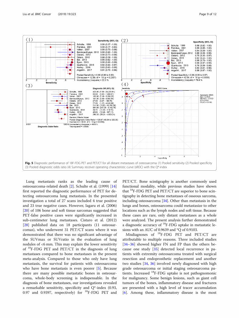
Lung metastasis ranks as the leading cause ofosteosarcoma-related death [2]. Schulte et al. (1999) [14]first reported the diagnostic performance of PET for de-tecting osteosarcoma lung metastasis. In the presentedinvestigation a total of 27 scans included 4 true positiveand 23 true negative cases. However, Iagaru et al. (2006)[20] of 106 bone and soft tissue sarcomas suggested thatPET-false positive cases were significantly increased insub-centimeter lung metastases. Cistaro et al. (2012)[28] published data on 18 participants (11 osteosar-comas), who underwent 31 PET/CT scans where it wasdemonstrated that there was no significant advantage ofthe SUVmax or SUVratio in the evaluation of lungnodules of <6 mm. This may explain the lower sensitivityof 18F-FDG PET and PET/CT in the diagnosis of lungmetastases compared to bone metastases in the presentmeta-analysis. Compared to those who only have lungmetastasis, the survival for patients with osteosarcomawho have bone metastasis is even poorer [5]. Becausethere are many possible metastatic bones in osteosar-coma, whole-body screening is indispensable. In thediagnosis of bone metastases, our investigations revealeda remarkable sensitivity, specificity and Q*-index (0.93,0.97 and 0.9397, respectively) for 18F-FDG PET and
PET/CT. Bone scintigraphy is another commonly usedfunctional modality, while previous studies have shownthat 18F-FDG PET and PET/CT are superior to bone scin-tigraphy in detecting bone metastases of osseous sarcoma,including osteosarcoma [34]. Other than metastasis in thelungs and bones, osteosarcoma could metastasize to otherlocations such as the lymph nodes and soft tissue. Becausethese cases are rare, only distant metastases as a wholewere analyzed. The present analysis further demonstrateda diagnostic accuracy of 18F-FDG uptake in metastatic le-sions with an AUC of 0.9639 and *Q of 0.9103.Misdiagnoses of 18F-FDG PET and PET/CT are
attributable to multiple reasons. Three included studies[34–36] showed higher FN and FP than the others be-cause one study [35] detected local recurrence in pa-tients with extremity osteosarcoma treated with surgicalresection and endoprosthetic replacement and anothertwo studies [34, 36] involved newly diagnosed with highgrade osteosarcoma or initial staging osteosarcoma pa-tients. Increased 18F-FDG uptake is not pathognomonicfor malignancy. Some benign lesions, such as giant celltumors of the bones, inflammatory disease and fracturesare presented with a high level of tracer accumulation[6]. Among these, inflammatory disease is the most
Fig. 5 Diagnostic performance of 18F-FDG PET and PET/CT for all distant metastases of osteosarcoma: (1) Pooled sensitivity (2) Pooled specificity(3) Pooled diagnostic odds ratio (4) Summary receiver operating characteristic curve (sROC) with the Q*-index
Liu et al. BMC Cancer (2019) 19:323 Page 9 of 12

commonly encountered causes of a false positive. There-fore, the findings of 18F-FDG PET/CT for malignant le-sions should be confirmed with a histopathologyexamination or follow-up. Meanwhile, some false nega-tive cases are unavoidable. One cause is the nonspecific18F-FDG uptake in malignant diseases and asymmetric18F-FDG distribution. Second, because of the limitedspatial resolution of 18F-FDG PET, some occult orsub-centimeter lesions could not be identified. Currently,the high cost of 18F-FDG PET and PET/CT discouragestheir use in developing countries [52–54]. However, consid-ering their satisfactory performance, as demonstrated bythe present investigation, and the poor prognosis of osteo-sarcoma, especially for those with recurrence or distantmetastases, patients may benefit from evaluation with18F-FDG PET or PET/CT.Some limitations of this meta-analysis merit conside-
rations. First, the present analysis was a meta-analysisand systemic review; therefore, we were only able toanalyze questions on the study level instead of on thepatient level. Second, as a result of a lack of publicationsassessing the effectiveness of 18F-FDG PET and PET/CTfor detecting osteosarcoma, several sub-group analysesin our investigation were performed on a small numberof studies. Third, there was methodological variability inthe included studies, such as in the SUV, reference stan-dards tests and duration of follow-up. Furthermore,some studies were assessed using 18F-FDG PET, whileothers were assessed with PET/CT. Finally, some infor-mation was not available in the included studies.
ConclusionsIn summary, our comprehensive meta-analysis of publica-tions demonstrated that 18F-FDG PET/CT have an excel-lent accuracy in the diagnosis, staging and recurrencemonitoring of osteosarcoma. 18F-FDG-avid lesions shouldbe further examined in osteosarcoma, especially for suspi-cious lung lesions. To support the current conclusions,larger-scale trials should now be conducted.
Abbreviations18F-FDG: 18F-fluorodeoxyglucose; AUC: Area under the curve; CI: Confidenceinterval; CT: Computed tomography; DOR: Diagnostic odd ratio; FN: False-negative; FP: False-positive; MRI: Magnetic resonance imaging; NLR: Negativelikelihood ratio; PET: Positron emission tomography; PLR: Positive likelihoodratio; PRISMA: Preferred Reporting Items for Systematic Reviews and Meta-Analyses; sROC: The summary receiver operating characteristic curve;SUV: Standardized uptake value; TN: True negative; TP: True-positive
AcknowledgementsI would like to express my very great appreciation to Roland M. Klar for hisvaluable and constructive suggestions during the planning and developmentof this work.
FundingThis study was funded by the Key R & D program in Shandong Province(Jinlei Dong, NO. 2016GSF201214) and the China Scholarship Council(Fanxiao Liu, No. 201808080126). The funding agencies were not involved in
the design of the study and collection, analysis, and interpretation of dataand in writing the manuscript.
Availability of data and materialsAll data analyzed during this study are included in this published article.
Authors’ contributionsLFX contributed to the idea of this study. LFX, ZQY and DJL searchedliteratures and screened them independently. Any disagreement was solvedby consulting the senior authors (DJL). LFX, ZQY and DJL screened data fromthe eleven final articles and make Tables. LFX and ZQY played an importantrole in analyzing the outcomes. LFX and ZQY conducted the data analysesand make graphs. LFX, ZQY, ZDS and DJL wrote the first draft. LFX, ZQY, ZDSand DJL revised the manuscript. LFX, ZQY, ZDS and DJL polished the draft.LFX, ZQY, ZDS and DJL approved the final version.
Ethics approval and consent to participateThis article does not contain any studies with human participants or animalsperformed by any of the authors.
Consent for publicationNot applicable.
Competing interestsThe authors declare that they have no competing interests.
Publisher’s NoteSpringer Nature remains neutral with regard to jurisdictional claims in publishedmaps and institutional affiliations.
Author details1Department of Orthopaedics, Shandong Provincial Hospital affiliated toShandong University, No.324, Road Jing Wu Wei Qi, Jinan 250021, Shandong,China. 2Department of Orthopaedic Surgery, Physical Medicine andRehabilitation, University Hospital of Munich (LMU), Campus Grosshadern,Marchioninistrasse 23, 81377 Munich, Germany. 3Department ofOrthopeadics, Qilu Hospital, Shandong University, Jinan, Shandong, China.
Received: 22 November 2018 Accepted: 19 March 2019
References1. Biermann JS, Adkins DR, Agulnik M, Benjamin RS, Brigman B, Butrynski JE,
Cheong D, Chow W, Curry WT, Frassica DA, et al. National comprehensivecancer network. J Natal Comp Can Newt. 2013;11(6):688–723.
2. Mirabella L, Trios RJ, Savage SA. Osteosarcoma incidence and survival ratesfrom 1973 to 2004: data from the surveillance, epidemiology, and endresults program. Cancer. 2009;115(7):1531–43.
3. Su W, Lai Z, Wu F, Lin Y, Mo Y, Yang Z, Wu J. Clinical efficacy ofpreoperative chemotherapy with or without ifosfamide in patients withosteosarcoma of the extremity: meta-analysis of randomized controlledtrials. Med Oncol. 2015;32(2):481.
4. Yin K, Liao Q, Zhong D, Ding J, Niu B, Long Q, Ding D. Meta-analysis of limbsalvage versus amputation for treating high-grade and localizedosteosarcoma in patients with pathological fracture. Exp Ther Med. 2012;4(5):889–94.
5. Takeuchi A, Lewis VO, Satcher RL, Moon BS, Lin PP. What are the factorsthat affect survival and relapse after local recurrence of osteosarcoma? ClinOrthop Relat Res. 2014;472(10):3188–95.
6. Peller PJ. Role of positron emission tomography/computed tomography inbone malignancies. Radiol Clin N Am. 2013;51(5):845–64.
7. Palmerini E, Colangeli M, Nanni C, Fanti S, Marchesi E, Paioli A, Picci P,Cambioli S, et al. The role of FDG PET/CT in patients treated withneoadjuvant chemotherapy for localized bone sarcomas. Eur J Nucl MedMol Imaging. 2017;44(2):215–23.
8. Quartuccio N, Treglia G, Salsano M, Mattoli MV, Muoio B, Piccardo A, LopciE, Cistaro A. The role of Fluorine-18-Fluorodeoxyglucose positron emissiontomography in staging and restaging of patients with osteosarcoma. RadiolOncol. 2013;47(2):97–102.
9. Liu F, Zhang Q, Zhu D, Li Z, Li J, Wang B, Zhou D, Dong J. Performanceof positron emission tomography and positron emission tomography/
Liu et al. BMC Cancer (2019) 19:323 Page 10 of 12

computed tomography using Fluorine-18-Fluorodeoxyglucose for thediagnosis, staging, and recurrence assessment of bone sarcoma: a systematicreview and meta-analysis. Medicine (Baltimore). 2015;94(36):e1462.
10. Huang T, Li F, Yan Z, Ma Y, Xiong F, Cai X, Zhang Q, Liu F, et al. Effectiveness of18F-FDG PET/CT in the diagnosis, staging and recurrence monitoring of Ewingsarcoma family of tumors: a meta-analysis of 23 studies. Medicine (Baltimore).2018;97(48):e13457.
11. Whiting P, Rutjes AW, Reitsma JB, Bossuyt PM, Kleijnen J. Thedevelopment of QUADAS: a tool for the quality assessment of studiesof diagnostic accuracy included in systematic reviews. BMC Med ResMethodol. 2003;3:25.
12. Liberati A, Altman DG, Tetzlaff J, Mulrow C, Gøtzsche PC, Ioannidis JP, ClarkeM, Devereaux PJ, et al. Preferred reporting items for systematic reviews andmeta-analyses: the PRISMA statement. PLoS Med. 2009;6(7):e1000097.
13. Kole AC, Nieweg OE, Hoekstra HJ, van Horn JR, Koops HS, Vaalburg W.Fluorine-18-fluorodeoxyglucose assessment of glucose metabolism in bonetumors. J Nucl Med. 1998;39(5):810–5.
14. Schulte M, Brecht-Krauss D, Werner M, Hartwig E, Sarkar MR, Keppler P,Kotzerke J, Guhlmann A, et al. Evaluation of neoadjuvant therapy responseof osteogenic sarcoma using FDG PET. J Nucl Med. 1999;40(10):1637–43.
15. Schulte M, Brecht-Krauss D, Heymer B, Guhlmann A, Hartwig E, Sarkar MR,Diederichs CG, Von Baer A, et al. Grading of tumors and tumorlike lesions ofbone: evaluation by FDG PET. J Nucl Med. 2000;41(10):1695–701.
16. Franzius C, Sciuk J, Daldrup-Link HE, Jürgens H, Schober O. FDG-PET fordetection of osseous metastases from malignant primary bone tumours:comparison with bone scintigraphy. Eur J Nucl Med. 2000;27(9):1305–11.
17. Franzius C, Daldrup-Link HE, Sciuk J, Rummeny EJ, Bielack S, Jürgens H, SchoberO. FDG-PET for detection of pulmonary metastases from malignant primary bonetumors: comparison with spiral CT. Ann Oncol. 2001;12(4):479–86.
18. Franzius C, Daldrup-Link HE, Wagner-Bohn A, Sciuk J, Heindel WL, JürgensH, Schober O. FDG-PET for detection of recurrences from malignant primarybone tumors: comparison with conventional imaging. Ann Oncol. 2002;13(1):157–60.
19. Yanagawa T, Watanabe H, Inoue T, Ahmed AR, Tomiyoshi K, Shinozaki T,Oriuchi N, Endo K, et al. Carbon-11 choline positron emission tomographyin musculoskeletal tumors: comparison with fluorine-18 fluorodeoxyglucosepositron emission tomography. J Comput Assist Tomogr. 2003;27(2):175–82.
20. Kneisl JS, Patt JC, Johnson JC, Zuger JH. Is PET useful in detecting occultnonpulmonary metastases in pediatric bone sarcomas? Clin Orthop RelatRes. 2006;450:101–4.
21. Iagaru A, Quon A, McDougall IR, Gambhir SS. F-18 FDG PET/CT evaluation ofosseous and soft tissue sarcomas. Clin Nucl Med. 2006;31(12):754–60.
22. Völker T, Denecke T, Steffen I, Misch D, Schönberger S, Plotkin M, Ruf J,Furth C, et al. Positron emission tomography for staging of pediatricsarcoma patients: results of a prospective multicenter trial. J Clin Oncol.2007;25(34):5435–41.
23. Shin DS, Shon OJ, Han DS, Choi JH, Chun KA, Cho IH. The clinical efficacy of18F-FDG-PET/CT in benign and malignant musculoskeletal tumors. AnnNucl Med. 2008;22(7):603–9.
24. Charest M, Hickeson M, Lisbona R, Novales-Diaz JA, Derbekyan V,Turcotte RE. FDG PET/CT imaging in primary osseous and soft tissuesarcomas: a retrospective review of 212 cases. Eur J Nucl Med MolImaging. 2009;36(12):1944–51.
25. Hawkins DS, Conrad EU 3rd, Butrynski JE, Schuetze SM, Eary JF. [F-18]-fluorodeoxy-D-glucose-positron emission tomography response isassociated with outcome for extremity osteosarcoma in children and youngadults. Cancer. 2009;115(15):3519–25.
26. Benz MR, Czernin J, Tap WD, Eckardt JJ, Seeger LL, Allen-Auerbach MS, DrySM, Phelps ME, Weber WA, Eilber FC. FDG-PET/CT imaging predictshistopathologic treatment responses after neoadjuvant therapy in adultprimary bone sarcomas. Sarcoma. 2010;2010:143540.
27. Lindholm P, Sutinen E, Oikonen V, Mattila K, Tarkkanen M, Kallajoki M, Aro H,Böhling T, et al. PET imaging of blood flow and glucose metabolism inlocalized musculoskeletal tumors of the extremities. Nucl Med Biol. 2011;38(2):295–300.
28. Cistaro A, Lopci E, Gastaldo L, Fania P, Brach Del Prever A, Fagioli F. The role of18F-FDG PET/CT in the metabolic characterization of lung nodules in pediatricpatients with bone sarcoma. Pediatr Blood Cancer. 2012;59(7):1206–10.
29. Iagaru A, Quon A, McDougall IR, Gambhir SS. Clinical experience of 18F-FDGPET/CT in soft tissue and osseous sarcomas. UHOD-Uluslararasi Hematoloji-Onkoloji Dergisi. 2012;22(3):163–9.
30. Bandopadhyaya GP, Gupta P, Singh A, Shukla J, Rastogi S, Kumar R,Malhotra A. (99m) Tc-DMSA (V) in evaluation of osteosarcoma: comparativestudies with (18) F-FDG PET/CT in detection of primary and malignantlesions. ISRN Oncol. 2012;2012:371830.
31. Byun BH, Kong CB, Lim I, Kim BI, Choi CW, Song WS, Cho WH, Jeon DG,et al. Comparison of (18) F-FDG PET/CT and (99 m) Tc-MDP bonescintigraphy for detection of bone metastasis in osteosarcoma. SkeletRadiol. 2013;42(12):1673–81.
32. Kong CB, Byun BH, Lim I, Choi CW, Lim SM, Song WS, Cho WH, Jeon DG,et al. 18F-FDG PET SUVmax as an indicator of histopathologic response afterneoadjuvant chemotherapy in extremity osteosarcoma. Eur J Nucl Med MolImaging. 2013;40(5):728–36.
33. Bai CJ, Zhu R, Fang ZW. A comparison between preoperative PET/CT andCT images of long bone osteosarcoma of the lower extremity. Acad JSecond Mil. 2013;34(1):100–3.
34. Quartuccio N, Fox J, Kuk D, Wexler LH, Baldari S, Cistaro A, Schöder H.Pediatric bone sarcoma: diagnostic performance of 18F-FDG PET/CT versusconventional imaging for initial staging and follow-up. Am J Roentgenol.2015;204(1):153–60.
35. Chang KJ, Kong CB, Cho WH, Jeon DG, Lee SY, Lim I, Lim SM. Usefulness ofincreased 18F-FDG uptake for detecting local recurrence in patients withextremity osteosarcoma treated with surgical resection and endoprostheticreplacement. Skelet Radiol. 2015;44(4):529–37.
36. Hurley C, McCarville MB, Shulkin BL, Mao S, Wu J, Navid F, Daw NC, PappoAS, et al. Comparison of 18F-FDG-PET-CT and bone scintigraphy forevaluation of osseous metastases in newly diagnosed and recurrentosteosarcoma. Pediatr Blood Cancer. 2016;63(8):1381–6.
37. Sharp SE, Shulkin BL, Gelfand MJ, McCarville MB. FDG PET/CT appearance oflocal osteosarcoma recurrences in pediatric patients. Pediatr Radiol. 2017;47(13):1800–8.
38. Angelini A, Ceci F, Castellucci P, Graziani T, Polverari G, Trovarelli G, Palmerini E,Ferrari S, et al. The role of 18F-FDG PET/CT in the detection of osteosarcomarecurrence. Eur J Nucl Med Mol Imaging. 2017;44(10):1712–20.
39. Choi YY, Kim JY, Yang SO. PET/CT in benign and malignant musculoskeletaltumors and tumor-like conditions. Semin Musculoskelet Radiol. 2014;18(2):133–48.
40. Hongtao L, Hui Z, Bingshun W, Xiaojin W, Zhiyu W, Shuier Z, Aina H,Yuanjue S, et al. 18F-FDG positron emission tomography for the assessmentof histological responseto neoadjuvant chemotherapy in osteosarcomas: ameta-analysis. Surg Oncol. 2012;21(4):e165–70.
41. Bastiaannet E, Groen H, Jager PL, Cobben DC, van der Graaf WT, VaalburgW, Hoekstra HJ. The value of FDG-PET in the detection, grading andresponse to therapy of soft tissue and bone sarcomas: a systematic reviewand meta-analysis. Cancer Treat Rev. 2004;30(1):83–101.
42. Costelloe CM, Chuang HH, Daw NC. PET/CT of osteosarcoma and Ewingsarcoma. Semin Roentgenol. 2017;52(4):255–68.
43. Boas FE, Fleischmann D. Evaluation of two iterative techniques for reducingmetal artifacts in computed tomography. Radiology. 2011;259(3):894–902.
44. Garcia R, Kim EE, Wong FC, Korkmaz M, Wong WH, Yang DJ, Podoloff DA.Comparison of fluorine-18-FDG PET and technetium-99m-MIBI SPECT inevaluation ofmusculoskeletal sarcomas. J Nucl Med. 1996;37(9):1476–9.
45. Zhuang H, Chacko TK, Hickeson M, Stevenson K, Feng Q, Ponzo F,Garino JP, Alavi A. Persistent non-specific FDG uptake on PET imagingfollowing hip arthroplasty. Eur J Nucl Med Mol Imaging. 2002;29(10):1328–33.
46. Heiba SI, Luo J, Sadek S, Macalental E, Cacavio A, Rosen G, Abdel-DayemHA. Attenuation-correction induced artifact in F-18 FDG PET imagingfollowing Total knee replacement. Clin Positron Imaging. 2000;3(6):237–9.
47. Zou H, Zhao Y. 18F-FDG PET-CT for detecting gastric cancer recurrence aftersurgical resection: ameta-analysis. Surg Oncol. 2013;22(3):162–6.
48. Poulou LS, Ziakas PD, Ziogas DC, Doxani C, Xyla V, Vakrinos G, Voulgarelis M,Thanos L. FDG-PET for detecting local tumor recurrence of ablated livermetastases: a diagnostic meta-analysis. Biomarkers. 2012;17(6):532–8.
49. Zhao H, Yao Y, Wang Z, Lin F, Sun Y, Chen P. Therapeutic effect ofpirarubicin-based chemotherapy for osteosarcoma patients with lungmetastasis. J Chemother. 2010;22(2):119–24.
50. Bacci G, Longhi A, Bertoni F, Briccoli A, Versari M, Pignotti E, Picci P. Bonemetastases in osteosarcoma patients treated with neoadjuvant or adjuvantchemotherapy: the Rizzoli experience in 52 patients. Acta Orthop. 2006;77(6):938–43.
51. San-Julian M, Diaz-de-Rada P, Noain E, Sierrasesumaga L. Bone metastasesfrom osteosarcoma. Int Orthop. 2003;27(2):117–20.
Liu et al. BMC Cancer (2019) 19:323 Page 11 of 12

52. Wang YT, Huang G. Is FDG PET/CT cost-effective for pre-operation stagingof potentially operative non-small cell lung cancer? - from Chinesehealthcare system perspective. Eur J Radiol. 2012;81(8):e903–9.
53. Keith CJ, Miles KA, Griffiths MR, Wong D, Pitman AG, Hicks RJ. Solitarypulmonary nodules: accuracy and cost-effectiveness of sodium iodide FDG-PET using Australian data. Eur J Nucl Med Mol Imaging. 2002;29(8):1016–23.
54. Cafarella TA. How to make PET cost effective. J Nucl Med. 1995;36(5):42N.
Liu et al. BMC Cancer (2019) 19:323 Page 12 of 12