effector mechanisms of protection against pseudomonas ...iai.asm.org/content/69/5/3295.full.pdf ·...
TRANSCRIPT
INFECTION AND IMMUNITY,0019-9567/01/$04.0010 DOI: 10.1128/IAI.69.5.3295–3304.2001
May 2001, p. 3295–3304 Vol. 69, No. 5
Copyright © 2001, American Society for Microbiology. All Rights Reserved.
Effector Mechanisms of Protection against Pseudomonasaeruginosa Keratitis in Immunized Rats
A. THAKUR,1* J. KYD,2 M. XUE,1 M. D. P. WILLCOX,1 AND A. CRIPPS2
Cooperative Research Center for Eye Research and Technology, The University of New South Wales, Sydney,New South Wales 2052,1 and Gadi Research Centre, Division of Science and Design,
University of Canberra, Canberra ACT 2601,2 Australia
Received 10 October 2000/Returned for modification 13 December 2000/Accepted 13 February 2001
Pseudomonas aeruginosa is an opportunistic pathogen which causes sight-threatening corneal infections inhumans. The purpose of this study was to evaluate various immunization routes that may provide protectionagainst Pseudomonas keratitis and to define the molecular mechanisms involved in the protection. Sprague-Dawley rats (10 to 12 weeks old) were immunized using paraformaldehyde-killed P. aeruginosa (strain 6206) viaoral, nasal, and intra-Peyer’s patch (IPP) routes followed by an ocular topical booster dose. Scratched corneaswere challenged with an infective dose of P. aeruginosa. Following clinical examination, eyes were enucleated forhistology, polymorphonuclear leukocyte (PMN) quantitation, bacterial count, enzyme-linked immunosorbentassay, and RNase protection assay. PMN infiltration was higher early (4 h) during the infection in immunizedrats than in nonimmunized rats. Later during the infection, the number of PMNs diminished in immunizedrats while in nonimmunized animals the number of PMNs continued to increase. Bacteria were cleared muchfaster from immunized groups than from the nonimmunized group, and the nasally immunized group had themost efficacious response among the immunized groups. Nasal and IPP immunization groups had increasedcytokine expression of interleukin-2 (IL-2) and IL-5 and differed from each other for IL-6. All three immunizedgroups had significantly reduced IL-1b levels when compared with the nonimmunized rats and a significantlyaltered profile for CINC-1 expression. This study has shown that the route of immunization modulates theinflammatory response to ocular P. aeruginosa infection, thus affecting the severity of keratitis and adversepathology, with nasal immunization being the most effective.
Corneal ulceration as a result of bacterial infection is apotentially devastating disease which may lead to permanentscarring of the cornea and loss of visual acuity or vision. Thepathogenesis is considered to be multifactorial and includesnumerous bacterial proteases, toxins, and other virulence fac-tors as well as mediators produced by a host’s own inflamma-tory responses (17, 32). Pseudomonas aeruginosa is a frequentlyisolated pathogen from bacterial keratitis and accounts for70% of soft contact lens-associated cases (31). Once infectionis initiated it is often difficult to control because of its progres-sive nature and/or the possible resistance to antibiotics of theinfecting bacteria. Even if the infection responds to antibiot-ics, inflammation can persist. Polymorphonuclear leukocytes(PMNs) are the major inflammatory cells that migrate into thecorneal stroma early after the onset of infection (16). AlthoughPMNs are required for the removal of viable bacteria from thetissue, their continued presence may lead to extensive cornealdamage.
Protective mechanisms against bacterial infection may in-clude recruitment of phagocytic cells, specific B- and T-cellresponses, and the presence of antigen-specific antibodies. Pre-vious studies using passive transfer of monoclonal antibodiesto outer membrane proteins of P. aeruginosa and immune seraproduced during corneal infection have shown that passiveimmunization can provide partial protection against infection
(26, 38). Similarly, active immunization with lipopolysaccarideand elastase can protect the cornea to some degree againstbacterial infection (19). Immunization via nonocular routes(subcutaneous and intraperitoneal) with peptide antigens ofherpes simplex virus has been shown to protect mice againstcorneal challenge with herpes simplex virus (14). These studiessuggest that considerable protection can be achieved by ma-nipulating the formulation of vaccines and immunizationroutes and schedules. However, effector mechanisms of immu-nity against P. aeruginosa infection in the eye remain poorlyunderstood. Thus, understanding effector mechanisms canhelp in designing strategies for better management of sight-threatening corneal inflammation.
Cytokines play an important role in inflammatory and im-mune responses. They have both beneficial and detrimentalinfluences. Various cytokines have been shown to enhanceimmunoglobulin A (IgA) antibody responses, especially theimmunosuppressive cytokines interleukin-4 (IL-4), IL-10, andtransforming growth factor beta (7). IL-5 and IL-6 induceIgA-committed B cells to terminally differentiate into IgAplasma cells (3). Synthesis and secretion of the secretory com-ponent is stimulated by tumor necrosis factor alpha and -beta,IL-1a, and IL-1b (15). On the other hand, proinflammatorycytokines produced during bacterial infection regulate PMNrecruitment by inducing chemokines. Recent studies haveshown that IL-1b and macrophage inflammatory protein 2(murine IL-8 homolog) are major cytokines involved in thedirect and indirect recruitment of PMNs (18, 29). Incornealinfections with P. aeruginosa, the host’s own inflammatory re-sponse is primarily derived from stimulated PMNs (32), and
* Corresponding author. Mailing address: Cooperative ResearchCenter for Eye Research and Technology, The University of NewSouth Wales, Sydney NSW 2052, Australia. Phone: 61-2-9385 7531.Fax: 61-2-9385 7401. E-mail: [email protected].
3295
on July 8, 2018 by guesthttp://iai.asm
.org/D
ownloaded from

the inappropriate production of inflammatory cytokines possi-bly contributes to corneal damage. Effective immunizationshould protect the host not only by facilitating effective re-moval of bacteria but also by controlling the inflammatoryprocess through appropriate cytokine expression and release.
The purpose of this study was to evaluate the various routes(ocular topical [OT], oral, nasal, and intra-Peyer’s patch [IPP])that can provide significant protection against P. aeruginosakeratitis. Further, we attempted to define the mechanisms in-volved in protection against acute bacterial ocular infections.
MATERIALS AND METHODS
Animal model. Sprague-Dawley (inbred) rats of 10 to 12 weeks of age wereused in this study. Eye swabs were taken from each rat for bacteriological cultureprior to the study, and rats that were not carrying P. aeruginosa were used.Baseline measurements of corneal integrity that included slit lamp biomicros-copy were performed on all rats.
Bacterial strain and growth conditions. The cytotoxic strain 6206 of P. aerugi-nosa was used. Strain 6206 was isolated from a human corneal ulcer and classifiedas a cytotoxic strain on the basis of its interaction with corneal epithelial cells invitro (8). Bacteria were grown in 10 ml of tryptone soy broth (Oxoid Ltd., Sydney,Australia) overnight at 37°C, harvested and washed three times in sterile phos-phate-buffered saline (PBS), and resuspended in PBS prior to use.
Vaccine. Vaccine was prepared by exposing P. aeruginosa strain 6206 (2 3 1010
CFU/ml) to 1% (wt/vol) paraformaldehyde (Sigma Chemical Co., Sydney, Aus-tralia) in PBS (pH 7.4) for 2 h at 37°C. After incubation, bacteria were washedthree times in sterile PBS. For oral, nasal, and OT immunization, paraformal-dehyde-killed bacteria were suspended in PBS to a concentration of 2 3 1010
CFU/ml. Paraformaldehyde-killed bacteria emulsified at a 1:1 ratio with incom-plete Freund’s adjuvant (Pierce, Sydney, Australia) were used to immunize ratsvia their intestinal Peyer’s patches.
Immunization. The primary mucosal immunization protocols were describedelsewhere (9). In this study the following four immunization schedules wereincluded: (i) combined IPP-OT immunization, (ii) combined oral-OT immuni-zation, (iii) combined nasal-OT immunization, and (iv) OT immunization only.The OT immunization was included because local booster doses have beenshown to be necessary for an optimal response in other systems (36). For eachimmunization group, 16 rats (3 animals for histology, 3 for enzyme-linked im-munosorbent assays [ELISAs] and bacterial counts, 3 for PMN quantitation, 3for lymphocyte proliferation assay [mesentric lymph nodes] and antigen-specificantibody detection [blood and tears], and 4 for mRNA quantitation) were usedat each time point. Test groups were anesthetized by inhalation of isoflurane(Cenvet, Sydney, Australia).
(i) Peyer’s patch immunization. The delivery procedure involved performinga laparotomy to expose the small intestine and delivery of a small volume (50 ml)of the inoculum subserosally to each Peyer’s patch. The incision was closed bysuturing the abdominal wall and skin.
(ii) Oral immunization. Daily doses (2 3 1010 CFU/ml) of paraformaldehyde-killed bacteria suspended in PBS were administered on days 1 to 5 and then days10 to 14 in a 0.5-ml volume via an infant feeding tube.
(iii) Nasal immunization. Killed bacteria (2 3 1010 CFU/ml) suspended inPBS were administered on days 1 to 3 and then days 7 to 10 intranasally in avolume of 0.2 ml.
(iv) OT booster dose. The tear fluid was blotted from the corner of the eye, and5 ml of vaccine (paraformaldehyde-killed bacteria suspended in PBS) was deliv-ered onto the corneal surface on the 7th day after completion of oral and nasalimmunization and 14 days after IPP immunization.
Animal infection. After completion of the immunization schedule and 7 dayspostbooster, rats were anesthetized and the left and right corneas were scratchedusing a 26-gauge needle. Left scratched corneas were challenged topically with2 3 106 live bacteria (P. aeruginosa strain 6206) in a 5-ml dose, while the righteyes served as scratch controls.
Clinical examination. Anesthetized animals were examined at 4, 8, and 24 hand 3, 5, and 7 days postinfection using a slit lamp biomicroscope to grade theseverity of infection. The following anterior segment variables were assessed: (i)corneal infiltrate density, grades 0 to 4, where 0 corresponds to none, 1 corre-sponds to very slight (iris detail visible), 2 corresponds to slight (iris detail partlyobscured), 3 corresponds to moderate (iris detail not visible), and 4 correspondsto severe (opaque); (ii) depth of infiltrates, 0 to 100%, where 100% means thefull corneal thickness shows infiltrates; (iii) extent of infiltrates, 0 to 100%, where
100% corresponds to full corneal coverage; (iv) epithelial defect size, 0 to 4 mm,where 4.0 mm means full epithelial loss; (v) epithelial defect depth, 0 to 100%,where 100% means a defect involving the full epithelial thickness; and (vi) edemaseverity, 0 to 4, where 0 corresponds to none, 1 corresponds to very slight, 2corresponds to slight, 3 corresponds to moderate, and 4 corresponds to severe.The anterior chamber reaction was graded on the basis of cells (grades 0 to 4),flare (grades 0 to 4), fibrinotic membrane presence or absence, hypopyon pres-ence or absence, and hyphema presence or absence. A composite corneal diseasescore was derived from the sum of the first five variables and a maximum totalcorneal score would be the total of each grade for each variable (i.e., 20).
Antigen-specific IgG and IgA detection by ELISA. Animals were examined foran antibody response for 3 weeks after immunization. Eye wash or blood sampleswere collected each week to monitor the effect of the vaccine. Rats were bledfrom the lateral tail vein once per week after immunization to detect IgG inserum. Tears were collected by washing eyes with 20 ml of PBS (pH 7.4) to detectocular IgA. Specific antibody to P. aeruginosa was measured by ELISA. Bacterialantigen was prepared from bacteria grown overnight on 10 nutrient agar plates.Cells were collected, washed, and resuspended in 5 ml of PBS. Suspendedbacteria were sonicated using a small probe assembly. Sonication (BransonSonifier 250; Branson Ultrasonics Corp., Danbury, Conn.) was performed withthe amplitude set at 6 m for 3 cycles of 30 s each on ice. Sonicated bacteria werecentrifuged at 10,000 3 g for 15 min, and the supernatant was used as a crudepolyvalent antigen. ELISA plates were coated by adding 100 ml of polyvalentantigen diluted 1:1,000 in carbonate and bicarbonate buffer (pH 9.6) and wereincubated at 4°C overnight. After completion of incubation, plates were washedand blocked with blocking buffer (5% skim milk and 0.05% Tween 20). Dilutedserum or eye wash (control sera, 1:100; immune sera, 1:1000; control eye wash,1:10; and immune eye wash, 1:100) was added in 100-ml volumes. IgG and IgAwere probed using peroxidase-conjugated goat anti-rat IgA and IgG (Pharmin-gen, Sydney, Australia). Antibody present in samples was detected by addingcolor substrate tetramethyl benzidine, and the reaction was detected at A450.
Bacterial enumeration. Clearance of P. aeruginosa from infected corneas wasmonitored by assessing the number of viable bacteria in whole eye homogenatesat 4, 8, and 24 h and 3, 5, and 7 days postinfection. Small aliquots (20 ml induplicate) of serial dilutions were plated onto nutrient agar plates. Plates wereincubated for 18 h at 37°C. Results were expressed as the mean CFU/cornea 6standard errors of the means (SEMs).
PMN quantitation. Samples were assayed for myeloperoxidase (MPO) activityas previously described (13). Briefly, the whole eye collected at various timepoints (4, 8, and 24 h and 3, 5, and 7 days) was homogenized in 1 ml of hexadecyltrimethylammonium bromide (HTAB) buffer (0.5% HTAB in 50 mM phosphatebuffer, pH 6.0) and sonicated for 10 s in an ice bath. The samples were freeze-thawed three times and centrifuged at 8000 3 g for 20 min. Supernatant (0.1 ml)was mixed with 2.9 ml of 50 mM phosphate buffer (pH 6.0) containing 0.167 mgof O-dianisidine hydrochloride per ml and 0.0005% hydrogen peroxide. Thechange in absorbance at 460 nm was monitored continuously for 5 min in aspectrophotometer (Unicam; Selby Bioscientific, Sydney, Australia). One unit ofMPO activity was determined to be equivalent to approximately 2 3 105
PMNs/ml (4).Lymphocyte proliferation assay. The lymphocyte proliferation assay was per-
formed as described by Kyd et al. (21). Briefly, lymphocytes were obtained bypassing mesenteric lymph nodes through a steel sieve and washing them in cold,sterile PBS supplemented with calcium, magnesium (CSL Biosciences, Sydney,Australia), 5% fetal calf serum, 100 U of penicillin/ml, 100 mg of streptomycin/ml, and 0.25 mg of amphotericin B/ml (CSL Biosciences). Viable cells werecounted by trypan blue exclusion. Cells were resuspended in culture mediumRPMI 1640 (CSL Biosciences) containing HEPES (pH 7.2), 5 3 1025 M b-mer-captoethanol (ICN, Sydney, Australia), 2 mM L-glutamine, 5% fetal calf serum,and penicillin, streptomycin, and amphotericin B (as described above) at a finalconcentration of 106 cells/ml. Polyvalent antigen was diluted in culture mediumin a 10-fold dilution series and filter sterilized. The cell suspension and antigenwere cultured in triplicate in a final volume of 0.2 ml/well. Lymphocyte prolif-eration was determined by [3H]thymidine (Amersham Australia, Sydney, Aus-tralia) incorporation for the last 8 h of a 4-day culture by counting radioactivityin a scintillation counter. Results were calculated by subtraction of the back-ground counts (radioactivity) from the geometric means (counts) of triplicatewells.
Histopathology of rat corneas. Rats were sacrificed at 4, 8, and 24 h and 3, 5,and 7 days postinfection and corneas were fixed in 2.5% (vol/vol) glutaraldehydein 0.1 M sodium cacodylate (pH 7.4) at 4°C for 4 h. Fixed tissues were washedthree times with PBS and dehydrated in graded ethanol (30, 50, 70, and 90%).Tissues were left at least 1 day in the infiltrating solution (90% ethanol andhistoresin at a 1:1 ratio) before they were embedded in Historesin Plus (Leica,
3296 THAKUR ET AL. INFECT. IMMUN.
on July 8, 2018 by guesthttp://iai.asm
.org/D
ownloaded from

Sydney, Australia). Sections of 3 mm in thickness (Leica RM 2155) were stainedwith toluidine blue and examined under a light microscope for the presence ofinfiltrating leukocytes and epithelial defects.
RNA purification and RNase protection assay. RNA was extracted from wholerat eyes collected at different time points in Tri-solution (Sigma-Aldrich, Sydney,Australia). RNA was isolated using standard methods of phenol-chloroformextraction and ethanol precipitation from homogenized eyes. Concentration wasdetected by measuring the absorbance at 260 nm. Various cytokines were de-tected using a multiprobe RNase protection assay (Pharmingen). Briefly, a mix-ture of 32P-labeled antisense riboprobe was generated from a cytokine template.Total RNA isolated from whole rat eyes was hybridized with 32P-labeled ribo-probe at 56°C overnight. After completion of hybridization, the samples weredigested with T1 nuclease and proteinase K. Protected fragments were purifiedby phenol-chloroform extraction followed by ethanol precipitation. Protectedhybridized RNA samples were air dried and reconstituted in 2 ml of loadingbuffer, and the samples were resolved on a 4.5% polyacrylamide sequencing gel.After completion, the gel was transferred onto filter paper, dried, and exposed toX-ray film (Kodak X-omat; Sigma-Aldrich) overnight at 270°C. Film was thendeveloped and bands were identified by comparing molecular weights to a cyto-kine template (rCK-1). Relative quantities were determined using Multi-analystsoftware (Bio-Rad, Sydney, Australia).
Cytokine and chemokine protein detection by ELISA. Cytokine levels weremeasured in ocular homogenates of challenged eyes of immunized and nonim-munized animals at different time points using commercially available ELISAkits (R & D Systems, Minneapolis, Minn.). Samples for ELISA were prepared byhomogenizing the whole rat eye in sterile PBS. Homogenates were centrifuged at1,800 3 g for 20 min at 4°C. The resulting supernatants were used to quantitateCINC-1 (human IL-8 homolog), IL-1b, IL-6, IL-4, IL-10, and IL-2 proteins.Samples diluted 1:5 in the sample diluting buffer were added in duplicate wells.Samples were analyzed following the manufacturer’s instructions. The lowerdetection limit ranged between 5 and 20 pg/ml for different cytokines.
Statistical analysis. Statistical analysis of data was performed by using one wayanalysis of variance tests to assess the differences in cytokine gene and proteinexpression in the corneas of immunized and nonimmunized animals infected
with P. aeruginosa. In addition, Pearson’s correlations were sought betweenbacterial clearance and/or PMN recruitment and the levels of cytokines. Meandifferences were considered significant when P was #0.05.
RESULTS
Clinical Examination. (i) Nonimmunized animals. Controlnonimmunized rats challenged with P. aeruginosa strain 6206developed a predominantly edematous response at 24 h post-challenge. A single peripheral ring infiltrate covered 50 to 75%(grade 3) of the corneal diameter, and 75% (grade 3) of thestroma was involved, with moderate to severe density (grade3.5). Ulceration involved up to 25% (grade 1) of the cornealepithelial thickness. The anterior chamber reaction was mod-erate, and there was moderate conjunctival redness. The com-posite corneal score for the severity of disease was 10.5 6 2.1(Fig. 1). At 7 days postchallenge, the severity (6.5 6 1.2) of thedisease was reduced.
(ii) Oral immunization. The corneas of 25 to 50% of theimmunized animals were clear at 24 h postchallenge. Infectedcorneas showed complete or incomplete ring infiltrates at theperiphery, with moderate densities (grade 3). Infiltrates in-volved 40 to 50% (grades 2 to 2.5) of the stromal thickness and50% of the corneal diameter (grades 2 to 2.5), with overlyingepithelial defects. There was a mild to moderate anteriorchamber response and some hypopyon was seen. The compos-ite score for the severity of the disease was 8.0 6 1.5. At 7 days
FIG. 1. (A) Clinical examination of nonimmunized and immunized rat corneas inoculated with cytotoxic P. aeruginosa strain 6206. Panels: a,nonimmunized rat corneas showing densely packed infiltrates that appeared as a ring in the periphery and mid-periphery of the cornea at 24 hpostchallenge; b, orally immunized rat corneas (50%) showing dense infiltrates in the periphery of the cornea, with fewer infiltrates in the centralcornea; c, nasally immunized rat corneas (25%) showing few focal infiltrates but diffuse infiltrates all over the anterior corneal stroma; d,IPP-immunized rat corneas (25%) showing dense infiltrates in the mid-periphery of the corneal stroma. (B) Composite clinical scores fornonimmunized and immunized rat corneas challenged with cytotoxic P. aeruginosa strain 6206 at various time points. Mean differences wereconsidered significant when P was #0.05. C, nonimmunized; OI, orally immunized; NI, nasally immunized; IPP, IPP immunized.
VOL. 69, 2001 IMMUNITY TO PSEUDOMONAS KERATITIS 3297
on July 8, 2018 by guesthttp://iai.asm
.org/D
ownloaded from

postchallenge, the severity of the disease was reduced (5.2 61.2) (Fig. 1).
(iii) Nasal immunization. At 24 h postchallenge, 75% of theanimals showed clear, healthy corneas and infected animalsshowed a few focal and diffuse infiltrates and no epithelialdefects. The composite score for the severity of disease was5.5 6 1.2. At 7 days postchallenge, the corneas of nasallyimmunized rats appeared normal (Fig. 1).
(iv) IPP immunization. The corneas of most IPP-immunizedanimals (50 to 75%) were normal 24 h after challenge withstrain 6206. Infected corneas were edematous, a few focalstromal infiltrates covered 25% (grades 1.5 to 2.0) of the cor-neal diameter, and 40% of infected corneas had stromal in-volvement (grades 2 to 2.5) with mild densities (grades 2 to2.5). There was no epithelial defect present. In these animalsan anterior chamber examination revealed a fibrinous reaction(grades 2 to 3). The composite score for the severity of diseasewas 6.5 6 1.5. At 7 days postchallenge, the corneas appearednormal (Fig. 1).
Histological examination. (i) Nonimmunized (control) ani-mals. There was massive PMN infiltration streaming from thelimbus and conjunctiva to the mid-periphery (densely packed)of the corneal stroma and fewer PMNs in the central cornea at
24 h postchallenge with strain 6206 in nonimmunized animals.The PMNs were lined up at the Descemet’s membrane. Bac-teria could be seen at the wound site and throughout thestroma. The epithelial defect was moderate (Fig. 2). At 7 dayspostchallenge, the infiltrates could be seen throughout thecorneal stroma but the density was much less compared to thatat 24 h postchallenge. New vessel growth was evident, and theepithelium was healed completely.
(ii) Oral immunization. The corneas of immunized rats thatdeveloped infection (50 to 75%) after challenge with strain6206 showed PMN infiltration, with PMN streaming from thelimbus to the periphery of the corneal stroma. Bacteria couldbe seen at the wound site and anterior stroma. A moderateepithelial defect was present (Fig. 2). At 7 days postchallenge,the infiltrates were still present in diffuse and focal patches andbacteria could not be seen in the corneal stroma.
(iii) Nasal immunization. The corneas of intranasally immu-nized rats showed diffuse infiltration throughout the cornealstroma. The epithelium was intact (Fig. 2). At 7 days postchal-lenge, the corneal histology appeared normal.
(iv) IPP immunization. Immunized animals (25 to 50%)challenged with strain 6206 showed focal patches of infiltrationin the stroma at 24 h postchallenge. Infected corneas were
FIG. 2. Histological examination at 24 h postchallenge of nonimmunized and immunized rat corneas inoculated with cytotoxic P. aeruginosastrain 6206. Panels: a, corneas of nonimmunized rats showing massive PMN infiltration streaming through the limbus and conjunctiva into themid-periphery of the corneal stroma, with fewer PMNs in the central cornea, bacteria that could be seen throughout the corneal stroma, andthinned epithelium in the periphery and mid-periphery of the cornea; b, corneas of orally immunized rats (50%) showing infiltrates in the peripheryof the cornea, with fewer PMNs in the central cornea and healed epithelium at the original scratch site; c, corneas of nasally immunized rats (25%)showing diffuse infiltrates all over the corneal stroma, with the scratch site being healed completely and with no epithelial defect seen at 24 h postchallenge; d, IPP-immunized rat corneas (25%) showing few patches of focal infiltrates in the mid-periphery of the corneal stroma and diffuseinfiltrates in the periphery of the cornea, with bacteria that could be seen near focal infiltrates in the corneal stroma.
3298 THAKUR ET AL. INFECT. IMMUN.
on July 8, 2018 by guesthttp://iai.asm
.org/D
ownloaded from

edematous, and no epithelial defect was present (Fig. 2). At 7days postchallenge, very few infiltrates were seen in the cornealstroma.
Evidence for the presence of antigen-specific antibody intear fluid and serum of immunized rats. The antibody re-sponse following immunization was measured by ELISA. An-tigen-specific IgA was measured every week for 3 weeks afterOT, nasal, oral, and IPP immunization. All immunizationroutes elicited significantly higher levels (P , 0.0001) of anti-gen-specific IgA antibodies in tear fluid than control nonim-munized rats. The most vigorous response was seen 3 weeksafter immunization. There was no significant difference foundin IgA antibody levels between the immunization groups (Fig.3A). Antigen-specific IgG antibodies in serum were present insignificantly higher (P , 0.0001) levels in nasally, orally, andIPP-immunized groups compared to OT-immunized and con-trol nonimmunized rats. The peak response was seen 3 weeksafter immunization in all immunized groups. There was nosignificant difference in IgG antibody levels found between thenasally, orally, and IPP-immunized groups (Fig. 3B).
Evidence for rapid bacterial clearance in immunizedgroups. Viable counts of the infected eye from immunized andnonimmunized animals were performed at 4 h postchallengeand continued for up to 7 days. All immunized groups showedrapid clearance of bacteria. Significantly lower numbers ofbacterial cells were present in nasally (P 5 0.03), IPP- (P 50.045), and orally (P 5 0.048) immunized animals at 24 hpostchallenge than in nonimmunized animals. Bacteria couldnot be recovered from nasally immunized groups by day 3, andby day 5 all immunized groups lacked recoverable bacteria.Bacterial cells could not be cultured from clinically clear cor-neas of IPP- orally, and nasally immunized rats (Fig. 4).
Effect of immunization on PMN infiltration. The MPO ac-tivity in experimental groups was calculated by subtracting theMPO activity of the normal eye (21 3 103 6 4.6 3 103).Comparison of MPO activity in immunized and nonimmunizedanimals showed significantly higher (nasal, P 5 0.03; IPP, P 50.035; oral, P 5 0.05) infiltration levels of PMNs in all threeimmunized groups at 4 h post challenge which were signifi-cantly diminished (nasal, P , 0.01; IPP, P , 0.001, P , 0.001)at 24 h post challenge compared to nonimmunized rats. Thelevels of PMNs in nonimmunized rats peaked at 24 h postchal-lenge and remained elevated for up to 7 days (Fig. 5).
Enhanced antigen-specific lymphocyte proliferation in im-munized animals. Lymphocytes isolated from mesentericlymph nodes from immunized and nonimmunized rats werecultured with killed bacteria (at 1:10 and 1:100 antigen dilu-tions) to assess the levels of antigen-specific lymphocyte re-sponses. Antigen-specific proliferation was significantly higher(1:100 dilution, P , 0.0001) in immunized groups than innonimmunized rats. Lymphocytes isolated from nasally andIPP-immunized animals showed significantly higher (P ,0.001) proliferation in the presence of killed bacteria than inorally immunized animals (Fig. 6).
Differential profile of cytokine mRNA expression in immu-nized groups. The rCK-1 template with multiple probes (IL-1b, IL-4, IL-5, IL-6, IL-2, and IL-10) was used to detect mRNAin immunized and nonimmunized groups. Immunized groups
FIG. 3. P. aeruginosa-specific IgA and IgG antibodies in tears andserum of nonimmunized (C) and immunized animals (immunized withparaformaldehyde-killed whole bacteria) at 1, 2, and 3 weeks afterimmunization. (A) Antigen-specific IgA antibodies in tears measuredby ELISA. (B) Antigen-specific IgG antibodies in serum. Each valuerepresents the mean 6 SEM for samples from three rats in each group.The ELISA titer corresponds to the absorbance at 405 nm. Meandifferences were considered significant (p) when P was #0.05. OI,orally immunized; NI, nasally immunized; IPP, IPP immunized.
FIG. 4. Bacterial number in whole eye of nonimmunized (C) andimmunized rats challenged with cytotoxic P. aeruginosa strain 6206 at4, 8, and 24 h and 3, 5, and 7 days postchallenge. For each time pointthree individual eyes were used for each group. Results are presentedas mean log10 number of CFU 6 SEM/eye. Mean differences wereconsidered significant (p) when P was #0.05. OI, orally immunized; NI,nasally immunized; IPP, IPP immunized.
VOL. 69, 2001 IMMUNITY TO PSEUDOMONAS KERATITIS 3299
on July 8, 2018 by guesthttp://iai.asm
.org/D
ownloaded from

showed differential mRNA expression compared to the non-immunized control group.
(i) Nonimmunized (control) rats. Various cytokines werepresent in the corneas of immunized and control rats infectedwith strain 6206. Transcripts of IL-1b and IL-4 were highlyupregulated, while IL-6 was upregulated to a lesser extent, at24 h postchallenge compared to immunized groups. IL-10 waspresent in significantly lower (P , 0.0003) levels than in im-munized groups. Transcripts of IL-2 and IL-5 were not de-tected at any time points.
(ii) Oral immunization. There was upregulation of IL-1bmRNA expression at 24 h postchallenge compared to otherimmunized (nasal and IPP) groups. IL-4 and IL-10 mRNAshowed similar expression patterns to those of other immu-nized animals. Similar to controls, IL-2 and IL-5 mRNAs werenot detected.
(iii) Nasal immunization. The expression profile of cytokinemRNA in nasally immunized rats differed from those of both
nonimmunized and orally immunized animals. Transcripts ofIL-2, IL-5, and IL-10 were upregulated, while IL-1b and IL-4mRNA were expressed at significantly lower (IL-1b, P ,0.0002; IL-4, P , 0.003) levels at 24 h postchallenge than incontrol nonimmunized animals. Expression of IL-6 mRNA wasbelow the detection limit at any time point.
(iv) IPP immunization. Rats immunized through IPP had asimilar pattern of cytokine mRNA expression to those thatwere immunized nasally, except for IL-6 expression. IPP-im-munized rats showed increased expression of IL-2, IL-5, andIL-10 mRNA and decreased expression of IL-1b, IL-4, andIL-6 at 24 h postchallenge compared to controls. Unlike na-sally immunized rats, IPP-immunized animals expressed bothIL-5 and IL-6 mRNA (Fig. 7).
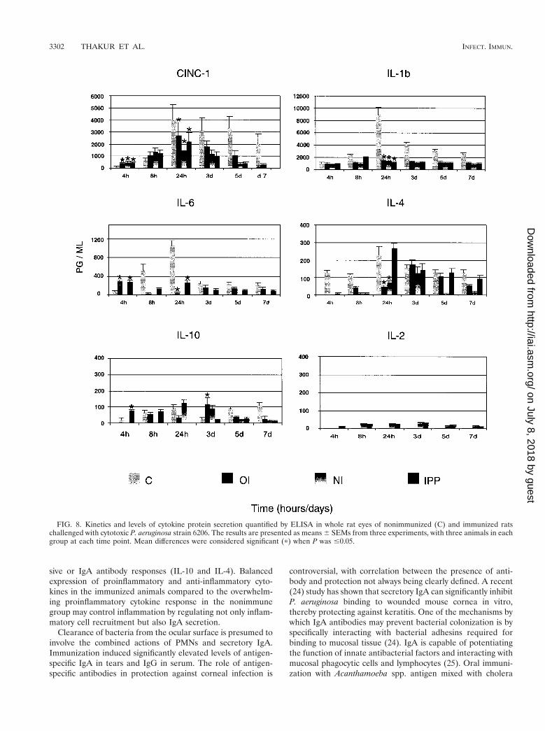
Effect of immunization on cytokine protein secretion. Theprotein levels were not determined for all cytokines (thoseprobed for mRNA) due to the limited availability of reagentsfor rats.
(i) Nonimmunized controls. In nonimmunized rats, neutro-phil chemoattractant CINC-1 protein levels were significantlylower (P , 0.04) early (4 h) during the infection and weresignificantly higher (P , 0.03) later (24 h) during the infectionthan those of immunized rats. Expression of CINC-1 proteinremained high up to 7 days postinfection. The amount of IL-1bprotein gradually increased and peaked at 24 h postchallengeand remained high up to 7 days postinfection. IL-6 protein alsopeaked at 24 h, diminished drastically at 3 days postinfection,and remained low up to 7 days postchallenge. Nonimmunizedrats showed high levels of IL-4 protein which peaked at 24 h(P , 0.03) postchallenge and remained high up to 7 dayspostinfection.
(ii) Oral immunization. Expression of CINC-1 protein wassignificantly higher (P , 0.04) early during the infection (4 and8 h) and significantly lower (P 5 0.013) by 24 h postinfectionthan that of nonimmunized control rats. IL-1b protein levelswere low throughout the period of infection compared to thoseof nonimmunized rats. The levels of IL-6 protein were signif-icantly higher (P , 0.033) in orally immunized rats at 4 hpostchallenge than those of nonimmunized rats. The pattern ofprotein expression was reversed at 8 h postinfection, with IL-6protein levels increasing dramatically in nonimmunized ani-mals. IL-10 and IL-4 proteins showed a biphasic pattern, withthe first peak appearing at 4 to 8 h and the second at 3 dayspostinfection.
(iii) Nasal immunization. The protein secretion pattern ofCINC-1 and IL-1b was similar to those of orally and IPP-immunized rats. IL-6 protein was below the limit of detection.IL-4 and IL-10 proteins were present late during the infection.IL-2 protein was present at most time points but at very lowlevels.
(iv) IPP immunization. The pattern of CINC-1 protein se-cretion was the same as in orally or nasally immunized rats.IL-1b and IL-6 levels were low throughout the period of in-fection compared to nonimmunized rats, except for the levelsof IL-6 at 4 h (P , 0.033). IL-4 protein was highly upregulatedat 24 h postinfection and diminished thereafter. Unlike innasally immunized rats, IL-10 protein showed a biphasic pat-tern peaking very early (4 to 8 h) and late (5 days) during theinfection (Fig. 8). IL-2 protein was present at all time points atvery low levels.
FIG. 5. MPO activity in whole eyes of nonimmunized (C) and im-munized rats challenged with cytotoxic P. aeruginosa strain 6206 at 4,8, and 24 h and 3, 5, and 7 days postchallenge. Three individual eyeswere used for each group at each time point. Results are reported asmean MPO activity 6 SEM/eye. Mean differences were consideredsignificant (p) when P was #0.05. OI, orally immunized; NI, nasallyimmunized; IPP, IPP immunized.
FIG. 6. P. aeruginosa-specific proliferation of lymphocytes isolatedfrom MLN of nonimmunized (C) and immunized rats. Mesentericlymph nodes from immunized rats were collected at 7 days post-booster immunization. Results are reported as means 6 SEMs oftriplicate cultures for each of three rats per group at two concentra-tions (1:10 and 1:100). Mean differences were considered significant(p) when P was #0.05. OI, orally immunized; NI, nasally immunized;IPP, IPP immunized.
3300 THAKUR ET AL. INFECT. IMMUN.
on July 8, 2018 by guesthttp://iai.asm
.org/D
ownloaded from

DISCUSSION
Our study showed that the route of immunization affects theseverity and persistence of microbial keratitis. Immunizationhas the potential to modulate the inflammatory response to aninfection. This modulation includes the production of chemicalsignals, cytokines and chemokines, with recruitment and acti-vation of cells involved in clearing the infection (29). This studyhas demonstrated that immunization changes the kinetics ofPMN infiltration, with immune groups having more rapid re-cruitment and resolution of PMNs in the cornea than thenonimmune group. Associated with this was a more rapidclearance of bacteria, differences in the levels of cytokinesexpressed and produced, and reduced adverse pathology. Inparticular, the IPP and intranasal immunization regimes withan OT boost provided the best protection from corneal ulcer-ation.
CINC-1 is a potent activator and attractant of neutrophils(27). Increased CINC-1 levels were detected earlier (4 to 8 h)postinfection in immunized rats than in nonimmunized rats,
with all groups peaking at 24 h postchallenge. However, de-spite the earlier increased production of CINC-1 in the immu-nized groups, the peak levels of CINC-1 were significantlylower in the immunized groups and also decreased far morerapidly. The changes in the CINC-1 levels corresponded to therecruitment and resolution profiles of the PMNs. The rate ofPMN recruitment in other disease settings has been associatedwith early bacterial clearance, such as enhanced respiratoryclearance of nontypeable Haemophilus influenzae followingmucosal immunization (5, 10). Persistence of PMNs in thenonimmune animals during the later stages of infection maycontribute to corneal scarring and perforation.
For the PMN response to be beneficial rather than detri-mental, a rapid resolution of PMN infiltration must occur.Immunization of rats against P. aeruginosa corneal infectionachieved a rapid resolution of PMN infiltrates. In addition tothe modulation of CINC-1 levels, there were reduced levels ofthe proinflammatory cytokines (IL-1b and IL-6) and similar orhigher levels of the cytokines associated with immunosuppres-
FIG. 7. Kinetics and levels of cytokine mRNA expression quantified by RNase protection assay in whole rat eyes of nonimmunized (C) andimmunized rats challenged with cytotoxic P. aeruginosa strain 6206. The graphs show means 6 SEMs from three experiments, with five animalsin each group at each time point, normalized with two housekeeping genes (L32 and GAPDH). Mean differences were considered significant (p)when P was #0.05. OI, orally immunized; NI, nasally immunized; IPP, IPP immunized.
VOL. 69, 2001 IMMUNITY TO PSEUDOMONAS KERATITIS 3301
on July 8, 2018 by guesthttp://iai.asm
.org/D
ownloaded from
sive or IgA antibody responses (IL-10 and IL-4). Balancedexpression of proinflammatory and anti-inflammatory cyto-kines in the immunized animals compared to the overwhelm-ing proinflammatory cytokine response in the nonimmunegroup may control inflammation by regulating not only inflam-matory cell recruitment but also IgA secretion.
Clearance of bacteria from the ocular surface is presumed toinvolve the combined actions of PMNs and secretory IgA.Immunization induced significantly elevated levels of antigen-specific IgA in tears and IgG in serum. The role of antigen-specific antibodies in protection against corneal infection is
controversial, with correlation between the presence of anti-body and protection not always being clearly defined. A recent(24) study has shown that secretory IgA can significantly inhibitP. aeruginosa binding to wounded mouse cornea in vitro,thereby protecting against keratitis. One of the mechanisms bywhich IgA antibodies may prevent bacterial colonization is byspecifically interacting with bacterial adhesins required forbinding to mucosal tissue (24). IgA is capable of potentiatingthe function of innate antibacterial factors and interacting withmucosal phagocytic cells and lymphocytes (25). Oral immuni-zation with Acanthamoeba spp. antigen mixed with cholera
FIG. 8. Kinetics and levels of cytokine protein secretion quantified by ELISA in whole rat eyes of nonimmunized (C) and immunized ratschallenged with cytotoxic P. aeruginosa strain 6206. The results are presented as means 6 SEMs from three experiments, with three animals in eachgroup at each time point. Mean differences were considered significant (p) when P was #0.05.
3302 THAKUR ET AL. INFECT. IMMUN.
on July 8, 2018 by guesthttp://iai.asm
.org/D
ownloaded from
toxin induces the production of parasite-specific IgA in muco-sal secretions and prevents corneal infection (23). Althoughantigen-specific IgG antibody appears to be important for op-sonophagocytosis (34), a correlation between the presence ofopsonizing antibodies and protection in vivo has not beenclearly determined to be an essential mechanism of effectiveimmunity (33, 35). There is also evidence that suggests thatsystemically derived IgG may also be capable of conferringprotection in the cornea (28). In addition to measuring signif-icant titers of antigen-specific IgA in tears, we have demon-strated the presence of a group of IgA-enhancing Th2-typecytokines (IL-4, IL-5, IL-6, and IL-10) which may provide anenvironment for preferential immunoglobulin class switchingfor IgA in the eye.
Previous studies using a rat model for pulmonary P. aerugi-nosa infection have shown that mucosal immunization signifi-cantly alters the profile of inflammatory cytokines produced inresponse to infection (5). Other evidence also suggests thatnasal and IPP immunization with mucosal adjuvant inducesdominant Th2 responses in nasal-associated lymphoid tissueand Peyer’s patches (12, 39). This study has shown that theroute of immunization changes the profile of cytokine expres-sion during P. aeruginosa corneal infection, with the most sig-nificant differences appearing in the nasal and IPP immuniza-tion groups. Expression of IL-2 and IL-5 were especiallyaltered, with nasally immunized rats expressing high levels ofIL-5 and baseline levels of IL-6 mRNA, with correspondingbaseline levels of IL-6 protein. In contrast, orally immunizedrats showed no IL-5 expression but had high IL-6 expressionand secretion, while IPP immunization resulted in the upregu-lation of both IL-5 and IL-6. IL-5 and IL-6 are known todifferentially influence the B-1 and B-2 lineage of plasma cells(2). Collectively, the data suggest that nasally immunized ani-mals may be producing IgA plasma cells of B-1 lineage, whichare IL-5 dependent and IL-6 independent (2), whereas orallyimmunized animals may be producing predominantly cells ofB-2 lineage. B-1 cells are physically and functionally unique Bcells producing antibodies to bacterial antigens such as lipo-polysaccharide and phosphocholine (1). B-1 cells mainly residein mucosal effector tissues, while conventional IgA1 B-2 cellsreside in mucosal inductive sites (39). Nasal-associated lym-phoid tissue functions as a primary inductive site for IgA an-tibody in tears by contributing triggered IgA-committed B cellsto the lacrimal gland (22). A recent study (30) has shown thata high frequency of IgA-committed B-1 cells occurs in thelacrimal gland (an effector site).
A role for T cells and cytokines produced by activated T cellsin protection from ocular bacterial infections has not beendemonstrated previously. Nasally and IPP-immunized rats in-duced antigen-specific lymphocyte responses, providing evi-dence that an antigen-specific T-lymphocyte response was in-duced by immunization and that these lymphocytes migratedfrom the site of immunization. Immunologically specific T cellsrecruit neutrophils in an antigen-dependent and dose-depen-dent fashion (6). Cytokines released by activated T cells maydirect the activity of nonspecific effector cells (21, 37). All ofthese studies have shown the involvement of T cells and cyto-kines in respiratory disease models. Evidence that supports therelevance of a CD41 Th1- versus Th2-type immune responsewas presented in a study that used a mouse P. aeruginosa
keratitis model. Data from this study suggest that Th2-respon-sive mice regulate inflammatory cellular infiltrates more effi-ciently by downregulating the inflammatory response, which inturn results in less corneal stromal damage (11, 20). Furtherstudies are required to define the importance of a T-cell re-sponse in protection against ocular infection.
This study has demonstrated that the immunization routemodulates the inflammatory response to ocular P. aeruginosainfection, thus affecting the severity of keratitis and adversepathology. The results show that immunization affects the rateof bacterial clearance and alters the profile of cytokines pro-duced in response to ocular infection, with nasal immunizationresulting in the most significant level of protection. The resultssuggest that the degree of protection afforded by immunizationmay depend upon the rapid recruitment of PMNs, the induc-tion of antigen-specific IgA, and the balanced production ofproinflammatory and immunosuppressive cytokines and thatT-cell responses may influence these events.
ACKNOWLEDGMENTS
This research was partly supported by the Australian Federal Gov-ernment through the Cooperative Research Centres Program.
We thank Reg Wong for excellent statistical analysis, Wen Wang fortechnical assistance, Denise Lawler and Robyn Lawler for helping withanimal experiments, and Philip Julian and Carol Woollcott for theirhelp in preparing illustrations.
REFERENCES
1. Aramaki, M., T. Nagasawa, T. Koeshi, and I. Ishikawa. 1998. Presence ofactivated B-1 cells in chronic inflamed gingival tissue. J. Clin. Immunol.18:421–429.
2. Bao, S., K. W. Beagley, A. M. Murray, V. Caristo, K. I. Matthaei, I. G. Young,and A. J. Husband. 1998. Intestinal IgA plasma cells of the B1 lineage areIL-5 dependent. Immunology 94:181–188.
3. Beagley, K. W., J. H. Eldridge, H. Kiyono, and F. Lee. 1989. Interleukins andIgA synthesis: human and murine IL-6 induce high rate of IgA secretion inIgA-committed B-cells. J. Exp. Med. 169:2133–2148.
4. Bradley, P. P., R. D. Christensen, and G. Rothstein. 1982. Cellular andextracellular myeloperoxidase in pyogenic inflammation. Blood 60:618–625.
5. Buret, A., M. L. Dunkley, G. Pang, R. L. Clancy, and A. W. Cripps. 1994.Pulmonary immunity to Pseudomonas aeruginosa in intestinally immunizedrats: role of alveolar macrophages, tumor necrosis factor, and interleukin-la.Infect. Immun. 62:5335–5343.
6. Campbell, P. A. 1990. The neutrophil, a professional killer of bacteria, maybe controlled by T cells. Clin. Exp. Immunol. 79:141–143.
7. Challacombe, S. J., and T. B. Tomasi, Jr. 1980. Systemic tolerance andsecretory immunity after oral immunization. J. Exp. Med. 152:1459–1472.
8. Cole, N., M. D. P. Willcox, S. M. J. Fleiszig, F. Stapleton, S. Bao, S. Tout, andA. Husband. 1998. Different strains of Pseudomonas aeruginosa isolated fromocular infections or inflammation display distinct corneal pathologies in ananimal model. Curr. Eye Res. 17:730–735.
9. Cripps, A. W., M. L. Dunkley, and R. L. Clancy. 1994. Mucosal and systemicimmunizations with killed Pseudomonas aeruginosa protect against acuterespiratory infection in rats. Infect. Immun. 62:1427–1436.
10. Foxwell, A. R. C., J. M. Kyd, and A. W. Cripps. 1998. Characterization of theimmunological response in the clearance of nontypable Haemophilus influ-enzae from the lung. Immunol. Cell Biol. 76:323–331.
11. Hazlett, L. D., S. McClellan, B. Kwon, and R. Barrett. 2000. Increasedseverity of Pseudomonas aeruginosa corneal infection in strains of micedesignated as Th1 versus Th2 responsive. J. Immunol. 41:805–810.
12. Hiroi, T., M. Yanagita, H. Iijima, K. Iwatani, T. Yoshida, K. Takatsu, and H.Kiyono. 1999. Deficiency of IL-5 receptor a-chain selectively influences thedevelopment of the common mucosal immune system independent IgA-producing B-1 cells in mucosa-associated tissue. J. Immunol. 162:821–828.
13. Hobden, J. A., S. A. Masinick, R. P. Barrett, and L. D. Hazlett. 1997.Proinflammatory cytokine deficiency and pathogenesis of Pseudomonasaeruginosa keratitis in aged mice. Infect. Immun. 65:2754–2758.
14. Inoue, Y., Y. Shimomura, and R. Manabe. 1992. Herpes simplex virus gly-coprotein D: protective immunity against murine herpetic keratitis. Investig.Ophthalmol. Vis. Sci. 31:411–418.
15. Kelleher, R. S., L. E. Hann, and J. A. Edwards. 1991. Endocrine, neural andimmune control of secretory component output by lacrimal gland acinarcells. J. Immunol. 146:3405–3412.
VOL. 69, 2001 IMMUNITY TO PSEUDOMONAS KERATITIS 3303
on July 8, 2018 by guesthttp://iai.asm
.org/D
ownloaded from

16. Kernacki, K. A., and R. S. Berk. 1994. Characterization of the inflammatoryresponse induced by corneal infection induced by Pseudomonas aeruginosa.J. Ocul. Pharmacol. 10:281–288.
17. Kernacki, K. A., J. A. Hobden, L. D. Hazlett, R. Friedman, and R. S. Berk.1995. In vivo bacterial protease production during Pseudomonas aeruginosacorneal infection. Investig. Ophthalmol. Vis. Sci. 36:1371–1379.
18. Kernacki, K. A., R. P. Barrett, J. A. Hobden, and L. D. Hazlett. 2000.Macrophage inflammatory protein-2 is a mediator of polymorphonuclearneutrophil influx in ocular bacterial infection. J. Immunol. 164:1037–1045.
19. Kreger, A. S., D. M. Lyerly, L. D. Hazlett, and R. S. Berk. 1986. Immuniza-tion against experimental Pseudomonas aeruginosa and Serratia marcescenskeratitis. Vaccination with lipopolysaccharide endotoxins and proteases. In-vestig. Ophthalmol. Vis. Sci. 27:932–939.
20. Kwon, B., and L. D. Hazlett. 1997. Association of CD41 T cell-dependentkeratitis with genetic susceptibility to Pseudomonas aeruginosa ocular infec-tion. J. Immunol. 159:6283–6290.
21. Kyd, J. M., M. L. Dunkley, and A. W. Cripps. 1995. Enhanced respiratoryclearance of nontypable Haemophilus influenzae following mucosal immuni-zation with P6 in rat model. Infect. Immun. 63:2931–2940.
22. Lather, D. M. R., R. F. Gill, and P. C. Montgomery. 1998. Inductive pathwaysleading to rat tear IgA antibody responses. Investig. Ophthalmol. Vis. Sci.39:1005–1011.
23. Leher, H. F., H. Alizadeh, W. M. Taylor, A. S. Shea, R. S. Silvany, F. V.Klink, M. J. Jager, and J. Y. Niederkorn. 1998. Role of mucosal IgA in theresistance to Acanthamoeba keratitis. Investig. Ophthalmol. Vis. Sci. 39:2666–2673.
24. Masinick, S. A., C. P. Montgomery, P. C. Montgomery, and L. D. Hazlett.1997. Secretory IgA inhibits Pseudomonas aeruginosa binding to cornea andprotects against keratitis. Investig. Ophthalmol. Vis. Sci. 38:910–918.
25. McGhee, J. R., and H. Kiyono. 1993. New perspective in vaccine develop-ment: mucosal immunity to infections. Infect. Agents Dis. 2:55–73.
26. Moon, M. M., L. D. Hazlett, R. E. Hancock, R. S. Berk, and R. Barrett. 1988.Monoclonal antibodies provided protection against ocular Pseudomonasaeruginosa infection. Investig. Ophthalmol. Vis. Sci. 29:1277–1284.
27. Mulligan, M. S., M. L. Jones, M. A. Bolanowski, M. P. Baganoff, W. L.Deppeler, D. M. Meyers, U. S. Ryan, and P. A. Ward. 1993. Inhibition of lunginflammatory reactions in rats by an anti-human IL-8 antibody. J. Immunol.150:5585–5595.
28. Preston, M. J., A. A. Gereceker, N. L. Koles, M. Pollack, and G. B. Pier. 1997.Prophylactic and therapeutic efficacy of immunoglobulin G antibodies toPseudomonas aeruginosa lipopolysaccharide against murine experimental
corneal infection. Investig. Ophthalmol. Vis. Sci. 38:1418–1425.29. Rudner, X. L., K. A. Kernacki, R. P. Barrett, and L. D. Hazlett. 2000.
Prolonged elevation of IL-1 in Pseudomonas aeruginosa ocular infectionregulates macrophage-inflammatory protein-2 production, polymorphonu-clear neutrophil persistence, and corneal perforation. J. Immunol. 164:6576–6582.
30. Saitoh-Inagawa, W., H. Takachika, M. Yanagita, H. Iijima, E. Uchio, S.Ohno, K. Aoki, and H. Kiyono. 2000. Unique characteristics of lacrimal glandas a part of mucosal immune network: high frequency of IgA-committed B-1cells and NK1.11 ab T cells. Investig. Ophthalmol. Vis. Sci. 41:138–144.
31. Schein, O. D., L. D. Ormerod, and E. Barraquer. 1989. Microbiology ofcontact lens associated keratitis. Cornea 8:281–285.
32. Steuhl, K. P., G. Doring, and H. J. Thiel. 1989. The significance of bacterialand host factors in corneal infections caused by Pseudomonas aeruginosa.Fortschr. Ophthalmol. 86:283–286.
33. Troelstra, A., L. Vogel, L. van Alphen, P. Eijk, H. Jansen, and J. Dankert.1994. Opsonic antibodies to outer membrane protein P2 of nonencapsulatedHaemophilus influenzae are strain specific. Infect. Immun. 62:779–784.
34. Verhoef, J., and M. R. Visser. 1993. Neutrophil phagocytosis and killing:normal function and microbial evasion, p. 108–137. In J. S. Abramson andJ. G. Wheeler (ed.), The neutrophil. Oxford University Press, New York,N.Y.
35. Wallace, F. J., R. L. Clancy, and A. W. Cripps. 1989. An animal modeldemonstration of enhanced clearance of non-typable Haemophilus influenzaefrom respiratory tract after antigen stimulation of gut associated lymphoidtissue. Am. Rev. Respir. Dis. 140:311–316.
36. Wallace, F. J., A. W. Cripps, R. L. Clancy, A. J. Husband, and C. S. Witt.1991. A role for intestinal T lymphocytes in bronchus mucosal immunity.Immunology 74:68–73.
37. Wallace, F. J., C. S. Witt, R. L. Clancy, A. W. Cripps, and A. J. Husband.1995. Protection against non-typable Haemophilus influenzae following sen-sitization of gut lymphoid tissue: role of specific antibody and phagocytes.Immunol. Cell Biol. 73:258–265.
38. Welsh, N. H., A. J. Rauch, and S. L. Graffin. 1984. Topical immunotherapyfor Pseudomonas keratitis in rabbits: use of anti-lipopolysaccharide plasma.Br. J. Ophthalmol. 68:828–832.
39. Yanagita, M., T. Hiroi, N. Kitagaki, S. Hamada, H. Ito, H. Shimauchi, H.Murakami, and H. Kiyono. 1999. NALT immunity: fimbriae-specific Th1and Th2 cell regulated IgA responses for the inhibition of bacterial attach-ment to epithelial cells and subsequent inflammatory cytokines. J. Immunol.162:3559–3565.
Editor: J. D. Clements
3304 THAKUR ET AL. INFECT. IMMUN.
on July 8, 2018 by guesthttp://iai.asm
.org/D
ownloaded from