effects of adenosine on lymphangiogenesis
TRANSCRIPT

Effects of Adenosine on LymphangiogenesisBenedicte Lenoir1, Daniel R. Wagner1,2, Silvia Blacher3, Graciela B. Sala-Newby4, Andrew C. Newby4,
Agnes Noel3, Yvan Devaux1*
1 Laboratory of Cardiovascular Research, Centre de Recherche Public de la Sante (CRP – Sante), Luxembourg, 2 Division of Cardiology, Centre Hospitalier Luxembourg,
Luxembourg, 3 Laboratory of Tumor and Development Biology, Groupe Interdisciplinaire de Genoproteomique Appliquee - Cancer, University of Liege, Liege, Belgium,
4 Bristol Heart Institute, Bristol Royal Infirmary, University of Bristol, Bristol, United Kingdom
Abstract
Background: The lymphatic system controls tissue homeostasis by draining protein-rich lymph to the vascular system.Lymphangiogenesis, the formation of lymphatic vessels, is a normal event in childhood but promotes tumor spread andmetastasis during adulthood. Blocking lymphangiogenesis may therefore be of therapeutic interest. Production ofadenosine is enhanced in the tumor environment and contributes to tumor progression through stimulation ofangiogenesis. In this study, we determined whether adenosine affects lymphangiogenesis.
Methods: Lymphatic endothelial cells (HMVEC-dLy) were cultured in presence of adenosine and their proliferation,migration and tube formation was assessed. Gelatin sponges embedded with the stable analogue of adenosine 2-chloroadenosine were implanted in mice ear and lymphangiogenesis was quantified. Mice were intravenously injected withadenoviruses containing expression vector for 59-endonucleotidase, which plays a major role in the formation of adenosine.
Results: In vitro, we observed that adenosine decreased the proliferation of lymphatic endothelial cells, their migration andtube formation. However, in vivo, gelatin sponges containing 2-chloro adenosine and implanted in mice ear displayed anelevated level of lymphangiogenesis (2.5-fold, p,0.001). Adenovirus-mediated over-expression of cytosolic 59-nucleotidaseIA stimulated lymphangiogenesis and the recruitment of macrophages in mouse liver. Proliferation of lymphatic endothelialcells was enhanced (2-fold, p,0.001) when incubated in the presence of conditioned medium from murine macrophages.
Conclusion: We have shown that adenosine stimulates lymphangiogenesis in vivo, presumably through a macrophage-mediated mechanism. This observation suggests that blockade of adenosine receptors may help in anti-cancer therapies.
Citation: Lenoir B, Wagner DR, Blacher S, Sala-Newby GB, Newby AC, et al. (2014) Effects of Adenosine on Lymphangiogenesis. PLoS ONE 9(3): e92715.doi:10.1371/journal.pone.0092715
Editor: Philippe Rouet, I2MC INSERM UMR U1048, France
Received December 2, 2013; Accepted February 25, 2014; Published March 20, 2014
Copyright: � 2014 Lenoir et al. This is an open-access article distributed under the terms of the Creative Commons Attribution License, which permitsunrestricted use, distribution, and reproduction in any medium, provided the original author and source are credited.
Funding: This study was funded by the Ministry of Higher Education and Health of Luxembourg. BL was supported by a fellowship from the National Fund ofResearch of Luxembourg (grant PhD-AFR-09-110). This work was also supported by grants from the Fondation contre le Cancer (foundation of public interest,Belgium), the Fonds speciaux de la Recherche (University of Liege), the Centre Anticancereux pres l’Universite de Liege, the Fonds Leon Fredericq (University ofLiege), the Interuniversity Attraction Poles Programme - Belgian Science Policy (Brussels, Belgium). The funders had no role in study design, data collection andanalysis, decision to publish, or preparation of the manuscript.
Competing Interests: The authors have declared that no competing interests exist.
* E-mail: [email protected]
Introduction
Lymphatic vessels are found all over the human body except in
certain tissues or organs such as epidermis, cartilage, brain,
cornea, bone marrow and retina. The lymphatic system controls
the homeostasis of tissue fluid by draining protein-rich lymph from
the tissues and organs back to the vascular system. It also
contributes to intestinal lipid absorption and to the transport of
lymphocytes and dendritic cells [1,2]. A deleterious role of the
lymphatic system has been evidenced in cancer, in which
lymphatic vessels participate in the promotion of tumor growth
and metastasis [3]. Also, dysfunction of lymphatic vessels in tumors
can reduce the efficacy of anti-cancer drugs [4,5].
Lymphangiogenesis is the formation of new lymphatic vessels, a
normal event in childhood. During adulthood, lymphangiogenesis
is associated with pathological conditions such as inflammation,
healing, graft rejection, auto-immune diseases and tumor progres-
sion [1,2,4,6]. Through the secretion of growth factors and pro-
lymphangiogenic cytokines, inflammatory cells stimulate lymphan-
giogenesis [2,6–8]. Macrophages are the main actors of inflam-
matory lymphangiogenesis [9], principally through the secretion of
vascular endothelial growth factor-C (VEGF-C) and VEGF-D
[2,7]. Other paracrine factors secreted by macrophages also share
pro-lymphangiogenic properties which drive the growth, morpho-
genesis and function of lymphatic endothelial cells (LEC) [10–12].
In addition, a subset of macrophages which possesses the ability to
transdifferentiate into LEC have been termed macrophage-
derived lymphatic progenitors (M-LEC) [1,2,10,13]. These mac-
rophages co-express the macrophage marker F4/80 and the
lymphatic marker podoplanin [11].
Modulation of lymphangiogenesis is expected to have some
therapeutic value in certain pathological conditions such as
lymphedema, tumor metastasis, Kaposi sarcoma and obesity
[14]. Blocking antibodies against VEGF receptor -3 (VEGFR-3),
the receptor of pro-lymphangiogenic VEGF-C, have recently
entered clinical trials as anti-angiogenic and anti-metastatic drugs
PLOS ONE | www.plosone.org 1 March 2014 | Volume 9 | Issue 3 | e92715

[15]. However, there is still a paucity of clinically applicable tools
to modulate lymphangiogenesis.
Adenosine is an ubiquitous and endogenous purine nucleoside
with a plethora of physiological functions [16]. Although
constitutive, the secretion of adenosine is increased under
metabolic stress such as hypoxia, inflammation and cancer. In
addition to its extensively documented role in inflammation,
adenosine has been shown to be a master regulator of angiogenesis
[17–21]. However, whether adenosine affects lymphangiogenesis
is unknown.
Adenosine binds to 4 types of receptors (A1, A2a, A2b and A3),
all belonging to class A family of G-protein coupled receptor
family [22]. Several pharmacological agents have been developed
to specifically activate or inhibit adenosine receptors. Thus,
adenosine receptors are appealing therapeutic targets [23].
In the present study, we hypothesized that adenosine may
regulate lymphangiogenesis. Using different in vitro and in vivo
models [24], we provide evidence that adenosine inhibits the
proliferation and migration of LEC in vitro but stimulates
lymphangiogenesis in vivo.
Materials and Methods
In vitro experimentsCell culture. Human adult dermal microvascular lymphatic
endothelial cells (LEC) were purchased from Lonza (HMVEC-
dLy; Braine-l9Alleud, Belgium) and used at passages 3 to 5. Cells
were cultured at 37uC in a 5% CO2 atmosphere in endothelial
growth microvascular (EGM2-MV) medium (Lonza) composed of
EBM2 medium with 5% FBS, hydrocortisone, h-FGF-B, VEGF,
R3-IGF-1, ascorbic acid, hEGF and GA 1000 [9]. For drug
treatments, cells were washed and cultured in EGM2-MV
medium supplemented with 2% FBS, and half of the medium
was renewed every 24 h. Adenosine (Sigma-Aldrich) was used at
concentration ranging from 0.1 mM to 10 mM and the adenosine
deaminase inhibitor EHNA (Sigma) was used at a concentration of
10 mM to increase adenosine half-life. CGS21680 and NECA
(Sigma-Aldrich) were used as preferential A2a and A2b agonists,
respectively.
Primary macrophages were isolated from peritoneal lavage of
C57BL/6 mice intraperitoneally injected with 4% thioglycollate
(Sigma-Aldrich, St. Louis, MO) 5 days earlier. After washing off
non-adherent cells, macrophages were cultured in serum-free
medium (RPMI-1640, Lonza) and conditioned medium was
collected.
Viability/cytotoxicity assay. Drug toxicity was assessed
with the live/dead viability/cytotoxicity kit (Molecular Probes,
Invitrogen) according to the manufacturer’s protocol. Six technical
replicates per test were performed.
Proliferation assay. LEC (46103) cultured in EBM2-MV
medium containing 2% FBS were treated with adenosine or with
50% conditioned medium from macrophages treated by adeno-
sine. Half of the medium was changed every 24 hours. Cell
proliferation was assessed using CyQUANT proliferation kit
(Lonza).
Boyden chamber assay. To study the migration of LEC, we
used Boyden chambers (Corning Inc., Corning, Amsterdam) with
filters of 8 mm pore size previously coated with 0.005% gelatin
(Type A, porcine skin, Sigma). Fifty thousand LEC were seeded in
the upper chamber, in EBM2-MV containing 2% FBS. Adenosine
at concentrations ranging from 0.1 mM to 10 mM was added in the
lower chamber. To study the migration of macrophages, murine
peritoneal macrophages were deposited on 5 mm pore size in
RPMI medium and incubated in presence of adenosine and
EHNA (10 mM). After 24 h for LEC and 4 h for macrophages,
cells in the upper chamber were carefully removed using cotton
buds and cells at the bottom of the membrane were fixed and
stained with 4% Giemsa. Quantification was performed by
counting the stained cells on the membrane. All assays included
3 technical replicates.
Wound healing assay. LEC (36104) were cultured in
EGM2-MV medium in a culture insert (Ibidi, Proxylab, Belgium).
When cells reached confluence, the insert was removed and the
wounded monolayers were washed with serum-free medium. Cells
were then treated with different concentrations of adenosine in
EBM2 containing 2% FBS. Culture plates were placed in a live
cell imaging platform (Cell IQ, Chip-man technologies, UK) and 3
pictures per well were acquired automatically each 30 min
according to fixed reference points. The width of the wound was
determined at time 0 and 8 h, 16 h and 24 h after experiment
onset. All assays included 3 technical replicates.
Tubulogenesis assay. LEC (86105) were seeded on a type I
collagen layer (1 mg/ml, Collagen R; Serva Electrophoresis,
Heidelberg, Germany) in a 6-well plate. Cells were grown for
24 hours in complete EGM2-MV medium. Medium was then
removed and a second layer of collagen was added over the cells.
Finally, collagen-embedded were incubated in 2 mL of EGM2-
MV medium containing or not adenosine. After 24 hours, 5
pictures per well were captured with a phase-contrast microscope
(Axiovert 25; Carl Zeiss) coupled to an Axiocam color digital
camera (Carl Zeiss) and tube length was measured [9].
Spheroid assay. LEC (1.56103 cells per well in 24 well
plates) were pre-cultured for 24 h in EBM-2 containing 0.24%
high viscosity methyl cellulose (Sigma-Aldrich) to form micro-
spheres. Subsequently, spheroids were collected, embedded in
collagen gels with 100 ng/ml PMA and maintained at 37uC for
24 h in EBM2-MV medium including 2% FBS, with or without
adenosine. Spheroids were examined by phase-contrast microsco-
py using an Axiovert 25 microscope equipped with a 20 NA 0.3
LD A-Plan lens and an Axiocam color digital camera (Carl Zeiss).
Images were captured at room temperature, using acquisition
software KS400 3.0. Cell migration was quantified by a
Table 1. Quantitative PCR primers.
Gene Primer sequence Annealing temperature Amplicon length
ADORA1 Sense: 59-GACCTACTTCCACACCTG-39 Anti-sense: 59-TCACCACCATCTTGTACC-39 58uC 140 bp
ADORA2A Sense: 59-TCTTCAGTCTCCTGGCCATC-39 Anti-sense: 59-GGGACCACATCCTCAAAGAG-39 64uC 244 bp
ADORA2B Sense: 59-CTCCATCTTCAGCCTTCTGG-39 Anti-sense: 59-ACAAGGCAGCAGCTTTCATT-39 58uC 234 bp
ADORA3 Sense: 59-TCATCTGCGTGGTCAAGC-39 Anti-sense: 59-CTGTAGAAGTGGATTGTGATGC-39 62uC 148 bp
b-actin Sense: 59-AGAAAATCTGGCACCACACC -39 Anti-sense: 59-GGGGTGTTGAAGGTCTCAAA-39 60uC 142 bp
doi:10.1371/journal.pone.0092715.t001
Adenosine and Lymphangiogenesis
PLOS ONE | www.plosone.org 2 March 2014 | Volume 9 | Issue 3 | e92715
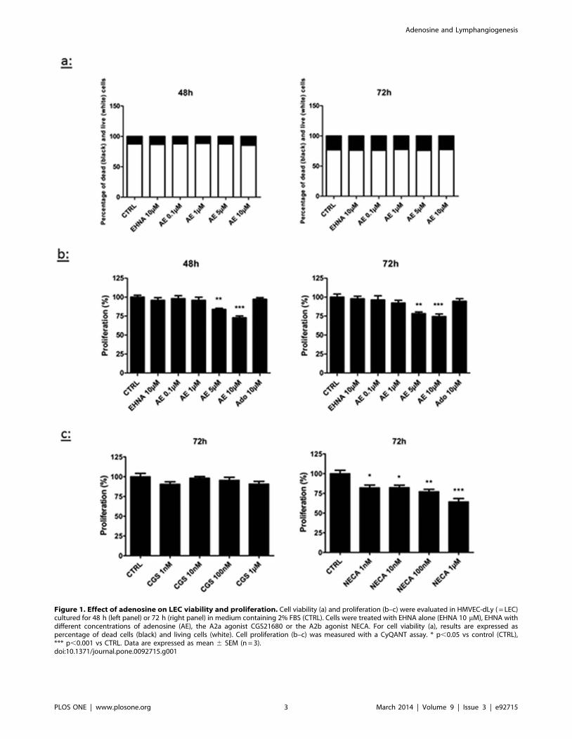
Figure 1. Effect of adenosine on LEC viability and proliferation. Cell viability (a) and proliferation (b–c) were evaluated in HMVEC-dLy ( = LEC)cultured for 48 h (left panel) or 72 h (right panel) in medium containing 2% FBS (CTRL). Cells were treated with EHNA alone (EHNA 10 mM), EHNA withdifferent concentrations of adenosine (AE), the A2a agonist CGS21680 or the A2b agonist NECA. For cell viability (a), results are expressed aspercentage of dead cells (black) and living cells (white). Cell proliferation (b–c) was measured with a CyQANT assay. * p,0.05 vs control (CTRL),*** p,0.001 vs CTRL. Data are expressed as mean 6 SEM (n = 3).doi:10.1371/journal.pone.0092715.g001
Adenosine and Lymphangiogenesis
PLOS ONE | www.plosone.org 3 March 2014 | Volume 9 | Issue 3 | e92715

computerized method determining the sprouting envelope area,
defined as the area of the minimal convex polygon containing the
whole spheroid and all sprouting cells.
Real-time quantitative PCR. Total RNA from cultured
cells was isolated using TriReagent and the RNeasy mini kit
(Qiagen, Venlo, Netherlands). Potential contaminating genomic
DNA was digested by DNase I treatment (Qiagen). One
microgram of total RNA was reverse-transcribed using the
SuperscriptH II Reverse Transcriptase (Invitrogen, Merelbeke,
Belgium). PCR primers were designed using the Beacon Designer
software (Premier Biosoft, Palo Alto, USA) and were chosen to
encompass an intron. PCR was performed using the iCycler and
the IQ SYBR Green Supermix (Biorad, Nazareth, Belgium). PCR
conditions were as follows: 3 min at 95uC, 30 s at 95uC and 1 min
annealing (40-fold). Optimal annealing temperature was deter-
mined for each primer pair (Table 1). Melting point analysis was
obtained after 80 cycles of 10 s from 55uC to 95uC. Each run
included negative reaction controls. b-actin was chosen as
housekeeping gene for normalization. Expression levels were
calculated by the relative quantification method (DDCt) using the
Genex software (Biorad) which takes into account primer pair
efficiency.
In vivo experimentsMice. Ten-week-old male C57BL/6 mice purchased from
Janvier (Le Genest St Isle, France) were used throughout this
study. Animal experiments were performed in compliance with the
local Animal Ethical Committee of the University of Liege (Liege,
Belgium) who specifically approved this study.
Collagen lymphangiogenesis assay. Gelatin sponges (Gel-
foam, Pfizer, Puurs, Belgium) were cut in small scares of
approximately 0.5 cm2. After incubation in 20 ml CADO
(0.003 mg/ml) (2-chloroadenosine) (Sigma), recombinant VEGF-
C (1 ng/ml) (R&D System, Oxon Abingdon, UK) or MRS1754
(0.2 mg/kg) (2.4 mg/ml) (Sigma) with or without CADO, sponges
were embedded in interstitial type I collagen gel (1.5 mg/mL;
Serva). Then sponges were implanted between the two skin layers
of mice ear. After 3 weeks, tissues were excised and sponges were
embedded and frozen in optimal cutting temperature (OCT)
compound. Immunofluorescent staining for mouse LYVE-1 (R&D
System) was performed using a secondary antibody labeled with
Alexa-Fluor 488 (Molecular Probes, Invitrogen). Cell nuclei were
counterstained with Dapi Fluoromount G (Southern Biotech).
Slices were scanned by Nanozoomer (Hamamatsu, Mont-Saint-
Guitbert, Belgium). A computer-assisted method of quantification
was used to determine the number of vessels per mm2. Each
experimental group contained 3 mice.
Adenoviral-mediated over-expression of cytosolic 59-
nucleotidase IA in mice. We used two adenoviral vectors
prepared at the Bristol Heart Institute (Bristol Royal Infirmary,
University of Bristol, United Kingdom) as previously described
[25]: a control vector containing a GFP cDNA as tag (Ad-GFP
vector) and a vector containing the coding sequence of pigeon cN-
IA (59-nucleotidase) without GFP cDNA (Ad-cN-IA vector). Virus
stocks were amplified, CsCl banded and titrated before use.
Vectors (Ad-cN-IA or Ad-GFP) (109 pfu/animal) or PBS were
injected through the tail vein. Mice were sacrificed 2 weeks later.
Liver was excised, fixed in 4% paraformaldehyde and embedded
in paraffin. Immunohistochemical stainings were performed for
the lymphatic vessel marker podoplanin (goat anti-mouse
podoplanin antibody, R&D System), the pan-leukocyte marker
CD45 (rat anti-mouse CD45 antibody, BD Pharmingen, BD
Bioscience, Belgium), and the macrophage marker F4/80 (rat anti-
mouse F4/80 antibody, AbD Serotec, Biorad, Dusseldorf,
Germany). For CD45 and F4/80 immunostainings, secondary
antibodies coupled to streptavidin/HRP (DAKO) were used. For
podoplanin, a tertiary antibody coupled to streptavidin/HRP
(DAKO) was used. A rabbit polyclonal antibody [26] was used for
cN-I immunostaining. Slices were counterstained with hematox-
ylin-eosin and scanned by Nanozoomer (Hamamatsu). Image
processing and signal quantification were performed using
Aphelion 3.2 software (Adcis, Saint-Contest, France) and image
analysis toolbox of Matlab 7.9 software (The Mathworks, Inc.,
Natick, MA).
Results
Lymphatic endothelial cells express adenosine receptorsFirst of all, we determined the expression profile of adenosine
receptors in human adult dermal microvascular lymphatic
endothelial cells (HMVEC-dLy = LEC) using quantitative PCR.
Lymphatic endothelial cells expressed the A2a and A2b adenosine
receptors, but not the A1 and A3 subtypes. Having verified that
LEC express adenosine receptors, we investigated the effect of
adenosine on different biological functions of these cells. In these
experiments performed in vitro, LEC were treated with various
concentrations of adenosine along with 10 mM of the adenosine
deaminase inhibitor EHNA. This drug slows down the degrada-
tion of adenosine into inosine, thereby sustaining the effects of
adenosine which has a very short half-life. This strategy has been
previously used and discussed [27–29].
Adenosine decreases the proliferation of LECFirst, we tested whether adenosine modulated LEC prolifera-
tion. Initial experiments were performed to address the toxicity of
adenosine and EHNA. Using the live/dead viability/cytotoxicity
assay, we observed that a combined treatment with adenosine and
EHNA did not affect cell viability for concentrations up to 10 mM
(Fig.1a). Then, we evaluated the proliferation of LEC with the
CyQUANT proliferation assay and we observed that treatments
Figure 2. Effect of adenosine on the migration of LEC inBoyden chamber. HMVEC-dLy were cultured in medium containing2% FBS (control condition, CTRL), with or without 10 mM EHNA and 0.1–10 mM adenosine (AE). Cell migration was assessed in a Boydenchamber assay 24 hours after treatment onset. There was no differenceon cell migration between all treatments. Results are expressed aspercentage of migrating cells (mean 6 SEM, n = 3).doi:10.1371/journal.pone.0092715.g002
Adenosine and Lymphangiogenesis
PLOS ONE | www.plosone.org 4 March 2014 | Volume 9 | Issue 3 | e92715

with 5 mM and 10 mM adenosine for 48 h and 72 h decreased the
proliferation rate (Fig.1b). This decrease reached 30% with 10 mM
adenosine after 72 h. Of note, no effect was observed before 48 h
(not shown). The inhibition of proliferation induced by adenosine
was reproduced by the A2b agonist NECA, but not by the A2a
agonist CGS21680 (Fig.1c).
Adenosine decreases the migration of LECThe effect of adenosine on the migration of LEC was evaluated
using two different methods. Firstly, we used a Boyden chamber
assay in which adenosine placed in the bottom chamber was used
as a chemoattractant. Adenosine did not affect the migration of
LEC (Fig.2). Secondly, we used a wound healing assay with inserts
allowing to standardize the width of the scar. As shown in Fig. 3,
Figure 3. Effect of adenosine on LEC migration in scratch test. HMVEC-dLy were treated during 24 h in medium containing 2% FBS, in thepresence of EHNA alone (EHNA, 10 mM) or EHNA with different concentrations of adenosine (AE). Pictures were taken at 0 h, 8 h, 16 h and 24 h afterinsert removing. Results are expressed as percentage of wound closure (percentage closure) (mean 6 SEM). * p,0.05 vs CTRL, ***p,0.001 vs CTRL.Each experiment was performed three times and a representative picture of each condition is shown.doi:10.1371/journal.pone.0092715.g003
Adenosine and Lymphangiogenesis
PLOS ONE | www.plosone.org 5 March 2014 | Volume 9 | Issue 3 | e92715

the scar was closed by 30% after 24 h, and this closure was
inhibited when cells were treated with 10 mM adenosine. EHNA
alone had no effect but it’s co-administration with adenosine was
necessary to observe a significant effect. Collectively, these data
show that adenosine itself is not a chemoattractant of LEC and
inhibits their migration.
Adenosine decreases tube formation from lymphaticendothelial cells
The impact of adenosine on the differentiation of LEC into
tube-like structures was investigated using two models. First, we
used a tubulogenesis assay in which cells were embedded between
two collagen layers. After 24 h, a network of tube-like structures
emanating from the LEC was observed in control condition
Figure 4. Effect of adenosine on LEC tube formation. Tube formation was assessed in two in vitro models, the tubulogenesis assay (a) and thespheroid assay (b). HMVEC-dLy were cultured in 2% FBS medium (CTRL) and treated or not with EHNA alone (EHNA 10 mM) or EHNA with differentconcentrations of adenosine (AE). Quantification of tube formation (a) and cell migration (b) was performed by a computerized method on picturestaken after 24 h of culture. The parameters measured are: the tubes branching (branching), the length of tube (length), the surface occupied by tube(surface), and the maximal length of tube (Lmax). * p,0.05 vs CTRL, **p,0.01 vs CTRL, *** p,0.001 vs CTRL. Each experiment was performed threetimes and representative pictures are shown. Data are expressed as mean 6 SEM.doi:10.1371/journal.pone.0092715.g004
Adenosine and Lymphangiogenesis
PLOS ONE | www.plosone.org 6 March 2014 | Volume 9 | Issue 3 | e92715

(Fig.4a). When cells were treated with adenosine however, the
vascular network was disorganized, and its surface and the
maximal length of the tubes were decreased. The effect was
maximal with 1 mM of adenosine (2-fold decrease). Second, we
used a spheroid assay in which micro-spheres of LEC were
embedded in a collagen gel. After 24 h, a network of tube-like
structures was visible in the control condition (Fig.4b). Adenosine
pre-treatment resulted in a reduced LEC outgrowth. Again, the
effect was maximal with 1 mM adenosine (2-fold decrease). EHNA
or adenosine alone had not effect. Together, these experiments
show that adenosine inhibits lymphangiogenesis in vitro.
A stable analog of adenosine increaseslymphangiogenesis in gelatin sponges in vivo
Following in vitro experiments, we sought to determine whether
adenosine affects lymphangiogenesis in vivo. We first used a
collagen lymphangiogenesis assay with gelatin sponges. Sponges
were embedded in collagen containing either the stable analogue
of adenosine 2-chloro adenosine (CADO), VEGF-C as a positive
control, or the A2b receptor antagonist MRS1754. Sponges were
implanted between the two skin layers of mice ear. PBS or
MRS1754 were injected at the apex of the ear every two days.
After 3 weeks, sponges were removed, frozen and sliced for
immunostaining for LYVE-1 lymphatic marker. As expected,
sponges containing VEGF-C displayed a higher level of lymphan-
giogenesis compared to control condition, as assessed by the area
of the sponges occupied by lymphatic vessels (Fig.5). Interestingly,
a 2.5-fold increase in lymphatic vessels was observed in CADO-
containing sponges, as compared to control. This increase was
blunted by MRS1754. Of note, administration of MRS1754 alone
did not affect lymphangiogenesis (not shown). These results show
that the stable analog of adenosine CADO is able to stimulate
lymphangiogenesis in vivo, presumably through the adenosine
A2b receptor.
Over-expression of 59-nucleotidase increaseslymphangiogenesis in the murine liver
To confirm the effect of adenosine on lymphangiogenesis
observed in gelatin sponges, we used an adenovirus overexpressing
59-nucleotidase (cN-IA), an isoform of the intracellular enzyme
which produces adenosine from adenosine monophosphate. As
control, we used a vector tagged with GFP (Fig.6a). Two weeks
after intravenous administration of viruses, an intense staining for
cN-IA was observed in the liver of mice treated with cN-IA vector,
particularly around lymphatic vessels (Fig.6b). A 2.5-fold increase
in podoplanin-positive lymphatic vessels was detected in the liver
of mice treated with cN-IA vector compared to mice treated with
the GFP vector (Fig.6c). This increase was paralleled by enhanced
expression of the pan-leukocyte marker CD45 (Fig.6d) and the
macrophage marker F4/80 (Fig.6e). Therefore, over-expression of
59-nucleotidase stimulates lymphangiogenesis and increases the
expression of macrophage markers in the liver.
Conditioned medium from macrophages increases theproliferation of LEC
To test a causal relationship between the presence of
macrophages in the liver and enhanced lymphangiogenesis in
mice injected with the adenovirus overexpressing 59-nucleotidase,
we exposed cultured LEC to conditioned medium from murine
peritoneal macrophages obtained from naıve mice (i.e. not treated
by the adenovirus). We observed a stimulation of the proliferation
rate of LEC, reaching a 2-fold increase compared to control
condition after 72 hours (Fig.7a).
Adenosine stimulates the migration of macrophagesFinally, we addressed whether adenosine affects the migratory
capability of macrophages. Using a Boyden chamber assay, we
were able to determine that adenosine activates the migration of
macrophages (Fig.7b).
Figure 5. Effects of adenosine on lymphangiogenesis in the invivo model of collagen sponge. Sponges were soaked with PBS ascontrol (CTRL), with VEGF-C (1 ng/ml) as positive control (VEGF-C), with20 ml CADO (3 ng/ml), a stable analog of adenosine, or with CADO inpresence of the A2b antagonist MRS1754 (2.4 mg/ml). Sponges wereimplanted between the two skin’s layers of ear’s mice for 3 weeks. Everyother day, PBS or MRS1754 were injected in the apex of the ear. Spongesections were stained with an anti-Lyve-1 antibody to detect lymphaticvessels (green) and Dapi to detect cell nucleus (blue). The graphcorresponds to computerized quantification of the surface occupied bylymphatics (vessel area). Data are expressed as mean 6 SEM (n = 6).** p,0.01 vs CTRL.doi:10.1371/journal.pone.0092715.g005
Adenosine and Lymphangiogenesis
PLOS ONE | www.plosone.org 7 March 2014 | Volume 9 | Issue 3 | e92715

Figure 6. Effect of adenosine on lymphatic vasculature and inflammatory cell recruitment in the liver. Control adenovirus expressingGFP (CTRL virus) or adenovirus carrying the sequence of 5-nucleotidase (cN-IA virus) were injected in the caudal vein of mice. PBS was used asnegative control (PBS). Mice were sacrificed 2 weeks after injection. The efficacy of virus transduction was shown by GFP immunostaining (a) and bycN-IA immunostaining (b)). Liver sections were stained with anti-podoplanin antibodies to detect lymphatic vessels (c), with anti-CD45 antibodies todetect inflammatory cells (d), and with anti-F4/80 antibodies to detect macrophages (e). Data are presented as mean 6 SEM (n = 15 for PBS, n = 13 forGFP and n = 12 for cN-IA).**p,0.01, *** p,0.001. Representative pictures are shown.doi:10.1371/journal.pone.0092715.g006
Adenosine and Lymphangiogenesis
PLOS ONE | www.plosone.org 8 March 2014 | Volume 9 | Issue 3 | e92715

Figure 7. Effect of medium conditioned from macrophages on LEC proliferation and effect of adenosine on macrophage migration.(a)Proliferation of LEC was evaluated after 24 h, 48 h or 72 h in medium containing 2% FBS (Control) or medium conditioned by murine peritonealmacrophages obtained from naıve mice (Conditioned medium). Cell proliferation was measured with a CyQANT assay. (b) Migration of macrophageswas evaluated using a Boyden chamber assay in which murine peritoneal macrophages obtained from naıve mice were incubated for 4 h with 2%FBS (Control) or adenosine with EHNA (10 mM each). Data are expressed as mean 6 SEM (n = 3). * p,0.05 vs CTRL, *** p,0.001 vs CTRL.doi:10.1371/journal.pone.0092715.g007
Adenosine and Lymphangiogenesis
PLOS ONE | www.plosone.org 9 March 2014 | Volume 9 | Issue 3 | e92715

Discussion
The present study was designed to investigate whether
adenosine modulates lymphangiogenesis. In vitro experiments
showed that adenosine inhibits the proliferation and migration of
cultured lymphatic endothelial cells. Most importantly, we
observed that adenosine stimulates lymphangiogenesis in vivo,
which might have therapeutic potential.
Since our initial hypothesis was that adenosine may directly
bind to adenosine receptors present at the surface of LEC, we
started our investigation with in vitro experiments involving
cultured human primary LEC. We observed that adenosine
consistently decreased cell proliferation and migration. This was
not a consequence of cell death since adenosine was not cytotoxic
for concentrations up to 10 mM. Adenosine had the optimal
inhibitory effect at concentrations ranging from 1 to 10 mM, which
are observed in the setting of pathological conditions such as
ischemia. Indeed, physiological concentrations of adenosine are
generally in the submicromolar range. Pharmacological in vitro
studies with agonists of adenosine receptors suggested that the A2b
receptor mediates the anti-proliferative effect of adenosine on
LEC.
Surprisingly, we observed opposite effects in the whole animal,
i.e. adenosine stimulated lymphangiogenesis. This observation was
made in mice implanted with a gelatin sponge containing the
stable analog of adenosine CADO, and in mice injected with an
adenoviral vector encoding the sequence of the 59-nucleotidase
which results in over-production of adenosine [25,26]. In mice
implanted with gelatin sponges, we observed a blockade of the
pro-lymphangiogenic effects of adenosine by MRS1754, an
antagonist of adenosine A2b receptor, supporting the involvement
of this sub-type of receptor in the effect of adenosine.
A possibility to explain the apparent discrepancy between in
vitro and in vivo experiments may be the need for a microenvi-
ronment. Indeed, our data showing enhanced presence of
macrophages in the liver where the adenovirus encoding 59-
nucleotidase accumulates suggest that adenosine stimulates
lymphangiogenesis indirectly through macrophages. Accordingly,
macrophages have been shown to drive lymphangiogenesis in
different pathological conditions by secreting growth factors such
as VEGF-C and VEGF-D [8,9,30,33]. We [17] and others [31–
36] have previously reported that adenosine is able to switch
macrophages from an inflammatory M1 phenotype to a pro-
angiogenic M2 phenotype associated with increased VEGF-A
secretion and decreased inflammatory factors production. Al-
though we failed to demonstrate that adenosine induces the
production of VEGF-C by cultured macrophages (not shown), the
possibility remains that adenosine may trigger the secretion of
other pro-lymphangiogenic factors by macrophages.
The involvement of macrophages in the stimulation of
lymphangiogenesis by adenosine is indirectly suggested by our
present data showing that conditioned medium from macrophag-
es, but not adenosine itself, stimulates LEC proliferation, and by
the capacity of adenosine to enhance the migration of macro-
phages. Furthermore, a recent report by Keshet’s group showed
that VEGF induced the recruitment of circulating Ly6Chi
monocytes to the liver and endowed them with pro-angiogenic
and pro-arteriogenic properties which stimulate vascularization
[37]. To which extent infiltrated monocytes/macrophages, resi-
dent Kupffer cells and M-LEC contribute to the stimulation of
lymphangiogenesis by adenosine is currently unknown.
A similar discrepancy in the in vitro and in vivo effects of
adenosine in LEC observed in the present study is less expected to
occur in vascular endothelial cells. Indeed, we previously reported
that adenosine stimulates the production of VEGF-A by cultured
macrophages [17] and it is known that adenosine has pro-
angiogenic properties in vivo (unpublished data and [21] for
review). Thus, adenosine directly and indirectly activates angio-
genesis, which confers its role in tissue vascularization and repair.
Knowledge of the effects of adenosine on lymphangiogenesis, in
addition to its effects on angiogenesis, finds its relevance in
multiple biomedical fields such as oncology and any disease with
an inflammatory component or a cellular stress. Indeed, under
conditions of stress, production of adenosine is increased and
adenosine receptors are activated [38,39]. Tumor cells secrete
adenosine in its environment [32] which can lead to 10- to 20-fold
higher concentrations of adenosine compared to normal tissues
[40]. Since lymphatic vessels participate in promotion of tumor
growth and metastasis [3], it is tempting to speculate that blockade
of adenosine receptors may result in decreased lymphangiogenesis
in tumors, thereby participating to inhibition of tumor promotion
[41]. While blocking antibodies against VEGFR-3 are currently
tested as anti-angiogenic and anti-metastatic drugs [15], the
potential for antagonists of adenosine receptors to inhibit
lymphangiogenesis and its deleterious consequences on tumor
development and anti-cancer therapies has not been addressed.
Finally, our findings may have some importance for non-cancer
liver diseases since we have observed that adenosine stimulates
lymphangiogenesis in the liver, and the lymphatic system is
associated with several liver pathologies such as liver fibrosis,
portal hypertension and cirrhotic ascites [42]. This is consistent
with recent reports showing that antagonism of adenosine A2a
receptor prevents and reverses liver fibrosis [43] and antagonism
of adenosine A1 receptor reduces mortality of cirrhotic rats [44].
In conclusion, our study shows for the first time that adenosine
stimulates lymphangiogenesis. This effect is dependent on the
microenvironment and may involve macrophages. This observa-
tion may be useful for anti-cancer therapies, since adenosine is
now recognized as a regulator of the complex interaction
occurring between immune, inflammatory and endothelial cells
[16].
Acknowledgments
We thank Isabelle Dasoul, Marie Dehuy, Emilie Feyereisen, Guy Roland,
Maud Theresine and Jill Tarlton for expert technical assistance.
Author Contributions
Conceived and designed the experiments: DRW YD AN. Performed the
experiments: BL SB. Analyzed the data: BL YD AN. Contributed
reagents/materials/analysis tools: GBS-N ACN. Wrote the paper: YD
AN BL.
References
1. Tammela T, Alitalo K (2010) Lymphangiogenesis: Molecular mechanisms and
future promise. Cell 140: 460–476.
2. Karpanen T, Alitalo K (2008) Molecular biology and pathology of lymphangio-
genesis. Annu Rev Pathol 3: 367–397.
3. Eklund L, Bry M, Alitalo K (2013) Mouse models for studying angiogenesis and
lymphangiogenesis in cancer. Molecular Oncology 7: 259–282.
4. Alitalo K (2011) The lymphatic vasculature in disease. Nat Med 17: 1371–1380.
5. Alitalo A, Detmar M (2012) Interaction of tumor cells and lymphatic vessels in
cancer progression. Oncogene 31: 4499–4508.
6. Paupert J, Sounni NE, Noel A (2011) Lymphangiogenesis in post-natal tissue
remodeling: lymphatic endothelial cell connection with its environment. Mol
Aspects Med 32: 146–158.
Adenosine and Lymphangiogenesis
PLOS ONE | www.plosone.org 10 March 2014 | Volume 9 | Issue 3 | e92715

7. Flister MJ, Wilber A, Hall KL, Iwata C, Miyazono K, et al. (2010) Inflammation
induces lymphangiogenesis through up-regulation of VEGFR-3 mediated byNF-kappaB and Prox1. Blood 115: 418–429.
8. Utrera-Barillas D, Castro-Manrreza M, Castellanos E, Gutierrez-Rodriguez M,
Arciniega-Ruiz de Esparza O, et al. (2010) The role of macrophages and mastcells in lymphangiogenesis and angiogenesis in cervical carcinogenesis. Exp Mol
Pathol 89: 190–196.9. Detry B, Blacher S, Erpicum C, Paupert J, Maertens L, et al. (2013) Sunitinib
inhibits inflammatory corneal lymphangiogenesis. Invest Ophthalmol Vis Sci 54:
3082–3093.10. Harvey NL, Gordon EJ (2012) Deciphering the roles of macrophages in
developmental and inflammation stimulated lymphangiogenesis. Vasc Cell 4: 15.11. Ran S, Montgomery KE (2012) Macrophage-Mediated Lymphangiogenesis:
The Emerging Role of Macrophages as Lymphatic Endothelial Progenitors.Cancers (Basel) 4: 618–657.
12. Kataru RP, Jung K, Jang C, Yang H, Schwendener RA, et al. (2009) Critical
role of CD11b+ macrophages and VEGF in inflammatory lymphangiogenesis,antigen clearance, and inflammation resolution. Blood 113: 5650–5659.
13. Kelley PM, Connor AL, Tempero RM (2013) Lymphatic vessel memorystimulated by recurrent inflammation. Am J Pathol 182: 2418–2428.
14. Norrmen C, Tammela T, Petrova TV, Alitalo K (2011) Biological basis of
therapeutic lymphangiogenesis. Circulation 123: 1335–1351.15. Tammela T, Zarkada G, Wallgard E, Murtomaki A, Suchting S, et al. (2008)
Blocking VEGFR-3 suppresses angiogenic sprouting and vascular networkformation. Nature 454: 656–660.
16. Antonioli L, Blandizzi C, Pacher P, Hasko G (2013) Immunity, inflammationand cancer: a leading role for adenosine. Nat Rev Cancer 13: 842–857.
17. Ernens I, Leonard F, Vausort M, Rolland-Turner M, Devaux Y, et al. (2010)
Adenosine up-regulates vascular endothelial growth factor in human macro-phages. Biochem Biophys Res Commun 392: 351–356.
18. Leonard F, Devaux Y, Vausort M, Ernens I, Rolland-Turner M, et al. (2011)Adenosine modifies the balance between membrane and soluble forms of Flt-1.
J Leukoc Biol 90: 199–204.
19. Azuaje F, Leonard F, Rolland-Turner M, Devaux Y, Wagner DR (2011) Proof-of-principle investigation of an algorithmic model of adenosine-mediated
angiogenesis. Theor Biol Med Model 8: 7.20. Auchampach JA (2007) Adenosine receptors and angiogenesis. Circ Res 101:
1075–1077.21. Feoktistov I, Biaggioni I, Cronstein BN (2009) Adenosine receptors in wound
healing, fibrosis and angiogenesis. Handb Exp Pharmacol: 383–397.
22. Goblyos A, Ijzerman AP (2009) Allosteric modulation of adenosine receptors.Purinergic Signal 5: 51–61.
23. Jacobson KA, Gao ZG (2006) Adenosine receptors as therapeutic targets. NatRev Drug Discov 5: 247–264.
24. Bruyere F, Noel A (2010) Lymphangiogenesis: in vitro and in vivo models.
Faseb J 24: 8–21.25. Sala-Newby GB, Freeman NV, Curto MA, Newby AC (2003) Metabolic and
functional consequences of cytosolic 59-nucleotidase-IA overexpression inneonatal rat cardiomyocytes. Am J Physiol Heart Circ Physiol 285: H991–998.
26. Sala-Newby GB, Skladanowski AC, Newby AC (1999) The mechanism ofadenosine formation in cells. Cloning of cytosolic 59-nucleotidase-I. J Biol Chem
274: 17789–17793.
27. Ernens I, Rouy D, Velot E, Devaux Y, Wagner DR (2006) Adenosine inhibits
matrix metalloproteinase-9 secretion by neutrophils: implication of A2A receptorand cAMP/PKA/Ca2+ pathway. Circ Res 99: 590–597.
28. Rolland-Turner M, Goretti E, Bousquenaud M, Leonard F, Nicolas C, et al.
(2013) Adenosine stimulates the migration of human endothelial progenitor cells.Role of CXCR4 and microRNA-150. PLoS ONE 8: e54135.
29. Velot E, Haas B, Leonard F, Ernens I, Rolland-Turner M, et al. (2008)Activation of the adenosine-A3 receptor stimulates matrix metalloproteinase-9
secretion by macrophages. Cardiovasc Res 80: 246–254.
30. Ji RC (2012) Macrophages are important mediators of either tumor- orinflammation-induced lymphangiogenesis. Cell Mol Life Sci 69: 897–914.
31. Hasko G, Pacher P (2012) Regulation of macrophage function by adenosine.Arterioscler Thromb Vasc Biol 32: 865–869.
32. Kumar V (2013) Adenosine as an endogenous immunoregulator in cancerpathogenesis: where to go? Purinergic Signal 9: 145–165.
33. Ferrante CJ, Pinhal-Enfield G, Elson G, Cronstein BN, Hasko G, et al. (2013)
The adenosine-dependent angiogenic switch of macrophages to an M2-likephenotype is independent of interleukin-4 receptor alpha (IL-4Ralpha) signaling.
Inflammation 36: 921–931.34. Olah ME, Caldwell CC (2003) Adenosine receptors and mammalian toll-like
receptors: synergism in macrophages. Mol Interv 3: 370–374.
35. Pinhal-Enfield G, Ramanathan M, Hasko G, Vogel SN, Salzman AL, et al.(2003) An angiogenic switch in macrophages involving synergy between Toll-like
receptors 2, 4, 7, and 9 and adenosine A(2A) receptors. Am J Pathol 163: 711–721.
36. Ramanathan M, Luo W, Csoka B, Hasko G, Lukashev D, et al. (2009)Differential regulation of HIF-1{alpha} isoforms in murine macrophages by
TLR4 and adenosine A2A receptor agonists. J Leukoc Biol 86: 681–689.
37. Avraham-Davidi I, Yona S, Grunewald M, Landsman L, Cochain C, et al.(2013) On-site education of VEGF-recruited monocytes improves their
performance as angiogenic and arteriogenic accessory cells. The Journal ofExperimental Medicine 210: 2611–2625.
38. Martin C, Leone M, Viviand X, Ayem ML, Guieu R (2000) High adenosine
plasma concentration as a prognostic index for outcome in patients with septicshock. Crit Care Med 28: 3198–3202.
39. Sperlagh B, Doda M, Baranyi M, Hasko G (2000) Ischemic-like conditionreleases norepinephrine and purines from different sources in superfused rat
spleen strips. J Neuroimmunol 111: 45–54.40. Blay J, White TD, Hoskin DW (1997) The extracellular fluid of solid carcinomas
contains immunosuppressive concentrations of adenosine. Cancer Res 57: 2602–
2605.41. Cekic C, Sag D, Li Y, Theodorescu D, Strieter RM, et al. (2012) Adenosine A2B
Receptor Blockade Slows Growth of Bladder and Breast Tumors. The Journal ofImmunology 188: 198–205.
42. Chung C, Iwakiri Y (2013) The lymphatic vascular system in liver diseases: its
role in ascites formation. Clin Mol Hepatol 19: 99–104.43. Chiang DJ, Roychowdhury S, Bush K, McMullen MR, Pisano S, et al. (2013)
Adenosine 2A Receptor Antagonist Prevented and Reversed Liver Fibrosis in aMouse Model of Ethanol-Exacerbated Liver Fibrosis. PLoS ONE 8: e69114.
44. Hocher B, Heiden S, von Websky K, Arafat AM, Rahnenfuhrer J, et al. (2011)Renal effects of the novel selective adenosine A1 receptor blocker SLV329 in
experimental liver cirrhosis in rats. PLoS One 6: e17891.
Adenosine and Lymphangiogenesis
PLOS ONE | www.plosone.org 11 March 2014 | Volume 9 | Issue 3 | e92715