effects of ampk activation on insulin sensitivity and...
TRANSCRIPT
Robby Zachariah Tom,1 Pablo M. Garcia-Roves,2 Rasmus J.O. Sjögren,1 Lake Q. Jiang,1 Maria H. Holmström,1
Atul S. Deshmukh,1 Elaine Vieira,1 Alexander V. Chibalin,1 Marie Björnholm,1 and Juleen R. Zierath1,2
Effects of AMPK Activationon Insulin Sensitivity andMetabolism in Leptin-Deficientob/ob MiceDiabetes 2014;63:1560–1571 | DOI: 10.2337/db13-0670
AMP-activated protein kinase (AMPK) is a heterotrimericcomplex, composed of a catalytic subunit (a) and tworegulatory subunits (b and g), which act as a metabolicsensor to regulate glucose and lipid metabolism. A mu-tation in the g3 subunit (AMPKg3
R225Q) increases basalAMPK phosphorylation, while concomitantly reducingsensitivity to AMP. AMPKg3
R225Q (g3R225Q) transgenic
mice are protected against dietary-induced triglycer-ide accumulation and insulin resistance. We deter-mined whether skeletal muscle–specific expressionof AMPKg3
R225Q prevents metabolic abnormalities inleptin-deficient ob/ob (ob/ob-g3
R225Q) mice. Glycogencontent was increased, triglyceride content was de-creased, and diacylglycerol and ceramide content wereunaltered in gastrocnemius muscle from ob/ob-g3
R225Q
mice, whereas glucose tolerance was unaltered. Insulin-stimulated glucose uptake in extensor digitorum longusmuscle during the euglycemic-hyperinsulinemic clampwas increased in lean g3
R225Q mice, but not inob/ob-g3
R225Q mice. Acetyl-CoA carboxylase phos-phorylation was increased in gastrocnemius musclefrom g3
R225Q mutant mice independent of adiposity.Glycogen and triglyceride content were decreasedafter leptin treatment (5 days) in ob/ob mice, but notin ob/ob-g3
R225Q mice. In conclusion, metabolic im-provements arising from muscle-specific expressionof AMPKg3
R225Q are insufficient to ameliorate insulin re-sistance and obesity in leptin-deficient mice. Central
defects due to leptin deficiency may override any meta-bolic benefit conferred by peripheral overexpression ofthe AMPKg3
R225Q mutation.
AMP-activated protein kinase (AMPK) is a heterotrimericcomplex, composed of a catalytic subunit (a1 or a2) andtwo regulatory subunits (b1 or b2 and g1, g2, or g3), whichacts as a metabolic sensor to regulate glucose and lipidmetabolism (1). AMPK is activated in response to changesin the intracellular AMP/ATP and ADP/ATP ratios in re-sponse to cellular stress or nutrient deprivation, changesin calcium concentration, or alterations in circulatinglevels of various hormones including leptin, cytokines,or adiponectin (2,3). Several lines of evidence highlightAMPK as an intertissue signal integrator among periph-eral tissues and the hypothalamus to control whole-bodyenergy and glucose homeostasis (4–10).
Acute activation of AMPK in peripheral tissues stim-ulates glucose uptake and lipid oxidation to produceenergy, while turning off energy-consuming processesincluding synthesis of glycogen, lipids, and proteins (2).Pharmacological activation of AMPK in rodents or humanswith insulin resistance or type 2 diabetes increases skeletalmuscle glucose uptake (5,7,11,12) by an insulin-independentmechanism (13). Although AMPK can form up to 12 uniqueheterotrimeric complexes based on the expression of
1Integrative Physiology, Department of Molecular Medicine and Surgery, KarolinskaInstitutet, SE-171 77 Stockholm, Sweden2Integrative Physiology, Department of Physiology and Pharmacology, KarolinskaInstitutet, SE-171 77 Stockholm, Sweden
Corresponding author: Juleen R. Zierath, [email protected].
Received 25 April 2013 and accepted 26 January 2014.
This article contains Supplementary Data online at http://diabetes.diabetesjournals.org/lookup/suppl/doi:10.2337/db13-0670/-/DC1.
R.Z.T. and P.M.G.-R. contributed equally to this work.
P.M.G.-R. is currently affiliated with the Diabetes and Obesity Laboratory, InstitutD’Investigacions Biomèdiques August Pi i Sunyer, Hospital Clínic, EstherKoplowitz Centre, and with CIBERDEM, Barcelona, Spain.
A.S.D. is currently affiliated with the Department of Proteomics and SignalTransduction, Max-Planck Institute of Biochemistry, Martinsried, Germany.
E.V. is currently affiliated with CIBERDEM, Barcelona, Spain.
© 2014 by the American Diabetes Association. See http://creativecommons.org/licenses/by-nc-nd/3.0/ for details.
1560 Diabetes Volume 63, May 2014
METABOLISM
different a/b/g subunits, only three complexes (a1/b2/g1, a2/b2/g1, and a2/b2/g3) are found in human skeletalmuscle (14). Expression profiling of the AMPK g-subunitsin human and rodent skeletal muscle highlights a spe-cialized role for the g3 isoform in glycolytic fibers (15).Expression of a naturally occurring mutant (R225Q)form of the AMPK g3-subunit (16) in COS7 cells increasesbasal AMPK phosphorylation, while concomitantly reduc-ing sensitivity to AMP (17). Moreover, AMPKg3
R225Q
(g3R225Q) transgenic mice have increased glycogen con-
tent and enhanced mitochondrial biogenesis, and areprotected against dietary-induced triglyceride accumula-tion and insulin resistance in glycolytic skeletal muscle(17,18). This phenotype is copied in humans expressingthe rare AMPKg3
R225W mutation, which increases basalAMPK activity and muscle glycogen content, and de-creases intramuscular triglyceride levels (19). Thus, lifelongexpression of activated forms of AMPKg3 may preventdisturbances in glucose and lipid homeostasis that are char-acteristic of obese people with insulin resistance or type 2diabetes (20,21).
The hypothalamus is a master regulator of food intakeand energy balance, and coordinates glucose and energyhomeostasis in response to the adipose-derived periph-eral hormone leptin (22). Hypothalamic AMPK signalingplays an important role in the regulation of food intake(4,8,9). Activation of hypothalamic AMPK by counter-regulatory hormones involved in appetite control aswell as by pharmacological AMPK activators increasesfood intake (4,9). Conversely, leptin inhibits hypotha-lamic AMPK signaling to reduce food intake and bodyweight, and increases AMPK signaling in peripheral tis-sues to promote lipid oxidation (4) and deplete triglycer-ide stores (23,24). Thus, an appropriate balance betweenAMPK signaling in central and peripheral tissues appearsto be important for glucose and energy homeostasis.Previously, we have provided evidence that muscle-specific g3
R225Q transgenic mice rendered obese andhyperleptinemic by a high-fat diet are protected againstexcessive intramuscular triglyceride accumulation andthe development of insulin resistance, presumably dueto increased AMPK activation and lipid oxidation in skel-etal muscle (17). This raises the question of whetherAMPK activation can improve defects in insulin actionand metabolism arising from severe obesity from eitherleptin deficiency or impaired leptin signaling. For exam-ple, in severely obese diabetic leptin receptor-deficientdb/db mice, GLUT4 overexpression can improve glucosetolerance (25) as well as skeletal muscle insulin sensitiv-ity (26). Furthermore, AMPK activation by AICAR treat-ment improves glucose homeostasis in rodents (5,7).Given the important role of leptin on energy balance,we determined whether glucose and energy homeosta-sis are improved by skeletal muscle overexpression ofthe mutant AMPKg3
R225Q subunit in leptin-deficientob/ob mice and whether the effects of AMPK are leptin-dependent.
RESEARCH DESIGN AND METHODS
ReagentsReagents were purchased from Sigma-Aldrich (St. Louis,MO), unless otherwise stated.
AnimalsWild-type (WT) and skeletal muscle–specific g3
R225Q
transgenic mice were generated as described previously(17). The transgenic g3
R225Q mutant mice were crossedwith heterozygous ob/+ mice to generate ob/+-g3
R225Q
mice. These ob/+-g3R225Q mice were bred with ob/+ mice
to generate the four mouse models studied in this report:lean WT, lean g3
R225Q, ob/ob-WT (ob/ob), and ob/ob-g3
R225Q mice. The ob/+ mice (on a C57BL/6J background)were purchased from Charles River Germany. ob/ob miceare obese and insulin-resistant because of a mutation inthe hormone leptin; the leptin receptor is, however, intactin ob/ob mice. All the animal experiments were approvedby the regional ethical committee on animal researchStockholm North, Sweden. Animals had free access towater and standard rodent chow (Lantmännen, Stock-holm, Sweden), and were maintained in a temperature-and light-controlled (12-h light/dark cycle) environment.Animals were cared for in accordance with regulations forthe protection of laboratory animals. Female and malemice were studied at 12–16 weeks of age.
Glycogen and Triglyceride DeterminationMice fasted for 4 h were anesthetized with Avertin (2,2,2-tribromoethanol 99% and tertiary amyl alcohol [1:1 w/v],500 mg/kg body weight) and gastrocnemius muscle andliver were removed, cleaned of fat and blood, and quicklyfrozen in liquid nitrogen. Glycogen content was deter-mined fluorometrically on HCl extracts as describedpreviously (17). Triglyceride content was determined us-ing a triglyceride/glycerol blanking kit (Roche DiagnosticsScandinavia, Bromma, Sweden) using Seronorm Lipid asa standard (17).
Intraperitoneal Glucose Tolerance TestAnimals were fasted for 4 h, and baseline glucose levelswere measured using a OneTouch Ultra glucose meter(LifeScan, Milpitas, CA). Blood samples were collectedfrom the tip of the tail. Glucose (1 g/kg) was injectedintraperitoneally, and blood glucose levels were measuredat 15, 30, 60, and 120 min after the injection. Blood wascollected at baseline and 15 min after the glucoseinjection to determine insulin levels. Plasma insulinconcentration was determined using an Ultra SensitiveInsulin ELISA Kit (Crystal Chem, Downers Grove, IL).
Tissue-Specific Glucose Uptake in Conscious MiceThe jugular vein was catheterized 5–7 days prior to the clampunder isoflurane anesthesia. On the day of experiment,animals were fasted for 4 h. Euglycemic-hyperinsulinemicclamps were performed on conscious lean WT and g3
R225Q
mice (10 mU insulin/kg/min) and ob/ob and ob/ob-g3R225Q
mice (75 mU insulin/kg/min) and insulin-stimulated glu-cose uptake in glycolytic extensor digitorum longus (EDL)
diabetes.diabetesjournals.org Zachariah Tom and Associates 1561
and gastrocnemius muscles was determined using 2-deoxy-D-[1-14C]-glucose (PerkinElmer, Waltham, MA) as a tracer(27). Results are reported in nanograms of glucose permilligram per minute.
Lipid Oxidation in Isolated Skeletal MusclePalmitate oxidation in isolated EDL muscle was analyzed asdescribed previously (28). EDL muscles were incubated ina Krebs-Henseleit buffer. After recovery, muscles were in-cubated for 2 h in 3H-palmitate (PerkinElmer). Palmitateoxidation was determined by analyzing the 3H-labeled watercontent using liquid scintillation counting. Results arereported in picomoles per milligram per minute.
Mitochondrial RespirationMitochondrial respiration in freshly isolated EDL mus-cle was determined using high-resolution respirometry(Oxygraph-2k; Oroboros Instruments, Innsbruck, Austria)as described previously (29,30). EDL muscles were dis-sected out, and fibers were gently separated under a micro-scope. Following saponin permeabilization in ice-coldrelaxing and biopsy preservation solution, tissues wereequilibrated in ice-cold mitochondrial respiration medium(MiRO5) and 1–2 mg of tissue was added to the respirom-etry chamber containing MiRO5. Leak respiration wasmeasured by adding malate and pyruvate in the absenceof ADP. Thereafter, ADP was added to measure oxidativephosphorylation. Respiration through complex I (C I) wasmeasured by the addition of glutamate followed by the addi-tion of succinate to measure C I+II respiration. Maximum fluxthrough the electron transfer system (ETS) was measuredby the addition of exogenous uncoupler carbonylcyanide-4-(trifluoromethoxy)-phenyl-hydrazone (ETS I+II). Rotenonewas used to inhibit electron transport through C I (ETS II).Absolute oxygen flux is expressed relative to tissue wetweight per second (picomoles of O2 per milligram persecond).
In Vivo Leptin TreatmentMice were acclimatized in individual cages for a periodof 2–3 days. They were injected with either saline or leptin(1 mg/kg, reconstituted in saline solution from PeproTech,Rocky Hill, NJ) at 1600 h for a period of 5 days. Foodconsumption and body weights were recorded daily. Onthe sixth day, after a 4-h fast, blood glucose level was de-termined, and the gastrocnemius muscle was dissected andimmediately frozen for determination of triglyceride, gly-cogen, diacylglycerol (DAG), and ceramide content, andWestern blot analysis.
Body Composition AnalysisBody composition (lean and fat mass) before and aftersaline and leptin treatment was determined in consciousmice using quantitative magnetic resonance imaging(EchoMRI, Houston, TX).
DAG and Ceramide ContentDAG and ceramide content were determined in gastroc-nemius muscle from 4-h fasted mice by conversion of
DAG and ceramides to phosphorylation products byexternally added DAG kinase from Escherichia coli (EnzoLife Sciences, Farmingdale, NY) in the presence of [g-32P]ATP as previously described (31).
Circulating Free Fatty AcidsPlasma free fatty acids were determined by a commerciallyavailable kit (Wako Chemicals, Dusseldorf, Germany) inob/ob and ob/ob-g3
R225Q mice fasted for 4 h.
Western Blot AnalysisGastrocnemius muscle was homogenized in ice-coldhomogenization buffer (NaCl 137 mmol/L, KCl 2.7mmol/L, MgCl2 1 mmol/L, Na4O7P2 5 mmol/L, NaF 10mmol/L, Triton X-100 1%, glycerol 10%, Tris pH 7.8,20 mmol/L, EDTA 1 mmol/L, phenylmethylsulfonyl fluo-ride 0.2 mmol/L, Na3VO4 0.5 mmol/L, and protease inhib-itor cocktail 31) (Calbiochem; Merck Millipore, Billerica,MA) using the TissueLyser (Qiagen, Hamburg, Germany).Protein content in the supernatant was determined us-ing the Pierce BCA protein assay kit (Thermo Scientific,Rockford, IL). Proteins were separated on a 4–12% Cri-terion XT Bis-Tris Precast Gel (Bio-Rad, Hercules, CA)and transferred to nitrocellulose membrane (100 V, 80min), then blocked in Tris-buffered saline with 0.02%Tween-20 containing 7.5% nonfat dry milk for 1 h atroom temperature. Membranes were incubated with pri-mary antibodies overnight at 4°C. MitoProfile total oxi-dative phosphorylation antibody cocktail was fromAbcam (Cambridge, U.K.). Abundance of the followingcomplex markers was determined; C I, NADH dehydro-genase (ubiquinone) 1 b subcomplex 8 (NDUFB8); C II,succinate dehydrogenase complex, subunit B, iron sulfur(SDHB); C III, ubiquinol cytochrome c reductase core pro-tein 2 (UQCRC2); C IV, cytochrome c oxidase I, mitochon-drial (MTCO1); and C V, ATP synthase, H+ transporting,mitochondrial F1 complex, a subunit 1 (ATP5A). GLUT4antibody was from Millipore (Temecula, CA). Phospho-AMPKaThr172, AMPKa, phospho-acetyl-CoA carboxylase(ACC) aSer79/bSer212, and ACCa/b antibodies were fromCell Signaling Technology (Danvers, MA). Phospho-ACCbSer219/221 and glyceraldehyde-3-phosphate dehy-drogenase (GAPDH) antibodies were from Santa CruzBiotechnology (Santa Cruz, CA). The AMPKg3 antibodywas a gift from Dr. Grahame Hardie (University of Dundee,Dundee, U.K.). Membranes were incubated with appropri-ate secondary antibody conjugated with horseradish per-oxidase (Bio-Rad). The immunoreactive proteins weredetected by enhanced chemiluminescence (Amersham,Arlington Heights, IL) and quantified by calibrated densitom-etry using Quantity One image analysis software (Bio-Rad).GAPDH was used as a loading control.
StatisticsStatistical analysis was performed by unpaired two-tailedStudent t test or two-way ANOVA, where applicable. Theg3
R225Q mice were compared with WT mice, and theob/ob-g3
R225Q mice were compared with the ob/ob, unless
1562 Insulin Sensitivity and AMPK Activation in ob/ob Mice Diabetes Volume 63, May 2014
otherwise stated. The effect of leptin treatment was com-pared with saline treatment in the lean and obese mousemodels. Results were considered statistically significant atP , 0.05.
RESULTS
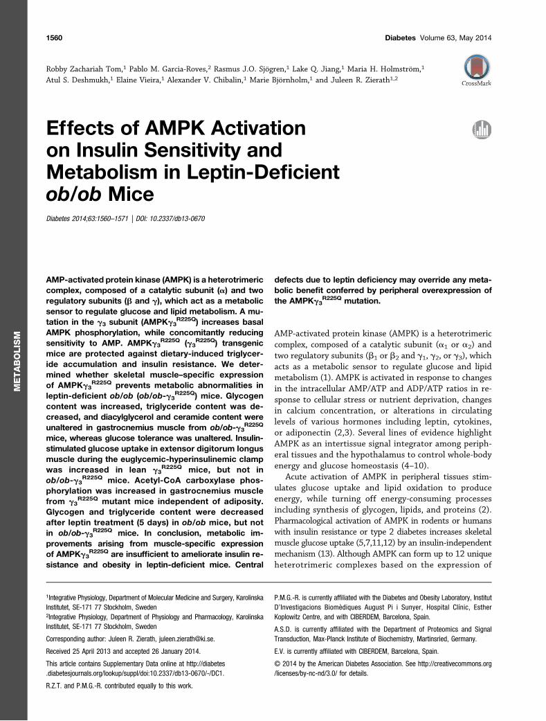
Glycogen Content in Skeletal MuscleGlycogen content was determined in the gastrocnemiusmuscle from 4-h fasted WT, lean g3
R225Q transgenic,ob/ob, and ob/ob-g3
R225Q transgenic mice. As previouslyreported (17), the AMPKg3
R225Q mutation increased gly-cogen content in lean mice (Fig. 1). WT and ob/ob micehave similar levels of glycogen content in the gastrocne-mius muscle. Consistent with the lean g3
R225Q transgenic
mice, glycogen content in the gastrocnemius muscle wasincreased in ob/ob-g3
R225Q transgenic mice compared withob/ob mice (Fig. 1).
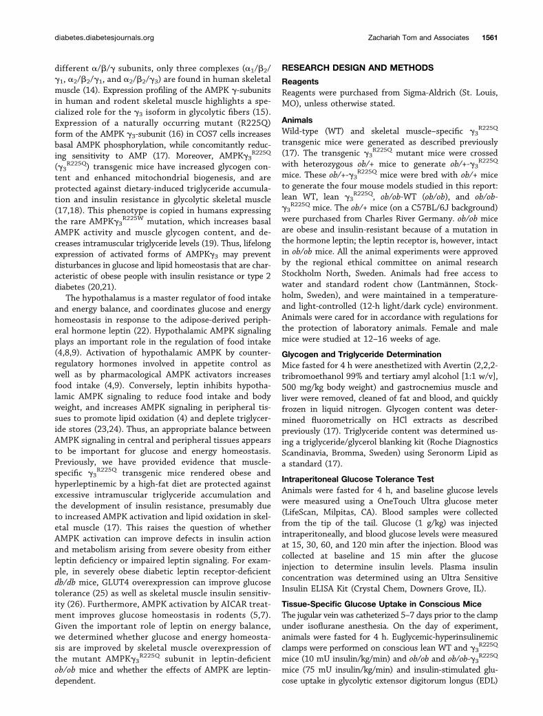
Glucose Tolerance in ob/ob-g3R225Q Transgenic Mice
Glucose tolerance was similar between ob/ob and ob/ob-g3
R225Q transgenic mice (Fig. 2A), consistent with ourpreviously observation that glucose tolerance is unalteredin lean WT and g3
R225Q transgenic mice (17). Plasma in-sulin concentrations determined at baseline and 15 minafter the glucose injection were similar between the ob/oband ob/ob-g3
R225Q transgenic mice (Fig. 2B).
In Vivo Insulin-Stimulated Glucose Uptake in SkeletalMuscleWe performed a euglycemic-hyperinsulinemic clamp toassess in vivo glucose uptake in EDL and gastrocnemiusmuscles. Lean WT and g3
R225Q transgenic mice were clampedusing an insulin infusion of 10 mU/kg/min (baseline plasmainsulin levels were 0.5 6 0.1 and 0.6 60.2 ng/mL, respec-tively, in WT and g3
R225Q mice, and levels achieved duringthe euglycemic-hyperinsulinemic clamp were 9.4 6 0.9and 11.3 6 1.2 ng/mL, respectively, in WT and g3
R225Q
mice (n = 6–9). ob/ob and ob/ob-g3R225Q transgenic mice
were clamped using an insulin infusion of 75 mU/kg/minbecause of their extreme insulin-resistant state (baselineplasma insulin levels in ob/ob and ob/ob-g3
R225Q micewere 15.1 6 5.6 and 9.3 61.6 ng/mL, respectively; levelsachieved in ob/ob and ob/ob-g3
R225Q mice during theeuglycemic-hyperinsulinemic clamp were 281.5 6 60.3and 338.1 6 17.9 ng/mL, respectively; n = 4). The bodyweight of lean mice (WT mice 31.6 6 0.8 g; g3
R225Q mice32.4 6 1.1 g; n = 10–12) and ob/ob mice (ob/ob 43.6 61.3 g; ob/ob-g3
R225Q 41.3 6 1.7 g; n = 6–9) without orwith the g3
R225Q transgene was unaltered. Four-hourfasted plasma glucose levels were similar between WTand g3
R225Q mice (8.4 6 0.4 and 9.8 6 0.6 mmol/L, re-spectively; n = 10–12) and ob/ob and ob/ob-g3
R225Q mice(9.3 6 0.7 and 11.4 6 1.8 mmol/L, respectively; n = 6–9).
Figure 1—Effect of the AMPKg3R225Q mutation on skeletal muscle
glycogen content. Glycogen content was measured in the gastroc-nemius muscle from 4-h–fasted mice in WT (n = 7), g3
R225Q (n = 6),ob/ob (n = 11), and ob/ob-g3
R225Q (n = 5) mice. Results are given asthe mean 6 SEM. *P < 0.05 vs. WT mice; †††P < 0.001 vs. ob/obmice.
Figure 2—Effect of the AMPKg3R225Q mutation on glucose tolerance. A: Intraperitoneal glucose tolerance tests (1 mg/kg glucose) were
performed in 4-h–fasted ob/ob mice (solid line, n = 9) and ob/ob-g3R225Q transgenic mice (dashed line, n = 15). B: Plasma insulin levels at
baseline (black bars) and 15 min after the glucose injection (white bars) in ob/ob and ob/ob-g3R225Q mice (n = 9–10). Results are given as the
mean 6 SEM.
diabetes.diabetesjournals.org Zachariah Tom and Associates 1563
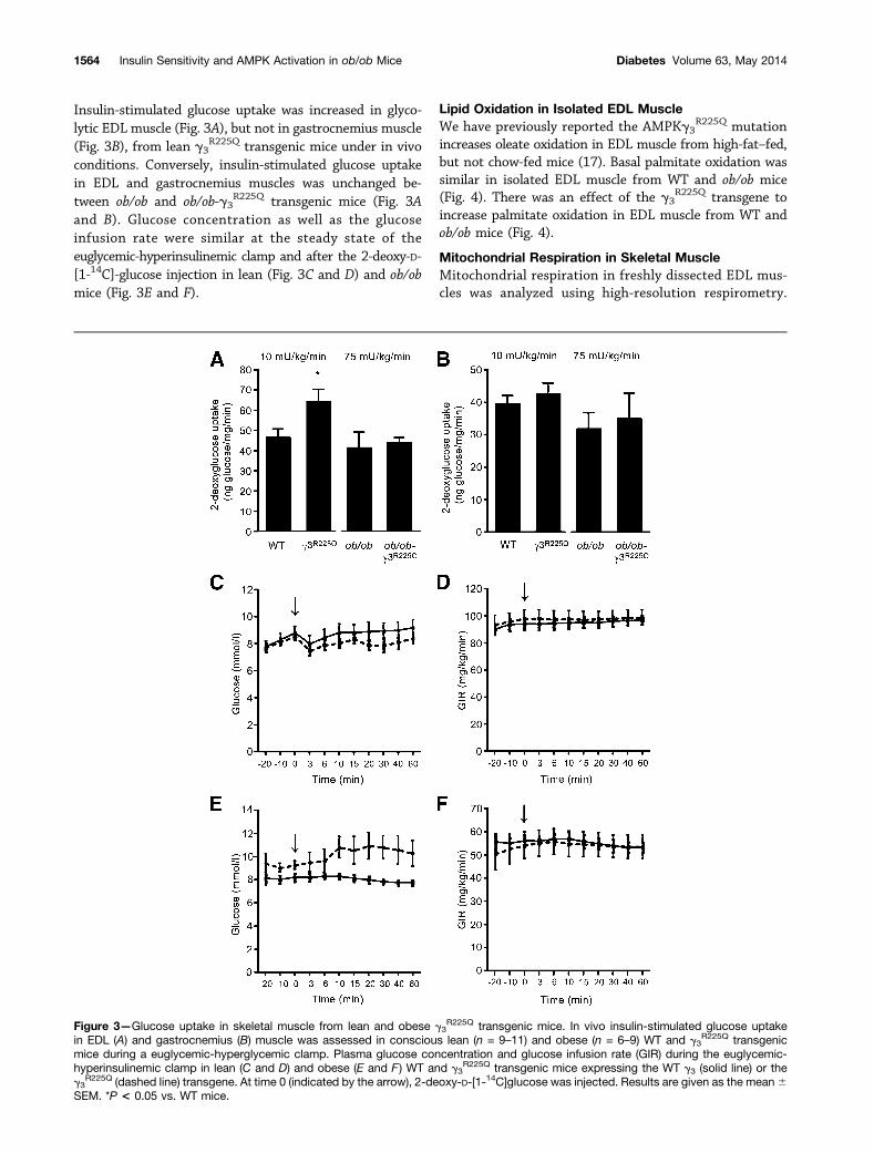
Insulin-stimulated glucose uptake was increased in glyco-lytic EDL muscle (Fig. 3A), but not in gastrocnemius muscle(Fig. 3B), from lean g3
R225Q transgenic mice under in vivoconditions. Conversely, insulin-stimulated glucose uptakein EDL and gastrocnemius muscles was unchanged be-tween ob/ob and ob/ob-g3
R225Q transgenic mice (Fig. 3Aand B). Glucose concentration as well as the glucoseinfusion rate were similar at the steady state of theeuglycemic-hyperinsulinemic clamp and after the 2-deoxy-D-[1-14C]-glucose injection in lean (Fig. 3C and D) and ob/obmice (Fig. 3E and F).
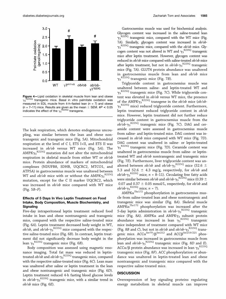
Lipid Oxidation in Isolated EDL MuscleWe have previously reported the AMPKg3
R225Q mutationincreases oleate oxidation in EDL muscle from high-fat–fed,but not chow-fed mice (17). Basal palmitate oxidation wassimilar in isolated EDL muscle from WT and ob/ob mice(Fig. 4). There was an effect of the g3
R225Q transgene toincrease palmitate oxidation in EDL muscle from WT andob/ob mice (Fig. 4).
Mitochondrial Respiration in Skeletal MuscleMitochondrial respiration in freshly dissected EDL mus-cles was analyzed using high-resolution respirometry.
Figure 3—Glucose uptake in skeletal muscle from lean and obese g3R225Q transgenic mice. In vivo insulin-stimulated glucose uptake
in EDL (A) and gastrocnemius (B) muscle was assessed in conscious lean (n = 9–11) and obese (n = 6–9) WT and g3R225Q transgenic
mice during a euglycemic-hyperglycemic clamp. Plasma glucose concentration and glucose infusion rate (GIR) during the euglycemic-hyperinsulinemic clamp in lean (C and D) and obese (E and F ) WT and g3
R225Q transgenic mice expressing the WT g3 (solid line) or theg3
R225Q (dashed line) transgene. At time 0 (indicated by the arrow), 2-deoxy-D-[1-14C]glucose was injected. Results are given as the mean6SEM. *P < 0.05 vs. WT mice.
1564 Insulin Sensitivity and AMPK Activation in ob/ob Mice Diabetes Volume 63, May 2014
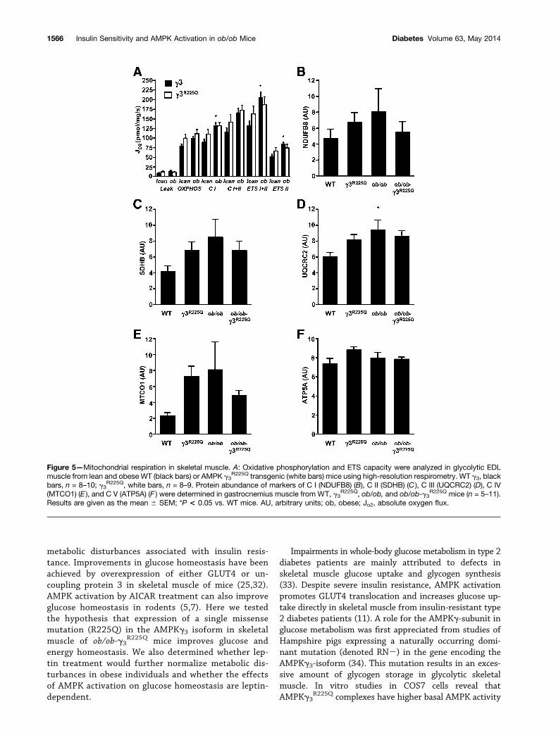
The leak respiration, which denotes endogenous uncou-pling, was similar between the lean and obese non-transgenic and transgenic mice (Fig. 5A). Mitochondrialrespiration at the level of C I, ETS I+II, and ETS II wasincreased in ob/ob versus WT mice (Fig. 5A). TheAMPKg3
R225Q mutation did not alter the mitochondrialrespiration in skeletal muscle from either WT or ob/obmice. Protein abundance of markers of mitochondrialcomplexes (NDUFB8, SDHB, UQCRC2, MTCO1, andATP5A) in gastrocnemius muscle was unaltered betweenWT and ob/ob mice with or without the AMPKg3
R225Q
mutation, except for the C II marker UQCRC2, whichwas increased in ob/ob mice compared with WT mice(Fig. 5B–F).
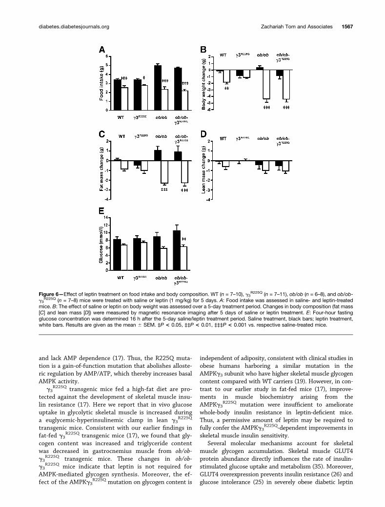
Effects of 5 Days In Vivo Leptin Treatment on FoodIntake, Body Composition, Muscle Biochemistry, andSignalingFive-day intraperitoneal leptin treatment reduced foodintake in lean and obese nontransgenic and transgenicmice, compared with the respective saline-treated mice(Fig. 6A). Leptin treatment decreased body weight in WT,ob/ob, and ob/ob-g3
R225Q mice compared with the respec-tive saline-treated mice (Fig. 6B). In contrast, leptin treat-ment did not significantly decrease body weight in thelean g3
R225Q transgenic mice (Fig. 6B).Body composition was assessed using magnetic reso-
nance imaging. Total fat mass was reduced in leptin-treated ob/ob and ob/ob-g3
R225Q transgenic mice, comparedwith the respective saline-treated mice (Fig. 6C). Lean masswas unaltered after saline or leptin treatment in the leanand obese nontransgenic and transgenic mice (Fig. 6D).Leptin treatment reduced 4-h fasting blood glucose levelsin ob/ob-g3
R225Q transgenic mice, with a similar trend inob/ob mice (Fig. 6E).
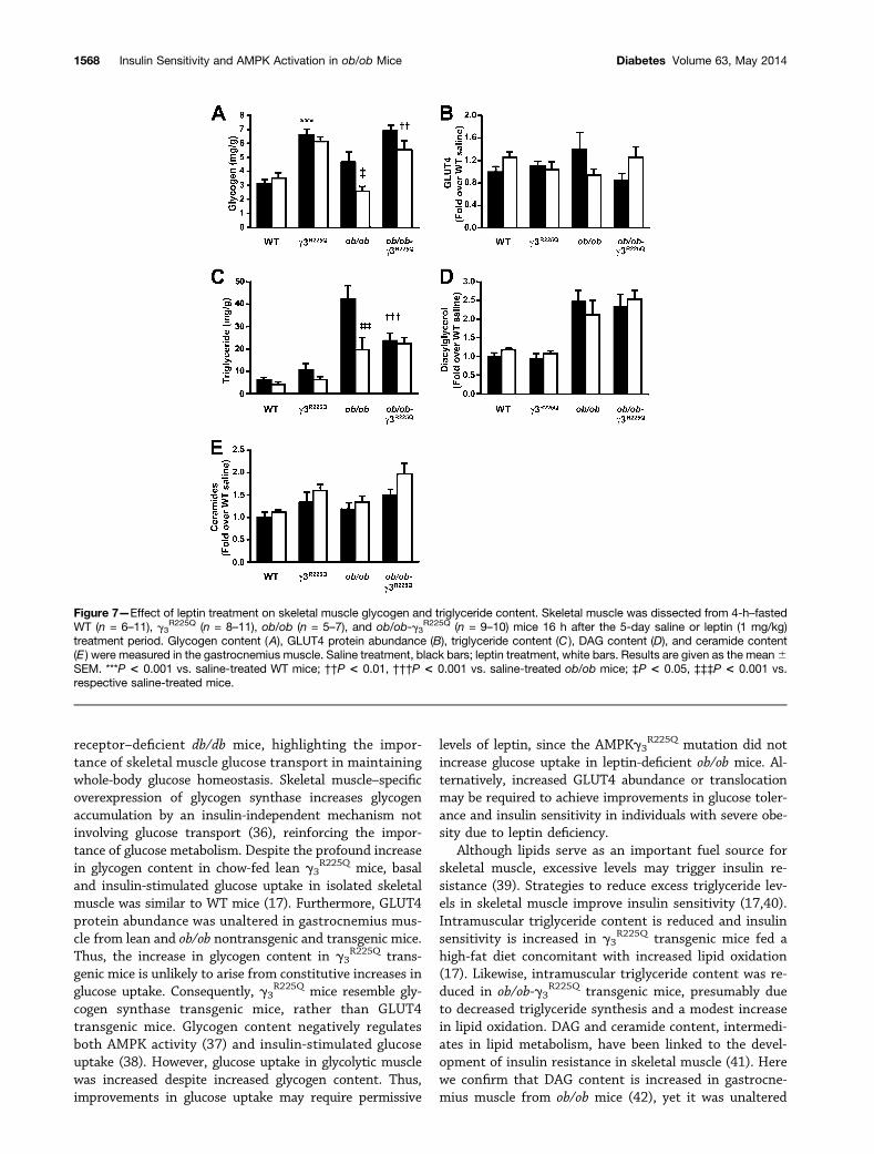
Gastrocnemius muscle was used for biochemical analysis.Glycogen content was increased in the saline-treated leang3
R225Q transgenic mice, compared with the WT mice (Fig.7A). Similarly, glycogen content was increased in ob/ob-g3
R225Q transgenic mice, compared with the ob/ob mice. Gly-cogen content was not altered in WT and g3
R225Q transgenicmice after leptin treatment. However, glycogen content wasreduced in ob/obmice compared with saline-treated ob/obmiceafter leptin treatment, but not in ob/ob-g3
R225Q transgenicmice (Fig. 7A). GLUT4 protein abundance was unalteredin gastrocnemius muscle from lean and ob/ob miceg3
R225Q transgenic mice (Fig. 7B).Triglyceride content in gastrocnemius muscle was
unaltered between saline- and leptin-treated WT andg3
R225Q transgenic mice (Fig. 7C). While triglyceride con-tent was elevated in ob/ob versus WT mice, the presenceof the AMPKg3
R225Q transgene in the ob/ob mice (ob/ob-g3
R225Q mice) reduced triglyceride content. Furthermore,leptin treatment reduced triglyceride content in ob/obmice. However, leptin treatment did not further reducetriglyceride content in gastrocnemius muscle from theob/ob-g3
R225Q transgenic mice (Fig. 7C). DAG and cer-amide content were assessed in gastrocnemius musclefrom saline- and leptin-treated mice. DAG content was in-creased in ob/ob mice compared with WT mice (Fig. 7D).DAG content was unaltered in saline- or leptin-treatedg3
R225Q transgenic mice (Fig. 7D). Ceramide content wasunaltered in gastrocnemius muscle from saline- or leptin-treated WT and ob/ob nontransgenic and transgenic mice(Fig. 7E). Furthermore, liver triglyceride content was un-altered between ob/ob and ob/ob-g3
R225Q mice (52.6 65.3 and 52.6 6 4.3 mg/g, respectively, for ob/ob andob/ob-g3
R225Q mice; n = 8–11). Circulating free fatty acidswere similar between ob/ob and ob/ob-g3
R225Q mice (0.3960.07 and 0.37 6 0.05 mmol/L, respectively, for ob/ob andob/ob-g3
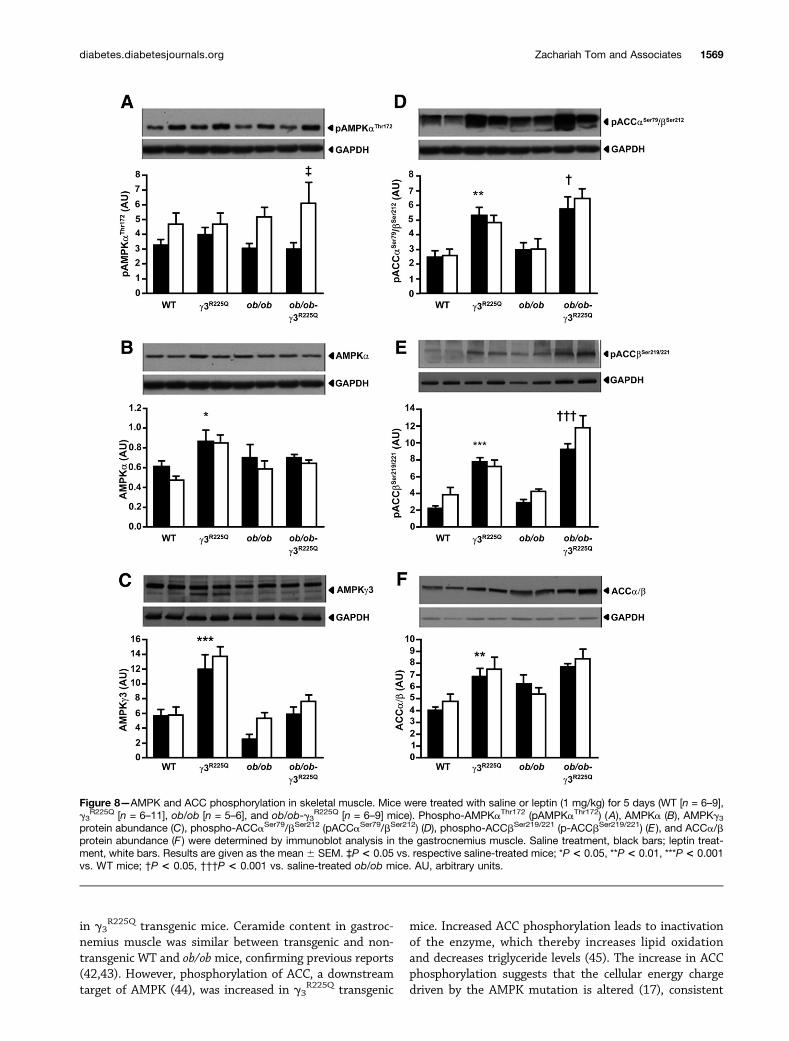
R225Q mice; n = 7).AMPKaThr172 phosphorylation in gastrocnemius mus-
cle from saline-treated lean and obese nontransgenic andtransgenic mice was similar (Fig. 8A). Skeletal muscleAMPKaThr172 phosphorylation was increased after the5-day leptin administration in ob/ob-g3
R225Q transgenicmice (Fig. 8A). AMPKa and AMPKg3 subunit proteinabundance was increased in lean g3
R225Q transgenicmice independent of treatment compared with WT mice(Fig. 8B and C), but not in ob/ob and ob/ob-g3
R225Q trans-genic mice. ACCaSer79/bSer212 and ACCbSer219/221 phos-phorylation was increased in gastrocnemius muscle fromlean and ob/ob-g3
R225Q transgenic mice (Fig. 8D and E).ACCa/b protein abundance was increased in lean g3
R225Q
transgenic mice (Fig. 8F). ACC phosphorylation or abun-dance was unaltered in leptin-treated lean and obesenontransgenic and transgenic mice compared with therespective saline-treated mice.
DISCUSSION
Overexpression of key signaling proteins regulatingenergy metabolism in skeletal muscle can improve
Figure 4—Lipid oxidation in skeletal muscle from lean and obeseg3
R225Q transgenic mice. Basal in vitro palmitate oxidation wasmeasured in EDL muscle from 4-h–fasted lean (n = 7) and obese(n = 7–11) mice. Results are given as the mean 6 SEM. #P < 0.05indicates the effect of the g3
R225Q transgene.
diabetes.diabetesjournals.org Zachariah Tom and Associates 1565
metabolic disturbances associated with insulin resis-tance. Improvements in glucose homeostasis have beenachieved by overexpression of either GLUT4 or un-coupling protein 3 in skeletal muscle of mice (25,32).AMPK activation by AICAR treatment can also improveglucose homeostasis in rodents (5,7). Here we testedthe hypothesis that expression of a single missensemutation (R225Q) in the AMPKg3 isoform in skeletalmuscle of ob/ob-g3
R225Q mice improves glucose andenergy homeostasis. We also determined whether lep-tin treatment would further normalize metabolic dis-turbances in obese individuals and whether the effectsof AMPK activation on glucose homeostasis are leptin-dependent.
Impairments in whole-body glucose metabolism in type 2diabetes patients are mainly attributed to defects inskeletal muscle glucose uptake and glycogen synthesis(33). Despite severe insulin resistance, AMPK activationpromotes GLUT4 translocation and increases glucose up-take directly in skeletal muscle from insulin-resistant type2 diabetes patients (11). A role for the AMPKg-subunit inglucose metabolism was first appreciated from studies ofHampshire pigs expressing a naturally occurring domi-nant mutation (denoted RN2) in the gene encoding theAMPKg3-isoform (34). This mutation results in an exces-sive amount of glycogen storage in glycolytic skeletalmuscle. In vitro studies in COS7 cells reveal thatAMPKg3
R225Q complexes have higher basal AMPK activity
Figure 5—Mitochondrial respiration in skeletal muscle. A: Oxidative phosphorylation and ETS capacity were analyzed in glycolytic EDLmuscle from lean and obese WT (black bars) or AMPK g3
R225Q transgenic (white bars) mice using high-resolution respirometry. WT g3, blackbars, n = 8–10; g3
R225Q, white bars, n = 8–9. Protein abundance of markers of C I (NDUFB8) (B), C II (SDHB) (C ), C III (UQCRC2) (D), C IV(MTCO1) (E ), and C V (ATP5A) (F ) were determined in gastrocnemius muscle from WT, g3
R225Q, ob/ob, and ob/ob-g3R225Q mice (n = 5–11).
Results are given as the mean 6 SEM; *P < 0.05 vs. WT mice. AU, arbitrary units; ob, obese; Jo2, absolute oxygen flux.
1566 Insulin Sensitivity and AMPK Activation in ob/ob Mice Diabetes Volume 63, May 2014
and lack AMP dependence (17). Thus, the R225Q muta-tion is a gain-of-function mutation that abolishes alloste-ric regulation by AMP/ATP, which thereby increases basalAMPK activity.
g3R225Q transgenic mice fed a high-fat diet are pro-
tected against the development of skeletal muscle insu-lin resistance (17). Here we report that in vivo glucoseuptake in glycolytic skeletal muscle is increased duringa euglycemic-hyperinsulinemic clamp in lean g3
R225Q
transgenic mice. Consistent with our earlier findings infat-fed g3
R225Q transgenic mice (17), we found that gly-cogen content was increased and triglyceride contentwas decreased in gastrocnemius muscle from ob/ob-g3
R225Q transgenic mice. These changes in ob/ob-g3
R225Q mice indicate that leptin is not required forAMPK-mediated glycogen synthesis. Moreover, the ef-fect of the AMPKg3
R225Q mutation on glycogen content is
independent of adiposity, consistent with clinical studies inobese humans harboring a similar mutation in theAMPKg3 subunit who have higher skeletal muscle glycogencontent compared with WT carriers (19). However, in con-trast to our earlier study in fat-fed mice (17), improve-ments in muscle biochemistry arising from theAMPKg3
R225Q mutation were insufficient to amelioratewhole-body insulin resistance in leptin-deficient mice.Thus, a permissive amount of leptin may be required tofully confer the AMPKg3
R225Q-dependent improvements inskeletal muscle insulin sensitivity.
Several molecular mechanisms account for skeletalmuscle glycogen accumulation. Skeletal muscle GLUT4protein abundance directly influences the rate of insulin-stimulated glucose uptake and metabolism (35). Moreover,GLUT4 overexpression prevents insulin resistance (26) andglucose intolerance (25) in severely obese diabetic leptin
Figure 6—Effect of leptin treatment on food intake and body composition. WT (n = 7–10), g3R225Q (n = 7–11), ob/ob (n = 6–8), and ob/ob-
g3R225Q (n = 7–8) mice were treated with saline or leptin (1 mg/kg) for 5 days. A: Food intake was assessed in saline- and leptin-treated
mice. B: The effect of saline or leptin on body weight was assessed over a 5-day treatment period. Changes in body composition (fat mass[C] and lean mass [D]) were measured by magnetic resonance imaging after 5 days of saline or leptin treatment. E: Four-hour fastingglucose concentration was determined 16 h after the 5-day saline/leptin treatment period. Saline treatment, black bars; leptin treatment,white bars. Results are given as the mean 6 SEM. ‡P < 0.05, ‡‡P < 0.01, ‡‡‡P < 0.001 vs. respective saline-treated mice.
diabetes.diabetesjournals.org Zachariah Tom and Associates 1567
receptor–deficient db/db mice, highlighting the impor-tance of skeletal muscle glucose transport in maintainingwhole-body glucose homeostasis. Skeletal muscle–specificoverexpression of glycogen synthase increases glycogenaccumulation by an insulin-independent mechanism notinvolving glucose transport (36), reinforcing the impor-tance of glucose metabolism. Despite the profound increasein glycogen content in chow-fed lean g3
R225Q mice, basaland insulin-stimulated glucose uptake in isolated skeletalmuscle was similar to WT mice (17). Furthermore, GLUT4protein abundance was unaltered in gastrocnemius mus-cle from lean and ob/ob nontransgenic and transgenic mice.Thus, the increase in glycogen content in g3
R225Q trans-genic mice is unlikely to arise from constitutive increases inglucose uptake. Consequently, g3
R225Q mice resemble gly-cogen synthase transgenic mice, rather than GLUT4transgenic mice. Glycogen content negatively regulatesboth AMPK activity (37) and insulin-stimulated glucoseuptake (38). However, glucose uptake in glycolytic musclewas increased despite increased glycogen content. Thus,improvements in glucose uptake may require permissive
levels of leptin, since the AMPKg3R225Q mutation did not
increase glucose uptake in leptin-deficient ob/ob mice. Al-ternatively, increased GLUT4 abundance or translocationmay be required to achieve improvements in glucose toler-ance and insulin sensitivity in individuals with severe obe-sity due to leptin deficiency.
Although lipids serve as an important fuel source forskeletal muscle, excessive levels may trigger insulin re-sistance (39). Strategies to reduce excess triglyceride lev-els in skeletal muscle improve insulin sensitivity (17,40).Intramuscular triglyceride content is reduced and insulinsensitivity is increased in g3
R225Q transgenic mice fed ahigh-fat diet concomitant with increased lipid oxidation(17). Likewise, intramuscular triglyceride content was re-duced in ob/ob-g3
R225Q transgenic mice, presumably dueto decreased triglyceride synthesis and a modest increasein lipid oxidation. DAG and ceramide content, intermedi-ates in lipid metabolism, have been linked to the devel-opment of insulin resistance in skeletal muscle (41). Herewe confirm that DAG content is increased in gastrocne-mius muscle from ob/ob mice (42), yet it was unaltered
Figure 7—Effect of leptin treatment on skeletal muscle glycogen and triglyceride content. Skeletal muscle was dissected from 4-h–fastedWT (n = 6–11), g3
R225Q (n = 8–11), ob/ob (n = 5–7), and ob/ob-g3R225Q (n = 9–10) mice 16 h after the 5-day saline or leptin (1 mg/kg)
treatment period. Glycogen content (A), GLUT4 protein abundance (B), triglyceride content (C ), DAG content (D), and ceramide content(E ) were measured in the gastrocnemius muscle. Saline treatment, black bars; leptin treatment, white bars. Results are given as the mean6SEM. ***P < 0.001 vs. saline-treated WT mice; ††P < 0.01, †††P < 0.001 vs. saline-treated ob/ob mice; ‡P < 0.05, ‡‡‡P < 0.001 vs.respective saline-treated mice.
1568 Insulin Sensitivity and AMPK Activation in ob/ob Mice Diabetes Volume 63, May 2014
in g3R225Q transgenic mice. Ceramide content in gastroc-
nemius muscle was similar between transgenic and non-transgenic WT and ob/ob mice, confirming previous reports(42,43). However, phosphorylation of ACC, a downstreamtarget of AMPK (44), was increased in g3
R225Q transgenic
mice. Increased ACC phosphorylation leads to inactivationof the enzyme, which thereby increases lipid oxidationand decreases triglyceride levels (45). The increase in ACCphosphorylation suggests that the cellular energy chargedriven by the AMPK mutation is altered (17), consistent
Figure 8—AMPK and ACC phosphorylation in skeletal muscle. Mice were treated with saline or leptin (1 mg/kg) for 5 days (WT [n = 6–9],g3
R225Q [n = 6–11], ob/ob [n = 5–6], and ob/ob-g3R225Q [n = 6–9] mice). Phospho-AMPKaThr172 (pAMPKaThr172) (A), AMPKa (B), AMPKg3
protein abundance (C), phospho-ACCaSer79/bSer212 (pACCaSer79/bSer212) (D), phospho-ACCbSer219/221 (p-ACCbSer219/221) (E ), and ACCa/bprotein abundance (F ) were determined by immunoblot analysis in the gastrocnemius muscle. Saline treatment, black bars; leptin treat-ment, white bars. Results are given as the mean 6 SEM. ‡P < 0.05 vs. respective saline-treated mice; *P < 0.05, **P < 0.01, ***P < 0.001vs. WT mice; †P < 0.05, †††P < 0.001 vs. saline-treated ob/ob mice. AU, arbitrary units.
diabetes.diabetesjournals.org Zachariah Tom and Associates 1569
with increased lipid oxidation in the g3R225Q transgenic
mice.Leptin influences AMPK signaling in central and
peripheral tissues. Leptin inhibits AMPK activity in thebrain and reduces food intake (4), while enhancing lipidmetabolism in skeletal muscle (46). Thus, we exploredwhether leptin treatment of ob/ob-g3
R225Q transgenicmice would lead to a further metabolic improvement im-posed by the AMPKg3
R225Q mutation. The AMPKg3R225Q
mutation did not alter the body weight response to leptintreatment in ob/ob mice. Thus, the metabolic differencesobserved between the nontransgenic and ob/ob-g3
R225Q
transgenic mice are directly related to the mutation,rather than to changes in food intake. Leptin treatmentimproved the fasting glucose level in ob/ob-g3
R225Q trans-genic mice, concomitant with a normalization of food in-take and a reduction in adiposity. Moreover, we foundthat skeletal muscle glycogen content was decreased inleptin-treated ob/ob mice, consistent with the effects ofleptin in decreasing glycogen synthesis in ob/ob mice (47).However, leptin treatment did not decrease skeletal mus-cle glycogen content in g3
R225Q transgenic lean and ob/obmice, indicating that the AMPKg3
R225Q mutation hasa dominant influence on fuel partitioning within skeletalmuscle, which may be overcome by hyperleptinemia. Al-though the concentration of leptin used in this study wassufficient to improve blood glucose and body weight,higher doses trigger a shift in substrate use such as thatobserved in fat-fed g3
R225Q mice (17).AMPK activation is linked to mitochondrial biogenesis,
providing a mechanism for the increased lipid oxidationobserved in fat-fed g3
R225Q transgenic mice (17). The in-crease in mitochondrial respiration in ob/ob mice confirmsour previous findings that obesity induces molecular adap-tations in glycolytic skeletal muscle to enhance mitochon-drial respiration (48). Nevertheless, these mice are severelyinsulin-resistant. Mitochondrial biogenesis is increased inglycolytic skeletal muscle from g3
R225Q transgenic mice,concomitant with increased expression of the coactivatorperoxisome proliferator–activated receptor g coactivator-1a and transcription factors that drive different mitochon-drial proteins expression (18). However, mitochondrialrespiration is unaltered between WT and g3
R225Q trans-genic mice (18), as well as ob/ob-g3
R225Q transgenic mice.The increase in skeletal muscle mitochondrial markers ing3
R225Q transgenic mice (18) may account for the increasein lipid oxidation in the g3
R225Q transgenic mice. However,the regulation of insulin sensitivity is complex and notentirely coupled to increased skeletal muscle mitochondrialcontent. Nevertheless, cultured myotubes from probandscarrying a homologous mutation (AMPKg3
R225W) reinforcethe profound effect of this mutation on glucose uptake andmetabolism, mitochondrial content, and oxidative capacity,and raise the clinical implications of mutations in theAMPKg3 subunit (49).
Defective leptin action leads to metabolic abnormalitiesassociated with obesity. The effects of leptin are partly
mediated via the AMPK pathway in central and peripheralsites. Here we show that the expression of a mutantform of the AMPKg3 subunit in glycolytic skeletal muscleincreases glycogen content and decreases intramusculartriglyceride levels. However, DAG and ceramide contentwere unaltered. The triglyceride depletion in ob/ob-g3
R225Q
transgenic mice does not appear to improve glucose utili-zation and insulin sensitivity in ob/ob-g3
R225Q mice. Thus,the lack of central leptin signaling may override thefavorable metabolic milieu conferred by peripheral overex-pression of the AMPKg3
R225Q mutation to improve glucoseand energy homeostasis. Further studies in hypothalamic-specific AMPK transgenic ob/obmice may clarify the centralrole of this protein kinase in the control of glucose andenergy homeostasis in leptin deficiency. Given our find-ings (Supplementary Table 1), targeting both peripheraland central AMPK actions may be required to improveglucose homeostasis.
Funding. This work was supported by grants from the European Foundationfor the Study of Diabetes, Swedish Research Council, Swedish Diabetes Associ-ation, Swedish Foundation for Strategic Research (INGVAR II), the European Re-search Council, Novo Nordisk Research Foundation, the Strategic ResearchProgramme in Diabetes at Karolinska Institutet, and Commission of the EuropeanCommunities (Contract no. LSHM-CT-2004-005272 EXGENESIS).Duality of Interest. No potential conflicts of interest relevant to this articlewere reported.Author Contributions. R.Z.T. researched the data, wrote the manuscript,and approved the final version of the manuscript. P.M.G.-R., A.V.C., and M.B.researched the data, reviewed and edited the manuscript, and approved the finalversion of the manuscript. R.J.O.S., L.Q.J., M.H.H., A.S.D., and E.V. researched thedata and approved the final version of the manuscript. J.R.Z. wrote the manuscriptand approved the final version of the manuscript. J.R.Z. is the guarantor of thiswork and, as such, had full access to all the data in the study and takesresponsibility for the integrity of the data and the accuracy of the data analysis.Prior Presentation. Parts of this study were presented in abstract form atthe Keystone Symposia Type 2 Diabetes, Insulin Resistance and MetabolicDysfunction, Keystone, CO, 12–17 January 2011, and at the EMBO | EMBLSymposium Diabetes and Obesity, Heidelberg, Germany, 13–16 September 2012.
References1. Hardie DG, Scott JW, Pan DA, Hudson ER. Management of cellular energyby the AMP-activated protein kinase system. FEBS Lett 2003;546:113–1202. Long YC, Zierath JR. AMP-activated protein kinase signaling in metabolicregulation. J Clin Invest 2006;116:1776–17833. Hardie DG, Ross FA, Hawley SA. AMPK: a nutrient and energy sensor thatmaintains energy homeostasis. Nat Rev Mol Cell Biol 2012;13:251–2624. Minokoshi Y, Alquier T, Furukawa N, et al. AMP-kinase regulates food in-take by responding to hormonal and nutrient signals in the hypothalamus. Nature2004;428:569–5745. Bergeron R, Previs SF, Cline GW, et al. Effect of 5-aminoimidazole-4-carboxamide-1-b-D-ribofuranoside infusion on in vivo glucose and lipid me-tabolism in lean and obese Zucker rats. Diabetes 2001;50:1076–10826. Fiedler M, Zierath JR, Selén G, Wallberg-Henriksson H, Liang Y, SakariassenKS. 5-aminoimidazole-4-carboxy-amide-1-beta-D-ribofuranoside treatmentameliorates hyperglycaemia and hyperinsulinaemia but not dyslipidaemia in KKAy-CETP mice. Diabetologia 2001;44:2180–21867. Song XM, Fiedler M, Galuska D, et al. 5-Aminoimidazole-4-carboxamideribonucleoside treatment improves glucose homeostasis in insulin-resistant di-abetic (ob/ob) mice. Diabetologia 2002;45:56–65
1570 Insulin Sensitivity and AMPK Activation in ob/ob Mice Diabetes Volume 63, May 2014
8. Claret M, Smith MA, Batterham RL, et al. AMPK is essential for energyhomeostasis regulation and glucose sensing by POMC and AgRP neurons. J ClinInvest 2007;117:2325–23369. Andersson U, Filipsson K, Abbott CR, et al. AMP-activated protein kinaseplays a role in the control of food intake. J Biol Chem 2004;279:12005–1200810. Cool B, Zinker B, Chiou W, et al. Identification and characterization ofa small molecule AMPK activator that treats key components of type 2 diabetesand the metabolic syndrome. Cell Metab 2006;3:403–41611. Koistinen HA, Galuska D, Chibalin AV, et al. 5-amino-imidazole carboxamideriboside increases glucose transport and cell-surface GLUT4 content in skeletalmuscle from subjects with type 2 diabetes. Diabetes 2003;52:1066–107212. Barnes BR, Ryder JW, Steiler TL, Fryer LG, Carling D, Zierath JR. Isoform-specific regulation of 59 AMP-activated protein kinase in skeletal muscle fromobese Zucker (fa/fa) rats in response to contraction. Diabetes 2002;51:2703–270813. Hayashi T, Hirshman MF, Kurth EJ, Winder WW, Goodyear LJ. Evidence for59 AMP-activated protein kinase mediation of the effect of muscle contraction onglucose transport. Diabetes 1998;47:1369–137314. Birk JB, Wojtaszewski JF. Predominant alpha2/beta2/gamma3 AMPKactivation during exercise in human skeletal muscle. J Physiol 2006;577:1021–103215. Mahlapuu M, Johansson C, Lindgren K, et al. Expression profiling of thegamma-subunit isoforms of AMP-activated protein kinase suggests a major rolefor gamma3 in white skeletal muscle. Am J Physiol Endocrinol Metab 2004;286:E194–E20016. Andersson L. Identification and characterization of AMPK gamma 3 muta-tions in the pig. Biochem Soc Trans 2003;31:232–23517. Barnes BR, Marklund S, Steiler TL, et al. The 59-AMP-activated proteinkinase gamma3 isoform has a key role in carbohydrate and lipid metabolism inglycolytic skeletal muscle. J Biol Chem 2004;279:38441–3844718. Garcia-Roves PM, Osler ME, Holmström MH, Zierath JR. Gain-of-functionR225Q mutation in AMP-activated protein kinase gamma3 subunit increasesmitochondrial biogenesis in glycolytic skeletal muscle. J Biol Chem 2008;283:35724–3573419. Costford SR, Kavaslar N, Ahituv N, et al. Gain-of-function R225W mutationin human AMPKgamma(3) causing increased glycogen and decreased tri-glyceride in skeletal muscle. PLoS One 2007;2:e90320. Osler ME, Zierath JR. Adenosine 59-monophosphate-activated protein ki-nase regulation of fatty acid oxidation in skeletal muscle. Endocrinology 2008;149:935–94121. Savage DB, Petersen KF, Shulman GI. Disordered lipid metabolism and thepathogenesis of insulin resistance. Physiol Rev 2007;87:507–52022. Halaas JL, Gajiwala KS, Maffei M, et al. Weight-reducing effects of theplasma protein encoded by the obese gene. Science 1995;269:543–54623. Unger RH, Zhou YT, Orci L. Regulation of fatty acid homeostasis in cells:novel role of leptin. Proc Natl Acad Sci U S A 1999;96:2327–233224. Lee Y, Wang MY, Kakuma T, et al. Liporegulation in diet-induced obesity.The antisteatotic role of hyperleptinemia. J Biol Chem 2001;276:5629–563525. Gibbs EM, Stock JL, McCoid SC, et al. Glycemic improvement in diabeticdb/db mice by overexpression of the human insulin-regulatable glucose trans-porter (GLUT4). J Clin Invest 1995;95:1512–151826. Brozinick JT Jr, McCoid SC, Reynolds TH, et al. GLUT4 overexpression indb/db mice dose-dependently ameliorates diabetes but is not a lifelong cure.Diabetes 2001;50:593–60027. Chibalin AV, Leng Y, Vieira E, et al. Downregulation of diacylglycerol kinase deltacontributes to hyperglycemia-induced insulin resistance. Cell 2008;132:375–38628. Chadt A, Leicht K, Deshmukh A, et al. Tbc1d1 mutation in lean mouse strainconfers leanness and protects from diet-induced obesity. Nat Genet 2008;40:1354–135929. Jiang LQ, Garcia-Roves PM, de Castro Barbosa T, Zierath JR. Constitu-tively active calcineurin in skeletal muscle increases endurance performance
and mitochondrial respiratory capacity. Am J Physiol Endocrinol Metab 2010;298:E8–E1630. Pesta D, Gnaiger E. High-resolution respirometry: OXPHOS protocols forhuman cells and permeabilized fibers from small biopsies of human muscle.Methods Mol Biol 2012;810:25–5831. Preiss J, Loomis CR, Bishop WR, Stein R, Niedel JE, Bell RM. Quantitativemeasurement of sn-1,2-diacylglycerols present in platelets, hepatocytes, and ras-and sis-transformed normal rat kidney cells. J Biol Chem 1986;261:8597–860032. Schrauwen P, Hardie DG, Roorda B, et al. Improved glucose homeostasis inmice overexpressing human UCP3: a role for AMP-kinase? Int J Obes Relat MetabDisord 2004;28:824–82833. Zierath JR, Krook A, Wallberg-Henriksson H. Insulin action in skeletalmuscle from patients with NIDDM. Mol Cell Biochem 1998;182:153–16034. Milan D, Jeon JT, Looft C, et al. A mutation in PRKAG3 associated withexcess glycogen content in pig skeletal muscle. Science 2000;288:1248–125135. Hansen PA, Gulve EA, Marshall BA, et al. Skeletal muscle glucose transportand metabolism are enhanced in transgenic mice overexpressing the Glut4glucose transporter. J Biol Chem 1995;270:1679–168436. Fogt DL, Pan S, Lee S, et al. Effect of glycogen synthase overexpression oninsulin-stimulated muscle glucose uptake and storage. Am J Physiol EndocrinolMetab 2004;286:E363–E36937. Steinberg GR, Watt MJ, McGee SL, et al. Reduced glycogen availability isassociated with increased AMPKalpha2 activity, nuclear AMPKalpha2 proteinabundance, and GLUT4 mRNA expression in contracting human skeletal muscle.Appl Physiol Nutr Metab 2006;31:302–31238. Fell RD, Terblanche SE, Ivy JL, Young JC, Holloszy JO. Effect of muscle glycogencontent on glucose uptake following exercise. J Appl Physiol 1982;52:434–43739. Sinha R, Dufour S, Petersen KF, et al. Assessment of skeletal muscle tri-glyceride content by 1H nuclear magnetic resonance spectroscopy in lean andobese adolescents: relationships to insulin sensitivity, total body fat, and centraladiposity. Diabetes 2002;51:1022–102740. Kim JK, Fillmore JJ, Gavrilova O, et al. Differential effects of rosiglitazone onskeletal muscle and liver insulin resistance in A-ZIP/F-1 fatless mice. Diabetes2003;52:1311–131841. Timmers S, Schrauwen P, de Vogel J. Muscular diacylglycerol metabolismand insulin resistance. Physiol Behav 2008;94:242–25142. Wendel AA, Li LO, Li Y, Cline GW, Shulman GI, Coleman RA. Glycerol-3-phosphate acyltransferase 1 deficiency in ob/ob mice diminishes hepaticsteatosis but does not protect against insulin resistance or obesity. Diabetes2010;59:1321–132943. Aerts JM, Ottenhoff R, Powlson AS, et al. Pharmacological inhibition ofglucosylceramide synthase enhances insulin sensitivity. Diabetes 2007;56:1341–134944. Park SH, Gammon SR, Knippers JD, Paulsen SR, Rubink DS, Winder WW.Phosphorylation-activity relationships of AMPK and acetyl-CoA carboxylase inmuscle. J Appl Physiol (1985) 2002;92:2475–248245. Fullerton MD, Galic S, Marcinko K, et al. Single phosphorylation sites inAcc1 and Acc2 regulate lipid homeostasis and the insulin-sensitizing effects ofmetformin. Nat Med 2013;19:1649–165446. Minokoshi Y, Kim YB, Peroni OD, et al. Leptin stimulates fatty-acid oxidationby activating AMP-activated protein kinase. Nature 2002;415:339–34347. Liu YL, Emilsson V, Cawthorne MA. Leptin inhibits glycogen synthesis in theisolated soleus muscle of obese (ob/ob) mice. FEBS Lett 1997;411:351–35548. Holmström MH, Tom RZ, Björnholm M, Garcia-Roves PM, Zierath JR. Effectof leptin treatment on mitochondrial function in obese leptin-deficient ob/ob mice.Metabolism 2013;62:1258–126749. Crawford SA, Costford SR, Aguer C, et al. Naturally occurring R225Wmutation of the gene encoding AMP-activated protein kinase (AMPK)gamma(3)results in increased oxidative capacity and glucose uptake in human primarymyotubes. Diabetologia 2010;53:1986–1997
diabetes.diabetesjournals.org Zachariah Tom and Associates 1571