effects of cyclin d-1 gene amplification and protein...
TRANSCRIPT

LUND UNIVERSITY
PO Box 117221 00 Lund+46 46-222 00 00
Effects of cyclin D-1 gene amplification and protein expression on time to recurrencein postmenopausal breast cancer patients treated with anastrozole or tamoxifen: aTransATAC study
Lundgren, Katja; Brown, Matthew; Pineda, Silvia; Cuzick, Jack; Salter, Janine; Zabaglo, Lila;Howell, Anthony; Dowsett, Mitch; Landberg, GoeranPublished in:Breast Cancer Research
DOI:10.1186/bcr3161
Published: 2012-01-01
Link to publication
Citation for published version (APA):Lundgren, K., Brown, M., Pineda, S., Cuzick, J., Salter, J., Zabaglo, L., ... Landberg, G. (2012). Effects of cyclinD-1 gene amplification and protein expression on time to recurrence in postmenopausal breast cancer patientstreated with anastrozole or tamoxifen: a TransATAC study. Breast Cancer Research, 14(2). DOI:10.1186/bcr3161
General rightsCopyright and moral rights for the publications made accessible in the public portal are retained by the authorsand/or other copyright owners and it is a condition of accessing publications that users recognise and abide by thelegal requirements associated with these rights.
• Users may download and print one copy of any publication from the public portal for the purpose of privatestudy or research. • You may not further distribute the material or use it for any profit-making activity or commercial gain • You may freely distribute the URL identifying the publication in the public portal

Take down policyIf you believe that this document breaches copyright please contact us providing details, and we will removeaccess to the work immediately and investigate your claim.
Download date: 16. Jul. 2018

RESEARCH ARTICLE Open Access
Effects of cyclin D1 gene amplification andprotein expression on time to recurrence inpostmenopausal breast cancer patients treatedwith anastrozole or tamoxifen: a TransATAC studyKatja Lundgren1,2, Matthew Brown2, Silvia Pineda3, Jack Cuzick3, Janine Salter4, Lila Zabaglo4, Anthony Howell2,Mitch Dowsett4,5 and Göran Landberg2,6*, for the TransATAC investigators
Abstract
Introduction: Gene amplification of CCND1 is observed in a subgroup of breast cancers with poor prognosis,whereas overexpression of the protein cyclin D1 has been linked to both worse and better clinical outcome.CCND1 amplification and protein overexpression have also been associated with resistance to treatment withtamoxifen or even to a potentially detrimental effect of tamoxifen.
Methods: To clarify these challenging and partly contrasting treatment predictive and prognostic links for cyclin D1
we analysed a large cohort of postmenopausal breast cancer patients randomised to receive either adjuvantanastrozole or tamoxifen, as part of the Arimidex, Tamoxifen, Alone or in Combination (ATAC) trial. The CCND1amplification status and protein expression of cyclin D1 were assessed by chromogenic in situ hybridisation andimmunohistochemistry, respectively, in 1,155 postmenopausal, oestrogen-receptor-positive breast cancer patientsincluded in the TransATAC substudy.
Results: Amplification of CCND1 was observed in 8.7% of the tumours and was associated with increased risk ofdisease recurrence (hazard ratio = 1.61; 95% confidence interval, 1.08 to 2.41) after adjustment for otherclinicopathological parameters. In contrast, nuclear expression of cyclin D1 protein was associated with decreasedrecurrence rate (hazard ratio = 0.6; 95% confidence interval, 0.39 to 0.92). The intensity of nuclear or cytoplasmicexpression was not of prognostic value. There was no significant interaction between cyclin D1 status and treatmentefficacy, ruling out any major detrimental effect of tamoxifen in CCND1-amplified postmenopausal breast cancer.
Conclusions: In summary, CCND1 amplification and low nuclear expression of cyclin D1 predicted poor clinicaloutcome in postmenopausal breast cancer patients treated with either anastrozole or tamoxifen.
Trial Registration: Current Controlled Trials ISRCTN18233230.
IntroductionHormone dependence is a fundamental hallmark of themajority of breast cancers, and tumour growth can beinhibited either by deprivation of circulating oestrogensor by antagonising the effect of these hormones on theirreceptors [1]. The selective oestrogen receptor (ER)
modulator tamoxifen has long been the most commonlyused adjuvant therapy for patients with advanced hor-mone-sensitive breast cancer [2]. In recent years, how-ever, aromatase inhibitors have become an alternativetreatment option for postmenopausal women with breastcancer. An aromatase inhibitor acts by interfering withthe enzyme that converts androgens to oestrogen, andreduces tumour and systemic oestrogen concentration[3]. The third-generation selective aromatase inhibitoranastrozole (Arimidex) reduces serum oestradiol tonanomolar concentrations [4]. The Arimidex, Tamoxifen,
* Correspondence: [email protected] Breast Cancer Research Unit, School of Cancer, EnablingSciences and Technology, University of Manchester, Manchester AcademicHealth Science Centre Paterson Institute for Cancer Research, The ChristieNHS Foundation Trust, Wilmslow Road, Manchester M20 4BX, UKFull list of author information is available at the end of the article
Lundgren et al. Breast Cancer Research 2012, 14:R57http://breast-cancer-research.com/content/14/2/R57
© 2012 Lundgren et al.; licensee BioMed Central Ltd. This is an open access article distributed under the terms of the CreativeCommons Attribution License (http://creativecommons.org/licenses/by/2.0), which permits unrestricted use, distribution, andreproduction in any medium, provided the original work is properly cited.

Alone or in Combination (ATAC) trial was designed tocompare the efficacy of anastrozole alone or in combina-tion with the established adjuvant treatment, tamoxifenfor 5 years, as adjuvant treatment for postmenopausalwomen with operable breast cancer [5]. The studydemonstrated that the efficacy of anastrozole was highercompared with tamoxifen alone, and also superior to thecombination of both agents [5,6]. After a median follow-up of 10 years, 5 years after completion of treatment, thesignificant advantage for anastrozole over tamoxifen asinitial adjuvant therapy for postmenopausal, ER-positivebreast cancer patients was confirmed [7].In breast cancer, genetic alterations such as amplifica-
tions and deletions occur within the tumour at high fre-quencies, and a number of these alterations are closelyrelated to poor clinical outcome. One such region ofamplification is 11q13, harbouring the cyclin D1 geneCCND1 [8-10]. Cyclin D1 plays a crucial role as a cellcycle regulator, promoting progression through the G1-Sphase, following complex formation with CDK4/6 andphosphorylation of the retinoblastoma protein [11].Various studies have described the oncogenic capacity ofcyclin D1 in vitro, and overexpression in vivo results intumour formation [12-14]. Overexpression of cyclin D1 isobserved in approximately 50% of breast cancers [15,16],and cyclin D1 is one of the most commonly overex-pressed proteins in this form of cancer. A number of stu-dies report cyclin D1 overexpression to be a predictor ofworse prognosis [17,18], while others have found an asso-ciation with an ER-positive phenotype and a better clini-cal outcome [19-23]. In about 15% of all primary breastcancers, overexpression is due to amplification of thecorresponding gene CCND1 [15,24,25], and this specificamplification has been linked to poor prognosis [23,26].Despite the presence of ERa, approximately 50% of
breast cancers develop resistance to hormonal treatment,a major clinical limitation of breast cancer therapy[27,28]. The mechanisms behind this phenomenon havebeen extensively studied, and imply a complex signallingnetwork governing ER function and interaction with var-ious co-regulators [29-32]. Cyclin D1 is one such co-fac-tor, known to interact with ERa and, independently ofoestrogen, activate the receptor and potentially modifyoestrogen/anti-oestrogen responses [33,34]. Overexpres-sion of cyclin D1 has been reported to result in a confor-mational change in ERa that induces receptor activationin the presence of the novel selective ER modulatorarzoxifene, which in turn promotes growth of MCF-7cells - indicating a change from antagonist to agonist[28]. This study also suggests that different mechanismsare required to confer resistance depending on the speci-fic anti-oestrogen administered, and that changes in theconformation of ERa play a crucial role in anti-hormonalinsensitivity. A similar study demonstrated that
overexpression of cyclin D1 reversed the growth inhibi-tory effect of tamoxifen in two ER-positive breast cancercell lines [35]. In line with these experimental findingswe have previously observed that cyclin D1 overexpres-sion was associated with tamoxifen resistance in preme-nopausal and postmenopausal breast cancer [21,36].Worryingly, amplification of CCND1 was further linkedto a potentially detrimental effect of tamoxifen in preme-nopausal breast cancer patients, when compared withrandomised control patients not receiving any adjuvanttherapy [36].The aim of our study was to characterise the associa-
tion between CCND1 amplification and cyclin D1 pro-tein expression and breast cancer recurrence in a largerandomised cohort of postmenopausal patients with ER-positive breast cancer treated with endocrine therapy. Inaddition, we aimed to assess whether there was a signifi-cant difference in response to anastrozole versus tamox-ifen according to cyclin D1 gene and protein status, andthereby to address any potentially unfavourable effectsof tamoxifen in subgroups of breast cancer defined byCCND1 amplification.
Materials and methodsPatientsThe ATAC trial originally evaluated the efficacy andsafety of 5 years of anastrozole, tamoxifen, or the com-bination of both treatments in postmenopausal patientspresenting with localised breast cancer [7]. For theTransATAC protocol, formalin-fixed, paraffin-embeddedblocks of the primary tumour were collected from asmany hormone-receptor-positive patients as possible,from the monotherapy trial arms [37]. The endpoint forthe analyses was any breast cancer recurrence and themedian follow-up time was 10 years. The original studywas performed according to the Declaration of Helsinkiafter approval by an institutional review board andethics committee, and informed consent was obtainedfrom all patients enrolled in the study.
ImmunohistochemistryNine tissue microarrays (TMAs) were used, each origin-ally including from 165 to 200 tumour tissue samplesfrom the patients included in the TransATAC study.This set of TMAs had one tissue core for each patient:a sample set was analysed for cyclin D1 protein expres-sion and a set was analysed for CCND1 copy number.For detailed description of the TMA assembly we referto our previously published study [37]. The TMA slideswere deparaffinised, rehydrated and microwave-treatedin target retrieval solution pH 9.9 (Dako, Glostrup, Den-mark), and were processed in an automated immunos-tainer (Techmate 500; Dako, Copenhagen, Denmark)using the Envision software (Dako, Glostrup, Denmark).
Lundgren et al. Breast Cancer Research 2012, 14:R57http://breast-cancer-research.com/content/14/2/R57
Page 2 of 11

The antibody employed was a mouse monoclonal anti-body reactive against human cyclin D1 (1:100, cloneDSC-6; Dako, Glostrup, Denmark).Staining of cyclin D1 was assessed as cytoplasmic stain-
ing intensity (0 to 2) as well as nuclear staining intensity (0to 3) and fraction-positive nuclei (0, < 1%, 1 to 9%, 10 to32%, 33 to 67% and > 67%) according to the Allred Score[38]. Evaluation was performed by two independent obser-vers (one a pathologist), with the pathologist’s score super-seding the other observer’s at consolidation. Conflictingobservations were low (< 5%) for all evaluations made. Allimmunohistochemical evaluations were performed with-out knowledge of tumour characteristics. In cases of noevaluation, the tumour cores were either nonrepresenta-tive (that is, no invasive tumour cells) or were missing.This study was carried out and is reported according toREMARK guidelines [39].
Chromogenic in situ hybridisationChromogenic in situ hybridisation (CISH) was performedaccording to the Zymed SPoT-Light Cyclin D1 Probe pro-tocol suited for CISH [40] using the SPoT-Light Cyclin D1
Amplification Probe (Zymed Laboratories, InvitrogenImmunodetection, San Francisco, CA, USA). Pretreatmentprocedures included heating and enzyme digestion to opti-mise the CISH performance. Nonamplified cases wereclassified as 0, cases with up to 8 copies classified as 1 and> 8 copies classified as 2. In statistical analyses, classifica-tions 1 and 2 were both included in the subgroup definedas amplified.
Statistical analysesThe primary endpoint for the analyses was time to recur-rence (TTR), also known as the recurrence-free interval.TTR was defined as the time from randomisation to firstlocoregional recurrence, distant recurrence or contralat-eral disease. Statistical analyses were performed accordingto a prespecified statistical analysis plan approved by theATAC Steering Committee. Cox proportional hazardsregression models were fitted to TTR, and hazard ratios(HRs) and associated 95% confidence intervals (CIs) wereestimated. The statistical tests employed for correlationsbetween cyclin D1 variables and clinicopathological para-meters were the Armitage’s trend test, the Wilcoxon test,the Goodman’s test and the Cuzick test [41]. Multiplehypothesis testing was not corrected for, thus marginal Pvalues should be interpreted with caution. The contribu-tion of cyclin D1 protein expression was analysed by thechange in likelihood ratio chi-squared test (one degree offreedom) univariately and multivariately, in addition to amodel with tumour size, nodal status, grade (central) andKi67 expression, for all patients and nonamplified patients.All hypothesis tests were conducted at the two-sided
P = 0.05 level. For detailed description of statistical ana-lyses, we refer to a previous report [42].
ResultsDistribution of CCND1 amplification status and cyclin D1
protein staining categoriesIn the ATAC trial, 5,880 hormone-receptor-positivebreast cancer patients were randomly assigned to receivethe monotherapy anastrozole or tamoxifen. For theTransATAC protocol, 1,868 patients from the monother-apy arms were initially included. In the present study,627 patients were not assessable for CCND1 amplifica-tion status due to missing or damaged tissue cores, and86 patients were excluded as they did not meet the studycriteria of being ER-positive, leaving 1,155 patients asses-sable for CCND1 amplification status. Out of the patientswith known amplification status, 1,054 (91.3%) exhibitednonamplified tumours and 101 (8.7%) were amplified(Figure 1a). High cyclin D1 cytoplasmic intensity wasobserved in 380 tumours (32.9%), high nuclear cyclin D1
intensity in 278 tumours (24.1%), and 190 tumours(16.5%) had > 67% nuclear fraction positivity as detailedin Additional file 1. CCND1 gene amplification was asso-ciated with a higher expression of nuclear cyclin D1 (P <0.001) as well as a higher fraction of positive nuclei (P <0.001), but was not significantly correlated to the amountof cytoplasmic protein (P = 0.063) (Table 1 and Figure1b). Furthermore, positive correlations were observedbetween nuclear and cytoplasmic components of cyclinD1 protein expression.
CCND1 gene amplification and patient prognosisInitially we studied the association between CCND1amplification status and TTR. Survival plots showedthat patients exhibiting CCND1-amplified tumours hadan increased risk of recurrence compared with patientsshowing nonamplified tumours (HR = 2.04; 95% CI,1.37 to 3.03; c1
2 = 10.51; P < 0.001, univariate) (Figure2a). Even when adjusting for the effects of tumour size,nodal status, grade (central) and Ki67 expression, ampli-fication of CCND1 was significantly associated with anincreased risk of recurrence (HR = 1.61; 95% CI, 1.08 to2.41; P = 0.03) (Table 2).
Cyclin D1 protein expression and patient prognosisWe next investigated how cyclin D1 protein localisationand expression was related to TTR. There was no signif-icant difference in TTR with relation to cytoplasmiccyclin D1 (Figure 2b) or cyclin D1 nuclear intensity (Fig-ure 2c and Table 2). Surprisingly, a greater fraction ofcyclin D1-positive nuclei was associated with longerTTR (HR = 0.60; 95% CI, 0.39 to 0.92; P = 0.03) whenadjusted for the effects of tumour size, nodal status,
Lundgren et al. Breast Cancer Research 2012, 14:R57http://breast-cancer-research.com/content/14/2/R57
Page 3 of 11

grade and Ki67 expression (Figures 2d, 3b and Table 2).Also, when subcategorising patients into high versuslower subgroups of cytoplasmic cyclin D1 there was atrend towards a significant difference in TTR (P =0.055; univariate, HR = 0.71; 95% CI, 0.51 to 0.98; P =0.039) (Figure 3a and Table 2). When focusing on thenon-amplified breast cancer samples, high cytoplasmiccyclin D1 protein expression was indeed associated witha better outcome (P = 0.005) (Figure 3c and Table 2).The data further indicated that the lowest fraction ofcyclin D1-positive nuclei (< 1%) was associated withshorter TTR compared with subgroups of higher per-centage positive nuclei, as illustrated in Figure 3b, d.Unfortunately, the number of CCND1-amplified caseswas too low to analyse survival according to nuclearprotein expression; that is, this subgroup contained nocases exhibiting a nuclear protein expression < 1%.
Interactions of cyclin D1 and treatment, nodal status andKi67Based on previous reports indicating that amplification ofthe CCND1 gene and cyclin D1 overexpression might beassociated with tamoxifen resistance or detrimentaleffects, we wanted to elucidate whether this could befurther clarified in the patient cohort of the presentstudy. The subgroup of patients treated with tamoxifenincluded 571 cases, and the anastrozole-treated subgroup
included 584 patients. For CCND1 amplification statusthere was no significant difference in TTR between ana-strozole-treated and tamoxifen-treated patients (Figure4a). Moreover, for nonamplified cases there was no dif-ference according to nodal status or Ki67 levels betweenthe treatment arms. For cytoplasmic cyclin D1 expressionthere was an association in TTR according to amplifica-tion status (HR = 2.8; 95% CI, 1.2 to 6.4 for the interac-tion) (Figure 4b). In nonamplified breast cancers,however, no significant difference in TTR was observedfor treatment (HR = 1.8; 95% CI, 0.8 to 3.9 for the inter-action), nodal status (HR = 1.5; 95% CI, 0.7 to 3.2) orKi67 levels (HR = 1.3; 95% CI, 0.6 to 3.0) (Figure 4b).Finally, for the nuclear fraction we observed an associa-tion in Ki67 levels for TTR (HR = 3.5; 95% CI, 1.0 to 12.3for the interaction) (Figure 4c).
Cyclin D1, CCND1 and clinicopathological dataThe CCND1 amplification status was positively corre-lated to tumour grade (P < 0.001) and proliferation(defined as Ki67 expression) (P < 0.001), but not tonodal status or tumour size (Table 1). Cytoplasmiccyclin D1 expression was inversely correlated to tumoursize (all patients; P = 0.01) and nodal status (P = 0.031),and was positively correlated to proliferation (P =0.021). Both nuclear staining intensity of cyclin D1 andfraction-positive nuclei were associated with higher
Figure 1 Chromogenic in situ hybridisation and immunohistochemical staining of breast cancer samples. (a) Two copies of the CCND1gene represents nonamplified patients (i). A copy number of 3 to 8 copies was considered a gain (ii). Amplified tumours often exhibit a veryhigh number of CCND1 gene copies (iii). (b) Low cytoplasmic staining without nuclear cyclin D1 expression (i). Low cyclin D1 expression incytoplasm and low fraction-positive nuclei displaying weak staining intensity of most nuclei (ii). Intermediate cytoplasmic expression andfraction-positive nuclei, with nuclei showing moderate staining intensity (iii). High cytoplasmic and nuclear expression of cyclin D1, with nuclearfraction > 67% (iv).
Lundgren et al. Breast Cancer Research 2012, 14:R57http://breast-cancer-research.com/content/14/2/R57
Page 4 of 11
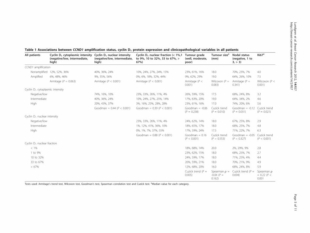
Table 1 Associations between CCND1 amplification status, cyclin D1 protein expression and clinicopathological variables in all patients
All patients Cyclin D1 cytoplasmic intensity(negative/low, intermediate,high)
Cyclin D1 nuclear intensity(negative/low, intermediate,high)
Cyclin D1 nuclear fraction (< 1%,1to 9%, 10 to 32%, 33 to 67%, >67%)
Tumour grade(well, moderate,poor)
Tumour sizea
(mm)Nodal status(negative, 1 to3, > 3)
Ki67a
CCND1 amplification
Nonamplified 12%, 52%, 36% 40%, 36%, 24% 10%, 24%, 27%, 24%, 15% 23%, 61%, 16% 18.0 70%, 23%, 7% 4.0
Amplified 6%, 48%, 46% 9%, 35%, 56% 0%, 6%, 18%, 32%, 44% 9%, 62%, 29% 19.0 64%, 26%, 10% 7.5
Armitage (P = 0.063) Armitage (P < 0.001) Armitage (P < 0.001) Armitage (P <0.001)
Wilcoxon (P =0.083)
Armitage (P =0.341)
Wilcoxon (P <0.001)
Cyclin D1 cytoplasmic intensity
Negative/low 74%, 16%, 10% 23%, 33%, 26%, 11%, 4% 26%, 59%, 15% 17.5 68%, 24%, 8% 3.2
Intermediate 40%, 36%, 24% 10%, 24%, 27%, 25%, 14% 17%, 63%, 20% 19.0 64%, 34%, 2% 4.6
High 20%, 43%, 37% 3%, 16%, 25%, 28%, 28% 23%, 61%, 16% 17.0 74%, 20%, 6% 5.6
Goodman = 0.44 (P < 0.001) Goodman = 0.39 (P < 0.001) Goodman = -0.06(P = 0.238)
Cuzick trend(P = 0.010)
Goodman = -0.12(P = 0.031)
Cuzick trend(P = 0.021)
Cyclin D1 nuclear intensity
Negative/low 23%, 33%, 26%, 11%, 4% 24%, 62%, 14% 18.0 67%, 25%, 8% 2.9
Intermediate 1%, 12%, 41%, 36%, 10% 18%, 65%, 17% 18.0 68%, 25%, 7% 4.8
High 0%, 1%, 7%, 37%, 55% 17%, 59%, 24% 17.5 71%, 22%, 7% 6.3
Goodman = 0.88 (P < 0.001) Goodman = 0.16(P < 0.001)
Cuzick trend(P = 0.553)
Goodman = -0.05(P = 0.327)
Cuzick trend(P < 0.001)
Cyclin D1 nuclear fraction
< 1% 18%, 68%, 14% 20.0 2%, 29%, 9% 2.8
1 to 9% 23%, 62%, 15% 18.0 68%, 25%, 7% 2.7
10 to 32% 24%, 59%, 17% 18.0 71%, 25%, 4% 4.4
33 to 67% 20%, 59%, 21% 18.0 70%, 21%, 9% 4.9
> 67% 12%, 68%, 20% 16.0 68%, 24%, 8% 5.9
Cuzick trend (P =0.005)
Spearman r =-0.04 (P =0.162)
Cuzick trend (P =0.694)
Spearman r= 0.22 (P <0.001
Tests used: Armitage’s trend test, Wilcoxon test, Goodman’s test, Spearman correlation test and Cuzick test. aMedian value for each category.
Lundgrenet
al.BreastCancer
Research2012,14:R57
http://breast-cancer-research.com/content/14/2/R57
Page5of
11

grade (P < 0.001 and P = 0.005 respectively) and higherproliferation rate (both P < 0.001).
Cyclin D1, proliferation and time to recurrenceKi67 expression is a common marker used to analyse theproliferation rate in tumour samples, and high prolifera-tion is linked to a more aggressive tumour phenotype. Sur-prisingly, despite a positive correlation between nuclearcyclin D1 expression and Ki67 expression (Table 1 and
Figure 5a), high expression of nuclear cyclin D1 was asso-ciated with an improved TTR compared with low expres-sion. To further illustrate how a combined proliferationand cyclin D1 assessment would be linked to TTR we sub-divided patients according to Ki67 expression and nuclearcyclin D1 status. Patients with tumours exhibiting lowKi67 expression in association with 1 to 100% cyclin D1-positive nuclei (high) were associated with a considerablylower risk of recurrence (P < 0.001) (Figure 5b) compared
Figure 2 Kaplan-Meier plots of recurrence risk over time in all patients. (a) Risk of recurrence was increased for patients exhibiting CCND1-amplified breast cancers compared with nonamplified. (b), (c) No significant difference was observed between varying intensities of cytoplasmicor nuclear cyclin D1. (d) Patients showing a nuclear fraction of cyclin D1 lower than 1% had an increased risk of recurrence compared withhigher expression.
Table 2 Cox proportional Hazards models for estimating the effect on time to recurrence
Univariate Multivariate
HR (95% CI) c12 P value HR (95% CI) c12 P value
All patients
CCND1, amplified vs. nonamplified 2.04 (1.37 to 3.03) 10.51 < 0.001 1.61 (1.08 to 2.41) 4.86 0.030
Cyclin D1 cytoplasmic intensity, high vs. remainder 0.71 (0.51 to 0.98) 4.43 0.039 0.78 (0.56 to 1.09) 2.15 0.143
Cyclin D1 nuclear intensity, high vs. remainder 0.91 (0.64 to 1.28) 0.30 0.588 0.85 (0.59 to 1.21) 0.84 0.360
Cyclin D1 nuclear fraction, 1 to 100% vs. < 1% 0.58 (0.38 to 0.90) 5.28 0.014 0.60 (0.39 to 0.92) 4.75 0.030
Nonamplified patients
Cyclin D1 cytoplasmic intensity, high vs. remainder 0.57 (0.39 to 0.82) 9.57 0.003 0.64 (0.44 to 0.95) 5.21 0.022
Cyclin D1 nuclear intensity, high vs. remainder 0.88 (0.60 to 1.32) 0.38 0.545 0.84 (0.55 to 1.26) 0.76 0.382
Cyclin D1 nuclear fraction, 1 to 100% vs. < 1% 0.52 (0.34 to 0.81) 7.29 0.004 0.54 (0.35 to 0.85) 6.36 0.012
The c12 value is based on the likelihood ratio test with the associated P values. Multivariate analyses were adjusted for tumour grade, tumour size, nodal status
and Ki67. CI, confidence interval; HR, hazard ratio.
Lundgren et al. Breast Cancer Research 2012, 14:R57http://breast-cancer-research.com/content/14/2/R57
Page 6 of 11

with the other subgroups. The subgroups of low cyclin D1
include quite low patient numbers and hence the resultsshould be interpreted with caution, but these results sug-gest that the expression of cyclin D1 affects disease out-come independently of proliferation status.
DiscussionAmplification of the CCND1 gene has been associatedwith a poor patient outcome in previous studies [19,26],whilst controversy regarding overexpression of cyclin D1
protein in relation to patient survival still exists. Cyclin D1
has been reported to be a prognostic marker in invasivebreast cancer and has been associated with both a lessaggressive ER-positive phenotype [20,22] and also with anadverse clinical outcome [18]. These conflicting findingscan potentially be explained by the low patient numbersanalysed and/or methodological discrepancies. To clarifythe importance of cyclin D1 in breast cancer we thereforeanalysed the expression of cyclin D1 in different subcellu-lar localisations, using a previously validated antibody [36],
as well as the gene amplification status by the well-estab-lished CISH technique in a large, well-characterised ran-domised patient cohort including more than 1,000patients with ER-positive breast cancers. Our data supportstudies indicating that low cyclin D1 protein expression aswell as CCND1 amplification are linked to tumour aggres-siveness and increased risk of disease recurrence in ER-positive postmenopausal breast cancer [23,26]. Similarfindings have been observed for HER2, where both highexpression linked to amplification and low expression arelinked to poor outcome [43].Amplification of CCND1 was observed in 8.7% of the
tumours, which is slightly lower than the frequency of 10to 15% generally reported, even though some groupshave demonstrated a lower percentage of CCND1-ampli-fied tumours [15,44,45]. The slightly lower fraction ofCCND1-amplified cases may be due to all patients beingER-positive or due to methodological differences and dif-ferent cutoff points for defining amplification betweenstudies. In addition, the use of TMAs has certain
Figure 3 Predicted risk of recurrence over time based on cytoplasmic intensity and nuclear fraction of cyclin D1, comparing twosubgroups. (a) In all patients, no significant difference in recurrence risk was observed between low and high cytoplasmic expression. (b)Patients showing a nuclear fraction of less than 1% positive cyclin D1 nuclei had an increased risk of recurrence compared with a fraction of 1 to100%, in all patients. (c) In patients exhibiting nonamplified tumours, high cytoplasmic expression was associated with a reduced risk ofrecurrence. (d) A fraction of cyclin D1-positive nuclei lower than 1% was associated with a higher recurrence risk also in patients showingnonamplified tumours.
Lundgren et al. Breast Cancer Research 2012, 14:R57http://breast-cancer-research.com/content/14/2/R57
Page 7 of 11
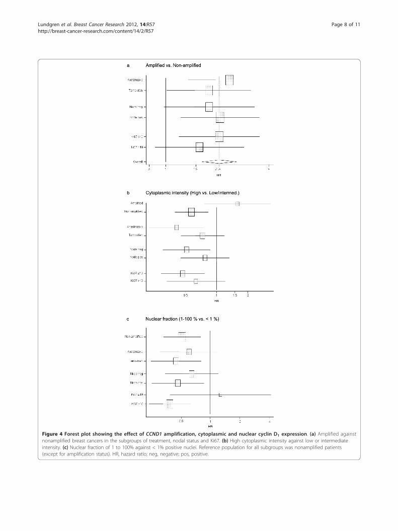
Figure 4 Forest plot showing the effect of CCND1 amplification, cytoplasmic and nuclear cyclin D1 expression. (a) Amplified againstnonamplified breast cancers in the subgroups of treatment, nodal status and Ki67. (b) High cytoplasmic intensity against low or intermediateintensity. (c) Nuclear fraction of 1 to 100% against < 1% positive nuclei. Reference population for all subgroups was nonamplified patients(except for amplification status). HR, hazard ratio; neg, negative; pos, positive.
Lundgren et al. Breast Cancer Research 2012, 14:R57http://breast-cancer-research.com/content/14/2/R57
Page 8 of 11

limitations; however, this technique is indispensablewhen analysing large patient materials and is today awell-accepted approach for large-scale tumour sampleanalysis. In agreement with previous studies, gene ampli-fication of CCND1 was associated with an overall adverseclinical outcome. The observed positive correlationbetween nuclear cyclin D1 expression and tumour gradeand proliferation suggests a link between cyclin D1 andaggressive disease. In contrast, both higher nuclearexpression and high cytoplasmic expression of cyclin D1
was instead associated with a decreased recurrence risk.Despite a positive association between cyclin D1 proteinand CCND1 amplification status, both low nuclear frac-tion of cyclin D1 and CCND1 amplification were linkedto earlier disease recurrence independently of other clini-copathological parameters - hence both factors serve asprognostic markers in endocrine-treated, ER-positivepostmenopausal breast cancer. In patients not displayingCCND1 amplification, cytoplasmic expression of cyclinD1 was also an independent marker for longer TTR, indi-cating that the true prognostic value of cyclin D1 proteinexpression may be obscured by the CCND1-amplifiedcases: the clinicopathological significance of cyclin D1
expression might thus be best considered separately foramplified and nonamplified cases.Apart from the role as a prognostic marker, cyclin D1
has been proposed as a predictive factor for tamoxifenresponse, as illustrated by poor clinical outcome inpatients with ER-positive tumours with high cyclin D1
expression treated with tamoxifen [46]. These findingstogether with numerous experimental reports [33-35]support that cyclin D1 overexpression might abrogate theresponse to tamoxifen, as previously reported by ourgroup and others [21,46]. Our earlier discoveries havenevertheless been made in cohorts where patients were
randomly assigned to receive either no adjuvant treat-ment or to receive tamoxifen [21,36]. In the presentstudy we compared the two endocrine therapies anastro-zole and tamoxifen in relation to disease recurrence andcyclin D1 status, but there were no untreated controlpatients. There was no significant difference in treatmentresponse between these two adjuvant therapies by strati-fication for cyclin D1 status, indicating that cyclin D1 isnot a predictive marker for differences in response toanastrozole versus tamoxifen. No conclusions can bedrawn, however, regarding cyclin D1 as a marker for gen-eral endocrine treatment resistance, since no untreatedpatients were available for analysis within the Trans-ATAC study. Moreover, differences in tamoxifenresponse in relation to cyclin D1 in postmenopausal ver-sus premenopausal breast cancer might exist. Our pre-vious study reporting a potential unfavourable effect oftamoxifen included premenopausal patients exclusively,whereas this study focused exclusively on postmenopau-sal breast cancer cases and has shown no detrimentaleffect since the results for the two endocrine therapieswere similar.The relationship between cyclin D1, proliferation and
prognosis is quite complex, with a positive correlationbetween cyclin D1 and Ki67 - Ki67 is associated withshorter TTR, while cyclin D1 is associated with longerTTR. Patients showing low expression of Ki67 had alonger TTR with the highest levels of cyclin D1 expres-sion, whereas patients exhibiting higher levels of Ki67had the shortest TTR with the lowest levels of cyclin D1
expression. Multivariate analysis identified the fractioncyclin D1-positive nuclei as a predictor of outcome inde-pendently of other clinicopathological parameters such asKi67. These results suggest that, irrespective of prolifera-tion status, intermediate to high expression of cyclin D1
Figure 5 Recurrence risk over time based on the expression of Ki67 and cyclin D1. (a) Expression of Ki67 increased with increasingpercentage of cyclin D1-positive nuclei in the breast tumours. (b) Kaplan-Meier plot showing that the combination of high Ki67 and low cyclinD1 expression was associated with high risk of recurrence (green). Patients exhibiting a high fraction of cyclin D1-positive tumour cell nuclei inconcurrence with low Ki67 expression showed the lowest risk of recurrence (red).
Lundgren et al. Breast Cancer Research 2012, 14:R57http://breast-cancer-research.com/content/14/2/R57
Page 9 of 11

results in a prolonged TTR in ER-positive, postmenopau-sal breast cancer patients. Similar results were observedin our previous study of randomised material from pre-menopausal breast cancer patients [47]. The relationshipbetween cyclin D1, proliferation and prognosis henceseems to be complex, and this could in part be explainedby potential additional functions for cyclin D1 unrelatedto proliferation control, as well as co-amplification andco-deletion of specific genes on chromosome 11q13, thelocus harbouring CCND1.
ConclusionsThis study confirms that the cyclin D1 status providesindependent prognostic information regarding ER-posi-tive, postmenopausal breast cancers, supporting its emer-ging role as a biomarker that might be useful in the clinic.Our results demonstrate that high expression of cyclin D1
was associated with a reduced risk of recurrences, whereasamplification of the CCND1 gene was linked to an aggres-sive disease. Finally, no difference in response to anastro-zole compared with tamoxifen was observed according toexpression of cyclin D1 gene amplification or proteinexpression.
Additional material
Additional file 1: Supplementary Table 1showing the distribution ofcyclin D1 staining categories.
AbbreviationsATAC: Arimidex, Tamoxifen, Alone or in Combination; CI: confidence interval;CISH: chromogenic in situ hybridisation; ER: oestrogen receptor; HR: hazardratio; TMA: tissue microarray; TTR: time to recurrence.
AcknowledgementsThe authors thank Elise Nilsson for excellent technical implementation. Thisstudy was supported by grants from the Swedish Cancer Society, theSwedish Research Council, the Knut and Alice Wallenberg Foundation,Malmö University Hospital Research and Cancer Funds, Lund UniversityResearch Funds, South Swedish and South-East Swedish Breast Cancergroups and Breakthrough Breast Cancer Unit, Manchester, UK.
Author details1Center for Molecular Pathology, Department of Laboratory Medicine, LundUniversity, Malmö University Hospital, SE-205 02 Malmö, Sweden.2Breakthrough Breast Cancer Research Unit, School of Cancer, EnablingSciences and Technology, University of Manchester, Manchester AcademicHealth Science Centre Paterson Institute for Cancer Research, The ChristieNHS Foundation Trust, Wilmslow Road, Manchester M20 4BX, UK. 3CancerResearch UK Centre for Epidemiology, Mathematics and Statistics, QueenMary University of London, Wolfson Institute of Preventive Medicine, LondonEC1M 6BQ, UK. 4Breakthrough Breast Cancer Research Centre, Institute ofCancer Research, 237 Fulham Road, London SW3 6JB, UK. 5Royal MarsdenHospital, 237 Fulham Road, London SW3 6JJ, UK. 6Sahlgrenska CancerCenter, University of Gothenburg, 405 30 Göteborg, Sweden.
Authors’ contributionsKL carried out the immunohistochemical and CISH assessments, participatedin the study design and drafted the manuscript. MB assisted with evaluationof the immunohistochemistry. SP performed the statistical analyses. JC
designed the clinical trial, assembled trial data, and planned and performedthe statistical analyses. JS assembled trial data. LZ assembled trial data. AHdesigned the clinical trial and collected clinical data. MD designed theclinical trial and collected clinical data, and initiated and conceived thecurrent study. GL initiated and conceived the study, supervisedinterpretation of the data, took part in the immunohistochemicalassessments and evaluation of CISH, and helped to draft the manuscript. Allauthors took part in data interpretation, writing the report and approval ofthe final version of the manuscript.
Competing interestsJC has acted as a consultant and/or on advisory boards for AstraZeneca andreceived commercial research grants from AstraZeneca. AH has acted as aconsultant and/or on advisory boards for AstraZeneca, GSK, Pfizer, Rocheand Amgen, and has received honoraria from AstraZeneca. MD receives paidadvisory boards and research funding from each of Novartis, AstraZenecaand Roche. The remaining authors declare that they have no competinginterests.
Received: 5 August 2011 Revised: 18 January 2012Accepted: 4 April 2012 Published: 4 April 2012
References1. Howell A, Dowsett M: Recent advances in endocrine therapy of breast
cancer. BMJ 1997, 315:863-866.2. Yoshida M, Haybittle JL, Davies C, Mason BH, Wilcken N, Ploner M,
Yosef HMA, Lobelle JP, Peek U, Powell J, Mauriac L, Piccart MJ, Price JJ,Hupperets PSGJ, Ragaz J, Weiss RB, Abu-Zahra HT, Portnoj SM, Riley D,Gordon NH, Davis HL, Naja A, Romestaing P, Dubois JB, Mace-Lesec’h J,Rambert P, Barkmanova J, Owen JR, Meier P, Swindell R: Effects ofchemotherapy and hormonal therapy for early breast cancer onrecurrence and 15-year survival: an overview of the randomised trials.Lancet 2005, 365:1687-1717.
3. Brodie AM, Njar VC: Aromatase inhibitors in advanced breast cancer:mechanism of action and clinical implications. J Steroid Biochem Mol Biol1998, 66:1-10.
4. Geisler J, King N, Dowsett M, Ottestad L, Lundgren S, Walton P,Kormeset PO, Lonning PE: Influence of anastrozole (Arimidex), a selective,non-steroidal aromatase inhibitor, on in vivo aromatisation and plasmaoestrogen levels in postmenopausal women with breast cancer. Br JCancer 1996, 74:1286-1291.
5. Baum M, Budzar AU, Cuzick J, Forbes J, Houghton JH, Klijn JG, Sahmoud T:Anastrozole alone or in combination with tamoxifen versus tamoxifenalone for adjuvant treatment of postmenopausal women with earlybreast cancer: first results of the ATAC randomised trial. Lancet 2002,359:2131-2139.
6. Howell A, Cuzick J, Baum M, Buzdar A, Dowsett M, Forbes JF, Hoctin-Boes G, Houghton J, Locker GY, Tobias JS: Results of the ATAC (Arimidex,Tamoxifen, Alone or in Combination) trial after completion of 5 years’adjuvant treatment for breast cancer. Lancet 2005, 365:60-62.
7. Cuzick J, Sestak I, Baum M, Buzdar A, Howell A, Dowsett M, Forbes JF:Effect of anastrozole and tamoxifen as adjuvant treatment for early-stage breast cancer: 10-year analysis of the ATAC trial. Lancet Oncol 2010,11:1135-1141.
8. Ormandy CJ, Musgrove EA, Hui R, Daly RJ, Sutherland RL: Cyclin D1, EMS1and 11q13 amplification in breast cancer. Breast Cancer Res Treat 2003,78:323-335.
9. Hui R, Ball JR, Macmillan RD, Kenny FS, Prall OW, Campbell DH, Cornish AL,McClelland RA, Daly RJ, Forbes JF, Musgrove EA, Robertson JF, Nicholson RI,Sutherland RL: EMS1 gene expression in primary breast cancer:relationship to cyclin D1 and oestrogen receptor expression and patientsurvival. Oncogene 1998, 17:1053-1059.
10. Brown LA, Johnson K, Leung S, Bismar TA, Benitez J, Foulkes WD,Huntsman DG: Co-amplification of CCND1 and EMSY is associated withan adverse outcome in ER-positive tamoxifen-treated breast cancers.Breast Cancer Res Treat 2010, 121:347-54.
11. Sherr CJ, Roberts JM: CDK inhibitors: positive and negative regulators ofG1-phase progression. Genes Dev 1999, 13:1501-1512.
12. Jiang W, Kahn SM, Zhou P, Zhang YJ, Cacace AM, Infante AS, Doi S,Santella RM, Weinstein IB: Overexpression of cyclin D1 in rat fibroblasts
Lundgren et al. Breast Cancer Research 2012, 14:R57http://breast-cancer-research.com/content/14/2/R57
Page 10 of 11

causes abnormalities in growth control, cell cycle progression and geneexpression. Oncogene 1993, 8:3447-3457.
13. Zhou P, Jiang W, Zhang YJ, Kahn SM, Schieren I, Santella RM, Weinstein IB:Antisense to cyclin D1 inhibits growth and reverses the transformedphenotype of human esophageal cancer cells. Oncogene 1995,11:571-580.
14. Wang TC, Cardiff RD, Zukerberg L, Lees E, Arnold A, Schmidt EV: Mammaryhyperplasia and carcinoma in MMTV-cyclin D1 transgenic mice. Nature1994, 369:669-671.
15. Gillett C, Fantl V, Smith R, Fisher C, Bartek J, Dickson C, Barnes D, Peters G:Amplification and overexpression of cyclin D1 in breast cancer detectedby immunohistochemical staining. Cancer Res 1994, 54:1812-1817.
16. Bartkova J, Lukas J, Muller H, Lutzhoft D, Strauss M, Bartek J: Cyclin D1
protein expression and function in human breast cancer. Int J Cancer1994, 57:353-361.
17. McIntosh GG, Anderson JJ, Milton I, Steward M, Parr AH, Thomas MD,Henry JA, Angus B, Lennard TW, Horne CH: Determination of theprognostic value of cyclin D1 overexpression in breast cancer. Oncogene1995, 11:885-891.
18. Kenny FS, Hui R, Musgrove EA, Gee JM, Blamey RW, Nicholson RI,Sutherland RL, Robertson JF: Overexpression of cyclin D1 messenger RNApredicts for poor prognosis in estrogen receptor-positive breast cancer.Clin Cancer Res 1999, 5:2069-2076.
19. Michalides R, Hageman P, van Tinteren H, Houben L, Wientjens E,Klompmaker R, Peterse J: A clinicopathological study on overexpressionof cyclin D1 and of p53 in a series of 248 patients with operable breastcancer. Br J Cancer 1996, 73:728-734.
20. Gillett C, Smith P, Gregory W, Richards M, Millis R, Peters G, Barnes D: CyclinD1 and prognosis in human breast cancer. Int J Cancer 1996, 69:92-99.
21. Stendahl M, Kronblad A, Ryden L, Emdin S, Bengtsson NO, Landberg G:Cyclin D1 overexpression is a negative predictive factor for tamoxifenresponse in postmenopausal breast cancer patients. Br J Cancer 2004,90:1942-1948.
22. Hwang TS, Han HS, Hong YC, Lee HJ, Paik NS: Prognostic value ofcombined analysis of cyclin D1 and estrogen receptor status in breastcancer patients. Pathol Int 2003, 53:74-80.
23. Naidu R, Wahab NA, Yadav MM, Kutty MK: Expression and amplification ofcyclin D1 in primary breast carcinomas: relationship withhistopathological types and clinico-pathological parameters. Oncol Rep2002, 9:409-416.
24. Dickson C, Fantl V, Gillett C, Brookes S, Bartek J, Smith R, Fisher C, Barnes D,Peters G: Amplification of chromosome band 11q13 and a role for cyclinD1 in human breast cancer. Cancer Lett 1995, 90:43-50.
25. Buckley MF, Sweeney KJ, Hamilton JA, Sini RL, Manning DL, Nicholson RI,deFazio A, Watts CK, Musgrove EA, Sutherland RL: Expression andamplification of cyclin genes in human breast cancer. Oncogene 1993,8:2127-2133.
26. Bieche I, Olivi M, Nogues C, Vidaud M, Lidereau R: Prognostic value ofCCND1 gene status in sporadic breast tumours, as determined by real-time quantitative PCR assays. Br J Cancer 2002, 86:580-586.
27. Clarke R, Leonessa F, Welch JN, Skaar TC: Cellular and molecularpharmacology of antiestrogen action and resistance. Pharmacol Rev 2001,53:25-71.
28. Zwart W, Rondaij M, Jalink K, Sharp ZD, Mancini MA, Neefjes J, Michalides R:Resistance to antiestrogen arzoxifene is mediated by overexpression ofcyclin D1. Mol Endocrinol 2009, 23:1335-1345.
29. Le Goff P, Montano MM, Schodin DJ, Katzenellenbogen BS:Phosphorylation of the human estrogen receptor. Identification ofhormone-regulated sites and examination of their influence ontranscriptional activity. J Biol Chem 1994, 269:4458-4466.
30. Kato S, Endoh H, Masuhiro Y, Kitamoto T, Uchiyama S, Sasaki H,Masushige S, Gotoh Y, Nishida E, Kawashima H, Metzger D, Chambon P:Activation of the estrogen receptor through phosphorylation bymitogen-activated protein kinase. Science 1995, 270:1491-1494.
31. Pietras RJ, Arboleda J, Reese DM, Wongvipat N, Pegram MD, Ramos L,Gorman CM, Parker MG, Sliwkowski MX, Slamon DJ: HER-2 tyrosine kinasepathway targets estrogen receptor and promotes hormone-independentgrowth in human breast cancer cells. Oncogene 1995, 10:2435-2446.
32. Holm C, Rayala S, Jirstrom K, Stal O, Kumar R, Landberg G: Associationbetween Pak1 expression and subcellular localization and tamoxifenresistance in breast cancer patients. J Natl Cancer Inst 2006, 98:671-680.
33. Zwijsen RM, Wientjens E, Klompmaker R, van der Sman J, Bernards R,Michalides RJ: CDK-independent activation of estrogen receptor by cyclinD1. Cell 1997, 88:405-415.
34. Zwijsen RM, Buckle RS, Hijmans EM, Loomans CJ, Bernards R: Ligand-independent recruitment of steroid receptor coactivators to estrogenreceptor by cyclin D1. Genes Dev 1998, 12:3488-3498.
35. Wilcken NR, Prall OW, Musgrove EA, Sutherland RL: Inducibleoverexpression of cyclin D1 in breast cancer cells reverses the growth-inhibitory effects of antiestrogens. Clin Cancer Res 1997, 3:849-854.
36. Jirstrom K, Stendahl M, Ryden L, Kronblad A, Bendahl PO, Stal O,Landberg G: Adverse effect of adjuvant tamoxifen in premenopausalbreast cancer with cyclin D1 gene amplification. Cancer Res 2005,65:8009-8016.
37. Dowsett M, Allred C, Knox J, Quinn E, Salter J, Wale C, Cuzick J, Houghton J,Williams N, Mallon E, Bishop H, Ellis I, Larsimont D, Sasano H, Carder P,Cussac AL, Knox F, Speirs V, Forbes J, Buzdar A: Relationship betweenquantitative estrogen and progesterone receptor expression and humanepidermal growth factor receptor 2 (HER-2) status with recurrence inthe Arimidex, Tamoxifen, Alone or in Combination trial. J Clin Oncol 2008,26:1059-1065.
38. Harvey JM, Clark GM, Osborne CK, Allred DC: Estrogen receptor status byimmunohistochemistry is superior to the ligand-binding assay forpredicting response to adjuvant endocrine therapy in breast cancer. JClin Oncol 1999, 17:1474-1481.
39. McShane LM, Altman DG, Sauerbrei W, Taube SE, Gion M, Clark GM:Reporting recommendations for tumor marker prognostic studies(REMARK). J Natl Cancer Inst 2005, 97:1180-1184.
40. Arnould L, Denoux Y, MacGrogan G, Penault-Llorca F, Fiche M, Treilleux I,Mathieu MC, Vincent-Salomon A, Vilain MO, Couturier J: Agreementbetween chromogenic in situ hybridisation (CISH) and FISH in thedetermination of HER2 status in breast cancer. Br J Cancer 2003,88:1587-1591.
41. Cuzick J: A Wilcoxon-type test for trend. Stat Med 1985, 4:87-90.42. Dowsett M, Cuzick J, Wale C, Forbes J, Mallon EA, Salter J, Quinn E,
Dunbier A, Baum M, Buzdar A, Howell A, Bugarini R, Baehner FL, Shak S:Prediction of risk of distant recurrence using the 21-gene recurrencescore in node-negative and node-positive postmenopausal patients withbreast cancer treated with anastrozole or tamoxifen: a TransATAC study.J Clin Oncol 28:1829-1834.
43. Camp RL, Dolled-Filhart M, King BL, Rimm DL: Quantitative analysis ofbreast cancer tissue microarrays shows that both high and normal levelsof HER2 expression are associated with poor outcome. Cancer Res 2003,63:1445-1448.
44. Weinstat-Saslow D, Merino MJ, Manrow RE, Lawrence JA, Bluth RF,Wittenbel KD, Simpson JF, Page DL, Steeg PS: Overexpression of cyclin DmRNA distinguishes invasive and in situ breast carcinomas from non-malignant lesions. Nat Med 1995, 1:1257-1260.
45. Zukerberg LR, Yang WI, Gadd M, Thor AD, Koerner FC, Schmidt EV,Arnold A: Cyclin D1 (PRAD1) protein expression in breast cancer:approximately one-third of infiltrating mammary carcinomas showoverexpression of the cyclin D1 oncogene. Mod Pathol 1995, 8:560-567.
46. Rudas M, Lehnert M, Huynh A, Jakesz R, Singer C, Lax S, Schippinger W,Dietze O, Greil R, Stiglbauer W, Kwasny W, Grill R, Stierer M, Gnant MF,Filipits M: Cyclin D1 expression in breast cancer patients receivingadjuvant tamoxifen-based therapy. Clin Cancer Res 2008, 14:1767-1774.
47. Lehn S, Tobin NP, Berglund P, Nilsson K, Sims AH, Jirstrom K, Harkonen P,Lamb R, Landberg G: Down-regulation of the oncogene cyclin D1
increases migratory capacity in breast cancer and is linked tounfavorable prognostic features. Am J Pathol 2010, 177:2886-2897.
doi:10.1186/bcr3161Cite this article as: Lundgren et al.: Effects of cyclin D1 geneamplification and protein expression on time to recurrence inpostmenopausal breast cancer patients treated with anastrozole ortamoxifen: a TransATAC study. Breast Cancer Research 2012 14:R57.
Lundgren et al. Breast Cancer Research 2012, 14:R57http://breast-cancer-research.com/content/14/2/R57
Page 11 of 11