effects of individual antibiotics and their mixtures akhyany degree project for master of science 60...
TRANSCRIPT

Marjan Akhyany
Degree project for Master of Science 60 hec
Department of Biological and Environmental SciencesUniversity of Gothenburg 2013
Effects of individualantibiotics and their
mixtureson single bacterial species, artificial and natural
microbial communities
http://www.rightparenting.com/article/understanding-antibiotics.html

Abstract Several pharmaceuticals including antibiotics are currently used to treat human and animal
disease. Antibiotics as a subgroup of pharmaceuticals could pass through Sewage Treatment Plants
and reach aquatic compartments of the environment and are present at very low concentrations
compared to the other chemicals. Presently, concern of occurrence, fate and environmental risks of
pharmaceutical including antibiotics have been increased. Empirical evidences on mixture eco‐
toxicity of chemicals sometimes demonstrate higher joint toxicity even at their individual non‐toxic
concentrations.
Since bacteria are most sensitive organism to antibiotics, the authors of this study aimed to
present the current toxicity of antibiotics to bacteria in single substance and mixture exposure
scenarios. Selection of antibiotics was based on a study by Andreozzi et al. 2002 which seven
antibiotics from different therapeutic classes were detected in effluents of three STPs in Sweden,
Italy and France as worst cases, namely Ofloxacin, Lomefloxacin, Enoxacin, Norfloxacin and
Ciprofloxacin (Quinolones) and Trimethoprim and Sulfamethoxazole (Sulfonamides) with
concentrations between 0.04 and 1.61 nmol/L, in addition to 14 non‐antibiotics.
Single substance toxicities were evaluated using a standard bacteria assay (Pseudomonas putida),
a new approach using artificial community (B.O.D.seed), and natural biofilms (Limnic periphyton
communities) under controlled exposure in laboratory. All substances demonstrated toxicities to
bacteria where Quinolones (most hazardous substances was Ciprofloxacin) showed more toxicity
compared to Sulfonamides. In term of single substance toxicity, the bioassays were sorted in
different order which seems to be substance dependent. Evaluating the single substance toxicity in
three different bioassays also demonstrated looking at the whole curve is essential in order to
determine the toxicity of a substance.
Mixture toxicities were evaluated using the first two bioassays. The mixture at the concentration
at which it is present in the selected effluents had no visible effect .Therefore, no impact of mixture
of antibiotics at their realistic concentrations in natural environment (which is comparatively lower
than effluents of STPs) would be considered for bacterial groups of the communities. Since bacteria
are most sensitive part of the natural communities to antibiotics and no adverse effects were
observed for bacteria, there is no concern for non‐bacterial parts such as invertebrates and fish
which are less sensitive to antibiotics.
In addition mixture toxicities of tested antibiotics were predicted by using two classic concepts
namely Concentration Addition (CA) and Independent Action (IA). Both CA and IA concepts
underestimate the mixture toxicities. This fact is in contrast to earlier studies in ecotoxicity of
chemicals. Higher predictive power was also illustrated for CA concept.

2
Furthermore, the results of mixture tests are valid only for the current mixture of the substances.
Therefore, presence of other chemicals including non‐antibiotics group of pharmaceuticals, heavy
metal and biocides should be also taken in to the account.

3
TableofContents
Introduction .......................................................................................................................................... 5
Pharmaceutical in the environment .......................................................................................................... 5
Antibiotics .................................................................................................................................................. 6
Environmental Risk Assessment (ERA) of antibiotics ........................................................................ 8
Exposure scenario and the compounds .................................................................................................. 8
Mixture toxicity concepts ....................................................................................................................... 12
Study organism and communities ......................................................................................................... 14
Aims of the thesis .................................................................................................................................... 17
Materials and methods .............................................................................................................. 18
Preparation of the test solutions ........................................................................................................... 18
Endpoint .................................................................................................................................................... 18
Toxicity tests ............................................................................................................................................ 19
Pseudomonas putida test ........................................................................................................................ 19
Artificial communities’ tests .................................................................................................................. 19
Experimental design- method development ........................................................................................ 20
Limnic periphyton community test (Swift) .......................................................................................... 23
Data treatment .......................................................................................................................................... 25
Results .................................................................................................................................................. 27
Control experiment ................................................................................................................................. 27
Single substance tests in Pseudomonas putida .................................................................................... 28
Mixture toxicity tests of antibiotics in Pseudomonas putida ............................................................ 30
Impact of non-antibiotic pharmaceuticals present in the effluents ................................................... 31
Single substance tests in B.O.D.seed artificial community ............................................................... 33
Mixture toxicity tests of antibiotics in B.O.D.seed artificial community ........................................ 36
Single substance tests in Limnic periphyton bacterial community ................................................... 37
Compared toxicities of four antibiotics to three different bioassays ................................................ 40
Predictability of the mixture toxicity by CA and IA concepts .......................................................... 43
Discussion & Conclusion ...................................................................................................... 49

4
Acknowledgments ......................................................................................................................... 52
References ......................................................................................................................................... 53
Appendix I .......................................................................................................................................... 55
Details of the media ............................................................................................................................... 55
Appendix II ........................................................................................................................................ 57
Toxicity of tested antibiotics to Pseudomonas putida in single substance exposure ..................... 57
Toxicity of tested antibiotics to B.O.D.seed in single substance exposure ..................................... 60
The parameters of Weibull fit model .................................................................................................... 63

5
Introduction
Pharmaceuticals in the environment
In general, pharmaceuticals are used to cure or prevent human and animal disease with
different way of application, followed by digestion, absorption, metabolism and excretion in
human or animal bodies.
Although releasing of the pharmaceuticals occurs for decades, the first report of presence of the
pharmaceuticals was at the beginning of 80’s (Andreozzi et al., 2002). Anyway, different
compartments of the environments are contaminated by pharmaceuticals. There are several
routes to enter the environment for pharmaceuticals in production, consumption and disposal as
shown in Figure 1. The human medicines could mainly end up in the environment via Sewage
treatment plants (STPs) streams. In contrast veterinary medicines, mainly antibacterial agents
might directly enter the environment. For instance, they are used directly to the water in
aquaculture (Boxall B.A., 2004). There are several other entering ways for the other sorts of the
pharmaceuticals such as runoff from applied sewage sludge and agricultural fields.
Figure 1: Indication of how pharmaceuticals enter the terrestrial and aquatic environment
(Boxall B.A., 2004)
The major reasons for the concern are that pharmaceuticals are designed to have a specific
action in their biological targets or affect a physiological process in organisms as well as their low

6
biodegradability within the human or animal bodies. This means, they could have inherent
properties that could also remain stable outside the human or animal bodies. Though,
Pharmaceutical might have also some unanticipated effects on non‐target organisms with the
same receptors with target organisms. From an environmental perspective, such persistence might
lead to persist in degradation in Sewage Treatment Plants as well and may cause environmental
impacts (EEA Technical report, 2010, P8). Pharmaceuticals are present in the environment in lower
than their low effect concentration (LOEC) (EEA Technical report, 2010, p17). Hence, continuous
releasing of pharmaceuticals to the environment expose the organisms to a low level of the
substance, but also chronic exposure would occur over a long period of time.
Concerns of using, exposure, fate and environmental impact of pharmaceuticals have been
increased during recent decades in Europe, due to developing European pharmaceutical market.
In Europe, the first attention to the impact of pharmaceuticals was in European Environmental
Agency (EEA) at the first years of 21th century. It has been confirmed by researches that human
and veterinary medicines could have environmental impacts (EEA Technical report, 2010, P5) via
unwanted effects on non‐target organisms that would alter ultimately the ecosystem function.
So far, the hazardous impacts of two compounds of pharmaceuticals have been documented
well including Ethinyl Estradiol (EE2) causing the feminisation of male fish and Diclofenac which
kills vultures in Asia (EEA Technical Report, 2010, p8). Unfortunately there are still knowledge gaps
for other pharmaceuticals like antibiotics and endocrine disrupters.
Antibiotics:
In general, there is no widely accepted definition for antibiotics. Antibiotics are originally any
chemical agents with biological activity against living organisms. In particular, antibiotics are a
specific therapeutic class of pharmaceuticals with inhibitory effect on microorganisms' growth
including bacteria, fungi and protozoa (parasite). Antibiotics without toxic effect on host are used
to prevent or treat the microbial infection in human, veterinary and aquaculture medicines. They
are even applied as a growth promoter in veterinary medicine (Kummerer, 2009, part I). According
to Wise 2002 the world wide consumption of antibiotics is estimated between 100,000 to 200,000
tons per year (Kummerer, 2009, part I reference in there).
They could be classified by their mode of action or chemical structure such as solubility,
hydrophobicity and hydrophilicity. These properties determine their distribution pattern and their
fate in Sewage Treatment Plants (STPs) streams. Hydrophobic substances take part in portioning in
the sludge while hydrophilic substances could pass through the STPs. Their biological activity and
toxicity might also change with pH value. Thus, log Kow of these substances are also pH dependent
(Kummerer, 2009, part I).

7
Antibiotics could have different source in the environment. They could be even produced by
natural bacteria of soil and sediments (Kummerer K., 2009, part I). Three main sources of the
antibiotics in the aquatic environments are STPs, agriculture farms and manure deposit form civil
area (Brosche S. et al, 2010). Total concentration of the antibiotics is also increased by direct
discharging from aquaculture, meat processing or even from pets. Despite antibiotics are used in
lower rates in hospitals, they have been detected in higher concentration in hospital effluents
compared to the municipal waste water (Kummerer K., 2009, part I).
Elimination of the antibiotics is done by different processes namely sorption, photolysis,
hydrolysis, thermolysis and biodegradation. To investigate the rate of sorption of antibiotics onto
sediment and sludge, their chemical and physical properties are necessarily. Antibiotics are very
different in sorption behaviour which is affected by the content of minerals and lipids in receiving
environment. Organic content has also effect on sorption rate (Boxall, 2004). Photolysis is the
major of abiotic elimination pathway in some of light sensitive antibiotics which is possible only in
clear surface water. The affectivity of the process could vary with light intensity and frequency,
latitude, pH and water hardness. Hydrolysis is also an effective pathway for some antibiotics like B‐
lactams which are rapidly hydrolysed in laboratory (Kummerer K., 2009 part I), but not for most of
the antibioticss, because they are designed persistent to hydrolysis in digesting system (Andreozzi
R., 2002).
Biodegradation is also a pathway to eliminate the antibiotics mainly by the bacteria which will
be discussed later.
Antibiotics are metabolised within the human or animal bodies (often in liver), they might be
more water soluble which can lead to more toxic substances than parent compounds. The
metabolic rates of the antibiotics within the human body are 30% in average which indicates 70%
of used antibiotics might emit to the waste water unchanged while they are still active (Kummerer
K., 2009, part I references in there). Despite they are metabolised or not, these two fractions after
passing through STPs could reach to the water compartments of the environment like surface
water, sediments or ground water. Moreover, physical and chemicals processes have an important
role in transferring of the antibiotic through different compartments which means they could be
bio transformed from one trophic level to another. Thus, all the relevant forms of the antibiotic
should be taken to the account such as degradation products, metabolites in addition to the parent
compounds (Backhaus T. et al, 2008, book chap, p263).
Several antibiotic therapeutic classes are detected in the water compartments. Although β‐
lactam class (β‐lactamase inhibitor) are the most common used therapeutic class, they are not
detected in aquatic environment. (Kummerer, 2009, part I references in there). In particular the
antibiotics from tetracyclines, sulphonamides and macrolides classes are present in the sediments.
(Kummerer, 2009, part I references in there).
8
Environmental Risk Assessment (ERA) of antibiotics
At 1980, the first requirement for providing the environmental risk assessments of
pharmaceuticals was demonstrated by Food and Drug Administration (FDA) in the USA, followed
by The EU at 1997. The risk assessments should be performed in terms of their effect on organisms
in aquatic and terrestrial ecosystems (Boxall B.A., 2004).
To date, there is a lack of data available on the risk assessment of the antibiotics. In most of the
cases Environmental risk assessment (ERA) were provided for the other class of the
pharmaceuticals. In addition, in a few ERAs with focus on antibiotics, the procedures were
undertaken in single substance exposure. Thus, the Predicted No‐Effect Concentration was
provided for single antibiotic.
Exposure scenario and the compounds
Selection of test chemicals in single substance exposure was based on Andreozzi R. et al. 2002.
According to this paper, seven different antibiotics have been detected in the effluents from STPs
in four European countries; Sweden, Greece, Italy and France. They appear in concentration range
between 0.01 and 0.58 µg/L (Tab 1).
The toxicity tests in single substances exposure are frequently done in eco‐toxicological
attempts. The real exposure scenario is mixture of different compound in various concentrations in
contaminated environment. Since single substance exposure is not environmentally relevant and it
is also possible to underestimate the hazard of exposure of the toxicants, investigating the toxicity
of the chemicals should be performed in mixture scenario. In addition, in most of the cases,
mixture of several compounds might have higher effects compared to the individuals even at their
concentrations with no significant effects (NOEC) (Backhaus T. et al, 2008, book chap, p264).
Selection of mixture scenarios was also based on Andreozzi 2002, in three STPs in Europe.
Additionally to the Swedish mixture, the two highest toxic scenarios were selected to be
investigated, which were Italian and French (Backhaus T. et al, 2013).
In order to determine the pure additivity, synergistic or antagonistic effects of the mixture
exposure, all the seven compounds were used in mixture toxicity tests in their real concentrations
in the effluents of three STPs in Europe (in dilution factor of ten between 0.01 and 1000),
regardless their toxicity in individual exposure.

9
Table 1: Antibiotics present in the effluents from STPs and their concentration (in µg/l) in
three selected STPs (Andreozzi R. et al., 2002) and their mode of actions
The compound and
the molecular
structure
CAS
num
Swede
(µg/L)
Italy
(µg/L)
France
(µg/L)
Mode of action Referenc
Trimethoprim
738‐70‐
5
0.05 0.04 0.02 Inhibits bacterial
DNA synthesis,
consequently inhibits bacterial growth
Web
page:
PubChem
Sulfamethoxazole
723‐46‐
6
0.02 0.01 0.07 Inhibits folic acid synthesis in susceptible bacteria,
consequently inhibits bacterial growth
Web
page:
PubChem
Ofloxacin
82419‐
36‐1
0.12 0.58 0.51 Prevents DNA replication
transcription, repair, and
recombination, inhibits
bacterial cell division
via inhibits DNA gyrase and
topoisomerase IV
Sato K.,
Et al,
1986
Lomefloxacin
98079‐
51‐7
0.13 0.32 0.19 Inhibits DNA replication and
transcription (via inhibits
DNA gyrase and
topoisomerase IV)
Web
page:
drug
bank
Enoxacin
74011‐
58‐8
0.01 0.03 0.01 Prevents DNA replication,
inhibits bacterial cell division
via inhibits DNA gyrase
Web
page:
drug
bank
Norfloxacin
70458‐
96‐7
0.03 0.07 0.08 Inhibits DNA replication
transcription, repair, and
recombination
via inhibits DNA gyrase
Web
page:
druglib
Ciprofloxacin
85721‐
33‐1
0.03 0.07 0.06 Inhibits DNA replication
transcription, repair, and
recombination
via inhibits DNA gyrase
and topoisomerase IV
Web
page:
toxnet
Antibiotics are mostly classified by their mode of action. They inhibit synthesis of the cell wall,
proteins or nucleic acids, also inhibit the membrane function or metabolism of the cell (Fig 2).

10
Figure 2: mechanism of action of the antibiotics from different therapeutic classes.
http://www.orthobullets.com/basic‐science/9059/antibiotic‐classification‐and‐mechanism
Trimethoprim and Sulfamethoxazole belong to Sulfonamides while Ofloxacin, Lomefloxacin,
Enoxacin, Norfloxacin and Ciprofloxacin are classified in Quinolones therapeutic class.
Trimethoprim is used to treat urinary tract infection. This antibiotic is also used to treat animals
in veterinary medicine and has synergistic effect in combination with Sulphametoxazole and
inhibits the synthesis of DNA in bacteria by binding to an enzyme that interferes in thymidine
synthesis (Web page: PubChem).
Sulphametoxazole is used to treat human bacterial infection and binds to an enzyme with is
crucial in purine synthesise. This compound has the same target with Trimethoprim (Web page:
PubChem).
Ofloxacin is used to treat infections in respiratory tract and skin, which high rate of parent
compound is observed unchanged in urine. This substance prevents replication of the DNA and
consequently cell division via acting on DNA gyrase and topoisomerase IV (Sato K., et al, 1986).
Lomefloxacin is mostly used in bacterial infections in respiratory and urinary tracts with the
common target site with Ofloxacin which were DNA gyrase and topoisomerase IV. (Web page: drug
bank)
Enoxacin is also used as antibiotics in the same infections mentioned above with the same
target; DNA gyrase. 40% of the compound is reported unchanged in urine. Enoxacin also might be
active against resistant bacteria with different mechanism of action (Web page: drug bank).

11
Norfloxacin and Ciprofloxacin are antibiotic against bacterial infection in mostly urine tract. The
target sites are two enzymes; DNA gyrase and topoisomerase IV (Web page: druglib) (Web page:
toxnet).
Several other non‐antibiotic group of the pharmaceuticals were also present in the effluents of
the STPs in Europe (Andreozzi R. et al., 2002) (Tab 2).
Table 2: Non‐antibiotic pharmaceuticals present in the effluents of three STPs and their
concentration (in µg/l) in Europe (Andreozzi R. et al., 2002)
The compound CAS num Sweden
(µg/L)
Italy
(µg/L)
France
(µg/L)
Therapeutic class
Gemfibrozil 25812‐30‐0 2.07 0.81 0.06 Lipid regulator
Fenofibrate 49562‐28‐9 n.d. 0.16 0.02
Bezafibrate 41859‐67‐0 n.d. n.d. 1.07
Clofibric acid 882‐09‐7 0.46 0.68 n.d.
Ibuprofen 15687‐27‐1 7.11 0.18 0.02 Antiphlogistics
Fenoprofen 34597‐40‐5 n.d. n.d. 0.19
Naproxen 22204‐53‐1 2.15 0.29 0.51
Ketoprofen 22071‐15‐4 n.d. n.d. 1.62
Diclofenac 15307‐86‐5 n.d. 0.47 0.25
Acebutolol 37517‐30‐9 <0.01 0.04 0.08 β ‐blocker
Metoprolol 37350‐58‐6 0.39 0.01 0.08
Oxprenolol 6452‐71‐7 n.d. 0.01 0.02
Propranolol 525‐66‐6 0.01 0.01 0.04
Carbamazepine 298‐46‐4 0.87 0.3 1.2 Antiepileptic
n.d.: not detected

12
Mixture toxicity concepts:
Regarding to continuous releasing of the different pharmaceuticals to the environment,
chemical mixture pattern are not constant in concentrations and combination. Thus the number of
realistic mixture scenario would be huge and performing such a huge number of tests is practically
non‐executable (Porsbring, 2009). In addition, results of the mixture tests are only meaningful for
the current tested mixture.
Because of mentioned items above and highly variation in qualitative and quantitative
combination in realistic exposure in the natural environment, using the predictive approaches is
suggested.
Two component based approaches were used in this study; Concentration Addition (CA) and
Independent Action (IA). These two concepts are able to predict the mixture toxicity of chemicals
where mechanisms of toxic action of the compounds are assumed similar in CA and dissimilar in
IA. Both mathematical concepts are based on individual toxicities of the compounds presented in
the mixture and their concentration in a mixture. It should be pointed out that both models could
be used for both retrospective and prospective assessments (Backhaus T., 2008, book chapter,
p267).
CA concept:
CA concept assumes every single substance can contribute to the whole toxicity, where any
substance can be exchanged or replaced by another while the overall toxicity will not change as far
as toxic unit (TU) of the compound does not change. TU is fraction of concentration of the
component in the mixture and the concentration of that compound which provoke a certain
percentage of effect. The assumptions for this model are similar mode of action for all the
components. Particularly for pharmaceuticals, they should have the same receptor sites. The
selected endpoint should be also on common for all the present components in the mixture. In
these conditions, the TU of the mixture is sum of every single TU of the component with their
concentrations in the mixture. CA can be calculated for a mixture of n compounds as;
ECx mix ∑ ‐1
(eq 1)
Where Pi stand for relative fraction of a compound in the mixture. ECxi denotes the
concentration of the compound which provoke x% effect in single exposure. For instance, if we
want to predict the EC50mix, all the EC50 values of the component are required. These ECxi values

13
were calculated from the concentration‐response curves. ECx(mix) is the concentration of the total
mixture that provoke x% effect.
IA concept:
In contrast IA concept assumes every single component affect the organisms independently,
which means the toxicity of each component is not affected by the presence of the other
chemicals in the mixture due to their dissimilar mechanisms of action. The same receptor sites and
common endpoint for all the present compounds in the mixture are assumptions of this model.
IA concept is formulated as;
Emix 1 ∏ 1
(eq 2)
Eci denotes the effect of the compound i at concentration ci in single exposure. These Eci values
are calculated from single exposure tests at the real concentration.
To clarify these two concepts, it is needed to mention that CA and IA are different in principles
which CA needs effect concentration (ECx) values which come from concentration‐response curves,
while IA based on single substance effect (Eci). CA model also predicts the effect concentration of
the mixture that provoke x% effect. In contrast, IA model predicts the mixture effect of n
compounds with certain concentrations in the mixture. Thus in addition to the data on qualitative
and quantitative mixture composition, required input data for both models could be derived from
concentration‐response curves (more details in material and method).
It is still debated that which predictive concepts might predict the toxicity more efficiently. In
EEA worksheet 2010 report num 4, Backhaus T, it was mentioned that these two models could
predict the joint toxicity rather well. In addition CA concept often predicts higher joint toxicity
compared to IA. (Same ref)
More over according to the predictions of these two models, which model could predict the
mixture toxicity better, similar or non‐similar mode of actions of the components could be derived
(Backhaus T., 2008, book chapter, p272).

14
Study organism and communities:
Bacteria are likely the most sensitive groups of microorganisms to the antibiotics. They are
present in different compartment of the natural environment which have essential role in sustain
the ecological balance and furthermore ecosystem function. Since the selected toxicant
(antibiotics) mostly affect the bacterial groups of the communities in aquatic compartments,
toxicity tests were decided to be performed on bacterial bioassays.
Selecting an appropriate end point is also crucial in toxicological experiments. In some cases in
environmentally relevant concentration, the adverse effects were detected in one endpoint while
no effect was observed in another endpoint (Kummerer K., 2009 part I). Growth inhibition rates of
bacteria were considered as an endpoint to present the results of the tests. This endpoint could be
measured by comparing the growth rate of exposed samples with non‐exposed controls.
From another perspective, the real exposed organisms are also in diverse community forms in
the natural environment; contain mixture of different spices of different trophic levels. In addition,
adverse effect on one trophic level might have severe effect on another trophic level, and
ultimately influence the ecosystem function. Thus, in order to investigate the effect of the toxicant
on community level which is more environmentally relevant, this attempt was performed from
single species tests following by gradually increasing the complexity to community levels.
Single species assay
Since a response of an ecosystem against entering chemicals is specific from one species to
another, there is no representative species in toxicology studies. Pseudomonas putida is one of the
most used species in toxicological tests among bacteria with a complex metabolism. This species is
selected due to the biological characteristics of the bacteria such as their ability to degrade organic
pollutants. Market availability, standard lab protocol and low level complexity to culture were the
commercial and practical reasons to select this species.
Pseudomonas putida belongs to Gamma proteobacteria class. This species is a rod‐shaped,
gram‐negative bacteria which lives in most soil and water environment compartment with aerobic
condition (Espinosa‐Urgel, M.,et al, 2000).

15
Figure 3: Picture of Pseudomonas putida (Dennis Kunkle Microscopy, Inc)
Underestimating the toxicity of a substance should be also considered due to delayed toxicity in
acute tests. The single species tests are performed with in exponential bacterial growing
population over a time period of 16 hours. This test might be considered as an acute test. In my
point of view that was chronic due to the test duration, which covers life cycles of several
generations. All the tests were performed under controlled condition such as temperature and
light (in the darkness).
Artificial community assay:
The most dominant process to eliminate the organic compounds in the environment is
biodegradation which is performed by microbial communities (Paixao S.M., et al, 2005). In
detecting the presence of the pollutants and monitoring the effect of the toxicant in the
environment, more complex biological assays in community level should be used. Several test
methods, including the standard test methods in International Organization for Standardization
(ISO, Geneva, Switzerland), are provided to indicate health of the aquatic environment, mostly in
Activated Sludge (AS) bacterial communities. The results are obtained by measuring the effect of
the pollutants on bacterial survival or growth, in addition to their ability to biodegradation or
bioremediation (Paixao S.M., et al, 2002).
The potential of artificial communities in ecotoxicological tests have been evaluated in several
scientific papers. For instance, in Paixao S. M. 2002 & 2005, the potential of artificial communities
was investigated by different parameter such as catabolic (carbon utilization) profile, reproduction
and respiration rate. The results indicated activated sludge (AS) test could be replaced by artificial
communities. It has been also well documented that two common commercial inocula namely,

16
Polyseed and B.O.D.seed are appropriate alternative to AS, due to no significant differences
between them and natural samples in the investigated parameter such as substrate utilization
profile (Paixao S.M., et al, 2005 & Khan E. A. et al, 2005, references in there)
Polyseed and B.O.D.seed are non‐pathogenic bacterial community in dehydrated capsule form
for microbial standard assay contained 100 mg of specialized bacterial culture. They are often used
as seed in biological (biochemical) oxygen demand (BOD) and biodegradable dissolved organic
carbon (BDOC) tests in degradation of industrial and municipal waste (Khan E. A. et al, 2005).
Polyseed and B.O.D.seed are used as a standard seeding material in BOD tests due to low cost,
consistently good results and their availability in the market.
In this study, these two artificial communities were applied in a new way. The further interest of
this step was evaluating the result of such a test with a community which contains only bacterial
species compared to the natural community with all the other non‐bacterial organisms.
Limnic periphyton community assay:
Fresh water is a water compartment of the natural environment worldwide; contain highly
biodivers communities including macroscopic fauna such as invertebrates, fishes and flora such as
algae and higher plants, as well as microorganisms. Bacteria, virus, fungi, protozoa and micro algae
are the present microorganisms' communities in fresh water which are also important from an
environmental perspective, due to their activities. Freshwater microorganisms have important role
in inorganic nutrients concentration and could affect the surrounding environment via transition of
the nutrients trough food webs (Sigee, 2005). In addition to their role in nutrient cycling, they also
contribute to primary production as well as removing the pollutant from water compartments.
Thus the function of microorganisms such as bacterial communities is important in ecosystem
health and function. Investigating the adverse effects of chemicals is critical which might lead to
deterioration of the freshwater environment (Sigee , 2005).
Periphyton communities are developed autotrophic and heterotrophic microbial, algal
communities on submerged surfaces in aquatic environment (Fig 4). This community also contains
the wide range of other organisms such as fungi and protozoa, which is too dynamic in their
number of species and species' abundance (Porsbring, 2009). Periphyton provides food for the
other trophic levels.

17
Figure 4: Picture of periphyton community, photo by Mats Kuylenstierna.
http://www.thomasbackhaus.eu/research/pharmaceuticals/
Ecological succession in these communities occurs as a consequence of their interaction
between consisting species, trophic levels, additionally to the surrounding environment (Porsbring,
2009).
Since all the species present in natural bacterial communities have different sensitivities, the
results from such a test provide more realistic interpretation.
Aims of the thesis:
The aims of this study were:
Identifying the most hazardous substance in single exposure and sort those substances
in terms of toxicity from higher to lower in Pseudomonas putida, B.O.D.seed artificial
communitiy and limnic periphyton communities.
Investigating the joint toxicity of seven antibiotics to Pseudomonas putida and
B.O.D.seed artificial community.
Analysing whether the mixture of antibiotics identified in the STPs effluents are toxic to
bacteria.
Evaluating the performance of CA and IA concepts, particularly in community level which
is more complex and include ecological succession.
To develop an ecotoxicological assay using artificial communities.

18
Materials & Methods
Preparation of the test solutions:
All seven selected substances were purchased from Sigma‐Aldrich (Tab 1)
Stock solutions of the compounds were prepared in methanol and stored in ‐20°C. For preparing
the test solution, specific amount of toxicant dissolved in methanol were added to 300 ml or 1 litre
bottles. After evaporating, the medium depending on the protocol of the test was added to re‐
dissolve the toxicant, followed by shaking on the shaker in the darkness overnight.
Prior to each test, dilution series were prepared by using the highest concentration test solution
diluted with the medium.
Endpoints:
For two bioassays; single species and artificial communities, bacterial growth rate is routinely
determined by recording the turbidity of the bacterial suspension. Hence the growth inhibition will
be evaluated by comparing measured Optical Density (OD) of the samples with unexposed controls
(ISO 10712). The Optical Density was measured by spectrometer at λ=700 nM wave length. This
value is directly related to biomass which means higher OD value indicated higher biomass and
consequently growth inhibition rate will be inversely related to OD which could be derived as a final
endpoint (ISO 10712).
For limnic periphyton communities’ tests, ability of bacterial communities in utilization of
different carbon source was considered as an endpoint. These changes might occur due to both
physiological and structural changes in the bacterial community and evaluated by investigating
colour development of the exposed bacterial communities in Biolog©Ecoplates. The Ecoplates
contain 31 different carbon sources in 96 wells while each well contains redox dye tetrazolium
violet. This material turns to purple due to electron transfer in substrate utilization of the bacteria
(Paixao S. M. 2007). The colour developments were measured by spectrometer at λ=700 and 595
nM wave length, due to absorbance of the plate which should be corrected with subtracting the
absorbance at 700nM from the absorbance at 595nM wave length.

19
Toxicity tests
Pseudomonas putida test:
To prepare the stock culture, dried capsule of the bacteria was rehydrated with a specific solution
in the sterile conditions according to the instruction from the manufacturer (DSMZ; Deutscha
Sammlung Von Mikroorganismen und Zellkulturen GmbH). After half an hour, the whole bacterial
suspension was added to 200 ml of the stirring nutrient solution in 250 ml E‐flask.
According to ISO 10712 two different solutions were used in this assay; namely nutrient solution
and test solution. These two solutions were prepared by using Milli‐Q water followed by autoclaving
and stored in the refrigerator (see details in appendix I).
To run the assay, the OD of the bacterial suspension should reach the value between 0.2 and 0.6.
It would occur in 1 week after the first inoculation of the dried capsule. This value was often less
than 0.2 (between 0.17 and 0.2) during these tests depending on the season.
To have fresh exponentially multiplying population, 200 µl of the previous bacterial stock should
be re‐inoculated daily with the freshly prepared nutrient solution stirring on stir bar at the room
temperature (approximately 23°C). This bacterial culture is usable for almost 4 weeks. In this study,
fresh re‐inoculated bacterial culture was used in all the tests.
The dilution series of the toxicant solutions also were prepared by using the test solution in 3.6
ml in scintillation vials. For all the single species tests triplicates for each concentration and six
untreated controls were considered.
In order to have comparable results, all the conditions were kept similar for all the tests including
temperature and even the initial OD of the bacterial culture. Since the bacterial culture growth rates
vary from one day to another, all the tests were performed using bacterial culture with OD values of
0.18 to 0.2 after 24 Hours incubation.
The inoculom of the bacteria with the OD of 0.0054 was prepared by diluting the bacterial culture
by nutrient solution. 400 µl of the bacterial suspension with this certain OD were added to each vial
which contained 3.6 ml of the test solution in different concentrations. The tests were run for 16
hours on the shaker at 350 rpm at room temperature (approximately 23°C). All the procedures were
performed in semi‐sterile conditions.
Artificial communities’ tests:
B.O.D.seed is developed and marketed by Fitzmaurice & Gray (1989) and M/s International
Biochemical Ltd., Berkshire, U.K, while Polyseed is developed in the USA and approved by the United
States Environmental Protection Agency (USEPA) (Web page: Polyseed).

20
Tryptic Soy Broth was used to prepare a medium for this test which is also known as Soybean‐
Casein Digest Medium. This medium is recommended to use for the tests for bacterial
contamination in cosmetics and blood culture in clinical application (See details in appendix I). OD of
pure TSB medium was measured as 0.049.
Experimental design- method development
Since these 2 artificial communities were used in a new setting in this study, five independent
tests were done under controlled conditions such as temperature and light (darkness) which were
recommended by the manufacturer. In order to have an idea on growth rate of the artificial
communities over a long period of time, the test durations were rather long in initial steps (up to 60
hours). The first three attempts were conducted with collaborating with one of the other master
student.
First experiment: An attempt to establish growth rate curve for Artificial bacterial Communities:
Polyseed and B.O.D.seed
Due to time limitations in OD measurements, several tests were performed in order to have a
fitted growth curve four bacterial communities. The first test was planned to be performed as
follow; Two capsules one each for Polyseed and B.O.D.seed were taken and the contents were
rehydrated in 100 ml of TSB in E‐ flasks with shaking at 150 rpm and incubated for 24 hours at room
temperature in two different experimental set up, one with decanting the supernatant and one
without decanting. After 24 hours of incubation, the bacterial culture was re inoculated by
transferring 1ml of the culture to 100 ml of fresh medium for each.
Further steps of this experiment was the rehydration the contents of one capsule each for
Polyseed and B.O.D.seed with 300ml of TSB medium, along with stirring for 60 minutes (Paixao et al.
2003, Paixao et al. 2007) followed by the same procedures as mentioned above with one exception;
adding filtration step to the sample without bran.
No difference in growth rate for filtered and unfiltered samples was observed for both Polyseed
and B.O.D.seed. Therefore, only the decanting step was recommended for the further experiments.
In addition, the presence of the bran could inhibit the growth in Polyseed and stimulate the growth
in B.O.D.seed, but only in exponential phase, which means both could reach the same ODs in the
stationary phase. This could be correlated that with difference in particle size of the bran; as
Polyseed contained larger bran particles than B.O.D.seed. Both could reach the stationary phase
after approximately 55 hours after inoculation. This test also indicated higher growth rate for
B.O.D.seed compared to Polyseed.

21
Second experiment: an assay to investigate the growth rate of Polyseed with Ciprofloxacin in
three different proportions of the culture and the growth medium
The contents of one capsule of Polyseed were rehydrated with 300ml of TSB medium, along with
stirring for 60 minutes. The solution was allowed to stand for 15 minutes to let the bran settle down.
The suspension was then re‐inoculated in three different proportions namely, 1:99, 5:95 and 10:90
using fresh TSB medium (1, 5 and 10 parts of culture of Polyseed, respectively in 99, 95 and 90 parts
of medium) for the controls. These three proportions were exposed to different concentration of
Ciprofloxacin; 1, 10, 100 and 1000nmol/L. All the three parts of the bacterial suspensions were kept
at 150 rpm at the room temperature (23°C) throughout the assay. For all the three proportions, the
measurement of three endpoints namely, Optical densities, Cell count by Flow cytometer and
Bacterial Respiration by Optodes were done.
According to the growth rate curves of these tests, 1:99 proportion was considered as a proper
proportion for the further tests. The delayed growth was observed in 1000nM samples, due to
succession in bacterial communities.
Unfortunately, results from the respiration rate measurements were not reasonable.
The cell counts and observation of cell size were done by Flow cytometer at 1 hour only for the
controls belonging to different proportions, and at 16, 21, 40 and 44 hours after re‐inoculation. The
last measurement was done only for 1000nmol/L treatment as it was the only growing sample at
that time. The cell sizes were different from the initial stage (bigger cells, R3 cluster) to the last stage
at 40 and 44 hours (smaller cells, R4 cluster) only in the controls. This should be mentioned here
that in 1000 nmol/L treatments with the delayed growth the present cells belong to the R3 cluster.
So Ciprofloxacin could affect the smaller bacteria.
Third experiment: an attempt to indicate the effect of Ciprofloxacin on Polyseed in 1:99 proportion
The experimental set up was the same as the second experiment except the proportion that 1:99
was selected to further tests and the bacterial community were exposed to Ciprofloxacin in 10, 100
and 1000 nmol/L. To fill the gap in growth rate of the bacteria, the OD values of the controls were
measured at different time compared to the previous experiment. The growth curves were fitted
fine with the new data from this test. The variations between the OD values were too high in
controls and the treatments in the later stages at 50 to 55 hours after inoculation.
The respiration rates also were measured in later stages of the experiment. The reason was to
confirm the growth and metabolic activity in the exposed community to higher concentration of the

22
toxicant were delayed. The outcomes were similar to the previous test such no good results were
observed from respiration measurements.
To have a look on the cell size distribution of the community in initial stage, surprisingly, there
was only one cluster (R4, bigger cells) in all the controls. Both R3 and R4clusters were also observed
in all the controls and the lowest treatments (10 nmol/L) at the middle stages of the experiment (33
hours); while the R3 cluster disappeared from all the samples at 53 hours. These finding were
concordant to the previous experiment. But an important point to mention is that these two
different types of clusters at one hour after inoculation could not be identified in this experiment.
The result of the cell count also demonstrated that the growth was delayed in higher concentration
compared to the lower concentrations and the controls.
Forth experiment: an assay to obtain growth behaviour of Polyseed in 1:99 proportions in three
replicates
To investigate the behaviour of the capsules from one brand the next experiment was performed
at the same set up with previous test in triplicates of Polyseed in 23°C at 150 rpm. The
measurements were from 20 hours after inoculation followed by hourly measurement until 30
hours.
The data from OD measurements and respiration rates did not provide any reliable data. The cell
counts which were done at 33 hours after inoculation also presented high variation between the
three capsules. Since all the conditions were the same for all the replicates, the cause of this
variation was unknown.
Fifth experiment: impact of volume of the samples in scintillation vials on growth rate
Since scintillation vial is more convenient to manage and due to several practical limitations such
as limited number of replicates in bigger container on the shakers, the test was performed in
scintillation vials. Since it was required to decide about the volume of the bacterial suspension and
investigate whether the volume had effect on the growth rate, the test was run in three different
volumes of bacterial suspension, namely 5, 10 and 15 ml in proportion of 1:99 at 350 rpm under the
incubator at 21°C. B.O.D.seed capsules were selected for the upcoming tests. The longer exponential
phase (up to 60 hours) was observed for this assay that might be due to the lower temperature
compared to the earlier tests. Lower growth was observed in 15 ml samples compared to 10 and 5
ml, due to smaller surface in the higher volume to transfer the air through the liquid. Due to limited
volume of bacterial suspension for sampling in 5 ml samples, volume of 10 ml was selected for the
upcoming tests.
Since the bacteria should be in exponential phase to be exposed, it was required to know the
growth curve of the bacterial community prior to perform another assay. To determine the test

23
duration, two further experiments were performed on B.O.D.seed with the same set up mentioned
above on the shaker under the incubator at 21°C.
According to the data from these two assays the bacteria started to grow approximately at 24 to
26 hours after inoculation and were growing up to 42 to 44 hours after inoculation. Then 36 and 44
hours after inoculation were selected as the test durations of the toxicity tests in artificial
communities’ bioassays.
Conclusion of method development tests:
B.O.D.seed has higher reproducibility (Manoharan A. Et al, 2000). This fact was confirmed with
results of the developing tests as well. Thus, B.O.D.seed in the TSB medium in scintillation vials in
volume of 10 ml and in 1:99 proportion at 350 rpm at 21°C was selected to perform further tests.
One hour after inoculation (the procedures described above) exposure was done in 3 replicates
for each treatment and 6 untreated controls.
The first OD measurement was 36 hours after exposure time followed by the second
measurement at 44 hours after exposure time.
The final outcomes were concentration‐response curves for each antibiotic and 3 mixture
scenarios at 36 hours after inoculation. The reason of this decision was decreasing the effects during
longer periods of time (44hours) in community level due to succession.
Limnic periphyton community test (Swift):
The Swift test is a bioassay to assess the effects of toxicants on succession in periphyton
community. From physiological to structural changes could be studied as a response of periphyton
communities to the toxicant (Porsbring T. Et al, 2007).
Four independent Swift tests were conducted in a small stream at the university research area
(Landvetter) during summer 2012.
Small round glass discs (1.5 cm2) on polyethylene sampling racks were used to have biofilms of
periphyton communities. After 7 to 9 days of submerging of racks contained 170 discs each in the
river at the depth of 30 cm, the visible biofilms were established. Those glasses were washed in
nitric acid and rinsed with distilled water prior to use. Before putting in the river, the racks were
immersed in 70% ethanol for 5 minutes.
Two test media were used in these tests, test medium and Z8 medium (See Details in appendix I).
The test medium was used to expose Priphyton to the toxicants with using filtered river water. Z8

24
medium was used to expose only the bacterial part of periphyton with using milli‐Q water. Both
media were enriched with nutrients, in order to have higher net production rate and phototrophic
succession in microbial communities (Porsbring T., 2009).
The river water were collected, filtered and stored in the dark chamber room at 4°C from one day
before each test. The filtration step was due to remove the other macro organisms or particles such
as phytoplankton which could use microorganisms as a food supply. Two different filters were used;
GF/D (pore size 2.7µm) and GF/F (pore size 0.7µm).
Preparations of the test media and dilution series were done daily and 24 hours prior to use. The
dilution series were prepared using test medium in 300ml bottles stored in the thermo‐constant
room, with 2 replicates for each treatment and four untreated controls.
The biofilms were transported to the lab in a black plastic container filled with river water
protected from light. The biofilms with damaged parts or with additional organisms were discarded.
Those with the same and healthy appearance were sorted. Eight discs per treatment were taken and
distributed into quadrangular glass containers (10×10×5 cm) with 200 ml of the test medium.
The incubation time of the experiment was 4 days while the test medium was replaced by the
fresh medium daily. The glass containers were located on a shaker (72 spm) in thermo‐constant
room with the ambient temperature (14‐18°C).
At the fourth day of incubation, 3 discs for each treatment were sampled in dilution series of the
toxicants in 20 ml of Z8 medium in scintillation vials. The pH value of Z8 medium was set
corresponding to the pH of the river water (approximately 7). In order to take the entire biofilms
from the surface of the discs, the samples were sonificated using a sonification bath and then were
filtered by paper tissue and distributed by multi‐channel pipette in Biolog©Ecoplates for each
treatment.
The measurements were done from 24 hours followed by 42, 48, 66, 72 and 96 hours after
preparation of the Ecoplates.
Due to presence of the toxicant, different wells in Ecoplate develop purple colour with different
intensities. These variations might be consequences of physiological changes (ability of the
community to utilize different carbon sources) or structural changes (different species composition).
After correcting the values from 700 and 595 nM wave length (AbsDye=Abs595 –Abs700) for each well,
average of the wells which contain only water were calculated namely AbsDye C1. This value was
needed to correct the absorbance of each well (Abscorr = AbsDye‐AbsDye C1). Then, average well colour
developments of each Ecoplate were calculated by summarizing all the ABscorr and dividing by 96.
The concentration response curves were prepared based on average well colour development of
each Ecoplate compared to the untreated controls.

25
Data treatment:
Calculation of the growth inhibition rate:
In single species and artificial community tests, inhibition rates of the exposed treatments were
calculated by using the OD values of the samples in following equation:
I 100
(eq 3)
Where I is inhibition rate in percentage, BC is average of the ODs of the controls at the end of the
test, Bn is average of the ODs of each treatments and B0 is the initial OD of the controls at time 0.
After calculating the inhibition rates these values were used to plot the inhibition rates versus
tested concentrations for each test.
Calculation of ECx values:
To determine the toxicity of a substance several parameter might be considered such as EC1 and
EC50 values. Since the concentration‐response curves are not symmetric in ECx values
(concentration of the substances which provoke x% of effect), it is needed to have a fitted curve. To
achieve this purpose there are different models to fit the curve, such as Probit, Weibull and Logistic.
The Weibull model is the most common used model, based on their advantage compared to the
classic Probit model (Backhaus T., 2008, book chapter, p271) which is formulated as:
E conc 1‐exp exp 10
(eq 4)
E(conc) denotes the effect from a certain concentration.
With applying the raw data (concentrations and inhibition rates) to a software named Nonlinear
Regression and curve fitting (available to download from webpage: http://www.nlreg.com/), a and b
parameters of the curve were calculated. Then with using these two parameters, the fitted curves
were plotted in excel sheets. The exact ECx values could be derived from these fitted curves.
These ECx values in addition to the fraction of each substance were used in CA model (see
Equation 1). Then ECxmix were calculated which provided required data to plot the CA predictive
curve.

26
To prepare IA predictive curve, molar amount of each compound in the mixture were calculated
using the predicted concentrations in CA and the fraction of each substance. Then, single substance
effect which are needed for IA model, were calculated for each substance with using a and b values.
According to the IA formula, estimated mixture effects were then calculated. Thus, the IA predictive
curve was plotted with using the estimated mixture effects versus mixture effect concentration.
At last three different curves were prepared; CA and IA predictive curves and the curve from
experimental observations. A comparison between these three curves was done to evaluate the
predictability of these two predictive concepts.

27
Results: In three tested biological levels, the complexity was increased from single species assay to
artificial community which contains only the bacteria to natural communities which contain the
other groups of organisms such as fungi and algae.
Overall, the toxicants had inhibition effect on growth of the exposed bacteria. In some cases
increasing in bacterial activities observed, which is indicated in these tests as a stimulating effect.
Despite this fact, the shapes of the curves were fine.
Control experiment:
Since high variation between growth rates of the controls and stimulating effect of some of the
toxicant were observed in Pseudomonas putida bioassay in initial steps, one test with all the controls
was done. This experiment was performed in order to investigate whether the bacteria could growth
with the same rate and whether the stimulation in growth is a side effect of experimental set up.
The test was run in 24 samples which were distributed in 2 shakers. Higher turbidity were observed
on the samples located on one shaker which the reason might be higher temperature or different
speed of shaking that could lead to higher growth. Thus, for all further experiments, the samples
were located on one shaker. The results are shown in figure 5.
Figure 5: Optical density of the samples in the control experiment.
0.3
0.35
0.4
0.45
0.5
0.55
Op
tica
l Den
sity
The controls in 24 Scintilation vials
Ods of the controls

28
Single substance tests in Pseudomonas putida:
For Trimethoprim, Sulfamethoxazole and Ofloxacin, the tests were performed with bacterial
suspension which the controls grew at different rates with 10 to 20 percent variation during March
to May 2012. For the rest of the substances, most of the tests were performed in October and
November 2012. In these recent tests the variation of growth of the controls were relatively higher
compared to the earlier experiments. In addition the growth rates of the controls in these
experiments were also lower. Since all the conditions were similar, the only possibility might be the
temperature which was not constant in the lab from May to Oct. This variation in growth rate of
bacterial suspension had effect on toxicity which led to higher or lower toxicity of a substance.
Since Ciprofloxacin was the most toxic substance to Pseudomonas putida, the result of toxic test
of this substance is presented as a representative concentration‐response curve of tested antibiotics
in Figure 6.
Figure 6: Growth Inhibition of Pseudomonas putida caused by Ciprofloxacin.
The concentration‐response curves of 6 other tested antibiotics are presented in appendix II.
Since Pseudomonas putida seems to be comparatively tolerant to Trimethoprim and
Sulfamethoxazole compared to the other tested antibiotics, the comparative results of these 2 tests
are shown in figure 7.
‐20%
0%
20%
40%
60%
80%
100%
0.1 1 10 100
Inh
ibit
ion
Concentration (nmol/L)
Ciprofloxacin
Observations
Controls

29
Figure 7: Growth Inhibition of Pseudomonas putida caused by Trimethoprim and
Sulfamethoxazole.
In order to compare the toxicity in single substance exposure, the concentration‐response curves
are indicated in one figure (Fig 8).
Figure 8: Growth inhibition of Pseudomonas putida caused by seven tested antibiotics.
Comparative data on EC1 and EC50 values from Weibull fit model for each substance and the
effluent concentrations are presented in Table 3. This table indicates that all the components are
present in lower than EC1 (considered for single species bioassay) in the effluents of three STPs.
0%
10%
20%
30%
40%
50%
60%
70%
80%
90%
100%
1.E+03 1.E+04 1.E+05 1.E+06 1.E+07
Inh
ibit
ion
Concentration (nmol/L)
Sulfamethoxazole
Trimethoprim
0%
10%
20%
30%
40%
50%
60%
70%
80%
90%
100%
1 10 100 1000 10000 100000 1000000
Inh
ibit
ion
Concentration (nmol/L)
Ciprofloxacin
Norfloxacin
Ofloxacin
Enoxacin
Lomefloxacin
Sulfamethoxazole
Trimethoprim

30
Table 3: Toxicity of the tested antibiotics to Pseudomonas putida (EC1 and EC50), compared to
effluent concentrations
Substance Effluent conc (Sweden,
nmol/L)
Effluent conc (Italy,
nmol/L)
Effluent conc (France,nmol/L)
EC1 (nmol/L)
EC50(nmol/L)
Ciprofloxacin 0.09 0.21 0.18 7 35Norfloxacin 0.09 0.22 0.25 52 205Ofloxacin 0.33 1.61 1.41 89 340Enoxacin 0.03 0.09 0.03 70 390Lomefloxacin 0.37 0.91 0.54 120 495Sulfamethoxazole 0.08 0.04 0.28 <10000 49 500Trimethoprim 0.17 0.14 0.07 <10000 260 000
Given are the EC1 and EC50 values according to the Weibull fit (for parameter estimates see
table 1, appendix II). Effluent concentrations from Andreozzi et al. (2002)
Mixture toxicity tests of antibiotics in Pseudomonas putida:
In this step at least 2 independent experiments were conducted for each mixture scenario.
Comparative data are shown in Figure 9.
Figure 9: Growth inhibition of Pseudomonas putida caused by 3 mixture scenarios.
Higher toxicities were observed for Italian and French mixtures which the effect started at lower
dilution factor compared to the Swedish mixture of antibiotics. Italian and French Scenarios
followed almost similar pattern with almost similar EC50s, while distinction between the first two
curves and Swedish mixture curve gradually decreased in higher dilution factors. Unexpectedly, 99%
0%
10%
20%
30%
40%
50%
60%
70%
80%
90%
100%
1 10 100 1000
Inh
ibit
ion
Dilution factor
Swedish mixture
italian mixture
French mixture

31
effect was at lower dilution factor in Swedish scenario while 1% effect observed at relatively higher
dilution factor compared to the other mixture scenarios. This might be due to different ratio of the
substances in three mixture scenarios. For instance, the concentration of Trimethoprim was higher
in Swedish mixture while Lomefloxacin was present in the mixture of the antibiotics at higher
concentration in Italian mixture. Depending on the ratio (fraction) of the substances in the mixture,
this variation might be observed.
All the three mixture scenarios of the antibiotics provoked stimulating effect in the lower ranges.
The reason might be different time of the year and different growth rates, due to temperature which
the stimulating effect occurred in the samples with higher growth rates.
A comparative data for the different mixture scenarios are shown in Table 4.
Table 4: Toxicity of three mixtures of antibiotics to Pseudomonas putida (EC1, EC50 and E99)
Mixture scenario EC1 EC50 EC99
Italian 7 91 290 French 4 91 374 Swedish 56 160 253
Concentration given as the dilution factor in relation to the effluent concentration from Andreozzi
et al. (2002). Given are the EC1, EC50 and EC99 values according to the Weibull fit (for parameter
estimates see table 2, appendix II).
Impact of non-antibiotic pharmaceuticals present in the effluents:
According to Andreozzi et al., 2002, 14 non antibiotic pharmaceuticals were detected in the
effluents in the effluent of STPs in Europe (Tab 2). Though, one further experiment was conducted
due to investigating whether presence of those pharmaceuticals has effect on toxicity of mixtures
of the antibiotics. The experimental set up were the same as previous experiments in mixture test.
The results have been shown in 3 oncoming figures for Swedish, Italian and French mixtures
scenarios (Fig 10, 11 and 12).

32
Figure 10: Growth inhibition of Pseudomonas putida caused by Swedish mixture of antibiotics
including non‐antibiotic pharmaceuticals present in the effluents. Concentration given as the
dilution factor in relation to the effluent concentration (i.e. 1= effluent concentration)
In Swedish mixture (Fig. 10) a curve with lower slope was derived from the experiment with all
the pharmaceuticals compared to the curve from the test contained only antibiotic. Therefore, the
effect started very earlier in the test with all the pharmaceuticals which followed by lower EC50
value (from 160 to 110 in dilution factor of effluents concentration).
Lower steepness was also observed which indicated higher toxicity for the mixture of all the
pharmaceuticals in in Swedish mixture scenario.
Figure 11: Growth inhibition of Pseudomonas putida caused by Italian mixture of antibiotics
including non‐antibiotic pharmaceuticals present in the effluents. Concentration given as the
dilution factor in relation to the effluent concentration (i.e. 1= effluent concentration)
‐40%
‐20%
0%
20%
40%
60%
80%
100%
0.001 0.01 0.1 1 10 100 1000 10000
Inh
ibit
ion
Dilution factor of antibiotics in STP effluents in Sweden
only antibiotc mixture
Observations (onlyantibiotics)with the other pharmas
Observations with the otherpharmas
‐20%
0%
20%
40%
60%
80%
100%
0.001 0.01 0.1 1 10 100 1000 10000
Inh
ibit
ion
Dilution factor of antibiotics in STP effluents in Italy
only antibiotics mixture
Observations (onlyantibiotics mixture)with the other pharmas
Observations with the otherpharmas

33
In Italian mixture (Fig. 11) the curve was too steep which the data between dilution factors of 10
and 100 were less identical (the effect is approximately 1% in dilution factor of ten and 91% in the
next). The EC50 values did not change in this experiment, but this data is not reasonable due to the
lack of data mentioned above. Overall, no systematic differences were observed in the toxicity of
Italian mixture scenario with presence of non‐antibiotics pharmaceuticals.
Figure 12: Growth inhibition of Pseudomonas putida caused by French mixture of antibiotics
including non‐antibiotic pharmaceuticals present in the effluents. Concentration given as the
dilution factor in relation to the effluent concentration (i.e. 1= effluent concentration)
In French mixture (Fig. 12), presence of the non‐antibiotics increased the toxicity which EC50
value shifted from 91 to 53 in dilution factor of effluents concentration.
Although non‐antibiotics specifically could not have any impact on bacteria; the reason of these
shifts are still unknown. These observed changes in Swedish and French mixture scenarios might
be related (dependent) to the fraction of each substance in the mixture. Presence of the
pharmaceutical from other therapeutic classes also might cause some changes in water chemistry,
or might lead to some changes in bioavailability of the substances.
Single substance tests in B.O.D.seed artificial community:
At least two independent experiments were done for each substance. Overall, comparatively
higher variations were observed in the community level tests compared to single species tests. As
mentioned earlier, in some cases increasing in bacterial activities might be observed, due to their
ability to intensify nitrification which is regarded as a stimulating effect in these tests.
‐20%
0%
20%
40%
60%
80%
100%
0.001 0.01 0.1 1 10 100 1000 10000
Inh
ibit
ion
Dilution factor of antibiotics in STP effluents in France
only the antibiotics
Observations (only theantibiotics)
with the other pharmas
Observations with the otherpharmas

34
Ciprofloxacin was also the most toxic substance to B.O.D.seed. The concentration‐response
curve of this substance is presented in figure 13.
Figure 13: Growth inhibition of B.O.D.seed caused by Ciprofloxacin.
The concentration‐response curves of 6 other tested antibiotics are presented in appendix II.
As already lower toxicities for Trimethoprim and Sulfamethoxazole observed in single species
tests, bacteria in B.O.D.seed artificial community were also relatively resistant to these two
toxicants compared to the other tested antibiotics. The results are shown in Figure14.
Figure 14: Growth inhibition of B.O.D.seed caused by Trimethoprim and Sulfamethoxazole.
Comparative data on toxicity of tested antibiotics to B.O.D.seed are presented in Figure 15.
‐20%
0%
20%
40%
60%
80%
100%
0.01 0.1 1 10 100 1000
Inh
ibit
ion
Concentration (nmol/L)
Ciprofloxacin
Observations
Controls
0%
10%
20%
30%
40%
50%
60%
70%
80%
90%
100%
1.E+04 1.E+05 1.E+06
Inh
ibit
ion
Concentration (nmol/L)
Sulfamethoxazole
Trimethoprim

35
Figure 15: Growth inhibition of B.O.D.seed caused by seven tested antibiotics.
Comparative data are presented in Table 5. This table also indicates that all the toxicants are
present in lower than their EC1 (considered for B.O.D.seed artificial community) in the effluents of
STPs.
Table 5: Toxicity of the tested antibiotics to B.O.D.seed artificial community (EC1 and EC50),
compared to effluent concentrations
Substance Effluent conc. (Sweden, nmol/L)
Effluent conc.(Italy,
nmol/L)
Effluent conc.(France, nmol/L)
EC1 (nmol/L)
EC50(nmol/L)
Ciprofloxacin 0.09 0.21 0.18 45 57
Ofloxacin 0.33 1.61 1.41 38 123
Lomefloxacin 0.37 0.91 0.54 97 250
Enoxacin 0.03 0.09 0.03 230 410
Norfloxacin 0.09 0.22 0.25 185 425
Sulfamethoxazole 0.08 0.04 0.28 93 000 152 000
Trimethoprim 0.17 0.14 0.07 63 000 366 000
Given are the EC50 and EC1 values according to the Weibull fit (for parameter estimates see
table 1, appendix II). Effluent concentrations from Andreozzi et al. (2002)
0%
10%
20%
30%
40%
50%
60%
70%
80%
90%
100%
10 100 1000 10000 100000 1000000
Inh
ibit
ion
Concentration (nmol/L)
Ciprofloxacin
Ofloxacin
Lomefloxacin
Enoxacin
Norfloxacin
Sulfamethoxazole
Trimethoprim

36
Mixture toxicity tests of antibiotics in B.O.D.seed artificial community:
The mixture toxicity tests were also conducted in three mixture scenarios in this study. The
comparative results of mixture toxicity tests for B.O.D.seed artificial community are presented in
Figure 16.
Figure 16: Growth inhibition of B.O.D.seed caused by three mixtures Scenarios.
Higher toxicities were observed for Italian and French mixtures with relatively lower EC1s
compared to the Swedish mixture of antibiotics. Italian and French mixture scenarios followed
different pattern such distinction between the two curves gradually increased. An obvious
interspace was also observed between Swedish mixture curve and the first two mixture scenarios’
curves.
According to the data in Figure 16, toxicity of the selected mixtures in B.O.D.seed artificial
community could be sorted as:
Italy > France > Sweden
A comparative data (Tab 6) is presented to illustrate the toxicity of the mixture of antibiotics in
B.O.D.seed which indicates that effects start at 9 to 60 dilution factor of effluent concentrations.
0%
10%
20%
30%
40%
50%
60%
70%
80%
90%
100%
1 10 100 1000
Inh
ibit
ion
Dilution factor
Swedish mixture
Italian mixture
French mixture

37
Table 6: Toxicity of three mixtures of antibiotics to B.O.D.seed (EC1, EC50 and EC99)
Mixture scenario EC1 EC50 EC99
Italian 15 42 57
French 9 60 105
Swedish 60 160 210
Concentration given as the dilution factor in relation to the effluent concentration from
Andreozzi et al. (2002). Given are the EC1, EC50 and EC99 values according to the Weibull fit (for
parameter estimates see table 2, appendix II).
B.O.D.seed artificial community followed different mixture toxicity patterns compared to
Pseudomonas putida bioassay. In order to compare the toxicities of three mixtures of antibiotics in
two bioassays, comparative data are presented in Table 7.
Table 7: Compared toxicity of three mixtures of antibiotics to Pseudomonas putida and
B.O.D.seed artificial community (EC50)
Mixture scenarios Pseudomonas putida B.O.D.seed artificial community
Italian 91 42
French 91 60
Swedish 160 160
Concentration given as the dilution factor in relation to the effluent concentration from
Andreozzi et al. (2002).
Single substance tests in Limnic periphyton bacterial communities:
As mentioned earlier in materials and methods, in order to investigating the effect of the
selected antibiotics, Average Well Colour (AWC) development of each plate was considered as an
endpoint which were measured in Ecoplates by spectrometer at 24, 42, 48, 66, 72, 90 and 96 hours
after preparation of the Ecoplates. Since it was needed to consider test duration for these
experiments, a comparative data from different periods of time for one selected compound;
Ciprofloxacin are indicated in Figure 17.

38
Figure 17: Comparative AWC inhibitions caused by Ciprofloxacin in Limnic periphyton
community in Ecoplates during 96 hours incubation.
In fact, presented curves indicated the variation between the treatments and the controls. The
maximum variation obviously decreases during 48 to 90 hours measurements. The reason might
be earlier start and stop colour development in controls compared to the treatments with delayed
colour development due to presence of the toxicant. Thus, the variation between the curves of the
control and the treatments were gradually decreasing in longer periods of time.
Therefore, 42 hours was regarded as test duration to indicating the concentration‐response
curves for the other compounds. Comparative results are shown in Figure 18.
0%
10%
20%
30%
40%
50%
60%
70%
80%
90%
100%
0.01 0.1 1 10 100 1000
AW
C In
hib
itio
n
Concentration (nmol/L)
42hrs
48 hrs
66 hrs
72 hrs
90 hrs
96 hrs

39
Figure 18: AWC inhibition caused by 4 tested antibiotics in Limnic periphyton community at 42
hours incubation in Ecoplates.
Since the lowest tested concentrations were 0.1 for Ciprofloxacin and 10 nmol/L for three tested
antibiotics and the observed effects at those concentrations were higher than 1%, EC1s of the
substances could not be derived from these tests. Comparative data are shown in Table 8.
Table 8: Toxicity of the tested antibiotics to Limnic periphyton communities (EC1 and EC50),
compared to effluent concentrations
Substance Effluent conc.
(Sweden,
nmol/L)
Effluent conc.
(Italy,
nmol/L)
Effluent conc.
(France,
nmol/L)
EC1
(nmol/L)
EC50
(nmol/L)
Ciprofloxacin 0.09 0.21 0.18 <0.1 33
Norfloxacin 0.09 0.22 0.25 <10 185
Enoxacin 0.03 0.09 0.03 <10 350
Lomefloxacin 0.37 0.91 0.54 <10 1 070
Given are the EC50 and EC1 values according to the Weibull fit (for parameter estimates see table 1,
appendix II). Effluent concentrations from Andreozzi et al. (2002)
Two mixture scenarios namely Italian and French were selected to be performed in Swift test.
Unfortunately no reasonable data were derived from those Swift tests.
0%
10%
20%
30%
40%
50%
60%
70%
80%
90%
100%
0.01 0.1 1 10 100 1000 10000
Inh
ibit
ion
AW
C
Concentration (nmol/L)
Ciprofloxacin
Norfloxacin
Enoxacin
Lomefloxacin

40
Compared toxicities of four antibiotics to three different bioassays:
In order to have a comparison of the toxicity of tested antibiotics on different bioassay, four
oncoming figures are provided (Figure 19, 20, 21 and 22). Overall, Limnoc periphyton community
was more sensitive.
Figure 19: Comparative data on toxicity of Ciprofloxacin to different bioassays.
According to the data in Figure 19, although low variations were observed for Ciprofloxacin in
EC50 values in three different bioassays, there are relatively high variations in EC1 values. Almost
similar EC99 were observed in Pseudomonas putida and B.O.D.seed bioassays. Regarding the whole
curve, highest toxicity for Ciprofloxacin was observed in Limnic periphyton community followed by
Pseudomonas putida and B.O.D.seed artificial community.
0%
10%
20%
30%
40%
50%
60%
70%
80%
90%
100%
0.01 0.1 1 10 100 1000
Inh
ibit
ion
Concentration of Ciprofloxacin (nmol/L)
Pseudomonas
Bioseed
Periphyton

41
Figure 20: Comparative data on toxicity of Norfloxacin to different bioassays.
For Norfloxacin (Fig 20), highest toxicity was also observed in Limnic periphyton community.
EC50 values were almost similar for Pseudomonas putida and Limnic periphyton community while
this value was twice in B.O.D.seed bioassay. High variations were also observed in EC1 and EC99
values in three bioassays.
Figure 21: Comparative data on toxicity of Enoxacin to different bioassays.
0%
10%
20%
30%
40%
50%
60%
70%
80%
90%
100%
1 10 100 1000 10000
Inh
ibit
ion
Concentration of Norfloxacin (nmol/L)
Pseudomonas
Bioseed
Periphyton
0%
10%
20%
30%
40%
50%
60%
70%
80%
90%
100%
1 10 100 1000 10000
Inh
ibit
ion
Concentration of Enoxacin (nmol/L)
Pseudomonas
Bioseed
Periphyton

42
In Enoxacin tests (Fig21), most sensitive bioassay was Limnic periphyton. Lowest variations were
observed in EC50 values compared to three other substances. Highest steepness was observed in
B.O.D.seed which might be due to the same species sensitivities to Enoxacin in this bacterial
community.
Figure 22: Comparative data on toxicity of Lomefloxacin to different bioassays.
For Lomefloxacin (Fig 22), B.O.D.seed was the most sensitive bioassay followed by Pseudomonas
putida and Limnic periphyton community. Highest variation in EC values were observed compared
to three other substances.
Table 9 indicates the variations in EC1 and EC50 for four individual substances.
Table 9: Comparative effect concentration of the substances in different bioassays
The bioassay
Substance Pseudomonas
putida
B.O.D.seed artificial
community
Limnic periphyton
community
Ciprofloxacin EC1 7 45 <0,1
EC50 35 57 33
Norfloxacin EC1 52 185 <10
EC50 205 425 185
Enoxacin EC1 80 230 <10
EC50 450 410 350
Lomefloxacin EC1 120 97 <10
EC50 495 250 1 070
0%
10%
20%
30%
40%
50%
60%
70%
80%
90%
100%
1 10 100 1000 10000
Inh
ibit
ion
Concentration of Lomefloxacin (nmol/L)
Pseudomonas
Bioseed
Periphyton

43
Given are the EC1 and EC50 values according to the Weibull fit (for parameter estimates see table 1,
appendix II). Concentrations are in nmol/L.
Predictability of mixture toxicity by CA and IA concepts:
Since one of the Aims of this study was evaluating the predictability of CA and IA concept,
comparative curves have been shown in upcoming figures. The shapes of predicted concentration‐
response curves were reasonable.
In all the three mixture tests, both CA and IA underestimated the mixture toxicity of the
antibiotics. In addition, these results illustrated higher predictive power of CA concept compared
to IA concept.
In Pseudomonas putida bioassay:
Both concepts underestimated the mixture toxicity of tested antibiotics. CA concept could
predict the toxicity with higher power compared to IA concept. The results are shown in figures
presented below (Fig 23, 24 and 25).
Figure 23: Observed and predicted mixture toxicities of Swedish mixture of antibiotics in
Pseudomonas putida.
0%
10%
20%
30%
40%
50%
60%
70%
80%
90%
100%
10 100 1000
effe
ct
Dilution factor of antibiotics in STP effluents in Sweden
Treated samples
CA predictive curve
IA predictive curve

44
In Swedish mixture test (Fig 23), three curves were almost similar in EC1 value while the
variation between observed and predicted EC values gradually increased in upper parts of the
curves.
Figure 24: Observed and predicted mixture toxicities of Italian mixture of antibiotics in
Pseudomonas putida.
Figure 25: Observed and predicted mixture toxicities of French mixture of antibiotics in
Pseudomonas putida.
0%
10%
20%
30%
40%
50%
60%
70%
80%
90%
100%
1 10 100 1000
eff
ect
Dilution factor of antibiotics in STP effluents in Italy
treated samples
CA predictive curve
IA predictive curve
0%
10%
20%
30%
40%
50%
60%
70%
80%
90%
100%
1 10 100 1000
eff
ect
Dilution factor of antibiotics in STP effluents in France
Treated samples
CA predictive curve
IA predictive curve

45
In Italian and French mixtures (Fig 24 and 25), higher variations were detected in observed and
predicted EC1 values compared to Swedish mixture test. In both mixture scenarios CA could
predict mixture toxicity more accurately in upper part of the curve.
To evaluate the predictability of these two concepts, two effect‐concentrations are considered
to record, EC1 and EC50. Comparative EC1 and EC50s are presented in Table 10.
Table 10: comparative observed and predicted EC1 and EC50 in mixture tests in Pseudomonas
putida
EC1 EC50
Mixture
scenario
observed Predicted
by CA
Predicted
by IA
observed Predicted
by CA
Predicted
by IA
Swedish 55 52 94 155 238 440
Italian 7 53 101 90 235 478
French 4 53 101 90 235 453
Concentration given as the dilution factor in relation to the effluent concentration
High variations were observed in predicted and observed EC1 and EC50 values differ by factor of
1 to 7.6 for the mixture tests, with some exceptions in Italian and French mixture tests (surprisingly
too high in EC1 values). Table 11 shows the variation factors of predicted and observed EC1 and
EC50 values.
Table 11: Variation factors of predicted and observed EC1 and EC50 values in Pseudomonas
putida (Predicted value divided by observed value).
Variation factor
Mixture scenario EC1 predicted
by CA
EC1 predicted
by IA
EC50 predicted
by CA
EC50 predicted
by IA
Swedish 1 1.7 1.5 2,8
Italian 7.6 14.4 2.6 5.3
French 13.2 25.2 2.6 5
Concentration given as the dilution factor in relation to the effluent concentration
In B.O.D.seed artificial community bioassay:
In this bioassay also IA predicted mixture toxicity with obvious lower power compared to CA
model. The results are shown in following figures (Fig 26, 27 and 28).

46
Figure 26: Observed and predicted mixture toxicities of Swedish mixture of antibiotics in
B.O.D.seed.
The observed and predicted curve by CA and IA concepts had similar shapes in Swedish mixture
test (Fig 26) while CA predicted curve resembled the observation curve very closely.
Figure 27: Observed and predicted mixture toxicities of Italian mixture of antibiotics in
B.O.D.seed.
0%
10%
20%
30%
40%
50%
60%
70%
80%
90%
100%
10 100 1000
eff
ect
Dilution factor of antibiotics in STP effluents in Sweden
treated samples
CA predictive curve
IA predictive curve
0%
10%
20%
30%
40%
50%
60%
70%
80%
90%
100%
1 10 100 1000
eff
ect
Dilution factor of antibiotics in STP effluents in Italy
Treated samples
CA predictive curve
IA predictive curve

47
Figure 28: Observed and predicted mixture toxicities of French mixture of antibiotics in
B.O.D.seed.
In Italian and French mixture tests (Fig 27 and 28), two predictive curves were located closely
while predicted EC1s were rather close and higher variation for EC50s were observed. In these two
mixture scenarios, the predictive curves were shifted to the right which indicated higher
underestimations of mixture toxicities by CA and IA concepts compared to Swedish mixture.
In order to have an overview on the mixture toxicities, comparative EC1 and EC50 values are
presented in Table 12.
Table 12: comparative observed and predicted EC1 and EC50 in mixture tests in B.O.D.seed.
EC1 EC50
Mixture
enario
observed Predicted
by CA
Predicted
by IA
observed Predicted
by CA
Predicted
by IA
Swedish 61 72 126 160 184 390
Italian 15 55 77 42 150 240
French 9 55 72 60 150 222
Concentration given as the dilution factor in relation to the effluent concentration
Predicted EC1 and EC50 values were away from the observed values by factor of 1.2 to 6.2.
These data are shown in Table 13.
0%
10%
20%
30%
40%
50%
60%
70%
80%
90%
100%
1 10 100 1000
eff
ect
Dilution factor of antibiotics in STP effluents in France
treated samples
CA predictive curve
IA predictive curve

48
Table 13: Variation factor of predicted and observed EC1 and EC50 values in B.O.D.seed
(Predicted value divided by observed value).
Variation factor
Mixture
scenario
EC1 predicted
by CA
EC1 predicted
by IA
EC50 predicted
by CA
EC50 predicted
by IA
Swedish 1.2 2 1.1 2.4
Italian 3.6 5.1 3.6 5.7
French 6.2 8 2.5 3.7
Concentration given as the dilution factor in relation to the effluent concentration
The variation factors for observed and predicted EC values were between 1.1 and 6.2. These
variation factors for B.O.D.seed were relatively lower compared to Pseudomonas putida mixture
tests.

49
Discussion & Conclusions:
Identifying the most hazardous substance in single exposure and sort those substances in terms of toxicity: According to the data in Figure 8, toxicity of the selected antibiotics in Pseudomonad putida could
be sorted as:
Ciprofloxacin > Norfloxacin > Ofloxacin > Enoxacin > Lomefloxacin > Sulfamethoxazole >
Trimethoprim.
The seven substances were also divided in two groups in terms of toxicity to Pseudomonas putida
(first five and the last two substances in Figure 8). The first five compounds belong to Quinolones
therapeutic class with similar mode of actions, while Trimethoprim and Sulfamethoxazole belong to
Sulfonamides therapeutic class with similar mode of actions as well (Tab 1). Therefore, the toxicity of
the individual substance might be mode of action dependent.
In contrast, the toxicities of tested antibiotics to B.O.D.seed artificial community (Fig 15) could be
sorted as:
Ciprofloxacin > Ofloxacin > Lomefloxacin > Enoxacin > Norfloxacin > Sulfamethoxazole >
Trimethoprim.
In figure 15, the concentration‐response curves of the substances were located in two distinct
groups. As mentioned earlier, the reason for this pattern would be different mode of action of
those substances.
As a comparison in term of sensitivity presented in Table 3 and 5, the variations in EC50s were
between 35 to 495 in Pseudomonas putida and 57 to 425 in B.O.D.seed for Quinolones. For
Sulfonamides relatively similar pattern occurred. These facts indicated lower toxicity of the
antibiotics to B.O.D.seed artificial community. The main reason would be different biological
complexity. Biodiversity is one of the structural characteristics in the community level (B.O.D.seed)
which could be regarded from 2 aspects; number of different species and the number of organisms
of each species which are equally important (Kummerer K., 2009, part II). Biodiversity might lead to
elasticity and resilience of a bacterial community and consequently lower sensitivity as a response
to a toxicant. In community level, products of biodegradation of some bacteria could be utilized by
other species in the community. Furthermore lower sensitivity might be due to different media
which could cause some changes in water chemistry and finally alter bioavailability of the toxicant.
Casein and Soybean Meal in TSB media used for B.O.D.seed tests might cause lower bioavailability
of the toxicant.

50
According to the data on Figure 18, toxicity of the selected antibiotics in limnic periphyton
communities could be sorted as:
Ciprofloxacin > Norfloxacin > Enoxacin > Lomefloxacin.
The orders of toxicities were non‐similar to three bioassays. The probable reason behind this
finding might be different species sensitivity in two communities’ bioassays. Different order of
toxicity also seems to be substances dependent.
Investigating the joint toxicity of seven antibiotics:
The results of mixture tests shown in Table 7 (regarding EC50s) illustrated higher toxicity for
Italian and French mixture scenarios to B.O.D.seed artificial community compared to Pseudomonas
putida bioassay. The toxicities were almost similar in Swedish mixture scenario for both bioassays.
Since antibiotics are not similarly toxic to different bacteria in community level, in addition to the
factors in community test mentioned above (in single substance exposure discussion), these
variations might be dependent to the fraction (ratio) of the substances in different mixtures.
Analysing whether the mixture of antibiotics identified in the STPs effluents are toxic to bacteria.
According to the data from Table 4 and 6, the effects start at 4 to 56 dilution factor of effluent
concentration of antibiotics in Pseudomonas putida. For B.O.D.seed artificial community also the
effects start at 9 to 60 dilution factor of effluents concentrations of antibiotics. These observations
indicated the mixture at the concentration at which it is present in the selected effluents had no
visible effect. Thus, mixture of antibiotics at their concentration in natural environment (which is
substantially lower than their concentrations in the STPs effluents) has no impact on bacteria. This
should be pointed out here, no visible effect does not mean there is no effect in reality.
In addition, bacterial parts of the community in natural environment are most sensitive parts to
antibiotics. No adverse effect would be considered for non‐bacterial parts of the natural
communities which are less sensitive compared to microorganisms. Thus there is no concern for
multi‐cellular organisms from higher classification classes including invertebrates and fish in term of
mixture toxicity of tested antibiotics.

51
Evaluating the performance of CA and IA concepts:
According to the results shown in Figure 23 to 28, both CA and IA underestimated the mixture
toxicities of the antibiotics to Pseudomonas putida and B.O.D.seed artificial community bioassays.
In addition, these results illustrated higher predictive power of CA concept compared to IA
concept.
Overall, the variation factors in predicted and observed EC values presented in Table 11 and 13,
both CA and IA concept could predict the joint toxicity more accurately in B.O.D.seed artificial
community compared to Pseudomonas putida bioassay.
Compared toxicities of four antibiotics to three different bioassays:
According to the result shown in Figure 19 to 22, the concentration response curves were
different in steepness. Limnic periphyton community was the most sensitive bioassay with shallow
curve compared to the other bioassays. The reason of this finding might be different species
sensitivities in limnic periphyton community. The concentration response curves of B.O.D.seed
community were steeper compared to limnic periphyton that indicated likely similar sensitivity of
species in this artificial community which low changes in concentration could provoke high effect.
Furthermore some variations in EC50 values were observed for each substance shown in Table
9. Therefore conducting different bioassays with different endpoints are required to calculate EC50
value for a particular substance. In addition, selecting a bioassay as a proper test to determine the
toxicity of a substance is unenviable.
Furthermore, in Enoxaicn test relatively high variations were observed in EC1 values while EC50s
were almost similar in three different bioassays. This means calculating EC50 provides inadequate
data in ecotoxicological tests. Hence, looking at the whole concentration response curve is
essential in order to determine the toxicity of a substance.
Different sensitivity in different biological level (single species and community) were observed
which B.O.D.seed was more sensitive to Lomefloxacin, less sensitive to Norfloxacin and
Ciprofloxacin and almost similar in sensitivity to Enoxacin compared to single species bioassay.
These variations might be substance dependent.
52
Further directions:
The current study focuses on bacterial parts of the natural communities in limnic water and
specifically antibiotics as a sub group of pharmaceuticals at their concentration in the effluents of
STPs. According to the data in Figure 10, which lower EC1 and EC50 values observed in the mixture
test with all pharmaceutical present in the STPs effluents, presence of all different kind of
pharmaceuticals should be taken in to the account in estimating the mixture toxicity of the
antibiotics.
Moreover, the results of the mixture tests are valid only for the current mixture of the
substances. Therefore presence of the other chemicals in limnic water such as non‐antibiotics sub
group of pharmaceuticals, heavy metals and biocides should be taken in to the account in further
investigations. In addition, the other form of pharmaceuticals should also be considered, such as
metabolites and transformation products.
These three aspects should be also taken in to the account: antibiotics' development, handling
and waste water management. Thus, active ingredients of pharmaceuticals should be designed
optimistically for efficiency in human body as well as their degradability in water compartments of
the environment. In drug handling, the environmental aspects should be considered by changing in
prescription and disposal of the pharmaceuticals.
The environmental risk classification and environmental risk assessment of pharmaceuticals have
been provided in Stockholm County Council and confirms that to have such a scheme is beneficial
for patients and professionals’ health. (EEA Technical report, 2010, P5&8) The final aims of these
considerations might be increasing public awareness and improving the prescription of the
pharmaceuticals.
Acknowledgments: I would like to thank my colleagues, head of the lab for kindly providing peaceful environment
and everybody who contribute to the performance of this study.
I am also grateful to my supervisors, Thomas Backhaus and Åsa Arrhenius for the helpful
comments that improved this manuscript.

53
References:
Andreozzi R., Raffaele M., Nicklas P., (2002), Pharmaceuticals in STP effluents and their solar
photodegradation in aquatic environment, Chemosphere, vol 50, p 1319‐1330.
Backhaus T., Karlsson M., (2013), Screening level mixture risk assessment of pharmaceuticals in
STP effluents, Water Research, submitted for publication.
Backhaus T., Sumpter J., Blank H., 2008, Pharmaceuticals in the environment (main author Klaus
Kummerer) Chap 16: On the ecotoxicology of pharmaceuticals mixtures, p263‐282.
Boxall Alistair B.A., (2004), The environmental side effects of Medication, European Molecular
biology organisation report, vol 5, num 12, p 1110‐1116.
Brosche S., Backhaus T., (2010), Toxicity of five protein synthesis inhibiting antibiotics and their
mixture to limnic bacterial communities, Aquatic toxicology, vol 99, p457‐465.
Ciprofloxacin: http://toxnet.nlm.nih.gov/cgi‐bin/sis/search/r?dbs+hsdb:@term+@rn+85721‐33‐1 (8
March 2013)
Enoxacin: http://www.drugbank.ca/drugs/DB00467 (8 March 2013)
Espinosa‐Urgel, M., Salido, A., Ramos, J. 2000, Genetic Analysis of Functions Involved in Adhesion
of Pseudomonas putida to Seeds, Journal Of Bacteriology, May 2000, p. 2363–2369.
European Environmental Agency Technical report, Results of an EEA workshop, No 1, (2010),
Pharmaceuticals in the environment
Interlab instructions for use of Polyseed for BOD analysis.
ISO 10712, (1995), Water Quality: Pseudomonas putida growth inhibition test (Pseodomonas cell
multiplication inhibition test), International Standardization Organization, Geneva, Switzerland.
Khan E., Sy‐Savane O., Jittawattanarat R., (2005), Application of commercial biochemical oxygen
demand inocula for biodegradable dissolved organic carbon determination, water research 39, p
4824‐4834.
Kummerer K., Alexy R., Huttig J., Schöll A., (2004), Standardized tests fail to assess the effects of
antibiotics on environmental bacteria, Water Research, Volume 38, Issue 8, April 2004, Pages
2111‐2116
Kummerer K., (2009), Antibiotics in the aquatic environment – A review – Part I.

54
Kummerer K., (2009), Antibiotics in the aquatic environment – A review – Part II.
Lomefloxacin: http://www.drugbank.ca/drugs/DB00978 (8 March 2013)
Manoharan A.,Gangal V., Makhijani S. D., Sharma A., Kumar A., Kumar R., (2000), Validation of the
use of microbial consortium as standard seeding material in BOD determination, Hydobiologia,
vol:430, p77‐86.
Norfloxacin: http://www.druglib.com/activeingredient/norfloxacin/ (8 March 2013)
Paixao S.M.et al., (2002).Alternative Inocula as Activated Sludge Surrogate Culture for a Toxicity
Test. Environ Toxicol. 18: 37‐44
Paixao S.M.et al., (2007). Assessing Microbial Communities for a Metabolic Profile Similar to
Activated Sludge. Water Environ. Res., Vol 79, No 5, p536‐546.
Petchey O.L., Morin P.J., Hulot F.D., Loreau M., McGrady‐Steed J., Naeem S., (2002). Contributions
of aquatic model systems to our understanding of biodiversity and ecosystem functioning. In:
Loreau, M., Naeem, S., Inchausti, P., Biodiversity and Ecosystem Functioning. Oxford University
Press, Oxford, p. 127
Polyseed Web page : http://www.polyseed.com/main/polyseed.php (5 April 2012)
Porsbring T., (2009), On Toxicant‐Induced Succession in periphyton Communities: Effects of Single
Chemicals and Chemical Mixtures, PhD thesis, University of Gothenburg.
Sato K., Inoue Y., Fujii T., Aoyama H., (1986), Antibacterial activity of ofloxacin and its mode of
action , Infection, Volume 14, Issue 4 Supplement, p S226‐S230
Sigee D. C., (2005), Freshwater Microbiology: Biodiversity and Dynamic Interactions of
Microorganisms in the Aquatic Environment, University of Manchester, UK, John Wiley & Sons
Ltd, England
Sulfamethoxazole: http://pubchem.ncbi.nlm.nih.gov/summary/summary.cgi?cid=5329 (8 March
2013)
TIS Lab protocol; Toxicant Induced Succession, May 2011, Ecotoxicology from an Ecological
Perspective course , Gothenburg University.
Trimethoprim: http://pubchem.ncbi.nlm.nih.gov/summary/summary.cgi?cid=5578&loc=ec_rcs (8 March
2013)
TSB medium: http://www.neogen.com/Acumedia/pdf/ProdInfo/7164_PI.pdf (10 March 2013)
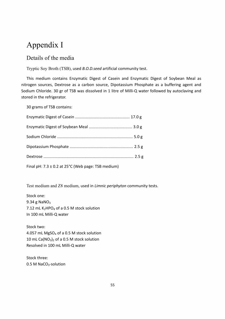
55
Appendix I
Details of the media
Tryptic Soy Broth (TSB), used B.O.D.seed artificial community test.
This medium contains Enzymatic Digest of Casein and Enzymatic Digest of Soybean Meal as
nitrogen sources, Dextrose as a carbon source, Dipotassium Phosphate as a buffering agent and
Sodium Chloride. 30 gr of TSB was dissolved in 1 litre of Milli‐Q water followed by autoclaving and
stored in the refrigerator.
30 grams of TSB contains:
Enzymatic Digest of Casein ................................................. 17.0 g
Enzymatic Digest of Soybean Meal ....................................... 3.0 g
Sodium Chloride .................................................................... 5.0 g
Dipotassium Phosphate ......................................................... 2.5 g
Dextrose ................................................................................. 2.5 g
Final pH: 7.3 ± 0.2 at 25°C (Web page: TSB medium)
Test medium and Z8 medium, used in Limnic periphyton community tests.
Stock one:
9.34 g NaNO3
7.12 mL K2HPO4 of a 0.5 M stock solution
In 100 mL Milli‐Q water
Stock two:
4.057 mL MgSO4 of a 0.5 M stock solution
10 mL Ca(NO3)2 of a 0.5 M stock solution
Resolved in 100 mL Milli‐Q water
Stock three:
0.5 M NaCO3‐solution

56
Stock four:
Micronutrient solution
compound
mg/
100 mL Milli‐Q Water
H3BO3 310
MnSO4 * 4H2O 223
Na2WO4 * 2 H2O 3.3
(NH4)6Mo7O24 * 4 H2O 8.8
KBr 11.9
KJ 8.3
ZnSO4 * 7 H2O 28.7
Cd(NO3)2 * 4 H2O 15.4
Co(NO3)2 * 6 H2O 14.6
CuSO4 * 5 H2O 12.5
NiSO4(NH4)2SO4 * 6 H2O 19.8
Cr(NO3)3 * 9 H2O 4.1
VOSO4 * 2 H2O 2
Al2(SO4)3KSO4 * 24 H2O 47.4
0.5 mL of Stock 1, 0.5 mL of Stock 2, 40 L of Stock 3 and 8 L of Stock 4 were added per litre of Milli‐Q water for Z8 and filtered river water for test solution in Swift test.

57
Appendix II
Toxicity of tested antibiotics to Pseudomonas putida in single substance exposure:
Figure 1: Growth Inhibition of Pseudomonas putida caused by Enoxacin
Figure 2: Growth Inhibition of Pseudomonas putida caused by Lomefloxacin
‐20%
0%
20%
40%
60%
80%
100%
10 100 1000
Inh
ibit
ion
Concentration (nmol/L)
Enoxacin
Controls
Observartions
‐40%
‐20%
0%
20%
40%
60%
80%
100%
10 100 1000
Inh
ibit
ion
Concentration (nmol/L)
Lomefloxacin
Controls
Observations

58
Figure 3: Growth Inhibition of Pseudomonas putida caused by Norfloxacin
Figure 4: Growth Inhibition of Pseudomonas putida caused by Ofloxacin
‐40%
‐20%
0%
20%
40%
60%
80%
100%
10 100 1000
Inh
ibit
ion
Concentration (nmol/L)
Norfloxacin
Controls
Observations
‐20%
0%
20%
40%
60%
80%
100%
1 10 100 1000
Inh
ibit
ion
Concentration (nmol/L)
Ofloxacin
Controls
Observations

59
Figure 5: Growth Inhibition of Pseudomonas putida caused by Sulfamethoxazole
Figure 6: Growth Inhibition of Pseudomonas putida caused by Trimethoprim
‐20%
0%
20%
40%
60%
80%
100%
1000 10000 100000 1000000
Inh
ibit
ion
Concentration (nmol/L)
Sulfamethoxazole
Controls
Observations
‐20%
0%
20%
40%
60%
80%
100%
1000 10000 100000 1000000
Inh
ibit
ion
Concentration (nmol/L)
Trimethoprim
Controls
Observations

60
Toxicity of tested antibiotics to B.O.D.seed in single substance exposure:
Figure 7: Growth Inhibition of B.O.D.seed artificial community caused by Enoxacin
Figure 8: Growth Inhibition of B.O.D.seed artificial community caused by Lomefloxacin
‐20%
0%
20%
40%
60%
80%
100%
1 10 100 1000
Inh
ibit
ion
Concentration (nmol/L)
Enoxacin
Controls
Observations
‐20%
0%
20%
40%
60%
80%
100%
0.1 1 10 100 1000 10000 100000
Inh
ibit
ion
Concentration (nmol/L)
lomefloxacin
Controls
Observations

61
Figure 9: Growth Inhibition of B.O.D.seed artificial community caused by Norfloxacin
Figure 10: Growth Inhibition of B.O.D.seed artificial community caused by Ofloxacin
‐20%
0%
20%
40%
60%
80%
100%
0.1 1 10 100 1000 10000
Inh
ibit
ion
Concentration (nmol/L)
Norfloxacin
Controls
Observations
‐20%
0%
20%
40%
60%
80%
100%
0.1 1 10 100 1000
Inh
ibit
ion
Concentration (nmol/L)
Ofloxacin
Controls
Observations

62
Figure 11: Growth Inhibition of B.O.D.seed artificial community caused by Trimethoprim
Figure 12: Growth Inhibition of B.O.D.seed artificial community caused by Sulfamethoxazole
‐20%
0%
20%
40%
60%
80%
100%
1.E+03 1.E+04 1.E+05 1.E+06
Inh
ibit
ion
Concentration (nmol/L)
Trimethoprim
Controls
Observations
‐20%
0%
20%
40%
60%
80%
100%
1.E+02 1.E+03 1.E+04 1.E+05 1.E+06
Inh
ibit
ion
Concentration (nmol/L)
Sulfamethoxazole
Controls
Observations

63
The parameters of Weibull fit model:
Table 1: a and b parameters in Weibull fit model in single exposure tests.
Pseudomonsas putida B.O.D.seed artificial community
Limnic Periphyon communities
Weibull parameters
a b a b a b
Ciprofloxacin ‐9.66 6.01 ‐73.56 41.64 ‐1,67 0,56Norfloxacin ‐16.79 7.09 ‐17.93 8.41 ‐2,69 1,03Ofloxacin ‐18.61 7.21 ‐25.09 10.32 ‐‐ ‐‐Enoxacin ‐15.07 5.67 ‐45.54 17.34 ‐3,63 1,29Lomefloxacin ‐19.11 6.96 ‐29.23 10.87 ‐4,86 1,49Sulfamethoxazole ‐12.16 2.51 ‐105.04 20.26 ‐‐ ‐‐Trimethoprim 12.87 2.31 ‐30.68 5.45 ‐‐ ‐‐
Table 2: a and b parameters in Weibull fit model in mixture tests.
Pseudomonsas putida B.O.D.seed artificial community
Weibull parameters
a b a b
Swedish mixture ‐21.01 9.41 ‐22.35 9.96
Italian mixture ‐7.75 3.78 ‐15.31 9.20
French mixture ‐6.44 3.10 ‐9.47 5.13