redalyc.the effects of nutrient artery ligation on the ... · the effects of nutrient artery...
TRANSCRIPT

Revista Científica
ISSN: 0798-2259
Universidad del Zulia
Venezuela
Alvarado Morillo, Manuel
The effects of nutrient artery ligation on the development of the distal ulnar metaphysis of the dog
Revista Científica, vol. XIV, núm. 6, diciembre, 2004, p. 0
Universidad del Zulia
Maracaibo, Venezuela
Available in: http://www.redalyc.org/articulo.oa?id=95914608
How to cite
Complete issue
More information about this article
Journal's homepage in redalyc.org
Scientific Information System
Network of Scientific Journals from Latin America, the Caribbean, Spain and Portugal
Non-profit academic project, developed under the open access initiative

THE EFFECTS OF NUTRIENT ARTERY LIGATION ON THE DEVELOPMENT OF THE DISTAL ULNAR METAPHYSIS OF THE DOG
Manuel Alvarado Morillo
Clinical Research Unit - Faculty of Veterinary Sciences University of Zulia - Medical
Surgical Department. P.O.Box 15157 (C.C. Galerias Mall) Maracaibo, Venezuela. E-mail: [email protected]
ABSTRACT
A total of 18 dogs were used in this study. They were obtained from the Veterinary
Hospital of the University of Pennsylvania, Comparative Cardiovascular Studies Unit and
from a dog pound of the Veterinary Policlinic, University of Zulia, Faculty of Veterinary
Sciences. Ablation of the nutrient artery by ligation of the palmar interosseous artery was
performed in 10 dogs; 6 one-month-old dogs; 2 two-month-old dogs; 2 three-month-old
dogs. Radiographic examination of the forelimbs at a weekly interval was performed in
these 10 dogs from one to six weeks postoperatively. The opposite normal front leg was
used as a control. Six dogs were sacrificed at one month of age and arteriograms of the
forelimbs were made. The radiographic changes observed during the first postoperative
week were an increased in density and diameter of the distal ulnar metaphysis, increased
width of the distal ulnar physis, and a decrease in length of the ulnar diaphysis. These
radiographic changes were observed decreasing in severity during a period of five
postoperative weeks. At six weeks postoperatively the distal ulna was similar to the
control. Between 3 to 5 weeks postoperatively the metaphyseal density of the ulna was
decreased and irregular. The decrease in metaphyseal density resulted from a wedge-
shaped retained cartilaginous tissue core wich extended proximally into the metaphysis. A
diminished blood supply to that area was observed angiographically and histologically in
all dogs. The changes observed in the distal ulnar metaphysis in histologic sections and
radiographs are comparable to those observed in the distal metaphysis of the ulna in
young giant dogs with retained enchondral cartilage. The radiographic and histological
findings observed in this study suggests that the lesion observed in rapidly-growing young
giant dogs in the distal ulna is due to a diminished blood supply to the metaphyseal area.
The study was designed with the specific objective to determine the effect on bone growth
after ablation of the nutrient artery of the ulna of the dog.
Key words: Dog, ligation, nutrient artery, ulna.
Efectos de la Ligadura de la Arteria Nutritiva sobre el Desarrollo de la Metafisis
Página 1 de 22veterinaria completa-14
25/01/2006file://C:\SciELO\serial\rc\v14n6\body\art_08.htm
Distal de la Ulna del Perro
RESUMEN
Un total de 18 perros fueron utilizados en éste estudio. Éstos fueron obtenidos de la
Unidad de Estudios Comparativos Cardiovasculares del Hospital Veterinario de la
Universidad de Pensylvania y de la Perrera de la Policlínica Veterinaria Universitaria de la
Facultad de Ciencias Veterinarias de la Universidad del Zulia. La ablación de la arteria
palmar interosea fue realizada en 10 perros. Seis (6) de un mes de edad, dos (2) de dos
meses de edad, y dos (2) de tres meses de edad. El examen radiográfico semanal de los
miembros toráxicos fue realizado en estos 10 perros desde la primera hasta la sexta
semana posterior a la cirugía. El miembro toráxico contralateral fue utilizado como control.
Seis (6) perros fueron eutanasiados al mes de edad y arteriogramas de los miembros
toráxicos fueron realizados. Los cambios radiográficos observados durante la primera
semana post operativa fueron: incremento de la densidad y en el di ámetro de la metafisis
distal de la ulna, incremento en el grosor del cartílago de crecimiento distal de la ulna, y
disminución de la longitud de la di áfisis ulnar. Éstos cambios radiográficos fueron
observados disminuyendo en severidad, durante un período de cinco (5) semanas post
operatorias. A las seis (6) semanas post quirúrgicas, la parte distal de la ulna fue similar
al control. Entre las tres (3) y cinco (5) semanas post operatorias la densidad metafisiaria
distal de la ulna estaba disminuida e irregular. La disminución de la densidad de la
metáfisis resultó ser debido a un núcleo o segmento de tejido cartilaginoso en forma de
cuña que se extendía proximalmente dentro de la metafisis. Una disminución del riego
sanguíneo hacia esa área fue observada angiográficamente e histológicamente en todos
los perros. Los cambios observados en secciones histológicas en la metafisis distal de la
ulna y en las radiografías son comparables con aquellas observadas en la metáfisis distal
de la ulna en perros jovenes de raza gigante con retención de cartílago encondral. Los
hallazgos radiográficos e histológicos observados en el presente estudio sugieren que la
lesión observada en perros de raza gigante de rápido crecimiento en la ulna distal es
debido a una disminución de riego sanguíneo en el área de la metáfisis. Este estudio fue
diseñado con el objetivo específico de determinar el efecto sobre el crecimiento óseo
posterior a la ablación de la arteria nutritiva de la ulna del perro.
Palabras clave: Perro, ligadura, arteria nutritiva, ulna.
Recibido: 26 / 04 / 2004. Aceptado: 22 / 09 / 2004.
INTRODUCTION
Página 2 de 22veterinaria completa-14
25/01/2006file://C:\SciELO\serial\rc\v14n6\body\art_08.htm
The long bone is a tube with thick walls of compact material. The ends, enlarged and
covered with articular cartilage, are formed of spongy bone contained in a thin shell of
compact bone. There is a central medullary cavity. Too often bone has been looked upon
as an inert material and likened to a piece of wood to be sawed, planed, or nailed without
thought given to its structure or physical properties. Bone is a living tissue wich has a
number of important functions [ 22, 23].
Bone is formed by osteoblasts and resorbed or destroyed by osteoclasts. Both of these
cells are active within the skeletal system throughout life. During osteogenesis many
factors affect the development of diseases such as achondroplasia, multiple epiphyseal
dysplasia, rickets, and osteogenesis imperfecta [ 22, 23].
Most of the skeleton forms as a result of enchondral ossification. Enchondral ossification
can be followed by observing the series of events that transform the cartilage mold of a
limb into bone. In the various sites where bones are to form, the mesenchymal tissue
defferentiates into cartilage [22, 23].
The cartilage forms by genetic direction into specific cartilage models which are to
become bones. The cartilage model increases in length and diameter by interstitial growth
(cell division). Each bone model is surrounded by a close-fitting membrane, the
perichondrium. The cells of the perichondrium form two ill -defined layers. The outer layer
differentiates into fibroblasts which form collagen, the connective tissue sheath. The inner
layer which sorrounds the cartilage mold does not differentiate into fibroblasts. The
nutrition is supplied by diffusion of fluids throught the perichondrium [20, 23].
Diffusion of nutrients from the sorrounding tissue throught the perichondrium is an
efficient and adequate method for a distance of 1-2 mm from either side of the cartilage
mold. When the cartilage mold size exceeds the functional limits of diffusion, the cartilage
cells become progressively separated at the center of the cartilage model. The separated
cells become hypertrophic and begin to degenerate. They develop vacuoles, and alkaline
phosphatase appears in the surrounding matrix [ 20, 22, 23].
The matrix is formed bay a mixture of highly reactive elements such as beta-proteins,
collagen, and soluble polysaccharides, the best known of which are the chondroitins [20,
22, 24].
The nutrient artery, a single vessel, penetrates the perichondrium exactly at the center
of the length of the bone model.
Página 3 de 22veterinaria completa-14
25/01/2006file://C:\SciELO\serial\rc\v14n6\body\art_08.htm
Calcification of the altered matrix begins at the periphery of the cartilage mold and
progresses to a point near and always ahead of the vessels. Calcification allows vascular
progression because it causes cell disintegration by cutting cells off from their nutrients.
As a result of positive chemotaxis, peripheral vascular proliferation occurs toward the area
of the cartilage mold occupied by degenerating chondrocytes and altered matrix [20, 22,
24]. A vascular front of branches from the nutrient artery advances into the calcified
matrix, removing the hypertrophied degenerated cartilage cells and some of the calcified
cartilage. As they continue to advance, the vascular tufts penetrate more hypertrophied
cartilage cells [22]. Very soon, around the advancing vessels, the first osteoblasts are
deposited against the calcified walls of the phypertrophied cavities occupied previously by
degenerative chondrocytes and altered matrix. Preliminary bone formation occurs when
the matrix or “osteoid” surrounding the osteoblasts becomes mineralizaed [20, 22, 23].
Bone, with its enclosed cells (osteocytes) is thus formed. By such a mechanism
provisional bone is laid down along the shaft of the long bone cartilage model until the
zone where the growth cartilage is to appear later is reached [20, 22, 23].
Growth deformities involving the dog’s radius and ulna are common. Numerous authors
[10, 11, 12, 15] report the clinical and radiographic signs and treatments of these
deformities. These deformities include: premature closure of the distal ulnar and/or radial
physis; retained hypertrophied enchondral cartilage of the distal urnal metaphysis.
Several authors hypothesize a vascular insufficiency as the cause for these deformities
[1, 7, 14, 15]. The deformities result in a complex orthopedic problem characterized by
shortening of the leg, deviation of the distal aspect of the limb (a disparity in the lenght of
the radius and ulna), and luxation and/or degenerative joint disease of the elbow and
carpal joints.
Several investigators hypothesize a growth disturbance can result from a reduction of
acceleration in the rate of enchondral bone formation in the physis of either the ulna or
radius, while growth continues at a normal rate in the other bone. The end results are
bowing of the radius and ulna and deformity of the carpus, metacarpus, and phalanges
[14, 15].
Radiographic examination is essential for evaluation of these forelimb deformities.
Deformities of the forelimb of large and giant dogs have been reported [3, 11, 12, 15,
17].
Página 4 de 22veterinaria completa-14
25/01/2006file://C:\SciELO\serial\rc\v14n6\body\art_08.htm
The radiographic and histologic changes associated with the development of the bones of
the forelimb in the giant dog have been described for both the normal and abnormal
foreleg [15]. The formation and fusión of the centers of ossification in the developing
skeleton of the cat have been reported [19]. The normal radiographic appearance of the
forelimb bones in the dog from birth to maturity has not been documented. The changes
seen radiographically following premature closure of the distal ulnar physis are shortening
of the ulna and curvature of the forelimb [2, 10, 15]. An overgrowth of the radius may
also contribute to the lateral deviation of the paw [15]. Premature closure of the distal
radial physis in dogs is seen less commonly than premature closure of the distal ulnar
physis [10, 17]. Premature closure of the proximal radial and/or ulnar physis has not been
reported in the dog. In man, injury of the distal radial and ulnar physes causes deformities
similar to those seen in the dog [13, 16, 18]. In man the epiphyseal vessels nourish the
stem cells of the distal physis of the long bones. If this network of vessels is damaged the
growth potential of these physes is impared. Interruption of the metaphyseal vascular
system by fracture tends to result in a transient growth problem even when there is good
reduction of the fracture. A longitudinal growth disturbance of the distal physis may result
from this transient alteration in the rate of enchondral bone formation in the ulna or radius
even if growth continues at a normal rate in the other physis of the same bone [14, 21,
22].
Another entity in the dog is a cartilaginous core with irregular margins which occurs in
the distal metaphysis of the ulna. Histologic examination of this area in dogs shows
increased numbers of unaligned hypertrophied cartilage cells. There is a diminished
amount of vascular tissue invading these hypertrophied cartilage cells [ 6, 15].
In rabbits, metaphyseal ischemia result in an increased lenght of hypertrophyc cartilage
cells which extended deeply into the metaphysis with a total lack of calcification of the
intercolumnar matrix [22, 23]. The radiographic and histologic similarity of hypertrophied
cartilage cells in the dog compared with the rabbit would suggest a similar vascular
ischemic cause in the dog.
Bone Circulation
During bone growth the perichondrium is transformed into periosteum. Bone is
completely covered on the outside by periosteum except at the sites of articulations;
these areas are covered by hyaline cartilage. The inside of the medullary cavity is lined by
the membranous endosteum which covers all the cancellous bone. This endosteum has
but a single layer of cells whereas the periosteum is composed of an inner or cambium
Página 5 de 22veterinaria completa-14
25/01/2006file://C:\SciELO\serial\rc\v14n6\body\art_08.htm
layer and an outer fibrous layer. Much of the blood supply to bone is provided by the
endosteum and periosteum. In long bones the blood circulation is further enhanced by the
presence of a nutrient artery which enters the bone along its diaphysis [22, 23].
This vessel gives off branches to the Haversian system and terminates in the medullary
canal in the form of sinusoidal capillaries. Osteocytes receive their nourishment by way of
the Haversian canals, the transverse channels (Volkma´s canals), and finally by diffusion
throught the canaliculi. Through the bathing of bone cells by blood and lymph there is a
constant exchange of nutritional elements and wastematter comparable to the exchange
taking place in any other cells of the body. The process of tearing down and building up
goes on throughout life, but is particulary noticeable in bone that has been injured or
subjected to undue stress [20, 22, 23]
In this study, the most important aspect of bone circulation to be analyzed is the
nutrient artery of the ulna which gives off the metaphyseal vessels that terminate in many
straight branches which penetrate the degenerating cartilage columns during the
transformation of mineralized cartilage to bone. Interruption of the nutrient vessels has no
effect on chondrogenesis in the physis. The physis increases in thickness since cartilage
producton continues, although transformation of cartilage into bone ceases. It is quite
evident that bone is a complex substance and under certain circumtances behaves in a
most amazing yet logical manner. The person who handles this substance with
understanding will greatly improve his chance of succes.
MATERIALS AND METHODS
In this experiment the dogs that were used were medium size dogs of slow growth rate.
The major portion of this study is concerned with attempts to determine the effects of
ligating the palmar interosseous artery (which gives off the nutrient artery of the ulna) on
the development of the distal ulnar methaphysis and physis. A sample of dogs was
obtained from two populations: one from the Comparative Cardiovascular Studies Unit,
University of Pennsylvania, School of Veterinary Medicine; one from a dog pound of the
Veterinary Policlinic, University of Zulia, Faculty of Veterinary Sciences.
Both forelegs of all puppies were radiographed at weekly intervals between one month
and five months of age.
Group 1
At two months of age two dogs with congenital heart disease but normal forelegs were
Página 6 de 22veterinaria completa-14
25/01/2006file://C:\SciELO\serial\rc\v14n6\body\art_08.htm
euthanized and post-mortem arteriograms of the radius and ulna were made. Heparin
was injected intravenously immediately prior to euthanasia with intravenous pentobarbital
to prevent the formation of clots within the vessels.
For the post-mortem arteriograms the forelimbs were removed by cutting through the
muscles connecting the limb to the trunk and neck. Immediately after death, contrast
material* was injected manually into the brachial artery through an angiographic catheter.
The catheter was removed, the artery ligated, and survey radiographs were made.
Dissection and photography of the vessels were performed to correlate the gross
anatomy with the radiographs.
Group 2
In six normal one-month-old puppies ablation of the right ulnar nutrient artery was
accomplished by surgical ligation of the palmar interosseous artery. The left foreleg was
the control. Radiographs of both front legs were taken just prior to the ligation and at
weekly intervals after ligation.
In two 8-week-old dogs and two 12-week-old dogs ligation of the left nutrient artery was
performed and radiographs were taken weekly until 20 weeks of age (5 months). During
this time interval arteriograms of both front legs were performed.
Physical examination including creatine levels and complete blood counts** were
performed in the normal puppies to exclude any sick dog from the project. All puppies in
Groups 1 and 2 were fed with the same diet*** in the same manner. At the time of
nutrient artery ablation bones were labelled by the administration of tetracycline in vivo
[4, 5]. Tetracycline has the property of becoming bound in osteoblasts at the site of active
growth. The tetracycline labelled cells can be detected because they fluoresce under
ultraviolet light [8, 9]. Oxytetracycline hydrochloride**** was administered intravenously
at a dose rate of 44 mg per kg. bodyweight.
The arteriographic technique used in Group 2 was the same as that described for Group
1. All bones were examined macroscopically and histologically, particularly at the distal
aspects of the radius and ulna.
Radiographic Examination
A 300 mA/126 kVp and a 200 mA 120 kVp x-ray machines were used for radiography.
Página 7 de 22veterinaria completa-14
25/01/2006file://C:\SciELO\serial\rc\v14n6\body\art_08.htm
Craniocaudal and lateral radiographs of the forelimbs were taken using a standardized
technique chart. Intensifying screens were used for the live animals. Kodak nonscreen
high detail AA Industrial film was used for the post-mortem radiographic studies.
The radiographs were developed in a Kodak X-omat MGN 90 second developing system
and in a mannual technique. They were interpreted on a conventional view box.
Significance
The radius and ulna each have two physes, each one growing at different rate. Normal
development of the radius and ulna in the dog depends upon proportionate longitudinal
growth. If injury or other stimuli delay or accelerate the development of one of these four
physes and cause disproportionate growth, bone deformity and malalignment of the limb
will occur. An insufficient blood supply to the ulna has been hypothesized as a cause for at
least one type of primary ulnar deformity seen in the dog [15, 22]. The blood supply to
the distal ulnar metaphysis comes from the nutrient artery which enters the bone in the
proximal metaphysis.
In this study it has documented the effects of ligating the palmar interosseous artery
which gives off the nutrient artery of the ulna as a cause of a retained core of
cartilaginous tissue at the level of the distal midportion of the ulnar metaphysis.
RESULTS
A. Gross radiographic changes in the distal ulna after ablation of the nutrient artery by
ligation of the palmar interosseous artery in six 4-weeks-old dogs.
The right forelimbs were used for the surgical procedure. The left forelimbs were the
controls. The left ulnae and radii appeared radiographically and histologically normal
during the entire experiment.
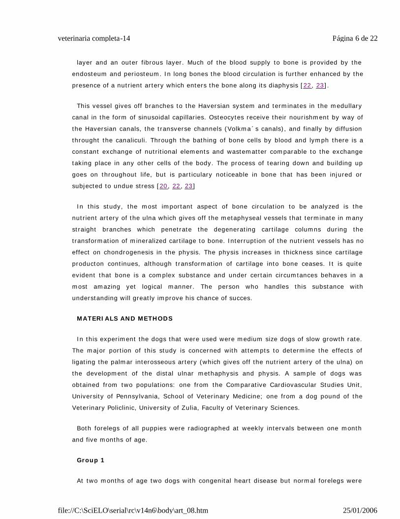
Surgery done at four weeks of age (observations made in six dogs) One week post-
operatively (FIG. 1).
Radiographically the bone density of both radii and ulnae was normal except for the
distal metaphysis of the right ulna. The right distal ulnar metaphysis was very irregular,
greater in diameter than the control, and heterogeneous in density. The lenght of the
ulnar diaphysis was 0.5 to 0.6cm shorter tan the control.
Página 8 de 22veterinaria completa-14
25/01/2006file://C:\SciELO\serial\rc\v14n6\body\art_08.htm
The right radius did not have any abnormalities. The vascular supply from the nutrient
artery of the radius was not compromised in any dog.
Two weeks post-operatively (observations made in six dogs) (FIG. 2).
Página 9 de 22veterinaria completa-14
25/01/2006file://C:\SciELO\serial\rc\v14n6\body\art_08.htm
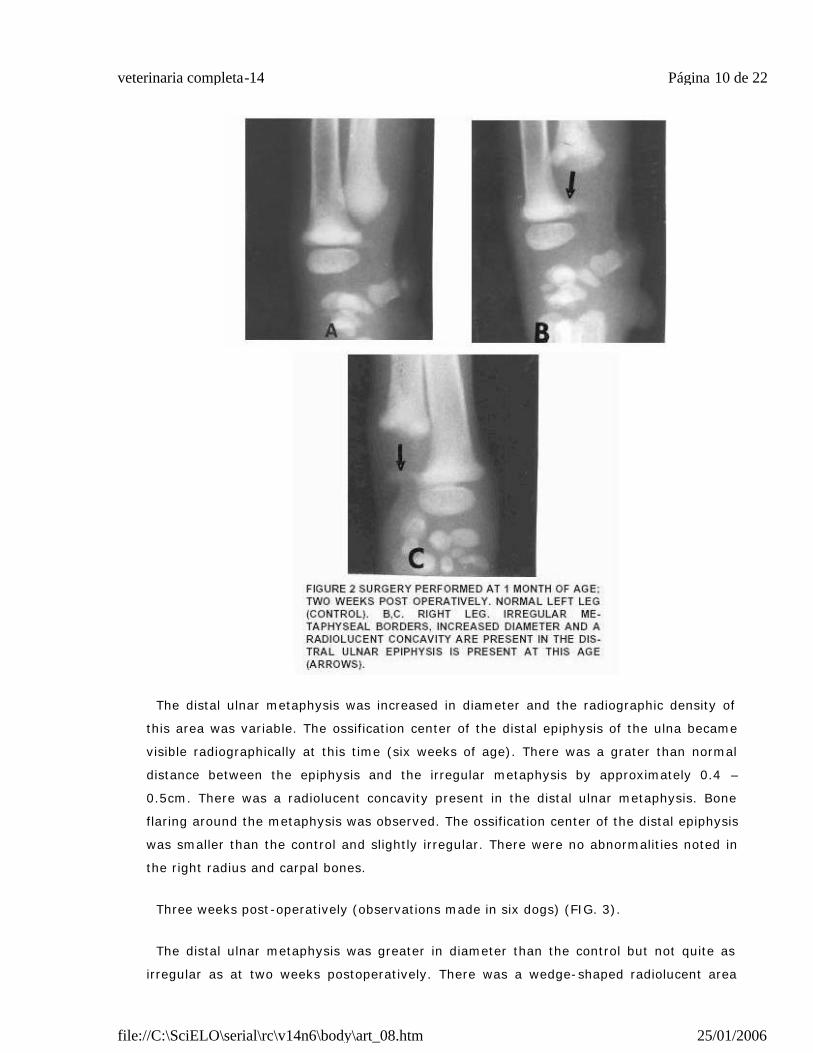
The distal ulnar metaphysis was increased in diameter and the radiographic density of
this area was variable. The ossification center of the distal epiphysis of the ulna became
visible radiographically at this time (six weeks of age). There was a grater than normal
distance between the epiphysis and the irregular metaphysis by approximately 0.4 –
0.5cm. There was a radiolucent concavity present in the distal ulnar metaphysis. Bone
flaring around the metaphysis was observed. The ossification center of the distal epiphysis
was smaller than the control and slightly irregular. There were no abnormalities noted in
the right radius and carpal bones.
Three weeks post-operatively (observations made in six dogs) (FIG. 3).
The distal ulnar metaphysis was greater in diameter than the control but not quite as
irregular as at two weeks postoperatively. There was a wedge-shaped radiolucent area
Página 10 de 22veterinaria completa-14
25/01/2006file://C:\SciELO\serial\rc\v14n6\body\art_08.htm
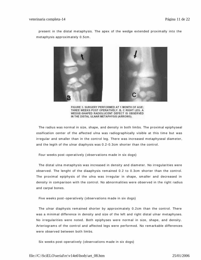
present in the distal metaphysis. The apex of the wedge extended proximally into the
metaphysis approximately 0.5cm.
The radius was normal in size, shape, and density in both limbs. The proximal epiphyseal
ossification center of the affected ulna was radiographically visible at this time but was
irregular and smaller than in the control leg. There was increased metaphyseal diameter,
and the legth of the ulnar diaphysis was 0.2-0.3cm shorter than the control.
Four weeks post-operatively (observations made in six dogs)
The distal ulna metaphysis was increased in density and diameter. No irregularities were
observed. The lenght of the diaaphysis remained 0.2 to 0.3cm shorter than the control.
The proximal epiphysis of the ulna was irregular in shape, smaller and decreased in
density in comparison with the control. No abnormalities were observed in the right radius
and carpal bones.
Five weeks post-operatively (observations made in six dogs)
The ulnar diaphysis remained shorter by approximately 0.2cm than the control. There
was a minimal difference in density and size of the left and right distal ulnar metaphyses.
No irregularities were noted. Both epiphyses were normal in size, shape, and density.
Arteriograms of the control and affected legs were performed. No remarkable differences
were observed between both limbs.
Six weeks post-operatively (observations made in six dogs)
Página 11 de 22veterinaria completa-14
25/01/2006file://C:\SciELO\serial\rc\v14n6\body\art_08.htm
Both radial and ulnae were radiographically similar in shape, size, and density. No
abnormallities were observed in any of the areas where they were present previously. At 3
months of age (eight weeks post -operatively) all six dogs were radiographically normal in
their forelimb. No deformity or lameness was observed.
B. Gross Radiographic changes in the distal ulnar metaphysis after ablation of the
nutrient artery by ligation of the palmar interosseou artery in two 8-week-old dogs.
One week post-operatively (observations made in two dogs)
The distal physis of the affected ulna was aproximately twice the diameter of the control.
The distal metaphysis was very irregular and increased in diameter. The entire leght of the
ulnar diaphysis was 0.4 to 0.5cm shorter than the control. An increased density was
observed in the distal metaphysis. The radius of the control and affected legs were similar
in size, shape, and density.
Two weeks post operatively (observations made in two dogs) (FIG. 4).
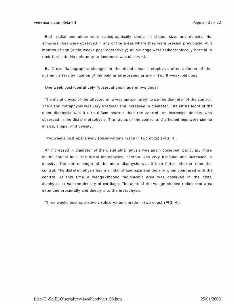
An increased in diameter of the distal ulnar physis was again observed, pariculary more
in the cranial half. The distal inetaphyseal contour was very irregular and increased in
density. The entire lenght of the ulnar diaphysis was 0.3 to 0.4cm shorter than the
control. The distal epiphysis had a similar shape, size and density when compared with the
control. At this time a wedge-shaped radioluceflt area was observed in the distal
diaphysis. It had the density of cartilage. The apex of the wedge-shaped radiolucent area
extended proximally and deeply into the metaphysis.
Three weeks post operatively (observations made in two dogs) (FIG. 4).
Página 12 de 22veterinaria completa-14
25/01/2006file://C:\SciELO\serial\rc\v14n6\body\art_08.htm
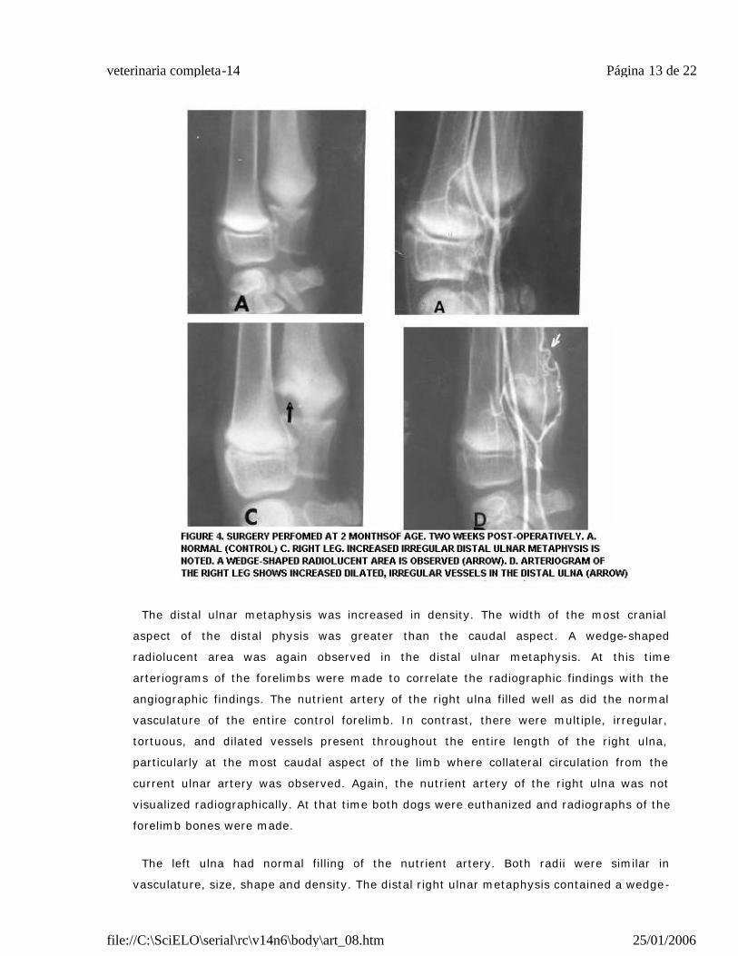
The distal ulnar metaphysis was increased in density. The width of the most cranial
aspect of the distal physis was greater than the caudal aspect. A wedge-shaped
radiolucent area was again observed in the distal ulnar metaphysis. At this time
arteriograms of the forelimbs were made to correlate the radiographic findings with the
angiographic findings. The nutrient artery of the right ulna filled well as did the normal
vasculature of the entire control forelimb. In contrast, there were multiple, irregular,
tortuous, and dilated vessels present throughout the entire length of the right ulna,
particularly at the most caudal aspect of the limb where collateral circulation from the
current ulnar artery was observed. Again, the nutrient artery of the right ulna was not
visualized radiographically. At that time both dogs were euthanized and radiographs of the
forelimb bones were made.
The left ulna had normal filling of the nutrient artery. Both radii were similar in
vasculature, size, shape and density. The distal right ulnar metaphysis contained a wedge-
Página 13 de 22veterinaria completa-14
25/01/2006file://C:\SciELO\serial\rc\v14n6\body\art_08.htm
shaped segment of cartilaginous tissue at the level of its cranial and mid portion. The
right ulnar metaphysis was irregular and greater in diameter than the control.
C. Gross radiographic changes in the distal ulnar metaphysis after ablation of the
nutrient artery by ligation of the palmar interosseous artery in two 12-weeks-old dogs.
One week post-operatively (observations made in two dogs).
The left leg was selected for the surgical procedure, the right leg was the control. The
left distal ulnar metaphysis was increased in density and diameter. The distal ulnar physis
was 2-3 time as wide as the control one. The proximal epiphysis of the left ulna was
decreased in density. The left radius was similar in size, shape, and density to the control.
No abnormalities were observed in the distal epiphysis. The entire length of the left ulnar
diaphysis was 0.5 to 0.6cm shorter than the control.
Two weeks post operatively (observations made in two dogs).
The left distal ulnar metaphysis was irregular in contour and increased in density and
diameter. Slight flaring of the caudal border of the physis was observed. Increased width
of the distal physis was present. There was a concave radiolucent area in the mid portion
of the distal ulnar metaphysis. This concave radiolucent area extended proximally into the
metaphysis about 0.8cm. The distal ulnar epiphysis was slightly irregular at its most distal
aspect. The left ulnar diaphysis was 0.4 to 0.5cm shorter than the right one. The radius in
both legs was normal.
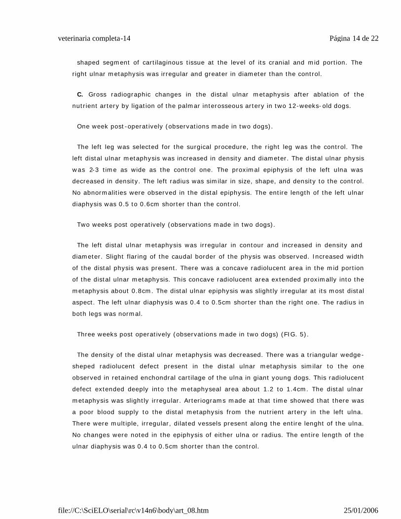
Three weeks post operatively (observations made in two dogs) (FIG. 5).
The density of the distal ulnar metaphysis was decreased. There was a triangular wedge-
sheped radiolucent defect present in the distal ulnar metaphysis similar to the one
observed in retained enchondral cartilage of the ulna in giant young dogs. This radiolucent
defect extended deeply into the metaphyseal area about 1.2 to 1.4cm. The distal ulnar
metaphysis was slightly irregular. Arteriograms made at that time showed that there was
a poor blood supply to the distal metaphysis from the nutrient artery in the left ulna.
There were multiple, irregular, dilated vessels present along the entire lenght of the ulna.
No changes were noted in the epiphysis of either ulna or radius. The entire length of the
ulnar diaphysis was 0.4 to 0.5cm shorter than the control.
Página 14 de 22veterinaria completa-14
25/01/2006file://C:\SciELO\serial\rc\v14n6\body\art_08.htm
Four weeks post-operatively (observations made in two dogs)
There was a decrease in density of the left distal ulnar metaphysis.
The radiolucent defect seen at three weeks was very well outlined and had a sclerotic
border. At the apex of the radiolucent defect there was a transverse to semioblique
opaque line suggesting a stress line fracture or scar tissue line. The entire length of the
ulnar diaphysis was 0.3 to 0.4cm shorter than the control. No deformities were observed.
Six weeks post-operatively (observations made in two dogs)
Both ulnae were similar in length and shape. The distal metaphysis of the left ulna
showed the same radiopaque line observed on week previously. The control ulna was
Página 15 de 22veterinaria completa-14
25/01/2006file://C:\SciELO\serial\rc\v14n6\body\art_08.htm
normal. At that time both dogs were euthanized for histological examination and
measurements of the bones.
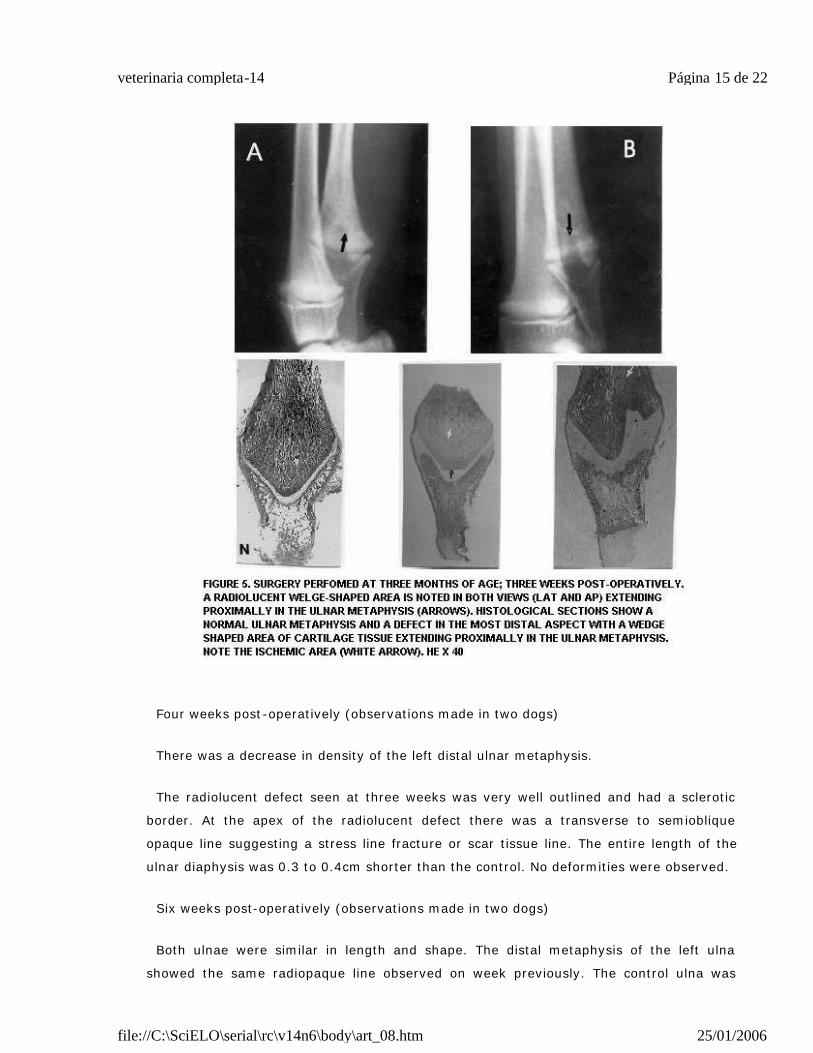
Interruption of the metaphyseal vascular system by fracture tends to result in a transient
growth problem even when there is good reduction of the fracture. Figure 6 shows a
clinical case of a retained enchondral cartilage in a 3.5 month old dog three weeks after a
traumatic injury.
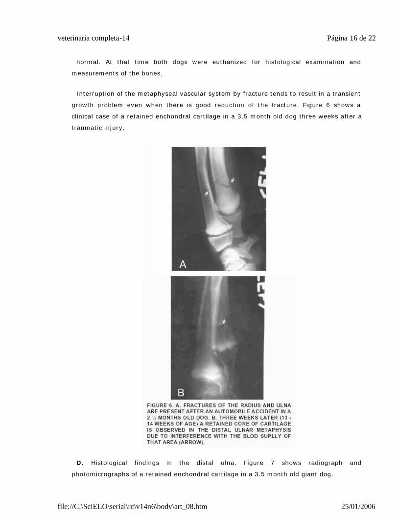
D. Histological findings in the distal ulna. Figure 7 shows radiograph and
photomicrographs of a retained enchondral cartilage in a 3.5 month old giant dog.
Página 16 de 22veterinaria completa-14
25/01/2006file://C:\SciELO\serial\rc\v14n6\body\art_08.htm
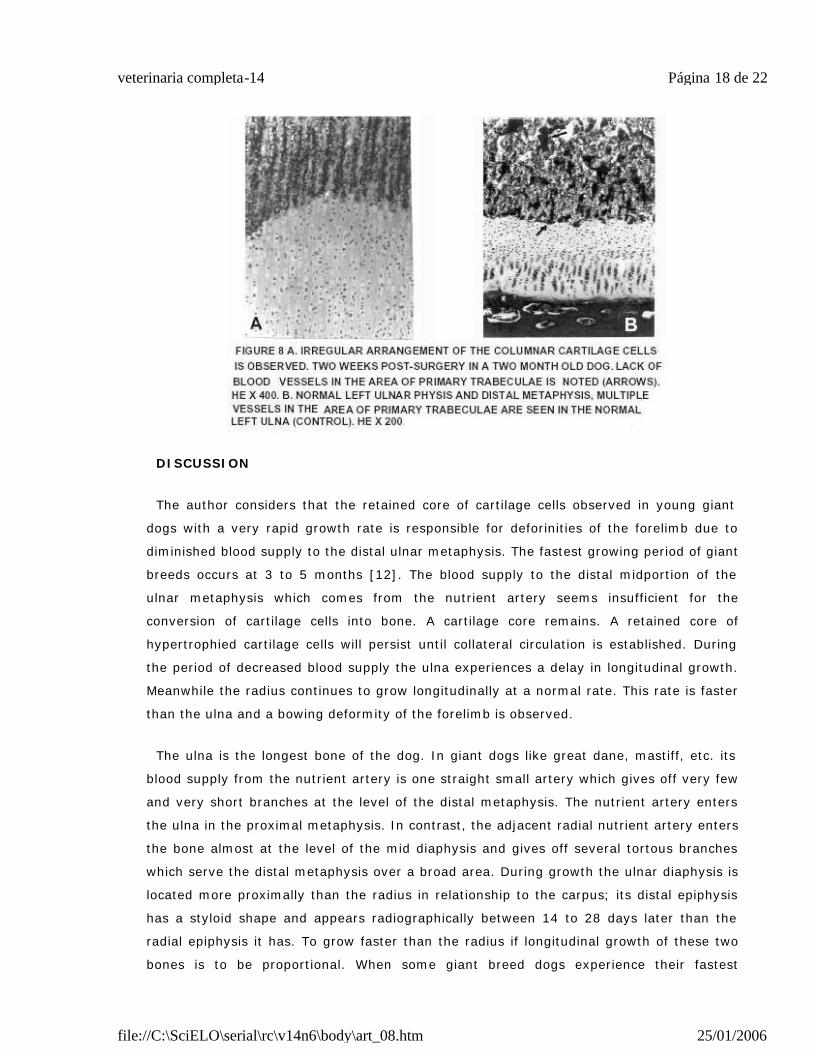
The majority of the bone specimens were submitted for histologic evaluation five weeks
post-operatively. Two 2-month-old dogs were euthanized at two and three weeks post-
surgery and histological examination of the forelimb bones was done. In these two dogs
ulnae there was increase in width of the distal physis. There was a retained core of
cartilaginous tissue present in the distal metaphysis (FIG. 8). The columnar cartilage cells
were increased in number and extended proximally in the metaphysis. The metaphyseal
primary bone trabeculae were irregular in shape and variable in size. There was lack of
blood vessels at the level of the distal portion of the metaphysis where the extension of
columnar cartilage cells was present. The blood supply of both sides to the increased
retained cartilaginous tissue and adjacent to the bone cortices was normal. There was
disorganization in the arrangement and location of the primary trabeculae. An increased
amount of the interlacunar matrix at the level of the distal metaphysis was present. The
lack of blood vessels al the level of the distal mid portion of the metaphysis together with
the extension of columnar cells and increased amount of interlacunar matrix, became
more visible from the first to the fifth week post-operatively. Three weeks after surgery in
three-month-old dogs there was and increased number of unaligned hypertrophied
cartilage cells and a diminished amount of vascular tissue invading the cartilaginous
tissue. There were also enlarged empty lacunae in the distal metaphysis representing
empty blood vessels. All the histological changes observed in all the dogs in the distal
ulnar metaphysis and physis after ablation of the nutrient artery of the ulna are similar to
those changes present in young growing giant dogs in the distal ulnar metaphysis. The
radiographic changes observed in the distal ulnar metaphysis and physis post-operatively
were correlated with the histological findings in all the dogs.
Página 17 de 22veterinaria completa-14
25/01/2006file://C:\SciELO\serial\rc\v14n6\body\art_08.htm
DISCUSSION
The author considers that the retained core of cartilage cells observed in young giant
dogs with a very rapid growth rate is responsible for deforinities of the forelimb due to
diminished blood supply to the distal ulnar metaphysis. The fastest growing period of giant
breeds occurs at 3 to 5 months [12]. The blood supply to the distal midportion of the
ulnar metaphysis which comes from the nutrient artery seems insufficient for the
conversion of cartilage cells into bone. A cartilage core remains. A retained core of
hypertrophied cartilage cells will persist until collateral circulation is established. During
the period of decreased blood supply the ulna experiences a delay in longitudinal growth.
Meanwhile the radius continues to grow longitudinally at a normal rate. This rate is faster
than the ulna and a bowing deformity of the forelimb is observed.
The ulna is the longest bone of the dog. In giant dogs like great dane, mastiff, etc. its
blood supply from the nutrient artery is one straight small artery which gives off very few
and very short branches at the level of the distal metaphysis. The nutrient artery enters
the ulna in the proximal metaphysis. In contrast, the adjacent radial nutrient artery enters
the bone almost at the level of the mid diaphysis and gives off several tortous branches
which serve the distal metaphysis over a broad area. During growth the ulnar diaphysis is
located more proximally than the radius in relationship to the carpus; its distal epiphysis
has a styloid shape and appears radiographically between 14 to 28 days later than the
radial epiphysis it has. To grow faster than the radius if longitudinal growth of these two
bones is to be proportional. When some giant breed dogs experience their fastest
Página 18 de 22veterinaria completa-14
25/01/2006file://C:\SciELO\serial\rc\v14n6\body\art_08.htm
longitudinal growth rate, the metaphyseal vessels of the ulna are not long enough in
comparision with the radius to maintain proportional growth. Anatomically the ulna differs
from the radius. The distal epiphysis is conical in shape, long, and grows much more
actively than that of the distal radius. The proximal ulnar physis is relatively inactive and
contributes very little to the ulnar lenght.
CONCLUSIONS
The changes wich ocurred in the distal metaphysis and distal physis of the ulna following
ablation of the nutrient artery by ligation of the palmar interosseous artery are considered
to result from a diminished blood supply to the metaphyseal region. The areas which were
more sensitive to the diminished blood supply were the zone of ossification of the distal
physis. In these areas the matrix and cartilage cells did not undergo changes in
preparation for thir transformation into bone, and the osteoclastic activity in the
resorption phase in the metaphysis did not occur until collateral circulation was
established. The diminished blood supply to the zone of ossification of the distal ulnar
physis resulted in interference with matrix elaboration and bone formation. The distal
ulnar physis therefore was wider than normal and the projection of hypertrophied cartilage
cells into the mid portion of the metaphyseal area was present. The first changes
observed radiographically were widening of the distal physis accompanied by an increased
metaphyseal density and multiple irregularities along the most distal aspect of the
metaphysis which were noted four days post-operatively. The irregularities in the distal
metaphyseal region were less obvious at 15 to 20 days post-operatively. A wedge-shaped
radiolucent defect with its apex extending deeply into the metaphyseal region was
observed from 20 to 40 days (4-6 weeks) after surgery. This radiolucent area corresponds
to that observed histologically where the diminished vascular tissue and increased number
of columnar cartilage cells were present.
The increased metaphyseal density present from two days after surgery became less
obvious at 5 to 6 weeks post-operatively when it resembled the normal leg. At seven
weeks post-operatively the entire lenght and width of the ulna was similar to the normal
control leg, apparently due to establishment of the collateral blood supply.
The increase in density of the metaphyseal region noted from 4 days to 35-40 days post-
operatively resulted from a persistence of mineralized cartilage cells due to diminished
blood supply to that area. As the collateral circulation was sufficient to invade the
mineralized cartilage cells, normal appearing bone trabeculae developed and the
radiographic appearence of the distal metaphysis became normal. The wedge-shaped
Página 19 de 22veterinaria completa-14
25/01/2006file://C:\SciELO\serial\rc\v14n6\body\art_08.htm
radiolucent area observed at 3 to 5 weeks post-operatively in the distal ulnar metaphysis
was similar to that observed in young growing giant dogs where forelimb deformities are
considered to be a common problem. One of these deformities occurring in giant dogs is
associated with a retained hypertrophied core of cartilage cells in the distal ulnar
metaphysis. In this study it has demonstrated that by ablation of the ulnar nutrient artery
a diminished blood supply to the distal ulnar metaphysis produces a similar retained core
of cartilage cells. In this experiment with medium size dogs no leg deformities were
observed.
* Microtrast (micro-opaque cream) 70% w/w Barium Sulphate mixed with Berlin Blue 2.5%. PICKER
CORPORATION, MEDICAL PRODUCTS DIVISION, CLEVELAND, OHIO, U.S.A
** Creatine levels were within normal limits in all dogs (0.8 to 1.2mgr%). Complete blood counts were within the
normal range (W.B.C. 5.500 to 9.800 per mm3). R.B.C. 5 to 7.8 millions per mm3
*** Wayne Dog Food, Allied Mills, Inc., Chicago, Illinois, U.S.A.
**** Liquamycin, Department of Veterinary Medicine, Pfizer, Inc, New York, N.Y., U.S.A.
BIBLIOGRAPHIC REFERENCES
[1] BRINKER, W. Fractures In: Small animal osthopedics and Fracture treatment,
2nd Ed. W.B. Saunders Company Philadelphia. 711pp. 1990.
[2] CARLSON, W.D. Veterinary Radiology , 2nd Ed. Lea and Febiger, Phipadelphia,
453pp. 1967.
[3] DE ANGELIS, M.; OLDS, R.; STOLL, S.; PRATA, R. SINIBALDI, K. Repairs of
fractures of Radius and Ulna in small dogs, J.A.A.H.A. 9:436-541. 1973.
[4] HARRIS, W.H.; JACKSON, R.H.; JOWSEY, J. “The In Vivo distribution of
tetracyclines in Canine Bone”. J. Bone and Joint Surgery. 44A: 1308-1320. 1962.
[5] HOLMES, J.R.R. “Tetracycline Fluorescence in Bone”. Vet. Record 75: 37-40. 1963.
[6] KELLY, P. Anatomy Phisiology and Pathology of the Blood supply of J. Bone and
Joint Surgery 59:840-866, 1968.
[7] MacCALLUM, J.F.; KRAMER, L.L.; LENER, D.J.; HUSKA, R.E. “Prenatal and Post-
Página 20 de 22veterinaria completa-14
25/01/2006file://C:\SciELO\serial\rc\v14n6\body\art_08.htm
natal Tetracycline Labeling in Equine and Bovine Ossification”. Am. J. Vet. Res. 33
(6):1277-1284. 1972.
[8] MacCALLUM, J.F.; LATSHAW, K.W.; KELLY, E.R. “Identification of Post Natal
Ossification Sites - A Contribution to Radiographic Interpretation ”. Br. Vet. J. 127:
83-87. 1971.
[9] MILCH, R.A.; RALL, D.P.; TOBIE, J.E. “Fluorescence of Tetracycline Antibiotics in
BONE”: J. Bone and Joint Surgery , 40A: 897-910. 1958.
[10] NEWTON, C.S. “Surgical Management of Distal Ulnar Physeal Growth Disturbance
in Dogs”. J. Am. Vet. Med. Assoc. 164: 479 -487. 1974.
[11] O´BRIEN, T.R. “Development Deformities Due to Arrested Epiphyseal Growth”.
Vet. Clin. of North Amer. 1: 441 -454. 1971.
[12] O´BRIEN, T.R.; MORGAN, J.P.; SUTER, P.F. “Epiphyseal Plate Injury in the Dog. A
Radiographic Study in Growth Disturbances in the Forelimb”. J. Small And Pract.
12: 19-36. 1971.
[13] PETERSON, C.A.; PETERSON, H.A. “Analysis of the Incidence of Injuries to the
Epiphyseal Growth Plate”. The Journal of Trauma 12: 275-281. 1972.
[14] RANG, M. The Growth Plate and its Disorders. E. & S. Livingstone, Ltd.,
Edinburg and London. 1-34pp. 1969.
[15] RISER, W.; SHIRER, F.D. “Normal and Abnormal Growth of the Distal Foreleg in
Large and Giant Dogs”, J.A.V.R.S., VI, (1): 50-64pp. 1965.
[16] SALTER, R.B.; HARRIS, R. “Injuries Involving the Epiphyseal Plate”, The of Bone
and Joint Surgery, 45A, (3) 587-621. 1963.
[17] SKAGGS, S.; DeANGELIS, M.P.; ROSEN, H. “Deformities Due to Premature Closure
of the Distal Ulna in Fourteen Dogs. A Radiographic Evaluation”. J.Am.And
Hosp.Assoc. 9: 496-500. 1973.
[18] SKAWINA, A.; LITWIN, J.; GROCZYCA, J.; MIODONSKI, A. The Vascular System of
Human Fetal Long Bones a Scanning Electron Microscopy Study of Corrossion Cast.
J. Anat. 185: 369-376. 1994.
Página 21 de 22veterinaria completa-14
25/01/2006file://C:\SciELO\serial\rc\v14n6\body\art_08.htm
[19] SMITH, N.R. "The Developing Skeleton"J.A.V.R.S. IX, (1): 30-35.1968.
[20] STREETER, G.L. “Developmental Horizons in Human Embryos” (Fourth Issue). A
Review of the Histogenesis of Cartilage and Bone. Contributions to Embryology.
220: 150-185. 1950.
[21] SZENTIMREY, D.; FOWELER; D. The Anatomic Basis of a Free Vascularized Bone
Graft Based on the Canine Distal Ulna. Vet. Surg. 23:529-533. 1994.
[22] TRUETA, J. Studies of the Developmental Decay of the Human Frame.
Philadelphia, Pennsylvania, London: W.B. Saunders Company. 124-168pp. 1968.
[23] TRUETA, J.; CALADIA, A. A study of Blood Supply of the Long Bones. Surg. Gyn.
Obst. 118: 484-492. 1964.
[24] VAUGHAN, J.M. The Physiologiy of Bone: Clarendon Press, Oxford. 1-80pp.
1970.
Página 22 de 22veterinaria completa-14
25/01/2006file://C:\SciELO\serial\rc\v14n6\body\art_08.htm