effects of pomegranate extract … of pomegranate extract supplementation on cardiovascular disease...
TRANSCRIPT
EFFECTS OF POMEGRANATE EXTRACT SUPPLEMENTATION ON
CARDIOVASCULAR DISEASE RISK AND PHYSICAL FUNCTION IN
PATIENTS WITH CHRONIC RENAL FAILURE
BY
PEI-TZU WU
DISSERTATION
Submitted in partial fulfillment of the requirements
for the degree of Doctor of Philosophy in Kinesiology
in the Graduate College of the
University of Illinois at Urbana-Champaign, 2013
Urbana, Illinois
Doctoral Committee:
Associate Professor Kenneth Wilund, Chair
Professor Bo Fernhall
Professor Elvira de Mejia
Associate Professor Shane Phillips
ii
ABSTRACT
BACKGROUND: Patients with chronic kidney disease (CKD) undergoing maintenance
hemodialysis therapy suffer from a variety of co-morbid conditions that greatly decrease physical
function and increase cardiovascular disease (CVD) mortality. Oxidative stress has been
implicated in increasing CVD risk and declining muscle function in hemodialysis patients, but
little is known about the efficacy of antioxidant treatment in this population. A recent one year
intervention in dialysis patients found that consumption of pomegranate juice, a rich source of
polyphenol antioxidants, significantly lowered serum markers of inflammation and oxidative
stress, reduced carotid atherosclerosis, and lowered the prevalence of hospitalizations due to
infection. Despite these potential benefits, pomegranate juice is normally contraindicated in
hemodialysis patients because its high potassium content may contribute to
hyperkalemia-induced cardiac dysfunction. As a result, the efficacy of alternative antioxidant
therapies needs to be investigated.
PURPOSE: The purpose of this study was to evaluate the effect of 6-month oral
supplementation with a pomegranate extract containing a high concentration of antioxidant
polyphenols, but low potassium content, on cardiovascular risk, physical function, oxidative
stress and inflammation in hemodialysis patients.

iii
METHODS: Thirty-three hemodialysis patients were recruited (20 men, 13 women; 54.3±2.1
years). Subjects were randomized to pomegranate (POM, n=16) or placebo (CON, n=17) group.
At baseline and 6 months following the start of the intervention, cardiovascular risk was assessed
by measuring arterial structure and function using a combination of vascular ultrasound and
arterial tonometry, as well as circulating markers of oxidative stress and inflammation. In
addition, a variety of tests were used to assess muscle strength and physical function.
RESULTS: Systolic and diastolic blood pressure (BP) were reduced by 24.2±13.7 mmHg and
10.9±5.3 mmHg, respectively, in POM (p<0.05), but did not change in CON. However, the BP
differences in the POM group were no longer significant after controlling for baseline BP levels.
Paraoxonase-1 activity, a measure of antioxidant capacity, increased by 26.6% (p<0.05) in POM,
compared to no significant change in CON. However, pomegranate supplementation had no
effect on other markers of CVD risk (e.g., β stiffness index, augmentation index, pulse wave
velocity, or carotid intima-media thickness (CIMT)), serum markers of inflammation and
oxidative stress, or measures of physical function and muscle strength.
CONCLUSION: This data suggests that while pomegranate extract supplementation may
reduce blood pressure in hemodialysis patients, it does not improve other markers of
cardiovascular risk, physical function or muscle strength.

iv
ACKNOWLEDGMENTS
This project would not have been possible without the support of many people in many
ways, and I have many people to acknowledge for their help during my time at the University of
Illinois. I would like to thank my advisor, Dr. Kenneth Wilund, for instilling in me the qualities
of being a good researcher. His infectious enthusiasm has been the major driving force through
my graduate career. I would like to thank my co-advisor, Dr. Bo Fernhall, for his guidance and
continuous support of my research over the years. I would like to express my sincere gratitude to
both of them for their patience, motivation, and immense knowledge. I could not have imagined
having better advisors for my Ph.D. study. I also would like to thank my committee members, Dr.
Elvira de Mejia, Dr. Shane Phillips and Dr. Jean Holley, for their encouragement, insightful
comments and invaluable advice.
My sincere thanks also go to the members of Wilund Lab: Emily Tomayko, Harry Chung,
Jinny Jeong, Peter Fitschen, Brandon Kistler, Hank Park, Annabel Biruete, and Kyle Leyshon,
for the stimulating discussion in the office, for the restless days we were working together on
testing subjects, and for all the fun we have had in the last five years. I would like to thank
Barbara Yudell, Elizabeth Jeanes, and Kristin Wiens for coordinating and managing the studies
in our lab. Their willingness to help is important to the success of this project. I also would like
v
to thank Pratibha Garg, Ann Jun, and the team at University of Illinois, Chicago, for handling so
many duties of the study at our Chicago clinics. I thank the undergraduate students in our lab for
helping process blood samples and covering the shifts at the dialysis clinics.
I would like to thank the members of de Mejia Lab, as well as the members of Fernhall
Lab, especially Michelle Johnson, Humin Yan and Sushant Ranadive, for providing me training
and helping me solve many technical problems. I am also thankful to the staff of the dialysis
clinics for their technical assistance.
I would like to thank PomWonderful, LLC, for their funding of this research. Also, many
thanks to the subjects whose participation made this study possible.
Last but not the least, I would like to thank my family and friends, who unconditionally
supported me through every step of the process. I would especially like to thank my parents,
Ming-Kuo Wu and Li-Yun Chen, for their faith in me and allowing me to be as ambitious as I
wanted. I am blessed to have a wonderful sister, Pei-Ching Wu, who provided me with unending
encouragement and some much needed entertainment during the stressful episodes I have had
during my Ph.D. study.
vi
TABLE OF CONTENTS
CHAPTER 1 INTRODUCTION .................................................................................................... 1
CHAPTER 2 LITERATURE REVIEW ......................................................................................... 5
CHAPTER 3 RESEARCH DESIGN AND METHODS .............................................................. 24
CHAPTER 4 RESULTS ............................................................................................................... 45
CHAPTER 5 DISCUSSION ......................................................................................................... 55
CHAPTER 6 CONCLUSIONS .................................................................................................... 64
REFERENCES ............................................................................................................................. 66

1
CHAPTER 1
INTRODUCTION
The prevalence of chronic kidney disease (CKD) in the United States has increased
dramatically in the past decade. In particular, the prevalence of advanced kidney disease, defined
as end-stage renal disease or kidney failure (CKD stage 5) treated with dialysis or transplantation,
increased by nearly 20% in the US in the decade following 20001. Compared to age- and
gender-matched healthy controls , patients with end-stage renal disease treated with hemodialysis
have a 10- to 30-fold increased risk of cardiovascular disease (CVD) mortality, which constitutes
58% of all-cause deaths in this population. Most patients with impaired renal function die of
CVD rather than progress to renal failure, and the risk increases as renal function worsens2-4
.
Patients treated with hemodialysis also have greatly reduced levels of physical functioning.
Muscle catabolism and wasting are especially common and lead to reduced muscle strength.
Physical inactivity exacerbates these functional declines, and also promotes CVD. This cycle of
disease and disability greatly reduces quality of life and increases mortality rates in CKD
patients5.

2
Excessive oxidative stress is believed to underlie many of the cardiovascular complications
in this population, including increases in carotid intima-media thickness (CIMT), arterial
stiffness, atherosclerosis, vascular calcification, and left ventricular hypertrophy (LVH)6-8
. In
addition, studies have shown that increased oxidative stress contributes to inflammation which
promotes skeletal muscle protein catabolism and atrophy in patients with kidney failure4, 9
. This
suggests that antioxidant therapy could be efficacious in CKD patients, potentially to attenuate
declines in both cardiovascular disease risk and muscle function.
In most populations, studies examining the effects of antioxidants (e.g., vitamin C, E, and
β-carotene) on CVD risk have been disappointing10
. In contrast, several studies in hemodialysis
patients have shown that antioxidants may be effective in reducing CVD events11, 12
. This
indicates that antioxidant therapy may be more efficacious in hemodialysis patients than in other
populations, possibly due to their excessive oxidative stress or other unique characteristics of the
disease. However, few studies have examined this question.
Although the efficacy of antioxidant supplementation for primary or secondary prevention
of muscle atrophy or dysfunction is limited, recent research indicates that novel antioxidants
such as N-acetylcysteine13
, as well as isolated polyphenolic compounds14, 15
which have higher
antioxidant activity than standard supplements, can indeed improve muscle strength or reduce
muscle catabolism. In particular, a recent study by Trombold et al.14
found that 9 days of

3
pomegranate extract supplementation improved the recovery of isometric strength after eccentric
exercise in healthy males. However, no studies to date have assessed the effects of antioxidant
supplementation, or pomegranate specifically, on muscle strength or function in CKD patients.
Pomegranate fruit is enriched in polyphenols with high antioxidant capacities. Pomegranate
juice was recently shown to improve CVD risk in patients with carotid artery stenosis by
reducing CIMT in association with reductions in blood pressure and serum oxidative stress16
, and
also has anti-inflammatory effects17, 18
. In addition, a recent study in hemodialysis patients
found that consumption of a very modest amount of pomegranate juice (~100ml/day) 3 days per
week for one year significantly reduced markers of inflammation and oxidative stress, carotid
atherosclerosis, and hospitalizations due to infection19
. However, the patients in this study were
required to consume the pomegranate juice immediately prior to their dialysis sessions so that the
high potassium load from the juice could be removed in the dialysate. A potentially efficacious
alternative strategy is to supplement patients with a pomegranate extract containing abundant
antioxidant polyphenols, but only trace amounts of potassium.
The objective of this research was to determine if daily oral supplementation with a
pomegranate extract can lower CVD risk and attenuate declines in physical function in
hemodialysis patients. To examine this, we conducted a double-blind, placebo controlled
clinical trial in which 33 patients receiving maintenance hemodialysis therapy were randomized
4
to the following groups for 6 months: 1) Control/placebo (CON; N=17), or 2) Daily
supplementation with a 1,000mg capsule of a pomegranate extract (POM; N=16). We
hypothesized that POM supplementation would: 1) reduce systemic markers of inflammation and
oxidative stress; 2) reduce arterial stiffness and CIMT; and 3) attenuate declines in physical
function and strength. Results from this study will help determine if pomegranate extract is an
efficacious means of preventing the development and progression of CKD co-morbidities.
5
CHAPTER 2
LITERATURE REVIEW
2.1 Chronic Kidney Disease: Consequences of a Growing Epidemic
The prevalence of chronic kidney disease (CKD) in the United States has increased
dramatically in the past decade. In particular, the incidence of advanced kidney disease, defined
as kidney failure (glomerular filtration rate (GFR) < 15 ml/min/1.73 m2 body-surface area)
treated with dialysis or transplantation, increased in incidence by 43% in the United States in the
decade following 199120
. Advanced CKD is associated with a variety of metabolic disturbances
that increase morbidity and mortality. In addition, oxidative stress/antioxidant imbalance, muscle
wasting, and cardiovascular complications are especially common, and these co-morbidities
greatly reduce physical function and quality of life in dialysis patients. As a result, new strategies
aimed at improving the health and quality of life of dialysis patients are needed.
2.2 Cardiovascular Disease – The Leading Cause of Death in CKD Patients
CVD is the leading cause of death in CKD patients, and the risk increases as the severity of
the CKD increases. Greater than 58% of premature deaths in dialysis patients have been

6
attributable to CVD (reviewed in21
). This increased CVD mortality is due to a variety of
inter-related factors, including excessive vascular calcification (VC)22-24
. VC is part of a
remodeling of the vascular wall in CVD that promotes arterial and aortic stiffness25
, as well as
other deleterious functional outcomes, including increases in arterial wall intima-media thickness,
endothelial dysfunction, changes in atherosclerotic plaque stability, and a variety of clinical end
points, such as LVH25, 26
.
Studies have demonstrated that arterial stiffness is associated with LVH and an independent
predictor of all-cause and cardiovascular mortality in dialysis patients27-30
. In patients with stage
5 CKD, with the progression of anemia, a decrease in the blood viscosity, and the creation of
arteriovenous (AV) shunt, the peripheral resistances are most frequently normal or lower. Thus,
the principal pressure factors opposing LV ejection are arterial stiffness and early return of wave
reflections27, 31
. In patients with CKD, arterial function alterations are associated with arterial
remodeling including intima-media hypertrophy of arteries26
, characterized by stiffening of the
arteries. Arterial stiffness is a major determinant of left ventricular pressure overload and of
abnormal coronary perfusion in this population32
.
Traditional pharmacological therapies used to treat or prevent CVD in patients with normal
renal function (e.g., lipid lowering medications) are generally less effective in CKD patients,
possibly due to the excessive CVD risk observed in this population which is not fully accounted
7
for by traditional CVD risk factors33
. A part of the extra risk is also attributable to renal-specific
risk factors, related to uremia, including inflammation, malnutrition, and oxidative stress34
.
Elevated plasma concentration of pro-inflammatory cytokines and of the acute-phase reactant
c-reactive protein (CRP) are among the strongest risk predictors of subsequent cardiovascular
morbidity and mortality in uremic patients35-37
.
2.3 Muscle Catabolism in Dialysis Patients
CKD Patients treated with hemodialysis are characterized by a severely limited exercise
capacity38
, reduced skeletal muscle strength and loss of lean muscle mass39
. Evidence suggests
that muscle atrophy in dialysis patients is the result of an enhanced muscle proteolysis40
, and it is
associated with several metabolic abnormalities that stimulate protein degradation41
. Metabolic
acidosis, commonly occurring in dialysis patients, causes loss of muscle mass by activating
irreversible oxidation of essential, branched-chain amino acids 42
. This suggests that reduction in
muscle wasting might be achieved with correction of acidosis or the resulting oxidative stress in
dialysis patients43
.
Insulin resistance, another common complication in dialysis patients, could also accelerate
muscle atrophy by activating the ubiquitin-proteasome system in muscle 44
. The
(ATP)-dependent ubiquitin-proteasome proteolytic pathway is considered to be the major
8
process to remove damaged proteins produced by genetic alterations or by oxidative stress.
Evidence for this includes the presence of higher levels of the transcription of ubiquitin and
subunits of the proteasome44-46
. Two ubiquitin ligases, atrogin-1 (also known as MAFbx) and
MuRF-1, are found specifically in muscle, and their expression increases dramatically in
catabolic states, causing loss of muscle protein47
. Additional evidence linking the
ubiquitin-proteasome proteolytic pathway to protein degradation in catabolism is the finding that
the increase in protein degradation in the muscle of rats with CKD can be blocked by inhibitors
of the proteasome45, 46
.
A third mechanism causing loss of protein stores is that the dialysis procedure itself
stimulates protein degradation in muscle 48
. The most convincing evidence that suggests
increased protein breakdown induced by hemodialysis was provided by Gutierrez et al.49
. These
investigators showed increased amino acid release from the leg during sham hemodialysis in
normal subjects using a bioincompatible dialyzer, but not with biocompatible membranes.
Furthermore, evidence indicates that levels of circulating inflammatory cytokines are elevated
during kidney disease progression, and these cytokines, such as tumor necrosis factor-alpha
(TNF-α) and interleukin-6 (IL-6), have been suggested to be responsible for muscle catabolism50,
51 via up-regulation of the ubiquitin-proteasome proteolytic pathway
52.
9
Finally, oxidative stress, an imbalance between free radical formation and antioxidant
status, is the major contributor to increased levels of circulating inflammatory cytokines and
associated with accelerated protein catabolism in dialysis patients by up-regulating elements of
the ubiquitin conjugation pathway53
. In summary, there are several oxidative balance
abnormalities in skeletal muscle of CKD patients that can stimulate protein degradation and lead
to loss of muscle mass.
2.4 Chronic Inflammation and Oxidative Stress in CKD
As increased oxidative stress and inflammation are both common features of CKD, it has
been suggested that they may be mechanistically linked54-56
. Excessive oxidative stress is
believed to be involved in triggering the inflammatory process by elevating circulating levels of
acute phase proteins such as CRP and pro-inflammatory cytokines (especially IL-6 and TNF-α)7,
36, 57, and is also a significant contributor to the accelerated pathology associated with CKD
58-60.
Oxidative stress, resulting from the imbalance of reactive oxygen species (ROS) production
and anti-oxidant defense mechanisms, is correlated with the level of renal dysfunction among
patients with CKD7, 61
. The increased oxidative stress in CKD patients is mainly attributed to the
retention of oxidized solute due to the loss of filtration function of the kidney, and the increase in
oxidative stress becomes more pronounced as renal function deteriorates58, 62
. In addition,

10
blood-membrane interaction during hemodialysis triggers circulating neutrophils to produce high
amounts of ROS, including superoxide anion, hydrogen peroxide, hydroxyl radical, and
hypochlorous acid63
. Therefore, hemodialysis treatment has been proposed to impose additional
oxidative stress on patients with end-stage renal disease because of the imbalance between ROS
production and antioxidant defense mechanisms.
ROS, which are generated both physiologically and pathologically, cause damage to cellular
constituents, including membrane lipids, proteins, and DNA. In recent studies, the plasma
concentration of oxidative stress markers such as malondialdehyde (MDA), a byproduct of the
peroxidation of polyunsaturated fatty acids, and oxidized low-density lipoprotein (ox-LDL) were
noted to be significantly increased in dialysis patients and have also been used to predict
mortality in uremic patients64-66
. Studies also demonstrate significantly elevated serum
concentrations of CRP and F2-isoprostane, a biomarker of lipid oxidation, in hemodialysis
patients compared with patients with normal kidney function67, 68
.
Plasma levels of protein carbonyls and advanced oxidation products of proteins (AOPP), an
index of oxidant-mediated protein damage, are significantly increased in uremic patients69, 70
. In
addition, AOPP levels were found closely related to advanced glycation end products (AGEs)
and monocyte activation markers in dialysis patients71
. Finally, AOPP was identified as a marker
of oxidative stress and a potent trigger of the monocyte respiratory burst in this population.
11
In contrast to lipids and proteins, the reactions of DNA with various oxidants have not been
well studied in hemodialysis patients. Phagocytes are activated after contact with
bioincompatible dialyzer membranes, so, leukocytes of patients undergoing chronic
hemodialysis may be useful for monitoring changes in the level of cellular DNA oxidation as
they are not only the source, but also the target of endogenously produced ROS and free radicals.
Among the many types of modifications induced by ROS, 8-hydroxy-2’-deoxyguanosine
(8-OHdG) is one of the most abundant oxidative products of DNA72, 73
, and is capable of
reflecting extremely low levels of oxidative DNA damage and is therefore useful as a surrogate
marker of oxidative stress in hemodialysis patients74
. Hemodialysis patients exhibit increased
oxidative DNA damage, compared to age- and sex-matched healthy individuals. Furthermore,
endothelial function has been shown to be negatively correlated with 8-OHdG in hemodialysis
patients75
.
2.5 Impairment in Antioxidant Defense System
Several intracellular and extracellular antioxidant systems have evolved to detoxify specific
free radicals and other oxidants. However, antioxidant mechanisms that serve as a safeguard
against ROS seem to be impaired in hemodialysis patients, and deficiencies in different
components, including reduced levels of enzymatic antioxidants (e.g., superoxide dismutase
12
(SOD), glutathione peroxidase (GSH-Px), and catalase (CAT))76, 77
and extracellular antioxidant
defense elements (such as reduced glutathione (GSH))78, 79
. Furthermore, decreased levels of
hydrophilic and lipophilic antioxidant vitamins in uremia have also been observed34, 80, 81
; if
antioxidant defense is compromised, elevated ROS formation in the body will lead to increased
damage.
2.6 Effects of Single Hemodialysis Sessions
Hemodialysis is a non-selective process clearing solute solely based on molecular weight, a
sieving property of the membrane and protein bound capacity. Consequently, hemodialysis
induces solute losses, including antioxidants. In addition, hemodialysis per se has been suggested
to induce oxidative stress, with ROS being generated on the surface of dialysis membranes82-84
.
Indeed, it has been well documented that even a single session of hemodialysis significantly
increases lipid peroxides and decreases antioxidants85-88
.
Contribution of the dialyzer membrane in the production of inflammation and ROS has
been studied in acute hemodialysis conditions by comparing cellulosic (cuprophane) and
synthetic (polysulfone) dialyzer membranes63, 89
. Plasma levels of inflammatory cytokines (such
as IL-6 and TNF-α) are elevated in dialysis patients, and their expression was exacerbated further
even after a single dialysis treatment90, 91
. Although there are controversial results in the literature

13
indicating that hemodialysis could improve lipid profiles in hemodialysis patients92
, others have
indicated an increase in lipid peroxidation during hemodialysis89, 93
. The majority of ROS
generated during a single dialysis session is hydrogen peroxide91
which causes subsequently lipid
peroxidation, which enhances the susceptibility of LDL oxidation and is recognized as the major
initiating event in the genesis of atherosclerosis. Besides the alterations in lipid levels,
hemodialysis treatment also contributes to increased oxidative stress at the protein level89, 94
.
ROS can not only inactivate mitochondrial enzymes or damage lipid, protein and DNA, but
also actively react with nitric oxide (NO), a principal endothelial-derived relaxing factor, to yield
the powerful oxidant, peroxynitrite (ONOO−). The inactivation of NO by ROS creates NO
deficiency and could play a role in the arterial stiffness.
Interestingly, NO production during hemodialysis is increased rather than decreased95-98
.
Most studies indicate that NO increases early during dialysis, possibly due to shear stress or
leukocyte activation by the dialysis membrane82, 83, 95, 96, 99
; however, a significant drop in NO
levels usually occurs at the end of dialysis98, 100
, probably due to its elimination during the
dialysis session, and returned to high levels between the sessions. The increased NO production
is, however, attacked by superoxide and other ROS released as a result of bioincompatibility of
the dialyzer membrane. This results in formation of ONOO−
which release hydroxyl radicals.
The hydroxyl radicals in the presence of fatty acids released by the lipolytic effect of heparin

14
perpetuate the oxidative stress and cause the ONOO− diverted toward lipid peroxidation. As a
result, hemodialysis can be seen as a situation of continuous production of NO, with
simultaneous inactivation by ROS, leading to endothelial dysfunction101
.
Given the intimate causal interaction between oxidative stress and inflammation,
interventions that aim to alleviate oxidative stress can potentially ameliorate inflammation.
Several clinical studies have attempted to treat oxidative stress and inflammation in this
population by a variety of antioxidant regimens. Many of these studies have employed vitamin E
or C given either orally or parenterally10, 79, 102
; however, these studies have produced conflicting
results and lack of acute responses to single antioxidant intervention. These data may be partially
explained by a vitamin C-related increase in catalytic iron and oxalate that favors prooxidant
activity; different doses and route of antioxidant administration and different markers of
oxidative stress may also help explain the results.
2.7 Oxidative Stress, Antioxidants and CVD in CKD
Elevated oxidative stress is a common feature in CKD patients that likely contributes to
their excessive CVD risk34, 103-105
. Increased oxidative stress results in vascular injury and a
variety of functional CVD outcomes, including increases in arterial stiffness106
, vascular
dysfunction107
, CIMT108
, and vascular calcification109
(Figure 1).
15
Superoxide anion, a primary ROS which is generated from a one-electron reduction of
molecular oxygen, may inactivate nitric oxide (NO) and diminish its bioavailability, thus
ultimately lead to endothelial dysfunction110, 111
. Endothelial dysfunction is especially important
in renal disease, given the nature of accelerated risk of CVD in this population.
Because inflammation, oxidative stress and the pathogenesis of CVD are closely
intertwined, and oxidation products may mediate inflammation in CKD patients, antioxidant
supplementation is of particular interest12, 81
. Due to the excessive oxidative stress in patients
with advanced CKD, antioxidants may be particularly effective in this population.
Despite a demonstrated lack of efficacy of vitamin E in most clinical trials, studies recently
showed that oral vitamin E supplementation reduced the incidence of myocardial infarction11
and
improved oxidative stress79
in CKD patients. Similarly, Tepel et al showed that twice daily
supplementation with 600mg of the antioxidant N-Acetylcysteine (NAC) reduced cardiovascular
endpoints by 40% in CKD patients12
. Yet, few studies have linked the reduced oxidative stress to
the functional cardiovascular outcomes in CKD patients.
Most previous trials assessing the efficacy of antioxidants have used single compounds such
as β-carotene, vitamin A, vitamin C, vitamin E, and selenium, each of which has rather limited
substrate specificity. Also, antioxidants vary significantly in their capacity to scavenge free
radicals, chelate transition metal ions, and/or inhibit oxygenase/oxidase activity. A more

16
efficacious approach may be to use combinations of antioxidants with diverse substrate
specificities.
2.8 Effects of Antioxidants on Muscular Performance
Several lines of evidence link ROS to muscle atrophy via reductive-oxidative (redox)
control of proteolysis. Importantly, a growing number of studies suggest that antioxidants can
serve as therapeutic agents in delaying the rate of muscle atrophy.
Abundant research demonstrated that oxidative stress contributes to muscle atrophy, and this
phenomenon could be delayed by exogenous antioxidants.
A recent cross-sectional study supports the hypothesis that oxidative stress is associated
with the loss of muscle mass in older adults112
. Findings from this study showed that higher
plasma concentrations of antioxidants were associated with reduced risk of low grip, hip, and
knee strength. Similarly, reduced muscle damage in claudicants was found with daily
administration of vitamin E (200mg) and vitamin C (500 mg) for 4 weeks113
. These findings
suggest that antioxidants may play a role in preventing oxidative protein modification in muscle
and preserving muscle function. The work of Appell et al. revealed that there was less oxidative
stress in the vitamin E supplemented muscles than the control group in rats, and eight days of
immobilization lead to a 35% atrophy, while with vitamin E the muscles atrophied only by

17
12%114
. This difference can be attributed to the action of vitamin E as a scavenger for free
radicals and, on the other hand, to an atrophy promoting effect of oxidative stress.
Another study also showed that Trolox attenuates the mechanical ventilation-induced
diaphragmatic contractility deficit. Mechanical ventilation is associated with a rapid onset of
protein oxidation in diaphragm fibers, and has been directly linked to activation of the ubiquitin–
proteasome system of proteolysis. However, diaphragmatic proteolysis did not differ between
controls and mechanical ventilation-Trolox animals. Moreover, proteasome activity in the
diaphragm was elevated in the mechanical ventilation animals, while Trolox treatment attenuated
this mechanical ventilation-induced rise in protease activity115
.
Hauer et al. 13
performed a randomized, placebo-controlled trial in the elderly with 13
weeks of 200 mg N-Acetylcysteine (NAC) supplementation. NAC is a precursor for glutathione,
an important antioxidant that protects cells against oxidative stress. NAC administration resulted
in improvement in muscle strength and decrease in TNF-α level, and suggesting a potential
anti-inflammatory role. In addition, a recent study showed that supplementation with
ellagitannins from pomegranate extract significantly improved isometric strength 2-3 days
following damaging eccentric elbow flexion exercise in recreationally active males14, 15
. The
recovery of strength during the 24- to 48-h period was more rapid, and strength was significantly
higher in the subjects who consumed 500mL of pomegranate extract beverage, compared to the

18
placebo group. Together, these findings suggest that antioxidant supplementation, including
pomegranate extract, may improve muscular performance. Furthermore, the effect of antioxidant
supplementation on the muscular performance might be mediated through changes in the levels
of inflammation and oxidative stress.
2.9 Pomegranate – A Rich Source of Potent Antioxidants
A specific source of antioxidants with therapeutic potential in dialysis patients is
pomegranate fruit (Punica granatum L.), which contains a variety of polyphenols and other
antioxidants, including punicalagin, anthocyanins, ellagic acid and gallic acid, that have
particularly potent and diverse antioxidant capacities116, 117
. The antioxidant capacity of
pomegranate juice was shown to be three times higher than that of red wine and green tea, based
on the evaluation of the free-radical scavenging and iron reducing capacity of the juices116
.
Pomegranate also has been reported to inhibit lipid peroxidation118
, and pomegranate
showed better protection than vitamin E and NAC against oxidative stress119
. In addition,
pomegranate may inhibit inflammation-stimulated c-Jun N-terminal kinases (JNK)- and
extracellular signal-regulated kinase (ERK)- mitogen-activated protein kinase (MAPK)
activation through the suppression of nuclear factor kappa-light-chain-enhancer of activated B
cells (NK-kB) activity17
, increasing endothelial NO synthase expression120, 121
, and reducing

19
atherosclerotic lesions122
. But unlike most studies using single antioxidants, several small clinical
trials indicate that pomegranate juice or extracts also help reduce CVD risk in humans.
Recently, Aviram et al. showed that 1 year of pomegranate juice consumption by patients
with severe internal carotid artery stenosis reduced CIMT by 30%, increased antioxidant
capacity by 130%, and substantially inhibited LDL-lipid peroxidation16
. In a follow-up study
with subjects at moderate risk for CHD, 1 year of pomegranate juice consumption attenuated
anterior wall CIMT progression, which was thicker at baseline than posterior wall CIMT, while
pomegranate juice showed little or no effect on progression rates in the posterior wall CIMT123
.
Furthermore, in the subgroup of patients with the greatest risk of CVD, those in the pomegranate
juice group had less CIMT progression versus control subjects. This finding suggests that
pomegranate may be most efficacious in subjects with the greater oxidative stress and/or CVD
risk. In addition, pomegranate juice and extract consumption in diabetics resulted in a significant
improvement in markers of oxidative stress and atherogenesis without worsening their diabetic
parameters124, 125
. Another potential benefit of pomegranate is that it has been shown to increase
endogenous antioxidant activity16
, suggesting another potential mechanism for its
cardioprotective effects.
Evidence for the clinical benefits of pomegranate juice in the patients undergoing
hemodialysis was recently reported by Shema-Didi et al.19
Hemodialysis patients who consumed
20
100 mL of pomegranate juice 3 times a week during dialysis sessions for 12 months had
significant reductions in neutrophil, IL-6 and oxidized fibrinogen, while the placebo group had
no change in levels of any of the markers. In addition, hospitalizations for cardiovascular causes
were reduced by 40% in patients randomized to pomegranate juice, but did not change in the
placebo group. However, pomegranate juice contains high concentrations of potassium, creating
a potential for potassium overload in dialysis patients. No studies to date have examined the
efficacy of pomegranate extract, which contains much lower concentrations of potassium than
juice, on CVD risk or physical function in this population.
Taken together, these data indicate that antioxidant intervention may be more efficacious in
patients with excessive oxidative stress, such as patients undergoing hemodialysis treatment. In
addition, pomegranate extracts may be more effective than single antioxidants in reducing CVD
risk and attenuating muscle wasting in clinical populations.
2.10 Safety and Drug Interactions
Drug-dietary supplement interactions have been a concern in clinical research, and
conflicting data regarding pomegranate's impact on hepatic P450 2C9 (CYP2C9) enzymes have
been published. Case reports of patients experiencing potential drug toxicities while drinking
pomegranate juice have been reported. One patient on warfarin had an increase prothrombin time

21
after introducing pomegranate juice into the diet126
. Another subject experienced rhabdomyolysis
after drinking pomegranate while on rosuvastatin and zetia combination therapy127
. Both case
reports, however, were confounded by preexisting conditions that may have explained observed
results. A recent study investigated the effect of pomegranate juice and pomegranate extracts on
inhibiting human CYP2C9 activity by using flurbiprofen as a probe compound to profile
CYP2C9128
. This study showed that pomegranate juice and pomegranate extracts inhibit
CYP2C9 activity in vitro, but not in vivo. This finding indicates that subjects on CYP2C9-related
medications can consume pomegranate juice or extract with little risk of drug-supplements
interaction.

22
Figure Legends
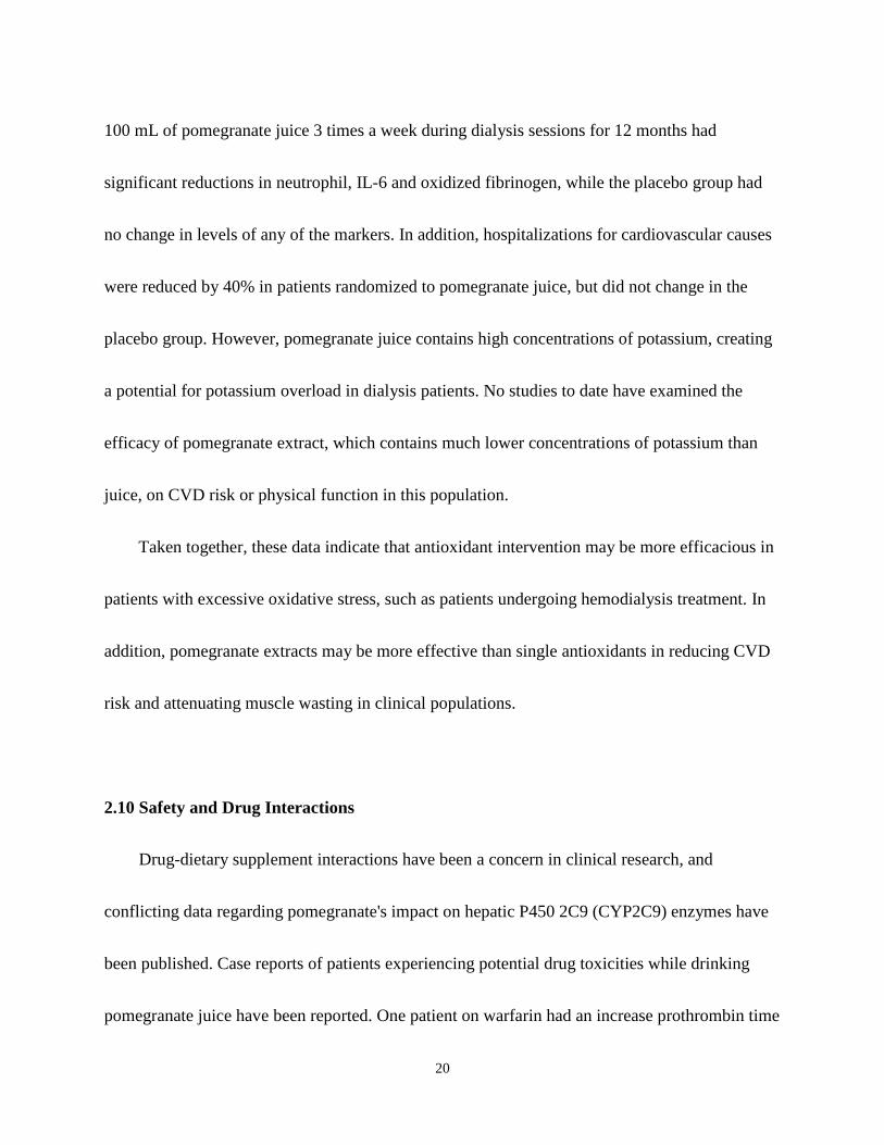
Figure 1 Mechanisms responsible for oxidative stress-induced increases in cardiovascular
disease risk (modified from105, 129
). Cardiovascular disease is one of the major complications of
CKD patients undergoing dialysis. Other common co-morbidities in this population, such as
hypertension, dyslipidemia, hyperglycemia, are centrally linked to increased oxidative stress and
reactive oxygen species (ROS). Elevated oxidative stress and ROS can cause oxidation of
tetrahydrobiopterin (BH4), a cofactor for the production of nitric oxide (NO) by endothelial
nitric oxide synthases (eNOs), and consequently lead to uncoupling of eNOS. This reaction
generates superoxide anion and causes decreased NO production and bioavailablilty. Superoxide
anion can cause lipid peroxidation, accumulation of which contributes to endothelial
dysfunction. Nitric oxide (NO) has been shown to be a potent vasodilator. Oxidative
stress-induced reductions in NO production and bioavailability also promote endothelial
dysfunction. This can impair microvascular vasodilatory capacity, elevate inflammatory levels,
and increase arterial stiffness. Furthermore, these responses result in the development of
cardiovascular complications, including atherogenesis, left ventricular hypertrophy and
dysfunction, and myocardial ischemia.
23
Figure 1 Mechanisms responsible for oxidative stress-induced increases in cardiovascular
disease risk (modified from105, 129
).
ROS, reactive oxygen species; BH4, tetrahydrobiopterin; NO, nitric oxide; eNOS, endothelial nitric oxide synthase.

24
CHAPTER 3
RESEARCH DESIGN AND METHODS
3.1 Study Overview
Patients with renal failure receiving maintenance hemodialysis therapy (CKD stage 5) at
local dialysis clinics were recruited and randomly assigned to one of two groups for 6 months: 1)
Usual care/control (CON; N=14); or 2) Daily oral supplementation with a purified pomegranate
extract (POM; N=13). Patients in the POM group ingested a 1,000mg capsule of a pomegranate
extract every day for 6 months. Patients in the CON group ingested a placebo capsule using the
same protocol. At baseline and 6 months following the start of the supplementation period, all
patients had blood collected to measure markers related to inflammation, oxidative stress, and
antioxidant capacity. The subjects also came in for clinical testing at each time point to measure
factors related to cardiovascular disease (CVD) risk and physical function. A study timeline is
shown in Figure 2.
3.2 Subjects, Recruitment, Screening, and Selection

25
Thirty-three patients with CKD stage 5 (GFR < 15 ml/minute/1.73 m2) receiving
hemodialysis therapy for more than 3 months were recruited on a rolling basis from the
hemodialysis clinics in Champaign and Chicago, IL. Patients were recruited through
advertisements that were placed in the clinics, as well as through brochures provided to subjects
by the medical staff and research members. Those expressing an interest in the study signed a
form indicating they would like to be contacted by a member of the research staff to acquire
more information. During the screening, subjects were informed of the study purpose, the risks
and benefits pertaining to their participation, the randomization process, the proposed testing,
and the necessary time commitment. Patients were screened for eligibility by administering a
medical history questionnaire (see exclusion criteria below), and an informed consent document
was provided for each individual to sign upon their agreement to participate. A physician
clearance for each subject was signed by their nephrologists. All protocols were approved by the
Institutional Review Board of the University of Illinois.
3.3 Inclusion/Exclusion Criteria
Inclusion/exclusion criteria for volunteers include the following: 1) Subjects must be
diagnosed with CKD stage 5, and had been receiving hemodialysis treatment 3 times per week
for ≥ 3 months. This criteria was chosen based on research indicating oxidative stress increases

26
as renal function deteriorates130
, and our working hypothesis is that pomegranate
supplementation would be most efficacious in individuals with excessive oxidative stress. 2)
Subjects must be ≥ 30 years of age. As previous studies have shown arterial stiffness increases
significantly after 30 years of age in CKD patients (unpublished observations from our
laboratory), and limiting the recruiting pool to those ≥ 30 years of age will reduce the number of
recruited subjects with little or undetectable arterial stiffness. 3) Subjects must be willing to be
randomized to the control or intervention groups. 4) Subjects must receive medical clearance
from their primary care physician to participate. 5) Subjects must NOT be consuming any form
of antioxidant supplementation.
3.4 Group Assignment
Following recruitment, screening, and baseline testing (see below), 33 eligible subjects were
randomized to the CON or POM groups as described below. Both the subjects and study
personnel involved in data collection were blinded to the group assignment. We used a simple
randomization procedure as follows: the eligible subjects were randomly sorted into pairs. In
each pair, a random permutation of the subjects is used to assign them to the two groups (CON
or POM). Under this randomization procedure, each subject has equal probability of being
assigned to each group.

27
3.5 Intervention Protocol
Patients in the POM group ingested a 1,000mg capsule of a purified pomegranate
polyphenol extract (POMx™, POM Wonderful, Inc. Los Angeles, CA;
http://www.pompills.com/pills/product_pills.aspx) 7 days a week for 6 months. POMx pills are a
concentrated blend of polyphenols extracted from whole pomegranates that have been safety
tested by the Food and Drug Administration. The no-observed-adverse-effect level (NOAEL) of
POMx was considered to be 1500 mg/kg of body weight (unpublished data, POM Wonderful
LLC., 2006), which is approximately 100~125-fold more than what was being provided in this
study. Research also showed that 1,000mg (610~755mg of gallic acid equivalents) of
pomegranate extract is sufficient to provide antioxidant effects through a reduction in
thiobarbituric acid reactive substances (TBARS), a byproduct of lipid peroxidation, in
overweight subjects131
.
POMx is derived from pomegranates grown in California (Paramount Farms). POMx is
prepared from inner and outer pomegranate peels, seeds and juice, pressed to produce a powder
with a high concentration of polyphenols. The pomegranate extract is 98% (980 mg) of the pill’s
active ingredient. The extract is composed of 5.7% ellagic acid, 25.6% punicalin/punicalagin,
and 68.4% other polyphenols. The remaining 2% of the drug is magnesium stearate. The
28
matching placebo pills contain 0mg of gallic acid equivalents, and the ingredients include
cellulose, caramel, beet root, magnesium stearate, and silicon (source: PomWonderful,
unpublished data). The study drugs were stored at the Agricultural Engineering Sciences
Building. Dry storage is required and shelf life is 18 months at or below 25˚C.
We chose to use the POMx pills instead of pomegranate juice because the juice contains
significant amounts of potassium (430mg per 8 ounces or 12% of the recommended Daily Value
for healthy adults), which is restricted in CKD patients, while the POMx pills have marginal
levels (<0.998 mg per 1,000mg pill) of this mineral. Hyperkalemia, the condition of
overabundance of potassium in the extracellular compartment, is common in patients with
end-stage renal disease. Excess intake of potassium through dietary indiscretion and oral
supplementation are the most common causes of hyperkalemia in dialysis patients. The effect of
hyperkalemia on cardiac conductivity is its most feared clinical consequence, symptoms
including slow pulse, irregular heartbeat, heart failure, and even sudden death. Severe
hyperkalemia may also profoundly affect skeletal muscle, manifesting as motor weakness and
parasthesia.
Individuals in the CON group ingested a non-caloric placebo capsule using the same
protocol. Patients in both groups received a pill bottle containing a month’s supply of capsules
(POM or placebo) prior to the beginning of each month. They were asked to bring their pill
29
bottles to the dialysis clinics on a monthly basis so the number of remaining pills can be counted
for compliance purposes. If a participant was non-compliant, defined as completing less than
75% of the available supplements (and does not respond to repeated attempts to increase
consumption), their data was not included in the analysis.
3.6 Baseline Testing and Measurements
3.6.1 Anthropometric Measures
Barefoot standing height was measured to the nearest 0.1 cm with a stadiometer and body
weight was measured on a balance scale with shoes and superfluous outer garments (e.g.,
jackets) removed. Waist circumference was measured as the minimum circumference between
the top of the iliac crest and the distal end of the rib cage along the mid-axillary line. An
additional measure of waist circumference was also taken at the umbilicus, and the average of
the 2 waist circumference measurements was used in all analysis. Hip circumference was
measured at the maximal hip circumference. All measurements for a given participant were taken
in triplicate and averaged.
3.6.2 Blood Chemistry

30
A small amount (20 – 30 ml) of blood was collected from each subject at baseline and 6
months for analyses described below. Plasma and serum from these extra samples was collected
by centrifugation, divided into 350ul aliquots, and stored at -80°C until analyzed.
3.6.2.1 Oxidative Stress Biomarkers
Advanced oxidation protein products (AOPP) are uremic toxins that are a marker of protein
oxidative stress created through the reaction of plasma proteins with chlorinated oxidants, such
as chloramines or hypochlorous acid. Concentration of AOPP in the plasma was determined
using the semi-automated method as previously described69
. Briefly, AOPP was measured by
spectrophotometry on a microplate reader (GENios Pro, Tecan, Männedorf, Switzerland) and
calibrated with chloramine-T (Sigma, St. Louis, MO) solutions that in the presence of potassium
iodide absorb at 340 nm. In test wells, 200 µl of plasma diluted 1/5 in PBS was placed on a
96-well microtiter plate, and 20 µl of acetic acid was added. In standard wells, 10 µl of 1.16 M
potassium iodide (Sigma, St. Louis, MO) was added to 200 µl of chloramine-T solution (0–400
µmol/liter) followed by 20 µl of acetic acid. The absorbance of the reaction mixture is
immediately read at 340 nm on the microplate reader against a blank containing 200 µl of PBS,
10 µl of potassium iodide, and 20 µl of acetic acid. AOPP concentrations were expressed as
micromoles per liter of chloramine-T equivalents.
31
Oxidized low-density lipoprotein (ox-LDL), a marker of lipoprotein-associated oxidative
stress132
, was measured in triplicate using commercially available ELISA kits (Mercodia,
Uppsala, Sweden).
8-hydroxy-2’-deoxyguanosine (8-OHdG) is produced by oxidative damage of DNA by
reactive oxygen and nitrogen species. It serves as a biomarker of DNA oxidation, and was
measured in triplicate using commercially available ELISA kits (Trevigen, Gaithersburg, MD).
3.6.2.2 Antioxidant Capacity
The Oxygen Radical Absorbance Capacity (ORAC) assay was performed according to
published methods by Prior et al.133
Fluorescein reacts with free radicals generated by
2,2’-azobis(2-amidinopropane) dihydrochloride (AAPH) yielding a non-fluorescent product.
Loss of fluorescence was measured over time in fluorescent plate reader, FLx800tbi (Bio-Tek,
Winooski, VT), at 37 °C and sensitivity 60. Readings were made with excitation 485 nm and
emission 528 nm. The area under the curve (AUC) was calculated using the following equation:
AUC = 0.5 + f1 / f0 + fi / f0 + …+ 0.5 (fn / f0)
Net AUC = AUCsample – AUCblank

32
Where AUC is area under the curve, f1 is fluorescence of first reading (2 min); f0 is fluorescence
of reading time zero and fn are n fluorescence readings. Areas under the curve were compared to
a standard antioxidant, Trolox (vitamin E analog). Results were expressed as μM Trolox
equivalents.
High-density lipoprotein (HDL) associated paraoxonase-1 (PON-1) is an antioxidant enzyme
that protects lipoproteins from oxidation, and is considered a primary anti-atherosclerotic
component of HDL119, 134, 135
. Previous research has shown that pomegranate supplementation
increases PON-1 activity in diabetic and healthy subjects124, 136
. PON-1 activity, including
arylesterase (monoesterase), paraoxonase (triesterase), and lactonase, was measured to assess
serum antioxidant capacity as described16, 137-139
.
Lactonase, paraoxonase, and arylesterase activity was measured kinetically at 37°C using
dihydrocoumarin, paraoxon, and phenyl acetate as a substrate, respectively. PON-1 activity was
measured using a microplate reader (GENios Pro, Tecan, Männedorf, Switzerland), and the
activity of lactonase, paraoxonase, and arylesterase was reported as units per milliliter, where 1
U is defined as 1 mmol of dihydrocoumarin, paraoxon, and phenyl acetate hydrolyzed per
minute, respectively.
3.6.2.3 Markers of Inflammation

33
Serum CRP and IL-6 were measured in triplicate using commercially available ELISA kits
(CRP:Alpco, Salem, NH ; IL-6: R&D Systems, Minneapolis, MN). To control for variation
between runs, baseline and 6-month samples from each subject were analyzed simultaneously.
Published intra-assay coefficients of variation were less than 5% for each assay.
3.6.3 Cardiovascular Disease Risk Measures
3.6.3.1 Blood Pressure
Brachial systolic (SBP) and diastolic (DBP) blood pressure was obtained using standard
methods following a 10-minute period of quiet supine rest in a dimly lit room. Brachial blood
pressure was measured using an automatic digital blood pressure monitor (Omron IntelliSense
HEM-907XL, IL). All the BP measurements were repeated and the average of the two values,
1-minute apart, was recorded and used for analysis. If the values differed by ≥ 5mmHg, a third
measurement was obtained and the two closest values were averaged. Mean arterial pressure was
calculated by DBP + 1/3 (SBP-DBP).
3.6.3.2 Carotid Artery Stiffness
A combination of ultrasound imaging of the common carotid artery with simultaneous
arterial tonometry of the carotid artery (for estimation of carotid blood pressure) allows for a
34
non-invasive determination of carotid arterial compliance. Carotid ultrasound images were
obtained from the common carotid artery, 1-2 cm proximal to the carotid bifurcation using a 7-13
MHz linear array transducer with a sampling rate of 1,000 Hz. B-Mode and M-mode images
were obtained and displayed simultaneously and automated wall tracking software was used to
detect changes in lumen size as described above. Electronic calipers were applied to the arterial
wall using the B-mode image and wall tracking is conducted using the M-mode image in real
time. Changes in lumen size between systole and diastole is recorded for 12 seconds and an
ensemble average beat is constructed from which measurements of arterial compliance and
stiffness was conducted. -stiffness index (β) was calculated with lumen size in systole and
diastole. The calculation for is shown below:
= In (Ps – Pd) / (Ds – Dd)
Ps=systolic pressure, Pd=diastolic pressure, Ds = maximum vessel diameter, and Dd = minimum
vessel diameter.
Carotid blood pressure was estimated using simultaneous arterial tonometry on the
contralateral carotid artery. A high fidelity pencil probe strain gauge transducer (Millar
Instruments, Huston, Texas) was interfaced with acquisition software (SphygmoCor, AtCor
35
Medical, Sydney, Australia). The carotid waveform was calibrated against brachial artery
pressure to derive carotid artery pressure using standard transfer functions (SphygmoCor, AtCor
Medical, Sydney, Australia).
3.6.3.3 Carotid Intima-Media Thickness (CIMT)
CIMT were measured using a high-resolution ultrasound system (Aloka Alpha 7, Japan)
with a 7-13 MHz linear array transducer. The common carotid artery was imaged at the proximal
1-2 cm straight portion. The carotid bifurcation or bulb was used as a reference point to
standardize the position of the CIMT measurements between baseline and final testing when
possible. All images were analyzed in a 10mm window which is 10mm distal to the bulb using
Carotid Analyzer (Medical Imaging Applications, LLC, Coralville, IA) software. The
intima-media thickness was defined as the distance between the leading edge of the
lumen-intima interface to the leading edge of the media-adventitia interface of the far wall of the
carotid artery. All measurements and analysis were made by the same trained investigator
blinded to group assignment.
3.6.3.4 Wave Reflection

36
Applanation tonometry was performed using a high-fidelity strain-gauge transducer
(SphygmoCor; AtCor Medical, Sydney, Australia) on the radial artery to obtain pressure
waveforms. Using a generalized validated transfer function140
, a central aortic pressure
waveform was reconstructed from the radial artery pressure waveform. Augmentation index
(AIx) was calculated as the ratio of amplitude of the pressure wave above its systolic shoulder
(i.e., the difference between the early and late systolic peaks of the arterial waveform) to the total
pulse pressure. The result was expressed as a percentage and was used as an index of aortic
pressure wave–reflection intensity. Because AIx is influenced by HR141
, AIx values were also
normalized to a HR of 75 bpm.
3.6.3.5 Aortic Pulse Wave Velocity (PWV)
PWV was measured following current guidelines142
. Using the same high-fidelity
strain-gauge transducer (Sphygmocor, AtCor Medical, Australia) as in the wave reflection
measurements, pressure waveforms were taken first at the right common carotid artery and then
at the right femoral artery. Consecutive waveforms were captured for a 10-s epoch. Simultaneous
ECG gating, as a timing marker, was assessed via a 3-lead CM5 configuration and further used
to obtain HR143
. The foot of the pressure wave was identified automatically, removing potential
observer bias, using an algorithm that detects the initial upstroke via a line tangent to the initial
37
systolic upstroke point of the pressure tracing and an intersecting horizontal line through the
minimum point144
.
Distances from the suprasternal notch to the femoral artery and from the carotid artery to the
suprasternal notch were measured as straight lines with a tape measure and recorded to the
nearest mm. The distance from the carotid artery to the suprasternal notch was then subtracted
from the distance between the suprasternal notch and femoral artery to account for differences in
the direction of pulse wave propagation.
Aortic PWV was calculated from the distances between measurement points and the
measured time delay between 10 proximal and distal waveforms. The peak of the R wave
recorded from the ECG was used as a timing marker. Values obtained from the carotid to the
femoral artery (PWV) were taken as an index of “central” arterial stiffness. Integral software
assessed pulse wave quality (strength of pulse wave signal, pulse height variation, pulse length
variation, and base line variation) and standard deviation of mean time differences
(SphygmoCor, AtCor Medical, Sydney, Australia).
3.6.3.6 Echocardiography
Echocardiography was performed using a multifrequency (1.5-4.25 MHz) transthoracic
transducer to assess parameters related to cardiac function and systemic vascular resistance. Left
38
ventricular stroke volume was assessed with the Simpson‘s biplane technique. Cardiac output
was calculated by multiplying the stroke volume by the heart rate. Systemic vascular resistance
was calculated by dividing the mean arterial pressure by the cardiac output. To minimize the
effect of variations in fluid volume in hemodialysis patients, studies were performed 18-24 hours
after a hemodialysis session at all testing timepoints.
3.6.4 Physical Performance Measures
3.6.4.1 Functional Fitness Testing
Subjects underwent a battery of tests to assess functional fitness, including: 1) Chair Stand
Test where subjects were asked to stand up from a seated position as many times as possible
within 30 seconds; 2) Arm Curl Test in which subjects were asked to complete as many arm curls
as possible during 30 seconds using either a 5-pound dumbbell for females, an 8-pound dumbbell
for males; 3) Chair Sit and Reach Test that asked subjects to reach forward with both arms to try
and touch their extended leg to assess flexibility; 4) Back Scratch Test where subjects tried to
touch their middle fingers behind their back to assess upper body flexibility; and 5) 8 Foot
Up-and-Go Test in which subjects were asked to walk around a cone placed 8 feet away from
their chair and back again during a timed trial.

39
3.6.4.2 Shuttle Walk Test
Each subject underwent a shuttle walk test to assess physical performance. This is a
progressive test in which patients walk back and forth continuously on a 10-meter course. The
walking is paced by beeps which are programmed - the subject should be at the end of the course
by each beep. They maintain each speed for one minute and then the pace is increased. The
speeds increase so that in each successive minute the speeds are as follows: 1.12, 1.54, 1.88,
2.26, 2.64, 3.02, 3.4, 3.78, 4.16, 4.54, 4.92, and 5.3 miles per hour. The patient continues until
they do not reach the end of the 10-meter course by the beep and the total distance covered is
calculated. Measures of physical performance such as this shuttle walk test are frequently used to
assess function in older and diseased people instead of more objective measures of aerobic
capacity such as VO2max testing. This is due to functional limitations like muscle weakness and
shortness of breath that prevent these individuals from achieving standard criteria used in
assessment of these more objective tests145
. This shuttle walk test is well established as a part of
the guidelines for assessment of fitness in patients with chronic pulmonary disease146
, and is
often preferred to the six-minute walk test because it is paced, and therefore more objective.
3.6.4.3 Muscle Strength Testing

40
A Biodex System 3 Pro dynamometer (Biodex Corp., Shirley, NY) was used to assess
hamstring and quadriceps strength. The reliability and reproducibility of this device is well
established.147
The subject sat upright on the Biodex chair with the axis of the dynamometer
corresponding to the knee joint axis. Once the patient was positioned, the shoulder, waist, thigh,
and lower leg proximal to the ankle were secured with straps. To avoid substitution and
compensation of other muscle groups, hip belts and a chest restraint were used to prevent hip
flexion and extension. Isolated isokinetic muscle torque of the knee joint in flexion (hamstring
muscles) and extension (quadriceps muscles) were evaluated at a speed of 60 degrees per
second.147
There were 2 sets of 6 repetitions, and resting interval of 3 minutes between sets. For
all tests, participants were verbally encouraged to perform as vigorously as possible. Total
strength was defined as the sum of peak torque of knee extension and flexion.
3.7 Final Testing
Six months following the start of the intervention period, all subjects repeated the same
testing procedures performed at baseline. This included anthropometric measures, vascular
ultrasound measurements, physical function assessment, and blood draws. All testing was
conducted by study personnel blinded to the subject’s group assignment. Several factors,
including blood pressure, heart rate, circulating hormones, and vascular smooth muscle reactivity,
41
have been implicated in the circadian pattern of cardiovascular events148
. Thus, each testing and
measurement session described above occurred approximately at the same time of day within a
2-hour window as the baseline testing to ensure conformity between baseline and final testing
measures.
3.8 Data Management, Statistical Analysis and Power Estimates
3.8.1 Data Management
Data management and quality control was managed using a Microsoft Excel spreadsheet.
All data that were not downloaded directly, such as data from serum assays, were entered and
double checked for accuracy using duplicate entry forms and compare features programmed in
Microsoft Excel. Confidentiality of data was maintained by using only subject identification
numbers for data entry. The master document linking participant names and identification
numbers and group assignment were maintained in a separate password-protected file on a
secure network accessible only by the mentor (Dr. Wilund); therefore, access to the data was
restricted.
3.8.2 Statistical Analysis

42
All statistical analysis was performed using SPSS software version 19.0 (IBM Corporation,
Armonk, NY) and significance was based on a two-tailed alpha value of 0.05. Each participant
was randomly assigned to one of the two groups (CON, POM) with equal probability.
Compliance was defined as ingestion of at least 75% of the prescribed POM or placebo
supplements. The data were screened for the presence of outliers in each of the two treatment
groups. This was accomplished by using box plots and frequency distributions.
The general analytic framework for the statistical analysis was a Group*Time mixed model
ANOVA. The model included the subject as a random effect, and the fixed effects include Group
as the main effect, and Group*Time as a 2-way interaction. Group (CON or POM) was used as a
between-subjects factor, and Time (baseline and 6 months) as a within-subjects factor. A
Chi-square test was used to analyze differences in gender, diabetic and smoking status between
groups. Correlation analysis was also performed to assess the relationship between selected
variables of interest.

43
Figure Legends
Figure 2 Study timeline. Subjects were recruited on a rolling basis and randomly assigned to
one of the two groups (CON or POM). Patients completed all testing measures at baseline and 6
months after starting the supplementation period.

44
Figure 2 Study timeline.
Recruiting,
Screening,
Baseline
Testing
CON
Final
Testing
POM
O
Randomization Month 0 Month 6

45
CHAPTER 4
RESULTS
4.1 Characteristics of Study Subjects
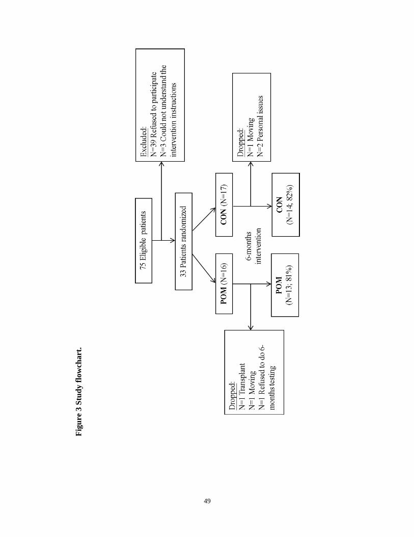
Thirty-three of the 75 patients that were recruited agreed to participate in the study. These
patients were randomly assigned to 2 groups to receive either pomegranate extracts (POM) or
placebo (CON). Among the 33 enrolled subjects, 6 (POM: N=3; CON: N=3) dropped out of the
study, including 2 that moved to other cities, 1 that received a kidney transplant, 1 that refused to
complete the 6-month testing, and 2 that were unable to finish the study due to personal issues.
The dropout rate was 19% for the POM group, compared to 18% for the CON group (Figure 3).
Adverse events such as stomach upset or other GI-related effects were not observed among
subjects. Twenty-seven subjects finished the study, and their 6-month compliance to the study
intervention (% of pills consumed) was 95.9±1.0% and 98.2±0.6% in POM and CON,
respectively (p = 0.07).
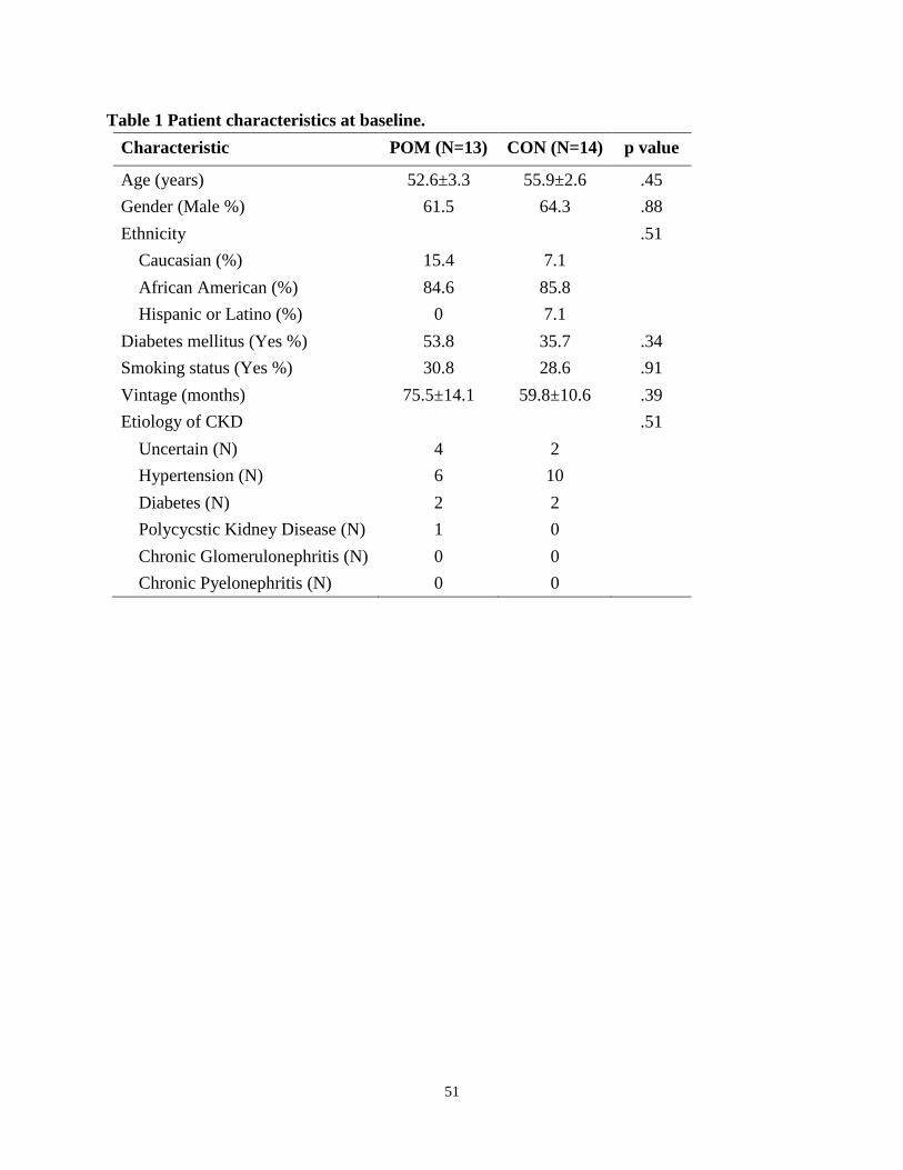
As shown in Table 1, general patient characteristics in the POM and CON groups were
similar at baseline. There were no significant differences between groups in gender, diabetes or

46
smoking status. However, baseline systolic and diastolic blood pressures were both significantly
higher in POM, compared to CON.
4.2 Cardiovascular Outcomes
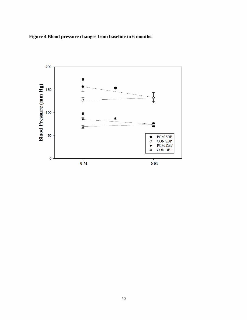
There was a significant interaction between group and time for systolic and diastolic blood
pressure, as they decreased by 12.8±0.1% (F1, 25 = 6.00; p < 0.05) and 11.2±0.1% (F1, 25 = 7.80; p
< 0.05), respectively, in POM, but did not change in CON (Figure 4). However, the changes in
blood pressure in the POM group were no longer significant after controlling for subject’s
baseline blood pressure. There were no interactive or main effects of group and time on heart
rate, CIMT, PWV, -stiffness, augmentation index, stroke volume, cardiac output, or systemic
vascular resistance (Table 2).
4.3 Physical Performance Outcomes
There were no interactive or main effects of activity group and time on gait speed, shuttle
walk time, functional fitness tests, or muscle strength (Table 2).
4.4 Markers of Inflammation and Oxidative Stress

47
There were no interactive or main effects of activity group and time on CRP, IL-6 8-OHdG,
AOPP, ox-LDL, ORAC, arylesterase, or paraoxonase (Table 2). There was a significant
interaction between group and time for lactonase activity, as it increased by 5.4±1.7 kU/L, or
26.6%, (F1, 25 = 4.58, p < 0.05) in POM, compared to no change in CON .
The change in total cholesterol was positively correlated with the change of ox-LDL (r
=0.49, p = 0.01) and the change in AOPP (r = 0.42, p = 0.03). The change in ox-LDL was
inversely correlated with the change in ORAC (r = -.38, p = 0.04), and positively correlated with
the change in 8-OHdG (r = 0.50, p = 0.01). No other significant correlations between major
outcome variables were found.

48
Figure Legends
Figure 3 Study flowchart. Thirty-three patients were recruited and randomly assigned to either
pomegranate extracts (POM) or placebo (CON). Six patients (POM: N=3; CON: N=3) dropped
out of the study. The dropout rate was 19% for the POM group, compared to 18% for the CON
group. Twenty-seven subjects finished the 6-month intervention.
Figure 4 Blood pressure changes from baseline to 6 months. Patients in both the POM and
CON groups had blood pressure measured at baseline and 6-month testing. SBP and DBP both
decreased from baseline to final testing in the POM group (p< 0.05), but did not change in the
CON group. 0M and 6M indicate baseline and 6-month testing time points, respectively. Values
are given as mean±S.E. Mean; Data was analyzed by 2-way ANOVA. * indicates p<0.05 for
group x time interaction effect. # indicates p < 0.05 for baseline levels between groups.
49
Fig
ure
3 S
tud
y f
low
chart
.
50
Figure 4 Blood pressure changes from baseline to 6 months.
51
Table 1 Patient characteristics at baseline.
Characteristic POM (N=13) CON (N=14) p value
Age (years) 52.6±3.3 55.9±2.6 .45
Gender (Male %) 61.5 64.3 .88
Ethnicity .51
Caucasian (%) 15.4 7.1
African American (%) 84.6 85.8
Hispanic or Latino (%) 0 7.1
Diabetes mellitus (Yes %) 53.8 35.7 .34
Smoking status (Yes %) 30.8 28.6 .91
Vintage (months) 75.5±14.1 59.8±10.6 .39
Etiology of CKD .51
Uncertain (N) 4 2
Hypertension (N) 6 10
Diabetes (N) 2 2
Polycycstic Kidney Disease (N) 1 0
Chronic Glomerulonephritis (N) 0 0
Chronic Pyelonephritis (N) 0 0
52
Table 2 Outcome variables at baseline and final testing.
Outcomes POM (N=13) CON (N=14)
N 0M 6M N 0M 6M
Weight (kg) 12 96.6±8.0 89.9±6.4 14 87.7±8.0 88.7±7.6
Height (cm) 11 167.3±2.9 167.0±2.7 14 167.3±2.1 167.4±2.0
BMI (kg/m2) 11 32.3±2.1 31.7±2.1 14 31.1±2.5 31.5±2.4
Body fat percentage (%) 11 29.1±3.1 28.9±3.1 13 29.6±2.6 29.5±2.5
Waist circumference (cm) 13 110.0±5.7 107.3±5.1 14 105.7±6.5 106.9±6.5
Albumin (g/dl) 13 4.0±0.1 4.0±0.1 14 3.9±0.1 4.0±0.1
Kt/V 6 1.6±0.1 1.6±0.1 11 1.6±0.1 1.6±0.1
Cardiovascular outcomes
SBP (mmHg) 13 156.8±10.1# 132.7±11.5* 14 127.1±6.1 132.6±8.2
DBP (mmHg) 13 85.8±4.0# 74.8±4.4* 14 69.2±3.3 73.5±3.7
Mean arterial pressure (mmHg) 13 109.3±5.6# 96.9±5.7* 14 88.4±3.8 93.4±4.6
Heart rate (beats/min) 13 72.9±3.3 68.0±8.2 14 69.3±3.6 68.6±2.9
PWV (m/s) 13 10.2±1.2 11.5±1.6 11 8.6±0.6 10.4±1.4
CIMT (mm) 12 0.54±0.10 0.76±0.04 14 0.70±0.10 0.74±0.06
β-stiffness 12 12.0±2.1 11.3±1.7 12 10.4±1.7 11.6±1.6
Augmentation index (%) 10 32.0±2.1 34.1±5.6 14 23.6±2.2 21.1±3.6
Stroke volume (ml/beat) 10 52.5±4.4 57.5±6.0 12 53.9±4.0 51.0±6.2
Cardiac output (L/min) 10 3.9±0.4 4.3±0.4 12 3.4±0.2 3.4±0.4
Systemic vascular resistance
(mmHg/L/min) 10 30.1±3.6 24.7±2.4 12 27.5±2.2 33.6±5.8

53
Table 2 (cont.)
Outcomes POM (N=13) CON (N=14)
Physical performance outcomes N 0M 6M N 0M 6M
Walk speed (sec) 12 12.4±1.1 13.4±1.2 14 12.0±1.2 11.7±0.9
Shuttle walk time (sec) 12 234.8±44.5 207.8±40.8 14 268.3±33.3 262.7±31.1
Peak torque RKF (ft*lb) 12 39.2±4.7 43.5±7.7 14 34.8±3.7 36.0±5.4
Peak torque RKE (ft*lb) 12 67.6±8.7 75.6±11.4 14 64.5±6.6 76.3±10.6
Peak torque LKF (ft*lb) 12 37.3±5.7 40.9±5.8 14 31.4±4.1 37.0±5.3
Peak torque LKE (ft*lb) 12 69.7±9.8 70.0±9.0 14 65.8±7.9 79.6±12.1
Chair stand (repetitions) 12 11.3±2.4 9.5±1.9 13 10.5±1.2 10.2±1.1
Arm curl (repetitions) 12 14.4±2.3 13.7±2.6 14 14.6±1.3 14.4±1.1
Sit and reach (inches) 12 -4.1±1.0 -3.3±0.9 13 -2.9±1.4 -2.5±1.3
Back scratch (inches) 13 -5.8±1.7 -4.6±1.3 14 -4.9±1.3 -5.7±1.2
8-foot up and go (seconds) 12 9.4±1.6 10.3±2.2 14 8.3±1.4 7.6±0.8
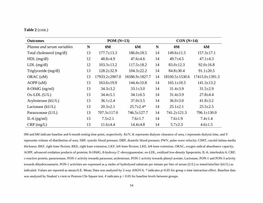
54
Table 2 (cont.)
0M and 6M indicate baseline and 6-month testing time point, respectively. Kt/V, K represents dialyzer clearance of urea, t represents dialysis time, and V
represents volume of distribution of urea. SBP, systolic blood pressure; DBP, diastolic blood pressure; PWV, pulse wave velocity; CIMT, carotid intima-media
thickness; RKF, right knee flexion; RKE, right knee extension; LKF, left knee flexion; LKE, left knee extension; ORAC, oxygen radical absorbance capacity;
AOPP, advanced oxidation products of proteins; 8-OHdG, 8-hydroxy-2’-deoxyguanosine; ox-LDL, oxidized low-density lipoprotein; IL-6, interleukin 6; CRP,
c-reactive protein; paraoxonase, PON-1 activity towards paraoxon; arylesterase, PON-1 activity towards phenyl acetate; Lactonase, PON-1 and PON-3 activity
towards dihydrocoumarin. PON-1 activities are expressed as µ moles of hydrolyzed substrate per minute per liter of serum (U/L) or mmol/min/liter (kU/L) as
indicated. Values are reported as mean±S.E. Mean; Data was analyzed by 2-way ANOVA. * indicates p<0.05 for group x time interaction effect. Baseline data
was analyzed by Student’s t-test or Pearson Chi-Square test. # indicates p < 0.05 for baseline levels between groups.
Outcomes POM (N=13) CON (N=14)
Plasma and serum variables N 0M 6M N 0M 6M
Total cholesterol (mg/dl) 13 177.7±13.3 186.0±18.5 14 149.6±11.5 157.3±17.1
HDL (mg/dl) 12 48.8±4.9 47.6±4.6 14 49.7±4.5 47.1±4.3
LDL (mg/dl) 12 103.3±13.2 117.5±18.2 14 83.0±12.3 92.0±16.8
Triglyceride (mg/dl) 13 128.2±32.9 104.3±22.2 14 84.8±30.4 91.1±20.5
ORAC (uM) 13 17933.2±2907.0 16586.9±1827.7 14 18100.5±1530.6 17415.0±1391.3
AOPP (uM) 13 163.6±19.9 144.4±10.8 14 165.1±10.5 141.3±13.2
8-OHdG (ng/ml) 13 34.3±3.2 33.1±3.0 14 31.4±3.9 31.5±2.9
Ox-LDL (U/L) 13 34.4±5.1 34.1±6.5 14 31.4±3.9 27.8±4.4
Arylesterase (kU/L) 13 36.1±2.4 37.0±3.5 14 36.0±3.0 41.8±3.2
Lactonase (kU/L) 13 20.3±2.1 25.7±2.4* 14 25.1±2.1 25.5±2.5
Paraoxonase (U/L) 13 707.3±117.0 746.5±127.7 14 741.2±121.3 796.1±130.0
IL-6 (pg/ml) 13 7.5±2.1 7.6±1.7 14 7.6±1.9 7.4±1.4
CRP (mg/L) 13 11.6±4.4 14.4±4.8 14 5.7±2.3 4.6±1.5

55
CHAPTER 5
DISCUSSION
The present study is the first study to investigate the effect of pomegranate extract on
cardiovascular health and physical function in patients with chronic kidney failure. Our primary
finding was that both systolic and diastolic blood pressure was significantly reduced in patients
after 6 months of pomegranate supplementation. However, because baseline blood pressures
were higher in the POM group, it cannot be ruled out that these changes may have been due to a
regression to the mean. Pomegranate supplementation had no effect on any other metrics of
cardiovascular risk or physical function in our patients. Taken together, this data suggests that
pomegranate extract supplementation may provide limited benefits in terms of reducing the
development or progression of co-morbidities in patients with chronic renal failure.
Oxidative stress is believed to affect blood pressure through a variety of mechanisms149, 150
.
Increased oxidative stress contributes to vascular dysfunction by reducing the bioavailability of
nitric oxide (NO), which is a major vasodilator (reviewed in151
). Oxidation of L-arginine and
tetrahydrobiopterin (BH4), which are two essential cofactors for endothelial NO synthase
(eNOS), results in the decreased formation of NO. In addition, oxidative stress in vascular smooth

56
muscle cells (VSMCs) can cause cell proliferation or apoptosis, and further contributes to changes
in blood vessel diameter and blood pressure152
. Antioxidants have been shown to improve
vascular dysfunction induced by excessive oxidative stress151
. Antioxidant supplementation, such
as vitamin C or E, has shown to protect against endothelial damage by upregulating eNOs and
scavenging reactive oxygen species153
. It is well-established that polyphenolic antioxidants, such
as red wine and pomegranate, can function as ROS scavengers, enhance the production and
bioavailability of NO, thereby improving vascular resistance and possibly may respond to changes
in blood pressure16, 105, 154-157
. Though not statistically significant, there was a trend for a
reduction in TPR in the POM group (p = 0.11 for group x time interaction), suggesting that
changes in vascular resistance may have partially contributed to the BP reduction in POM. In
addition, several studies have demonstrated reductions in blood pressure following pomegranate
juice consumption in hypertensive subjects and in patients with carotid artery stenosis16, 158, 159
,
so there is precedent for the blood pressure reductions that we found following pomegranate
extract supplementation in our patients. This also suggests that the blood pressure changes we
found may not have been solely due to a regression to the mean. Aviram et al. identified a
potential mechanism for the blood pressure lowering effect of pomegranate, suggesting that it
directly interacts with serum angiotensin converting enzyme (ACE). They incubated human
serum with different concentrations of pomegranate juice, and found a pomegranate juice

57
dose-dependent inhibitory effect on serum ACE activity159
. However, they found no correlation
between the reduction in ACE activity and BP, suggesting the reduction in ACE activity may not
be the major factor contributing to pomegranate-induced BP changes.
Research also suggests that pomegranate provides additional cardiovascular benefits besides
reducing blood pressure. CIMT has been used as a validated surrogate end-point for
atherosclerosis and vascular disease risk19, 160
. Aviram et al. found a 30% reduction in CIMT
among subjects with severe carotid artery stenosis after 1 year of pomegranate juice
consumption16
. Pomegranate juice was also reported to decrease the CIMT progression rates in
subjects at moderate risk for coronary heart disease123
, and improved a composite score of CIMT
in hemodialysis patients19
. Despite these previous studies showing beneficial effects of
pomegranate juice on CIMT in clinical populations, the present study did not find CIMT
improvements with 6 months of pomegranate extract consumption. This discrepancy in the
CIMT findings might be due to different forms of supplementation (i.e., powder extracts versus
juice), as well as a shorter period of intervention (i.e., 6 months versus more than 1 year) in the
present study. Although Aviram et al.16
found a reduction in CIMT as soon as 3 months of
pomegranate juice consumption in subjects with severe carotid stenosis, our dialysis patients had
relatively thinner CIMT at baseline (0.54mm CIMT in our study versus 1.52mm CIMT in

58
Aviram et al.), which might be another potential reason that we did not see changes in CIMT
after 6 months of pomegranate extract supplementation.
Our findings are contrary to prior studies suggesting antioxidant supplementation, such as
vitamin C and/or E, would reduce vascular disease risk by decreasing arterial stiffness in subjects
with diabetes or hypertension161, 162
. However, Dohadwala et al. showed that PWV did not
change after a 4-week intervention with cranberry juice, which contains abundant polyphenolic
compounds, in patients with coronary artery disease163
. Although there is little evidence of
reducing arterial stiffness by pomegranate supplements in clinical populations, consumption of
pomegranate juice for 4 weeks had no effect on PWV in healthy subjects164
. Longer clinical trials
are needed to investigate the effects of polyphenolic compounds on arterial stiffness to confirm
these findings.
Although studies have shown that low plasma antioxidant levels (e.g., tocopherol,
carotenoids) are associated with poor skeletal muscle strength and impaired physical
performance in older adults112, 165, 166
, to our knowledge, only a few studies have directly
investigated the effects of antioxidant supplementation on these metrics. A recent study by Hauer
et al. found that N-acetylcysteine (NAC) combined with resistance exercise training improved
muscle strength in the elderly after 13 weeks13
. However, these data must be interpreted with
caution because plasma antioxidant levels were not reported to confirm the relationship between

59
changes of antioxidant level and improved muscle performance. In addition, the improvements in
physical performance and muscular strength were not only observed in the group with NAC
supplementation combined with resistance exercise, but also in the group with placebo combined
with resistance training. These results suggested the functional improvements may be due to
resistance training response or learning effects, instead of antioxidant benefits solely.
Pomegranate juice has been used as an antioxidant supplement to improve recovery of
elbow flexor strength, as well as to attenuate soreness, after a single damaging bout of eccentric
exercise in healthy subjects14, 15
. However, our data did not show that pomegranate
supplementation chronically improves physical function or muscle strength in dialysis patients.
This inconsistency may be due to the different populations and functional measures that were
assessed, or the fact that we assessed chronic changes in function, as opposed to recovery from a
single bout of exercise.
Paraoxonase-1 (PON1) is the most potent HDL-associated antioxidant enzyme, and uremic
toxins may play a mechanistic role in PON1 inactivation167
. Past studies have reported widely
variable PON-1 activity in hemodialysis patients137, 168-170
, which may be explained in part by
differences in subject’s BMI, nutritional status, CRP levels, vintages of hemodialysis, or
differences in assay methods used in each study170
. However, we did not find any relationship
between PON-1 activity and patient’s BMI, dialysis vintages, albumin or CRP levels.

60
Pomegranate juice has shown to increase the paraoxonase, arylesterase and lactonase
activities of PON-1123, 131
. PON-1 hydrolyzes organophosphate compounds (e.g., paraoxon) and
aromatic carboxylic acid esters (e.g., phenyl acetate), and possesses peroxidase-like activity that
can contribute to its protective effect against lipoprotein oxidation135, 171
. PON-1 also provides its
anti-atherogenic properties through a homocysteine-thiolactonase activity134, 172
. Among
arylesterase, paraoxonase and lactonase, we only found an increase in lactonase activity in the
POM group. Although these results differ from some published studies showing that PON-1
activity toward other substrates improved after pomegranate juice consumption124, 136
, they are
consistent with the recently accumulating evidence suggesting that the lactonase activity is the
most significant explanation for PON-1’s atheroprotective properties137, 173-175
.
PON-1 activity largely depends on its association with HDL. However, we did not find an
association between the change in HDL cholesterol concentration and change of PON-1
activities in the present study, suggesting that the change in lactonase activity is not related
solely to changes in HDL concentration in hemodialysis patients. This finding is in agreement
with those by Gugliucci et al., which showed no correlation between lactonase activity and HDL
levels in CKD patients, and might be due to the inhibition of enzyme catalysis and synthesis by
uremic toxins137
.

61
Punicalagin, an ellagitannin found in pomegranate, accounts for 70-89% of the antioxidant
activity of pomegranate juice116, 176
, and studies have shown that pomegranate juice and extracts
have similar bioavailabilities. Our 1,000-mg pomegranate extract contains approximately 755 mg
of gallic acid equivalents177
. This is similar to a typical 8-ounce serving of pomegranate juice,
which contains 857 mg of gallic acid equivalents. As a result, we chose to use the pomegranate
extract in this study, as opposed to the juice, due to the high content of potassium in pomegranate
juice, and restrictions on the intake of these minerals in dialysis patients.
Despite very similar antioxidant contents in juice and extracts, prior work on ellagitannin
absorption yielded variable results. Seeram et al. showed that ellagic acid concentration in
plasma reached its peak concentration 2-3 hours after consuming pomegranate extract, compared
to a peak plasma ellagic acid concentration at 1 hour following consumption of pomegranate
juice177
. By contrast, Cerda et al. found that neither punicalagin nor ellagic acid were detected in
plasma after consuming 1 liter of pomegranate juice per day for 5 days178
. The different
absorption rates between pomegranate juice and extracts may contribute to the inconsistent
findings from the current study, compared to the previous work published by Shema-Didi et al.19
,
who reported that pomegranate juice provided anti-inflammatory and cardioprotective benefits in
hemodialysis patients.

62
A potential explanation for why we observed no POM-induced change in circulating
markers of inflammation or oxidative stress in the present study may be due to the abnormal
distribution of microflora in hemodialysis patients. Cerda et al. suggested that the poor
absorption of ellagic acid was explained by its poor solubility at physiological pH in the
intestine, and its poor ability to bind to intestinal epithelium179, 180
. Cerda also suggested that the
cardiovascular protective effects of pomegranate might be due to metabolites produced from
pomegranate digestion by intestinal microflora, such as urolithins, rather than to the polyphenolic
concentration present in the pomegranate products178
. Excessive uremic toxins, CKD-related
comorbidities, medications, and irregular dietary patterns, which are all conditions commonly
seen in hemodialysis patients, may cause an abnormal distribution of microflora in the
gastrointestinal tract and change of gastrointestinal permeability181
. Indeed, we found no
difference in ORAC, a measure of serum total antioxidant capacity, between groups at 1.5 and 3
hours after pomegranate extract or placebo ingestion (data not shown). The abnormal distribution
of microflora in CKD patients might result in a reduction in antioxidant absorption, thereby
attenuating the potential antioxidant, and anti-inflammatory effects of pomegranate in
hemodialysis patients. Further research is needed to investigate the concentration of ellagic acid
in plasma after ingesting pomegranate products to determine the absorption rate of antioxidant in
patients undergoing hemodialysis treatment.
63
Limitations
There are several limitations to our study. Primarily, the sample size was relatively limited,
thereby reducing the statistical power for the analysis. This also inhibited us from controlling for
subjects’ medication status and intercurrent illness, both of which may have affected our primary
outcomes. In addition, we did not measure plasma levels of ellagic acid to confirm whether the
subjects were actually taking pills during the intervention.
64
CHAPTER 6
CONCLUSIONS
Chronic kidney disease patients undergoing hemodialysis therapy suffer from a variety of
co-morbidities that may be mechanistically linked, such as reduced exercise capacity, increased
cardiovascular risk, and elevated inflammation and oxidative stress. These co-morbidities not
only contribute to the low quality of life, but also increase the mortality rate in this population.
Because current pharmacological therapies have failed to significantly reduce uremic-related
co-morbidities in hemodialysis patients, the present study was designed to determine the effects
of pomegranate supplementation, a rich source of polyphenol antioxidants, on CVD risk,
physical performance, inflammation and oxidative stress. The present study suggests modest
benefits of 6-months pomegranate extract supplementation in patients undergoing hemodialysis,
including (1) possible reductions in blood pressure, and (2) an increase in serum antioxidant
activity. However, pomegranate extract supplementation had no effect on other markers of
cardiovascular risk, or muscle strength and function. Though past studies demonstrated relatively
robust cardioprotective effects of pomegranate juice consumption in various clinical populations,

65
including hemodialysis patients16, 19, 123, 125, 158, 159
, our data indicates that the benefits of
pomegranate extract supplementation may be limited in hemodialysis patients.

66
REFERENCES
1. U.S. Renal Data System USRDS Annual Data Report: atlas of chronic kidney disease and
end-stage renal disease in the United States. National Institutes of Health, National
Institute of Diabetes and Digestive and Kidney Diseases, Bethesda, MD. 2010.
2. Muntner P, He J, Hamm L, Loria C, Whelton PK. Renal insufficiency and subsequent
death resulting from cardiovascular disease in the United States. J Am Soc Nephrol. Mar
2002;13(3):745-753.
3. K/DOQI clinical practice guidelines for chronic kidney disease: evaluation, classification,
and stratification. Am J Kidney Dis. Feb 2002;39(2 Suppl 1):S1-266.
4. Sarnak MJ, Levey AS, Schoolwerth AC, Coresh J, Culleton B, Hamm LL, McCullough
PA, Kasiske BL, Kelepouris E, Klag MJ, Parfrey P, Pfeffer M, Raij L, Spinosa DJ,
Wilson PW. Kidney disease as a risk factor for development of cardiovascular disease: a
statement from the American Heart Association Councils on Kidney in Cardiovascular
Disease, High Blood Pressure Research, Clinical Cardiology, and Epidemiology and
Prevention. Circulation. Oct 28 2003;108(17):2154-2169.
5. Bergstrom J. Nutrition and mortality in hemodialysis. J Am Soc Nephrol. Nov
1995;6(5):1329-1341.
6. Caglar K, Hakim RM, Ikizler TA. Approaches to the reversal of malnutrition,
inflammation, and atherosclerosis in end-stage renal disease. Nutr Rev. Nov
2002;60(11):378-387.
7. Cachofeiro V, Goicochea M, de Vinuesa SG, Oubina P, Lahera V, Luno J. Oxidative
stress and inflammation, a link between chronic kidney disease and cardiovascular
disease. Kidney International. Dec 2008(111):S4-9.
8. Himmelfarb J. Oxidative stress in hemodialysis. Contrib Nephrol. 2008;161:132-137.
9. Bergstrom J, Lindholm B, Lacson E, Jr., Owen W, Jr., Lowrie EG, Glassock RJ, Ikizler
TA, Wessels FJ, Moldawer LL, Wanner C, Zimmermann J. What are the causes and
consequences of the chronic inflammatory state in chronic dialysis patients? Semin Dial.
May-Jun 2000;13(3):163-175.
10. Bjelakovic G, Nikolova D, Gluud LL, Simonetti RG, Gluud C. Antioxidant supplements
for prevention of mortality in healthy participants and patients with various diseases.
Cochrane Database Syst Rev. 2008(2):CD007176.
11. Boaz M, Smetana S, Weinstein T, Matas Z, Gafter U, Iaina A, Knecht A, Weissgarten Y,
Brunner D, Fainaru M, Green MS. Secondary prevention with antioxidants of

67
cardiovascular disease in endstage renal disease (SPACE): randomised
placebo-controlled trial. Lancet. Oct 7 2000;356(9237):1213-1218.
12. Tepel M, van der Giet M, Statz M, Jankowski J, Zidek W. The antioxidant acetylcysteine
reduces cardiovascular events in patients with end-stage renal failure: a randomized,
controlled trial. Circulation. Feb 25 2003;107(7):992-995.
13. Hauer K, Hildebrandt W, Sehl Y, Edler L, Oster P, Droge W. Improvement in muscular
performance and decrease in tumor necrosis factor level in old age after antioxidant
treatment. Journal of Molecular Medicine (Berlin, Germany). Feb 2003;81(2):118-125.
14. Trombold JR, Reinfeld AS, Casler JR, Coyle EF. The effect of pomegranate juice
supplementation on strength and soreness after eccentric exercise. Journal of Strength
and Conditioning Research / National Strength & Conditioning Association.
Jul;25(7):1782-1788.
15. Trombold JR, Barnes JN, Critchley L, Coyle EF. Ellagitannin Consumption Improves
Strength Recovery 2-3 Days after Eccentric Exercise. Med Sci Sports Exerc. Nov 23
2009.
16. Aviram M, Rosenblat M, Gaitini D, Nitecki S, Hoffman A, Dornfeld L, Volkova N,
Presser D, Attias J, Liker H, Hayek T. Pomegranate juice consumption for 3 years by
patients with carotid artery stenosis reduces common carotid intima-media thickness,
blood pressure and LDL oxidation. Clin Nutr. Jun 2004;23(3):423-433.
17. Rasheed Z, Akhtar N, Anbazhagan AN, Ramamurthy S, Shukla M, Haqqi TM.
Polyphenol-rich pomegranate fruit extract (POMx) suppresses PMACI-induced
expression of pro-inflammatory cytokines by inhibiting the activation of MAP Kinases
and NF-kappaB in human KU812 cells. J Inflamm (Lond). 2009;6:1.
18. de Nigris F, Balestrieri ML, Williams-Ignarro S, D'Armiento FP, Fiorito C, Ignarro LJ,
Napoli C. The influence of pomegranate fruit extract in comparison to regular
pomegranate juice and seed oil on nitric oxide and arterial function in obese Zucker rats.
Nitric Oxide. Aug 2007;17(1):50-54.
19. Shema-Didi L, Sela S, Ore L, Shapiro G, Geron R, Moshe G, Kristal B. One year of
pomegranate juice intake decreases oxidative stress, inflammation, and incidence of
infections in hemodialysis patients: a randomized placebo-controlled trial. Free Radic
Biol Med. Jul 15;53(2):297-304.
20. USRDS. USRDS 2006 Annual Data Report: atlas of end-stage renal disease in the United
States. National Institutes of Health, National Institue of Diabetes and Digestive and
Kidney Diseases; Bethesda, MD. 2007.

68
21. Tonelli M, Wiebe N, Culleton B, House A, Rabbat C, Fok M, McAlister F, Garg AX.
Chronic kidney disease and mortality risk: a systematic review. J Am Soc Nephrol. Jul
2006;17(7):2034-2047.
22. Goodman WG, London G, Amann K, Block GA, Giachelli C, Hruska KA, Ketteler M,
Levin A, Massy Z, McCarron DA, Raggi P, Shanahan CM, Yorioka N. Vascular
calcification in chronic kidney disease. American Journal of Kidney Diseases. Mar
2004;43(3):572-579.
23. Hujairi NM, Afzali B, Goldsmith DJ. Cardiac calcification in renal patients: what we do
and don't know. American Journal of Kidney Diseases. Feb 2004;43(2):234-243.
24. Moe SM, Chen NX. Pathophysiology of vascular calcification in chronic kidney disease.
Circ Res. Sep 17 2004;95(6):560-567.
25. Guerin AP, London GM, Marchais SJ, Metivier F. Arterial stiffening and vascular
calcifications in end-stage renal disease. Nephrol Dial Transplant. Jul
2000;15(7):1014-1021.
26. London GM, Guerin AP, Marchais SJ, Pannier B, Safar ME, Day M, Metivier F. Cardiac
and arterial interactions in end-stage renal disease. Kidney International. Aug
1996;50(2):600-608.
27. London GM. Left ventricular alterations and end-stage renal disease. Nephrol Dial
Transplant. 2002;17 Suppl 1:29-36.
28. Matsumoto Y, Hamada M, Hiwada K. Aortic distensibility is closely related to the
progression of left ventricular hypertrophy in patients receiving hemodialysis. Angiology.
Nov 2000;51(11):933-941.
29. London GM, Blacher J, Pannier B, Guerin AP, Marchais SJ, Safar ME. Arterial wave
reflections and survival in end-stage renal failure. Hypertension. Sep
2001;38(3):434-438.
30. Toussaint ND, Kerr PG. Vascular calcification and arterial stiffness in chronic kidney
disease: implications and management. Nephrology (Carlton, Vic. Oct
2007;12(5):500-509.
31. London G, Guerin A, Pannier B, Marchais S, Benetos A, Safar M. Increased systolic
pressure in chronic uremia: role of arterial wave reflections. Hypertension. Jul
1992;20(1):10-19.
32. London GM. Cardiovascular calcifications in uremic patients: clinical impact on
cardiovascular function. J Am Soc Nephrol. Sep 2003;14(9 Suppl 4):S305-309.
33. Kaisar MO, Isbel NM, Johnson DW. Recent clinical trials of pharmacologic
cardiovascular interventions in patients with chronic kidney disease. Rev Recent Clin
Trials. May 2008;3(2):79-88.

69
34. Locatelli F, Canaud B, Eckardt KU, Stenvinkel P, Wanner C, Zoccali C. Oxidative stress
in end-stage renal disease: an emerging threat to patient outcome. Nephrol Dial
Transplant. Jul 2003;18(7):1272-1280.
35. Stenvinkel P. Malnutrition and chronic inflammation as risk factors for cardiovascular
disease in chronic renal failure. Blood Purification. 2001;19(2):143-151.
36. Stenvinkel P, Barany P, Heimburger O, Pecoits-Filho R, Lindholm B. Mortality,
malnutrition, and atherosclerosis in ESRD: what is the role of interleukin-6? Kidney Int
Suppl. May 2002(80):103-108.
37. Ikizler TA, Wingard RL, Harvell J, Shyr Y, Hakim RM. Association of morbidity with
markers of nutrition and inflammation in chronic hemodialysis patients: a prospective
study. Kidney Int. May 1999;55(5):1945-1951.
38. Johansen KL. Physical functioning and exercise capacity in patients on dialysis. Adv Ren
Replace Ther. Apr 1999;6(2):141-148.
39. Johansen KL, Shubert T, Doyle J, Soher B, Sakkas GK, Kent-Braun JA. Muscle atrophy
in patients receiving hemodialysis: effects on muscle strength, muscle quality, and
physical function. Kidney International. Jan 2003;63(1):291-297.
40. Du J, Hu Z, Mitch WE. Cellular signals activating muscle proteolysis in chronic kidney
disease: a two-stage process. The international journal of biochemistry & cell biology.
Oct 2005;37(10):2147-2155.
41. Mitch WE. Malnutrition: a frequent misdiagnosis for hemodialysis patients. The Journal
of clinical investigation. Aug 2002;110(4):437-439.
42. Graham KA, Reaich D, Channon SM, Downie S, Goodship TH. Correction of acidosis in
hemodialysis decreases whole-body protein degradation. J Am Soc Nephrol. Apr
1997;8(4):632-637.
43. Kraut JA, Kurtz I. Metabolic acidosis of CKD: diagnosis, clinical characteristics, and
treatment. Am J Kidney Dis. Jun 2005;45(6):978-993.
44. Mitch WE, Bailey JL, Wang X, Jurkovitz C, Newby D, Price SR. Evaluation of signals
activating ubiquitin-proteasome proteolysis in a model of muscle wasting. The American
journal of physiology. May 1999;276(5 Pt 1):C1132-1138.
45. Bailey JL, Wang X, England BK, Price SR, Ding X, Mitch WE. The acidosis of chronic
renal failure activates muscle proteolysis in rats by augmenting transcription of genes
encoding proteins of the ATP-dependent ubiquitin-proteasome pathway. The Journal of
Clinical Investigation. Mar 15 1996;97(6):1447-1453.
46. Price SR, Bailey JL, Wang X, Jurkovitz C, England BK, Ding X, Phillips LS, Mitch WE.
Muscle wasting in insulinopenic rats results from activation of the ATP-dependent,
70
ubiquitin-proteasome proteolytic pathway by a mechanism including gene transcription.
The Journal of Clinical Investigation. Oct 15 1996;98(8):1703-1708.
47. Lecker SH, Solomon V, Mitch WE, Goldberg AL. Muscle protein breakdown and the
critical role of the ubiquitin-proteasome pathway in normal and disease states. J Nutr. Jan
1999;129(1S Suppl):227S-237S.
48. Ikizler TA, Pupim LB, Brouillette JR, Levenhagen DK, Farmer K, Hakim RM, Flakoll PJ.
Hemodialysis stimulates muscle and whole body protein loss and alters substrate
oxidation. Am J Physiol Endocrinol Metab. Jan 2002;282(1):E107-116.
49. Gutierrez A, Alvestrand A, Wahren J, Bergstrom J. Effect of in vivo contact between
blood and dialysis membranes on protein catabolism in humans. Kidney Int. Sep
1990;38(3):487-494.
50. Goodman MN. Tumor necrosis factor induces skeletal muscle protein breakdown in rats.
The American journal of physiology. May 1991;260(5 Pt 1):E727-730.
51. Goodman MN. Interleukin-6 induces skeletal muscle protein breakdown in rats.
Proceedings of the Society for Experimental Biology and Medicine. Society for
Experimental Biology and Medicine (New York, N.Y. Feb 1994;205(2):182-185.
52. Tisdale MJ. The ubiquitin-proteasome pathway as a therapeutic target for muscle wasting.
The Journal of Supportive Oncology. May-Jun 2005;3(3):209-217.
53. Muscaritoli M, Molfino A, Bollea MR, Rossi Fanelli F. Malnutrition and wasting in renal
disease. Current Opinion in Clinical Nutrition and Metabolic Care. Jul
2009;12(4):378-383.
54. Ghiadoni L, Cupisti A, Huang Y, Mattei P, Cardinal H, Favilla S, Rindi P, Barsotti G,
Taddei S, Salvetti A. Endothelial dysfunction and oxidative stress in chronic renal failure.
J Nephrol. Jul-Aug 2004;17(4):512-519.
55. Annuk M, Soveri I, Zilmer M, Lind L, Hulthe J, Fellstrom B. Endothelial function, CRP
and oxidative stress in chronic kidney disease. J Nephrol. Nov-Dec 2005;18(6):721-726.
56. Oberg BP, McMenamin E, Lucas FL, McMonagle E, Morrow J, Ikizler TA, Himmelfarb
J. Increased prevalence of oxidant stress and inflammation in patients with moderate to
severe chronic kidney disease. Kidney Int. Mar 2004;65(3):1009-1016.
57. Li N, Karin M. Is NF-kappaB the sensor of oxidative stress? Faseb J. Jul
1999;13(10):1137-1143.
58. Roberts MA, Hare DL, Ratnaike S, Ierino FL. Cardiovascular biomarkers in CKD:
pathophysiology and implications for clinical management of cardiac disease. Am J
Kidney Dis. Sep 2006;48(3):341-360.
59. Stenvinkel P, Ketteler M, Johnson RJ, Lindholm B, Pecoits-Filho R, Riella M,
Heimburger O, Cederholm T, Girndt M. IL-10, IL-6, and TNF-alpha: central factors in

71
the altered cytokine network of uremia--the good, the bad, and the ugly. Kidney Int. Apr
2005;67(4):1216-1233.
60. Okamura DM, Himmelfarb J. Tipping the redox balance of oxidative stress in fibrogenic
pathways in chronic kidney disease. Pediatr Nephrol. Dec 2009;24(12):2309-2319.
61. Dounousi E, Papavasiliou E, Makedou A, Ioannou K, Katopodis KP, Tselepis A,
Siamopoulos KC, Tsakiris D. Oxidative stress is progressively enhanced with advancing
stages of CKD. Am J Kidney Dis. Nov 2006;48(5):752-760.
62. Terawaki H, Yoshimura K, Hasegawa T, Matsuyama Y, Negawa T, Yamada K,
Matsushima M, Nakayama M, Hosoya T, Era S. Oxidative stress is enhanced in
correlation with renal dysfunction: examination with the redox state of albumin. Kidney
Int. Nov 2004;66(5):1988-1993.
63. Cristol JP, Canaud B, Rabesandratana H, Gaillard I, Serre A, Mion C. Enhancement of
reactive oxygen species production and cell surface markers expression due to
haemodialysis. Nephrol Dial Transplant. 1994;9(4):389-394.
64. Jung HH, Choi DH, Lee SH. Serum malondialdehyde and coronary artery disease in
hemodialysis patients. American Journal of Nephrology. Sep-Oct 2004;24(5):537-542.
65. McCullough PA, Li S, Jurkovitz CT, Stevens L, Collins AJ, Chen SC, Norris KC,
McFarlane S, Johnson B, Shlipak MG, Obialo CI, Brown WW, Vassalotti J,
Whaley-Connell AT, Brenner RM, Bakris GL. Chronic kidney disease, prevalence of
premature cardiovascular disease, and relationship to short-term mortality. American
Heart Journal. Aug 2008;156(2):277-283.
66. Loughrey CM, Young IS, Lightbody JH, McMaster D, McNamee PT, Trimble ER.
Oxidative stress in haemodialysis. Qjm. Nov 1994;87(11):679-683.
67. Handelman GJ, Walter MF, Adhikarla R, Gross J, Dallal GE, Levin NW, Blumberg JB.
Elevated plasma F2-isoprostanes in patients on long-term hemodialysis. Kidney Int. May
2001;59(5):1960-1966.
68. Ikizler TA, Morrow JD, Roberts LJ, Evanson JA, Becker B, Hakim RM, Shyr Y,
Himmelfarb J. Plasma F2-isoprostane levels are elevated in chronic hemodialysis patients.
Clinical Nephrology. Sep 2002;58(3):190-197.
69. Witko-Sarsat V, Friedlander M, Capeillere-Blandin C, Nguyen-Khoa T, Nguyen AT,
Zingraff J, Jungers P, Descamps-Latscha B. Advanced oxidation protein products as a
novel marker of oxidative stress in uremia. Kidney Int. May 1996;49(5):1304-1313.
70. Miyata T, Ueda Y, Yamada Y, Izuhara Y, Wada T, Jadoul M, Saito A, Kurokawa K, van
Ypersele de Strihou C. Accumulation of carbonyls accelerates the formation of
pentosidine, an advanced glycation end product: carbonyl stress in uremia. J Am Soc
Nephrol. Dec 1998;9(12):2349-2356.
72
71. Witko-Sarsat V, Friedlander M, Nguyen Khoa T, Capeillere-Blandin C, Nguyen AT,
Canteloup S, Dayer JM, Jungers P, Drueke T, Descamps-Latscha B. Advanced oxidation
protein products as novel mediators of inflammation and monocyte activation in chronic
renal failure. J Immunol. Sep 1 1998;161(5):2524-2532.
72. Ames BN. Micronutrients prevent cancer and delay aging. Toxicology Letters. Dec 28
1998;102-103:5-18.
73. Ames BN, Shigenaga MK, Hagen TM. Oxidants, antioxidants, and the degenerative
diseases of aging. Proceedings of the National Academy of Sciences of the United States
of America. Sep 1 1993;90(17):7915-7922.
74. Tarng DC, Huang TP, Wei YH, Liu TY, Chen HW, Wen Chen T, Yang WC.
8-hydroxy-2'-deoxyguanosine of leukocyte DNA as a marker of oxidative stress in
chronic hemodialysis patients. Am J Kidney Dis. Nov 2000;36(5):934-944.
75. Kaya Y, Ari E, Demir H, Soylemez N, Cebi A, Alp H, Bakan E, Gecit I, Asicioglu E,
Beytur A. Accelerated atherosclerosis in haemodialysis patients; correlation of
endothelial function with oxidative DNA damage. Nephrol Dial Transplant. Aug 5.
76. Dobashi K, Ghosh B, Orak JK, Singh I, Singh AK. Kidney ischemia-reperfusion:
modulation of antioxidant defenses. Molecular and Cellular Biochemistry. Feb
2000;205(1-2):1-11.
77. Yilmaz MI, Saglam M, Caglar K, Cakir E, Sonmez A, Ozgurtas T, Aydin A, Eyileten T,
Ozcan O, Acikel C, Tasar M, Genctoy G, Erbil K, Vural A, Zoccali C. The determinants
of endothelial dysfunction in CKD: oxidative stress and asymmetric dimethylarginine.
Am J Kidney Dis. Jan 2006;47(1):42-50.
78. Dursun B, Dursun E, Capraz I, Ozben T, Apaydin A, Suleymanlar G. Are uremia,
diabetes, and atherosclerosis linked with impaired antioxidant mechanisms? J Investig
Med. Feb 2008;56(2):545-552.
79. Cristol JP, Bosc JY, Badiou S, Leblanc M, Lorrho R, Descomps B, Canaud B.
Erythropoietin and oxidative stress in haemodialysis: beneficial effects of vitamin E
supplementation. Nephrol Dial Transplant. Nov 1997;12(11):2312-2317.
80. Galli F, Canestrari F, Buoncristiani U. Biological effects of oxidant stress in
haemodialysis: the possible roles of vitamin E. Blood Purification. 1999;17(2-3):79-94.
81. Bohm V, Tiroke K, Schneider S, Sperschneider H, Stein G, Bitsch R. Vitamin C status of
patients with chronic renal failure, dialysis patients and patients after renal transplantation.
International Journal for Vitamin and Nutrition Research. Internationale Zeitschrift fur
Vitamin- und Ernahrungsforschung. 1997;67(4):262-266.
82. Salem M, Ivanovich P, Mujais S. Biocompatibility of dialysis membranes. Contributions
to Nephrology. 1993;103:55-64.

73
83. Ritz E, Deppisch R, Stier E, Hansch G. Atherogenesis and cardiac death: are they related
to dialysis procedure and biocompatibility? Nephrol Dial Transplant. 1994;9 Suppl
2:165-172.
84. Lazarus JM, Owen WF. Role of bioincompatibility in dialysis morbidity and mortality.
Am J Kidney Dis. Dec 1994;24(6):1019-1032.
85. Peuchant E, Carbonneau MA, Dubourg L, Thomas MJ, Perromat A, Vallot C, Clerc M.
Lipoperoxidation in plasma and red blood cells of patients undergoing haemodialysis:
vitamins A, E, and iron status. Free Radical Biology & Medicine. Mar
1994;16(3):339-346.
86. Jackson P, Loughrey CM, Lightbody JH, McNamee PT, Young IS. Effect of
hemodialysis on total antioxidant capacity and serum antioxidants in patients with
chronic renal failure. Clinical Chemistry. Aug 1995;41(8 Pt 1):1135-1138.
87. Daschner M, Lenhartz H, Botticher D, Schaefer F, Wollschlager M, Mehls O,
Leichsenring M. Influence of dialysis on plasma lipid peroxidation products and
antioxidant levels. Kidney Int. Oct 1996;50(4):1268-1272.
88. Hultqvist M, Hegbrant J, Nilsson-Thorell C, Lindholm T, Nilsson P, Linden T,
Hultqvist-Bengtsson U. Plasma concentrations of vitamin C, vitamin E and/or
malondialdehyde as markers of oxygen free radical production during hemodialysis.
Clinical Nephrology. Jan 1997;47(1):37-46.
89. Varan HI, Dursun B, Dursun E, Ozben T, Suleymanlar G. Acute effects of hemodialysis
on oxidative stress parameters in chronic uremic patients: comparison of two dialysis
membranes. International Journal of Nephrology and Renovascular Disease.3:39-45.
90. Descamps-Latscha B, Herbelin A, Nguyen AT, Roux-Lombard P, Zingraff J, Moynot A,
Verger C, Dahmane D, de Groote D, Jungers P, et al. Balance between IL-1 beta,
TNF-alpha, and their specific inhibitors in chronic renal failure and maintenance dialysis.
Relationships with activation markers of T cells, B cells, and monocytes. J Immunol. Jan
15 1995;154(2):882-892.
91. Raj DS, Boivin MA, Dominic EA, Boyd A, Roy PK, Rihani T, Tzamaloukas AH, Shah
VO, Moseley P. Haemodialysis induces mitochondrial dysfunction and apoptosis. Eur J
Clin Invest. Dec 2007;37(12):971-977.
92. Schulz T, Schiffl H, Scheithe R, Hrboticky N, Lorenz R. Preserved antioxidative defense
of lipoproteins in renal failure and during hemodialysis. Am J Kidney Dis. Apr
1995;25(4):564-571.
93. Tetta C, Biasioli S, Schiavon R, Inguaggiato P, David S, Panichi V, Wratten ML. An
overview of haemodialysis and oxidant stress. Blood Purification. 1999;17(2-3):118-126.

74
94. Dursun E, Dursun B, Suleymanlar G, Ozben T. Carbonyl stress in chronic renal failure:
the effect of haemodialysis. Annals of Clinical Biochemistry. Jan 2005;42(Pt 1):64-66.
95. Rysz J, Luciak M, Kedziora J, Blaszczyk J, Sibinska E. Nitric oxide release in the
peripheral blood during hemodialysis. Kidney Int. Jan 1997;51(1):294-300.
96. Roccatello D, Mengozzi G, Alfieri V, Pignone E, Menegatti E, Cavalli G, Cesano G,
Rossi D, Formica M, Inconis T, Martina G, Paradisi L, Sena LM, Piccoli G. Early
increase in blood nitric oxide, detected by electron paramagnetic resonance as
nitrosylhaemoglobin, in haemodialysis. Nephrol Dial Transplant. Feb
1997;12(2):292-297.
97. Amore A, Bonaudo R, Ghigo D, Arese M, Costamagna C, Cirina P, Gianoglio B,
Perugini L, Coppo R. Enhanced production of nitric oxide by blood-dialysis membrane
interaction. J Am Soc Nephrol. Oct 1995;6(4):1278-1283.
98. Caimi G, Carollo C, Montana M, Iatrino R, Bondi B, Lo Presti R. Nitric oxide
metabolites, leukocyte activation markers and oxidative status in dialyzed subjects. Blood
Purification. 2009;27(2):194-198.
99. Beckman JS, Koppenol WH. Nitric oxide, superoxide, and peroxynitrite: the good, the
bad, and ugly. The American Journal of Physiology. Nov 1996;271(5 Pt 1):C1424-1437.
100. Clermont G, Lecour S, Lahet J, Siohan P, Vergely C, Chevet D, Rifle G, Rochette L.
Alteration in plasma antioxidant capacities in chronic renal failure and hemodialysis
patients: a possible explanation for the increased cardiovascular risk in these patients.
Cardiovascular Research. Aug 18 2000;47(3):618-623.
101. Errakonda PR, Paladugu R, Bitla AR, Musturu SM, Lakshman J, Pemmaraju SR,
Vishnubhotla S. Effect of a single hemodialysis session on endothelial dysfunction. J
Nephrol. Jan-Feb;24(1):83-90.
102. MRC/BHF Heart Protection Study of antioxidant vitamin supplementation in 20,536
high-risk individuals: a randomised placebo-controlled trial. Lancet. Jul 6
2002;360(9326):23-33.
103. Tepel M, Echelmeyer M, Orie NN, Zidek W. Increased intracellular reactive oxygen
species in patients with end-stage renal failure: effect of hemodialysis. Kidney
International. Aug 2000;58(2):867-872.
104. Vaziri ND, Oveisi F, Ding Y. Role of increased oxygen free radical activity in the
pathogenesis of uremic hypertension. Kidney International. Jun 1998;53(6):1748-1754.
105. Rodrigo R, Bosco C. Oxidative stress and protective effects of polyphenols: comparative
studies in human and rodent kidney. A review. Comp Biochem Physiol C Toxicol
Pharmacol. Mar-Apr 2006;142(3-4):317-327.
75
106. Kals J, Kampus P, Kals M, Pulges A, Teesalu R, Zilmer K, Kullisaar T, Salum T, Eha J,
Zilmer M. Inflammation and oxidative stress are associated differently with endothelial
function and arterial stiffness in healthy subjects and in patients with atherosclerosis.
Scand J Clin Lab Invest. Apr 9 2008:1-8.
107. Pennathur S, Heinecke JW. Oxidative stress and endothelial dysfunction in vascular
disease. Curr Diab Rep. Aug 2007;7(4):257-264.
108. Lind L, Andersson J, Ronn M, Gustavsson T, Holdfelt P, Hulthe J, Elmgren A, Zilmer K,
Zilmer M. Brachial artery intima-media thickness and echogenicity in relation to lipids
and markers of oxidative stress in elderly subjects: the prospective investigation of the
vasculature in Uppsala Seniors (PIVUS) Study. Lipids. Feb 2008;43(2):133-141.
109. You H, Yang H, Zhu Q, Li M, Xue J, Gu Y, Lin S, Ding F. Advanced oxidation protein
products induce vascular calcification by promoting osteoblastic trans-differentiation of
smooth muscle cells via oxidative stress and ERK pathway. Ren Fail.
2009;31(4):313-319.
110. Kojda G, Harrison D. Interactions between NO and reactive oxygen species:
pathophysiological importance in atherosclerosis, hypertension, diabetes and heart failure.
Cardiovascular Research. Aug 15 1999;43(3):562-571.
111. Doughan AK, Harrison DG, Dikalov SI. Molecular mechanisms of angiotensin
II-mediated mitochondrial dysfunction: linking mitochondrial oxidative damage and
vascular endothelial dysfunction. Circulation Research. Feb 29 2008;102(4):488-496.
112. Semba RD, Blaum C, Guralnik JM, Moncrief DT, Ricks MO, Fried LP. Carotenoid and
vitamin E status are associated with indicators of sarcopenia among older women living
in the community. Aging Clinical and Experimental Research. Dec 2003;15(6):482-487.
113. Wijnen MH, Coolen SA, Vader HL, Reijenga JC, Huf FA, Roumen RM. Antioxidants
reduce oxidative stress in claudicants. The Journal of Surgical Research. Apr
2001;96(2):183-187.
114. Appell HJ, Duarte JA, Soares JM. Supplementation of vitamin E may attenuate skeletal
muscle immobilization atrophy. Int J Sports Med. Apr 1997;18(3):157-160.
115. Betters JL, Criswell DS, Shanely RA, Van Gammeren D, Falk D, Deruisseau KC,
Deering M, Yimlamai T, Powers SK. Trolox attenuates mechanical ventilation-induced
diaphragmatic dysfunction and proteolysis. American Journal of Respiratory and Critical
Care Medicine. Dec 1 2004;170(11):1179-1184.
116. Gil MI, Tomas-Barberan FA, Hess-Pierce B, Holcroft DM, Kader AA. Antioxidant
activity of pomegranate juice and its relationship with phenolic composition and
processing. J Agric Food Chem. Oct 2000;48(10):4581-4589.
76
117. Seeram NP, Aviram M, Zhang Y, Henning SM, Feng L, Dreher M, Heber D. Comparison
of antioxidant potency of commonly consumed polyphenol-rich beverages in the United
States. J Agric Food Chem. Feb 27 2008;56(4):1415-1422.
118. Priyadarsini KI, Khopde SM, Kumar SS, Mohan H. Free radical studies of ellagic acid, a
natural phenolic antioxidant. Journal of Agricultural and Food Chemistry. Mar 27
2002;50(7):2200-2206.
119. Rosenblat M, Aviram M. Paraoxonases role in the prevention of cardiovascular diseases.
Biofactors. Jan-Feb 2009;35(1):98-104.
120. de Nigris F, Williams-Ignarro S, Lerman LO, Crimi E, Botti C, Mansueto G, D'Armiento
FP, De Rosa G, Sica V, Ignarro LJ, Napoli C. Beneficial effects of pomegranate juice on
oxidation-sensitive genes and endothelial nitric oxide synthase activity at sites of
perturbed shear stress. Proceedings of the National Academy of Sciences of the United
States of America. Mar 29 2005;102(13):4896-4901.
121. de Nigris F, Williams-Ignarro S, Sica V, Lerman LO, D'Armiento FP, Byrns RE,
Casamassimi A, Carpentiero D, Schiano C, Sumi D, Fiorito C, Ignarro LJ, Napoli C.
Effects of a pomegranate fruit extract rich in punicalagin on oxidation-sensitive genes
and eNOS activity at sites of perturbed shear stress and atherogenesis. Cardiovascular
Research. Jan 15 2007;73(2):414-423.
122. Aviram M, Dornfeld L, Rosenblat M, Volkova N, Kaplan M, Coleman R, Hayek T,
Presser D, Fuhrman B. Pomegranate juice consumption reduces oxidative stress,
atherogenic modifications to LDL, and platelet aggregation: studies in humans and in
atherosclerotic apolipoprotein E-deficient mice. Am J Clin Nutr. May
2000;71(5):1062-1076.
123. Davidson MH, Maki KC, Dicklin MR, Feinstein SB, Witchger M, Bell M, McGuire DK,
Provost JC, Liker H, Aviram M. Effects of consumption of pomegranate juice on carotid
intima-media thickness in men and women at moderate risk for coronary heart disease.
Am J Cardiol. Oct 1 2009;104(7):936-942.
124. Rock W, Rosenblat M, Miller-Lotan R, Levy AP, Elias M, Aviram M. Consumption of
wonderful variety pomegranate juice and extract by diabetic patients increases
paraoxonase 1 association with high-density lipoprotein and stimulates its catalytic
activities. J Agric Food Chem. Sep 24 2008;56(18):8704-8713.
125. Rosenblat M, Hayek T, Aviram M. Anti-oxidative effects of pomegranate juice (PJ)
consumption by diabetic patients on serum and on macrophages. Atherosclerosis. Aug
2006;187(2):363-371.
126. Komperda KE. Potential interaction between pomegranate juice and warfarin.
Pharmacotherapy. Aug 2009;29(8):1002-1006.
77
127. Sorokin AV, Duncan B, Panetta R, Thompson PD. Rhabdomyolysis associated with
pomegranate juice consumption. Am J Cardiol. Sep 1 2006;98(5):705-706.
128. Hanley MJ, Masse G, Harmatz JS, Court MH, Greenblatt DJ. Pomegranate juice and
pomegranate extract do not impair oral clearance of flurbiprofen in human volunteers:
divergence from in vitro results. Clinical Pharmacology and Therapeutics.
Nov;92(5):651-657.
129. Chew GT, Watts GF. Coenzyme Q10 and diabetic endotheliopathy: oxidative stress and
the 'recoupling hypothesis'. Qjm. Aug 2004;97(8):537-548.
130. Nakayama K, Nakayama M, Iwabuchi M, Terawaki H, Sato T, Kohno M, Ito S. Plasma
alpha-oxoaldehyde levels in diabetic and nondiabetic chronic kidney disease patients.
American Journal of Nephrology. 2008;28(6):871-878.
131. Heber D, Seeram NP, Wyatt H, Henning SM, Zhang Y, Ogden LG, Dreher M, Hill JO.
Safety and antioxidant activity of a pomegranate ellagitannin-enriched polyphenol dietary
supplement in overweight individuals with increased waist size. Journal of Agricultural
and Food Chemistry. Nov 28 2007;55(24):10050-10054.
132. Aviram M. Interaction of oxidized low density lipoprotein with macrophages in
atherosclerosis, and the antiatherogenicity of antioxidants. Eur J Clin Chem Clin
Biochem. Aug 1996;34(8):599-608.
133. Prior RL, Hoang H, Gu L, Wu X, Bacchiocca M, Howard L, Hampsch-Woodill M,
Huang D, Ou B, Jacob R. Assays for hydrophilic and lipophilic antioxidant capacity
(oxygen radical absorbance capacity (ORAC(FL))) of plasma and other biological and
food samples. Journal of Agricultural and Food Chemistry. May 21
2003;51(11):3273-3279.
134. Aviram M, Rosenblat M. Paraoxonases 1, 2, and 3, oxidative stress, and macrophage
foam cell formation during atherosclerosis development. Free Radic Biol Med. Nov 1
2004;37(9):1304-1316.
135. Aviram M, Rosenblat M, Bisgaier CL, Newton RS, Primo-Parmo SL, La Du BN.
Paraoxonase inhibits high-density lipoprotein oxidation and preserves its functions. A
possible peroxidative role for paraoxonase. J Clin Invest. Apr 15 1998;101(8):1581-1590.
136. Rosenblat M, Volkova N, Attias J, Mahamid R, Aviram M. Consumption of
polyphenolic-rich beverages (mostly pomegranate and black currant juices) by healthy
subjects for a short term increased serum antioxidant status, and the serum's ability to
attenuate macrophage cholesterol accumulation. Food & Function. Oct;1(1):99-109.
137. Gugliucci A, Kinugasa E, Kotani K, Caccavello R, Kimura S. Serum paraoxonase 1
(PON1) lactonase activity is lower in end-stage renal disease patients than in healthy
control subjects and increases after hemodialysis. Clin Chem Lab Med. Jan;49(1):61-67.

78
138. Hernandez AF, Gil F, Leno E, Lopez O, Rodrigo L, Pla A. Interaction between human
serum esterases and environmental metal compounds. Neurotoxicology. Jul
2009;30(4):628-635.
139. Richter RJ, Jampsa RL, Jarvik GP, Costa LG, Furlong CE. Determination of paraoxonase
1 status and genotypes at specific polymorphic sites. Current Protocols in Toxicology.
May 2004;Chapter 4:Unit4 12.
140. Pauca AL, O'Rourke MF, Kon ND. Prospective evaluation of a method for estimating
ascending aortic pressure from the radial artery pressure waveform. Hypertension. Oct
2001;38(4):932-937.
141. Wilkinson IB, MacCallum H, Flint L, Cockcroft JR, Newby DE, Webb DJ. The influence
of heart rate on augmentation index and central arterial pressure in humans. J Physiol.
May 15 2000;525 Pt 1:263-270.
142. Van Bortel LM, Duprez D, Starmans-Kool MJ, Safar ME, Giannattasio C, Cockcroft J,
Kaiser DR, Thuillez C. Clinical applications of arterial stiffness, Task Force III:
recommendations for user procedures. Am J Hypertens. May 2002;15(5):445-452.
143. Wilkinson IB, Hall IR, MacCallum H, Mackenzie IS, McEniery CM, van der Arend BJ,
Shu YE, MacKay LS, Webb DJ, Cockcroft JR. Pulse-wave analysis: clinical evaluation
of a noninvasive, widely applicable method for assessing endothelial function.
Arterioscler Thromb Vasc Biol. Jan 2002;22(1):147-152.
144. Chiu YC, Arand PW, Shroff SG, Feldman T, Carroll JD. Determination of pulse wave
velocities with computerized algorithms. Am Heart J. May 1991;121(5):1460-1470.
145. Painter P, Carlson L, Carey S, Paul SM, Myll J. Physical functioning and health-related
quality-of-life changes with exercise training in hemodialysis patients. American Journal
of Kidney Diseases. Mar 2000;35(3):482-492.
146. Brown CD, Wise RA. Field tests of exercise in COPD: the six-minute walk test and the
shuttle walk test. Copd. Sep 2007;4(3):217-223.
147. Drouin JM, Valovich-mcLeod TC, Shultz SJ, Gansneder BM, Perrin DH. Reliability and
validity of the Biodex system 3 pro isokinetic dynamometer velocity, torque and position
measurements. European Journal of Applied Physiology. Jan 2004;91(1):22-29.
148. Panza JA, Epstein SE, Quyyumi AA. Circadian variation in vascular tone and its relation
to alpha-sympathetic vasoconstrictor activity. The New England Journal of Medicine. Oct
3 1991;325(14):986-990.
149. Rodrigo R, Passalacqua W, Araya J, Orellana M, Rivera G. Implications of oxidative
stress and homocysteine in the pathophysiology of essential hypertension. Journal of
Cardiovascular Pharmacology. Oct 2003;42(4):453-461.

79
150. Lassegue B, Griendling KK. Reactive oxygen species in hypertension; An update. Am J
Hypertens. Sep 2004;17(9):852-860.
151. Rodrigo R, Gonzalez J, Paoletto F. The role of oxidative stress in the pathophysiology of
hypertension. Hypertens Res. Apr;34(4):431-440.
152. Rao GN, Berk BC. Active oxygen species stimulate vascular smooth muscle cell growth
and proto-oncogene expression. Circ Res. Mar 1992;70(3):593-599.
153. Piccoli C, Quarato G, D'Aprile A, Montemurno E, Scrima R, Ripoli M, Gomaraschi M,
Cirillo P, Boffoli D, Calabresi L, Gesualdo L, Capitanio N. Native LDL-induced
oxidative stress in human proximal tubular cells: multiple players involved. Journal of
Cellular and Molecular Medicine. Feb;15(2):375-395.
154. Galley HF, Thornton J, Howdle PD, Walker BE, Webster NR. Combination oral
antioxidant supplementation reduces blood pressure. Clin Sci (Lond). Apr
1997;92(4):361-365.
155. Duarte J, Andriambeloson E, Diebolt M, Andriantsitohaina R. Wine polyphenols
stimulate superoxide anion production to promote calcium signaling and
endothelial-dependent vasodilatation. Physiological research / Academia Scientiarum
Bohemoslovaca. 2004;53(6):595-602.
156. Zenebe W, Pechanova O, Andriantsitohaina R. Red wine polyphenols induce
vasorelaxation by increased nitric oxide bioactivity. Physiological research / Academia
Scientiarum Bohemoslovaca. 2003;52(4):425-432.
157. Kitiyakara C, Wilcox CS. Antioxidants for hypertension. Curr Opin Nephrol Hypertens.
Sep 1998;7(5):531-538.
158. Asgary S, Sahebkar A, Afshani MR, Keshvari M, Haghjooyjavanmard S,
Rafieian-Kopaei M. Clinical Evaluation of Blood Pressure Lowering, Endothelial
Function Improving, Hypolipidemic and Anti-Inflammatory Effects of Pomegranate
Juice in Hypertensive Subjects. Phytother Res. Mar 21.
159. Aviram M, Dornfeld L. Pomegranate juice consumption inhibits serum angiotensin
converting enzyme activity and reduces systolic blood pressure. Atherosclerosis. Sep
2001;158(1):195-198.
160. Poredos P. Intima-media thickness: indicator of cardiovascular risk and measure of the
extent of atherosclerosis. Vascular Medicine (London, England). Feb 2004;9(1):46-54.
161. Mullan BA, Young IS, Fee H, McCance DR. Ascorbic acid reduces blood pressure and
arterial stiffness in type 2 diabetes. Hypertension. Dec 2002;40(6):804-809.
162. Plantinga Y, Ghiadoni L, Magagna A, Giannarelli C, Franzoni F, Taddei S, Salvetti A.
Supplementation with vitamins C and E improves arterial stiffness and endothelial
function in essential hypertensive patients. Am J Hypertens. Apr 2007;20(4):392-397.

80
163. Dohadwala MM, Holbrook M, Hamburg NM, Shenouda SM, Chung WB, Titas M, Kluge
MA, Wang N, Palmisano J, Milbury PE, Blumberg JB, Vita JA. Effects of cranberry
juice consumption on vascular function in patients with coronary artery disease. Am J
Clin Nutr. May;93(5):934-940.
164. Lynn A, Hamadeh H, Leung WC, Russell JM, Barker ME. Effects of pomegranate juice
supplementation on pulse wave velocity and blood pressure in healthy young and
middle-aged men and women. Plant Foods for Human Nutrition (Dordrecht,
Netherlands). Sep;67(3):309-314.
165. Cesari M, Pahor M, Bartali B, Cherubini A, Penninx BW, Williams GR, Atkinson H,
Martin A, Guralnik JM, Ferrucci L. Antioxidants and physical performance in elderly
persons: the Invecchiare in Chianti (InCHIANTI) study. Am J Clin Nutr. Feb
2004;79(2):289-294.
166. Walston J, Xue Q, Semba RD, Ferrucci L, Cappola AR, Ricks M, Guralnik J, Fried LP.
Serum antioxidants, inflammation, and total mortality in older women. Am J Epidemiol.
Jan 1 2006;163(1):18-26.
167. Gugliucci A, Kotani K, Kimura S. Paraoxonase 1 in chronic kidney failure. Journal of
Lipids.2012:726048.
168. Ferretti G, Bacchetti T, Masciangelo S, Pallotta G. Lipid peroxidation in hemodialysis
patients: effect of vitamin C supplementation. Clin Biochem. Apr 2008;41(6):381-386.
169. Horoz M, Kiykim AA, Cimen B, Erdem A. The influence of hemodialysis membrane
permeability on serum paraoxonase-1 activity and oxidative status parameters. Artificial
Organs. Oct;35(10):923-929.
170. Sztanek F, Seres I, Harangi M, Locsey L, Koncsos P, Paragh G. Effect of nutritional
status on human paraoxonase-1 activity in patients with chronic kidney disease. Kidney &
Blood Pressure Research.36(1):310-319.
171. Aviram M, Billecke S, Sorenson R, Bisgaier C, Newton R, Rosenblat M, Erogul J, Hsu C,
Dunlop C, La Du B. Paraoxonase active site required for protection against LDL
oxidation involves its free sulfhydryl group and is different from that required for its
arylesterase/paraoxonase activities: selective action of human paraoxonase allozymes Q
and R. Arterioscler Thromb Vasc Biol. Oct 1998;18(10):1617-1624.
172. Jakubowski H. The role of paraoxonase 1 in the detoxification of homocysteine
thiolactone. Advances in Experimental Medicine and Biology.660:113-127.
173. Tavori H, Khatib S, Aviram M, Vaya J. Characterization of the PON1 active site using
modeling simulation, in relation to PON1 lactonase activity. Bioorganic & Medicinal
Chemistry. Aug 1 2008;16(15):7504-7509.

81
174. Tavori H, Vaya J, Aviram M. Paraoxonase 1 attenuates human plaque atherogenicity:
relevance to the enzyme lactonase activity. Advances in Experimental Medicine and
Biology.660:99-111.
175. Vos E. Homocysteine levels, paraoxonase 1 (PON1) activity, and cardiovascular risk.
JAMA. Jul 9 2008;300(2):168-169; author reply 169.
176. Tzulker R, Glazer I, Bar-Ilan I, Holland D, Aviram M, Amir R. Antioxidant activity,
polyphenol content, and related compounds in different fruit juices and homogenates
prepared from 29 different pomegranate accessions. J Agric Food Chem. Nov 14
2007;55(23):9559-9570.
177. Seeram NP, Zhang Y, McKeever R, Henning SM, Lee RP, Suchard MA, Li Z, Chen S,
Thames G, Zerlin A, Nguyen M, Wang D, Dreher M, Heber D. Pomegranate juice and
extracts provide similar levels of plasma and urinary ellagitannin metabolites in human
subjects. J Med Food. Jun 2008;11(2):390-394.
178. Cerda B, Espin JC, Parra S, Martinez P, Tomas-Barberan FA. The potent in vitro
antioxidant ellagitannins from pomegranate juice are metabolised into bioavailable but
poor antioxidant hydroxy-6H-dibenzopyran-6-one derivatives by the colonic microflora
of healthy humans. European Journal of Nutrition. Aug 2004;43(4):205-220.
179. Seeram NP, Lee R, Heber D. Bioavailability of ellagic acid in human plasma after
consumption of ellagitannins from pomegranate (Punica granatum L.) juice. Clin Chim
Acta. Oct 2004;348(1-2):63-68.
180. Whitley AC, Stoner GD, Darby MV, Walle T. Intestinal epithelial cell accumulation of
the cancer preventive polyphenol ellagic acid--extensive binding to protein and DNA.
Biochem Pharmacol. Sep 15 2003;66(6):907-915.
181. Evenepoel P, Meijers BK, Bammens BR, Verbeke K. Uremic toxins originating from
colonic microbial metabolism. Kidney International. Dec 2009(114):S12-19.