effects of pure and ozonated sunflower seed oil helianthus
TRANSCRIPT
Di Filippo et al. 2020. Brazilian Journal of Veterinary Medicine, 42, e113520. DOI: 10.29374/2527-2179.bjvm113520 1/8
p-ISSN 0100-2430e-ISSN 2527-2179
SCIENTIFIC ARTICLE
Copyright Di Filippo et al. This is an Open Access article distributed under the terms of the Creative Commons Attribution Non-Commercial License which permits unrestricted non-commercial use, distribution, and reproduction in any medium provided the original work is properly cited.
Effects of pure and ozonated sunflower seed oil (Helianthus annuus) on hypergranulation tissue formation, infection and healing of equine lower limb woundsEfeitos do óleo de semente de girassol puro e ozonizado (Helianthus annuus) na formação de tecido de granulação, infecção e cicatrização de feridas nos membros locomotores de equinos
Paula Alessandra Di Filippo1* , Luiza Maria Feitosa Ribeiro2 , Francielli Pereira Gobbi3 , Gabriela Bravim Lemos4 , Rachel Bittencourt Ribeiro Rodrigues5 , Hassan Jerdy6 , Larissa Carvalho da Silva7 , Inácio Silva Viana8 & Célia Raquel Quirino9 1Veterinarian, DSc., Laboratório de Clínica e Cirurgia Animal (LCCA), Universidade Estadual do Norte Fluminense “Darcy Ribeiro” (UENF), Campos dos Goytacazes, RJ, Brasil.2Veterinarian, Resident. Programa de residência em Clínica e Cirurgia de Equinos, UENF, Campos dos Goytacazes, RJ, Brazil.3Veterinarian, MSc, Programa de Pós-Graduação em Ciência Animal (PPGCA), UENF, Campos dos Goytacazes, RJ, Brasil.4Veterinarian MSc, Autonomus, Domingos Martins, ES, Brasil.5Veterinarian, DSc., Laboratório de Morfologia e Patologia Animal (LMPA), UENF, Campos dos Goytacazes, RJ, Brasil.6Veterinarian, DSc., LMPA, UENF, Campos dos Goytacazes, RJ, Brasil.7Curso de Graduação em Medicina Veterinária, UENF, Campos dos Goytacazes, RJ, Brasil.8Veterinarian, MSc, Programa de Pós-Graduação em Cirurgia Veterinária (PPCV), Universidade Estadual Paulista “Júlio de Mesquita Filho” (UNESP), Jaboticabal, SP, Brasil.9Veterinarian, DSc., Laboratório de Reprodução e Melhoramento Genético Animal (LRMGA), UENF, Campos dos Goytacazes, RJ, Brasil.
How to cite: Di Filippo, P. A., Ribeiro, L. M. F., Gobbi, F. P., Lemos, G. B., Rodrigues, R. B. R., Jerdy, H., Silva, L. C., Viana, I. S., & Quirino C. R. (2020). Effects of pure and ozonated sunflower seed oil (Helianthus annuus) on hypergranulation tissue formation, infection and healing of equine lower limb wounds. Brazilian Journal of Veterinary Medicine, 42, e113520. http://dx.doi.org/10.29374/2527-2179.bjvm113520
Financial support: Universidade Estadual do Norte Fluminense Darcy Ribeiro – UENF.Conflict of interests: No conflict of interests declared concerning the publication of this article.Received: July 06, 2020.Accepted: October 01, 2020.The study was carried out at Laboratório de Clínica e Cirurgia Animal – LCCA, Universidade Estadual do Norte Fluminense Darcy Ribeiro – UENF, Campos dos Goytacazes, RJ, Brasil.
*Correspondence Paula Alessandra Di Filippo Laboratório de Clínica e Cirurgia Animal – LCCA, Universidade Estadual do Norte Fluminense Darcy Ribeiro – UENF Avenida Alberto Lamego, n 2000, Bairro Parque Califórnia CEP 28013-602 - Campos dos Goytacazes (RJ), Brasil E-mail: [email protected]
AbstractThis study was undertaken to evaluate the therapeutic effects of topical application of pure sunflower seed oil (oil group) and ozonated sunflower seed oil (ozone group) on acute cutaneous wound healing in eight healthy horses. The control group was treated with 0.9% sodium chloride. Two wounds were surgically produced on each horse on the proximal epiphysis of the metacarpus on the dorsal aspect of each forelimb. Wound area and contraction were measured on days 0, 3, 7, 14 and 21. Biopsy samples were taken to evaluate epithelial hyperplasia, inflammation, exuberant granulation tissue, exudate type, angiogenesis, and fibroplasia. Culture swabs were also collected. Wound contraction at 21 days for the oil group was 52.68%, for the ozone group was 72.16%, and for the control group (0.9% sodium chloride) was 34.80%. The ozone group had a significantly smaller wound size and a residual wound area than the control and the oil groups, on days 14 and 21. The control-wounds and oil-wounds had suppurative exudate and the presence of Streptococcus zooepidemicus. Neutrophilic inflammatory infiltrate was observed in all groups. Granulation tissue was observed on day 7 in all groups, but exuberant granulation tissue was observed only in the control group on days 14 and 21. Reepithelialization was observed on day 14 in the ozone group. In conclusion, these results demonstrate that topical application of ozonated sunflower seed oil accelerates acute cutaneous wound repair in horses, preventing hypergranulation tissue and infection, and that it is superior to treatment with pure sunflower seed oil and 0.9% sodium chloride.
Keywords: wound, healing, equine, phytotherapy, ozone.
ResumoEste estudo foi realizado para avaliar os efeitos terapêuticos da aplicação tópica de óleo de semente de girassol puro (grupo óleo) e óleo de semente de girassol ozonizado (grupo ozônio) na cicatrização cutânea em oito equinos saudáveis. As feridas do grupo controle foram tratadas com solução de cloreto de sódio 0,9%. Duas feridas foram produzidas cirurgicamente em cada cavalo na epífise proximal do metacarpo no aspecto dorsal de cada membro anterior. A área da ferida e a contração foram medidas nos dias 0, 3, 7, 14
Di Filippo et al. 2020. Brazilian Journal of Veterinary Medicine, 42, e113520. DOI: 10.29374/2527-2179.bjvm113520 2/8
Effects of pure and ozonated sunflower seed oil (Helianthus annuus) on hypergranulation tissue formation, infection and healing of equine lower limb wounds
e 21. Foram coletadas amostras de biópsia para avaliar hiperplasia epitelial, inflamação, tecido de granulação exuberante, tipo de exsudato, angiogênese e fibroplasia. Swabs de cultura também foram coletadas. A contração da ferida aos 21 dias no grupo óleo foi de 52,68%, no grupo ozônio foi de 72,16% e no grupo controle (cloreto de sódio a 0,9%) foi de 34,80%. O tamanho e área residual da ferida, do grupo ozônio foram significativamente menores do que o grupo controle e óleo nos dias 14 e 21. As feridas do grupo controle e do óleo puro apresentaram exsudato supurativo e presença de Streptococcus zooepidemicus. Infiltrado inflamatório neutrofílico foi observado em todos os grupos. Tecido de granulação foi observado no dia 7 em todos os grupos, mas a presença de tecido de granulação exuberante foi observada apenas no grupo controle nos dias 14 e 21. Foi observada reepitelização no dia 14 no grupo ozônio. Em conclusão, esses resultados demonstram que a aplicação tópica de óleo de semente de girassol ozonizado acelera o reparo cutâneo agudo de feridas em equinos, prevenindo o excesso de tecido de granulação e infecções, mostrando-se superior ao tratamento com óleo de semente de girassol puro.
Palavras-chave: ferida, cicatrização, equino, fitoterápico, ozônio.
IntroductionSkin is the largest organ in vertebrates and is important for defense and survival (Crovetti et al.,
2004). Disturbances of the normal anatomic structure and functional integrity of the skin are common in equines and can be described as wounds (Caston, 2012). Wound healing is a coordinated dynamic tissue repair process that involves the interaction of multiple cell types, chemokines, cytokines, and growth factors wounds (Maxson et al., 2012). Growth factors are endogenous signaling molecules that regulate cell responses for the wound healing processes of migration, proliferation, and differentiation (Macri & Clark, 2009). If this mechanism is interrupted, chronic, non-healing wounds or excessive granulation tissue formation can appear, prolonging the chronic inflammatory phase. Excessive granulation tissue is common on the distal limbs of equines and this exuberant tissue may halt healing by impeding epithelialization and wound contraction (Bertone, 1989; Theoret et al., 2002; Tracey et al., 2014).
In recent years, ozone (O3) has been widely recognized as one of the best bactericidal, antiviral and antifungal agents (Al-Dalain et al., 2001; Valacchi et al., 2005; Travagli et al., 2010). As such, it has been used as a clinical therapeutic agent for chronic wounds (Burgassi et al., 2009). It is now accepted that O3 does not penetrate the cells but reacts instantaneously with polyunsaturated fatty acids to form reactive oxygen species, such as hydrogen peroxide, which via the activation of redox transcription factors can induce the synthesis of growth factors and accelerate cell cycling (Valacchi et al., 2005). It has been suggested that ozonated oil can be a way to slowly deliver O3 messengers to the target tissue. Such an effect is due to the oil’s ability to stabilize O3 as ozonides between the double bonds of unsaturated fatty acids (Valacchi et al., 2005; Travagli et al., 2010). Sunflower seeds contain oleic acid and abundant unsaturated fatty acids, most prominently linoleic acid, which is a precursor of arachidonic acid (Glasgow & Eling, 1990; Ziboh, 1996). Arachidonic acid is a potent mediator of substances that stimulate local neovascularization, cellular migration, fibroblastic proliferation and differentiation and extracellular matrix synthesis (Marques et al., 2004; Magalhães et al., 2008).
Recent studies have shown the horses share anatomical skin features with humans and naturally suffer from wounds reminiscent of human conditions, making them an ideal animal to study particular skin pathologies. Thus using the horse as a model studying wound healing processes and evaluating new therapies will improve wound management protocols for both humans and horses (Harman et al., 2019). Ozonated oil has been used empirically for the treatment of equine wounds, but there have been few studies concerning the therapeutic effects of ozonated sunflower seed oil especially on the distal portion of the limb.
The present study was designed to evaluate the therapeutic effect of topical application of ozonated sunflower seed oil on acute cutaneous wound healing in horses in comparison with pure sunflower seed oil, and to elucidate its therapeutic mechanisms associated with wound healing in horses. We hypothesized that both treatments would safely modulate distal limb wound healing by increasing reepithelialization and contraction without creating exuberant granulation tissue or delaying wound closure.
Di Filippo et al. 2020. Brazilian Journal of Veterinary Medicine, 42, e113520. DOI: 10.29374/2527-2179.bjvm113520 3/8
Effects of pure and ozonated sunflower seed oil (Helianthus annuus) on hypergranulation tissue formation, infection and healing of equine lower limb wounds
Materials and methods
HorsesThe study was approved from the Ethics and Welfare Committee (protocol n 328). Eight
crossbred horses (2 geldings, 6 mares), mean age 4.5 y (range: 4 to 5 y), average weight 403 kg (range: 380 to 430 kg) with no abnormal findings on physical examination and complete blood cell count, were used. There were no visible or palpable abnormalities of their metacarpal regions. During the study, the animals were maintained in stalls and fed grass hay ad libitum.
Wound creationHorses were sedated with a bolus of detomidine (Detomidin®; Syntec, Brazil), 20 to 40 µg/kg
body weight (BW), IV, and butorphanol tartrate (Torbugesic, Fort Dodge Animal Health, Charles City, IA, USA), 10 mg IV and placed in restraining stocks. A ring block near the proximal epiphysis of the metacarpus was achieved using 2% lidocaine without epinephrine.
The metacarpi were clipped and cleaned with a solution of 5% povidone-iodine and 70% isopropyl alcohol. A 2.0 cm2 square template was used to ensure consistent wound surface area. Two wounds were created with a scalpel on the dorsal surface of each third metacarpal bone, centered between the metacarpophalangeal and carpometacarpal joints, one proximal and the other distal, 4 cm apart. A full-thickness skin incision was created and the dermis and subcutis were removed (n = 32 wounds).
To control hemorrhage, the limbs were bandaged with non-adherent dressing held in place with elastic gauze, followed by cotton padding, gauze bandage and Vetrap bandaging tape.
Peri-operative careEach horse received phenylbutazone (Equipalazone, Marcolab, Duque de Caxias, Rio de
Janeiro, Brazil), 4.4 mg/kg BW, SID, IV, for 3 days following surgery. All wounds remained bandaged throughout the study period. Treatment began 12 hours after surgery and was repeated daily until total wound healing.
Experimental designIn four horses the proximal wounds of right side received ozonated sunflower seed oil (ozone
group), and the distal wounds were rinsed with 0.9% sodium chloride (control group). In the contralateral limb the proximal wounds received pure sunflower seed oil (oil group), and the distal wounds 0.9% sodium chloride (control group). A similar sequence was used in the other four horses, however the ozonated sunflower seed oil was used in the left limb and pure sunflower seed oil in the right limb. In these animals the distal wounds received treatment with ozonated sunflower seed oil (ozone group) or pure sunflower seed oil (oil group) and the proximal wounds received physiological solution.
Three milliliters of each solution were applied once a day (every 24 hours) on the wounds. Ozonized sunflower seed oil (ozone group) was produced by an ozone generator (Ozone & Life model 0&L 3.0 RM). The sunflower seed oil used was Sinhá (Caramuru, Maringá, Brazil).
Wound measurementsSubjective macroscopic evaluation (existence of edema, hyperemia, exudate, granulation tissue,
reepithelialization and crust) and objective area measurement (Nikon D5200 18-55mm Tokyo, Japan) by a pachymeter (Mitutoyo model 500-196-20, accuracy ± 0.01 mm) were performed 12 hours after surgery (0) and on days 3, 7, 14, and 21 afterward. A standardized sterile ruler was included in each photograph to allow for digital calibration of the photographs. All photographs were cropped to remove identifying labels and numbered between 1 and 128.
The observer was blinded to treatment group in all macroscopic and histopathological examinations. Wound area was estimated using the following equation: A = π x R x r, where “A” represents the area, “R” represents larger measure, and “r” represents the smaller measure (Magalhães et al., 2008).
Di Filippo et al. 2020. Brazilian Journal of Veterinary Medicine, 42, e113520. DOI: 10.29374/2527-2179.bjvm113520 4/8
Effects of pure and ozonated sunflower seed oil (Helianthus annuus) on hypergranulation tissue formation, infection and healing of equine lower limb wounds
The degree of wound contraction was calculated by the equation: Contraction (%) = 100 x ((F0 - FA)/F0), where F0 represents original area (0) of the wound and FA represents the area of the wound at the time of evaluation (3, 7, 14, or 21 days), expressed as a percentage (Ramsey et al., 1995). The area measurement and macroscopic observations were performed only on non-biopsied sides.
Biopsy samplingBiopsy samples were obtained on days 3, 7, 14, and 21. A 6 mm punch biopsy instrument
(Brasmed Equipamentos Veterinários, Paulínia, SP, Brazil) was used to collect tissue samples. Samples were placed immediately in 10% formalin, routinely processed, and stained with hematoxylin and eosin. A blinded observer examined all biopsy samples in fixed sections. The parameters epithelial hyperplasia (reepithelialization), inflammation infiltrate, granulation tissue, exudate type and angiogenesis were subjectively evaluated.
CulturesCulture swabs of the wounds were obtained on days 3, 7 and 21 of treatment, prior to biopsy
sample collection, by rolling a sterile cotton culture swab across the center of the wound and then sealing it in the provided culturette container. Samples were submitted to aerobic and anaerobic culture.
Statistical analysisStatistical analysis of data was performed using the MIXED procedure in SAS 9.2 software (SAS
Institute, Cary, North Carolina, USA). Wound area data were submitted to analysis of variance to check the effects of day (0, 3, 7, 14 and 21) and the treatments (control group, pure sunflower seed oil and ozonated sunflower seed oil) besides the simple interaction. Since no interaction was found, the final model only included the fixed effects. Means comparisons for the treatments and days were made using the Student t-test with a level of significance at P < 0.05.
ResultsNone of 32 wounds were completely healed by day 21: complete healing time was 25 days
for ozone group; 27 days for oil group and 30 days for control group. A significant reduction of the wound area was evident for all groups during the study period (Table 1). Compared with the control group, the oil group and the ozone group showed significant reductions of wound area at days 7, 14 and 21. Among the treatments, the ozone group showed a significant reduction of wound area compared to the oil group on days 14 and 21 (P<0.05). The residual wound area, on days 3, 7, 14 and 21 were to control group 94.8%, 90.9%, 86.8%, and 65.2%, to the oil group 85%, 68.6%, 61% and 47.25% and to the ozone group 75.4%, 53.1%, 49.1%, and 27.7% respectively. Thus, there was a significant difference in the residual wound area between the oil group and the control group on days 7, 14 and 21. The ozone group showed a significantly decreased residual wound area compared to the oil group and the control group (P<0.05).
Table 1. Mean values of wound area (cm2) on day 21 for metacarpal wounds treated with 0.9% sodium chloride (control group), pure sunflower seed oil (oil group), and ozonated sunflower seed oil (ozone group).
Treatment Day 0 Day 3 Day 7 Day 14 Day 21
Control 5.19±0.00aA 5.02±0.18abA 4.81±0.08abA 4.57±0.25bA 3.45±0.85cA
Oil 5.20±0.00aA 4.50±0.65bA 3.63±0.37cB 3.23±0.25cB 2.50±0.67dB
Ozone 5.19±0.00aA 3.99±0.98bA 2.81±0.86bB 2.60±0.33bcC 1.47±0.36cC
Note: Different lowercase letters in the same row are significantly different between moments (P < 0.05) by ANOVA. Different uppercase letters in the same column are significantly different between groups (P < 0.05) by the t-test.
Wound contraction at 21 days for the control group was 34.80%, while for the oil group was 52.68% and for the ozone group it was 72.16%. There were significant differences in wound contraction between groups at days 7, 14 and 21 (Table 2). On day 3, contraction was significantly greater in the ozone group.
Di Filippo et al. 2020. Brazilian Journal of Veterinary Medicine, 42, e113520. DOI: 10.29374/2527-2179.bjvm113520 5/8
Effects of pure and ozonated sunflower seed oil (Helianthus annuus) on hypergranulation tissue formation, infection and healing of equine lower limb wounds
Table 2. Mean values of contraction (%) on day 21 for metacarpal wounds treated with 0.9% sodium chloride (control group), pure sunflower seed oil (oil group), and ozonated sunflower seed oil (ozone group).
Treatment Day 3 Day 7 Day 14 Day 21
Control 5.01±3.47cbB 9.07±1.63cbC 13.48±4.81bC 34.80±16.22aC
Oil 20.03±10.58cAB 31.31±7.11cbB 38.76±4.88bB 52.68±12.66aB
Ozone 32.99±18.56cA 46.75±16.33bcA 55.29±14.86abA 72.16±7.00aA
Note: Different lowercase letters in the same row are significantly different between moments (P < 0.05) by ANOVA. Different uppercase letters in the same column are significantly different between groups (P < 0.05) by the t-test.
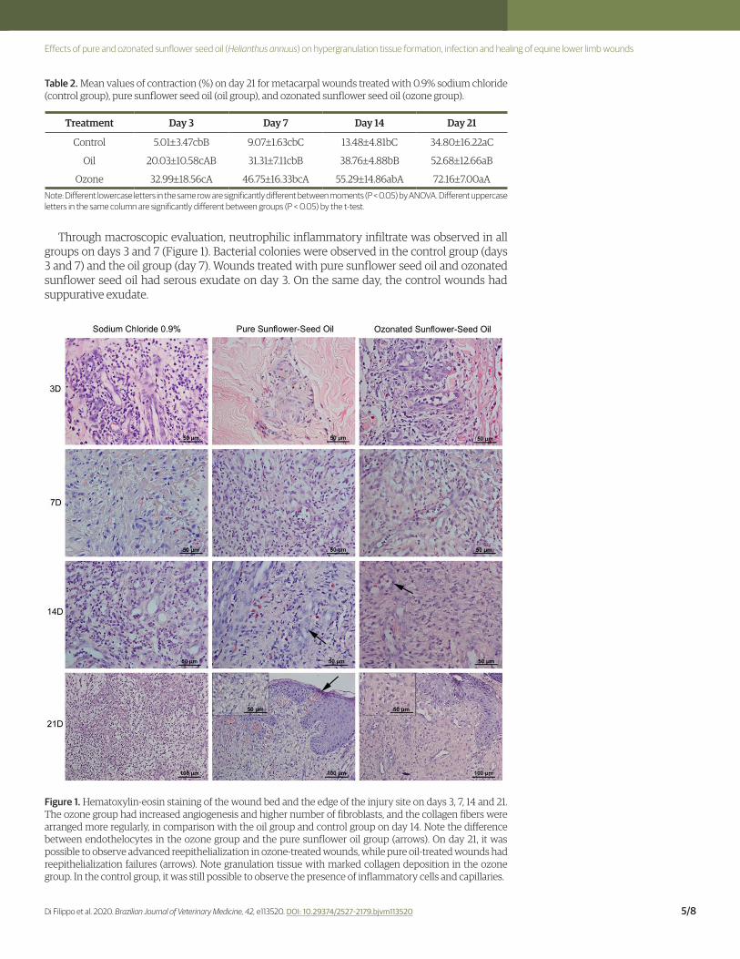
Through macroscopic evaluation, neutrophilic inflammatory infiltrate was observed in all groups on days 3 and 7 (Figure 1). Bacterial colonies were observed in the control group (days 3 and 7) and the oil group (day 7). Wounds treated with pure sunflower seed oil and ozonated sunflower seed oil had serous exudate on day 3. On the same day, the control wounds had suppurative exudate.
Figure 1. Hematoxylin-eosin staining of the wound bed and the edge of the injury site on days 3, 7, 14 and 21. The ozone group had increased angiogenesis and higher number of fibroblasts, and the collagen fibers were arranged more regularly, in comparison with the oil group and control group on day 14. Note the difference between endothelocytes in the ozone group and the pure sunflower oil group (arrows). On day 21, it was possible to observe advanced reepithelialization in ozone-treated wounds, while pure oil-treated wounds had reepithelialization failures (arrows). Note granulation tissue with marked collagen deposition in the ozone group. In the control group, it was still possible to observe the presence of inflammatory cells and capillaries.
Di Filippo et al. 2020. Brazilian Journal of Veterinary Medicine, 42, e113520. DOI: 10.29374/2527-2179.bjvm113520 6/8
Effects of pure and ozonated sunflower seed oil (Helianthus annuus) on hypergranulation tissue formation, infection and healing of equine lower limb wounds
Granulation tissue was observed on day 7 in all groups. Suppurative exudate was also observed on this day except in the ozone group. At day 14, the control wounds showed exuberant granulation tissue with crusts on the borders. Reepithelialization was observed on day 21 in the ozone group (Figure 1).
The main difference between the groups was observed on day 21 when the wounds of the ozone group were almost completely healed, whereas wounds in the control and oil groups still had granulation tissue. The wounds treated with 0.9% sodium chloride and sunflower seed oil had serous exudate on this day.
The culture results showed the presence of Streptococcus equi subsp. zooepidemicus in the wounds of the control group (days 3 and 7) and oil group (day 7).
DiscussionHypergranulation tissue, infection and delayed closure are common when equine lower limb
wounds heal by second intention (Bertone, 1989), but this study shows that ozonated sunflower seed oil is significantly more effective than pure sunflower seed oil and 0.9% sodium chloride in preventing hypergranulation and infection in equine lower limb wounds. In addition, wounds treated with ozonated oil presented contraction rates 20 and 38% higher than those treated with pure sunflower seed oil and 0.9% sodium chloride, respectively. Thus, these results demonstrate that O3 can enhance acute cutaneous wound healing in equines. Especially on days 14 and 21, both the wound size and residual wound area of the ozone group were diminished much more significantly. Similar results were observed after topical application of ozonated olive oil in a study of acute cutaneous wound healing in a guinea pig model (Kim et al., 2009). In that study, after creating full-thickness skin wounds on the backs of guinea pigs, the authors examined the wound healing effect of topically applied ozonated olive oil, as compared to the pure olive oil and no treatment. According to the authors, the results suggested that topical exposure to O3 can affect granulation tissue formation of the wound healing process rather than affecting immediate formation of blood clotting and recruitment of inflammatory cells during the inflammation phase.
As shown by hematoxylin-eosin staining, the relatively increased reepithelialization in the ozone group might be due to the fact that O3 acts directly or indirectly via collagen synthesis and fibroblast proliferation during granulation tissue formation and the early tissue remodeling phase of wound healing.
Fibroblasts have been shown to play important roles in reepithelialization, collagen fiber synthesis, extracellular matrix regeneration, remodeling of wounds, and the release of endogenous growth factors such as PDGF (platelet derived growth factor), TGF-β (transforming growth factor-β) and VEGF (vascular endothelial growth factors), as well as pro-proliferative mediators such as cyclin D1 (Kim et al., 2009; Valacchi et al., 2011).
In a guinea pig model, the increased expressions of PDGF, TGF-β and VEGF seen in the ozone group was correlated with increased staining intensity of collagen fibers and fibroblast proliferation (Kim et al., 2009). In addition, an increase of expression cyclin D1 was seen in cutaneous wounds made in the skin of SKH1 mice treated with ozonated sesame oil (Valacchi et al., 2011). Cyclin D1 is a key regulator released from macrophages, fibroblasts, and keratinocytes at the wound site and they participate in the regulation of reepithelialization, granulation tissue formation, collagen synthesis, and neovascularization (Martinez-Ferrer et al., 2010; Witzel et al., 2010). Both VEGF and cyclin D1 are genes under the control of the transcription factor NFkB, which has been shown to be activated in cutaneous tissue by O3 exposure (Valacchi et al., 2004). Therefore, it is possible that the topical release of either O3 or its mediators in the wound activates both NFkB, allowing its translocation to the nucleus, and the transcription of genes that play a key role in wound healing, such as cyclin D1 and VEGF. Ozone has a dose–effect relationship in wound healing between levels of oxidative stress and NFkB. A moderate level of oxidative stress activates the transcription factor whereas high levels of oxidative stress inhibit NFkB activation and consequently the rate of wound healing (Valacchi et al., 2011).
In open wounds, reepithelialization occurs after the development of a granulation bed, but the exuberant granulation tissue has an inhibitory effect on epithelialization and contraction, contributing to delayed closure and increased scar tissue formation (Theoret et al., 2002; Tracey et al., 2014). Wound contraction is a vital component of healing and the exuberant granulation tissue
Di Filippo et al. 2020. Brazilian Journal of Veterinary Medicine, 42, e113520. DOI: 10.29374/2527-2179.bjvm113520 7/8
Effects of pure and ozonated sunflower seed oil (Helianthus annuus) on hypergranulation tissue formation, infection and healing of equine lower limb wounds
observed only in the control wounds could explain why these wounds presented the worst results in this study. In addition, it is known that sunflower oil contains linoleic acid, a precursor of arachidonic acid, which is important in the inflammatory cascade (prostaglandins, thromboxanes, and leukotrienes).
These substances act as inflammatory mediators, stimulating local neovascularization, cellular migration, and fibroblastic proliferation and differentiation along with extracellular matrix synthesis (Glasgow & Eling, 1990). Acceleration of the healing process with a reduction in the wound area and faster contraction of the borders with more rapid development of granulation tissue were observed in wounds treated with topical application of sunflower seed oil (Marques et al., 2004; Oliveira Junior et al., 2012). Similar results were seen in our study in the wounds treated with pure sunflower seed oil compared to those treated with 0.9% sodium chloride. Oil wounds had a higher rate of contraction and therefore a smaller residual area (Tables 1 and 2).
Ozone (O3) is widely recognized as one of the best bactericidal agents (Al-Dalain et al., 2001; Valacchi et al., 2005, 2011), so this could explain the negative findings of culture in ozone-treated wounds, as well as in the absence of bacterial colonies and suppurative exudate in these wounds. The use of ozonated oil not only prevents infection, but also stimulates the initial tissue reconstruction by increasing cell proliferation and new vascularization. In fact, in the ozonated treatment there was more effective and earlier reepithelialization, which associated with the higher contraction rate resulted in a smaller residual area (Table 1 and 2). The bacteria that were isolated in this study are common environmental contaminants. Similar culture results were obtained in other studies using a similar wound model (Tracey et al., 2014; Morgan et al., 2009). However, our results differ from those obtained by other researchers, who reported that the most common wound-isolated bacteria were Pseudomonas aeruginosa and Staphylococcus epidermis (Freeman et al., 2009; Westgate et al., 2011). Microbial diversity varies between acute and chronic wounds, which can explain the differences between the studies. The opportunistic pathogenesis of isolates such as Pseudomonas may result in biofilm formation. In this respect, recognizing and understanding biofilms in equine wounds would help to guide appropriate management strategies for the treatment of these wounds (Westgate et al., 2011).
ConclusionsOur results show that topical application of ozonated sunflower seed oil is highly effective in
accelerating acute cutaneous wound repair, preventing hypergranulation tissue and infection in equine lower limb wounds, and that it is better than treatment with pure sunflower seed oil.
AcknowledgementsUENF, State University of North Fluminense Darcy Ribeiro.
ReferencesAl-Dalain, S. M., Martinez, G., Candelario-Jalil, E., Menendez, S., Re, L., Giuliani, A., & Leon, O. S. (2001). Ozone
treatment reduces markers of oxidative and endothelial damage in an experimental diabetes model in rats. Pharmacological Research, 44(5), 391-396. http://dx.doi.org/10.1006/phrs.2001.0867.
Bertone, A. L. (1989). Management of exuberant granulation tissue. Veterinary Clinics of North America: Equine Practice, 5(3), 551-562. http://dx.doi.org/10.1016/s0749-0739(17)30574-6.
Burgassi, S., Zanardi, I., Travagli, V., Montomoli, E., & Bocci, V. (2009). How much ozone bactericidal activity is compromised by plasma components? Journal of Applied Microbiology, 106(5), 1715-1721. http://dx.doi.org/10.1111/j.1365-2672.2008.04141.x.
Caston, S. S. (2012). Wound care in horses. The Veterinary Clinics of North America. Equine Practice, 28(1), 83-100. http://dx.doi.org/10.1016/j.cveq.2012.01.001. PMid:22640581.
Crovetti, G., Martinelli, G., Issi, M., Barone, M., Guizzardi, M., Campanati, B., Moroni, M., & Carabelli, A. (2004). Platelet gel for healing cutaneous chronic wounds. Transfusion and Apheresis Science, 30(2), 145-151. http://dx.doi.org/10.1016/j.transci.2004.01.004. PMid:15062754.
Freeman, K., Woods, E., Welsby, S., Percival, S., & Cochrane, C. A. (2009). Biofilm evidence and the microbial diversity of horse wounds. Canadian Journal of Microbiology, 55(2), 197-202. http://dx.doi.org/10.1139/W08-115. PMid:19295652.
Glasgow, W. C., & Eling, G. T. (1990). Epidermal growth factor simulates linoleic acid metabolic in BAB/C 3T3 fibroblasts. Molecular Pharmacology, 38(4), 503-510. PMID: 2233691.
Di Filippo et al. 2020. Brazilian Journal of Veterinary Medicine, 42, e113520. DOI: 10.29374/2527-2179.bjvm113520 8/8
Effects of pure and ozonated sunflower seed oil (Helianthus annuus) on hypergranulation tissue formation, infection and healing of equine lower limb wounds
Harman, R. M., Theoret, C. L., & Van de Walle, G. R. (2019). The horse as a model for the study of cutaneous wound healing. Advances in Wound Care, 1-19. In press. http://dx.doi.org/10.1089/wound.2018.0883.
Kim, H. S., Noh, S. U., Han, Y. W., Kim, K. M., Kang, H., Kim, H. O., & Park, Y. M. (2009). Therapeutic effects of topical application of ozone on acute cutaneous wound healing. Journal of Korean Medical Science, 24, 368-374. http://dx.doi.org/10.3346/jkms.2009.24.3.368.
Macri, L., & Clark, R. A. (2009). Tissue engineering for cutaneous wounds: Selecting the proper time and space for growth factors, cells and the extracellular matrix. Skin Pharmacology and Physiology, 22, 83-93. http://dx.doi.org/10.1159/000178867.
Magalhães, M. S., Fechine, F. V., Macedo, R. N., Monteiro, D. L., Oliveira, C. C., Brito, G. A., Moraes, M. E., & Moraes, M. O. (2008). Effect of a combination of medium chain triglycerides, linoleic acid, soy lecithin and vitamins A and E on wound healing in rats. Acta Cirurgica Brasileira, 23(3), 262-269. http://dx.doi.org/10.1590/S0102-86502008000300009. PMid:18552998.
Marques, S. R., Peixoto, C. A., Messias, J. B., Albuquerque, A. R., & Silva Junior, V. A. (2004). The effects of topical application of sunflower seed oil on open wound healing in lambs. Acta Cirurgica Brasileira, 19(3), 196-205. http://dx.doi.org/10.1590/S0102-86502004000300005.
Martinez-Ferrer, M., Afshar-Sherif, A. R., Uwamariya, C., Crombrugghe, B., Davidson, J. M., & Bhowmick, N. A. (2010). Dermal transforming growth factor-beta responsiveness mediates wound contraction and epithelial closure. The American Journal of Pathology, 176, 98-107. http://dx.doi.org/10.2353/ajpath.2010.090283.
Maxson, S., Lopez, E. A., Yoo, D., Danilkovitch-Miagkova, A., & Leroux, M. A. (2012). Concise review: Role of mesenchymal stem cells in wound repair. Stem Cells Translational Medicine, 1, 142-149. http://dx.doi.org/10.5966/sctm.2011-0018.
Morgan, D. D., McClure, S., Yaeger, M. J., Schumacher, J., & Evans, R. B. (2009). Effects of extracorporeal shock wave therapy on wounds of the distal portion of the limbs in horses. Journal of the American Veterinary Medical Association, 234(9), 1154-1161. http://dx.doi.org/10.2460/javma.234.9.1154. PMid:19405886.
Oliveira Junior, L. A. T., Souza, V. R. S., Endringer, D. C., Hendrickson, D. A., & Coelho, C. S. (2012). Effects of topical application of sunflower seed oil on experimentally induced wounds in horses. Journal of Equine Veterinary Science, 32(3), 139-145. http://dx.doi.org/10.1016/j.jevs.2011.08.006.
Ramsey, D. T., Pope, E. R., Wagner-Mann, C., Berg, J. N., & Swaim, S. F. (1995). Effects of three occlusive dressing materials on healing of full-thickness skin wounds in dogs. American Journal of Veterinary Research, 56(7), 941-949. PMid:7574165.
Theoret, C. L., Barber, S. M., Moyana, T. N., & Gordon, J. R. (2002). Preliminary observations on expression of transforming growth factors beta1 and beta3 in equine full-thickness skin wounds healing normally or with exuberant granulation tissue. Veterinary Surgery, 31, 266-273. http://dx.doi.org/10.1053/jvet.2002.32394.
Tracey, A. K., Alcott, C. J., Schleining, J. A., Safayi, S., Zaback, P. C., Hostetter, J. M., & Reinertson, E. L. (2014). The effects of topical oxygen therapy on equine distal limb dermal wound healing. The Canadian Veterinary Journal. La Revue Veterinaire Canadienne, 55(12), 1146-1152. PMid:25477541.
Travagli, V., Zanardi, I., Valacchi, G., & Bocci, V. (2010). Ozone and ozonated oils in skin diseases: a review. Mediators of Inflammation, 2010, 610418. http://dx.doi.org/10.1155/2010/610418. PMid:20671923.
Valacchi, G., Fortino, V., & Bocci, V. (2005). The dual action of ozone on the skin. British Journal of Dermatology, 153, 1096-100. http://dx.doi.org/10.1111/j.1365-2133.2005.06939.x
Valacchi, G., Lim, Y., Belmonte, G., Miracco, C., Zanardi, I., Bocci, V., & Travagli, V. (2011). Ozonated sesame oil enhances cutaneous wound healing in SKH1 mice. Wound Repair and Regeneration, 19, 107-115. http://dx.doi.org/10.1111/j.1524-475X.2010.00649.x.
Valacchi, G., Pagnin, E., Corbacho, A. M., Olano, E., Davis, P. A., Packer, L., & Cross, C. E. (2004). In vivo ozone exposure induces antioxidant/stress-related responses in murine lung and skin. Free Radical Biology and Medicine, 36, 673-681. http://dx.doi.org/10.1016/j.freeradbiomed.2003.12.005.
Westgate, S. J., Percival, S. L., Knottenbelt, D. C., Clegg, P. D., & Cochrane, C. A. (2011). Microbiology of equine wounds and evidence of bacterial biofilms. Veterinary Microbiology, 150(1-2), 152-159. http://dx.doi.org/10.1016/j.vetmic.2011.01.003. PMid:21273008.
Witzel, I. I., Koh, L. F., & Perkins, N. D. (2010). Regulation of cyclin D1 gene expression. Biochemical Society Transactions, 38(Pt 1), 217-222. http://dx.doi.org/10.1042/BST0380217. PMid:20074063.
Ziboh, V. A. (1996). The significance of polyunsaturated fatty acids in cutaneous biology. Lipids, 31(1, Suppl), S249-S53. http://dx.doi.org/10.1007/BF02637085. PMid:8729128.