effects of red and white ginseng preparations on
TRANSCRIPT

pharmaceuticals
Article
Effects of Red and White Ginseng Preparations on ElectricalActivity of the Brain in Elderly Subjects: A Randomized,Double-Blind, Placebo-Controlled, Three-ArmedCross-Over Study
Wilfried Dimpfel 1,2,*, Pierre-Antoine Mariage 3 and Alexander G. Panossian 4,5
�����������������
Citation: Dimpfel, W.; Mariage, P.-A.;
Panossian, A.G. Effects of Red and
White Ginseng Preparations on
Electrical Activity of the Brain in
Elderly Subjects: A Randomized,
Double-Blind, Placebo-Controlled,
Three-Armed Cross-Over Study.
Pharmaceuticals 2021, 14, 182.
https://doi.org/10.3390/ph14030182
Academic Editor: Daniela De Vita
Received: 29 January 2021
Accepted: 20 February 2021
Published: 25 February 2021
Publisher’s Note: MDPI stays neutral
with regard to jurisdictional claims in
published maps and institutional affil-
iations.
Copyright: © 2021 by the authors.
Licensee MDPI, Basel, Switzerland.
This article is an open access article
distributed under the terms and
conditions of the Creative Commons
Attribution (CC BY) license (https://
creativecommons.org/licenses/by/
4.0/).
1 Departmrent of Pharmacology, Justus-Liebig-University Giessen, Germany c/o A 4164 Schwarzenberg amBöhmerwald, Panoramaweg 21, Schwarzenberg am Böhmerwald, Övre-Österrike, 4164 Österrike, Austria
2 NeuroCode AG, D-35578 Wetzlar, Germany3 Botalys SA, 8 Quai des Usines, 7800 Ath, Belgium; [email protected] Department of Research & Development, Phytomed AB, Bofinkvagen 1, 31275 Vaxtorp, Sweden;
[email protected] Department of Science & Education, EuroPharmaUSA, Green Bay, WI 54311, USA* Correspondence: [email protected]
Abstract: Background: Recently, the superior efficacy of hydroponically cultivated red ginsengpreparation HRG80® compared to wild growing white ginseng (WG) in preventing stress-inducedsymptoms related to the daily work situation of healthy subjects was reported. The aim of this studywas to compare the effects of HRG80®, WG, and placebo on the electrical activity in the brain ofelderly human subjects during relaxation and mental challenges. Methods: Changes in the electroen-cephalogram (EEG) frequency ranges of 17 different brain regions were measured after single andrepeated administration of HRG80®, WG, and placebo across a four-week randomized, double-blind,placebo-controlled three-armed cross-over trial. Results: Both red and white ginseng preparationshad a strong impact on brain activity, with different effects on various brain regions depending onthe mental load during relaxation and cognitive tasks associated with memory, attention, and mentalperformance. Both ginseng preparations exhibited significant effects on spectral powers comparedto placebo, reflecting an activating action. The spectral changes in the quantitative EEG induced byHRG80® indicated an improvement in mood as well as calming effects, evidenced by the modulationof β2 waves, representing changes in GABA-ergic neurotransmission. HRG80® attenuated δ/θpowers during relaxation, suggesting the potential improvement of pathologically enhanced spectralpower in aging. Conclusion: The results of this study suggest that both hydroponically cultivated redand wild growing white ginseng have similar beneficial effects on the cognitive functions of elderlysubjects, as reflected by electric brain activity, but their modes of action on the brain are different.
Keywords: elderly subjects; Panax ginseng; quantitative EEG; brain; discriminant analysis; cognition
1. Introduction
Ginseng Radix and Rhizoma (Panax ginseng Meyer) have been traditionally used inChina, Korea, and Japan for thousands of years “to promote vitality and healthy aging,to enhance cognitive function, mental activity, general weakness, and enhance longevity”with long-term intake. It has been used primarily as a tonic to stimulate weak bodiesand to maintain homeostasis, which is a contemporary term associated with adaptivestress syndrome, adaptability and adaptogen concepts [1–7]. According to the WHO [8],medicinal uses of ginseng include prophylactic and restorative treatment for enhancementof mental and physical performance, in cases of weakness, exhaustion, tiredness, andloss of concentration, and during convalescence. In Europe, most ginseng preparationsare used as tonics in cases of tiredness, weakness, and decreased mental and physical
Pharmaceuticals 2021, 14, 182. https://doi.org/10.3390/ph14030182 https://www.mdpi.com/journal/pharmaceuticals
Pharmaceuticals 2021, 14, 182 2 of 18
capacity [9]. However, these and other international monographs [10–12] have been basedon mixed data, without differentiating results from quite different preparations obtainedfrom air-dried white ginseng (known in China as Renshen) or steamed at 100 ◦C to redginseng (Hongshen) [13]. Chemical composition and pharmacological activity of whiteand red ginseng are obviously different [14–19].
Recently, the efficacy of white ginseng (WG) and hydroponically cultivated red ginseng(HRG80®) preparations in preventing stress-induced symptoms associated with the daily worksituation of healthy subjects was reported [18,19]. HRG80® was more effective compared to thatwith the WG and placebo regarding attention, memory, and perceived stress scores after singleand repeated administrations for 5 and 12 days [18,19]. HRG80® was also more active thanWG in inducing excitatory neurotransmission of rat hippocampal slice preparations in an exvivo model of long-term potentiation (LTP), reflecting time and spatial dependent memory [20].It is well known that mental challenges are associated with significant changes in synapticneurotransmission and differential electrical activity recorded at six electroencephalogram(EEG) brain frequencies (δ, θ, α1, α2, β1, and β2) in 17 different brain regions, which in turncorrelate with various cognitive functions and mental diseases [21,22]. Furthermore, accordingto Dimpfel et al., 2014, high spectral δ and θ power (in general and specifically at frontotemporalregions of brain) is characteristic of mild cognitive impairment (MCI) [21]. The primary aim ofpresent study was to detect possible differences in the effects of the same preparations (WGand HRG80®) on electrical activity of the brain of elderly human subjects in normal (relaxing)condition and during mental challenges (cognitive tasks), after repeated administration of testarticles for four weeks.
2. Results2.1. Baseline Data of Study Participants, Their Disposition and Treatment Compliance
Overall, 31 participants were assessed for eligibility; all met the inclusion criteria.Thirty patients were recruited in the study, of which all patients completed treatment andwere included in the final analysis, Figure 1. One subject was withdrawn from the study(screening dropout) due to hypertension not related to trial medication. The treatmentcompliance calculated by counting of unused capsules was 98.97% (Placebo = 99.17%,HRG80® = 98.42%, WG = 99.32%).
Baseline demographic and efficacy outcome measures are shown in Table 1.
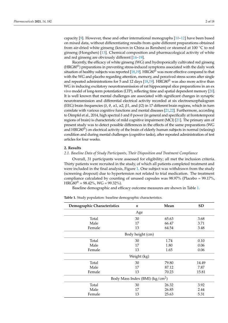
Table 1. Study population: baseline demographic characteristics.
Demographic Characteristics n Mean SD
Age
Total 30 65.63 3.68Male 17 66.47 3.71
Female 13 64.54 3.48
Body height (cm)
Total 30 1.74 0.10Male 17 1.80 0.06
Female 13 1.65 0.06
Weight (kg)
Total 30 79.80 14.49Male 17 87.12 7.87
Female 13 70.23 15.81
Body Mass Index (BMI) (kg/cm2)
Total 30 26.32 3.92Male 17 26.85 2.44
Female 13 25.63 5.31

Pharmaceuticals 2021, 14, 182 3 of 18
Pharmaceuticals 2021, 14, x FOR PEER REVIEW 3 of 19
Total 30 26.32 3.92 Male 17 26.85 2.44
Female 13 25.63 5.31
Figure 1. CONSORT flow chart of the disposition of participants into three arms on all steps of the study: screening, randomization, allocation, treatment, and data analysis. WG, white ginseng; HRG80®, hydroponically cultivated red gin-seng.
2.2. Efficacy of Treatment 2.2.1. Efficacy Outcome Measures and Endpoints
The primary efficacy outcomes were the responses of electric brain activity measured as spectral power in 17 different brain regions within six specially defined frequency ranges (i.e., δ, θ, α1, α2, β1, and β2) during relaxation as well as during the following psychometric tests of cognitive performance: concentration test for attention (d2 test), memory test (ME test), and calculation performance test (CPT).
The primary efficacy endpoint was achieved by comparison of the changes in neu-ronal electrical activity of the brain from baseline to the end of the treatment with WG, HRG80®, and placebo for four weeks.
Figure 1. CONSORT flow chart of the disposition of participants into three arms on all steps ofthe study: screening, randomization, allocation, treatment, and data analysis. WG, white ginseng;HRG80®, hydroponically cultivated red ginseng.
2.2. Efficacy of Treatment2.2.1. Efficacy Outcome Measures and Endpoints
The primary efficacy outcomes were the responses of electric brain activity measuredas spectral power in 17 different brain regions within six specially defined frequency ranges(i.e., δ, θ, α1, α2, β1, and β2) during relaxation as well as during the following psychometrictests of cognitive performance: concentration test for attention (d2 test), memory test (MEtest), and calculation performance test (CPT).
The primary efficacy endpoint was achieved by comparison of the changes in neuronalelectrical activity of the brain from baseline to the end of the treatment with WG, HRG80®,and placebo for four weeks.
2.2.2. Quantitative EEG Results after Acute and Repetitive Dosing
Electric brain activity was recorded under several different conditions. The firstrecording was always in a relaxed state with open eyes. After this, three different mentalchallenges were presented during quantitative EEG recordings (Figure 2).

Pharmaceuticals 2021, 14, 182 4 of 18
Pharmaceuticals 2021, 14, x FOR PEER REVIEW 4 of 19
2.2.2. Quantitative EEG Results after Acute and Repetitive Dosing Electric brain activity was recorded under several different conditions. The first re-
cording was always in a relaxed state with open eyes. After this, three different mental challenges were presented during quantitative EEG recordings (Figure 2).
Figure 2. Changes in the electric brain maps 2 h after the ingestion of placebo, WG, or HRG80® under different recording conditions on the first day of treatment. The right column in section represents the left hemisphere, while the left column represents the right hemisphere. Please note changes in the frontotemporal areas are more pronounced in the left hemisphere (frontal area is in the middle).
The electric maps constructed after the intake of the placebo or one of the two ginseng preparations showed somewhat stronger differences for HRG80® in comparison to pla-cebo with respect to the frontotemporal brain areas under the eyes open recording condi-tion and during the performance of the d2 test on the first day.
Under recording conditions, the CPT and memory test after ingestion of either of the ginseng preparations induced similar patterns of change; however, they were only slightly different from that of placebo. The details are provided in Figure 2.
Figure 2. Changes in the electric brain maps 2 h after the ingestion of placebo, WG, or HRG80® underdifferent recording conditions on the first day of treatment. The right column in section representsthe left hemisphere, while the left column represents the right hemisphere. Please note changes inthe frontotemporal areas are more pronounced in the left hemisphere (frontal area is in the middle).
The electric maps constructed after the intake of the placebo or one of the two ginsengpreparations showed somewhat stronger differences for HRG80® in comparison to placebowith respect to the frontotemporal brain areas under the eyes open recording conditionand during the performance of the d2 test on the first day.
Under recording conditions, the CPT and memory test after ingestion of either of theginseng preparations induced similar patterns of change; however, they were only slightlydifferent from that of placebo. The details are provided in Figure 2.
The EEG data were initially documented as absolute spectral power (µV2) for eachelectrode position (brain area) and each frequency range (δ–β2). The absolute power valuesfrom the baseline recording with respect to all recording conditions were then set to 100%.Drug-induced changes were documented as a pre-post intake comparison of the changesin percentage of these baseline values for every recording condition. When comparingthe median values at the baseline (time 0) before all treatments, no major differences weredetected. Thus, the starting values of the outcome measures were comparable. Drug-induced changes were documented as a comparison of the pre-post intake values as apercentage of the baseline values for each condition (Figures 3 and 4).

Pharmaceuticals 2021, 14, 182 5 of 18
On the last experimental day after the repetitive administration of HRG80® for fourweeks, statistically significant decreases in spectral α1 power in the frontal lobe Fz, F8,and the temporal lobe T6, as well as α2 power in the central lobe C3, were observed 2 hafter intake of HRG80® during relaxation (Figure 3). With respect to the intake of placeboduring relaxation, hardly any change was observed, whereas repetitive administration ofWG during this period resulted in a statistically significant decrease in spectral α and β1power in the parietal lobe at electrode position P4 (Figure 3).
As expected, quite different patterns were observed in the experiments when brainelectrical activity was recorded during the different psychometric tests; namely, the d2attention test, CPT (calculation test), and memory test. Under the recording conditionsof the d2 test, WG changed spectral power only at the electrode positions C3, T3, and O1.HRG80® induced statistically significant spectral changes in seven electrode positions; thedetails are documented in Figure 4.
A quite different response was observed in the CPT, where an increase in δ powerwas recorded in the frontal F3 lobe with decreases in θ power in the frontal F8 and α2power in the temporal T5 location. Similarly, decreases in θ power in Cz and F8 as well asδ power in the Cz brain region were recorded during performance of the memory test. TheEEG fingerprint response to WG treatment was different from that of HRG80® in all threetests, although there were some similarities, e.g., only three of the 17 brain regions weresignificantly affected (decrease in α1 and α2 spectral powers) during the d2 test comparedto the placebo. On the contrary, all the δ, θ, α1, α2, β1, and β2 spectral powers weresignificantly decreased in 12 of the 17 brain regions during the CTP, and the δ, θ, α2, β1,and β2 spectral powers were significantly decreased in 14 of the 17 brain regions duringthe memory test compared to the placebo. The details are provided in Figure 4.
Pharmaceuticals 2021, 14, x FOR PEER REVIEW 5 of 19
The EEG data were initially documented as absolute spectral power (µV2) for each electrode position (brain area) and each frequency range (δ–β2). The absolute power val-ues from the baseline recording with respect to all recording conditions were then set to 100%. Drug-induced changes were documented as a pre–post intake comparison of the changes in percentage of these baseline values for every recording condition. When com-paring the median values at the baseline (time 0) before all treatments, no major differ-ences were detected. Thus, the starting values of the outcome measures were comparable. Drug-induced changes were documented as a comparison of the pre–post intake values as a percentage of the baseline values for each condition (Figures 3 and 4).
Figure 3. Spectral power differences under the eyes open recording condition in all brain regions, as represented by the electrode positions, on the last experimental day (repetitive) with the administration of placebo, WG, or HRG80®. C, cen-tral; P, parietal; O, occipital; F, frontal; T, temporal. Even numbers indicate a location in the right hemisphere, while odd numbers indicate the left hemisphere. Frequencies: red, δ; orange, θ; yellow, α1; green, α2; turquoise, β1; blue, β2. The spectral power (between 40% and 140% on the ordinate of the bar graph) was averaged over 6 min and plotted against the pre-drug value (values at 0 min set as 100%), thus reflecting the effect of the placebo, WG, or HRG80®. * p < 0.12, ** p < 0.05, and *** p < 0.01 (nonparametric sign test) between the placebo, WG, or HRG80®. The direction of change is marked by + or − underneath the relevant bar.
On the last experimental day after the repetitive administration of HRG80® for four weeks, statistically significant decreases in spectral α1 power in the frontal lobe Fz, F8, and the temporal lobe T6, as well as α2 power in the central lobe C3, were observed 2 h after intake of HRG80® during relaxation (Figure 3). With respect to the intake of placebo dur-ing relaxation, hardly any change was observed, whereas repetitive administration of WG during this period resulted in a statistically significant decrease in spectral α and β1 power in the parietal lobe at electrode position P4 (Figure 3).
Figure 3. Spectral power differences under the eyes open recording condition in all brain regions, as represented by theelectrode positions, on the last experimental day (repetitive) with the administration of placebo, WG, or HRG80®. C, central;P, parietal; O, occipital; F, frontal; T, temporal. Even numbers indicate a location in the right hemisphere, while odd numbersindicate the left hemisphere. Frequencies: red, δ; orange, θ; yellow, α1; green, α2; turquoise, β1; blue, β2. The spectralpower (between 40% and 140% on the ordinate of the bar graph) was averaged over 6 min and plotted against the pre-drugvalue (values at 0 min set as 100%), thus reflecting the effect of the placebo, WG, or HRG80®. * p < 0.12, ** p < 0.05, and*** p < 0.01 (nonparametric sign test) between the placebo, WG, or HRG80®. The direction of change is marked by + or −underneath the relevant bar.

Pharmaceuticals 2021, 14, 182 6 of 18Pharmaceuticals 2021, 14, x FOR PEER REVIEW 6 of 19
Figure 4. Cont.

Pharmaceuticals 2021, 14, 182 7 of 18Pharmaceuticals 2021, 14, x FOR PEER REVIEW 7 of 19
Figure 4. Spectral power differences in all brain regions, as represented by the electrode positions, under the recording conditions for the concentration test for attention (d2) test, the calculation performance test (CPT), and the memory test on the last experimental day with the administration of the placebo, WG, or HRG80®. C, central; P, parietal; O, occipital; F, frontal; T, temporal. Even numbers are located in the right hemisphere, and odd numbers in the left hemisphere. Fre-quencies: red, δ; orange, θ; yellow, α1; green, α2; turquoise, β1; blue, β2; h, hours. The spectral power (between 40% and 140% on the ordinate of the bar graph) was averaged over 5 min and plotted against the pre-drug value (at 0 min set to 100%), thus reflecting the effects of placebo, WG, or HRG80®. Statistical significance (sign test) between placebo, WG, or HRG80® is indicated by stars. * p < 0.10, ** p < 0.05, and *** p < 0.01. The direction of change is marked by + or − underneath the bar graphic.
As expected, quite different patterns were observed in the experiments when brain electrical activity was recorded during the different psychometric tests; namely, the d2 attention test, CPT (calculation test), and memory test. Under the recording conditions of the d2 test, WG changed spectral power only at the electrode positions C3, T3, and O1. HRG80® induced statistically significant spectral changes in seven electrode positions; the details are documented in Figure 4.
A quite different response was observed in the CPT, where an increase in δ power was recorded in the frontal F3 lobe with decreases in θ power in the frontal F8 and α2 power in the temporal T5 location. Similarly, decreases in θ power in Cz and F8 as well as δ power in the Cz brain region were recorded during performance of the memory test. The EEG fingerprint response to WG treatment was different from that of HRG80® in all three tests, although there were some similarities, e.g., only three of the 17 brain regions were significantly affected (decrease in α1 and α2 spectral powers) during the d2 test com-pared to the placebo. On the contrary, all the δ, θ, α1, α2, β1, and β2 spectral powers were significantly decreased in 12 of the 17 brain regions during the CTP, and the δ, θ, α2, β1, and β2 spectral powers were significantly decreased in 14 of the 17 brain regions during the memory test compared to the placebo. The details are provided in Figure 4.
2.2.3. Efficacy of Red and White Ginseng Documented by Discriminant Analysis All 102 parameters from the EEG recordings (17 electrode positions × 6 frequency
ranges) during the eyes open condition were fed into the linear discriminant analysis con-taining data from several preparations earlier tested with identical methodology [23]. The results of both ginseng preparations projected in close vicinity of each other and were not well separated, also showing a similar color; altogether, this indicates that they have a similar action. The data following acute intake were similar to the effects of Adaptra Forte,
Figure 4. Spectral power differences in all brain regions, as represented by the electrode positions, under the recordingconditions for the concentration test for attention (d2) test, the calculation performance test (CPT), and the memory test onthe last experimental day with the administration of the placebo, WG, or HRG80®. C, central; P, parietal; O, occipital; F,frontal; T, temporal. Even numbers are located in the right hemisphere, and odd numbers in the left hemisphere. Frequencies:red, δ; orange, θ; yellow, α1; green, α2; turquoise, β1; blue, β2; h, hours. The spectral power (between 40% and 140% on theordinate of the bar graph) was averaged over 5 min and plotted against the pre-drug value (at 0 min set to 100%), thusreflecting the effects of placebo, WG, or HRG80®. Statistical significance (sign test) between placebo, WG, or HRG80® isindicated by stars. * p < 0.10, ** p < 0.05, and *** p < 0.01. The direction of change is marked by + or − underneath thebar graphic.
2.2.3. Efficacy of Red and White Ginseng Documented by Discriminant Analysis
All 102 parameters from the EEG recordings (17 electrode positions × 6 frequencyranges) during the eyes open condition were fed into the linear discriminant analysiscontaining data from several preparations earlier tested with identical methodology [23].The results of both ginseng preparations projected in close vicinity of each other and werenot well separated, also showing a similar color; altogether, this indicates that they havea similar action. The data following acute intake were similar to the effects of AdaptraForte, another adaptogenic preparation [23]. The stimulating preparations Zembrin® andmemoLoges® appear more toward the front, whereas HRG80® and White Panax ginsengcapsules appear toward the back. The details are shown in Figure 5.

Pharmaceuticals 2021, 14, 182 8 of 18
Pharmaceuticals 2021, 14, x FOR PEER REVIEW 8 of 19
another adaptogenic preparation [23]. The stimulating preparations Zembrin® and memo-Loges® appear more toward the front, whereas HRG80® and White Panax ginseng cap-sules appear toward the back. The details are shown in Figure 5.
Figure 5. Results of the discriminant analysis. Acute effect on the first day of administration dur-ing the eyes open recording condition. White ginseng and HRG80® are projected as near neigh-bors. Results from first three discriminant functions are displayed with respect to space (x, y, and z coordinates). Results from the next three functions are displayed as an additive color mixture. If the preparations are projected as rather close neighbors, they cannot be well discriminated from one another, which means that they have a similar effect or can be used for similar clinical indica-tions.
2.3. Safety Evaluation Thirty elderly subjects were administered HRG80®, WG, or placebo preparations at
a daily dose of two capsules per day for four weeks. One participant dropped out during the screening, and there were no study dropouts.
In total, four adverse events (AEs) were observed in three patients during the treat-ment with HRG80®, WG, and placebo capsules in phases A, B, and C of the study; for details, see Appendix A. These AEs were neither serious nor related to the study medica-tion.
Overall, the treatment was well tolerated: all EEG measurements were within the normal range, and physical examinations did not reveal any deviation from normality. No organic disease emerged during the study; for details, see Appendix A.
3. DiscussionIn the relaxation state, repeated administration of HRG80® decreased the α1 and α2
spectral powers in six locations compared to the placebo, while WG decreased the α power in only one location and the β1 spectral power in two locations, Table 2.
Table 2. Statistically significant (p < 0.05) or conspicuous (p < 0.10) effects of HRG80® and WG compared to placebo on spectral power (α1, α2, β1, β2, δ, and θ) under different recording condi-tions (relaxation and during the d2 test, CPT, and memory test) on the last experimental day. Brain regions are represented by the electrode positions. F, frontal; T, temporal; C, central; P, parietal; O, occipital. The direction of change is marked by ↑ or ↓ symbols.
Condition Ginseng δ θ α1 α2 β1 β2 Mediator Ach NE 5-HT DA Glu GABA
Relaxation HRG80®
↓ Fz, F7,F8, T6
↓ C3, O1
WG P4 P4 ↓ P4, O2
Figure 5. Results of the discriminant analysis. Acute effect on the first day of administration during the eyes open recordingcondition. White ginseng and HRG80® are projected as near neighbors. Results from first three discriminant functionsare displayed with respect to space (x, y, and z coordinates). Results from the next three functions are displayed as anadditive color mixture. If the preparations are projected as rather close neighbors, they cannot be well discriminated fromone another, which means that they have a similar effect or can be used for similar clinical indications.
2.3. Safety Evaluation
Thirty elderly subjects were administered HRG80®, WG, or placebo preparations at adaily dose of two capsules per day for four weeks. One participant dropped out during thescreening, and there were no study dropouts.
In total, four adverse events (AEs) were observed in three patients during the treatmentwith HRG80®, WG, and placebo capsules in phases A, B, and C of the study; for details,see Appendix A. These AEs were neither serious nor related to the study medication.
Overall, the treatment was well tolerated: all EEG measurements were within thenormal range, and physical examinations did not reveal any deviation from normality. Noorganic disease emerged during the study; for details, see Appendix A.
3. Discussion
In the relaxation state, repeated administration of HRG80® decreased the α1 and α2spectral powers in six locations compared to the placebo, while WG decreased the α powerin only one location and the β1 spectral power in two locations, Table 2.
Alpha1 spectral power is known to be associated with serotonin (5HT)-mediatedneurotransmission. Thus, an increase in α1 power is positively correlated with relaxationand deep satisfaction (calming effect), whereas a decrease signalizes a higher activatedstate; meanwhile, α2 power is mainly associated with dopaminergic (DA)-mediated neuro-transmission. A decrease in α2 is positively correlated with a stimulating effect, such as thatinduced by amphetamine, having an influence on mood and motivation that is possiblyrelated to the pleasure–reward pathway. Attenuation of these waves must be interpretedas a potential positive action on mood. β1 power is associated with the modulation ofglutamatergic (Glu) = mediated neurotransmission. A decrease in β1 power signalizes anactivating effect (Table 2).

Pharmaceuticals 2021, 14, 182 9 of 18
Table 2. Statistically significant (p < 0.05) or conspicuous (p < 0.10) effects of HRG80® and WG compared to placebo onspectral power (α1, α2, β1, β2, δ, and θ) under different recording conditions (relaxation and during the d2 test, CPT, andmemory test) on the last experimental day. Brain regions are represented by the electrode positions. F, frontal; T, temporal;C, central; P, parietal; O, occipital. The direction of change is marked by ↑ or ↓ symbols.
Condition Ginseng δ θ α1 α2 β1 β2
Mediator Ach NE 5-HT DA Glu GABA
RelaxationHRG80® ↓Fz, F7, F8, T6 ↓C3, O1
WG P4 P4 ↓P4, O2
D2 testHRG80® ↓F4 ↓F4, F7, F8 ↓F4, F8, T5 ↓F8, Pz
WG ↓C3, O1 T3
CPTHRG80® ↑F3, ↓F8 ↓F8, O2 ↓T5 ↓Cz, P4, O2
WG ↓P4 ↓F7, Pz, O2 ↓T5, T6,C3, C4 ↓F7, T5, P4, ↓F3, P3, P4, O1,
O2↓F3, F7, T3, C3,
O1, O2
Memory testHRG80® ↓F7, F8, Cz, O1 ↓F3, F8, Cz, ↓P4 ↓Fz, F8 ↓C3
WG ↓T3, T6, O2 ↓F7, T5, T6, C3,C4, P3, P4
↓F7, C3, C4, P4,O2
↓F3, F7, F8, T3,C3, P3, C4 ↓F3, T3, P3, ↓T3, T5, C3, C4,
P3
Since the electric activity of the frontal brain seems to be especially important formental performance, analyzing this activity during treatment with drugs is essential. Eventhough both of the ginseng preparations seemed to generally act in a similar way duringrelaxation based on the results of the discriminant analysis, some differences were detectedunder the different challenging recording conditions and with respect to brain locations.
A major difference between the effects of HRG80® and WG was observed duringrelaxation. Whereas HRG80® reduced the spectral power in three frontal brain regions,WG did not affect any of the frontal locations under this recording condition (see Table 2).The effects of both ginseng preparations on brain activity depended considerably on thetype of cognitive activity. During the d2 test, seven frontal brain locations were involved,in contrast to the effect of WG (no frontal regions involved). During the CPT, HRG80®
modulated three frontal regions, whereas WG had an influence on five frontal brain regions.However, HRG80® affected more long waves, whereas WG mainly had an influence onhigh frequencies. During the memory test, six frontal regions were involved in response toboth ginseng preparations.
These data are in accordance with the results of a recent study, where the beneficial ef-fects of HRG80® on stress were demonstrated in psychometric tests of healthy subjects [18].These data are also in line with the observations in this study of increased δ/θ powersin elderly human subjects with MCI [21] since ginseng seems to reduce this pathologicalpower. Table 2 shows that HRG80®, in general, also attenuated δ/θ powers in elderlyhuman subjects during the memory test, but increased δ power at electrode position F3during the CPT, indicating better performance during this test. The δ spectral power isknown to be mainly associated with acetylcholine (Ach)-mediated neurotransmission; theactivation of δ power in the presence of mental challenges is positively correlated withan improvement in learning, memory, and concentration, while the θ spectral power ismainly associated with the activation of norepinephrine (NE)-mediated neurotransmissionrelated to attention. The β2 spectral power is mainly associated with the activation ofGABA-mediated neurotransmission, which inhibits excessive excitation and is positivelycorrelated with sedation and calmness: Strong sedative molecules such as diazepam inducelarge increases of β2 power. Therefore, ginseng seems to lead to more activation.
In contrast to red ginseng, WG inhibited one spectral frequency (α1) in only oneelectrode position (C3) during the d2 test for attention, where it was significantly lesseffective in comparison to HRG80® (see also Mariage et al. [18]). During the present clinicaltrial, WG attenuated the electric power of all EEG waves during the memory tests (seeTable 3) and was as effective as HRG80® in the memory test (see also Mariage et al. [18]),where Ach-mediated neurotransmission plays a major role. WG also attenuated the powerof all EEG waves during both the CPT and memory test, but in different regions of the brain.

Pharmaceuticals 2021, 14, 182 10 of 18
Table 3. Challenges during the EEG recordings on the experimental days.
Challenges during qEEG Recordings on Study Days A, B, C, D, E and F at Baseline(0 h) and 120 min (2 h) after Intake of Two Capsules of HRG80®, WG, or the Placebo
Eyes open (Eo) 6 minEyes closed (Ec) 4 min
Concentration test (d2 test) 5 minMemory test (ME test) 5 min
Calculation performance test (CPT) 5 minTotal time excluding instructing of subjects 25 min
Note: h, hour.
In summary, both ginseng preparations acted in a similar and statistically significantmanner in terms of the effects on electric brain activity of older subjects in comparison withplacebo. However, some differences were also observed between red and white ginsengwith respect to the involved brain regions, especially for the frontal lobe.
4. Materials and Methods4.1. Participant Eligibility and Study Population
This clinical trial was performed at the contract research organization (CRO) Neu-roCode AG (Wetzlar, Germany) with governing approval of an ethical committee at Lan-desärztekammer Hessen, Germany (approval date: 25 September 2019; protocol number:PP_0319_EuroPharma Ginseng HRG80® Final V1 from 3 July 2019). This study was con-ducted in accordance with the current version of the Declaration of Helsinki (52nd WMAGeneral Assembly, Edinburgh, Scotland, October 2000). The trial was conducted in agree-ment with the International Conference on Harmonization (ICH) guidelines on GoodClinical Practice (GCP). This trial was registered at ClinicalTrials.gov (accessed on 23 Febru-ary 2021) (Identifier: 04167449; https://www.clinicaltrials.gov/ct2/show/NCT04167449(accessed on 23 February 2021), Effects of Korean Red Ginseng Extract on Electrical BrainActivity in Elderly Subjects—Full Text View—ClinicalTrials.gov (accessed on 23 February2021); last assessed on 26 February 2021). All participants gave written informed consentto participate in this study.
Thirty-one elderly volunteers of both genders aged 60–75 years were assessed foreligibility and enrolled in this study from October 2019 to March 2020. They were informedabout the objectives, technical procedures, potential risks, and benefits of the study, andwere asked to terminate taking dietary supplements that may have affected their cognitivefunctions at least two weeks prior to beginning the study. The inclusion and exclusioncriteria were the same as in our previously reported study [23]—Pharmaceuticals 2020, 13,45; doi:10.3390/ph13030045.
4.2. Study Design
This was a randomized, double-blind, placebo-controlled, three-arm crossover trialto compare the efficacy of red P. ginseng Meyer root preparation HRG80® to traditionallyharvested six-year-old white P. ginseng Meyer root (WG) and a placebo in elderly subjects.The effects of both ginseng preparations and placebo capsules, taken for four weeks, werestudied (Figure 1 and Table 3).
4.3. Intervention and Comparator
The dietary supplement used in this trial was a 418 mg HRG80® capsule (Botalys SA,8 quai des usines, 7800 Ath, Belgium) containing 209 mg of hydroponically cultivated P.ginseng Meyer dry root powder (HRG80®) corresponding to 31.7 mg of total ginsenosidesRg1, Re, Rf, Rh1, Rg2, Rb1, Rc, Rb2, Rd, Rg6, Rh4, Rg3, PPT, Rk1, C(K), Rh2, Rh3, and PPD,including 25.9 mg of rare ginsenosides Rh1, Rg2, Rg6, Rh4, Rg3, PPT, Rk1, C(K), Rh2, Rh3,and PPD; 209 mg of inactive excipient rice flour.
Pharmaceuticals 2021, 14, 182 11 of 18
Details of phytochemical analysis including HPLC profile are shown in our recentpublication [18].
The second dietary supplement used in this trial was a generic 418 mg WG capsulecontaining 382 mg of wild growing white P. ginseng Meyer dry root powder (WG) corre-sponding to 9.8 mg of total ginsenosides Rg1, Re, Rf, Rh1, Rg2, Rb1, Rc, Rb2, Rd, Rg6, Rh4,Rg3, PPT, Rk1, C(K), Rh2, Rh3, and PPD, including 3.06 mg of rare ginsenosides Rh1, Rg2,Rg6, Rh4, Rg3, PPT, Rk1, C(K), Rh2, Rh3, and PPD, calculated as ginsenoside Rb1 (BotalysSA, Ath, Belgium).
The visually identical placebo capsules each contained 418 mg of rice flour. The labelincluded the drug name, study code, and storage conditions. Reference samples wereretained and stored at Botalys SA.
The participants received a package containing either WG, HRG80®, or placebocapsules. They were instructed to take two capsules in the morning with water for fourweeks. After a four-week washout period, all participants started to undergo further testpreparation for the next four-week period.
Randomization, Blinding, Allocation Concealment, and Evaluation of Compliance
Study preparations were randomly labeled by a qualified pharmacist (QP) and therandom sequence of treatment codes was retained at the manufacturing site until the studywas completed. Randomization, blinding, and allocation concealment were performed aspreviously reported [18], ensuring a double-blind design.
The time administration of investigational products was under the investigator’scontrol, assuring 100% compliance. It was calculated by counting the remaining capsulesfor each subject from the first to the last day of the study and verified by the study monitorat the end of the study. Unused capsules (two capsules in each package) were returned tothe sponsor.
4.4. Study Procedures and Follow-up
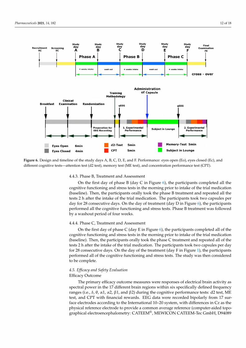
During the visits, the patient was isolated in a quiet, darkened room, sitting relaxed ina comfortable chair. EEG data were recorded twice: at baseline before drug administration(eyes open for 6 min followed by eyes closed for 4 min, during the d2 test for 5 min, theME test for 5 min, and the CPT for 5 min) and 120 min after drug administration (Figure 6).These test conditions were validated and standardized in our previous studies. Betweenmeasurements, the subjects relaxed in the leisure room. The experiments took place at thesame time of day, starting from 07:00.
4.4.1. Phase A, Screening and Training
On visit 1, the participants were checked for eligibility and informed about the detailsof the study, including restrictions—do not consume more than one cup of coffee a day (inthe morning) during the study and do not take medicines or dietary supplements that mayhave potential effects on their cognitive functions. They signed written informed consentand passed a routine medical examination to be eligible for inclusion in the trial.
4.4.2. Phase A, Treatment and Assessment
On day A (Figure 6), the participants completed all the cognitive functioning and stresstests in the morning (baseline). Then, the Principal Investigator (PI) randomly assignedthe participants to a treatment. The participants orally took the phase A treatment andrepeated all the tests 2 h after intake of the trial medication. The participants took twocapsules per day for 28 consecutive days. On the day of treatment (day B in Figure 6), theparticipants performed all the cognitive functioning and stress tests. Phase A treatmentwas followed by a washout period of four weeks.
Pharmaceuticals 2021, 14, 182 12 of 18Pharmaceuticals 2021, 14, x FOR PEER REVIEW 12 of 19
Figure 6. Design and timeline of the study days A, B, C, D, E, and F. Performance: eyes open (Eo), eyes closed (Ec), and different cognitive tests—attention test (d2 test), memory test (ME test), and concentration performance test (CPT).
4.4.1. Phase A, Screening and Training On visit 1, the participants were checked for eligibility and informed about the details
of the study, including restrictions—do not consume more than one cup of coffee a day (in the morning) during the study and do not take medicines or dietary supplements that may have potential effects on their cognitive functions. They signed written informed con-sent and passed a routine medical examination to be eligible for inclusion in the trial.
4.4.2. Phase A, Treatment and Assessment On day A (Figure 6), the participants completed all the cognitive functioning and
stress tests in the morning (baseline). Then, the Principal Investigator (PI) randomly as-signed the participants to a treatment. The participants orally took the phase A treatment and repeated all the tests 2 h after intake of the trial medication. The participants took two capsules per day for 28 consecutive days. On the day of treatment (day B in Figure 6), the participants performed all the cognitive functioning and stress tests. Phase A treatment was followed by a washout period of four weeks.
4.4.3. Phase B, Treatment and Assessment On the first day of phase B (day C in Figure 6), the participants completed all the
cognitive functioning and stress tests in the morning prior to intake of the trial medication (baseline). Then, the participants orally took the phase B treatment and repeated all the tests 2 h after the intake of the trial medication. The participants took two capsules per day for 28 consecutive days. On the day of treatment (day D in Figure 6), the participants performed all the cognitive functioning and stress tests. Phase B treatment was followed by a washout period of four weeks.
Figure 6. Design and timeline of the study days A, B, C, D, E, and F. Performance: eyes open (Eo), eyes closed (Ec), anddifferent cognitive tests—attention test (d2 test), memory test (ME test), and concentration performance test (CPT).
4.4.3. Phase B, Treatment and Assessment
On the first day of phase B (day C in Figure 6), the participants completed all thecognitive functioning and stress tests in the morning prior to intake of the trial medication(baseline). Then, the participants orally took the phase B treatment and repeated all thetests 2 h after the intake of the trial medication. The participants took two capsules perday for 28 consecutive days. On the day of treatment (day D in Figure 6), the participantsperformed all the cognitive functioning and stress tests. Phase B treatment was followedby a washout period of four weeks.
4.4.4. Phase C, Treatment and Assessment
On the first day of phase C (day E in Figure 6), the participants completed all of thecognitive functioning and stress tests in the morning prior to intake of the trial medication(baseline). Then, the participants orally took the phase C treatment and repeated all of thetests 2 h after the intake of the trial medication. The participants took two capsules per dayfor 28 consecutive days. On the day of the treatment (day F in Figure 1), the participantsperformed all of the cognitive functioning and stress tests. The study was then consideredto be complete.
4.5. Efficacy and Safety EvaluationEfficacy Outcome
The primary efficacy outcome measures were responses of electrical brain activity asspectral power in the 17 different brain regions within six specifically defined frequencyranges (i.e., δ, θ, α1, α2, β1, and β2) during the cognitive performance tests: d2 test, MEtest, and CPT with financial rewards. EEG data were recorded bipolarly from 17 sur-face electrodes according to the International 10–20 system, with differences in Cz as thephysical reference electrode to provide a common average reference (computer-aided topo-graphical electroencephalometry: CATEEM®, MEWICON CATEEM-Tec GmbH, D94089

Pharmaceuticals 2021, 14, 182 13 of 18
Neureichenau, Germany) using an electro-cap. For a detailed description of the procedure,please refer to reference [23].
The signals of all 99 electrode positions (17 real and 82 virtual) were subject to fastFourier transformation (FFT) based on 4 s sweeps of data epochs (Hanning window).The data were analyzed from 1.25 to 35 Hz using CATEEM® software. In this software,the resulting frequency spectra were divided into six frequency bands: δ (1.25–4.50 Hz),θ (4.75–6.75 Hz), α1 (7.00–9.50 Hz), α2 (9.75–12.50 Hz), β1 (12.75–18.50 Hz), and β2(18.75–35.00 Hz) [24]. Frequency analysis was based on absolute spectral power valuesand calculated as source density [24,25].
4.6. Safety Outcomes
Safety outcome measures—the occurrence and severity of AEs—were recorded fromthe date of randomization until the end of the trial. All AEs observed by a subject orinvestigators were recorded during the trial in detail, e.g., description of the event, onset(date and time), resolution (date and time), maximum intensity, action taken, outcome,and causality.
4.7. Sample Size Considerations
Thirty patients were enrolled per treatment phase. The sample size was not deter-mined during this pilot study. It was estimated based on the results of our previous studiescarried out under a similar experimental design.
4.8. Statistical Analysis
Statistical analysis was done as previously reported [23]. Electrical activity of the brainwas measured as absolute spectral power (µV2) in various experimental conditions.
Data from the first recording (baseline) were set as 100% and electrophysiologicalchanges produced by the placebo or the WG and HRG80® capsules are reported as percent-age changes. WG and HRG80® capsules versus the placebo were compared by evaluatingthe baseline recording of the last day in comparison to the recording 2 h after the intake. Anonparametric sign test was used for the comparison between the effects of the placebo,WG, and HRG80®. The p-values are provided for the statistics of the exploratory study.
Linear discriminant analysis according to Fischer was used for comparison with otherdrugs of herbal preparations, as previously reported [23].
5. Conclusions
Despite numerous studies conducted during the last few decades, ginseng is notyet recognized in Europe and the US as a medicinal product with well-established use.That is mainly due to the inconsistency of the results of clinical studies, where ginsengpreparations were not identical, and therefore reproducible efficacy from one study toanother has not been achieved. Reproducible efficacy and quality of wild ginseng cannotbe achieved because its environmental conditions are not regulated. Standardized methodsof cultivation and processing are required to assure reproducible quality and efficacy.The efficacy of cultivated preparations must be validated in clinical studies on humans,where the effects of cultivated ginseng (particularly hydroponically cultivated) on thecentral nervous system are compared with the effects of wild ginseng. To the best of ourknowledge, the only publication on this topic is the publication of the results of our recentof study of red ginseng preparation HRG80®, where superior efficacy of hydroponicallycultivated red ginseng compared to wild growing ginseng on the cognitive functions ofhealthy subjects was demonstrated (Mariage et al., 2020). However, no direct evidence onthe brain activity of HRG80® was available.
In this study, we, for the first time, demonstrate that:
• Red ginseng has an effect on the CNS in humans• Red ginseng has an effect on the CNS in elderly subjects with mild cognitive impairments

Pharmaceuticals 2021, 14, 182 14 of 18
• Cultivated in standard conditions, red ginseng has an effect on the CNS of elderlysubjects with mild cognitive impairments
• Hydroponically cultivated in standard conditions, red ginseng has an effect on theCNS in elderly subjects with mild cognitive impairments
• The overall effects of white and red ginseng on the electrical activity of the brain aredifferent, suggesting different pharmacological activity of the red and white ginsengpreparations
• A treatment duration of 4 weeks seems to be sufficient to uncover the action of ginsengon the activity of the human brain
No side effects were observed, suggesting also longer treatment periods withoutunwanted actions.
CONSORT 2010 Checklist is available in Appendix B.
Author Contributions: Project administration, methodology, investigation, formal analysis, valida-tion, data curation, software, resources, writing—original draft, editing, W.D.; Conceptualization,writing, editing, visualization, supervision, A.G.P.; Resources, funding acquisition, supervision,reviewing, P.-A.M. All authors have read and agreed to the published version of the manuscript.
Funding: This work was funded in part by EuroPharmaUSA (Green Bay, WI, USA) and BotalysSA (Ath, Belgium). The sponsors of the research were Terrence Lemerond, EuroPharmaUSA Inc.,USA; Pierre-Antoine Mariage, Botalys SA, Belgium; Paul-Evence Coppee, Botalys SA, Belgium (grantnumber 2019-02).
Institutional Review Board Statement: The study protocol (PP_0319_EuroPharma) and all of its amend-ments were reviewed and fully approved by Landesärztekammer Hessen, an independent ethics commit-tee in Hessen (Hanauer Landstraße 152, D-60314 Frankfurt am Main, Germany; Chairman: Prof. Dr. med.Sebastian Harder) on 13 July and 24 September 2019; case number 2019-1310-evBO.
Informed Consent Statement: All volunteers provided written informed consent to participate inthis study. The participants signed the consent form to state that the information had been explainedand that they understood. They received a copy of the form, while the original copy was maintainedin a confidential file in the investigator’s records.
Data Availability Statement: Data sharing not applicable due to privacy and force majeure reasons.
Acknowledgments: The author acknowledges the support of EuroPharmaUSA for the supply ofinvestigational products, their characterization, and material support. The author thanks JenniferHansgate and Terry Lemerond for reviewing the manuscript and providing critical comments. Theauthor thanks med. Klaus Koch for the recruitment of participants, the implementation of thetreatment, and performing the clinical examinations, as well as Sabine Gradl for quality assurance.Additionally, the author thanks Luiza Panosyan for monitoring the study and ensuring the imple-mentation of ICH guidelines in order to conduct the study according to Good Clinical Practice (GCP).The author is also grateful to all of the participants of the study.
Conflicts of Interest: P.A.M. declares a conflict of interest since he is co-CEO at Botalys SA (Ath,Belgium); A.P. has an independent-contractor agreement with EuroPharmaUSA Inc. and is headof the research and development company Phytomed AB. W.D. declares no conflicts of interestsand no significant financial benefits from any pharmaceutical company. The funders had no role inthe design of the study; in the collection, analyses, or interpretation of data; in the writing of themanuscript; or in the decision to publish the results.
Appendix A. Adverse Events, Tolerability and Vital Signs
Appendix A.1. Summary of Adverse Events
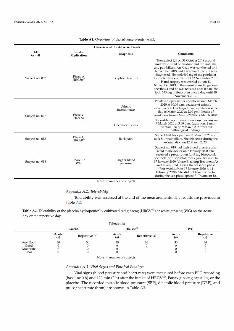
Adverse events (AEs) were detected in three subjects receiving the HRG80®, WG, orplacebo capsules, as shown in Table A1. The AEs were not related to the study medication.
Pharmaceuticals 2021, 14, 182 15 of 18
Table A1. Overview of the adverse events (AEs).
Overview of the Adverse Events
AE(n = 4)
StudyMedication Diagnosis Comments
Subject no. 007 Phase AHRG80® Scaphoid fracture
The subject fell on 31 October 2019 aroundmidday in front of his door and did not take
any painkillers. An X-ray was conducted on 1November 2019 and a scaphoid fracture wasdiagnosed. He took 600 mg of the painkiller
ibuprofen twice a day until 13 November 2019.Hand surgery was carried out on 13
November 2019 in the morning under generalanesthesia and he was released at 2:00 p.m. Hetook 600 mg of ibuprofen once a day until 18
November 2019.
Subject no. 007 Phase CPlacebo
Urinaryincontinence
Prostate biopsy under anesthesia on 6 March2020 at 10:00 a.m. because of urinary
incontinence. Discharge from hospital on sameday (6 March 2020 at 2.00 pm). Intake of
painkillers from 6 March 2020 to 7 March 2020.
Unconsciousness
The sudden occurrence of unconsciousness on7 March 2020 at 3:00 p.m. (duration: 2 min).
Examination on 9 March 2020 withoutpathological findings.
Subject no. 013 Phase CHRG80® Back pain
Subject had back pain on 11 March 2020 andtook four painkillers. She felt better during the
examination on 12 March 2020.
Subject no. 018 Phase B/WG
Higher bloodpressure
Subject no. 018 had high blood pressure andwent to the doctor on 7 January 2020. Shereceived a prescription for 5 mg bisoprolol.
She took the bisoprolol from 7 January 2020 to17 January 2020 (phase B, taking Treatment A)
and as required during the washout phase(four weeks, from 17 January 2020 to 11
February 2020). She did not take bisoprololduring the last phase (phase 3, Treatment B).
Note: n, number of subjects.
Appendix A.2. Tolerability
Tolerability was assessed at the end of the measurements. The results are provided inTable A2.
Table A2. Tolerability of the placebo hydroponically cultivated red ginseng (HRG80®) or white ginseng (WG) on the acuteday or the repetitive day.
Tolerability
Placebo HRG80® WG
Acute(n) Repetitive (n) Acute
(n) Repetitive (n) Acute(n)
Repetitive(n)
Very Good 30 30 30 30 30 30Good 0 0 0 0 0 0
Moderate 0 0 0 0 0 0Poor 0 0 0 0 0 0
Note: n, number of subjects.
Appendix A.3. Vital Signs and Physical Findings
Vital signs (blood pressure and heart rate) were measured before each EEG recording(baseline 0 h) and 120 min (2 h) after the intake of HRG80®, Panax ginseng capsules, or theplacebo. The recorded systolic blood pressure (SBP), diastolic blood pressure (DBP), andpulse/heart rate (bpm) are shown in Table A3.
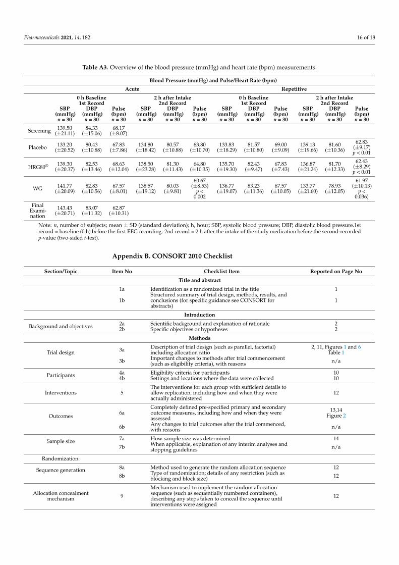
Pharmaceuticals 2021, 14, 182 16 of 18
Table A3. Overview of the blood pressure (mmHg) and heart rate (bpm) measurements.
Blood Pressure (mmHg) and Pulse/Heart Rate (bpm)
Acute Repetitive
0 h Baseline 2 h after Intake 0 h Baseline 2 h after Intake1st Record 2nd Record 1st Record 2nd Record
SBP DBP Pulse SBP DBP Pulse SBP DBP Pulse SBP DBP Pulse(mmHg) (mmHg) (bpm) (mmHg) (mmHg) (bpm) (mmHg) (mmHg) (bpm) (mmHg) (mmHg) (bpm)n = 30 n = 30 n = 30 n = 30 n = 30 n = 30 n = 30 n = 30 n = 30 n = 30 n = 30 n = 30
Screening 139.50(±21.11)
84.33(±15.06)
68.17(±8.07)
Placebo 133.20(±20.52)
80.43(±10.88)
67.83(±7.86)
134.80(±18.42)
80.57(±10.88)
63.80(±10.70)
133.83(±18.29)
81.57(±10.80)
69.00(±9.09)
139.13(±19.66)
81.60(±10.36)
62.83(±9.17)p < 0.01
HRG80® 139.30(±20.37)
82.53(±13.46)
68.63(±12.04)
138.50(±23.28)
81.30(±11.43)
64.80(±10.35)
135.70(±19.30)
82.43(±9.47)
67.83(±7.43)
136.87(±21.24)
81.70(±12.33)
62.43(±8.29)p < 0.01
WG 141.77(±20.09)
82.83(±10.56)
67.57(±8.01)
138.57(±19.12)
80.03(±9.81)
60.67(±8.53)
p <0.002
136.77(±19.07)
83.23(±11.36)
67.57(±10.05)
133.77(±21.60)
78.93(±12.05)
61.97(±10.13)
p <0.036)
FinalExami-nation
143.43(±20.71)
83.07(±11.32)
62.87(±10.31)
Note: n, number of subjects; mean ± SD (standard deviation); h, hour; SBP, systolic blood pressure; DBP, diastolic blood pressure.1strecord = baseline (0 h) before the first EEG recording. 2nd record = 2 h after the intake of the study medication before the second-recordedp-value (two-sided t-test).
Appendix B. CONSORT 2010 Checklist
Section/Topic Item No Checklist Item Reported on Page No
Title and abstract
1a Identification as a randomized trial in the title 1
1bStructured summary of trial design, methods, results, andconclusions (for specific guidance see CONSORT forabstracts)
1
Introduction
Background and objectives 2a Scientific background and explanation of rationale 22b Specific objectives or hypotheses 2
Methods
Trial design 3a Description of trial design (such as parallel, factorial)including allocation ratio
2, 11, Figures 1 and 6Table 1
3b Important changes to methods after trial commencement(such as eligibility criteria), with reasons n/a
Participants 4a Eligibility criteria for participants 104b Settings and locations where the data were collected 10
Interventions 5The interventions for each group with sufficient details toallow replication, including how and when they wereactually administered
12
Outcomes 6aCompletely defined pre-specified primary and secondaryoutcome measures, including how and when they wereassessed
13,14Figure 2
6b Any changes to trial outcomes after the trial commenced,with reasons n/a
Sample size 7a How sample size was determined 14
7b When applicable, explanation of any interim analyses andstopping guidelines n/a
Randomization:
Sequence generation 8a Method used to generate the random allocation sequence 12
8b Type of randomization; details of any restriction (such asblocking and block size) 12
Allocation concealmentmechanism 9
Mechanism used to implement the random allocationsequence (such as sequentially numbered containers),describing any steps taken to conceal the sequence untilinterventions were assigned
12

Pharmaceuticals 2021, 14, 182 17 of 18
Section/Topic Item No Checklist Item Reported on Page No
Implementation 10Who generated the random allocation sequence, whoenrolled participants, and who assigned participants tointerventions
12
Blinding 11aIf done, who was blinded after assignment to interventions(for example, participants, care providers, those assessingoutcomes) and how
12
11b If relevant, description of the similarity of interventions 12
Statistical methods12a Statistical methods used to compare groups for primary and
secondary outcomes 14
12b Methods for additional analyses, such as subgroup analysesand adjusted analyses n/a
Results
Participant flow (a diagramis strongly recommended)
13aFor each group, the numbers of participants who wererandomly assigned, received intended treatment, and wereanalyzed for the primary outcome
Figure 1
13b For each group, losses and exclusions after randomization,together with reasons n/a
Recruitment 14a Dates defining the periods of recruitment and follow-up 10, Figure 614b Why the trial ended or was stopped n/a
Baseline data 15 A table showing baseline demographic and clinicalcharacteristics for each group Table 1
Numbers analyzed 16For each group, number of participants (denominator)included in each analysis and whether the analysis was byoriginal assigned groups
Figure 1
Outcomes and estimation17a
For each primary and secondary outcome, results for eachgroup, and the estimated effect size and its precision (such as95% confidence interval)
9–13Figures 2–4
Table 2
17b For binary outcomes, presentation of both absolute andrelative effect sizes is recommended
Figures 2–4Appendix A
Ancillary analyses 18Results of any other analyses performed, including subgroupanalyses and adjusted analyses, distinguishing pre-specifiedfrom exploratory
n/a
Harms 19 All important harms or unintended effects in each group 14, Appendix A
Discussion
Limitations 20 Trial limitations, addressing sources of potential bias,imprecision, and, if relevant, multiplicity of analyses 9
Generalizability 21 Generalizability (external validity, applicability) of the trialfindings 8,9
Interpretation 22 Interpretation consistent with results, balancing benefits andharms, and considering other relevant evidence 8, 9
Other information
Registration 23 Registration number and name of trial registry 10
Protocol 24 Where the full trial protocol can be accessed, if available 15
Funding 25 Sources of funding and other support (such as supply ofdrugs), role of funders 15
References1. National Pharmacopoeia Committee. Pharmacopoeia of the People’s Republic of China; National Pharmacopoeia Committee: Beijing,
China, 2010.2. Panossian, A.G.; Efferth, T.; Shikov, A.N.; Pozharitskaya, O.N.; Kuchta, K.; Mukherjee, P.K.; Banerjee, S.; Heinrich, M.; Wu, W.;
Guo, D.A.; et al. Evolution of the adaptogenic concept from traditional use to medical systems: Pharmacology of stress- andaging-related diseases. Med. Res. Rev. 2021, 41, 630–703. [CrossRef] [PubMed]
3. Liao, L.Y.; He, Y.F.; Li, L.; Meng, H.; Dong, Y.M.; Yi, F.; Xiao, P.G. A preliminary review of studies on adaptogens: Comparison oftheir bioactivity in TCM with that of ginseng-like herbs used worldwide. Chin. Med. 2018, 13, 57. [CrossRef] [PubMed]
4. Patel, S.; Rauf, A. Adaptogenic herb ginseng (Panax) as medical food: Status quo and future prospects. Biomed. Pharmacother.2017, 85, 120–127. [CrossRef] [PubMed]
5. Baeg, I.H.; So, S.H. The world ginseng market and the ginseng (Korea). J. Ginseng Res. 2013, 37, 1–7. [CrossRef] [PubMed]6. Radad, K.; Gille, G.; Liu, L.; Rausch, W.D. Use of ginseng in medicine with emphasis on neurodegenerative disorders. J. Pharmacol.
Sci. 2006, 100, 175–186. [CrossRef] [PubMed]7. So, S.H.; Lee, J.W.; Kim, Y.S.; Hyun, S.H.; Han, C.K. Red ginseng monograph. J. Ginseng Res. 2018, 42, 549–561. [CrossRef]
[PubMed]
Pharmaceuticals 2021, 14, 182 18 of 18
8. World Health Organization (WHO), Radix Ginseng. WHO Monographs on Medicinal Plants Commonly Used in the Newly IndependentStates (NIS); World Health Organization: Genova, Switzerland, 2010; pp. 141–160.
9. European Medicines Agency. EMA/HMPC/321232/2012. Assessment Report on Panax Ginseng C.A. Meyer, Radix. Based on Article 16d(1), Article 16f and Article 16h of Directive 2001/83/EC as Amended (Traditional Use); European Medicines Agency: Amsterdam, TheNetherlands, 2014.
10. Blumenthal, M. The ABC Clinical Guide to Herbs; Theime: Austin, TX, USA, 2003; p. 211e25.11. Panax ginseng. Monograph. Altern Med. Rev. 2009, 14, 172.e6.12. Blumenthal, M.; Goldberg, A.; Brinkmann, J. Herbal Medicine: Expanded Commission E Monographs; Integrative Medicine Commu-
nications: Austin, TX, USA, 2000; p. 170.e7.13. European Directorate for the Quality of Medicines & Health Care. Ginseng radix. In European Pharmacopoeia, Monograph
01/2008:1523; European Directorate for the Quality of Medicines & Health Care: Strasburg, France, 2008.14. Zhou, Q.L.; Zhu, D.N.; Yang, X.W.; Xu, W.; Wang, Y.P. Development and validation of a UFLC-MS/MS method for simultaneous
quantification of sixty-six saponins and their six aglycones: Application to comparative analysis of red ginseng and white ginseng.J. Pharm. Biomed. Anal. 2018, 159, 153–165. [CrossRef] [PubMed]
15. Lim, C.Y.; Moon, J.M.; Kim, B.Y.; Lim, S.H.; Lee, G.S.; Yu, H.S.; Cho, S.I. Comparative study of Korean White Ginseng and KoreanRed Ginseng on efficacies of OVA-induced asthma model in mice. J. Ginseng Res. 2015, 39, 38–45. [CrossRef] [PubMed]
16. Han, M.J.; Kim, D.H. Effects of Red and Fermented Ginseng and Ginsenosides on Allergic Disorders. Biomolecules 2020, 10, 634.[CrossRef] [PubMed]
17. Yoon, D.; Shin, W.C.; Lee, Y.S.; Kim, S.; Baek, N.I.; Lee, D.Y. A Comparative Study on Processed Panax ginseng Products UsingHR-MAS NMR-Based Metabolomics. Molecules 2020, 25, 1390. [CrossRef] [PubMed]
18. Mariage, P.A.; Hovhannisyan, A.; Panossian, A.G. Efficacy of Panax ginseng Meyer Herbal Preparation HRG80 in Preventingand Mitigating Stress-Induced Failure of Cognitive Functions in Healthy Subjects: A Pilot, Randomized, Double-Blind, Placebo-Controlled Crossover Trial. Pharmaceuticals 2020, 13, 57. [CrossRef] [PubMed]
19. Lemerond, T.; Panossian, A.G. Panax ginseng Meyer Herbal Preparation HRG80 for Preventing and Mitigating Stress-InducedFailure of Cognitive Functions in Healthy Subjects. J. Altern Complement Integr. Med. 2020, 6, 100.
20. Dimpfel, W.; Schombert, L.; Panossian, A.G. Panax Ginseng Preparations Enhance Long Term Potentiation in Rat HippocampalSlices by Glutamatergic NMDA and Kainate Receptor Mediated Transmission. J. Altern Complement Integr. Med. 2020, 6, 106.
21. Dimpfel, W. Neurophysiological Biomarker of Mild Cognitive Impairment. Adv. Alzheimers Dis. 2014, 3, 64–77. [CrossRef]22. Dimpfel, W.; Schombert, L.; Biller, A. Psychophysiological Effects of Sideritis and Bacopa Extract and Three Combinations
Thereof—A Quantitative EEG Study in Subjects Suffering from Mild Cognitive Impairment (MCI). Adv. Alzheimers Dis. 2016, 5,1–22. [CrossRef]
23. Dimpfel, W.; Schombert, L.; Keplinger-Dimpfel, I.K.; Panossian, A. Effects of an Adaptogenic Extract on Electrical Activity ofthe Brain in Elderly Subjects with Mild Cognitive Impairment: A Randomized, Double-Blind, Placebo-Controlled, Two-ArmedCross-Over Study. Pharmaceuticals 2020, 13, 45. [CrossRef] [PubMed]
24. Schober, F.; Schellenberg, R.; Dimpfel, W. Reflection of Mental Exercise in the Dynamic Quantitative Topographical EEG.Neuropsychobiolgy 1995, 31, 98–112. [CrossRef] [PubMed]
25. Dimpfel, W.; Hofmann, H.C.; Prohaska, A.; Schober, F.; Schellenberg, R. Source density analysis of functional topographical EEG:Monitoring of cognitive drug action. Eur. J. Med. Res. 1996, 1, 283–290. [PubMed]