effects ofx-radiation onmouse fetus …jnm.snmjournals.org/content/10/2/68.full.pdfcerebral lesions,...
TRANSCRIPT

Eiposures to ionizing radiation can threaten healthand life, increase mutations and damage the fetus.The type and extent of lesions produced in the fetusdepend greatly on the gestational stage and the doseof ionizing radiation received (1—4). Most tissuesshow greatest sensitivity to irradiation during theearly stage of organogenesis, but the brain continuesto be very susceptible to damage during its prolongedperiod of differentiation and growth. For understandable reasons interest in irradiation effects onliving organisms has increased greatly in recent dccades, and many pertinent papers have been published. It would be beyond the scope of this articleto review the vast literature on the subject; interestedreaders are referred to original articles and specialreviews (1—13).
The purpose of this investigation is twofold: First,it is a complementary study to Part I, “Abnormalities in children exposed to x-radiation during variousstages of gestation : tentative timetable of radiationinjury to the human fetus―(8) . The existing publications could not fill the particular need of providingtime-specific pathological correlates for interpretation of the findings in these 26 children exposed toirradiation in utero. Second, this investigation is intended to provide semiquantitative data on differential sensitivity to irradiation of phylogeneticallydifferent cerebral structures. These particular findings are important in interpreting one of the mostcommon features in surviving children after irradiation in utero : type and degree of mental retardation (8).
MATERIAL AND METhODS
The experiments were carried out on the WebsterSwiss albino strain of mice. Five to six-months-oldfertile females were mated between 7 pm and 9 amthe next morning. The fetal age on that morning was
considered as zero day although the actual age wasin a range between 0 and 14 hr. Twelve groups ofpregnant females totaling 152 were irradiated oneach consecutive day between the 7th and 18th dayof gestation. To prevent loss of some abnormal newboms due to cannibalism, Caesarean section dclivcries were carried out in all instances on the 19thday of gestation (spontaneous birth in mice occurson the 19th and 20th day of pregnancy) . The litterand contents of the uterus were examined with lowpower magnification for gross abnormalities. Fourtransverse blocks from the head and two from thethorax were processed for histological studies. Thesections were cut at 8 microns and stained withhemotoxylin-cosin. In selected sections, trichromeand silver stains were also used. To obtain someinformation on acute cell damage, a few of theirradiated mice were sacrificed 12 hr after irradiation and fetuses processed for microscopic studies.
All mice were exposed to 200 R. The parametersof irradiation were: 200 kv constant potential, 0.25mm Cu + 0.55 mm Al filter, HVL 0.9 mm Cu,source distance to midline of animals 54 cm, cxposure rate in air 60 R/min. Irradiation exposurerate was measured in air with a Victoreen dosimeter
next to the mouse uterus before, during and afterthe animals were exposed. The mice were maintainedin specially designed Lucite box containers with alead plate to shield the head and upper one fourthof the thorax of the pregnant mice. Stained serialsections of the brain of normal control animals of17, 18, 19 and 20 days of gestation were availableat the time microscopic examination of the brains
Received Feb. 27, 1968; revision accepted July 18, 1968.For reprints contact: A. S. Dekaban, Section of Child
Neurology, National Institute of Neurological Diseases andBlindness, Building 10, Room 4N-250, National Institutesof Health, Bethesda,Maryland 20014.
68 JOURNAL OF NUCLEAR MEDICINE
EFFECTS OF X-RADIATION ON MOUSE FETUS DURING
GESTATION: EMPHASIS ON DISTRIBUTION OF
CEREBRAL LESIONS, PART II
Anatole S. Dekaban
National Institute of Neurological Diseases and Blindness,National Institutes of Health, Bethesda, Maryland
by on July 1, 2018. For personal use only. jnm.snmjournals.org Downloaded from
IrradiatedNo. preg
Totalno.MeanNo.
animalsalive& qppar
No. grosslyapparentabnormalitiesSmall
orKinkedgest.nantanilitterentlydeformedLargeorshortdaysmicemalssizenormal
Stunted Dysraphismheadheadtail Others
of irradiated animals was made. This allowed us tocompare the stage of development of various structures of the brain in irradiated and nonirradiatedmice.
RESULTS
Because it has been shown previously that irradiation of pregnant mice up to 7 days of gestationleads to a high mortality rate among fetuses whilethe few survivors are apparently normal (2,13) , webegan irradiation in this series with the 7th day ofpregnancy.
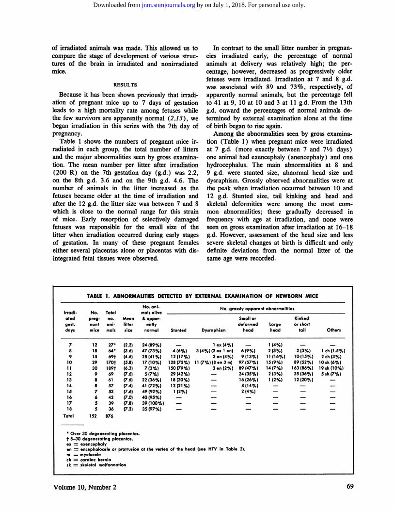
Table 1 shows the numbers of pregnant mice irradiated in each group, the total number of littersand the major abnormalities seen by gross examination. The mean number per litter after irradiation(200 R) on the 7th gestation day (g.d.) was 2.2,on the 8th g.d. 3.6 and on the 9th g.d. 4.6. Thenumber of animals in the litter increased as thefetuses became older at the time of irradiation andafter the 12 g.d. the litter size was between 7 and 8which is close to the normal range for this strainof mice. Early resorption of selectively damagedfetuses was responsible for the small size of thelitter when irradiation occurred during early stagesof gestation. In many of these pregnant femaleseither several placentas alone or placentas with disintegrated fetal tissues were observed.
In contrast to the small litter number in pregnancies irradiated early, the percentage of normalanimals at delivery was relatively high; the percentage, however, decreased as progressively olderfetuses were irradiated. Irradiation at 7 and 8 g.d.was associated with 89 and 73 % , respectively, ofapparently normal animals, but the percentage fellto 41 at 9, 10 at 10 and 3 at 11 g.d. From the 13thg.d. onward the percentages of normal animals determined by external examination alone at the timeof birth began to rise again.
Among the abnormalities seen by gross examination (Table 1) when pregnant mice were irradiatedat 7 g.d. (more exactly between 7 and 7½ days)one animal had exencephaly (anencephaly) and onehydrocephalus. The main abnormalities at 8 and9 g.d. were stunted size, abnormal head size anddysraphism. Grossly observed abnormalities were atthe peak when irradiation occurred between 10 and12 g.d. Stunted size, tail kinking and head andskeletal deformities were among the most common abnormalities; these gradually decreased in
frequency with age at irradiation, and none wereseen on gross examination after irradiation at 16—18g.d. However, assessment of the head size and lesssevere skeletal changes at birth is difficult and onlydefinite deviations from the normal litter of thesame age were recorded.
TABLE 1. ABNORMALITIES DETECTEDBY EXTERNAL EXAMINATION OF NEWBORN MICE
12181529309887655
152
1ex(4%)3(4%)(2ex 1 en)
3 en (4%)11 (7%)(8en3m)
3 en (2%)
1 (4%)2(3%)
11(16%)15(9%)14(7%)2(3%)1(2%)
789
101112131415161718
Total
2764*69t
170f189t69615753423936
(2.2)(3.6)(4.6)(5.8)(6.3)(7.6)(7.6)(7.4)(7.6)(7.0)(7.8)(7.2)
24(89%)47(73%)28 (41 0/s)17(10%)7(3%)5(7%,)22(36%)41 (72%)49(92%)40(95%)39(100%)35(97%)
4(6%)12(17%)
128(75%)150(79%)29(42%)18(30%)12(21%)
1(2%)
6(9%)9(13%)97(57%)89(47%)24(35%)16(26%)8(14%)2(4%)
2(3%)10 (15%)89(52%)
163(86%)25 (36%)12 (20%)
1 ch(1.5%)2ch(3%)
10 sk (6%)19 sk (10%)Ssk(7%)
876
* Over 30 degenerating placentas.
t 8—30degeneratingplacenta:.cx = exencephalyen encephalocele or protrusion at the vertex of the head (see HTV in Table 2).m myelocelech cardiac herniask skeletal malformation
69Volume 10, Number 2
by on July 1, 2018. For personal use only. jnm.snmjournals.org Downloaded from
Smallbrain,TotalMacer.otherwiseIrradiatedno.&
notNo. abnorm.Malform.&normalgest.daysbrainsstudiedbr&nsfotherlesionsHydroceph.structure72712(8%)1—
DEKABAN
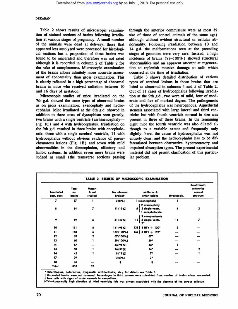
Table 2 shows results of microscopic examination of stained sections of brains following irradiation at various stages of pregnancy. A small numberof the animals were dead at delivery; those thatappeared less autolyzed were processed for histological sections but a proportion of these brains wasfound to be maccrated and therefore was not ratedalthough it is recorded in column 2 of Table 2 forthe sake of completeness. Microscopic examinationof the brains allows infinitely more accurate assessment of abnormality than gross examination. Thisis clearly reflected in a high percentage of abnormalbrains in mice who received radiation between 10and 16 days of gestation.
Microscopic studies of mice irradiated on the7th g.d. showed the same types of abnormal brainsas on gross examination: exencephaly and hydro-.cephalus. Mice irradiated at the 8th g.d. showed inaddition to three cases of dysraphism seen grossly,two brains with a single ventricle (arrhinencephaly—.Fig. 1C) and 4 with hydrocephalus. Irradiation onthe 9th g.d. resulted in three brains with encephalo-.cele, three with a single cerebral ventricle, 11 withhydrocephalus without obvious evidence of parenchymatous lesions (Fig. 1B) and seven with mildabnormalities in the diencephalon, olfactory andlimbic systems. In addition seven more brains werejudged as small (the transverse sections passing
through the anterior commissure were at most ½size of those of control animals of the same age)although without evident structural or cellular abnormality. Following irradiation between 10 and14 g.d. the malformations seen at the precedingstages of gestation were very rare. Instead, a highincidence of brains (98—100%) showed structuralabnormalities and an apparent attempt at regeneration to replenish massive damage to cells whichoccurred at the time of irradiation.
Table 3 shows detailed distribution of varioustypes of cerebral lesions in those brains that arelisted as abnormal in columns 4 and 5 of Table 2.Out of 11 cases of hydrocephalus following irradiaton at the 9th g.d., two were of mild, four of modcrate and five of marked degree. The pathogenesisof the hydrocephalus was heterogenous. Aqueductalstenosis associated with large lateral and third yentricles but with fourth ventricle normal in size waspresent in three of these brains. In the remainingeight mice the fourth ventricle was also dilated although to a variable extent and frequently . onlyslightly; here, the cause of hydrocephalus was notentirely clear, and the hydrocephalus has to be differentiated between obstructiye, hypersecretory andimpaired absorption types. The present experimentalmaterial did not permit clarification of this particular problem.
TABLE 2. RESULTSOF MICROSCOPIC EXAMINATION
1 (exencephaly)
( 2 exencephaly11(19%) 5j 2slngIev.ntr.
â€1̃ encephalocele
( 3.ncephalocel.31 (49%) 13@ 3 sIngle ventr.
â€7̃*
8 64 7 4 2
9 69 5 11 7
10 151 8 141(98%) 138(8HTV+130* 3
11 168 6 162(100%) 162(3HTV+159 —
12 69 2 67(100%) 67
13 60 1 59(100%) 59
14 57 — 56(98%) 55@ 1 —15 53 1 26(50%) 24* 2
16 42 1 8(19%) 7* 1
17 39 — 2(5%) 2*
18 36 — * t — —
Total 835 32
* Heterotopias, deformities, dysgenetic architectonics, etc.: for details see Table 3.
t Maccratedbrainswerenotassessed.Percentag.sIn thirdcolumnwerecalculatedfromnumberof brainsminusmaccrated.* Rarecells with signsof acutenecrosisIn neopalllum.HTV—Abnormally high situation of third ventricle; this was always associated with the absence of th. corpus callosum.
70 JOURNAL OF NUCLEAR MEDICINE
by on July 1, 2018. For personal use only. jnm.snmjournals.org Downloaded from
EFFECT OF X-RADIATION ON BRAIN
,,
@$.@ \j@
@ . - -.@ -@ .. ,@.- . .@@@ .‘.,-.@..@... . .. ‘@@ .@ .,.- .@ :•“. @‘@:@
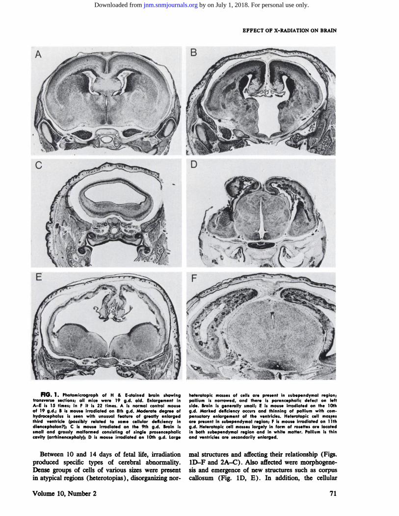
FIG. 1. Photomicrographof H & E-stalnedbrain showingtransverse sections; all mice were 19 g.d. old. Enlargement inA—Eis 15 times; in F it is 22 times. A Is normal control mouseof 19 g.d.; B is mouse irradiated on 8th g.d. Moderate degree ofhydrocephalus is seen with unusual feature of greatly enlargedthird ventricle (possibly related to some cellular deficiency Indiencephalon?); C is mouse irradiated on the 9th g.d. Brain Issmall and grossly malformed consisting of single prosencephallccavity (arrhinencephaly); D Is mouse irradiated on 10th g.d. Large
heterotopic massesof cells are present in subependymal region:pallium is narrowed, and there is porencephalic defect on leftside. Brain is generally small; E is mouse irradiated on the 10thg.d. Marked deficiency occursand thinning of pallium with cornpensatory enlargement of the ventricles. Heterotopic cell massesare present in subependymalregion; F Is mouseIrradiated on 1lthgd. Heterotopic cell masseslargely in form of rosettesare locatedin both subependymal region and In white matter. Pallium is thinand ventricles are secondarily enlarged.
mal structures and affecting their relationship (Figs.iD—Fand 2A—C).Also affected were morphogenesis and emergence of new structures such as corpuscallosum (Fig. 1D, E). In addition, the cellular
71
Between 10 and 14 days of fetal life, irradiationproduced specific types of cerebral abnormality.Dense groups of cells of various sizes were presentin atypical regions (heterotopias), disorganizing nor
Volume 10, Number 2
by on July 1, 2018. For personal use only. jnm.snmjournals.org Downloaded from

Irradl.No. abnor.Damage,repair,
regen., malform. &othersHeterotopic
cellatedbrainsmasses:rosettes,gest.from
col. 3,Malform. of earlycords, clumps;Second ventr.Small groupsofatiplcaldaysTable2organogenesisdamage & repairdilatationcells; mm. archit. imperf.
DEKABAN
TABLE 3. DETAILED DISTRIBUTION OF TYPES OF ABNORMAUT1ES FROM TABLE 2
7 2(8%) 2{
8 9(16%) 9
9 24(37%) 17{
1 exencephaly1 hydroceph.2 exencephaly2 single ventr.1 encephalocele4 hydroceph.3 encephaloceles3 single ventr.
11 hydroceph.mild 2mod. 4mark. 5
2 few (dienc. & olf.) 5 (dienc. & olf.)
( [email protected]@ mod.am@t.
â€m̃any( smallam't.
1611 mod.am@t.â€m̃any( smallam@t.
67@ mod.am@t.â€m̃any( smallam@t.
59@ [email protected]' many
( few55@ smallam@t.
â€m̃od.am't.
19 mild 1831 121 mod. 3383 mark. 7012 ( mild 2138 159@ mod. 42
111 ‘mark. 9642 ( mild 4216 67@ mod. 169 ‘mark. 9
24 ( mild 1829 53@ mod. 29
6 @mark. 639 (mm. 314 7@ mild 22 ‘mod. 2
10 141(98%) 11 {@ hydroceph. mod.8 HTV'
11 162(100%) 3 HTV
12 67(100%)
13 59(100%)
14 56(98%)
2 c 24 mm. lmperf.neopal.— 6@ 2 mild imperf. neopal.
— 8 mm. imperf. neopal.
— 2 mm. imperf. neopal.
15 26(50%)
161718
8(19%)2(5%)
t
S HTV—High situation of the third ventricle.
t Rarenecroticcells.
complement of many structures was deficient, thedegree of low cellularity being greatest when irradiation occurred on 10—12g.d.
The heterotopias present in fetuses irradiated onthe 10th day consisted of large, solid islands of immature cells located mainly in the subependymalregion (Fig. 1D, E). In fetuses irradiated on the1lth and 12th g.d. heterotopias were extensive andconsisted of numerous cones of cells with centrallumen and radially arranged cells imitating theneural tube (Fig. lF, Fig. 2A) . In addition, clustersof cells and large groups of cells in a form of solidcords were seen. When heterotopic cell masses werepresent in the cortex and basal ganglia, they produced distortion of these structures. In many brainsthe presence of heterotopias, which can be considered an attempt at regeneration, was not sufficientto supplement destroyed cellular elements at thetime of irradiation and as a result of this compensatory ventricular enlargement occurred (Fig. 1E, F).
Irradiation during the 13th and 14th g.d. also ledto a high incidence of cerebral abnormality but theheterotopias were much smaller and discrete andthe nervous parenchyma less deficient (Fig. 2B);here ventricular dilatation, if present, was mild. Irradiation during 15—17 g.d. was associated only withisolated minor architectonic imperfections in the neocortex and rarely with a small nest of cells (Fig. 2C).Irradiated fetuses on the 18th g.d. were sacrificed24 hr later. Here, the microscopic lesions were veryrare and consisted of scattered necrotic cells in theneopalhium. It must be stressed, however, that prescut experimental data are derived from the microscopic examination at the time of birth when certaincerebral structures have not yet reached their mature condition. Cowen and Geller (9) and Hicks(10) have found abnormalities in the brain of adultrats which were irradiated during last days of fetallife.
The degree of compensatory ventricular dilata
72 JOURNAL OF NUCLEAR MEDICINE
by on July 1, 2018. For personal use only. jnm.snmjournals.org Downloaded from
EFFECT OF X-RADIATION ON BRAIN
ton corresponded to the amount of initial tissuedamage, extent of regeneration and stage of gestationwhen ionizing radiation was given. The details canbe seen by comparing the data in column 4 and 5of Table 3 in the corresponding gestational days.
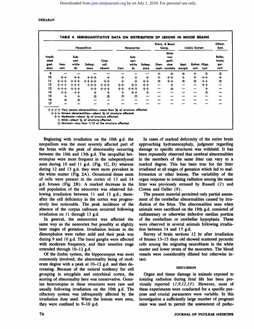
A preliminary survey of the brain sections revealed that many of the observed lesions tended togroup in a rather characteristic topographic distribution which changed depending on the gestationalstage at the time irradiation took place. It was therefore decided to record the distribution of varioustypes of lesions in the major brain subdivisions,noting also predominant involvement of the subependymal region, white matter and cortex. In general, the brain can be conveniently subdivided intothe following functional systems: (1 ) neopallium(neocortex—all dorsolateral cortex above the rhinalfissure; underlying white matter including corpuscallosum), (2) mesocortex (cingulate and retrosplenial region), (3) diencephalon and basal ganglia,(4) limbic system (hippocampal formation, entorhinal and prepyriform cortex, amygdala and septal
A
nuclei), (5 ) olfactory system (olfactory bulbs, tractsand tubercie; pyriform lobe) and (6) brainstem including cerebellum.
The last of the six subdivisions received little attention because the cerebellum begins to develop latein the fetal stage and its differentiation continuesthrough the early postnatal life whereas for the purpose of this study the animals were sacrificed on the19th day c?fgestation. Moreover, to properly assessbrainstem lesions, it would have been necessary to
include many intermediary stages between the dayof irradiation and delivery at 19 days. Nevertheless,brief reference to early cerebellar lesions will bemade.
Table 4 contains semiquantitative data on theseverity and distribution of abnormalities in themain subdivisions of the brain after irradiation atvarious stages of pregnancy. In a small proportionof brains, irradiation up to the 9th day of gestationcaused organ-type malformation but few focal lesions; these were located only in the diencephalon,limbic and olfactory systems.
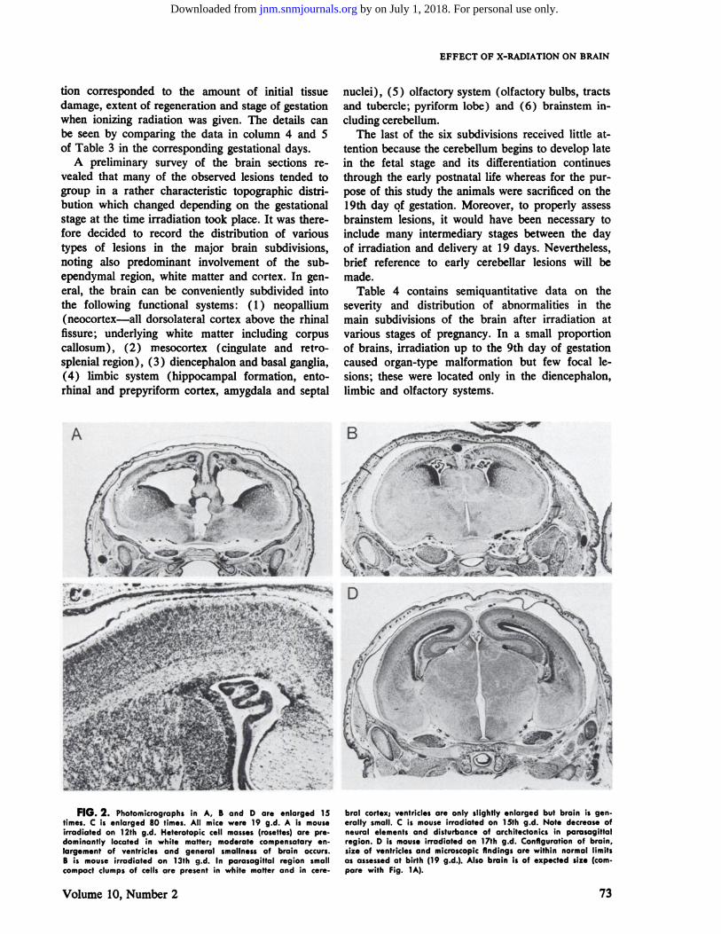
FIG. 2. Photomicrographsin A, B and D are enlarged15times. C is enlarged 80 times. All mice were 19 g.d. A is mouseirradiated on 12th g.d. Heterotopic cell masses (rosettes) are pre.dominantly located in white matter; moderate compensatory enlargement of ventricles and general smallness of brain occurs.B is mouse irradiated on 13th g.d. In parasagittal region smallcompact clumps of cells are present in white matter and in cere
bral cortex; ventricle: are only slightly enlarged but brain is generally small. C is mouse irradiated on 15th g.d. Note decrease ofneural elements and disturbance of architectonics in parasagittalregion. D is mouse irradiated on 17th g.d. Configuration of brain,size of ventriclesand microscopicfindings are within normal limitsas assessedat birth (19 g.d.). Also brain is of expected size (campare with Fig. 1A).
Volume 10, Number 2 73
by on July 1, 2018. For personal use only. jnm.snmjournals.org Downloaded from
-NeopalliumMesocortexDienc.& BasalGang.LImbic System @Olfact.Syst.Irradi
atedgest.daysNeo cort.Sub
cod.white
M.Subep.zoneCorp.call.
absent.Sub.
cart.white
Cod. M.Subep.zoneStria
turnpalli
Dien- dumceph. caudateSept.amygd.Enthor.
Hipp.cart. syst.Bulbs,
tracts,pyr.coil.
DEKABAN
TABLE 4. SEMIQUANTITATIVE DATA ON DISTRIBUTION OF LESIONS IN MOUSE BRAINS
+ ± ± + ±+ ++ + +++± ++ ± ±++- + —
— ± — — +
— — — — -I-
4-
+-5.
9 —10 ++11 +++12 +++13 +++14 ++15 +16 ±17 —
+++++++++++
+++-5-
+++++++
+++++
+-5-
+++
++++++
+4-
++
++++
+-I-
++
+++++
++-5-
+++++++
+
++++ Verysevereabnormalities—morethan3/4of structureaffected.+++ Sever.abnormalities—about1/3of structureaffected.
++ Moderate—about‘/@ofstructureaffected.+ Mild—about‘/of structureaffected.±Minimal—lessthan 1/10 of the structureaffected.
Beginning with irradiation on the 10th g.d. theneopallium was the most severely affected part ofthe brain with the peak of abnormality occurringbetween the 10th and 13th g.d. The neopallial beterotopias were most frequent in the subependymalzone during 10 and 11 g.d. (Fig. 1C, D) whereasduring 12 and 13 g.d. they were more prevalent inthe white matter (Fig. 2A) . Occasional dense nestsof cells were present in the cortex of 13 and 14g.d. fetuses (Fig. 2B). A marked decrease in thecell population of the neocortex was observed following irradiation between 11 and 13 g.d. ; thereafter the cell deficiency in the cortex was progressively less noticeable. The peak incidence of theabsence of the corpus callosum occurred followingirradiation on 11 through 13 g.d.
In general, the mesocortex was affected thesame way as the neocortex but possibly at slightlylater stages of gestation. Irradiation lesions in thediencephalon were rather mild and their peak wasduring 9 and 10 g.d. The basal ganglia were affectedwith moderate frequency, and their sensitive stageextended through 10—12g.d.
Of the limbic system, the hippocampus was mostcommonly involved, the abnormality being of modcrate degree with a peak at 10—12g.d. and then decreasing. Because of the natural tendency for cellgrouping in amygdala and entorhinal cortex, thescoring of abnormality here was conservative. Genuinc heterotopias in these structures were rare andusually following irradiation on the 10th g.d. Theolfactory system was infrequently affected by theirradiation dose used. When the lesions were seen,they were confined to 9—10g.d.
In cases of marked deformity of the entire brainapproaching hydranencephaly, judgment regardingdamage to specific structures was withheld. It hasbeen repeatedly observed that cerebral abnormalitiesin the members of the same litter can vary to amarked degree. This has been true for the litterirradiated at all stages of gestation which led to malformation or other lesions. The variabifity of theorgan response to ionizing radiation among the samelitter was previously stressed by Russell (2) andCowen and Geller (9).
The present material permitted only partial assessment of the cerebellar abnormalities caused by irradiation of the fetus. The abnormalities seen whenanimals were sacrificed on the 19th g.d. consisted ofrudimentary or otherwise defective median portionof the cerebellum or cerebellar hypoplasia. Thesewere observed in several animals following irradiation between 14 and 17 g.d.
Survey of brain sections 12 hr after irradiationof fetuses 13—15 days old showed scattered pycnoticcells among the migrating neuroblasts in the whitematter and lower strata of the neocortex. The bloodvessels were considerably dilated but otherwise intact.
DISCUSSION
Organ and tissue damage in animals exposed toionizing radiation during fetal life has been previously reported (1,9,12,13). However, most ofthese experiments were conducted for a specific purpose and crucial parameters were variable. In thisinvestigation a sufficiently large number of pregnantmice was used to permit the assessment of patho
74 JOURNAL OF NUCLEAR MEDICINE
by on July 1, 2018. For personal use only. jnm.snmjournals.org Downloaded from
EFFECT OF X-RADIATION ON BRAIN
logical lesions in 12 groups of litters between 7 and18 g.d. In this study, the gestational age at the timeof irradiation was the main variable. Duration ofgestation after irradiation was dependent on the
main variable since all 12 groups of animals weredelivered on the 19th day of pregnancy. The studywas designed to provide as closely comparable dataas possible to the available human material, especially for help in interpretation of the most commonand serious condition: mental retardation associatedwith microcephaly resulting from irradiation of thepregnant mothers (8).
Observations of Russell (2) that a medium dose(100—300 R) of irradiation during earlier stagesof gestation led to apparently normal newborn animals but a small size of litter were confirmed. Thereason for this state is not clear but it is possiblethat the damaging effect of irradiation is sufficientlygreat for certain fetuses of the same litter to causetheir death (in multiple pregnancies slight asynchrony of development is to be expected), whereasthe others who survive the insult go on to developnormally. It may be recalled that the primitivestreak stage in the mouse embryo occurs at 6½g.d. (14) ; the neural plate appears at 7½ and theneural tube begins to form at 8 g.d. At 9½ daysafter conception the mouse brain is at one cerebralvesicle stage. Thus, organ formation in the mouseembryo does not commence until about the 8th gestational day and the primitive embryonic layers(still pluripotential) are probably more able to regenerate. Gross organ-type malformation of thebrain without evident secondary lesions from the initial damage occurred when irradiation was given at8 and 9 days of gestation. Here, the abnormalitiesincluded exencephaly, arrhinenceplialy, grossly hypoplastic brain and hydrocephalus. Thus irradiationat this stage caused sufficient damage to some animals to cause fetal death (see Table 1) , the othersescaped without apparent abnormality of the brain,and a proportion suffered damage to the cephalicneural folds (8 days of gestation) or neural tube (9days of gestation) which led to dysraphism or prevented formation of two cerebral hemispheres fromthe stage of a single prosencephalic vesicle. In asmall proportion of brains, minimal abnormalitieswere also seen in the diencephalon and the primaryolfactory area.
Irradiation between 10 and 13 days of gestationproduced startling abnormalities. Here, predominantlesions, as seen at the time of birth, consisted of agreatly reduced amount of cerebral tissue with cornpensatory enlargement of the ventricles, presence
of heterotopias and disorganization of architectonicpatterns in various structures. As can be seen in
Table 2, columns 3, 4 and 5, the total incidence of10—13 g.d. irradiated fetuses whose brains show suchabnormalities is nearly 100% . At this gestationalage the six main subdivisions of the brain were present—but in a primitive stage—and their furtherdifferentiation was in progress. The least advancedwere neopalhium, mesocortex and basal ganglia,whereas the brainstem, primary olfactory system andmost of the hippocampal formation were considerably more developed. As recorded in the results(see also Table 4) the early differentiating structures such as the olfactory system, diencephalon,
septum and enthorhinal cortex showed greatest resistance to irradiation injury. The basal ganglia andhippocampus were moderately sensitive during 10—12 g.d. The neopallium and also mesocortex werethe most sensitive structures to irradiation with thepeak between 10 and 14 g.d. It is probable thatgreater susceptibility to irradiation damage of certan brain structures may depend (among others) ontheir long proliferative phase and faster cell divisions.The mechanism of production of heterotopias maybe related to the disruption of the long fibrous dcments which act as “pathfinders―for migratingneuroblasts to the cerebral cortex.
Comparison of gestational stages of the mouseand man can be made only with great approximaton. The following is based on available studies offetal brain development in man (15,16) and on ourunpublished material on mice. The brain develop-.ment of a 7-day-old mouse fetus is roughly comparable to the brain of a 3-week-old human fetus.The brain of an 8—9-day-oldmouse fetus corresponds in its development to the human fetus brainof about 3—6weeks of gestation. The brain development in a 10—13-day-old mouse fetus is comparableto the human fetal brain of about 7—16weeks. Thestage of brain differentiation in a 14-17-day-oldmouse fetus corresponds roughly to the human fetusbrain of 17—26weeks of gestation. The brain ofthe mouse fetus from 17 to 20 gestation days approaches and finally reaches term; at this stage theneopallium and cerebellum of the mouse appear less
advanced in development than the same structuresin a full-term child.
Using informations obtained from the present cxperimental study in the mouse, a tentative prediction of the type of brain lesion in the human fetusfollowing irradiation at various stages of gestationis offered. Since little is known about the relationship between the x-ray dose range and the resultingbrain damage in the fetus of man, the predictionsmade in this paragraph refer to “amoderate tohigh dose.―The scant informations available (8)indicates that a dose in man defined in this way
Volume 10, Number 2 75
by on July 1, 2018. For personal use only. jnm.snmjournals.org Downloaded from
DEKABAN
would be over 200 R but its upper limit is difficultto estimate; perhaps it would be in the vicinity of600—800R. The high dose of total-body irradiationof the fetus must be contrasted with the maternaldose to a relatively small portion of total body.Because of the transpiacental exchanges, the fetusis in a better survival situation than those exposed
to ionizing radiation postnatally. Thus, if an irradiation dose defined in this way was given to a humanfetus during the first 3 weeks or after 6 months ofgestation, the probability that the child would begrossly mentally abnormal (if he survived) couldbe considered not very great. If irradiation of thehuman fetus occurred between 3 and 6 weeks ofgestation, a considerable proportion of the childrenborn would be expected to have severe organ-typecerebral malformations. These would include various degrees of dysraphism, single ventricle brain,microencephaly and hydrocephalus. Irradiation ofthe human fetus between 7 and 16 weeks of gestation would probably lead to severe cerebral lesionsin all exposed children; the lesions would be of thesecondary type such as heterotopias, deficient anddeformed cerebral structures, cell depletion and compensatory dilatation of the ventricles. On the whole,the amount of nervous tissue would be reduced, andthe brain would be small. The extent of abnormalitywould be greater between 8—12weeks of gestationthan between 13 and 16 weeks. Irradiation of thehuman fetus between 17 and 24 weeks of gestationcould be expected to produce relatively mild cerebra! abnormalities involving predominantly the neopallium. Such abnormalities would probably includeslight depletion of the neural elements and disturbance of cortical architecture. In 17—20weeks of gestation possibly small heterotopic cell nests may alsobe present in the subcortical white matter. At thisstage the cerebral ventricles would be expected tobe normal or only slightly dilated. The cerebellarabnormality, however, could be still considerableowing to the late differentiation of this organ.
It must be admitted that very little human pathological material is available to validate these assumptions. However, a few interesting examples can becited. The child described by Johnson (17) receivedover 400 R at 5—6weeks of gestation and died at1 year of age. The pathological findings consistedof hydrocephalus from aqueductal stenosis and deficient frontal lobes. Glass (18) reported postmortemfindings in a child whose mother received a therapeutic abortion dose to her pelvis at the 3rd andsubsequent months of pregnancy. The main findingswere stunted stature and hypoplasia of the brain with“warty―and small cerebellar folia. The patient ofHardoUin (19) received an enormous irradiation
dose (about 22,000 R were delivered to the lowerabdomen and pubic region of the mother) at 6months of gestation. The infant died 24 hr afterbirth; the autopsy findings consisted of anemia, generalized hemorrhages and atrophy of the lymphoidtissue. The patient reported by Dekaban (8) re
ceived over 900 R at about 3 months of gestation.The main findings included hypoplasia of the cerebellar vermis as evidenced by pneumoencephalogram,microcephaly, stunted growth and mental retardation. The findings in these four cases seem to fitfairly well in the predicted categories of cerebrallesions, which were anticipated from the study ofbrain abnormalities in irradiated mouse fetuses atvarious stages of gestation.
SUMMARY
A total of 152 female mice were irradiated onconsecutive days of pregnancy between the 7th and18th gestational day (g.d.) . This resulted in 876irradiated newborns. Exposure to irradiation on the7th and 8th g.d. led to death and absorption of themajority of the fetuses; however, those which survived were largely normal at birth and only a smallproportion had organ-type brain abnormality. Irradiation on 9 g.d. produced an increased numberof cerebral abnormalities which included dysraphism,hydrocephalus, microencephaly and arrhinencephaly.Following irradiation between 10 and 12 g.d. entirelydifferent cerebral lesions were found to occur;they included heterotopias, deformities of variousstructures, cellular deficiency and compensatory dila
tation of the ventricles. From the 13th g.d. onwardthese lesions became smaller and less frequent, andafter the 15th g.d. only mi!d architectonic imperfec
tions were present. The heterotopias were mostprominent in the subependymal region when irradiation occurred on the 10th and 1ith g.d., in the whitematter after exposure on the 12th g.d. and in thecerebral cortext following irradiation on the 13thand 14th g.d.
Different functional regions (subdivisions) of thebrain showed a varied degree of sensitivity to irradiation in general and especially in different gestationalstages. Diencephalon, olfactory system and a partof the limbic system had a low sensitivity in the rangeof irradiation used; the hippocampus and basal ganglia were moderately sensitive between 10 and 12g.d. The neopallium and mesocortex were the mostsensitive structures and severe damage was causedby irradiation between 10 and 13 g.d. while milderabnormalities occurred after exposure between 14and 16 g.d. Most of the mice with cerebral lesionshad small heads and their bodies were stunted. Tentative correlation of the stages of brain development
76 JOURNAL OF NUCLEAR MEDICINE
by on July 1, 2018. For personal use only. jnm.snmjournals.org Downloaded from
EFFECT OF X-RADIATION ON BRAIN
in the mouse and man has been offered. Also a prediction of the type of cerebral lesions that may resultfrom irradiation of the human fetus at various stagesof pregnancy is suggested.
ACKNOWLEDGMENT
The author wishes to express his appreciation to MissMarie Kendall for her efficient assistance in preparation ofanimals and carrying out histological procedures and toR. W. Swain for irradiation of the experimental animals.
REFERENCES
1. KAVEN, A. : Röntgenmodifikationenbei Mäusen.Ztschr. f. menschl. Vererb.-u. Konstitutionslehre 22:238,1938.
2. RUSSELL, L. B. : X-ray induced developmental abnormalities in the mouse and their use in the analysis of embryological patterns. I. External and gross visceral changes.I. Exp. Zoo!. 114:545, 1950.
3. Hzcxs, S. P.: Acute necrosisand malformations ofdeveloping mammalian brain caused by x-ray. Proc. Soc.Erp. Biol. Med. 75:485, 1950.
4. KRIEGEL, H., LANGENDORFF, H. AND SHIBATA, K. : DieBeeinflussung der Embryonalentwicklung bei der Maus nacheiner Röntgenbestrahlung. Strahlentherapie 119 :349, 1962.
5. MURPHY, D. P. : The outcome of 625 pregnancies inwomen subjected to pelvic radium or roentgen irradiation.Am. I. Obstet. Gynecol. 18:179, 1929.
6. GOLDSTEIN, L. : Radiogenic microcephaly—a surveyof nineteen recorded cases, with special reference to ophthalmic defects. Arch. Neurol. Psychiat. 24: 102, 1930.
7. PLUMMER,G.: Anomaliesoccurringin childrencxposed in utero to the atomic bomb in Hiroshima. Pediatrics10:687, 1952.
8. DEKABAN, A. S. : Abnormalities in children exposedto x-radiation during various stages of gestation; tentativetimetable of radiation injury to the human fetus, Part 1.I. Nuc!. Med. 9:471, 1968.
9. COWEN, D. AND GELLER, L. M. : Long-term pathological effects of prenatal x-irradiation on the central nervous system of the rat. I. Neuropathol. Exp. Neuro!. 19:488,1960.
10. D'AMATO, C. J. AND Hicxs, S. P. : Effects of lowlevels of ionizing radiation on the developing cerebral cortex of the rat. Neurology 15:1,104, 1965.
11. WERBOFF,J.: ConferenceProceedings:Prenatal irradiation Effects on CNS Development and Postnatal Behavior. The Jackson Laboratory, Bar Harbor, Maine, 1963.
12. Roizn.@,L., RUGH,R. ANDKAUFMAN,M. D. : Neuropathologic investigations of the x-irradiated embryo ratbrain. I. Neuropath. Erp. Neurol. 21 :219, 1962.
13. RUSSELL,L. B.: X-ray-induced developmental abnormalities in the mouse and their use in the analysis ofembryological patterns. I. Exp. Zool. 131 :329, 1956.
14. SNELL, 0. D. : Biology of the Laboratory Mouse.Dover Publications, New York, 1941.
15. HOCHSTETrER, F. : Beiträge zur Entwicklungsgeschichte des menschlichen Gehirns, Franz Deuticke, Wienund Leipzig, 1919.
16. BARTELMEZ, G. W. AND DEICABAN, A. S. : The earlydevelopment of the human brain. Cam. inst. Wash. Pub!.621, Contribs. Embryo!. 37 :13, 1962.
17. Jom@rsoN,F. E. : Injury of the child by roentgen rayduring pregnancy. I. Pediat. 13 :894, 1938.
18. GLASS,S. J.: Dwarfism associatedwith microcephalicidiocy and renal rickets. I. Clin. Endocrino!. Metab. 4:47,1944.
19. }Luwotimi, M. D. AND BRAULT, M. : Tumeur sarcomateuse du bassin chez une secondipare de 29 ans;radiothérapie profonde; césarienne a sept mois et demisui@ie de Porro; mort rapide de l'enfant avec graves lesionsviscérales dues aux rayons X. Bull. Soc. d'Obstet. et deGynec.16:105,1927.
STATEMENT OF OWNERSHIP, MANAGEMENT AND CIRCULATION (Act of Octob.r 23, 1962; $.ctlon 4369, Title 39, UnIted StatesCode).
1. Date of filing: October 1, 1968.2. ThIs of publication: Journal of Nuclear Medicine.3. Frequencyof Issue: Monthly.4. Location of known office of publication (Street, city, county, stat., zip code): 211 E. 43rd St., New York, N.Y. 10017.5. Locationof headquartersof general businessofficesof the publishers(not printers):211 E. 43rd St., New York, N.Y. 10017.6. Names and addresses of publisher, editor and managing editor: Publisher—TheSociety of Nuclear Medicine, 211 E. 43rd St., N.w
York, N.Y. 10017. Editor—GeorgeThoma, M.D., St. LouisUniv., 1504 5. Grand Blvd., St. Louis,Mo. 63104. Managing editor—MargaretGlos, 211 E. 43rd St., New York, N.Y. 10017.
7. Owner (if owned by a corporation, its name and address must be stated and also immediately thereunder the names and addressesof stockholders owning or holding 1 percent or more of total amount of stock. If not owned by a corporation, the names and addressesof the individual owners must be given): If owned by a partnership or other unincorporated firm, its name and address as well as thatof each individual must be given: The Society of Nuclear Medicine, 211 E. 43rd St., New York, N.Y. The Journal of Nuclear Medicineis the official publication of the Society of Nuclear Medicine. The corporation is nonprofit and there are no stockholders.
8. Known bondholders, mortgagees and other security holders owning or holding 1 percent or more of total amount of bonds, martgages or other securities: None.
9. For completion by nonprofit organizations authorized to mail at special rates: The purpose, function and nonprofit status ofthis organization and the exempt status for Federal income tax purposes have not changed during the preceding 12 months.
10. Extent and nature of circulation. (A) total number of copes printed: average during preceding 12 months—5,125; actualnumber of copies printed in October 1968—5,750.(B) Paid circulation: Non.. Mail subscriptions: average numb.r—5,265;actual numhar in Octob.r—5,499. (C) Total paid circulation: average number—5,265;actual number in October—5,499. (D) Free distribution: average number—91; actual number in October—91.(E) Total distribution:average number 5,356; actual number in October—5,590.(F)Office use, left-over, unaccounted, spoiled after printing: avrage number—69; actual number in Octob.r—160. (G) Total: average numb.r—5,425; actual number in October—5,750.
Volume 10, Number 2 77
by on July 1, 2018. For personal use only. jnm.snmjournals.org Downloaded from
1969;10:68-77.J Nucl Med. Anatole S. Dekaban Cerebral Lesions, Part IIEffects of X-Radiation on Mouse Fetus during Gestation: Emphasis on Distribution of
http://jnm.snmjournals.org/content/10/2/68This article and updated information are available at:
http://jnm.snmjournals.org/site/subscriptions/online.xhtml
Information about subscriptions to JNM can be found at:
http://jnm.snmjournals.org/site/misc/permission.xhtmlInformation about reproducing figures, tables, or other portions of this article can be found online at:
(Print ISSN: 0161-5505, Online ISSN: 2159-662X)1850 Samuel Morse Drive, Reston, VA 20190.SNMMI | Society of Nuclear Medicine and Molecular Imaging
is published monthly.The Journal of Nuclear Medicine
© Copyright 1969 SNMMI; all rights reserved.
by on July 1, 2018. For personal use only. jnm.snmjournals.org Downloaded from